

PhD thesis - Biologisk Institut - Københavns Universitet
PhD thesis - Biologisk Institut - Københavns Universitet
PhD thesis - Biologisk Institut - Københavns Universitet

Create successful ePaper yourself
Turn your PDF publications into a flip-book with our unique Google optimized e-Paper software.
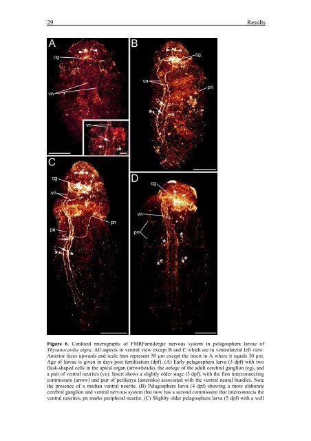
29 ResultsFigure 6. Confocal micrographs of FMRFamidergic nervous system in pelagosphera larvae ofThysanocardia nigra. All aspects in ventral view except B and C which are in ventrolateral left view.Anterior faces upwards and scale bars represent 50 µm except the insert in A where it equals 30 µm.Age of larvae is given in days post fertilisation (dpf). (A) Early pelagosphera larva (3 dpf) with twoflask-shaped cells in the apical organ (arrowheads), the anlage of the adult cerebral ganglion (cg), anda pair of ventral neurites (vn). Insert shows a slightly older stage (3 dpf), with the first interconnectingcommissure (arrow) and pair of perikarya (asterisks) associated with the ventral neural bundles. Notethe presence of a median ventral neurite. (B) Pelagosphera larva (4 dpf) showing a more elaboratecerebral ganglion and ventral nervous system that now has a second commissure that interconnects theventral neurites; pn marks peripheral neurite. (C) Slighlty older pelagosphera larva (5 dpf) with a well


