

PhD thesis - Biologisk Institut - Københavns Universitet
PhD thesis - Biologisk Institut - Københavns Universitet
PhD thesis - Biologisk Institut - Københavns Universitet

You also want an ePaper? Increase the reach of your titles
YUMPU automatically turns print PDFs into web optimized ePapers that Google loves.
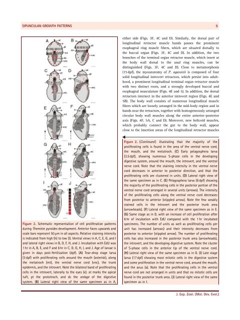
SIPUNCULAN GROWTH PATTERNS 5either side (Figs. 3F, 4C and D). Similarly, the dorsal pair oflongitudinal retractor muscle bands passes the prominentesophageal ring muscle fibers, which are situated dorsally tothe buccal organ (Figs. 3F, 4C and D). In addition, the twobranches of the terminal organ retractor muscle, which insert atthe body wall dorsal to the anal ring muscles, can bedistinguished (Figs. 3F, 4C and D). Close to metamorphosis(15 dpf), the myoanatomy of P. agassizii is composed of foursolid longitudinal introvert retractors, which persist into adulthood,a prominent longitudinal terminal organ retractor musclewith two distinct roots, and a strongly developed buccal andesophageal musculature (Figs. 4E and 5). In addition, the dorsalretractors intersect in the anterior introvert region (Figs. 4E and5B). The body wall consists of numerous longitudinal musclefibers which are loosely arranged in the mid-body region and inbands near the retractors, together with homogeneously arrangedcircular body wall muscles along the entire anterior–posterioraxis (Figs. 4F, 5A, C and D). Moreover, new helicoid muscles,which probably connect the gut to the body wall, appearclose to the insertion areas of the longitudinal retractor musclesFigure 2. Schematic representation of cell proliferation patternsduring Themiste pyroides development. Anterior faces upwards andscale bars represent 50 mm in all aspects. Relative staining intensityis indicated from high (h) to low (l). Ventral views in A, C, E, G, and Iand lateral right views in B, D, F, H, and J. Incubation with EdU was1hrinA,B,E,andFand6hrinC,D,G,H,I,andJ.Ageoflarvaeisgiven in days post-fertilization (dpf). (A) Tear-drop stage larva(3 dpf) with proliferating cells around the mouth (asterisk), alongthe metatroch (mt), the ventral nerve cord (vnc), the trunkepidermis, and the introvert. Note the bilateral band of proliferatingcells in the introvert, laterally to the eyes (e). at marks the apicaltuft, pt the prototroch, and ds the anlage of the digestivesystem. (B) Lateral right view of the same specimen as in A,Figure 2. (Continued ) illustrating that the majority of theproliferating cells is found in the area of the ventral nerve cord,the mouth, and the metatroch. (C) Early pelagosphera larva(3.5 dpf), showing numerous S-phase cells in the developingdigestive system, around the mouth, the introvert, and the ventralnerve cord. Note that the staining intensity in the ventral nervecord decreases in anterior to posterior direction, and that theproliferating cells are clustered in units. (D) Lateral right view ofthe same specimen as in C. (E) Pelagosphera larva (8 dpf) showingthe majority of the proliferating cells in the posterior portion of theventral nerve cord arranged in several units (arrows). The intensityof the proliferating cells along the ventral nerve cord decreasesfrom posterior to anterior (stippled arrow). Note the few weaklystained cells in the introvert and the posterior trunk area(arrowheads). (F) Lateral right view of the same specimen as in E.(G) Same stage as in D, with an increase of cell proliferation after6 hr of incubation with EdU compared with the 1 hr incubatedspecimens. The number of units as well as proliferating cells perunit has increased (arrows) and their intensity decreases fromposterior to anterior (stippled arrow). The number of proliferatingcells has also increased in the posterior trunk area (arrowheads),the introvert, and the developing digestive system. Note the clusterof S-phase cells in the anterior tip of the ventral nerve cord.(H) Lateral right view of the same specimen as in G. (I) Latestagelarva (17 dpf) showing most mitotic cells in the digestive systemand some proliferation in the ventral nerve cord, around the mouth,and the anus (a). Note that the proliferating cells in the ventralnerve cord are not arranged in units and that no mitotic cells arefound in the posterior trunk area. (J) Lateral right view of the samespecimen as in I.J. Exp. Zool. (Mol. Dev. Evol.)


