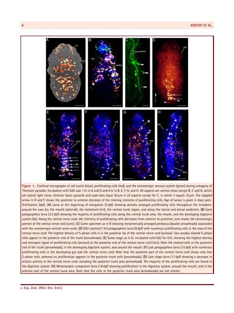
SIPUNCULAN GROWTH PATTERNS 3Denmark) in 0.1 M PBS in the dark for 24 hr at room temperature.Thereafter, the samples were washed three times for 15 min andmounted in Fluoromount G on glass slides. Some specimens weredouble labeled for DAPI and F-actin.Analysis and digital image acquisition was performed on aLeica DM IRBE microscope equipped with a Leica TCS SP2confocal unit (Leica Microsystems, Wetzlar, Germany). Opticalsections taken at intervals of 0.5–0.2 mm were digitally mergedinto maximum projection images. Images were further processedwith Photoshop 9.0.2 (Adobe Systems, San Jose, CA) to adjustcontrast and brightness. 3D reconstructions were created fromselected confocal stacks using isosurface algorithms of theimaging software Imaris v. 4.1 (Bitplane, Zürich, Switzerland).Digital line drawings were created with Corel Draw 11.0 (CorelCorporation, Ottawa, Ontario, Canada).RESULTSCell Proliferation During Larval Development of T. pyroides and T. nigraDividing cells were labeled with the thymidine analogue EdUduring development of T. pyroides and T. nigra. Combination ofEdU and DAPI labeling enabled identification of proliferatingcells and their location in the specimens (Fig. 1). Subsequentclones of S-phase cells that have incorporated EdU showprogressive loss of staining intensity in the respective daughtercells, which allows following the direction of proliferation(Fig. 1B, D, E; cf. Chehrehasa et al., 2009).In the trochophore stages, mitotic cells are scattered throughoutthe ectoderm (not shown). At the beginning of elongation(‘‘teardrop stage’’; age: 3 dpf), proliferating cells are denselyarranged around the eyes, mouth, metatroch, ventral trunk region,and along the lateral and dorsal epidermis (Figs. 1A, 2A and B).Moreover, the S-phase cells are symmetrically arranged in twolateral stripes in the introvert (Figs. 1A and 2A). Slightly later(3.5 dpf), proliferating cells appear in the area of the developingdigestive system (Figs. 1B, 2C and D). The highest number ofproliferating cells is found along the developing ventral nervecord (Figs. 1B, 2C and D), which at this stage has three pairs ofserotonergic perikarya associated with the serotonergic nervecords (Fig. 1C). Moreover, intensity loss toward the posterior poleof the ventral trunk region indicates an anterior to posteriordirection of cell proliferation (Figs. 1B, 2C and D). As in the 1 hrincubated pelagosphera larvae aged 8 dpf, few units of proliferatingcells appear in the posterior tip of the ventral nerve cord. Thesignal intensity of these cells decreases from posterior to anterior(Figs. 1D, 2E and F). In addition, few weakly stained mitotic cellsappear in the developing digestive system and in the posteriortrunk region (Figs. 1D, 2E and F). As expected, 8 dpf oldpelagosphera larvae that had been exposed to EdU for 6 hr showa higher number of proliferating cells in the ventral nerve cord,the developing digestive system, around the mouth, and in twobilateral stripes in the introvert (Figs. 1E, 2G and H). Moreover, thenumber of S-phase cells has increased in the posterior trunkregion, and more units of proliferating cells appear in theposterior part of the ventral nerve cord (Figs. 1E, 2G, H). Lateral tothe ventral nerve cord, bilateral patches of stained nuclei seem tomigrate into the developing digestive system (Figs. 1E, 2G and H).As in the 1 hr incubated specimens, the 6 hr incubated pelagospheralarvae show the same pattern of intensity decrease fromposterior to anterior (Figs. 1E, 2G and H). As the larvae grow, mostproliferating cells occur in the developing digestive system,around the anus, and in the anterior part of the ventral trunk area(Figs. 1F, 2F, G). We found no units of S-phase cells in theposterior ventral trunk region (Figs. 1F, G, 2I, J). In metamorphiccompetent larvae (18 dpf), the majority of proliferating cells isdetected in the anterior trunk, the introvert, and the digestivesystem (Fig. 1H). Post-metamorphic stages show proliferating cellsmostly in the introvert and tentacle anlagen (not shown). Overall,the distribution pattern of proliferating cells during ontogeny ofT. pyroides and T. nigra does not unambiguously reveal atextbook-like posterior growth zone. However, a distinct tissueformation zone appears temporarily in the posterior ventral trunkregion in the pelagosphera stage.Myogenesis in P. agassiziiIn the early trochophore larva of P. agassizii (i.e., at 6 dpf),numerous circular body wall muscles appear simultaneously,together with the anlagen of the longitudinal retractor muscles(Figs. 3A inset and 4A). Slightly later, the longitudinal retractorscan be distinguished as one pair of ventral, one pair of dorsal,and one single unpaired terminal organ retractor muscle(Fig. 3B). In addition, the mouth opening is visible and situatedanterior to the anlage of the buccal musculature (Fig. 3B).The ventral and dorsal retractor muscles and the terminal organretractor muscle are well developed in the late trochophore larvabefore the onset of longitudinal growth at 7 dpf (Figs. 3C and 4B).At the same time, numerous circular muscles of the future buccalmusculature emerge posteriorly to the mouth opening (Figs. 3Cand 4B). In addition, the entire length of the larval trunk iscovered by circular body wall muscles (Figs. 3C and 4B). Theirnumber remains constant during the early phases of elongation(7–8 dpf) (Fig. 3D and E). By contrast, additional longitudinalbody wall and introvert retractor muscle fibers start to form(Fig. 3D and E). At the same time, the esophageal ring, intestinalring, and longitudinal muscle fibers, together with the anal ringmuscles of the digestive system, are established (Figs. 3D, E and4C). As the pelagosphera larva grows (9–12 dpf), new circularbody wall muscles are synchronously added along the entireanterior–posterior axis by fission from existing myocytes(Figs. 3F and 4D). Accordingly, the formation of new circularbody wall muscle fibers does not occur in a directionalanterior–posterior process. The ventral longitudinal retractormuscles contribute to the muscles that encircle the mouthopening and surround the musculature of the buccal organ fromJ. Exp. Zool. (Mol. Dev. Evol.)
4KRISTOF ET AL.Figure 1. Confocal micrographs of cell nuclei (blue), proliferating cells (red), and the serotonergic nervous system (green) during ontogeny ofThemiste pyroides. Incubation with EdU was 1 hr in A and D and 6 hr in B, E, F, G, and H. All aspects are ventral views except B, C and G, whichare lateral right views. Anterior faces upwards and scale bars equal 50 mm in all aspects except for C, in which it equals 10 mm. The stippledarrow in D and E shows the posterior to anterior decrease of the staining intensity of proliferating cells. Age of larvae is given in days postfertilization(dpf). (A) Larva at the beginning of elongation (3 dpf) showing densely arranged proliferating cells throughout the ectoderm,around the eyes (e), the mouth (asterisk), the metatroch (mt), the ventral trunk region, and along the lateral and dorsal epidermis. (B) Earlypelagosphera larva (3.5 dpf) showing the majority of proliferating cells along the ventral trunk area, the mouth, and the developing digestivesystem (ds). Along the ventral nerve cord, the intensity of proliferating cells decreases from anterior to posterior; svnc marks the serotonergicportion of the ventral nerve cord (svnc). (C) Same specimen as in B showing metamerically arranged perikarya (double arrowheads) associatedwith the serotonergic ventral nerve cords. (D) EdU-stained (1 hr) pelagosphera larva (8 dpf) with numerous proliferating cells in the area of theventral nerve cord. The highest density of S-phase cells is in the posterior tip of the ventral nerve cord (arrows). Two weakly stained S-phasecells appear in the posterior end of the trunk (arrowheads). (E) Same stage as in D, incubated with EdU for 6 hr, showing the highest densityand strongest signal of proliferating cells (arrows) at the posterior end of the ventral nerve cord (vnc). Note the stained cells at the posteriorend of the trunk (arrowheads), in the developing digestive system, and around the mouth. (F) Late pelagosphera larva (15 dpf) with numerousproliferating cells in the developing gut and the ventral nerve cord. Note that the posterior part of the ventral nerve cord shows only fewS-phase cells, whereas no proliferation appears in the posterior trunk cells (arrowheads). (G) Late stage larva (17 dpf) showing a decrease inmitotic activity in the ventral nerve cord, including the posterior trunk area (arrowhead). The majority of the proliferating cells are found inthe digestive system. (H) Metamorphic competent larva (18 dpf) showing proliferation in the digestive system, around the mouth, and in theanterior part of the ventral trunk area. Note that the cells in the posterior trunk area (arrowheads) are not mitotic.J. Exp. Zool. (Mol. Dev. Evol.)