- Page 1 and 2: AT MICROFICHEREFERENCELIBRARYA proj
- Page 3 and 4: This book aims to bring the mini~mu
- Page 5 and 6: To all those who might soeasily be
- Page 7 and 8: Oxford University Press, E& House,
- Page 9 and 10: PrefaceThere is believed to be a ne
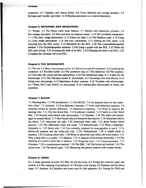
- Page 11: ContentsChapter1. INTRODUCTION1.1 H
- Page 15 and 16: How to use this book 1 1.11 1 Intro
- Page 17 and 18: Honesty and responsibility 1 1.2bes
- Page 19 and 20: Some special words 1 1.3with. A row
- Page 21 and 22: Centrifuging and filtering 1 1.5/th
- Page 23 and 24: Cells 1 1.9the pH is about Il. At p
- Page 25 and 26: Micro-organisms 1 1 .l 1made from t
- Page 27 and 28: Stains 1 1.16difficult to see. We u
- Page 29 and 30: Stklization 1 1.20This kills all th
- Page 31 and 32: The pressure cooker 1 1.21The 3-way
- Page 33 and 34: Laboratory infection 1 1.23the plun
- Page 35 and 36: 2 1 Equipment andChemicals2.1 The e
- Page 37 and 38: Special equipment in the main list
- Page 39 and 40: Special equipment in the main list
- Page 41 and 42: .I‘:; ‘1 :....’ ;.,. . .I, _(
- Page 43 and 44: Chemicals 1 2.4heating things on th
- Page 45 and 46: Stock and spares 1 2.6chamber 0.1 m
- Page 47 and 48: 3.1 Benches and shelvesThere are ma
- Page 49 and 50: P,,,.” y- ,.* 9.; _, ,,.Bottled g
- Page 51 and 52: The Bunsen burner 1 3.5the tin draw
- Page 53 and 54: .,;-./I ,__( .if you have no diamon
- Page 55 and 56: Making and using Pasteur pipettes 1
- Page 57 and 58: Some practical details 1 3.11and in
- Page 59 and 60: Washing equipment 1 3.12Find a piec
- Page 61 and 62: Stains and reagents 1 3.15Keep all
- Page 63 and 64:
Buffers 1 3.20aa wash bottle and th
- Page 65 and 66:
Buffers 1 3.253.25 MAKING CRYSTAL V
- Page 67 and 68:
Buffers 1 3.37/fill the bottle with
- Page 69 and 70:
A description of Figure 3-11 1 3.46
- Page 71 and 72:
_.:;:;r- ^>,< ’ “- , _:’ , ,,
- Page 73 and 74:
4 1 Records and Specimens4.1 Record
- Page 75 and 76:
; -. . . _specimen is lahelled and
- Page 77 and 78:
“‘MAKING A SEQUESTRENEPOLYTUBEA
- Page 79 and 80:
Cross infection and syringe jaundic
- Page 81 and 82:
: 1 _;;:.,.“ -. y ‘, ;Send&g sp
- Page 83 and 84:
5 1 Weighing and MeasuringWEIGHT5.1
- Page 85 and 86:
The Ohaus triple beam balance 1 ‘
- Page 87 and 88:
: ,!“&;‘.: ; ), : IIPipettes a
- Page 89 and 90:
I’- >- Why and how we measure col
- Page 91 and 92:
The Lovibond comparator 1 5.10THE L
- Page 93 and 94:
The ‘Grey wedge’ 1 5.11The wedg
- Page 95 and 96:
Ii’ used for the blood urea. THE
- Page 97 and 98:
-Measuring colour with electricity
- Page 99 and 100:
Oxyhaemoglobin methods 1 5.2T arefr
- Page 101 and 102:
When an EEL goes wrong 1 5.22This i
- Page 103 and 104:
When an EEL goes wrong 1 5.22BEFORE
- Page 105 and 106:
6 1 The Microscope6.1 The pm your m
- Page 107 and 108:
How a microscope works 1 6.2may not
- Page 109 and 110:
FROM ON TOPTHE CONDENSERB FROM THE
- Page 111 and 112:
‘z:.! y,*. 4:f.‘> &&jg;y: -: r_
- Page 113 and 114:
The objectives j 6.7all these are t
- Page 115 and 116:
.i’i (. 1. ” .The eyepiece 1 6.
- Page 117 and 118:
., -.-Lights for the microscope 1 6
- Page 119 and 120:
Knowing your microscope 1 6.13foeos
- Page 121 and 122:
Specimens of poor contrast 1 6.15ey
- Page 123 and 124:
Troubles with microscopes 1 6.166.1
- Page 125 and 126:
Some ‘DOS’ and ‘Don’ts’ i
- Page 127 and 128:
.,:‘=--L&,??6;,* >.: -- ,I i .~:.
- Page 129 and 130:
The haematocrit 7.2If you do. you w
- Page 131 and 132:
z -.The haematocrit 1 7.2instrument
- Page 133 and 134:
An anaemia chart for the ‘Under F
- Page 135 and 136:
Causes of anaemia 1 7.5both the hae
- Page 137 and 138:
The thin blood film 1 7.11pale conj
- Page 139 and 140:
7 1 BloodA film is always too thick
- Page 141 and 142:
Faults in a thin blood film 1 7.13T
- Page 143 and 144:
Platelets 1 7.15NEUTROPHILMETAMYELO
- Page 145 and 146:
How blood cells are formed in the m
- Page 147 and 148:
The red cells of anaemic patients a
- Page 149 and 150:
The differential white cell count 1
- Page 151 and 152:
What ahaemoglobinopathy is 1 7.24ME
- Page 153 and 154:
Two solubility methods for haemoglo
- Page 155 and 156:
Sickle-cell anaemia and the sickle-
- Page 157 and 158:
THE TOTAL WHITE CELL COUNT7.29 Coun
- Page 159 and 160:
Counting white cells 1 7.29THESE AR
- Page 161 and 162:
What an abnormal total white cell c
- Page 163 and 164:
I1.‘.)?6),.I~_I/” ,1,~ Why a th
- Page 165 and 166:
Ilymphocyteb 0 ayoung trophozoites
- Page 167 and 168:
P.vivax P.malariae P.falciparum P.o
- Page 169 and 170:
Sleeping sickness or trypanosomiasi
- Page 171 and 172:
A concentration method ! 7.38for ex
- Page 173 and 174:
The ESR 1 7.39make several of these
- Page 175 and 176:
1”ig; I , _ :-!. * ip ’ _Make s
- Page 177 and 178:
,.,;+fi :_ . .2 The blood sugar 1 7
- Page 179 and 180:
,,:Measuring the plasma acetone wit
- Page 181 and 182:
8 1 Urine8.1 A clean specimen of ur
- Page 183 and 184:
“:. * -,y,: .&$.,i.:, .’ ; I 1
- Page 185 and 186:
;,‘-j_‘. ..‘I I: ^I .p-: 1.
- Page 187 and 188:
,- ;>. .-:I_. -.._). Jaundice and s
- Page 189 and 190:
p-7-- -~- -by’ ”I!$: ‘-/!& Ii
- Page 191 and 192:
Testingthe urine for sulphones1 8.1
- Page 193 and 194:
_/Looking at the centrifuged deposi
- Page 195 and 196:
aliSCH!STOSOME OVA 3deadSchistosoma
- Page 197 and 198:
Three kinds of movement 1 8.148.14
- Page 199 and 200:
Looking for the ova of Schistosoma
- Page 201 and 202:
Diagnosing meningitis 1 9.3IF THE P
- Page 203 and 204:
Equipment for lum’bar puncture 1
- Page 205 and 206:
Cells 1 9.9of CSF to flow out of th
- Page 207 and 208:
Stained films 1 9.11protein that yo
- Page 209 and 210:
,(The CSF protein 1 9.1316 and 17.
- Page 211 and 212:
i r: .,_‘.,i, I,>‘,i,.2c:,, y-,
- Page 213 and 214:
Cerebral mil-aria 1 9.20blood has g
- Page 215 and 216:
The saline stool smear 1 10.2aent t
- Page 217 and 218:
,L- ;.:
- Page 219 and 220:
spin the suspensionfor about five m
- Page 221 and 222:
testtube-I piece ofSellotapelay the
- Page 223 and 224:
;: “,Some common ova1 10.5Trichur
- Page 225 and 226:
important thing about these organis
- Page 227 and 228:
E. histolytica and E. co/i 1 10.7E.
- Page 229 and 230:
.-.i- ; .>_; ,’ .:Identifying E.
- Page 231 and 232:
Measuring the pH of a stool and tes
- Page 233 and 234:
,-?‘,C ,:-. 1,I”.&‘.;-,__,-_
- Page 235 and 236:
find a purulent(pus like) pieceof t
- Page 237 and 238:
Examining the sputum for helminth o
- Page 239 and 240:
this is the centrifuged depositfrom
- Page 241 and 242:
Classifying leprosy 1 11 .l laIn no
- Page 243 and 244:
Classifying leprosy 1 11.1 laThese
- Page 245 and 246:
‘,y,d+“,> &^-”,.,-:>. I -_’
- Page 247 and 248:
$i&i: :: -&ll _ ~*”ii’1~11’ 1
- Page 249 and 250:
-.,‘&A, ,1 ’ .C’. ‘--Lymph
- Page 251 and 252:
‘ h-7 1‘, ,,.->$ ,’ .: 1,:. +
- Page 253 and 254:
This chapter makes extensive use of
- Page 255 and 256:
this is a tube of \anti-A serum, it
- Page 257 and 258:
.bthis is awash-IvlttlPof salinewit
- Page 259 and 260:
Cross-matching or compatibility tes
- Page 261 and 262:
Rhesus grouping 1 12.712.7 Rhesus g
- Page 263 and 264:
guard tubethis rubber discgoes insi
- Page 265 and 266:
B,>Sharpening needles 1 12.10Rubber
- Page 267 and 268:
The pilot bottle 1 12.11ment and st
- Page 269 and 270:
Taking blood 1 12.12this pair of fo
- Page 271 and 272:
The Uganda Mobile Team 1 12.13Red C
- Page 273 and 274:
I-;.~: : ,.,!r’-Storing blood: th
- Page 275 and 276:
13 1 For Pathologists, Stores Offic
- Page 277 and 278:
Upgrading peripheral laboratories 1
- Page 279 and 280:
,.‘. ,. .”Gelieral stores requi
- Page 281 and 282:
,h,,.-.ISpecialequipmentin the main
- Page 283 and 284:
,, . .ML 50ML5 1ML 52ML 53ML 54ML 5
- Page 285 and 286:
:~!16; ,z..: ;.,,,j, .,, ,I__ ,”F
- Page 287 and 288:
I
- Page 289 and 290:
Choice 13. ‘Dextrostix’ 1 13.26
- Page 291 and 292:
p... ‘,( I’ ‘8a..:g& >,&Ii II
- Page 293 and 294:
VocabularyIndexautoclave. A machine
- Page 295 and 296:
Vocabulary Indexcy~toplasm. IIe com
- Page 297 and 298:
VocabularyIndexGram’s method. A w
- Page 299 and 300:
Vocabularyindexmeasuring qiinder (M
- Page 301 and 302:
Vocabulary IndexPlasmodium, fatcipa
- Page 303 and 304:
VocabularyIndexrelapse. When a pati
- Page 305 and 306:
VocabularyIndextest rube (ML 48). A
- Page 307 and 308:
_.,,:,. I _. , ,( * , ),), OI.,I‘
- Page 309 and 310:
Haemoglobin g%increasinganaemiavery
- Page 311 and 312:
“-;
- Page 313 and 314:
10 11 12These are fresh unstained w
- Page 315 and 316:
30 31 32 33Plasmodium malariae. 30.
- Page 317 and 318:
51 and 52. These are the same speci
- Page 319 and 320:
65 66 67All the ova on this page ar
- Page 321 and 322:
,;^3ROTOZOA. ETC. IN THE STOOLS PLA
- Page 323:
101 102101. This shows red staining


