Biochemistry/Molecular Biology - ARVO
Biochemistry/Molecular Biology - ARVO
Biochemistry/Molecular Biology - ARVO
You also want an ePaper? Increase the reach of your titles
YUMPU automatically turns print PDFs into web optimized ePapers that Google loves.
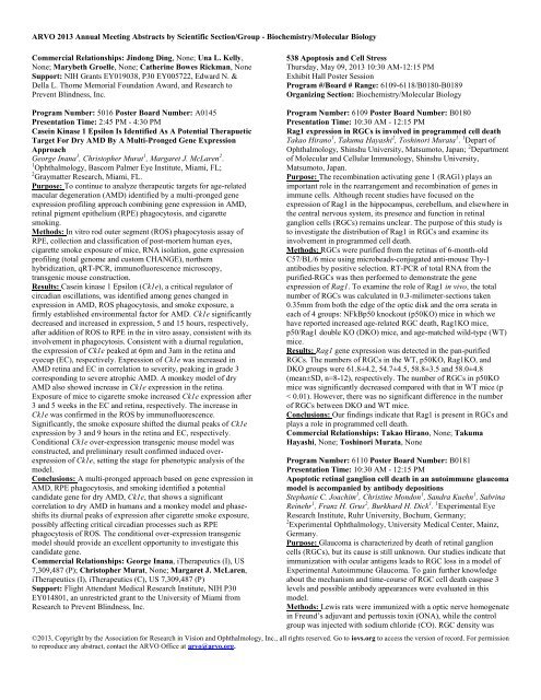
<strong>ARVO</strong> 2013 Annual Meeting Abstracts by Scientific Section/Group - <strong>Biochemistry</strong>/<strong>Molecular</strong> <strong>Biology</strong>Commercial Relationships: Jindong Ding, None; Una L. Kelly,None; Marybeth Groelle, None; Catherine Bowes Rickman, NoneSupport: NIH Grants EY019038, P30 EY005722, Edward N. &Della L. Thome Memorial Foundation Award, and Research toPrevent Blindness, Inc.538 Apoptosis and Cell StressThursday, May 09, 2013 10:30 AM-12:15 PMExhibit Hall Poster SessionProgram #/Board # Range: 6109-6118/B0180-B0189Organizing Section: <strong>Biochemistry</strong>/<strong>Molecular</strong> <strong>Biology</strong>Program Number: 5016 Poster Board Number: A0145Presentation Time: 2:45 PM - 4:30 PMCasein Kinase 1 Epsilon Is Identified As A Potential TherapueticTarget For Dry AMD By A Multi-Pronged Gene ExpressionApproachGeorge Inana 1 , Christopher Murat 1 , Margaret J. McLaren 2 .1 Ophthalmology, Bascom Palmer Eye Institute, Miami, FL;2 Graymatter Research, Miami, FL.Purpose: To continue to analyze therapeutic targets for age-relatedmacular degeneration (AMD) identified by a multi-pronged geneexpression profiling approach combining gene expression in AMD,retinal pigment epithelium (RPE) phagocytosis, and cigarettesmoking.Methods: In vitro rod outer segment (ROS) phagocytosis assay ofRPE, collection and classification of post-mortem human eyes,cigarette smoke exposure of mice, RNA isolation, gene expressionprofiling (total genome and custom CHANGE), northernhybridization, qRT-PCR, immunofluorescence microscopy,transgenic mouse construction.Results: Casein kinase 1 Epsilon (Ck1e), a critical regulator ofcircadian oscillations, was identified among genes changed inexpression in AMD, ROS phagocytosis, and smoke exposure, afirmly established environmental factor for AMD. Ck1e significantlydecreased and increased in expression, 5 and 15 hours, respectively,after addition of ROS to RPE in the in vitro assay, consistent with itsinvolvement in phagocytosis. Consistent with a diurnal regulation,the expression of Ck1e peaked at 6pm and 3am in the retina andeyecup (EC), respectively. Expression of Ck1e was increased inAMD retina and EC in correlation to severity, peaking in grade 3corresponding to severe atrophic AMD. A monkey model of dryAMD also showed increase in Ck1e expression in the retina.Exposure of mice to cigarette smoke increased Ck1e expression after3 and 5 weeks in the EC and retina, respectively. The increase inCk1e was confirmed in the ROS by immunofluorescence.Significantly, the smoke exposure shifted the diurnal peaks of Ck1eexpression by 3 and 9 hours in the retina and EC, respectively.Conditional Ck1e over-expression transgenic mouse model wasconstructed, and preliminary result confirmed induced overexpressionof Ck1e, setting the stage for phenotypic analysis of themodel.Conclusions: A multi-pronged approach based on gene expression inAMD, RPE phagocytosis, and smoking identified a potentialcandidate gene for dry AMD, Ck1e, that shows a significantcorrelation to dry AMD in humans and a monkey model and phaseshiftsits diurnal peaks of expression after cigarette smoke exposure,possibly affecting critical circadian processes such as RPEphagocytosis of ROS. The conditional over-expression transgenicmodel should provide an excellent opportunity to investigate thiscandidate gene.Commercial Relationships: George Inana, iTherapeutics (I), US7,309,487 (P); Christopher Murat, None; Margaret J. McLaren,iTherapeutics (I), iTherapeutics (C), US 7,309,487 (P)Support: Flight Attendant Medical Research Institute, NIH P30EY014801, an unrestricted grant to the University of Miami fromResearch to Prevent Blindness, Inc.Program Number: 6109 Poster Board Number: B0180Presentation Time: 10:30 AM - 12:15 PMRag1 expression in RGCs is involved in programmed cell deathTakao Hirano 1 , Takuma Hayashi 2 , Toshinori Murata 1 . 1 Depart ofOphthalmology, Shinshu University, Matsumoto, Japan; 2 Departmentof <strong>Molecular</strong> and Cellular Immunology, Shinshu University,Matsumoto, Japan.Purpose: The recombination activating gene 1 (RAG1) plays animportant role in the rearrangement and recombination of genes inimmune cells. Although recent studies have focused on theexpression of Rag1 in the hippocampus, cerebellum, and elsewhere inthe central nervous system, its presence and function in retinalganglion cells (RGCs) remains unclear. The purpose of this study isto investigate the distribution of Rag1 in RGCs and examine itsinvolvement in programmed cell death.Methods: RGCs were purified from the retinas of 6-month-oldC57/BL/6 mice using microbeads-conjugated anti-mouse Thy-1antibodies by positive selection. RT-PCR of total RNA from thepurified-RGCs was then performed to demonstrate the geneexpression of Rag1. To examine the role of Rag1 in vivo, the totalnumber of RGCs was calculated in 0.3-milimeter-sections taken0.35mm from both the edge of the optic disk and the orra serata ineach of 4 groups: NFkBp50 knockout (p50KO) mice in which wehave reported increased age-related RGC death, Rag1KO mice,p50/Rag1 double KO (DKO) mice, and age-matched wild-type (WT)mice.Results: Rag1 gene expression was detected in the pan-purifiedRGCs. The numbers of RGCs in the WT, p50KO, Rag1KO, andDKO groups were 61.8±4.2, 54.7±4.5, 58.8±3.5 and 58.0±4.8(mean±SD, n=8-12), respectively. The number of RGCs in p50KOmice was significantly decreased compared with that in WT mice (p< 0.01). However, there was no significant difference in the numberof RGCs between DKO and WT mice.Conclusions: Our findings indicate that Rag1 is present in RGCs andplays a role in programmed cell death.Commercial Relationships: Takao Hirano, None; TakumaHayashi, None; Toshinori Murata, NoneProgram Number: 6110 Poster Board Number: B0181Presentation Time: 10:30 AM - 12:15 PMApoptotic retinal ganglion cell death in an autoimmune glaucomamodel is accompanied by antibody depositionsStephanie C. Joachim 1 , Christine Mondon 1 , Sandra Kuehn 1 , SabrinaReinehr 1 , Franz H. Grus 2 , Burkhard H. Dick 1 . 1 Experimental EyeResearch Institute, Ruhr University, Bochum, Germany;2 Experimental Ophthalmology, University Medical Center, Mainz,Germany.Purpose: Glaucoma is characterized by death of retinal ganglioncells (RGCs), but its cause is still unknown. Our studies indicate thatimmunization with ocular antigens leads to RGC loss in a model ofExperimental Autoimmune Glaucoma. To gain further knowledgeabout the mechanism and time-course of RGC cell death caspase 3levels and possible antibody appearances were evaluated in thismodel.Methods: Lewis rats were immunized with a optic nerve homogenatein Freund’s adjuvant and pertussis toxin (ONA), while the controlgroup was injected with sodium chloride (CO). RGC density was©2013, Copyright by the Association for Research in Vision and Ophthalmology, Inc., all rights reserved. Go to iovs.org to access the version of record. For permissionto reproduce any abstract, contact the <strong>ARVO</strong> Office at arvo@arvo.org.


