<strong>ARVO</strong> 2013 Annual Meeting Abstracts by Scientific Section/Group - <strong>Biochemistry</strong>/<strong>Molecular</strong> <strong>Biology</strong>Commercial Relationships: Jiang Qian, None; Jun Wan, None;Verity F. Oliver, None; Donald J. Zack, Alcon (C), Merck (F),Allergan (C); Shannath L. Merbs, NoneSupport: NIH Grants EY021897 EY018703 and Unrestricted fundsfrom RPBProgram Number: 2616Presentation Time: 9:30 AM - 9:45 AMHistone marks predict cell plasticity of the adult human retinalpigment epitheliumTimothy A. Blenkinsop 1 , Alvaro Rada-Iglesias 2 , Joanna Wysocka 2 ,Sally Temple 1 . 1 Macular Degeneration Program, Neural Stem CellInstitute, Rensselaer, NY; 2 Developmental <strong>Biology</strong>, StanfordUniversity, Stanford, CA.Purpose: Retinal pigment epithelial (RPE) cells are one of the fewcell types well recognized to change fate in lower vertebrates. Werecently showed adult human RPE as old as 99 years differentiatedown mesenchymal and neural lineages using commercially availabledifferentiation media. Addressing whether this was due to themultipotency of the RPE cells or to a contaminating cell type, forexample MSCs, in the cultures was critical. Therefore, wedemonstrated that single, cloned RPE cells exhibit this ability,irrefutably demonstrating that they are multipotent cells. Wetherefore sought to understand the basis for this observed plasticity.Histone marks at promoter and enhancer sites can reveal both genesthat are actively being transcribed - i.e. active, and genes that arepoised - i.e. currently inactive, but can be activated by underappropriate conditions. Recently histone H3K27me3 mark has beenshown to be enriched in regions several kilobases upstream from thepromoter around enhancer regions that are poised in humanembryonic stem cells (hESCs). In contrast, histone mark H3K27acmarks similar locations when the downstream gene is active.Therefore, we hypothesized that these two marks can predict genechanges from being poised to active.Methods: We cultured human retinal pigment epithelium intopolarized layers and verified their terminal differentiation throughtransepithelial resistance (TER) in Ωcm2. These monolayersexpressed TER of > 200Ωcm2 similar to native RPE. We alsoconfirmed their identity immunohistochemically, by quantitativePCR. We then conducted H3K27ac and H3K27me3 Chromatinimmunoprecipitation-sequencing (ChIP-seq) on adult human RPEmonolayers.Results: We found RPE specific genes posses H3K27ac marksupstream of their promotor regions, whereas the genes we have foundto increase expression during differentiation, for example RUNX2during osteogenic conditions, possess the poised mark H3K27me3upstream of their promotor.Conclusions: These results suggest the histone marks can predict notonly the cell identity, but also the poised mark can predict RPEplasticity.Commercial Relationships: Timothy A. Blenkinsop, None; AlvaroRada-Iglesias, None; Joanna Wysocka, None; Sally Temple,Athghin Biotech (I)Support: Ey022079-01Program Number: 2617Presentation Time: 9:45 AM - 10:00 AMThe MLL1 Histone Methyltransferease is Essential forDevelopment of Photoreceptor FunctionDiana S. Brightman, Ray Suzuki, Shiming Chen. Ophthalmology andVisual Sciences, Washington University in St. Louis, St. Louis, MO.Purpose: Development and maintenance of retinal photoreceptorfunction requires precisely controlled gene expression. This isregulated by both photoreceptor-specific and general transcriptionregulators, including histone modification enzymes. Members of theMLL family of histone H3K4 methyltransferases are expressed inmouse photoreceptors. The most prominent of these is MLL1 whoseexpression increases between postnatal day 2 (P2) and P14, a criticalperiod for photoreceptor terminal differentiation. MLL1 expressiondepends on the key photoreceptor-specific transcription factor CRX,suggesting a potential role of MLL1 in the CRX regulatory pathway.We determined the role of MLL1 in photoreceptor gene expression,development and survival using a loss-of-function approach.Methods: To avoid embryonic lethality, Cre-loxP-mediatedconditional knockout (CKO) was used. Mice with Mll1 floxed alleles(Mll1 fl/fl ) were crossed with Crx-Cre mice to create Mll1 CKO in thedeveloping retina. Mll1 CKO mice show morphological changes inthe retina by H&E staining, immunohistochemistry (IHC) andelectron microscopy (EM), and visual function changes byelectroretinogram (ERG).Results: Cre activity was found in all cell layers of the developingMll1 CKO retina, in a superior-to-inferior gradient. As a result, Mll1expression was uniformly depleted in the superior region, where allretinal layers were significantly thinner by morphometry. IHC andEM analyses show abnormal superior outer plexiform layer (OPL)synapses. The intensity of the presynaptic markers V-GLUT1 andCTBP2 were markedly decreased at the OPL. Calbindin-positivehorizontal cells were also reduced, indicative of postsynaptic defects.Consistent with these defects, both dark and light-adapted ERGs of 1-month-old Mll1 CKO mice were significantly decreased, suggestingdefects in rod, cone, and inner retina functions. The morphologicaland functional changes are stable to 6 months of age, suggesting adevelopmental origin, not degeneration.Conclusions: MLL1 is required for the development of appropriateretinal structure and function. Additional Mll1 CKO usingphotoreceptor and INL cell-type-specific Cre lines are in progress todetermine cell autonomy and underlying molecular mechanisms.These studies will shed light on how general epigenetic modulatorscontribute to the regulation of cell-type specific gene expression anddevelopment.Commercial Relationships: Diana S. Brightman, None; RaySuzuki, None; Shiming Chen, NoneSupport: NIH Grant EY012543 (to SC), EY02687 (to WU-DOVS),RBP Lew R. Wasserman Award (to SC) and unrestricted funds (toWU-DOVS)Program Number: 2618Presentation Time: 10:00 AM - 10:15 AMRole of 5-hydroxymethylcytosine during postnatal retinaldevelopmentStylianos Michalakis 1 , Arshan Perera 1 , Susanne Koch 1 , MirkoWagner 2 , Lukas Windhager 3 , Kerstin Nagel-Wolfrum 4 , Tim M.Strom 5, 6 , Ralf Zimmer 3 , Thomas Carell 2 , Martin Biel 1 . 1 Center forIntegrated Protein Science at the Department of Pharmacy-Center forDrug Research, Ludwig-Maximilians-Universität München, Munich,Germany; 2 Center for Integrated Protein Science at the Department ofChemistry, Ludwig-Maximilians-Universität München, Munich,Germany; 3 Institute for Informatics, Ludwig-Maximilians-UniversitätMünchen, Munich, Germany; 4 Department of Cell and Matrix<strong>Biology</strong>, Institute of Zoology, Johannes Gutenberg University-Mainz,Mainz, Germany; 5 Institute of Human Genetics, TechnischeUniversität München, Munich, Germany; 6 Institute of HumanGenetics, Helmholtz Zentrum München, German Research Center forEnvironmental Health, Neuherberg, Germany.Purpose: 5-hydroxymethylcytosine (5hmC), also known as the sixthbase of the genome, is a recently discovered oxidative product of 5-©2013, Copyright by the Association for Research in Vision and Ophthalmology, Inc., all rights reserved. Go to iovs.org to access the version of record. For permissionto reproduce any abstract, contact the <strong>ARVO</strong> Office at arvo@arvo.org.
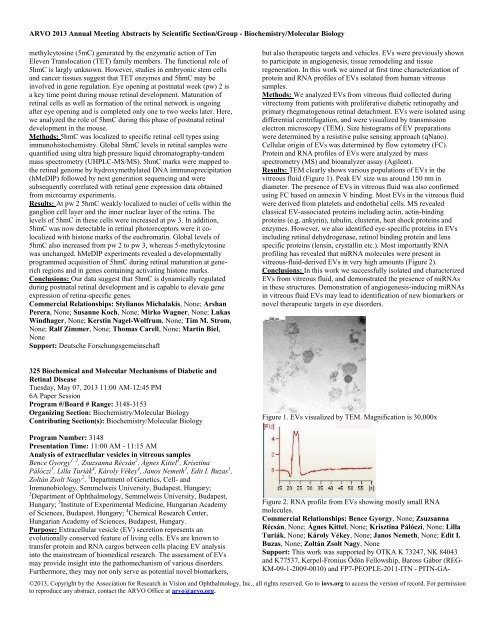
<strong>ARVO</strong> 2013 Annual Meeting Abstracts by Scientific Section/Group - <strong>Biochemistry</strong>/<strong>Molecular</strong> <strong>Biology</strong>methylcytosine (5mC) generated by the enzymatic action of TenEleven Translocation (TET) family members. The functional role of5hmC is largly unknown. However, studies in embryonic stem cellsand cancer tissues suggest that TET enzymes and 5hmC may beinvolved in gene regulation. Eye opening at postnatal week (pw) 2 isa key time point during mouse retinal development. Maturation ofretinal cells as well as formation of the retinal network is ongoingafter eye opening and is completed only one to two weeks later. Here,we analyzed the role of 5hmC during this phase of postnatal retinaldevelopment in the mouse.Methods: 5hmC was localized to specific retinal cell types usingimmunohistochemistry. Global 5hmC levels in retinal samples werequantified using ultra high pressure liquid chromatography-tandemmass spectrometry (UHPLC-MS/MS). 5hmC marks were mapped tothe retinal genome by hydroxymethylated DNA immunoprecipitation(hMeDIP) followed by next generation sequencing and weresubsequently correlated with retinal gene expression data obtainedfrom microarray experiments.Results: At pw 2 5hmC weakly localized to nuclei of cells within theganglion cell layer and the inner nuclear layer of the retina. Thelevels of 5hmC in these cells were increased at pw 3. In addition,5hmC was now detectable in retinal photoreceptors were it colocalizedwith histone marks of the euchromatin. Global levels of5hmC also increased from pw 2 to pw 3, whereas 5-methylcytosinewas unchanged. hMeDIP experiments revealed a developmentallyprogrammed acquisition of 5hmC during retinal maturation at generichregions and in genes containing activating histone marks.Conclusions: Our data suggest that 5hmC is dynamically regulatedduring postnatal retinal development and is capable to elevate geneexpression of retina-specific genes.Commercial Relationships: Stylianos Michalakis, None; ArshanPerera, None; Susanne Koch, None; Mirko Wagner, None; LukasWindhager, None; Kerstin Nagel-Wolfrum, None; Tim M. Strom,None; Ralf Zimmer, None; Thomas Carell, None; Martin Biel,NoneSupport: Deutsche Forschungsgemeinschaftbut also therapeutic targets and vehicles. EVs were previously shownto participate in angiogenesis, tissue remodeling and tissueregeneration. In this work we aimed at first time characterization ofprotein and RNA profiles of EVs isolated from human vitreoussamples.Methods: We analyzed EVs from vitreous fluid collected duringvitrectomy from patients with proliferative diabetic retinopathy andprimary rhegmatogenous retinal detachment. EVs were isolated usingdifferential centrifugation, and were visualized by transmissionelectron microscopy (TEM). Size histograms of EV preparationswere determined by a resistive pulse sensing approach (qNano).Cellular origin of EVs was determined by flow cytometry (FC).Protein and RNA profiles of EVs were analyzed by massspectrometry (MS) and bioanalyzer assay (Agilent).Results: TEM clearly shows various populations of EVs in thevitreous fluid (Figure 1). Peak EV size was around 150 nm indiameter. The presence of EVs in vitreous fluid was also confirmedusing FC based on annexin V binding. Most EVs in the vitreous fluidwere derived from platelets and endothelial cells. MS revealedclassical EV-associated proteins including actin, actin-bindingproteins (e.g. ankyrin), tubulin, clusterin, heat shock proteins andenzymes. However, we also identified eye-specific proteins in EVsincluding retinal dehydrogenase, retinol binding protein and lensspecific proteins (lensin, crystallin etc.). Most importantly RNAprofiling has revealed that miRNA molecules were present invitreous-fluid-derived EVs in very high amounts (Figure 2).Conclusions: In this work we successfully isolated and characterizedEVs from vitreous fluid, and demonstrated the presence of miRNAsin these structures. Demonstration of angiogenesis-inducing miRNAsin vitreous fluid EVs may lead to identification of new biomarkers ornovel therapeutic targets in eye disorders.325 Biochemical and <strong>Molecular</strong> Mechanisms of Diabetic andRetinal DiseaseTuesday, May 07, 2013 11:00 AM-12:45 PM6A Paper SessionProgram #/Board # Range: 3148-3153Organizing Section: <strong>Biochemistry</strong>/<strong>Molecular</strong> <strong>Biology</strong>Contributing Section(s): <strong>Biochemistry</strong>/<strong>Molecular</strong> <strong>Biology</strong>Figure 1. EVs visualized by TEM. Magnification is 30,000xProgram Number: 3148Presentation Time: 11:00 AM - 11:15 AMAnalysis of extracellular vesicles in vitreous samplesBence Gyorgy 1, 2 , Zsuzsanna Récsán 2 , Ágnes Kittel 3 , KrisztinaPálóczi 1 , Lilla Turiák 4 , Károly Vékey 4 , Janos Nemeth 2 , Edit I. Buzas 1 ,Zoltán Zsolt Nagy 2 . 1 Department of Genetics, Cell- andImmunobiology, Semmelweis University, Budapest, Hungary;2 Department of Ophthalmology, Semmelweis University, Budapest,Hungary; 3 Institute of Experimental Medicine, Hungarian Academyof Sciences, Budapest, Hungary; 4 Chemical Research Center,Hungarian Academy of Sciences, Budapest, Hungary.Purpose: Extracellular vesicle (EV) secretion represents anevolutionally conserved feature of living cells. EVs are known totransfer protein and RNA cargos between cells placing EV analysisinto the mainstream of biomedical research. The assessment of EVsmay provide insight into the pathomechanism of various disorders.Furthermore, they may not only serve as potential novel biomarkers,Figure 2. RNA profile from EVs showing mostly small RNAmolecules.Commercial Relationships: Bence Gyorgy, None; ZsuzsannaRécsán, None; Ágnes Kittel, None; Krisztina Pálóczi, None; LillaTuriák, None; Károly Vékey, None; Janos Nemeth, None; Edit I.Buzas, None; Zoltán Zsolt Nagy, NoneSupport: This work was supported by OTKA K 73247, NK 84043and K77537, Kerpel-Fronius Ödön Fellowship, Baross Gábor (REG-KM-09-1-2009-0010) and FP7-PEOPLE-2011-ITN - PITN-GA-©2013, Copyright by the Association for Research in Vision and Ophthalmology, Inc., all rights reserved. Go to iovs.org to access the version of record. For permissionto reproduce any abstract, contact the <strong>ARVO</strong> Office at arvo@arvo.org.