NBE CME programme for DNB consultants - National Board Of ...
NBE CME programme for DNB consultants - National Board Of ...
NBE CME programme for DNB consultants - National Board Of ...

Create successful ePaper yourself
Turn your PDF publications into a flip-book with our unique Google optimized e-Paper software.
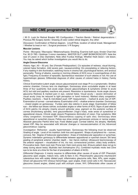
<strong>NBE</strong> <strong>CME</strong> <strong>programme</strong> <strong>for</strong> <strong>DNB</strong> <strong>consultants</strong>/ 90 D. Look <strong>for</strong> Retinal Breaks/ RD Configuration / Traction Bands / Retinal degenerations /Previous RD Surgery bands / Drawing of color coded retinal diagram; Any otherDiscussion- Confirmation of Retinal diagram; Lincoff Role; location of retinal break; Management/ Whether to treat or not / ; Surgical procedure; V R SurgeryMacular LesionsPoints - Micropsia / Macropsia / Metamorphopsia; Smoking; Examine both eyes; Amsler Chart AskFor; 90 D/ 78D / Goldman 3 mirror mandatory MASTER SLIT LAMP BIOMICROSCOPY; Note thesize of Lesion in Disc Diameters; Note other Fundus findings; Whether fresh lesion / old lesion;You may be asked which further investigations you would like to do;Angle Closure GlaucomaHistory- Age ( 40 – 50 yrs); Sex (Female Predisposition); C/o episodes of redness, visual blurring,colored halos <strong>for</strong>mation, mild severe pain, nausea/vomiting; H/o precipitating or relieving factors,if any reading in dim illumination, watching movie in cinema hall, psychological factors, and anxiouspersonality; Timing of attacks; evening or morning (Attacks of ACG occur in evenings/times of dimligh); Frequency & duration of episodes; Spontaneous resolution of such attacks or not; H/o use ofhypermetropic glasses; Differential diagnosis of other causes of colored halos in history; Familyhistory.Clinical Examination-Latent angle closure glaucoma/prod oval stage Pt is a symptomatic; Shallowanterior chamber; Convex lens iris diaphragm; Gonioscopy show occludable angles in at leastthree of four quadrants; Sub acute angle closure glaucomaSigns & symptoms similar to acuteACG, but mild and papillary reactions are present; Resolution is spontaneous. Acute angle closureglaucoma Redness & marked pain in eye, colored halos; Visual acuity – severe diminution ofvisual acuity (may be reduced to light perception or hand motions); Marked ciliary congestion;Pupillary reactions – fixed & mid-dilated pupil; IOP measurement – marked & acute rise of IOP;Examination of cornea – corneal edema; Examination of AC – shallow anterior chamber; Gonioscopy– closed angles on gonioscopy; Fundas optic disc edema in acute stage; Examination of felloweye – the fellow eye shows narrow, occludabel angles or other <strong>for</strong>ms of ACG; Signs of past attacksof ACG patchy iris atrophy (mainly around sphincter area, pigment on the posterior surface ofcornea (Krukenberg spindle), lens glaucomflecken (Vogt’s triad), wide areas of synedchial orappositional closure. Chronk angle closure Progressive diminution of vision; Few symptoms; Mildciliary congestion; Increased IOP; Glaucomatous cupping of optic disc; Gonioscopy showappositional or synechial closure; Fellow eye show similar gonioscopic pictures or narrow angles;Absolute glancoma Painful blind eye; Fixed dilated pupil; Corneal edema; Iris atrophy; Markedlyraised IOP; May be associated with leticular changes or neovascularisation of iris; Advancedglaucomatous cupping.Investigation- Refraction usually hypermetropic. Gonioscopy the following must be observedGrading of angle; Level of iris insertion, both true and apparent; Shape of peripheral iris – convex,concave, flat; Degree of trabecular pigmentation; Areas of iridotrabecular apposition or synechiae;Dynamic indentation gonioscpy to differentiate between appositional or syechial closure; Slit lampgrading of peripheral anterior chamber depth – Van Herick method; Ultrasound biomicroscopy ofthe anterior segment – visualization of iris, iris root, corneoscleral junction, ciliary body andlens.Provocative tests Dark room test; Prone test; Dark room-prone test; (Ensure patient does not go tosleep during above tests); Mydiratic test (homatropine 2%); Combined mydriatic miotic test (Oneeye to be done at a time <strong>for</strong> the fear of precipitation of attack of ACG).Differential Diagnosis- It mainly requires to differentiate other causes of acute of IOP. Phacolyticglaucoma open angles, mature lens, cells & flare in AC; Phacomorphic glaucoma intumescentcataractous lens with widely dilated pupil; inflammatory glaucoma cells, flare, KPs, open angles;Ghost cell glaucoma H/O vitreous hemorrhage, open angles, khaki cored cells in AC; Neovascularglaucoma closed angles, dilated pupil with ectropin uveae, NVI; Differentiate other causes of104