NBE CME programme for DNB consultants - National Board Of ...
NBE CME programme for DNB consultants - National Board Of ...
NBE CME programme for DNB consultants - National Board Of ...

Create successful ePaper yourself
Turn your PDF publications into a flip-book with our unique Google optimized e-Paper software.
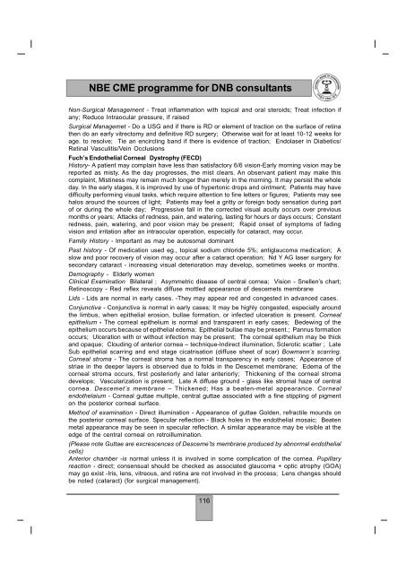
<strong>NBE</strong> <strong>CME</strong> <strong>programme</strong> <strong>for</strong> <strong>DNB</strong> <strong>consultants</strong>Non-Surgical Management - Treat inflammation with topical and oral steroids; Treat infection ifany; Reduce Intraocular pressure, if raisedSurgical Managemet - Do a USG and if there is RD or element of traction on the surface of retinathen do an early vitrectomy and definitive RD surgery; Otherwise wait <strong>for</strong> at least 10-12 weeks <strong>for</strong>age. to resolve; Tie an encircling band if there is evidence of traction; Endolaser in Diabetics/Retinal Vasculitis/Vein OcclusionsFuch’s Endothelial Corneal Dystrophy (FECD)History- A patient may complain have less than satisfactory 6/6 vision-Early morning vision may bereported as misty. As the day progresses, the mist clears. An observant patient may make thiscomplaint, Mistiness may remain much longer than merely in the morning. It may persist the wholeday. In the early stages, it is improved by use of hypertonic drops and ointment; Patients may havedifficulty per<strong>for</strong>ming visual tasks, which require attention to fine letters or figures; Patients may seehalos around the sources of light; Patients may feel a gritty or <strong>for</strong>eign body sensation during partof or during the whole day; Progressive fall in the corrected visual acuity occurs over previousmonths or years; Attacks of redness, pain, and watering, lasting <strong>for</strong> hours or days occurs; Constantredness, pain, watering, and poor vision may be present; Rapid onset of symptoms of fadingvision and irritation after an intraocular operation, especially <strong>for</strong> cataract, may occur.Family History - Important as may be autosomal dominantPast history - <strong>Of</strong> medication used eg., topical sodium chloride 5%; antiglaucoma medication; Aslow and poor recovery of vision may occur after a cataract operation; Nd Y AG laser surgery <strong>for</strong>secondary cataract - increasing visual deterioration may develop, sometimes weeks or months.Demography - Elderly womenClinical Examination Bilateral ; Asymmetric disease of central cornea; Vision - Snellen’s chart;Retinoscopy - Red reflex reveals diffuse mottled appearance of descemets membraneLids - Lids are normal in early cases. -They may appear red and congested in advanced cases.Conjunctiva - Conjunctiva is normal in early cases; It may be highly congested, especially aroundthe limbus, when epithelial erosion, bullae <strong>for</strong>mation, or infected ulceration is present. Cornealepithelium - The corneal epithelium is normal and transparent in early cases; Bedewing of theepithelium occurs because of epithelial edema; Epithelial bullae may be present.; Pannus <strong>for</strong>mationoccurs; Ulceration with or without infection may be present; The corneal epithelium may be thickand opaque; Clouding of anterior cornea – technique-Indirect illumination, Sclerotic scatter ; LateSub epithelial scarring and end stage cicatrisation (diffuse sheet of scar) Bowmann’s scarring.Corneal stroma - The corneal stroma has a normal transparency in early cases; Appearance ofstriae in the deeper layers is observed due to folds in the Descemet membrane; Edema of thecorneal stroma occurs, first posteriorly and later anteriorly; Thickening of the corneal stromadevelops; Vascularization is present; Late A diffuse ground - glass like stromal haze of centralcornea. Descemet’s membrane – Thickened; Has a beaten-metal appearance. Cornealendothelaium - Corneal guttae multiple, central guttae associated with a fine stippling of pigmenton the posterior corneal surface.Method of examination - Direct illumination - Appearance of guttae Golden, refractile mounds onthe posterior corneal surface. Specular reflection - Black holes in the endothelial mosaic; Beatenmetal appearance may be seen in specular reflection. A similar appearance may be visible at theedge of the central corneal on retroillumination.(Please note Guttae are excrescences of Desceme’ts membrane produced by abnormal endothelialcells)Anterior chamber -is normal unless it is involved in some complication of the cornea. Pupillaryreaction - direct; consensual should be checked as associated glaucoma + optic atrophy (GOA)may go exist -Iris, lens, vitreous, and retina are not involved in the process; Lens changes shouldbe noted (cataract) (<strong>for</strong> surgical management).116