NBE CME programme for DNB consultants - National Board Of ...
NBE CME programme for DNB consultants - National Board Of ...
NBE CME programme for DNB consultants - National Board Of ...
Create successful ePaper yourself
Turn your PDF publications into a flip-book with our unique Google optimized e-Paper software.
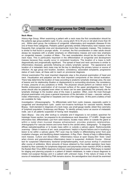
<strong>NBE</strong> <strong>CME</strong> <strong>programme</strong> <strong>for</strong> <strong>DNB</strong> <strong>consultants</strong>Neck MassHistory-Age Group, When examining a patient with a neck mass the first consideration should bethe patients age group pediatric (upto 15 yrs), young adult (16 to 45 yrs) or old adult (more than 40yrs). Within each group, the incidence of congenital, inflammatory and neoplastic diseases fit intoone of these three categories. Pediatric patient generally exhibits inflammatory neck masses morefrequently than congenital ones and developmental more than neoplastic masses. This incidenceis similar to that found in younger adults. In contrast, the first consideration in older adults shouldalways be neoplasia with a smaller emphasis on inflammatory masses and even less emphasison congenital masses.; Location of Mass - The next consideration should be the location of theneck mass. This is particularly important in the differentiation of congenital and developmentmasses because they usually occur in consistent locations. The location of a mass is bothdiagnostically and prognostically significant. The spread of head and neck carcinoma is similar toinflammatory diseases, generally following an orderly lymphatic spread. The appearance andlocation of a metastatic neck mass may be the key to identifying the primary tumour or source ofinfection. As age group and location, duration, consistency, associated complaints, progressivestatus of neck mass, all these add to reach on provisional diagnosis.Clinical examination-The most important diagnostic step is the physical examination of head andneck. Visualization and palpation are the most important components of the clinical evaluation.There help determine the location of mass according to anatomic lymphatic drainage area, the sizeof lesions and its relationship (fixation or displacement) to surrounding structures, the consistencyof mass, presume of any pulsations or thrills. The physician should per<strong>for</strong>m an indirect mirror offlexible endoscopics examination of all mucosal surface of the upper aerodigestive tract. Theseareas should also be palpated even when no lesion can be seen specifically the primarily site <strong>for</strong>lymphatic drainage to the location area of the mass is question. <strong>Of</strong>ten, even the most thoroughphysical examination only gives a general impression of the derivation of mass – vascular, salivary,nodal; inflammatory, congenital or neoplastic and not a firm diagnosis. At this point a battery of testare available <strong>for</strong> help.Investigation- Ultrasonography- To differentiate solid from cystic masses, especially useful incongenital and development cysts useful non-invasive technique <strong>for</strong> vascular lesions; NeedleBiopsy- Gold standard in diagnosis of a neck mass; use small gauge needle; obtain flow cytometryof lymphoid population; Endoscopy and Biopsy- To identify primary tumor as source of metastaticnode, use in all patients suspected of having neck neoplasia.Open Biopsy- Use only after workup is complete and if diagnosis is not evident; specimen <strong>for</strong>histologic frozen section, be prepared to do simultaneous neck dissection. CT & MRI- Single mostin<strong>for</strong>mative test; differentiates cyst from solid lesions; locates mass within or outside the gland orwithin a nodal chain mucosal disease enhancement, provides anatomic relationships.Arteriography-For vascular lesions and tumor fixed to the carotid artery. Sialography- To diagnosisdiffuse sialoadenopathies or to locate mass within or outside a salivery gland. Radionucleotidescanning- Obtain in lesions of ant. neck compartment, helpful in thyroid lesions and in localizing alesion to be within a salivary gland, PET scan may be helpful in differentiating tumor from postirradiation changes, and identifying distant metastasis. X-Ray, Plain- Rarely of help in differentiatingneck masses. Culture and Sensitivites- Inflammatory tissue at open biopsy.Skin Tests- Usedwhen chronic and granulomatous inflammatory lesion is suspected. Antibiotic Course- Clinicaltest <strong>for</strong> suspected inflammatory bacterial lymaphadenopathy, must pursue workup if unsolovedafter course of antibiotics. Micro amounts of tissue obtained by fine needle aspiration have beenstudied by flow cytometry <strong>for</strong> lymphoma diagnosis and polymerase chain reaction (PCR) to identifythe Epstein Barr Virus (EBV) diagnosis of primary nasopharyngeal carcinoma. FNA biopsy is needed<strong>for</strong> staging and planning purpose in a patient of distant metastasis, to make a tissue diagnosis toinitiate non-surgical therapy and in a patient with unknown mass. If the nature of mass or thesource of metastasis identified by FNA remains elusive, the aerodigestive tract should be examined97


