NBE CME programme for DNB consultants - National Board Of ...
NBE CME programme for DNB consultants - National Board Of ...
NBE CME programme for DNB consultants - National Board Of ...
You also want an ePaper? Increase the reach of your titles
YUMPU automatically turns print PDFs into web optimized ePapers that Google loves.
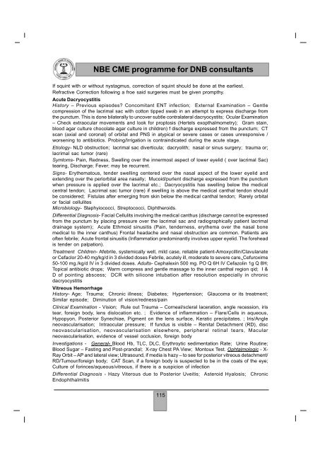
<strong>NBE</strong> <strong>CME</strong> <strong>programme</strong> <strong>for</strong> <strong>DNB</strong> <strong>consultants</strong>If squint with or without nystagmus, correction of squint should be done at the earliest.Refractive Correction following a froe said surgeries must be given prompthy.Acute DacryocystitisHistory – Previous episodes? Concomitant ENT infection; External Examination – Gentlecompression of the lacrimal sac with cotton tipped swab in an attempt to express discharge fromthe punctum. This is done bilaterally to uncover subtle contralateral dacryocystits; Ocular Examination– Check extraocular movements and look <strong>for</strong> proptosis (Hertels exopthalmometry); Gram stain,blood agar culture chocolate agar culture in children) f discharge expressed from the punctum; CTscan (axial and coronal) of orbital and PNS in atypical or severe cases or cases unresponsive /worsening to antibiotics. Probing/Irrigation is contraindicated during the acute stage.Etiology- NLD obstruction; lacrimal sac diverticula; dacryolith; nasal or sinus surgery; trauma or;lacrimal sac tumor (rare)Symtoms- Pain, Redness, Swelling over the innermost aspect of lower eyelid ( over lacrimal Sac)tearing, Discharge; Fever; may be recurrent.Signs- Erythematous, tender swelling centered over the nasal aspect of the lower eyelid andextending over the periorbital area nasally; Mucoid/purlent discharge expressed from the punctumwhen pressure is applied over the lacrimal etc.; Dacryocystitis has swelling below the medicalcentral tendon; Lacrimal sac tumor (rare) if swelling is above the medical canthal tendon shouldbe considered; Fistulas after emerging from skin below the medical canthal tendon; Rarely orbitalor facial cellulitesMicrobiology- Staphyiococci, Streptococci, Diphtheroids.Differential Diagnosis- Facial Cellulits involving the medical canthus (discharge cannot be expressedfrom the punctum by placing pressure over the lacrimal sac and radiographically patient lacrimaldrainage system); Acute Ethmoid sinusitis (Pain, tenderness, erythema over the nasal bonemedical to the inner canthus) Frontal headache and nasal obstruction are common. Patients areoften febrile; Acute frontal sinusitis (Inflammation predominantly involves upper eyelid. The <strong>for</strong>eheadis tender on palpation).Treatment Children- Afebrile, systemically well, mild case, reliable patient-Amoxycillin/Clavulanateor Cefaclor 20-40 mg/kg/d in 3 divided doses Febrile, acutely ill, moderate to severe care, Cefuroxime50-100 mg./kg/d IV in 3 divided doses. Adults- Cephalexin 500 mg. PO Q 6H IV Cefazolin 1g Q 8H;Topical antibiotic drops; Warm compress and gentle massage to the inner canthal region qid; I &D of pointing abscess; DCR with silicone intubation after resolution especially in chronicdacryocystitisVitreous HemorrhageHistory- Age; Trauma; Chronic illness; Diabetes; Hypertension; Glaucoma or its treatment;Similar episode; Diminution of vision/redness/painClinical Examination - Vision; Rule out Trauma – Corneal/scleral laceration, angle recession, iristear, <strong>for</strong>eign body, lens dislocation etc. ; Evidence of inflammation – Flare/Cells in aqueous,Hypopyon, Posterior Synechiae, Pigment on the lens surface, Keratic precipitates. ; Iris/Angleneovascularisation; Intraocular pressure; If fundus is visible – Renital Detachment (RD), discneovascularisation, neovascularisation elsewhere, peripheral retinal tears, Macularneovascularisation, evidence of vessel occlusion, <strong>for</strong>eign bodyInvestigations - General- Blood Hb, TLC, DLC, Erythroytic sedimentation Rate; Urine Routine;Blood Sugar – Fasting and Post-prandial; X-ray Chest PA View; Montoux Test. Ophtalmologic - X-Ray Orbit – AP and lateral view; Ultrasound, if media is hazy – to see <strong>for</strong> posterior vitreous detachment/RD/Tumour/<strong>for</strong>eign body; CAT Scan, if a <strong>for</strong>eign body is suspected to be in the coats of the eye;Culture of <strong>for</strong>inces/aqueous/vitreous, if there is a suspicion of infectionDifferential Diagnosis - Hazy Viterous due to Posterior Uveitis; Asteroid Hyalosis; ChronicEndophthalmitis115


