NBE CME programme for DNB consultants - National Board Of ...
NBE CME programme for DNB consultants - National Board Of ...
NBE CME programme for DNB consultants - National Board Of ...
You also want an ePaper? Increase the reach of your titles
YUMPU automatically turns print PDFs into web optimized ePapers that Google loves.
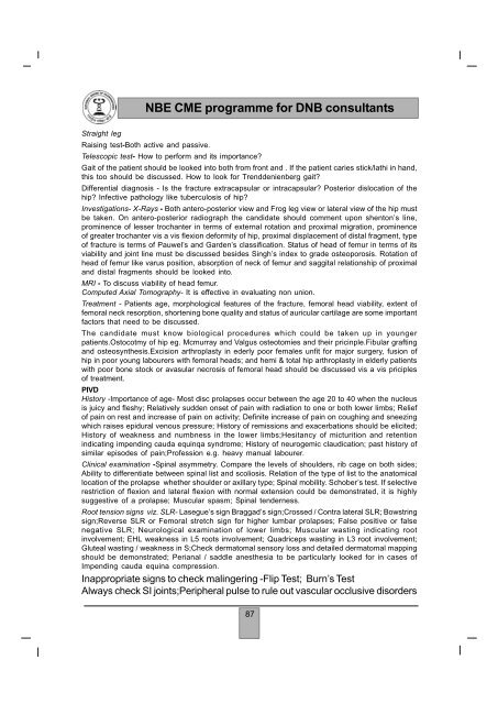
<strong>NBE</strong> <strong>CME</strong> <strong>programme</strong> <strong>for</strong> <strong>DNB</strong> <strong>consultants</strong>Straight legRaising test-Both active and passive.Telescopic test- How to per<strong>for</strong>m and its importance?Gait of the patient should be looked into both from front and . If the patient caries stick/lathi in hand,this too should be discussed. How to look <strong>for</strong> Trenddenienberg gait?Differential diagnosis - Is the fracture extracapsular or intracapsular? Posterior dislocation of thehip? Infective pathology like tuberculosis of hip?Investigations- X-Rays - Both antero-posterior view and Frog leg view or lateral view of the hip mustbe taken. On antero-posterior radiograph the candidate should comment upon shenton’s line,prominence of lesser trochanter in terms of external rotation and proximal migration, prominenceof greater trochanter vis a vis flexion de<strong>for</strong>mity of hip, proximal displacement of distal fragment, typeof fracture is terms of Pauwel’s and Garden’s classification. Status of head of femur in terms of itsviability and joint line must be discussed besides Singh’s index to grade osteoporosis. Rotation ofhead of femur like varus position, absorption of neck of femur and saggital relationship of proximaland distal fragments should be looked into.MRI - To discuss viability of head femur.Computed Axial Tomography- It is effective in evaluating non union.Treatment - Patients age, morphological features of the fracture, femoral head viability, extent offemoral neck resorption, shortening bone quality and status of auricular cartilage are some importantfactors that need to be discussed.The candidate must know biological procedures which could be taken up in youngerpatients.Ostocotmy of hip eg. Mcmurray and Valgus osteotomies and their pricinple.Fibular graftingand osteosynthesis.Excision arthroplasty in ederly poor females unfit <strong>for</strong> major surgery, fusion ofhip in poor young labourers with femoral heads; and hemi & total hip arthroplasty in elderly patientswith poor bone stock or avasular necrosis of femoral head should be discussed vis a vis priciplesof treatment.PIVDHistory -Importance of age- Most disc prolapses occur between the age 20 to 40 when the nucleusis juicy and fleshy; Relatively sudden onset of pain with radiation to one or both lower limbs; Reliefof pain on rest and increase of pain on activity; Definite increase of pain on coughing and sneezingwhich raises epidural venous pressure; History of remissions and exacerbations should be elicited;History of weakness and numbness in the lower limbs;Hesitancy of micturition and retentionindicating impending cauda equinqa syndrome; History of neurogemic claudication; past history ofsimilar episodes of pain;Profession e.g. heavy manual labourer.Clinical examination -Spinal asymmetry. Compare the levels of shoulders, rib cage on both sides;Ability to differentiate between spinal list and scoliosis. Relation of the type of list to the anatomicallocation of the prolapse whether shoulder or axillary type; Spinal mobility. Schober’s test. If selectiverestriction of flexion and lateral flexion with normal extension could be demonstrated, it is highlysuggestive of a prolapse; Muscular spasm; Spinal tenderness.Root tension signs viz. SLR- Lasegue’s sign Braggad’s sign;Crossed / Contra lateral SLR; Bowstringsign;Reverse SLR or Femoral stretch sign <strong>for</strong> higher lumbar prolapses; False positive or falsenegative SLR; Neurological examination of lower limbs; Muscular wasting indicating rootinvolvement; EHL weakness in L5 roots involvement; Quadriceps wasting in L3 root involvement;Gluteal wasting / weakness in S;Check dermatomal sensory loss and detailed dermatomal mappingshould be demonstrated; Perianal / saddle anesthesia to be particularly looked <strong>for</strong> in cases ofImpending cauda equina compression.Inappropriate signs to check malingering -Flip Test; Burn’s TestAlways check SI joints;Peripheral pulse to rule out vascular occlusive disorders87


