Peptidoglycan .Types of Bacterial Cell Walls and their Taxonomic ...
Peptidoglycan .Types of Bacterial Cell Walls and their Taxonomic ...
Peptidoglycan .Types of Bacterial Cell Walls and their Taxonomic ...

Create successful ePaper yourself
Turn your PDF publications into a flip-book with our unique Google optimized e-Paper software.
458 SCHLEIFER AND KANDLER<br />
BACTERIOL. REV.<br />
around the cell. With regard to the Gram<br />
reaction, the spirochaetes are presumably<br />
gram-negative. Thus, it was expected that the<br />
peptidoglycan would show the directly crosslinked<br />
m-Dpm type. But already qualitative<br />
studies on cell walls <strong>of</strong> Treponema reiteri have<br />
yielded ornithine instead <strong>of</strong> m-Dpm as the<br />
major diamino acid (382). Recent studies on the<br />
ultrastructure <strong>and</strong> chemical composition <strong>of</strong> the<br />
cell wall <strong>of</strong> Spirochaeta stenostrepta have confirmed<br />
the presence <strong>of</strong> L-Orn as constituent <strong>of</strong><br />
the peptidoglycan (166). Two layers <strong>of</strong> the cell<br />
wall <strong>of</strong> Spirochaeta stenostrepta were isolated<br />
<strong>and</strong> analyzed. The outermost <strong>of</strong> these two<br />
layers consists mainly <strong>of</strong> lipoprotein. The second<br />
layer was characterized as peptidoglycan.<br />
It is a thin structure or monolayer as in typical<br />
gram-negative organisms which retains the cylindrical<br />
<strong>and</strong> coiled shape <strong>of</strong> the cell. Studies on<br />
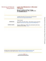
the amino acid sequence <strong>of</strong> the purified peptidoglycan<br />
revealed that the peptide subunits<br />
consist <strong>of</strong> tripeptides (L-Ala-y-D-Glu-L-Orn)<br />
<strong>and</strong> tetrapeptides (L-Ala-'y-D-Glu-L-Orn-D-Ala).<br />
The peptide subunits are directly cross-linked<br />
(Fig. 28). The primary structure <strong>of</strong> the peptidoglycan<br />
resembles that <strong>of</strong> other gram-negative<br />
bacteria with the only difference being that<br />
m-Dpm is replaced by L-Orn.<br />
Order Myxobacteriales. Since the myxobacteria<br />
are flexible cells, it has been assumed<br />
that the cell walls are quite different from those<br />
<strong>of</strong> eubacteria <strong>and</strong> that the peptidoglycan may<br />
be lacking. But even the first studies on the cell<br />
walls <strong>of</strong> myxobacterial strains showed that a<br />
peptidoglycan layer is probably present in <strong>their</strong><br />
walls (7, 239). More detailed studies on myxobacterial<br />
cell walls were reported by Verma<br />
<strong>and</strong> Martin (397) <strong>and</strong> by White et al. (415). The<br />
-G-M-G-<br />
I<br />
L- Ala<br />
4<br />
D-Glu<br />
'Y 6<br />
L-Orn +-D-Ala<br />
at<br />
L-Orn<br />
FIG. 28. Fragment <strong>of</strong> the primary structure <strong>of</strong> the<br />
peptidoglycan <strong>of</strong> Spirochaeta stenostrepta.<br />
results demonstrated that in all species so far<br />
examined (Cytophaga hutchinsonii, Sporocytophaga<br />
myxococcoides, <strong>and</strong> Myxococcus<br />
xanthus), the directly cross-linked, m-Dpmcontaining<br />
peptidoglycan type occurs (Fig. 6).<br />
In the case <strong>of</strong> C. hutchinsonii <strong>and</strong> S. myxococcoides,<br />
a discrete peptidoglycan layer was isolated<br />
(397). The vegetative cell walls <strong>of</strong> M.<br />
xanthus, on the other h<strong>and</strong>, completely disaggregated<br />
after treatment with trypsin <strong>and</strong> detergent.<br />
White et al. (415) suggested, therefore,<br />
that the rigid layer <strong>of</strong> the vegetative cell wall <strong>of</strong><br />
M. xanthus is not a continous peptidoglycan<br />
layer but consists <strong>of</strong> patches <strong>of</strong> peptidoglycan<br />
separated by nonpeptidoglycan material. This<br />
patchlike arrangement may be related to the<br />
flexibility <strong>of</strong> the myxobacterial cell. In the case<br />
<strong>of</strong> C. hutchinsonii <strong>and</strong> S. myxococcoides, however,<br />
the flexibility is explained by the occurrence<br />
<strong>of</strong> "naked tubes <strong>of</strong> murein (peptidoglycan)<br />
monolayers" (397).<br />
With the exception <strong>of</strong> the genus Cytophaga,<br />
all myxobacteria can form resting cells. These<br />
resting cells, the so-called microcysts, are produced<br />
from single vegetative cells. The microcysts<br />
are shorter than the vegetative cells<br />
<strong>and</strong> <strong>their</strong> cell walls are much thicker <strong>and</strong><br />
inflexible. In both Sporocytophaga <strong>and</strong> Myxococcus,<br />
not only the vegetative cell walls but<br />
also the cell walls <strong>of</strong> microcysts were studied.<br />
Verma <strong>and</strong> Martin (397) explained the transition<br />
<strong>of</strong> the flexible, vegetative cell wall <strong>of</strong><br />
Sporocytophaga to the rigid, thick microcyst<br />
cell wall by the superposition <strong>of</strong> several peptidoglycan<br />
layers. White et al. (415), on the<br />
other h<strong>and</strong>, found in Myxococcus, during the<br />
change from the vegetative cell to the microcyst,<br />
a temporary decrease in the cross-linkage<br />
<strong>of</strong> the peptidoglycan. They assume a high<br />
rate <strong>of</strong> turnover <strong>of</strong> the peptidoglycan during the<br />
formation <strong>of</strong> the microcyst <strong>and</strong> simultaneously<br />
some uncross-linked peptidoglycan appears. A<br />
newly synthesized extracellular layer <strong>of</strong> nonpeptidoglycan<br />
material serves to strengthen the<br />
microcyst wall. Moreover, the authors stated<br />
that "the patch-like arrangement <strong>of</strong> the peptidoglycan<br />
may be related to the change in<br />
shape when M. xanthus converts from the<br />
vegetative rod to the spherical cyst." They<br />
think that the nonpeptidoglycan areas perform<br />
a necessary role in the morphogenesis <strong>of</strong> the<br />
wall.<br />
FINAL REMARKS<br />
<strong>Taxonomic</strong> Implications <strong>of</strong> Other <strong>Cell</strong><br />
Wall Polymers<br />
Lipopolysaccharides. A good correlation<br />
between the structure <strong>of</strong> the lipopolysaccha-<br />
Downloaded from<br />
http://mmbr.asm.org/<br />
on December 3, 2012 by guest