Download PDF - Carl Zeiss
Download PDF - Carl Zeiss
Download PDF - Carl Zeiss

You also want an ePaper? Increase the reach of your titles
YUMPU automatically turns print PDFs into web optimized ePapers that Google loves.
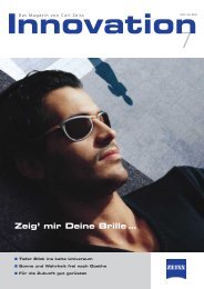
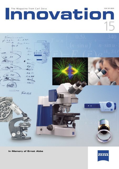
Innovation<br />
The Magazine from <strong>Carl</strong> <strong>Zeiss</strong><br />
In Memory of Ernst Abbe<br />
ISSN 1431-8059<br />
15
Contents<br />
2<br />
Editorial<br />
Formulas for Success. . . ❚ Dieter Brocksch 3<br />
In Memory of Ernst Abbe<br />
Ernst Abbe 4<br />
Microscope Lenses 8<br />
Numerical Aperture, Immersion and Useful Magnification ❚ Rainer Danz 12<br />
Highlights from the History of Immersion Objectives 16<br />
From the History of Microscopy:<br />
Abbe’s Diffraction Experiments ❚ Heinz Gundlach 18<br />
The Science of Light 24<br />
Stazione Zoologica Anton Dohrn, Naples, Italy 26<br />
Felix Anton Dohrn 29<br />
The Hall of Frescoes ❚ Christiane Groeben 30<br />
Bella Napoli 31<br />
From Users<br />
The Zebra Fish as a Model Organism for Developmental Biology 32<br />
SPIM – A New Microscope Procedure 34<br />
The Scourge of Back Pain –<br />
Treatment Methods and Innovations 38<br />
ZEISS in the Center for Book Preservation ❚ Manfred Schindler 42<br />
Across the Globe<br />
<strong>Carl</strong> <strong>Zeiss</strong> Archive Aids Ghanaian Project ❚ Peter Gluchi 46<br />
Prizes and Awards<br />
100 Years of Brock & Michelsen 48<br />
Award for NaT Working Project 49<br />
Product Report<br />
Digital Pathology: MIRAX SCAN 50<br />
UHRTEM 50<br />
Superlux Eye Xenon Illumination 50<br />
<strong>Carl</strong> <strong>Zeiss</strong> Optics in Nokia Mobile Phones 51<br />
Masthead 51<br />
Innovation 15, <strong>Carl</strong> <strong>Zeiss</strong> AG, 2005
Formulas for Success...<br />
Formulas describe the functions and processes of what<br />
happens in the world and our lives. It is often the small,<br />
insignificant formulas in particular that play a decisive<br />
role in what we know and in the functionality of modern<br />
instruments and examination methods.<br />
for the large...<br />
The fiftieth anniversary of the death of Albert Einstein<br />
(1879-1955) also marks the centennial of his theory of<br />
relativity; a theory that revolutionized perceptions, made<br />
the processes of life more understandable and began to<br />
explain the dimensions of time and space. The short formula,<br />
E =m·c 2, expresses the infinite complexity of our<br />
world. Einstein had contact with <strong>Zeiss</strong> throughout the<br />
course of his scientific activities. In 1925 he wrote to the<br />
company Anschütz in Kiel about producing a gyrocompass:<br />
“The difficulties of manufacturing are so great –<br />
accuracies of 10 -4 have to be achieved – that <strong>Zeiss</strong> is<br />
currently the only company capable of meeting the requirements.“<br />
... and small things in life.<br />
2005 also marks the 100th anniversary of the death of<br />
Ernst Abbes (1840-1905). Numerous events throughout<br />
2005 honor his many great achievements. His extensive<br />
examinations within the scope of his activities at <strong>Carl</strong><br />
<strong>Zeiss</strong>’ optical workshop also resulted in the formula for<br />
the resolution of a microscope: �<br />
d =<br />
2n sin �<br />
Innovation 15, <strong>Carl</strong> <strong>Zeiss</strong> AG, 2005<br />
Editorial<br />
It clearly and concisely describes the resolution of optical<br />
instruments using the visible spectrum of light and contributed<br />
to the improvement of optical devices.<br />
The same year also saw the passing of another German<br />
microscope manufacturer with connections to Abbe and<br />
<strong>Zeiss</strong>: Rudolf Winkel (1827-1905). During Abbe’s times,<br />
good microscopes were also built at Winkel’s workshop<br />
which was founded in Göttingen in 1857. Abbe visited<br />
Winkel’s workshop while he was a student in Göttingen.<br />
His visit in 1894 led to closer cooperation. In 1911, <strong>Zeiss</strong><br />
became Winkel’s chief partner. In October 1957, the<br />
firm R. Winkel GmbH became part of the <strong>Carl</strong> <strong>Zeiss</strong><br />
Foundation.<br />
The same year that Abbe died, Robert Koch (1843-<br />
1919) received the Nobel Prize for Medicine for his examinations<br />
and discoveries while researching tuberculosis. In<br />
1878, Robert Koch used the Abbe oil immersion system<br />
for the first time and was impressed by the “quantum<br />
leap“ made by the “<strong>Carl</strong> <strong>Zeiss</strong> Optical Workshop using<br />
Professor Abbe’s ingenious advice.“ In 1904, <strong>Carl</strong> <strong>Zeiss</strong><br />
management presented Robert Koch with the 1000 th<br />
1/12 objective lens for homogenous oil immersion.<br />
Many articles in this issue are dedicated to Ernst Abbe<br />
and his times. We reflect on what Ernst Abbe meant to<br />
<strong>Carl</strong> <strong>Zeiss</strong> and what he did for optics, and we take a special<br />
look at developments that were and continue to be<br />
significantly influenced by Ernst Abbe and his scientific<br />
results. This is emphasized by the image on the cover<br />
pages: an historical tribute to the more than 150 years of<br />
optical development with a focus on microscopy.<br />
July 2005<br />
Dr. Dieter Brocksch<br />
Scientist<br />
Entrepreneur<br />
Social Reformer<br />
3
In Memory of Ernst Abbe<br />
4<br />
Education and<br />
early years<br />
Ernst Abbe was born in Eisenach on<br />
January 23, 1840, as the son of<br />
master spinner and subsequent factory<br />
attendant Georg Adam Abbe<br />
and his wife Christina. He attended<br />
elementary school from 1846 to<br />
1850, after which he was a student<br />
at the local high school in Eisenach.<br />
He finished his school education in<br />
1857 and graduated with aboveaverage<br />
grades. In the period to<br />
1861 he studied mathematics and<br />
physics at the Universities of Jena<br />
In 2005, the book titled “Ernst<br />
Abbe – Scientist, Entrepreneur<br />
and Social Reformer” was<br />
published by Bussert &<br />
Stadeler, Jena Quedlinburg<br />
to mark the 100 th anniversary<br />
of Ernst Abbe’s death.<br />
[ISBN 3-932906-57-8]<br />
(1857-1859) and Göttingen (1859-<br />
1861). Abbe completed his studies by<br />
obtaining his doctorate in Göttingen<br />
on the subject “Experiential substantiation<br />
of the theorem of equivalence<br />
between heat and mechanical energy“.<br />
He subsequently worked for two<br />
years as a teacher at the Physics<br />
Association in Frankfurt/Main.<br />
Family and<br />
science<br />
Abbe’s mother died early in his life:<br />
July 14, 1857. During his studies, his<br />
father married for the second time, to<br />
the widow Eva Margarethe Liebetrau<br />
on November 11, 1859. After his<br />
time in Frankfurt, Abbe joined the<br />
Mathematical Association in Jena in<br />
1863. In the same year, he obtained<br />
his post-doctoral qualification as a<br />
lecturer with his paper on “the laws<br />
in the distribution of errors in observation<br />
series“. He taught as a private<br />
mathematics and physics lecturer at<br />
the University of Jena.<br />
In 1863 Abbe also became a member<br />
of the Jena Association of Medi-<br />
1857<br />
cine and Science, in which he gave a<br />
total of 45 lectures in the period to<br />
1895. From 1866, he was a freelance<br />
scientist with the court and university<br />
mechanic <strong>Carl</strong> <strong>Zeiss</strong> in Jena. In 1870<br />
Abbe formulated the famous sine<br />
condition subsequently named after<br />
him, a condition that must be met by<br />
any spherically corrected lens if it is<br />
also to be free from coma in<br />
the neighborhood of the lens axis in<br />
microscope image formation. In the<br />
same year he became an extraordinary<br />
professor at the University of<br />
Jena. On September 24, 1871, he<br />
married Elise Snell, the daughter of lecturer<br />
Prof. Snell, a mathematics and<br />
physics lecturer at the University of<br />
Jena. Three years later, the first child,<br />
his daughter Paula, was born. His father<br />
died on August 18, 1874. Abbe<br />
was director of Jena Observatory<br />
from 1877 to 1900. On May 1, 1878,<br />
he was appointed as an honorary<br />
member of the London Royal Microscopical<br />
Society. In June of the same<br />
year, he became an ordinary honorary<br />
professor at the University of<br />
Jena. He was awarded the title of Dr.<br />
Innovation 15, <strong>Carl</strong> <strong>Zeiss</strong> AG, 2005
1863 1870 1875<br />
med. h. c. by the University of Halle<br />
in 1883 and the title Dr. jur. h. c. by<br />
the University of Jena in 1886. In<br />
1900 Abbe became a corresponding<br />
member of the Imperial Austrian<br />
Academy of Science in Vienna. In<br />
1901 he was appointed as an honorary<br />
member of the Saxon Academy<br />
of Science and of the Academy of<br />
Science in Göttingen.<br />
Abbe as an<br />
entrepreneur<br />
The process of integrating science<br />
into industry already started in the<br />
1860s. In addition to <strong>Carl</strong> <strong>Zeiss</strong>,<br />
Siemens and Bayer were also pioneers<br />
of this development. By hiring<br />
scientific staff, Ernst Abbe was a<br />
decisive driving force behind this<br />
process at <strong>Zeiss</strong>: the integration of<br />
R&D into the company was an important<br />
step toward technology leadership.<br />
The training of capable employees<br />
and successors also played a<br />
significant role in the entrepreneurial<br />
and commercial areas. Competent<br />
staff and constant quality control<br />
Innovation 15, <strong>Carl</strong> <strong>Zeiss</strong> AG, 2005<br />
allowed the implementation of high<br />
quality standards. The corporate organization<br />
was successfully focused<br />
on growth by the clear allocation of<br />
responsibilities for scientific, technical<br />
and commercial staff.<br />
From 1872, all ZEISS microscopes<br />
were built in line with Abbe’s calculations.<br />
Three years later, in 1875,<br />
Abbe became a dormant partner in<br />
the Optical Workshop of <strong>Carl</strong> <strong>Zeiss</strong>.<br />
Abbe pledged not to increase his academic<br />
activity beyond the current<br />
measure and not to accept a professorship<br />
in Jena or elsewhere. One<br />
year later, he traveled to London to<br />
attend the international exposition of<br />
scientific instruments on behalf of<br />
the Prussian Department of Education.<br />
In 1878, due to his obligations<br />
at <strong>Zeiss</strong>, he turned down the offer of<br />
a post as professor in Berlin instigated<br />
by Hermann von Helmholtz. He<br />
also declined the offer of an ordinary<br />
professorship in Jena.<br />
He initially came into contact with<br />
Dr. Otto Schott in 1879. Their collaboration<br />
began one year later. In 1882<br />
a private glass laboratory was set up<br />
1880<br />
for Otto Schott in Jena. The following<br />
year saw the signing of the new partnership<br />
agreement in which Abbe<br />
became an active partner together<br />
with <strong>Carl</strong> and Roderich <strong>Zeiss</strong>.<br />
In 1884 the Glastechnische Laboratorium<br />
Schott & Gen. (later to become<br />
Jenaer Glaswerk Schott &<br />
Gen.) was founded by Otto Schott,<br />
Ernst Abbe, <strong>Carl</strong> <strong>Zeiss</strong> and Roderich<br />
<strong>Zeiss</strong>.<br />
In search of calcium fluoride for<br />
optical applications, Abbe traveled to<br />
Oltscherenalp in Switzerland for the<br />
first time in 1886. After the death of<br />
<strong>Carl</strong> <strong>Zeiss</strong> on December 3, 1888,<br />
Abbe became the sole owner of the<br />
<strong>Zeiss</strong> works in 1889. At the same<br />
time, he discontinued his teaching<br />
activities at the University of Jena.<br />
From 1890 onwards, Abbe expanded<br />
the product spectrum on an ongoing<br />
basis: measuring instruments (1890),<br />
camera lenses (1890), binoculars<br />
(1894), astronomical instruments<br />
(1897) and photogrammetric instruments<br />
(1901). As a result, the number<br />
of employees rose to over 2000<br />
by 1905.<br />
5
special<br />
6<br />
Global Player<br />
As many as 100 years ago, <strong>Carl</strong> <strong>Zeiss</strong> was already<br />
what would now be called a global player: the first<br />
sales branches were founded in London (1894),<br />
Vienna (1902) and St. Petersburg (1903). Today, the<br />
company has 15 production facilities in Germany,<br />
USA, Hungary, Switzerland, Mexico, Belarus<br />
and China as well as 35 sales organizations and<br />
100 agencies across the globe.<br />
Abbe’s persistence in his endeavors<br />
to also provide other manufacturers<br />
with new types of optical glass was<br />
of great help to the German optical<br />
industry. He was skeptical about the<br />
patenting of products, which he<br />
saw as an obstruction to scientific<br />
progress in general. Not until competitive<br />
pressure made it unavoidable did<br />
the patenting of camera lenses and<br />
binoculars begin. However, his early<br />
pioneering work remained accessible<br />
for general use. With the aid of<br />
Abbe’s comparator principle, instruments<br />
for the highly accurate measurement<br />
of workpieces were produced.<br />
These were important aids for<br />
instrument construction in Germany.<br />
Abbe retired in 1903. To mark this<br />
occasion, a torchlight procession took<br />
place with 1500 employees of the<br />
Foundation companies. Two years<br />
later, Ernst Abbe died on January 14,<br />
1905, after a long, serious illness.<br />
Abbe the social<br />
reformer and the<br />
<strong>Carl</strong> <strong>Zeiss</strong> Foundation<br />
Many companies introduced their<br />
social policy in the late 19 th century.<br />
1888 1901<br />
As a reformer, Abbe was far ahead of<br />
his times with his socio-political<br />
ideas. In 1889, in order to safeguard<br />
the existence of the enterprises <strong>Carl</strong><br />
<strong>Zeiss</strong> and SCHOTT irrespective of personal<br />
ownership interests, Abbe set<br />
up the <strong>Carl</strong> <strong>Zeiss</strong> Foundation which<br />
he made the sole owner of the <strong>Zeiss</strong><br />
works and partial owner of the<br />
SCHOTT works in 1891. In the same<br />
year, Abbe transferred his industrial<br />
assets to the <strong>Carl</strong> <strong>Zeiss</strong> Foundation<br />
With the appropriate compensation,<br />
Roderich <strong>Zeiss</strong> also transferred his<br />
shares to the Foundation, making it<br />
the sole owner of the firm <strong>Carl</strong> <strong>Zeiss</strong><br />
and partial owner (sole owner from<br />
1919) of Jena Glaswerk Schott &<br />
Gen. Until 1903, Abbe was the authorized<br />
representative and one of<br />
the three directors of the <strong>Carl</strong> <strong>Zeiss</strong><br />
Foundation. The corporate statute of<br />
the Foundation came into force in<br />
1896. The first supplementary statute<br />
followed just four years later.<br />
With his corporate statute of<br />
1896, Abbe gave the enterprise a<br />
unique constitution. In addition to its<br />
exceptionally progressive stipulations<br />
concerning corporate management<br />
and legally anchored labor relations,<br />
the constitution also reflected Abbe’s<br />
social commitment. For example, a<br />
council was set up to represent the<br />
interests of employees. Although this<br />
could not be seen as codetermination<br />
in the modern sense of the term, it<br />
did entitle the representatives to<br />
voice their opinion in all matters concerning<br />
the enterprise. Paid vacation,<br />
profit sharing, a documented entitlement<br />
to pension payments, continued<br />
pay in the event of illness and,<br />
from 1900, the eight-hour working<br />
day were further social milestones.<br />
This all made the Foundation enterprises<br />
<strong>Carl</strong> <strong>Zeiss</strong> and SCHOTT forerunners<br />
of modern social legislation.<br />
Tolerance was central to Ernst<br />
Abbe’s basic philosophy of life. Although<br />
he was certainly not a social<br />
democrat, it was important to him<br />
that this political party was able to<br />
evolve and develop freely. He was<br />
also vehemently against racism, a<br />
phenomenon which was already<br />
prevalent during his times. He ensured<br />
that no-one at <strong>Carl</strong> <strong>Zeiss</strong> suffered<br />
in any way due to their origin,<br />
religion or political affiliation. This<br />
attitude was reflected in the fact<br />
that two of his closest management<br />
Innovation 15, <strong>Carl</strong> <strong>Zeiss</strong> AG, 2005
1905<br />
colleagues, Siegfried Czapski and<br />
Rudolf Straubel, were Jewish citizens.<br />
Promoter of science<br />
and research<br />
Abbe always attached great importance<br />
to the promotion not only of<br />
science and research, but also of culture.<br />
As a private citizen, Abbe supported<br />
the university with anonymous<br />
donations. The university and<br />
the city of Jena were both sponsored<br />
by the <strong>Carl</strong> <strong>Zeiss</strong> Foundation after its<br />
creation. As early as 1886, the promotion<br />
of science and research began<br />
with the secret “endowment<br />
fund for scientific purposes“.<br />
Innovation 15, <strong>Carl</strong> <strong>Zeiss</strong> AG, 2005<br />
special<br />
Products for<br />
Science<br />
Shortly after the development of<br />
the new microscopes, decisive<br />
breakthroughs were made in the<br />
field of bacteriology. In 1904<br />
Robert Koch wrote: “I owe a large<br />
proportion of the success I have<br />
achieved for science to your excellent<br />
microscopes”. In the decades<br />
before the First World War medical<br />
research in Germany had a world<br />
standing that was paralleled only<br />
by the reputation enjoyed by<br />
ZEISS instruments. Emil Behring in<br />
the field of serology or Paul Ehrlich<br />
in the field of chemotherapy are<br />
only two examples of many. Needless<br />
to say, their success was not<br />
attributable to their instruments<br />
alone, but the microscopes did play<br />
an important role. The firm <strong>Carl</strong><br />
<strong>Zeiss</strong> also created products for the<br />
field of chemistry, some of which<br />
were customized solutions: the gas<br />
interferometer for Fritz Haber, for<br />
example.<br />
7
Microscope Objectives<br />
Figs 1-3:<br />
Early immerson objectives.<br />
William Hyde Wollaston<br />
(1766-1828),<br />
English physicist, chemist<br />
and philospher.<br />
8<br />
ao<br />
o<br />
The Wollaston prism of the DIC<br />
microscopy technique is named<br />
after him. Wollaston discovered<br />
the elements palladium and<br />
rhodium and was the first scientist<br />
to report about the dark<br />
lines in the solar spectrum.<br />
His way of viewing the geometric<br />
arrangement of atoms<br />
led him to crystallography and<br />
to the invention of the modern<br />
goniometer for the angular<br />
measurement of crystal surfaces.<br />
In 1806 he invented the<br />
Camera lucida, an optical aid<br />
for perspective drawing that<br />
allows the observation of an<br />
object on a drawing surface.<br />
In its basic structure, the<br />
Camera lucida is a glass prism<br />
with two reflecting surfaces<br />
inclined at an agle of 135° that<br />
generates the image of a scene<br />
at right angles to the eye of<br />
the observer.<br />
1 2 3<br />
The quality displayed by early<br />
microscope objectives was usually<br />
very modest. The images were<br />
slightly blurred. Producing a microscope<br />
objective required a lot<br />
of hard, intricate work until well<br />
into the second half of the 19 th<br />
century. The standard complex<br />
trial and error process needed to<br />
construct the optical systems was<br />
extremely time consuming and<br />
thus expensive.<br />
Sometime around 1810, Joseph Jackson<br />
Lister (1786-1869) referred to<br />
the connection between the angular<br />
aperture of the objective and the<br />
attainable resolution for the first<br />
time. Two years later, William Hyde<br />
Wollaston (1766-1828) improved the<br />
optics of the simple microscope:<br />
using Wollaston doublets, he intro-<br />
duced a new lens combination – it<br />
consisted of two plano-convex lenses<br />
with a stop in the middle. Sir David<br />
Brewster’s (1781-1868) idea of manufacturing<br />
objective lenses from diamonds<br />
was implemented in 1824 by<br />
Andrew Pritchard. Brewster recommended<br />
using oil immersion to secure<br />
achromatism in 1813. In 1816,<br />
Joseph von Fraunhofer (1770-1841)<br />
produced the first achromatic lens<br />
that could be used in microscopy. In<br />
1823, Paris based physicist Selligue<br />
combined up to four achromatic<br />
cemented elements into one objective<br />
– this was the breakthrough in<br />
the manufacture of achromatic microscope<br />
objectives with high resolution.<br />
In an age of increasing mechanization<br />
and the beginning of industrial<br />
manufacture, <strong>Carl</strong> <strong>Zeiss</strong> quickly<br />
recognized the link between theory<br />
and practice, between science and<br />
production required to effectively<br />
Innovation 15, <strong>Carl</strong> <strong>Zeiss</strong> AG, 2005
R 2<br />
manufacture powerful instruments<br />
that would be able to stand up to<br />
the pressures of competition. <strong>Zeiss</strong><br />
looked hard for a solution. The initial<br />
attempts to compute microscope objectives<br />
between 1850 and 1854 by<br />
himself and his friend, mathematician<br />
Friedrich Wilhelm Barfuss, however,<br />
did not return any noteworthy results.<br />
In 1866, <strong>Carl</strong> <strong>Zeiss</strong> approached a<br />
young, private tutor, Ernst Abbe, and<br />
requested help in developing improved<br />
microscope objectives. Over<br />
the next few years, Abbe developed<br />
the new theory of microscope image<br />
formation which is based on wave<br />
optics (theory of diffraction) and was<br />
published in 1873. During this time,<br />
the sine condition for imaging, which<br />
defines the resolution limits of a microscope,<br />
was also formulated. Using<br />
the new theory, Abbe calculated several<br />
new microscope objectives. The<br />
construction of measuring devices<br />
Innovation 15, <strong>Carl</strong> <strong>Zeiss</strong> AG, 2005<br />
d<br />
R 1<br />
f<br />
4<br />
S 1<br />
f<br />
S2 f<br />
which are required for efficient production<br />
of objectives with consistently<br />
high quality, finally enabled Abbe<br />
to produce objectives on a scientific<br />
basis. He expanded the division of<br />
labor and specialization of employees<br />
that <strong>Zeiss</strong> had started in the 1850s.<br />
Measuring machines and testing instruments<br />
such as a thickness gages,<br />
refractometers, spectrometers and<br />
apertometer later entered volume<br />
production.<br />
In his early works, Abbe already<br />
recognized that microscope objectives<br />
would only be able to achieve<br />
their full performance with the help<br />
5<br />
7<br />
f<br />
R 1<br />
d<br />
of new types of glass. This led Abbe<br />
to bring young glass maker Otto<br />
Schott to Jena in 1882. Two years<br />
later, Abbe and <strong>Zeiss</strong> became partners<br />
of the newly founded Glastechnische<br />
Laboratorium Schott &<br />
Genossen. The new Schott glass materials<br />
enabled Abbe to construct the<br />
apochromatic objective – the most<br />
powerful microscope objectives of the<br />
late 19 th century – in 1886.<br />
R 2<br />
6<br />
8<br />
Fig. 4:<br />
Positive lens element.<br />
Fig.5:<br />
Diagram showing image<br />
generation.<br />
Fig. 6:<br />
Negative lens element.<br />
Fig. 7:<br />
Chromatic aberration.<br />
Fig. 8:<br />
Spherical aberration.<br />
9
Fig. 9:<br />
Natural (left) and<br />
artificial fluorite.<br />
10<br />
9<br />
Reason for Abbe’s<br />
trip to Switzerland<br />
16 February 1889, lecture by Dr.<br />
Edmund von Fellenberg during a<br />
meeting of the Natural Research Society<br />
in Bern, Switzerland: About the<br />
Fluorite of Oltschenalp and its Technical<br />
Utilization. “An historical, scientific<br />
memorandum for later times”.<br />
…In our Alps calcium fluoride or<br />
fluorite is no rarity and is quite frequently<br />
found in the area of protogine<br />
(gneiss granite), the various<br />
types of gneiss and crystalline slate<br />
and sometimes with excellent coloring<br />
and interesting crystal shapes…<br />
In 1830, on a scree slope at the<br />
foot of the Oltschikopf (2235 m) on<br />
the Oltscheren mountain pasture,<br />
some Alpine enthusiasts discovered<br />
fragments of a shining, spathic<br />
mineral of outstanding transparency<br />
that they naturally thought to be<br />
rock crystal.…<br />
…The mineral emerged from<br />
oblivion again in the summer of<br />
1886. When visiting mineral inspector<br />
B. Wappler in Freiberg (Saxony)<br />
during his search for water-clear fluorite,<br />
Dr. Abbe, professor of physics at<br />
the University of Jena, had seen<br />
pieces of this material which Wappler<br />
had received from me in exchange<br />
for minerals from Saxony many years<br />
before.<br />
…Wappler indicated that he had<br />
received the pieces from me and<br />
correctly stated that they had been<br />
found in the lower Haslithal in the<br />
canton of Bern in Switzerland. Acting<br />
upon this information, Professor<br />
Abbe traveled immediately to<br />
Switzerland to visit me. He showed<br />
me a piece of transparent fluorite<br />
and asked me whether I could tell<br />
him where this mineral could be<br />
found in Switzerland.<br />
Source: Mitt. Naturf. Ges. Bern (1889)<br />
p.202-219<br />
Innovation 15, <strong>Carl</strong> <strong>Zeiss</strong> AG, 2005
Calcium fluoride or<br />
fluorite (CaF 2)<br />
Fluorite is a mineral of the halogenides<br />
class. It is not only a popular<br />
gem, but also an important raw material<br />
for the production of hydrofluoric<br />
acid, fluorine and fluxing agents<br />
(e.g. for the manufacture of aluminum)<br />
and for the etching of glass.<br />
Clear crystals are used as lens elements<br />
for optical instruments. Nowadays,<br />
artificially produced fluorite is<br />
used in optics. The name fluorite comes<br />
from the Latin word fluere (to<br />
flow) and resulted from the use of<br />
the mineral as a fluxing agent in the<br />
extraction of metals. Fluorite displays<br />
the colors purple, blue, green, yellow,<br />
colorless, brown, pink, black and reddish<br />
orange. At the end of the 19 th<br />
century Ernst Abbe was the first person<br />
to use natural fluorite crystals in<br />
the construction of microscope objective<br />
lenses in order to enhance<br />
chromatic correction.<br />
Oltscheren mountain<br />
pasture (1623 m)<br />
Hut no. 86 with the inscription “B<br />
1873 H*: edifice on a solid foundation,<br />
with a kitchen-cum-living room<br />
since 1996; saddle roof with …,<br />
three-room living area facing NE on<br />
the first floor (two rooms in the<br />
front, behind them a now unused<br />
kitchen and behind this a store and<br />
barn. Beside these a milking installation<br />
with 5 stands. The hut belonged<br />
to the <strong>Zeiss</strong> Works in Jena during<br />
the period when the company was<br />
mining for fluorite.<br />
Innovation 15, <strong>Carl</strong> <strong>Zeiss</strong> AG, 2005<br />
10<br />
Fig. 10:<br />
The Alpine house in Bielen<br />
(Bühlen) on the Oltscheren<br />
Alps in 1889. Ernst Abbe can<br />
be easily recognized in the<br />
enlarged section produced<br />
by Trinkler in 1930 using the<br />
best <strong>Carl</strong> <strong>Zeiss</strong> copying<br />
lenses available at that time.<br />
11
Numerical Aperture, Immersion and Use<br />
Fig. 1:<br />
Illustration of the invariance<br />
of the numerical aperture<br />
with regard to refraction at a<br />
plano-parallel glass plate<br />
(e. g. a coverslip).<br />
n 1 = 1.518 ,<br />
� 1 = 40° ,<br />
n 2 = 1.0 ,<br />
� 2 = 77.4° ,<br />
n 1 sin � 1 = n 2 sin � 2<br />
Fig. 2:<br />
Large-aperture dry objective:<br />
the sine condition has been<br />
met (explained in the text).<br />
Fig. 3:<br />
Same objective type: the sine<br />
condition has not been met;<br />
off-axis image points display<br />
pronounced coma<br />
(explained in the text).<br />
(Figures 2 and 3 by courtesy<br />
of M. Matthä, Göttingen,<br />
Germany).<br />
1) (R.W. Pohl aptly<br />
described spherical<br />
aberration as<br />
“…poor combination<br />
of axially symmetrical<br />
light bundles with a<br />
wide opening...”)<br />
n2 = 1.0 (air)<br />
Coverslip,<br />
n1 = 1.518<br />
Object(point) of<br />
refractive index n O<br />
12<br />
Causal connection<br />
between numerical<br />
aperture and<br />
resolution<br />
The key parameter of a microscope is<br />
known to be its ability to resolve<br />
minute object details, and not its<br />
magnification. To define the resolving<br />
power and its reciprocal value,<br />
the limit of resolution, Ernst Abbe<br />
coined the term numerical aperture<br />
(apertura [lat.] = opening, numerical<br />
aperture = dimensionless aperture).<br />
The numerical aperture is the product<br />
of the refractive index OR and the<br />
sine of half the angular aperture in<br />
the object space, and has one decisive<br />
advantage over the sole use of<br />
the parameter “angular aperture =<br />
2�”: its behavior is not changed by<br />
refraction at plano-parallel surfaces<br />
(e. g. coverslips).<br />
Snell’s law of sines<br />
(1)<br />
sin � 1 = n 2<br />
sin � 2 n 1<br />
allows easy proof of this invariance<br />
(Fig. 1):<br />
(2)<br />
n 1 · sin � 1 = n 2 · sin � 2 = ... = n i sin � i<br />
2� 1<br />
OP<br />
2� 2<br />
1<br />
Readers interested in mathematics<br />
might be surprised to see that Abbe<br />
used the sine instead of the tangent<br />
of half the angular aperture, as required<br />
by Gaussian image formation,<br />
for example. The latter, however, is<br />
only concerned with very narrow ray<br />
pencils, i. e. the sines and tangents<br />
of the angular apertures are interchangeable.<br />
After initial failures,<br />
Abbe quickly realized that a very<br />
specific condition must be met for<br />
microscopic imaging, namely the sine<br />
condition: if surface-elements are to<br />
be imaged without error by means of<br />
widely opened ray pencils, the ratio<br />
between the numerical apertures on<br />
the object and image sides must<br />
equal the lateral magnification, and<br />
thus must be constant. If this condition<br />
has been met and the spherical<br />
aberration 1) (aberratio [Lat.] = going<br />
astray) corrected, the image is described<br />
as “aplanatic” (� – ���������<br />
[Gr.] = not going astray); off-axis<br />
object points are imaged without coma.<br />
Fig. 2 shows the 3-dimensional,<br />
almost error-free diffraction image of<br />
an illuminated off-axis point (star<br />
test) that was captured with an objective<br />
meeting the sine condition. If<br />
the sine condition is not met, however,<br />
the image cannot be aplanatic,<br />
but displays pronounced coma (Fig. 3).<br />
There is a fundamental relationship<br />
between the sine condition and the<br />
resolving power: the angular separation<br />
2� min of two separately visible<br />
2 3<br />
object points (the “weights” on the<br />
dumbbell-shaped object 2∆y min in<br />
Fig. 4) must measure at least<br />
(3a)<br />
1.22 · �<br />
sin2�min =<br />
d<br />
since only then can the diffraction<br />
maximum of the one object point<br />
coincide with the diffraction minimum<br />
of the other, and hence both<br />
object points can only just be seen<br />
separately (diffraction at a circular<br />
pinhole or lens edge; the number<br />
1.22 is connected with the zero<br />
point of the Bessel function).<br />
In the microscope, however, the<br />
resolution limit 2∆y min (longitudinal<br />
dimension) and not the angular resolution<br />
is of prime interest. R. W. Pohl<br />
provides an amazingly simple derivation<br />
of the microscopic resolution<br />
limit from the sine condition:<br />
In accordance with Fig. 4, formula<br />
(3a) can also be written as follows:<br />
(3b)<br />
2∆y’ min 1.22 · �<br />
=<br />
b d<br />
According to Fig. 4, the numerical<br />
aperture on the image side can be<br />
represented as follows:<br />
Innovation 15, <strong>Carl</strong> <strong>Zeiss</strong> AG, 2005
ful Magnification<br />
(4)<br />
n BR · sin �’= d<br />
2b<br />
(In general, the image space is filled<br />
with air, i.e. n BR = 1.0)<br />
When the sine condition is taken into<br />
account,<br />
(5)<br />
n OR · sin � = 2∆y’ = const.<br />
n BR · sin �’ 2∆y<br />
the smallest, still resolvable distance<br />
between two object points, i.e. the<br />
resolution limit 2∆y min , is<br />
2∆ymin = 2∆y’ nBR · sin �’<br />
nOR · sin �<br />
2∆y min =<br />
(6)<br />
2∆y min =<br />
1.22 · b · � · d<br />
2 · b · d · n OR · sin �<br />
0.61 · �<br />
n OR · sin �<br />
The reciprocal value of (6) is described<br />
as resolving power, which<br />
should have as high a value as possible.<br />
The benefits<br />
of immersion<br />
The fundamental resolution formula<br />
(6) valid for objects which are not<br />
self-luminous states that the resolution<br />
limit depends on two factors,<br />
namely the wavelength � and the numerical<br />
aperture of the objective. If,<br />
therefore, the resolving power is to<br />
be increased or the resolution limit<br />
minimized accordingly, shorter wavelengths<br />
and a larger numerical aperture<br />
must be selected.<br />
Innovation 15, <strong>Carl</strong> <strong>Zeiss</strong> AG, 2005<br />
What should be done, however, if<br />
a very specific wavelength or white<br />
light must be used and if, with n OR =<br />
1.0, the maximum value of 0.95, for<br />
example, has already been allocated<br />
to the dry aperture? In such cases,<br />
immersion objectives are used, i. e.<br />
objectives whose front lens immerses<br />
(immergere [Lat.]) into a liquid, the<br />
optical data of which has been included<br />
in the objective’s computation.<br />
In the special case of homogeneous<br />
immersion, the refractive indices<br />
of the immersion liquid n 2 and<br />
the front lens n 3 have been matched<br />
for the centroid wavelength 2) in such<br />
a way that the rays emitted by an object<br />
point OP (Fig. 5) pass the immersion<br />
film without being refracted and<br />
can thus be absorbed by the front<br />
lens of the objective. In this case, the<br />
numerical aperture has been increased<br />
by the factor n 2 , i. e. the<br />
wavelength and therefore the resolution<br />
limit has decreased to 1/n 2 . This<br />
means that the numerical aperture of<br />
a dry objective and an immersion objective<br />
differs by the factor n 2 , provided<br />
the objectives can absorb rays<br />
of the same angular aperture. The<br />
standard numerical apertures of immersion<br />
objectives are 1.25 (water<br />
immersion), 1.30 glycerin immersion)<br />
and 1.40 (oil or homogeneous immersion).<br />
The values correspond to<br />
half the angular apertures � = 56°,<br />
59° and 68°; for dry objectives, the<br />
numerical apertures would be reduced<br />
to 0.83, 0.86 and 0.93.<br />
Another advantage of immersion<br />
objectives over dry objectives is their<br />
considerable reduction or even entire<br />
elimination of interfering reflected<br />
light produced at the front surfaces<br />
of the coverslip and the front lens of<br />
the objective.<br />
For the sake of completeness, we<br />
would also like to mention the immersion<br />
technique used to determine<br />
the refractive index of isolated solid<br />
bodies. The object to be measured is<br />
2��<br />
n 2 = 1.0 (air)<br />
1<br />
2<br />
2� 2�’<br />
Lens diameter d<br />
2�<br />
a b<br />
OP<br />
2�<br />
2�’<br />
Dry objective Immersion objective<br />
2��’<br />
4<br />
Objective front lens<br />
(e. g. BK 7,<br />
n3 = 1.518)<br />
Homogeneous<br />
immersion,<br />
n2 = 1.518 (oil)<br />
Coverslip,<br />
n1 = 1.518<br />
Object(point) of<br />
refractive index nO Fig. 4:<br />
The resolving power of the<br />
microscope (according to<br />
R.W. Pohl, 1941)<br />
2∆y object, 2∆y’ image,<br />
2� angular aperture<br />
on the object side,<br />
2�’ angular aperture<br />
on the image side,<br />
2� resolvable angle size<br />
on the object side,<br />
2�’ resolvable angle size<br />
on the image side.<br />
Fig. 5:<br />
Influence of the immersion<br />
medium on the numerical<br />
aperture of the objective.<br />
2� = Angular aperture<br />
of the objective<br />
(numerical aperture<br />
= n 2 • y sin �)<br />
1 = Limit angle of<br />
total reflection<br />
(= arcsin [n 2 /n 1 ]<br />
� 41°) reached;<br />
grazing light exit.<br />
2 = Total reflection<br />
5<br />
13
2) Due to the matching<br />
refractive indices of the<br />
front lens, the immersion<br />
liquid and the<br />
cover slip, the microscopist<br />
first tends not<br />
to make any difference<br />
between coverslipcorrected<br />
(e. g. HI<br />
100x/1,40 ∞/0.17)<br />
and non-corrected<br />
(e. g. HI 100x/1.40 ∞/0)<br />
immersion objectives.<br />
This is certainly possible<br />
in monochromatic light<br />
(centroid wavelength);<br />
in polychromatic light,<br />
however, immersion<br />
liquid and coverslip<br />
usually display different<br />
dispersions, i. e. the<br />
refractive indices<br />
depend on the<br />
wavelength to a<br />
greater or less extent.<br />
This effect becomes<br />
apparent in the<br />
microscope image with<br />
chromatic and spherical<br />
aberration. Therefore:<br />
always carefully note<br />
the correction state of<br />
the objective!<br />
14<br />
embedded in an immersion liquid,<br />
the refractive index of which approximates<br />
that of the object. Using a<br />
heating and cooling stage, the temperature<br />
is then varied until the refractive<br />
index of the liquid is identical<br />
to that of the object. The fact – derived<br />
from the Dulong-Petit law –<br />
that the temperature dependence of<br />
the refractive index of solids is significantly<br />
lower than that of liquids is<br />
very important here. The refractive<br />
index can be determined using either<br />
the “Becke line” or, if high accuracies<br />
are required, by interferometric means;<br />
please see the relevant literature.<br />
Useful<br />
magnification<br />
To enable the human eye to see two<br />
image points separately, an angular<br />
distance 2� of between 2 and 4 arc<br />
minutes, or<br />
(7)<br />
5.8 · 10 -4 ≤ 2� ≤ 11.6 · 10 -4<br />
must exist between these points,<br />
according to Ernst Abbe. If the lower<br />
limit of the overall magnification of<br />
the microscope is V u and the upper<br />
limit V o , where the overall magnification<br />
of the microscope V M equals the<br />
quotient of the apparent visual range<br />
of 250 mm and the overall focal<br />
length of the microscope f M , the calculation<br />
of V u and V o is easy:<br />
(8a)<br />
2∆y 2∆y [mm] Vu -4<br />
= = 5.8 · 10 (= 2’)<br />
fM 250<br />
(8b)<br />
2∆y 2∆y [mm] Vo -4<br />
= = 11.6 · 10 (= 4’)<br />
fM 250<br />
with � = 550 nm = 5.5 · 10 -4 mm<br />
and<br />
2∆ymin = �<br />
2 sin �<br />
=<br />
�<br />
2 nAObj finally result in<br />
(9a)<br />
V u · 5.5 · 10 -4 [mm] = 5.8 · 10 -4<br />
500 · nA Obj<br />
and<br />
(9b)<br />
V o · 5.5 · 10 -4 [mm] = 11.6 · 10 -4<br />
500 · nA Obj<br />
or<br />
(9c)<br />
V u ≈ 500 nA Obj<br />
and<br />
(9d)<br />
V o ≈ 1000 nA Obj<br />
Therefore, the performance of the<br />
microscope is meaningfully utilized<br />
only if the selected total magnification<br />
is no less than 500x and no<br />
more than 1000x the numerical aperture<br />
of the objective<br />
Our forefathers rightfully termed<br />
magnifications > 1000 nA Obj as<br />
“empty magnifications” because still<br />
smaller object details can no longer<br />
be expected to be resolved, which<br />
will result in ineffective over-magnification.<br />
Rainer Danz, <strong>Carl</strong> <strong>Zeiss</strong> AG,<br />
Göttingen Plant<br />
danz@zeiss.de<br />
Innovation 15, <strong>Carl</strong> <strong>Zeiss</strong> AG, 2005
Numerical<br />
Aperture<br />
The numerical aperture indicates the<br />
resolving power of an objective. Put<br />
more simply, the resolving power of<br />
an objective depends on how much<br />
light of a specimen structure reaches<br />
the objective. This amount of light<br />
depends on what is called the angular<br />
aperture of the objective. The<br />
larger the angular aperture, the better<br />
an objective will resolve the details<br />
of a specimen. However, it is not<br />
the angular aperture which is specified<br />
on the objective, but the numerical<br />
aperture.<br />
The numerical aperture is defined<br />
by the formula A = n * sin � (A: numerical<br />
aperture, n: refractive index<br />
of the medium between the coverslip<br />
and the front lens of the objective,<br />
sin �: half the aperture angle of the<br />
Front lens of the objective<br />
Objective<br />
with small<br />
angular<br />
aperture<br />
Front lens of the objective<br />
Air Immersion oil<br />
Cover slip<br />
Dry objective Oil immersion objective<br />
Object plane<br />
2�<br />
Objective<br />
with large<br />
angular<br />
aperture<br />
2�<br />
Innovation 15, <strong>Carl</strong> <strong>Zeiss</strong> AG, 2005<br />
objective). The formula for the numerical<br />
aperture shows that, in addition<br />
to the angular aperture of the<br />
objective, the refractive index of the<br />
medium between the coverslip and<br />
the objective is also used for the<br />
computation.<br />
In the case of dry objectives, the<br />
light rays are refracted away from the<br />
perpendicular on entering the air between<br />
the coverslip and the objective<br />
in accordance with the refraction<br />
law. Therefore, strongly inclined light<br />
rays no longer reach the objective<br />
and do not contribute to the resolution.<br />
With oil immersion objectives,<br />
immersion oil is inserted between the<br />
coverslip and the objective: even<br />
strongly inclined light rays reach the<br />
objective.<br />
details<br />
Resolving<br />
power<br />
The resolving power of an<br />
objective is the ability to<br />
show two object details<br />
separately from each other<br />
in the microscope image.<br />
The numerical aperture of the<br />
objective directly determines<br />
the resolving power: the<br />
higher the numerical aperture,<br />
the better the resolving<br />
power.<br />
The theoretically possible<br />
resolution in light microscopy<br />
is approx. 0.20 µm. The<br />
resolving power of an objective<br />
is defined by the formula<br />
d = �<br />
2 x A<br />
d: distance between two<br />
image points<br />
�: wavelength of light<br />
A: numerical aperture of<br />
the objective<br />
Refractive index<br />
of various media<br />
Refractive index n of the media<br />
through which the light ray<br />
passes in the beam path of<br />
a microscope.<br />
Air n = 1.000<br />
Glass n = 1.513<br />
Oil n = 1.516<br />
15
Highlights from the History of Immersion<br />
details<br />
16<br />
Immersion oil<br />
In the beginning, natural cedar<br />
oil was used. Gradual thickening<br />
leads to alteration of the<br />
refractive index over time.<br />
Exposed to air, it turns to resin<br />
and becomes solid.<br />
Nowadays, synthetic immersion<br />
oil with a constant refractive<br />
index is used. It does not harden<br />
in air and can therefore be<br />
stored longer.<br />
Robert Hooke (1635-1703) was the<br />
first to discuss the technique of<br />
immersion: “that if you would have a<br />
microscope with one single refraction,<br />
and consequently capable of<br />
the greatest clearness and brightness,<br />
spread a little of the fluid to be examined<br />
on a glass plate, bring this<br />
under one of the globules, and then<br />
move it gently upward till the fluid<br />
touches and adheres to the globule.”<br />
His “Lectures and Collections” from<br />
1678, published in his book “Microscopium”<br />
in the same year, thus<br />
marked the beginning of oil immersion<br />
objectives.<br />
Sir David Brewster (1781-1868)<br />
proposed the immersion of the objective<br />
in 1812. Around 1840, Giovanni<br />
Battista Amici (1786-1868) produced<br />
the first immersion objectives<br />
that were used with anis oil and had<br />
the same refractive index as glass.<br />
However, this type of immersion was<br />
not yet used to increase the aperture,<br />
but more to correct chromatic aberration.<br />
Amici had already recognized<br />
this problem during Brewster’s time.<br />
Because microscope slides were very<br />
expensive at the time, microscopists<br />
in the 19th century did not yet accept<br />
oil immersion. Amici gave up on oil<br />
immersion and converted to water<br />
immersion. A short time later in<br />
1853, he built the first water immersion<br />
objective and presented it in<br />
Paris in 1855.<br />
In 1858, Robert Tolles (1822-<br />
1883) built his first water immersion<br />
objective which had two exchangeable<br />
front lenses: one for working<br />
under dry conditions, the other for<br />
water immersion.<br />
Approximately 15 years later in<br />
1873, he constructed his famous<br />
1/10 objective for homogenous immersion.<br />
Edmund Hartnack (1826-<br />
1891), who in 1859 presented his<br />
first water immersion objective, also<br />
added a correction ring for the first<br />
time. Hartnack sold around 400<br />
models over the next five years. By<br />
1860, many German microscope<br />
manufacturers offered water immer-<br />
Innovation 15, <strong>Carl</strong> <strong>Zeiss</strong> AG, 2005
Objectives<br />
sion lenses, including Bruno Hasert in<br />
Eisenach, Kellner in Wetzlar, G&S<br />
Merz in Munich and Hugo Schroder<br />
in Hamburg. Hartnack’s immersion<br />
lenses, however, were considered the<br />
best.<br />
At the “Exposition Universelle” in<br />
1867 in Paris, Ernst Grundlach (1834-<br />
1908) presented his new glycerin immersion<br />
lens, claiming that he developed<br />
the lens because he wanted to<br />
use an immersion medium with a<br />
higher refractive index than water.<br />
In 1871, Tolles once again presented<br />
something new: he used<br />
Canada balsam as an immersion<br />
medium for homogenous immersion.<br />
His discovery that Canada balsam has<br />
the same refractive index as the<br />
crown glass that was standard at<br />
the time remained unused until Ernst<br />
Abbe discovered a suitable liquid in<br />
1877. The <strong>Zeiss</strong> Optical Works in Jena<br />
also produced initial water immersion<br />
objectives in 1871. In 1872, <strong>Carl</strong><br />
<strong>Zeiss</strong> introduced the Abbe water immersion<br />
objective. Three objectives<br />
Innovation 15, <strong>Carl</strong> <strong>Zeiss</strong> AG, 2005<br />
were offered in the <strong>Zeiss</strong> catalog at<br />
the time, all with an aperture of<br />
180°. They had different working distances,<br />
but also a numerical aperture<br />
of 1.0. Objective no. 3 was equipped<br />
with a correction ring.<br />
In August 1873, Robert Tolles built<br />
a three-lens objective for homogenous<br />
immersion in balsam with a numerical<br />
aperture of 1.25. It was the<br />
first homogenous immersion system<br />
for microscopes to be recognized at<br />
the time. In the same month, he produced<br />
his first objective for glycerin<br />
immersion with a numerical aperture<br />
of 1.27.<br />
In August 1877, <strong>Carl</strong> <strong>Zeiss</strong> began<br />
building Abbe’s oil immersion objectives<br />
which later became known as<br />
“homogenous” immersion. The design<br />
of the <strong>Zeiss</strong> oil immersion objectives<br />
was influenced by the work of<br />
J. W. Stephenson, which Abbe emphasized<br />
in a lecture to the Jena Society<br />
for Medicine and Natural Science<br />
in 1879.<br />
In 1879, Ernst Abbe published his<br />
details<br />
Glycerin<br />
1,2,3 propanetriol – is the most<br />
simple tertiary alcohol. The Greek<br />
word glykerós means “sweet”.<br />
The viscous, hygroscopic, sweettasting<br />
liquid boils at 290 °C and<br />
freezes at 18 °C. Glycerin can be<br />
mixed with water and lower-order<br />
alcohols. A mixture of water and<br />
glycerin is used in microscopy for<br />
immersion. It is mainly used in UV<br />
microscopy as glycerin transmits<br />
UV light.<br />
“On New Methods for Improving<br />
Spherical Correction” in the “Royal<br />
Microscopical Society” magazine. In<br />
this article, Abbe described the optics<br />
he used in his 1873 experiments. He<br />
also added that homogenous immersion<br />
systems make it possible to<br />
achieve an aperture at the limits of<br />
the optical materials used and available<br />
at the time. Robert Koch was<br />
one of the first to utilize the Abbe oil<br />
immersion objective and the Abbe<br />
condenser system for research purposes.<br />
In 1904, <strong>Carl</strong> <strong>Zeiss</strong> manufactured<br />
its 10,000th objective for<br />
homogenous oil immersion.<br />
17
From the History of Microscopy: Abbe’s<br />
18<br />
The history of microscopy started<br />
around 1590 with Dutch spectacle<br />
makers. The first “simple” microscopes<br />
date back to Antony von<br />
Leeuwenhoek (1632-1723) and<br />
his contemporaries. Leeuwenhoek<br />
built microscopes with a single,<br />
small lens displaying magnifications<br />
of up to 270x. This enabled<br />
him to discover protozoa (singlecelled<br />
organisms) as early as<br />
1683. The “combined” microscopes<br />
are attributed to the Englishman<br />
Robert Hooke (1635-<br />
1703), who is known to have<br />
used these for the first time.<br />
“Combined” microscopes consist<br />
of an objective lens and an eyepiece.<br />
Hooke already recognized<br />
the importance of microscope illumination<br />
at that time. However,<br />
the aberrations of both microscope<br />
types impaired precise<br />
observation. Nevertheless, they<br />
formed the base of the pioneering<br />
microscopic discoveries in the<br />
19th and 20th centuries.<br />
Until 1866 – the year when the cooperation<br />
between <strong>Carl</strong> <strong>Zeiss</strong> and<br />
Ernst Abbe began – microscopes, and<br />
the microscope objectives in particular,<br />
were made by trial and error,<br />
resulting not only in some microscopes<br />
with outstanding optical performance,<br />
but also in some with less<br />
desirable features. <strong>Carl</strong> <strong>Zeiss</strong> (1816-<br />
1905) and Ernst Abbe were aware<br />
that optimum and – above all –<br />
consistent performance would only<br />
be possible on a sound theoretical<br />
basis. The first calculations were<br />
made of the geometric beam path.<br />
To improve correction, Abbe used<br />
lower apertures than those of the<br />
objectives made by <strong>Zeiss</strong> until then.<br />
The results were not really satisfactory:<br />
fine specimen structures remained<br />
blurred, and their resolution was less<br />
good than that obtained with the<br />
old objectives with a wider angular<br />
aperture.<br />
Abbe’s key theory<br />
on microscope image<br />
formation<br />
In the end, it was <strong>Carl</strong> <strong>Zeiss</strong> who recognized<br />
the importance of a solid<br />
theoretical basis and who initiated<br />
and also financed Abbe’s research.<br />
The major breakthrough for the<br />
building of microscopes came with<br />
the theory of microscope image<br />
formation from Ernst Abbe (1840-<br />
1905). After countless calculations<br />
and experiments, Abbe realized that<br />
it is the diffraction image in the<br />
back focal plane of the objective<br />
that is decisive for image formation.<br />
In 1873, he wrote: “No microscope<br />
permits components (or the features<br />
of an existing structure) to be seen<br />
separately if these are so close to<br />
each other that even the first light<br />
bundle created by diffraction can<br />
no longer enter the objective simultaneously<br />
with the non-diffracted<br />
light cone.”<br />
Innovation 15, <strong>Carl</strong> <strong>Zeiss</strong> AG, 2005
Diffraction Trials<br />
This is also the core of Abbe’s theory<br />
of microscope image formation. His<br />
theory, based on the wave characteristics<br />
of light, shows that the<br />
maximum resolution is determined<br />
by half the wavelength of the light<br />
used, divided by half the numerical<br />
aperture.<br />
Abbe’s theory of image formation<br />
and resolution limits was rejected by<br />
many biologists and mainly by microscopists<br />
in England, who were too<br />
biased by the old microscopy technique<br />
of using strongly magnifying<br />
eyepieces, a method known as empty<br />
magnification.<br />
Finally, Abbe was able to prove his<br />
theory with a system of experiments<br />
he first demonstrated using the ZEISS<br />
Microscope VII. Robert Koch used<br />
the same Microscope VII for his discovery<br />
of the tuberculosis bacterium.<br />
Innovation 15, <strong>Carl</strong> <strong>Zeiss</strong> AG, 2005<br />
Microscope stand VII. Microscope stand I.<br />
Cover page of brochure on<br />
the diffraction apparatus.<br />
Abbe’s illumination apparatus.<br />
19
20<br />
1<br />
5<br />
2<br />
6<br />
7<br />
Diffraction<br />
experiments<br />
Even today, diffraction experiments<br />
still play a major role in training<br />
courses in microscopy, and make a<br />
major contribution to the understanding<br />
of modern microscopy<br />
techniques. Abbe created almost 60<br />
experiments to prove his theory of<br />
image formation. A diffraction plate<br />
with various objects engraved in the<br />
form of lines and dots is used as a<br />
specimen. The condenser is removed<br />
so that the light source lies at infinity<br />
and can be made to adopt a pointshaped<br />
structure by closing the luminous-field<br />
diaphragm (Fig. 1). Removal<br />
of the eyepiece or the use<br />
of an auxiliary microscope permits<br />
viewing of the images produced in<br />
the back focal plane of the objective.<br />
Innovation 15, <strong>Carl</strong> <strong>Zeiss</strong> AG, 2005<br />
3<br />
4
Single slit<br />
With a single slit (Fig. 2), a light strip<br />
(Fig. 3) appears in the back focal<br />
plane of the objective, which is perpendicular<br />
to the optical axis and<br />
displays the point-shaped light source<br />
in the center. This light strip is produced<br />
by the diffracted light waves<br />
at the edges of the slit.<br />
Double slit<br />
The double slit (Fig. 2) enables observation<br />
of the interference phenomenon:<br />
the image of the light source is<br />
located in the center, while bright<br />
and dark sections extend to the right<br />
and left in a regular sequence (Fig.<br />
4). The bright sections display characteristic<br />
color fringes (spectral colors).<br />
Abbe designated this image in the<br />
back focal plane of the objective as<br />
the diffraction spectrum.<br />
Innovation 15, <strong>Carl</strong> <strong>Zeiss</strong> AG, 2005<br />
Line grid with<br />
16 µm grating<br />
constant<br />
In the produced diffraction image of<br />
the 16 µm line grid (Fig. 5), the image<br />
of the light source, also called<br />
the zeroth maximum, and the first<br />
and second order secondary maxima<br />
are clearly separated from each other<br />
and imaged much more sharply (Fig.<br />
6) than is the case in Fig. 4. Furthermore,<br />
it is also evident that blue light<br />
is less diffracted than red light.<br />
Line grid with<br />
8 µm grating<br />
constant<br />
In the diffraction image of the 16 µm<br />
line grid (Fig. 5), the 1st secondary<br />
maximum is twice as distant from the<br />
0th maximum, and the 2nd secondary<br />
maximum is no longer visible at all<br />
(Fig. 7). From this, Abbe concluded<br />
that the closer the structures – or<br />
lines in this case – the more the light<br />
waves are diffracted, explaining why<br />
the 1st maximum in the 8 µm grid<br />
8<br />
9 10<br />
is twice as distant from the 0th<br />
maximum as in the 16 µm grid.<br />
Point grid<br />
In the diffraction image of the point<br />
grid (Fig. 8), the respective primary<br />
diffraction spectrum (Fig. 9) can be<br />
seen in the back focal plane of the<br />
objective. From this, Abbe concluded<br />
that each specimen forms a specific<br />
diffraction image of the light source.<br />
Influencing the<br />
diffraction image<br />
If an intermediate component with a<br />
slit is inserted between the objective<br />
and the nosepiece (Fig. 10), in which<br />
various stops can be inserted, parts<br />
of the diffraction image can be<br />
faded out. For more precise orientation,<br />
the intermediate component<br />
can be rotated through 360 degrees.<br />
This means that the primary diffraction<br />
image is changed by artificial<br />
means.<br />
Fig. 10:<br />
Abbe’s diffraction<br />
apparatus b.<br />
21
22<br />
14<br />
Point grid<br />
From the diffraction image of the<br />
point grid (Fig. 9), the 0th and the<br />
first secondary maxima in the horizontal<br />
axis are faded out via the slit.<br />
In the intermediate image, a line grid<br />
then appears in the perpendicular<br />
direction (Fig. 11), and at an angle of<br />
45° when the slit is oriented diagonally<br />
(Fig. 12). A particularly remarkable<br />
feature of the artificially produced<br />
45° grating is that the lines are<br />
closer to each other than in other images.<br />
This is bound to be the case because<br />
the secondary maxima in the<br />
45° diffraction image are further<br />
away from the 0th maximum than in<br />
the perpendicular or horizontal diffraction<br />
images.<br />
With these experiments, Abbe finally<br />
showed that the image in the<br />
microscope is created in the space<br />
between the primary diffraction image<br />
(back focal plane of the objective)<br />
and the intermediate image<br />
plane through interference of diffracted<br />
light waves.<br />
In a further experiment, Abbe<br />
demonstrated why the resolution<br />
formula is<br />
d=<br />
�<br />
2nx sin �<br />
numerical aperture.<br />
2 x n x sin � =<br />
Abbe pushed the point-shaped light<br />
source to the edge (known as<br />
oblique illumination): therefore, the<br />
1st secondary maximum can be twice<br />
as distant as in straight illumination,<br />
i. e. the distance d between two<br />
points or lines can be twice as small.<br />
On this basis, Abbe had already developed<br />
the illumination apparatus<br />
with focusing condenser in 1872 (Fig.<br />
14). In today’s microscopy, we use unilateral<br />
oblique illumination to achieve<br />
full resolution with the maximum<br />
condenser aperture.<br />
The theory of microscope image<br />
formation and its practical implementation<br />
defined the resolution limits<br />
and thus enabled the scientific construction<br />
of microscopes. Abbe could<br />
11 12<br />
then concentrate on the correction of<br />
spherical and chromatic aberration.<br />
He realized that this requires the<br />
development of special glass materials.<br />
His collaboration with the glass<br />
maker Otto Schott (1851-1935) started<br />
in 1879, and the first Apochromat<br />
objectives featuring high color fidelity<br />
were already launched in 1886.<br />
In 1893, August Köhler (1866-<br />
1948) developed his illumination<br />
technique with separate control of<br />
luminous-field diaphragm and condenser<br />
aperture on the basis of<br />
Abbe’s results. Köhler also developed<br />
the microscope with ultraviolet light,<br />
which was primarily built to increase<br />
the resolution by a factor of 2 relative<br />
to green light. Finally, the phase<br />
contrast technique from the Dutch<br />
physicist Frits Zernike also is attributable<br />
to manipulation in the back<br />
focal plane of the objective.<br />
With his theory and experiments,<br />
Ernst Abbe decisively shaped the development<br />
of microscopy in the 19th<br />
and 20th centuries. Numerous Nobel<br />
prizes are directly or indirectly con-<br />
Innovation 15, <strong>Carl</strong> <strong>Zeiss</strong> AG, 2005
Innovation 15, <strong>Carl</strong> <strong>Zeiss</strong> AG, 2005<br />
nected with microscopy. Abbe himself<br />
was twice nominated for the<br />
Nobel prize. Pioneering examinations<br />
in cell and molecular biology would<br />
not have been possible without<br />
modern light microscopy. Metallography<br />
would probably not have<br />
achieved its current status without<br />
microscopy, and semiconductor technology<br />
would probably not even exist<br />
at all.<br />
Heinz Gundlach, Heidenheim<br />
Image plane<br />
Back focal plane<br />
Specimen plane<br />
Image of the specimen<br />
Diffraction image of the specimen<br />
Specimen<br />
13<br />
23
24<br />
The Science of Light<br />
The importance of light is emphasized<br />
in the First Book of Moses in<br />
the Bible: “In the beginning, God<br />
created the heaven and the earth.<br />
And the earth was without form,<br />
and void; and darkness was upon<br />
the face of the deep. And the<br />
Spirit of God moved upon the<br />
face of the waters. And God said,<br />
Let there be light: and there was<br />
light. And God saw the light, that<br />
it was good: and God divided the<br />
light from the darkness.” Without<br />
light life is impossible. Light plays<br />
an important role in our lives.<br />
And the question of the “nature<br />
of light” is one that we have<br />
always endeavored to answer.<br />
Besides mechanics, optics would<br />
seem to be the oldest field in which<br />
scientific work has been conducted.<br />
The Babylonians, on the basis of their<br />
experience, were already applying<br />
the law of the rectilinear propagation<br />
of light around 5000 BC in the use of<br />
astronomical instruments.<br />
There is evidence of scientific<br />
study in the field of optics in Greece<br />
in the 6 th century BC: here the emphasis<br />
was on explaining the effect<br />
the visible object had on the eye. The<br />
various schools developed ideas that<br />
differed from each other to a greater<br />
or lesser extent and were generally<br />
rather imprecise.<br />
The predominant theory in Ancient<br />
Greece was the extramission<br />
theory, which can presumably be<br />
traced back to Pythagoras (570/560-<br />
480 BC) and was later supported<br />
in particular by Euclid (around 300<br />
BC) and Ptolemaeus (around 100-<br />
160 AD). The extramission theory<br />
assumed that we are able to see as<br />
a result of hot rays that emanate<br />
from our eyes towards an object. The<br />
resistance these rays meet with when<br />
they reach the cold object causes<br />
them to be sent back, enabling the<br />
information they have gathered to<br />
reach the eye. The ability of many animals<br />
to see at night was put forward<br />
in support of this theory.<br />
Aristotle (384-322 BC), in particular,<br />
had a different view. He believed<br />
that light is not something physical<br />
that moves between the object and<br />
the eye, but rather that the process<br />
of seeing is the result of the effect<br />
the object has on the eye by means<br />
of the medium between them (“the<br />
transparent”).<br />
In addition to the process of seeing<br />
in itself, the Greeks also studied<br />
the laws of geometric optics. It seems<br />
that Plato (424-347 BC) was aware<br />
of the law of reflection and he described<br />
the reflection of concave and<br />
cylindrical mirrors. The mention of<br />
oars bending in water indicates that<br />
the phenomenon of refraction was<br />
also familiar. The playwright Aristophanes<br />
(445-385 BC) described the<br />
effect of burning glasses (glass lenses<br />
or glass globes filled with water).<br />
Ptolemaeus, who summarized the<br />
entire optical knowledge of the ancient<br />
world and systematically examined<br />
the refraction of light, is perhaps<br />
the most important optical scientist<br />
of that time.<br />
During the Middle Ages, Christianity<br />
was not particularly open to<br />
science. It was the Arabs who not<br />
only collected and translated the ancient<br />
writings but also made their<br />
own scientific contributions. The<br />
most important Arab scientist was<br />
Abu Ali Al-Hasan Ibn Al-Haitham<br />
(965-1040), also known as Alhazen,<br />
Innovation 15, <strong>Carl</strong> <strong>Zeiss</strong> AG, 2005
who wrote more than 200 works on<br />
optics, astronomy and mathematics.<br />
His principal work, the “Book of Optics”,<br />
contained descriptions and explanations<br />
regarding light and vision.<br />
From the 12 th century, the main<br />
scientific focus shifted geographically<br />
from the East back to the West of<br />
the known world. Initially, however,<br />
only the works of Alhazen, Ptolemaeus<br />
and Euclid were translated<br />
and summarized. There is evidence<br />
that Roger Bacon (1214-1294), a Dominican<br />
monk, studied the camera<br />
obscura, which he recommended for<br />
observing solar eclipses. Bacon also<br />
predicted the development of eyeglasses<br />
and the telescope. Eyeglasses<br />
were apparently invented in Italy at<br />
the end of the 13 th century.<br />
At first, however, it was not<br />
known how they functioned exactly,<br />
since people did not know how the<br />
eye saw, nor how lenses worked.<br />
Giovanni Battista della Porta (1535-<br />
1615) compared the eye with a camera<br />
obscura. Father Franciscus Maurolycus<br />
(1494-1575) recognized that<br />
defective vision was caused by the<br />
incorrect curvature of the lens.<br />
Johannes Kepler (1571-1630) is<br />
Innovation 15, <strong>Carl</strong> <strong>Zeiss</strong> AG, 2005<br />
regarded as the founder of modern<br />
optics. He succeeded in correctly explaining<br />
how the camera obscura<br />
and the eye worked, including the<br />
lens and the retina. Thomas Harriott<br />
(1560-1621) was supposedly the first<br />
person to discover the law of refraction.<br />
In 1637, Rene Descartes (1596-<br />
1650) derived a theory relating to the<br />
Law of Refraction in his work “La<br />
Dioptrique”. He was one of the first<br />
to attempt to explain all optical laws<br />
and phenomena on the basis of the<br />
mechanical properties of the light<br />
source and the transparent medium.<br />
The work of Johannes Marcus Marci<br />
de Kronland (1595-1667) and<br />
Francesco Maria Grimaldi (1618-<br />
1663) gradually brought us closer to<br />
the wave theory of light, which was<br />
then supported emphatically by<br />
Robert Hooke (1635-1703). At the<br />
same time as Hooke, the Jesuit priest<br />
Ignace Gaston Pardies championed<br />
the theory of the wave nature of<br />
light. Nevertheless, it is Christiaan<br />
Huygens (1629-1695), with the Huygens’<br />
principle that was named after<br />
him, who is regarded as the true<br />
founder of the wave theory of light.<br />
In Huygens’ famous principle,<br />
every point on a forwardmoving<br />
wavefront is seen as<br />
a source of new waves. He used<br />
this principle to develop the<br />
wave theory of light. With<br />
a new method (1655) for the<br />
grinding and polishing of lenses,<br />
Huygens achieved an improvement<br />
in optical performance.<br />
This enabled him to discover<br />
the moon of Saturn and<br />
to make the first exact description<br />
of the rings of Saturn.<br />
To observe the night sky, Huygens<br />
developed the pendulum<br />
clock with an exact time measure.<br />
In 1656 he invented the<br />
Huygens telescope. He developed<br />
theories about centrifugal<br />
force in circular motion. These<br />
helped the English physicist<br />
Sir Isaac Newton to formulate<br />
the law of gravitation. In<br />
1678 Huygens discovered the<br />
polarization of light through<br />
birefringence in calcite.<br />
Christian Huygens<br />
(1629-1695)<br />
Dutch astronomer,<br />
mathematician, physicist<br />
and clockmaker.<br />
Birefringent calcite.<br />
25
Stazione Zoologica Anton Dohrn, Naples,<br />
Fig. 1:<br />
Group of researchers at<br />
the Stazione Zoologica at<br />
the end of the 19th century.<br />
26<br />
Since its inception, the zoological<br />
station has been devoted to basic<br />
research in biology for all its inter-disciplinary<br />
activities – from<br />
evolution and molecular biology<br />
to ecophysiology. The station celebrated<br />
its 125 th anniversary in<br />
1998. Named after founder, financier<br />
and first director, Anton<br />
Dohrn, the center took 18 months<br />
to complete, finishing in September<br />
1873. Located in Naples, Italy,<br />
the station served as an example<br />
for many other marine biological<br />
centers and institutes. This<br />
renowned list includes Woods<br />
Hole in the USA and Misaki in<br />
Japan, as well as the Kaiser Wilhelm<br />
Institutes which later became<br />
the Max Planck Institutes. A<br />
novelty when the station was<br />
founded, its internationality was<br />
fostered and ensured by influential<br />
scientists at the time, Charles<br />
Darwin and Rudolf Virchow,<br />
among others.<br />
Naturalists as well as philosophers<br />
have always shared a fascination for<br />
life under the sea. This curiosity, scientific<br />
observation and legends<br />
passed down through the years, led<br />
Pliny the Elder (23-79 A. D.) to pen<br />
Naturalis Historia. It attracted attention<br />
to sea life, an unending source<br />
of wondrous (Mirabilia) and fantastic<br />
monsters, a place of mystery and<br />
a constant source of life and beauty.<br />
To the German science scene in the<br />
19th century, which was marked by<br />
men such as Alexander von Humboldt,<br />
the ocean was a place of elementary<br />
life forms and a symbol of<br />
the endless search for knowledge.<br />
Marine life became the focus of<br />
interest of all those who recognized<br />
the philosophy of nature. Appearing<br />
in the “protoplasmic theory of life”,<br />
initial forms of cell theory looked for<br />
“elementary forms of matter”, the<br />
“primordial mud” teeming with onecelled<br />
organisms, in the depths of the<br />
oceans. The new generation of biologists<br />
from the 1850s and 1860s was<br />
well versed in “natural philosophy”<br />
and in Darwinism. They viewed the<br />
ocean as a source of knowledge and<br />
as an experiment of life. In the second<br />
half of the 19th century, the<br />
wealth of life forms, particularly simple<br />
forms, caused many scientists to<br />
see the oceans as a source of biological<br />
models, experimental objects and<br />
as a metaphor for fundamental biological<br />
problems such as the organizational<br />
plan of life, embryogenesis,<br />
general physiology, evolution and<br />
phylogeny.<br />
Johannes Müller (1801-1858), considered<br />
the founder of physiology<br />
and theoretical biology, raised interest<br />
in marine organisms for the understanding<br />
of fundamental biological<br />
problems. The concept of marine<br />
biology research published by Müller<br />
was a means of explaining the fundamental<br />
biological concept. The station’s<br />
first boat carried his name as<br />
an indication of its purpose. It was<br />
used to gather marine plants and<br />
animals.<br />
First marine<br />
institutes<br />
Zoologist and parasitologist P. M. van<br />
Beneden from Leuven, Netherlands<br />
founded the first marine laboratory in<br />
Ostende 1843. Similar initiatives in<br />
North America emphasized the need<br />
for such research institutes: Louis<br />
Innovation 15, <strong>Carl</strong> <strong>Zeiss</strong> AG, 2005<br />
1
Italy<br />
Agassiz founded the Anderson<br />
School of Natural History in 1873;<br />
Johns Hopkins University the Chesapeake<br />
Zoological Laboratory in 1878.<br />
The Woods Hole Oceanographic Institution<br />
opened in 1892.<br />
However, these were all university<br />
and institute field stations. They can<br />
also be split up into two categories:<br />
centers such as Naples were dedicated<br />
to research and practical training,<br />
while French and American institutes<br />
were used primarily for teaching.<br />
Deciding on Naples<br />
During his stay at the zoological center<br />
in Messina, Anton Dohrn realized<br />
that a permanent laboratory structure<br />
was required to study ocean life.<br />
This is where his dream of a “full<br />
house” for ocean research started:<br />
instant and permanent availability of<br />
instruments, laboratory stations, laboratory<br />
services, chemicals, books<br />
and much more.<br />
The diversity of life in the Gulf of<br />
Naples, the size of the city and its international<br />
reputation contributed to<br />
Dohrn’s decision to locate his center<br />
in Naples. With a mixture of imagination,<br />
willpower, diplomatic skill, luck<br />
and some friendly help from other<br />
scientists, artists and musicians, he<br />
was able to remove any doubts, uncertainties<br />
and misunderstandings,<br />
and convince the city to donate a<br />
piece of land directly beside the sea:<br />
a piece of land at one of the most<br />
beautiful places in Naples – Park Ville<br />
Reale. He promised to finance<br />
the zoological center himself. Dohrn<br />
knew exactly what he wanted. The<br />
cornerstone was laid in March 1872<br />
and construction of the station was<br />
completed in September 1873. He<br />
and his father paid for two-thirds of<br />
the construction costs. The remainder<br />
was financed by loans from friends.<br />
Although the center had just opened<br />
its doors, the first scientists headed<br />
to Naples in September 1873. The official<br />
inauguration was on April 14,<br />
Innovation 15, <strong>Carl</strong> <strong>Zeiss</strong> AG, 2005<br />
1875 and Anton Dohrn signed the<br />
contract with the City of Naples.<br />
Anton Dohrn’s<br />
idea of independence<br />
In order to increase the internationality<br />
of the center, and to ensure the<br />
economic – and therefore political –<br />
independence and freedom of research,<br />
Anton Dohrn introduced a<br />
series of innovative measures to<br />
finance projects. The system for<br />
renting work and research space<br />
deserves to be mentioned first. For<br />
an annual fee, partners such as universities,<br />
governments, private institutions,<br />
and even individuals were<br />
permitted to send a scientist to<br />
Naples for one year. Everything needed<br />
for unlimited research was available.<br />
Scientists could even use the<br />
knowledge of station employees.<br />
Everyone was free to pursue their<br />
projects and ideas. The fast and free<br />
exchange of ideas, methods, techniques<br />
and instruments, as well as<br />
the contact between scientists from<br />
different cultures, was decisive for<br />
the success of the system. In 1890,<br />
for example, 15 countries rented 36<br />
tables for one year. More than 2,200<br />
scientists from Europe and America<br />
had worked at the center by the time<br />
of Dohrn’s death in 1909.<br />
Internationality increased as a result<br />
of scientific publications. These<br />
included Notes from the Zoological<br />
Station in Naples (1879-1915), which<br />
later became the Pubblicazioni della<br />
Stazione Zoologica di Napoli (1924-<br />
1978) and has appeared as Marine<br />
Ecology since 1980. The Zoological<br />
Annual Report (1880-1915) also appeared<br />
for a short time. The Fauna<br />
and Flora of the Gulf of Naples<br />
monograph is still considered an<br />
outstanding work today.<br />
The station’s<br />
technical setup<br />
The station has always provided scientists<br />
with state-of-the-art research<br />
equipment which was acquired either<br />
as a gift or at a very low price. The<br />
latest developments from <strong>Carl</strong> <strong>Zeiss</strong><br />
were used, tested and presented to<br />
the guest scientists. Ernst Abbe, one<br />
of Anton Dohrn’s few close friends<br />
made it possible for the station to<br />
purchase <strong>Zeiss</strong> microscopes and other<br />
optical instruments at cut-rate prices.<br />
Station employees not only assisted<br />
in the optimization of the instruments,<br />
but also aided in distribution<br />
to the international science community.<br />
Employees and guest scientists<br />
constantly improved the scientific<br />
methods, techniques and instruments<br />
provided to the station.<br />
Gaius Plinius Secundus, better<br />
known as Pliny the Elder<br />
(Latin: Plinius maior), an<br />
ancient author and scientist,<br />
is best known for his scientific<br />
work Naturalis Historia. He<br />
was born around 23 A. D. in<br />
Novum Comum and died on<br />
August 24, 79, in Stabiae. The<br />
Naturalis Historia (occasionally<br />
Historia Naturalis) comprises<br />
a comprehensive encyclopedia<br />
of natural sciences and research.<br />
It is the oldest known<br />
complete systematic encyclopedia.<br />
The Naturalis Historia<br />
consists of 37 books with a<br />
total of 2,493 chapters. The<br />
bibliography cites almost 500<br />
authors, including 100 primary<br />
and almost 400 secondary<br />
sources.<br />
27
Fig. 2:<br />
Delivery note/invoice<br />
for Microscope Stand IV a<br />
No. 29057 from April 20,<br />
1898.<br />
Fig. 3:<br />
Theodor Boveri (1862-1915):<br />
the development of doubly<br />
(di-sperm) fertilized sea<br />
urchin eggs.<br />
28<br />
Well-known visitors<br />
The zoological station has never had<br />
its own research project. The structure<br />
of the center always reflected<br />
the interests of its guests. According<br />
to statements by Theodor Boveri,<br />
Dohrn always had an eye open for<br />
the importance of the different scientific<br />
aspects and their interaction in<br />
the overall scheme of things. This<br />
was the only way to attract the best<br />
scientists of the time to Naples.<br />
Nobel Prize winner Fridtjof Nansen<br />
was accepted by Dohrn in 1886 although<br />
he did not have a financial<br />
agreement with Norway. Nansen<br />
“found” a new field of interest: hotly<br />
2<br />
3<br />
debated by physiologists and histologists<br />
at the time, the relationship between<br />
ganglion cells and nerve<br />
fibers, the nature of nerve impulses<br />
and brain functions at a cellular level<br />
later became a traditional field of interest.<br />
A visit by Robert Koch to<br />
Naples in 1887 convinced Dohrn of<br />
the necessity of establishing a bacteriological<br />
laboratory. Developmental<br />
biology examinations on sea urchins<br />
were first conducted by Theodor<br />
Boveri in 1889 and later by Otto<br />
Warburg.<br />
Art and culture in<br />
a scientific facility<br />
The significance of the zoological<br />
station extends far beyond pure scientific<br />
aspects. It is also known for<br />
its humanitarian values and its cultural<br />
climate. For the majority of the<br />
many guest scientists, the “Naples<br />
experiment” was an impressive mixture<br />
of new research, human experience,<br />
acquiring new methods and<br />
the exchange of ideas and cultural<br />
differences. The zoological station is<br />
the only institution in the world<br />
where science, art and music were<br />
integral elements of a unique project,<br />
the complementary halves of a<br />
dream, from the beginning. The architectural<br />
design and the technicalscientific<br />
equipment are a perfect<br />
match. Art and music were an essential<br />
element of cultural life in the<br />
19th century. Dohrn wrote to E. B<br />
Wilson in 1900: “Phylogeny is a subtle<br />
thing. It requires not only the analytical<br />
powers of the researcher, but<br />
also the constructive imagination of<br />
the artist. Both must balance each<br />
other out, otherwise it does not succeed.”<br />
The Stazione<br />
Zoologica organism<br />
To this day, many complementary<br />
facts contribute to the uniqueness of<br />
the station: the high level of scientific<br />
activity, the active and constant exchange<br />
of information with the international<br />
scientific community, the<br />
flexible organizational structure and<br />
the associated independence of<br />
political and academic institutions,<br />
the incomparable library, the availability<br />
of state-of-the-art instruments,<br />
the cultural atmosphere and the<br />
creative dialogue between different<br />
cultures. Tradition and innovation<br />
have merged right from the beginning<br />
of the station and enabled the<br />
Stazione Zoologica “organism” to<br />
survive until now. The zoological<br />
station has been officially called<br />
“Stazione Zoologica Anton Dohrn”<br />
since 1982 and has almost 300<br />
employees.<br />
www.szn.it<br />
Innovation 15, <strong>Carl</strong> <strong>Zeiss</strong> AG, 2005
Felix Anton Dohrn<br />
Innovation 15, <strong>Carl</strong> <strong>Zeiss</strong> AG, 2005<br />
Anton Dohrn founded the zoological<br />
station in Naples in 1870.<br />
For decades it was the leading<br />
international center for marine<br />
research.<br />
Anton Dohrn was a zoologist and<br />
one of the first outstanding researchers<br />
of phylogeny. He came from a<br />
well-to-do family in the German city<br />
of Stettin. In his early childhood, he<br />
learned that art and science coexist<br />
and interact: his father <strong>Carl</strong> August<br />
(1806-1892) corresponded with artists,<br />
poets and scientists such as<br />
Alexander von Humboldt and Felix<br />
Mendelsohn. His children were required<br />
to “know their Goethe”, be<br />
knowledgeable about music and<br />
share his passion for science.<br />
Anton Dohrn, 1898,<br />
drawing by<br />
Johannes Martini.<br />
Dohrn studied in Königsberg, Bonn<br />
and Jena under Rudolf Virchow, Ernst<br />
Haeckel and <strong>Carl</strong> Gegenbaur. Following<br />
his studies in medicine and zoology,<br />
he became interested in the<br />
theories of Darwin. In 1870, he founded<br />
the zoological station for the<br />
research of marine fauna in Naples,<br />
Italy, one of the first marine research<br />
centers. He also studied the phylogeny<br />
of arthropods based on embryological<br />
and comparable anatomical data.<br />
Building on his insights, he was<br />
the first to suggest that vertebrates<br />
evolved from annelids. Furthermore,<br />
he described the “principle of the<br />
change of functions”.<br />
Dohrn earned his doctorate in<br />
1865 in Breslau. He qualified as a<br />
lecturer in 1868 in Jena and taught<br />
zoology until 1870. From 1874 until<br />
his death, he was director of the marine<br />
research station in Naples, Italy<br />
(Stazione Zoologica di Napoli). In<br />
1874 he married 16 year old Marie<br />
de Baranowska.<br />
Despite his nationality and cultural<br />
origins, Anton Dohrn received considerable<br />
support from the British<br />
natural science tradition during the<br />
realization of his dream of founding<br />
a marine biology laboratory. At the<br />
annual meeting of the British society<br />
in 1870 in Liverpool, a committee<br />
was founded with the intention of<br />
promoting the foundation of zoological<br />
stations in different regions of the<br />
world. In fact, it was this committee<br />
that increased the fame of Dohrn’s<br />
zoological station in Naples in the English-speaking<br />
world with regularly<br />
published reports and articles in Nature.<br />
Felix Anton Dohrn<br />
Born December 29, 1840,<br />
in Stettin,<br />
Died September 26, 1909,<br />
in Munich<br />
“…not only that Abbe provided<br />
new instruments at<br />
a very low price, or even<br />
donated them, he also received<br />
suggestions from there,<br />
the control of the practice,<br />
the critical thoughts of experience.<br />
The station was also<br />
a place where foreign researchers<br />
were introduced to<br />
and acquired <strong>Zeiss</strong> equipment,<br />
and then announced the<br />
splendor of superior German<br />
workmanship in their home<br />
countries. The station was<br />
almost a type of ideal export<br />
warehouse and Dohrn was<br />
delighted to write to Abbe<br />
that Balfour acquired this<br />
apparatus and he wants one<br />
for everyone…”<br />
Theodor Heuss was friends<br />
with Dohrn’s son Boguslav<br />
who, on the 100 th birthday of<br />
the great natural scientist in<br />
1940, requested that Heuss<br />
write a comprehensive<br />
summary of the life work<br />
of Anton Dohrn.<br />
From “Anton Dohrn: A Life for Science”<br />
by Theodor Heuss (1940)<br />
29
Fig. 1:<br />
View of the hall of frescoes<br />
at the Stazione Zoologica<br />
Anton Dohrn.<br />
Fig. 2:<br />
Desk in front of the<br />
east wall of the hall<br />
of frescoes with the<br />
“La Pergola” fresco.<br />
The Hall of Frescoes<br />
30<br />
An unexpected treasure is hidden<br />
above the aquarium and between<br />
the labs, technical equipment and<br />
offices at the Zoological Station<br />
in Naples: the Hall of Frescos, a<br />
room Anton Dohrn dedicated to<br />
music and entertainment.<br />
2<br />
Two German artists, Hans von<br />
Marées, and sculptor and architect<br />
Adolf von Hildebrand, decorated it<br />
with a fresco cycle that plays a<br />
unique role in the history of 19th<br />
century art.<br />
Hans von Marées (1837-1887)<br />
was one of the most influential German<br />
artists in the second half of the<br />
19th century. He developed an idealistic<br />
style of painting with clarity of<br />
form focused on people. As a painter<br />
he was attracted to the world of the<br />
antiques. As an art theoretician, he<br />
worked together with Adolf von<br />
Hildebrand (1847-1921), the leading<br />
sculptor of his time, on the theory of<br />
pure visibility.<br />
Following his studies and training<br />
in Berlin (1853-1855) and Munich<br />
(from 1857), and travels to Italy,<br />
Spain and France (1864-1866),<br />
Marées completed the frescoes in<br />
Naples which represent a high point<br />
of his work. They are the result of<br />
his meeting with friends of Anton<br />
Dohrn, with the world of the antiques<br />
and the sunny life on the<br />
Mediterranean.<br />
Hilderbrand studied in Nuremburg<br />
and Munich. In 1867 he accompanied<br />
his teacher, von Zumbusch, to<br />
Rome. From 1872 to 1897, he lived<br />
in Florence and focused his work on<br />
the sculpture of the Italian Renaissance.<br />
With his marked tectonic<br />
talent, he created fountains and<br />
monuments.<br />
Christiane Groeben, groeben@szn.it<br />
www.szn.it<br />
Innovation 15, <strong>Carl</strong> <strong>Zeiss</strong> AG, 2005<br />
1
Bella Napoli<br />
During his trip to Italy in 1786,<br />
Johann Wolfgang von Goethe<br />
also made a stop in Naples:<br />
“Everyone is on the street, sitting<br />
in the sun, as long as it shines.<br />
The Neapolitans believe they are<br />
living in paradise,” he wrote in<br />
February 1787.<br />
Naples is well known, beloved and<br />
popular and attracts many visitors. It<br />
is a city with its own character, a city<br />
of enchantment, entrapping everyone<br />
in its spell with the beauty of the<br />
sea, the magic of history, the extraordinary<br />
architecture and its friendly<br />
people.<br />
The city was founded sometime in<br />
the 8 th century B. C., most likely by<br />
inhabitants of the Greek Cumae<br />
colony. In the 17 th century, Naples<br />
boasted 300,000 citizens, making it<br />
the second largest European city<br />
after London. Today, Naples (Greek:<br />
nea polis: New City, Italian: Napoli) is<br />
the third largest city in Italy after<br />
Rome and Milan, and is the largest<br />
city in southern Italy. It is the capital<br />
of the Campania region. The city currently<br />
has approximately one million<br />
inhabitants; together with the sub-<br />
Innovation 15, <strong>Carl</strong> <strong>Zeiss</strong> AG, 2005<br />
urbs around three million. It is situated<br />
halfway between Mount Vesuvius<br />
and another volcanic region, the<br />
Campi Flegrei (Phlegraean Fields) on<br />
the Gulf of Naples.<br />
Science quickly found a home in<br />
the city: in 1224, Friedrich II von Hohenstaufen<br />
founded the University of<br />
Naples. For some, it is the loudest,<br />
most polluted and chaotic city; for<br />
others it is the most beautiful and<br />
lively. Five and six story apartment<br />
buildings existed as early as the 16 th<br />
century. It was the largest city in Europe<br />
and space was at a premium.<br />
Scholars came to the city at all times<br />
to revel in its artistic splendor.<br />
Naples is considered the birthplace<br />
of pizza. The recipe for a margherita<br />
has remained unchanged since the<br />
16th century: topped with only<br />
tomato sauce, mozzarella and basil,<br />
the pizzaiolo places the culinary<br />
delight into the wood-burning brick<br />
oven. Three minutes later, it is ready<br />
to eat and the next one takes its<br />
place.<br />
details<br />
See Naples<br />
and die<br />
“See Naples and die,” is an<br />
expression often used when<br />
someone is completely mesmerized<br />
by the beauty of something<br />
they have seen.<br />
It is based on the Italian saying<br />
“Vedi Napoli e poi muori”.<br />
In Italian it has a funny double<br />
meaning. It is a play on words<br />
with the name of a city,<br />
“Muori”, that is located just<br />
beyond Naples which can only<br />
be seen upon leaving, and the<br />
verb form “muori”, meaning to<br />
die. Enjoying a favorable climate,<br />
Naples is a special place: the<br />
Italians consider it a piece of<br />
heaven on earth, while the<br />
Germans and French saw it as<br />
the center of sorcery and black<br />
magic until the 19 th century.<br />
2<br />
Fig. 1:<br />
Menu from 1907<br />
with the<br />
Stazione Zoologica.<br />
Fig. 2:<br />
The Stazione Zoologica<br />
around 1873.<br />
1<br />
31
From Users<br />
The Zebra Fish as a Model Organism for<br />
Fig. 1:<br />
3-day-old zebra fish<br />
in red and green<br />
fluorescence: antibody<br />
labeled axon populations<br />
and GFP motor neurons<br />
NeoLumar S 1.5x<br />
150x magnification.<br />
Specimen:<br />
Prof. M. Bastmeyer,<br />
Dr.M.Marx,Friedrich<br />
Schiller University<br />
Jena, Germany.<br />
Photo:<br />
Dr. M. Zölffel, <strong>Carl</strong> <strong>Zeiss</strong>.<br />
32<br />
The zebra fish (danio rerio) is<br />
very easy to breed, requiring only<br />
three days to develop from an<br />
egg into a free-swimming larva.<br />
As the zebra fish remains transparent<br />
throughout its entire developmental<br />
period, it is an ideal<br />
organism for examinations of vertebrate<br />
organ development under<br />
the microscope. Examinations on<br />
zebra fish models lead to a better<br />
understanding of organ development<br />
of “man the vertebrate”<br />
and his diseases.<br />
In cancer research<br />
The zebra fish has already replaced<br />
the well-established model organism<br />
– the mouse – in cancer research: the<br />
mouse has a longer developmental<br />
cycle and the ontogenesis stages<br />
are less translucent than in the<br />
zebra fish. Until now, mice were used<br />
that develop cancer cells (e. g.<br />
leukemia) caused by genetic mutations.<br />
These cells are transfected<br />
with GFP using molecular biology<br />
techniques. The extremely powerful<br />
SteREO Lumar.V12 fluorescence<br />
stereo microscope enables scientists<br />
to optimally view and research the<br />
progression of the disease.<br />
For retinal diseases<br />
Degenerative changes to the retina<br />
are genetic diseases in people that<br />
cause photosensitive receptor cells<br />
to die. This is one of the most common<br />
causes of blindness in humans.<br />
Zebra fish suffer from similar genetic<br />
eye diseases. The development of<br />
the fish eye and the activation of the<br />
nerve fibers in the zebra fish’s eye<br />
are very similar to the human eye.<br />
The short development period of<br />
the zebra fish permits observation of<br />
these degenerative retinal processes<br />
with the SteREO Discovery.V12 and<br />
Lumar.V12 high-resolution stereomicroscopes<br />
as if in slow motion,<br />
thus enabling better research into<br />
the cause of blindness and possible<br />
treatment methods. The eyesight of<br />
zebra fish larva is examined using a<br />
special test. The stereomicroscope<br />
makes it possible to examine the<br />
developing eyes of blind as well<br />
as normally sighted fish and also<br />
compare them to each other.<br />
1<br />
The First Stereomicroscope<br />
Came from Jena<br />
It all started at the Weimar Courtyard<br />
in Jena in 1892. Under the leadership<br />
of Ernst Abbe and developmental<br />
biologist Ernst Haeckel, this was<br />
the regular meeting point for<br />
science employees from the university<br />
and the <strong>Zeiss</strong> Works. At one of<br />
these gatherings, American zoologist<br />
Horatio S. Greenough expressed his<br />
wish for a ”binocular microscope<br />
that renders true 3D images.”<br />
<strong>Carl</strong> <strong>Zeiss</strong> set to work on fulfilling<br />
this wish and constructed the first<br />
industrially manufactured stereomicroscope<br />
at the end of 1897 – the<br />
Greenough double microscope.<br />
Innovation 15, <strong>Carl</strong> <strong>Zeiss</strong> AG, 2005
Developmental Biology<br />
It had two tubes tilted towards each<br />
other at a convergence angle of 14<br />
degrees with objective lenses at the<br />
lower ends. <strong>Carl</strong> <strong>Zeiss</strong> ensured that<br />
the axes on the two lenses were in<br />
one plane, i.e. they actually intersected.<br />
Porro erecting prisms were used<br />
between the lenses and the eyepieces.<br />
These prisms ensure that images<br />
are upright and unreversed, i. e.<br />
the images can be viewed as they<br />
are in reality. This was also a demand<br />
from Greenough and the guarantee<br />
of a true orthoscopic impression<br />
when looking through the stereomicroscope,<br />
or dissecting microscope<br />
as it was called back then.<br />
The invention of the stereomicroscope<br />
at <strong>Carl</strong> <strong>Zeiss</strong> was an essential<br />
contribution to the rapid upswing in<br />
the still young developmental and<br />
Innovation 15, <strong>Carl</strong> <strong>Zeiss</strong> AG, 2005<br />
marine biology: the Greenough stereomicroscope<br />
enabled exact research<br />
into the lifecycle of many invertebrates<br />
(e. g. polyps, bristle worms,<br />
snails) for the first time. It also contributed<br />
considerably to the most important<br />
discoveries in developmental<br />
biology and genetics of the early 20 th<br />
century (Wilhelm Roux, Hans Spemann,<br />
Thomas Hunt Morgan).<br />
Today, the SteREO Lumar. V12 is<br />
setting new standards for the fluorescence<br />
microscope examination of<br />
complex issues related to developmental<br />
genetics in biological and<br />
clinical research.<br />
www.zeiss.de/micro<br />
2 3<br />
4<br />
5<br />
Fig. 2:<br />
Sketch of Greenough’s<br />
idea for a binocular<br />
microscope that renders<br />
true 3D images.<br />
Fig. 3:<br />
The Greenough<br />
double microscope<br />
from <strong>Carl</strong> <strong>Zeiss</strong>.<br />
Fig. 4:<br />
Dissecting microscope<br />
following the design<br />
of Paul Mayer.<br />
Fig. 5:<br />
SteREO Lumar.V12.<br />
33
The new high-tech microscope<br />
procedure, SPIM (Selective Plane<br />
Illumination Microscopy), permits<br />
fascinating insights into living organisms<br />
and makes it possible to<br />
observe processes – even those in<br />
deep-lying tissue layers. The new<br />
development has its roots in the<br />
theta microscope from the 1990s<br />
which was designed for examinations<br />
of large specimens with<br />
high 3D resolution. The fundamental<br />
light microscopy principle<br />
is fluorescence detection at an angle<br />
of 90° relative to the illumination<br />
axis. SPIM now unites the<br />
technology of the targeted illuminated<br />
plane in the specimen with<br />
the theta principle, thus permitting<br />
optical cutting.<br />
SPIM – A New Microscope Procedure<br />
34<br />
In the SPIM procedure, the specimen<br />
is no longer positioned on the microscope<br />
slide as usual, but in a liquidfilled<br />
specimen chamber which allows<br />
it to remain viable during the<br />
measurement. Rotating the specimen<br />
changes the illumination and detection<br />
axes relative to the specimen,<br />
permitting better detection of previously<br />
hidden or covered areas. Complex<br />
development processes such as<br />
formation of the eyes and brains of<br />
fish embryos or other specimens can<br />
be observed and documented.<br />
Innovation 15, <strong>Carl</strong> <strong>Zeiss</strong> AG, 2005
Innovation 15, <strong>Carl</strong> <strong>Zeiss</strong> AG, 2005<br />
1<br />
Jan Huisken,<br />
European Molecular<br />
Biology Laboratory (EMBL).<br />
Ernst Stelzer (front)<br />
and Jim Swoger,<br />
European Molecular<br />
Biology Laboratory (EMBL).<br />
180° 90°<br />
Fusion<br />
270° 0°<br />
2<br />
3<br />
Fig. 1:<br />
Medaka fish.<br />
Fig. 2:<br />
Pictures of Medaka fish<br />
embryos, head region,<br />
different perspectives.<br />
The last (or fifth) picture<br />
in a series shows the fusion<br />
of the data sets.<br />
Fig. 3:<br />
3D display of the picture<br />
series from Fig. 2.<br />
Display in various exposure<br />
angles. The center image<br />
shows a section through the<br />
fusion of the data set.<br />
35
36<br />
In the SPIM method, which is based<br />
on the theta principle, the specimen<br />
is illuminated from the side and not<br />
from above through the objective<br />
lens as before. With the traditional<br />
configuration, researchers obtained<br />
excellent resolution in the microscope<br />
slide, but the resolution perpendicular<br />
to the slide is worse. With the<br />
SPIM procedure, an extremely thin<br />
“light sheet” is generated in the<br />
specimen so that an optical sectional<br />
image is created. A special feature of<br />
SPIM is that only one plane is illumi-<br />
nated and observed at a time unlike<br />
in conventional or confocal microscopes<br />
in which the entire specimen<br />
is exposed for each plane. For example,<br />
if 100 planes have to be recorded,<br />
the radiation exposure to the<br />
specimen is reduced to 1% of what<br />
was previously required. This advantage<br />
can be used to significantly increase<br />
the period of observation. The<br />
sectional images can be recorded<br />
from several sides by moving or<br />
rotating the specimen. This makes<br />
hidden regions visible, allowing re-<br />
searchers to delve deeper into the<br />
tissue. The entire process is very fast<br />
and the image information generated<br />
can be pieced together using appropriate<br />
software to form a high-resolution<br />
3D image. It is the perfect<br />
complement to confocal and multiphoton<br />
3D imaging systems.<br />
www.embl.de/ExternalInfo/stelzer<br />
www.zeiss.de/micro<br />
4<br />
Innovation 15, <strong>Carl</strong> <strong>Zeiss</strong> AG, 2005
Innovation 15, <strong>Carl</strong> <strong>Zeiss</strong> AG, 2005<br />
pole<br />
cells<br />
A<br />
B<br />
y<br />
pole<br />
cells<br />
y<br />
x<br />
z<br />
Lightguide<br />
from laser<br />
B<br />
A<br />
200 �m<br />
Collimator<br />
somatic cells<br />
20�m<br />
yolk<br />
Illumination<br />
5<br />
6<br />
Cylindrical lens<br />
Light sheet<br />
Specimen<br />
Camera<br />
Tube lens<br />
Detection<br />
Filter<br />
Objective<br />
(a) (b) (c) (d)<br />
7<br />
8<br />
SPIM Principle:<br />
beam path and optical<br />
components.<br />
Fig. 4:<br />
Three-dimensional image<br />
rendition of the picture<br />
series from Fig. 5:<br />
left 180°, right fusion.<br />
Fig. 5:<br />
Drosophila embryo,<br />
pole cells, x-y plane.<br />
Fig. 6:<br />
Drosophila embryo,<br />
pole cells, y-z plane.<br />
Fig. 7:<br />
Picture series of Medaka<br />
fish embryos from various<br />
perspectives.<br />
Fig. 8:<br />
Drosophila larva:<br />
(a) Traditional image<br />
(b) Theta illumination<br />
of a single plane<br />
(c) Image stack<br />
(d) Image stack rotated<br />
180° around the<br />
vertical axis.<br />
37
The Scourge of Back Pain –<br />
Treatment Methods and Innovations<br />
Fig. 1:<br />
The ergonomic design of<br />
the surgical microscope<br />
allows the surgeon to work<br />
in an extremely comfortable<br />
position over longer<br />
periods.<br />
38<br />
Back pain is among the most common<br />
health complaints in industrialized<br />
countries. More than 30<br />
million people in Germany alone<br />
suffer from chronic or occasional<br />
back pain – tense muscles in<br />
the neck and shoulder regions,<br />
pinched nerves or dislocated vertebra.<br />
Over the long term, spinal pain<br />
can considerably affect the quality<br />
of life, and not just the elderly<br />
suffer. Signs of wear on the spinal<br />
column can already be seen from<br />
the age of 30 and onwards. The<br />
rise in the number of patients<br />
with spinal pain and the simultaneous<br />
cost pressures in healthcare<br />
are leading to numerous innovations<br />
in treatment methods.<br />
<strong>Carl</strong> <strong>Zeiss</strong> recently discussed<br />
this with Dr. H. Michael Mayer,<br />
one of the leading spine surgeons<br />
and medical director at the Orthozentrum<br />
in Munich.<br />
Dr. Mayer, what developments<br />
and changes have you noticed in<br />
recent years concerning spine<br />
diseases?<br />
The range of spine diseases has<br />
changed so that now primarily older<br />
people have to undergo surgery on<br />
the spine. There are many different<br />
types of disease that occur mainly in<br />
old age, such as a narrowing of the<br />
spinal canal, degenerative scoliosis<br />
and constriction of the nerve canals.<br />
Increasing life expectancy has resulted<br />
in many more patients with symptoms<br />
of wear on the spine who<br />
require treatment and surgery than<br />
in the past.<br />
What is the significance of this<br />
rise in the number of patients for<br />
spinal surgery and what are the<br />
consequences?<br />
As a result of this development,<br />
spinal surgery is becoming more and<br />
more important, leading to a constant<br />
increase in the surgical possibilities.<br />
With new techniques, e. g. artificial<br />
discs or minimally invasive surgery,<br />
it is now possible to intervene<br />
at a much earlier stage with innovative<br />
methods that are designed to<br />
preserve the mobility and dynamics<br />
of the spine, particularly with symptoms<br />
of wear.<br />
Considering the cost pressures<br />
facing the healthcare sector, what<br />
opportunities and innovations are<br />
particularly important from your<br />
point of view?<br />
Any procedure that is considered<br />
minimally invasive is particularly important,<br />
i. e. all microsurgical and endoscopic<br />
procedures. In fact, we have<br />
been using minimally invasive methods<br />
in Germany for 15 years. However,<br />
it was only after healthcare reform<br />
that the positive effects had their full<br />
impact. Patients are released earlier<br />
from the hospital, i. e. the more gentle<br />
the operation, the shorter the stay<br />
and the faster the patient can be<br />
rehabilitated.<br />
What is required for minimally<br />
invasive surgery?<br />
There can be no minimally invasive<br />
surgery without visualization systems.<br />
Minimally invasive surgery is only<br />
possible if optical aids are available,<br />
e. g. a surgical microscope. Through<br />
tiny incisions, these instruments pro-<br />
1<br />
Innovation 15, <strong>Carl</strong> <strong>Zeiss</strong> AG, 2005
Innovation 15, <strong>Carl</strong> <strong>Zeiss</strong> AG, 2005<br />
Fig. 2:<br />
The OPMI® Vario/NC33<br />
was developed exclusively<br />
for minimally invasive<br />
procedures on the spine.<br />
facts<br />
Spinal column<br />
Combined with a complex<br />
muscle system the S-shaped<br />
spinal column not only provides<br />
the body a support beam to<br />
walk upright, but also a high<br />
degree of mobility and elasticity.<br />
The entire spinal column consists<br />
of seven cervical, twelve<br />
thoracic, five lumbar, five sacral<br />
vertebrae and the coccyx consisting<br />
of three or four vertebrae.<br />
The vertebras are separated<br />
by discs which work as<br />
a buffer, thus enabling mobility<br />
of the back. The spinal column<br />
also contains a spinal canal in<br />
which the very sensitive spinal<br />
cord is located. This connects<br />
the brain with the body’s organs<br />
(peripheral nervous system).<br />
Minimally invasive<br />
surgery<br />
Unlike open surgery, minimally<br />
invasive techniques avoid using<br />
a large incision. Surgical instruments,<br />
e. g. a surgical microscope,<br />
require a small incision<br />
of around 2 cm. The new<br />
techniques deliver a range of<br />
advantages, including reduced<br />
pain, smaller scars and cost<br />
savings resulting from shorter<br />
hospital stays.<br />
39
40<br />
vide us with the necessary light and<br />
magnification – even in the depths of<br />
the thoracic and lumbar vertebrae.<br />
That is what is required. Without it,<br />
you cannot perform minimally invasive<br />
surgery.<br />
What are the advantages of minimally<br />
invasive surgery to patients?<br />
We have recorded lower perioperative<br />
morbidity with microsurgery,<br />
i. e. as a result of the minimally invasive<br />
access, there is less pain and<br />
blood loss, as well as shorter waiting<br />
times, hospital stays and rehabilitation<br />
periods. The techniques and instruments<br />
permit surgeons to work<br />
much more accurately and safely<br />
with fewer complications than with<br />
the naked eye.<br />
Who else benefits from this type<br />
of spinal surgery?<br />
Everyone involved benefits, first<br />
and foremost the patient, surgeon<br />
and the assistant. This procedure allows<br />
the surgeon to work more safely.<br />
Assistants profit because they generally<br />
see what the surgeon sees and<br />
can learn more easily through onsite<br />
teaching. Even the entire OR team<br />
benefits, including the nurses, as<br />
everyone is able to see exactly what<br />
is happening. Most of all, when you<br />
project the image onto a monitor or<br />
the wall everyone is able to see exactly<br />
what the doctor is doing. There<br />
is really no one who does not benefit<br />
one way or another.<br />
How common is this type of operation?<br />
Unfortunately, it is not as common<br />
as we would like. It goes without<br />
saying that microsurgery is much<br />
more common at neurosurgery centers<br />
than in orthopedic or spine<br />
trauma centers. Going from the<br />
number of signups for the courses<br />
that we conduct here in cooperation<br />
with <strong>Zeiss</strong> and the feedback from<br />
the many international guests at our<br />
hospital, it is obvious that it is not<br />
very widespread and there is a very<br />
high need for training.<br />
What can be done so that more<br />
doctors, clinics and patients can<br />
benefit?<br />
It is really just a lot of hard legwork.<br />
On the one hand, surgeons<br />
have to convince other surgeons of<br />
the benefits of microsurgery and<br />
minimally invasive surgery. This works<br />
best in the OR where they can see up<br />
close how this type of surgery works.<br />
On the other hand, there must be<br />
courses available, preferably cadaver<br />
workshops, in which the participants<br />
can actually practice these techniques<br />
and see for themselves how advantageous<br />
they really are. I don’t know<br />
anyone that has looked through a<br />
microscope, or has operated with a<br />
microscope, and then later returned<br />
to traditional surgery.<br />
How do you think spinal surgery<br />
will develop?<br />
Spine surgery is a relatively crisisproof,<br />
specialized field which will<br />
continue to grow. Within the surgical<br />
disciplines, it is one of the most dynamically<br />
growing specialized fields.<br />
This can be seen indirectly in the<br />
growth rates of the medical technology<br />
industry, the number of spine<br />
implants sold per year and the new<br />
procedures being developed. Further<br />
growth is expected, primarily because<br />
more and more surgeons are looking<br />
to sub-specialize. If you look at the<br />
number of knee specialists and hip<br />
specialists around the world, then<br />
look at the number of spinal column<br />
specialists, you can see that there is<br />
a significant gap.<br />
What do you envision for the<br />
future?<br />
It goes without saying that surgeons<br />
want still more flexibility. The<br />
image of the surgical incision should<br />
be visible regardless of the position<br />
of the surgeon’s head or the position<br />
of the monitor.<br />
Innovation 15, <strong>Carl</strong> <strong>Zeiss</strong> AG, 2005
It is possible to virtually project<br />
digitized images using head-mounted<br />
displays, in eyeglasses or even in<br />
space (augmented reality). Augmented<br />
reality uses certain technology to<br />
create a virtual, 3D image in space.<br />
This technology is currently used primarily<br />
in advertising films, but it is<br />
theoretically possible to use it for<br />
surgery.<br />
For example, you have a patient<br />
and look at the body’s interior<br />
through a certain opening. If you<br />
could project what you see inside to<br />
the surface of the body, it would be<br />
as if you were working with a normal<br />
open wound. You work in the direction<br />
you are looking and the interior<br />
of the body is projected on the patient’s<br />
skin or onto a monitor. This is,<br />
of course, something for the future<br />
that I think would increase comfort<br />
for the surgeon as well as the acceptance<br />
of minimally invasive surgery. It<br />
would be an ideal surgical technique.<br />
Dr. Mayer, thank you very much<br />
for an interesting and informative<br />
interview.<br />
Innovation 15, <strong>Carl</strong> <strong>Zeiss</strong> AG, 2005<br />
facts<br />
Intervertebral disc<br />
The intervertebral discs work as a buffer between each<br />
vertebra and maintain their spacing. The plasticity of<br />
the discs enables them to reduce the stress that affects<br />
the back caused by movement and strong forces. With<br />
increasing age, the fluidity of the disc deteriorates,<br />
thus reducing its elasticity. Nowadays, it is possible to<br />
implant artificial discs when severe signs of wear appear.<br />
Biocompatible disc prostheses implanted between<br />
the vertebra help to restore the natural anatomy of<br />
the body while preserving the mobility of the back.<br />
Constriction<br />
of the spinal canal<br />
Spinal canal stenosis is a narrowing of the spinal canal.<br />
Various diseases can appear depending on where<br />
the stenosis is. Most of them are associated with severe<br />
pain, organic malfunctions and numbness in the<br />
extremities.<br />
Spinal canal stenosis can<br />
also be congenital. It<br />
can also result from bone<br />
diseases, injuries or degenerative<br />
changes, through<br />
tumors on rare occasions.<br />
Discs degenerate considerably<br />
faster as the result<br />
of improper posture and<br />
biomechanical stress.<br />
Discs and tissue narrow<br />
the spinal canal. Spinal<br />
fluid builds up causing<br />
painful irritation of the<br />
spinal cord.<br />
41
ZEISS in the Center for Book Preservation<br />
42<br />
Books stacked pile-high, plans<br />
and files as far as the eye can<br />
see! Diffusing the air with the<br />
delicate mixture of smells – old<br />
leather, linen, bookbinding glue,<br />
paper, printer’s ink and patina –<br />
that usually greets experts when<br />
they enter historic libraries and<br />
archives. However, we are not<br />
inside one of those venerable old<br />
buildings; we are actually in an<br />
extremely modern building north<br />
of Leipzig, the “Center for Book<br />
Preservation” (ZFB).<br />
The ZFB originated from the<br />
two central archives, the Deutsche<br />
Bücherei and the Deutsche Bibliothek,<br />
which were amalgamated in<br />
Leipzig after the reunification of Germany.<br />
Since 1998, it has offered, as<br />
an independent institution, comprehensive<br />
services for the expert preservation<br />
of the valuable collections of<br />
books held in libraries, archives and<br />
museums. The center is able to draw<br />
on what may well be unique experi-<br />
ence in the area of paper restoration<br />
and combines this knowledge with<br />
research into and the development of<br />
new methods for preserving large<br />
quantities of books efficiently and rationally,<br />
a service for which there is<br />
considerable demand worldwide.<br />
The work of the ZFB is known internationally<br />
among experts in this<br />
field. In general, however, the center<br />
carries out its work and services<br />
largely outside the public domain.<br />
This changed suddenly in the fall of<br />
2004 when the ZFB contributed, using<br />
methods that it had developed,<br />
to saving one of the most valuable<br />
and historically irreplaceable collections<br />
of books to have been preserved<br />
in Germany. What happened?<br />
On the night of September 2,<br />
2004, a devastating fire destroyed<br />
large parts of the historic Herzogin<br />
Anna Amalia Library building in<br />
Weimar. Residents from that part<br />
of the town, employees and several<br />
hundred spontaneous helpers formed<br />
a human chain to save more than<br />
half of the treasures – autographs<br />
and books – from the burning<br />
Unesco world-heritage site. Around<br />
50,000 volumes from the library,<br />
passed from hand to hand, were rescued<br />
unscathed in this way. A further<br />
30,000 escaped the inferno, damaged<br />
to a greater or lesser extent.<br />
First deep-frozen,<br />
then dried<br />
and saved<br />
The latter, already singed by the fire<br />
and soaked by the water used to<br />
extinguish it, were given an initial<br />
emergency home at the ZFB. Here<br />
they were sorted and classified according<br />
to the extent of the damage:<br />
from Group One, virtually intact, to<br />
Group Six, almost completely destroyed.<br />
The treatment began with<br />
temporary storage in large cold<br />
chambers. Here, wrapped in muslin<br />
or fleece, and at a temperature of<br />
minus 20 degrees Celsius, each soaking<br />
wet book was transformed within<br />
Innovation 15, <strong>Carl</strong> <strong>Zeiss</strong> AG, 2005
a short time into a solid block of ice.<br />
This prevented any further distortion<br />
and stopped the spread of mold<br />
spores; what is more, it allowed<br />
the center to gain valuable time. Although<br />
the institute worked in three<br />
shifts, the careful and expert damage<br />
limitation process needed time more<br />
than anything else. Who would expect<br />
to be faced with tens of thousands<br />
of books, the survival of which<br />
hung in the balance from one hour<br />
to the next?<br />
In the second stage of the<br />
process, the books were freeze-dried<br />
– a method for which the ZFB has developed<br />
its own system for extracting<br />
the moisture from the books. If they<br />
were simply left to dry out naturally,<br />
the inks, colors and glues would run.<br />
The pages would stick together and<br />
become distorted and brittle. Additional,<br />
even more harmful problems<br />
would be added to the existing damage.<br />
Instead of this, freeze-drying<br />
prevents the moisture in the book<br />
block from thawing again in the con-<br />
Innovation 15, <strong>Carl</strong> <strong>Zeiss</strong> AG, 2005<br />
ventional sense once it has turned to<br />
ice. It ensures that the ice escapes as<br />
a gas, that is to say, in a dry form.<br />
A quantity of books weighing up<br />
to a tonne is locked in a low-pressure<br />
chamber, the internal temperature of<br />
the cooling pipes in the condenser<br />
is lowered to minus 196 degrees<br />
Celsius and the air pressure, which is<br />
normally around 1,000 millibars, is<br />
reduced to below 6 millibars. Rather<br />
than melting, under these conditions<br />
the ice begins to “evaporate”. In this<br />
consistency it can simply be removed<br />
by suction. Normal air pressure is<br />
restored in the chamber and the<br />
temperature is gradually raised to<br />
plus 20 degrees Celsius. Depending<br />
on the number and format of the<br />
books stored, the treatment process<br />
is often finished after only 3 days or<br />
fewer. The books are thoroughly<br />
dried out.<br />
The last stage of the treatment<br />
process is the manual removal of<br />
any remaining dirt. Below the suction<br />
systems of the work cubicles, in-<br />
stalled in rows, ZFB employees turned<br />
the pages one at a time and, using<br />
paintbrushes and fine brushes, carefully<br />
removed the mixture of dust<br />
originating from the fire ashes and<br />
lime plaster that the water used to<br />
extinguish the fire had washed from<br />
the shelves, ceilings and walls of the<br />
burning rooms into the books. With<br />
this after-care process, the ZFB’s rescue<br />
operation and task were complete.<br />
The patients treated in this way<br />
have now arrived back in Weimar,<br />
where the experts and restorers of<br />
the Anna-Amalia library are faced<br />
with the difficult decision of which<br />
further rehabilitation measures to<br />
carry out and with what priority.<br />
One thing is for sure: it will take<br />
many years and require considerable<br />
financial support before the unique,<br />
historic, cultural heritage that this<br />
collection represents is open once<br />
again to academics and the public.<br />
And, even then, the evidence of the<br />
fire will never be completely erased.<br />
43
Fig. 1:<br />
Damaged books from the<br />
Herzogin Anna Amalia<br />
Library in Weimar.<br />
Fig. 2:<br />
Mass-deacidification in<br />
which the books are<br />
soaked in an alkaline,<br />
non-aqueous solution.<br />
The treatment capacity<br />
totals over 100 tons a year.<br />
44<br />
Acid corrosion,<br />
the greatest concern<br />
and threat<br />
After this spectacular emergency operation,<br />
the ZFB returned to its “normal<br />
fields of activity” in the area of<br />
book preservation, where rescues following<br />
fire damage actually, or rather<br />
thankfully, form the exception.<br />
It is not bookworms, bark beetles,<br />
mold or improper handling that present<br />
the main threat to books as<br />
historic cultural possessions. When<br />
asked about the main issues in book<br />
preservation, Dr. Manfred Anders,<br />
CEO of the ZFB, named acid corrosion,<br />
which already threatens a good<br />
two thirds of all historically and culturally<br />
important collections of<br />
books, newspapers and documents<br />
worldwide, as the greatest problem.<br />
As a result of the growing demand<br />
for paper, experiments began<br />
using all kinds of ingredients as early<br />
as the 17th and 18th centuries to<br />
make the diminishing source materials<br />
for paper manufacture go further,<br />
1 2<br />
and to improve the glue and quality<br />
of finish. Today it is the acids in the<br />
glue which, combined with environmental<br />
influences, destroy the paper.<br />
They degrade the fibrous substances<br />
and cellulose which guarantee the<br />
mechanical stability of the books. The<br />
pages become fragile and brittle. This<br />
aging process is autocatalytic, meaning<br />
that it accelerates itself. Only<br />
efficient mass-deacidification is able<br />
to counteract this deterioration. The<br />
ZFB has developed what it calls the<br />
Papersave process for this purpose.<br />
During this process, books are saturated<br />
in an alkaline, non-aqueous solution<br />
and deacidified. In this way<br />
their life expectancy is extended by a<br />
factor of 4 to 5. Although massdeacidification<br />
(Papersave process) as<br />
a conservation treatment is able to<br />
delay damage, it is unable to reverse<br />
it. In addition, therefore, the ZFB also<br />
deals with all kinds of restoration<br />
measures: correcting ink corrosion,<br />
carrying out paper stabilization, fighting<br />
against mold and performing all<br />
forms of damage limitation – from<br />
recording the problem to remedying<br />
it – to the extent that these measures<br />
are possible on the basis of the latest<br />
research, findings and techniques.<br />
This has achieved amazing results – a<br />
scarcely decipherable hand-written<br />
musical notation by Beethoven, the<br />
virtually disintegrated first edition of<br />
a Luther bible as well as the plans for<br />
a Schinkel building drawn by the architect<br />
himself have been protected<br />
against further decay.<br />
Naturally, all decisions regarding<br />
how and with which methods the<br />
various deterioration processes can<br />
best be countered are preceded by<br />
thorough analyses of the actual and<br />
aging condition – using NIR spectroscopy,<br />
for example. It almost goes<br />
without saying, therefore, that <strong>Zeiss</strong><br />
is present in the Center for Book<br />
Preservation, acting in some ways as<br />
a partner, with instruments for expert<br />
scientific examination, measurement<br />
and determination of methodology.<br />
But why go to all this effort when<br />
everything can be recorded on microfilm<br />
and digitized, a task that, inci-<br />
Innovation 15, <strong>Carl</strong> <strong>Zeiss</strong> AG, 2005
dentally, is usually carried out in the<br />
ZFB in parallel to the restoration<br />
measures?<br />
Heritage is an<br />
ongoing duty<br />
Viewing words, sentences, images<br />
and drawings on a screen alone is<br />
just not the same as still being able<br />
to hold in your hand the pieces of<br />
paper on which, centuries ago, a<br />
good proportion of what forms the<br />
basis of our history and culture today<br />
was written down, drawn or printed.<br />
In any case, the passage of time and<br />
events have already destroyed or<br />
ruined much of this. What still remains<br />
should not be seen as a problem<br />
that we have inherited but as a<br />
duty, and the effort of preserving it<br />
must be regarded as being worthwhile,<br />
even for our own sakes.<br />
Manfred Schindler<br />
manfred.schindler@msw.de<br />
Innovation 15, <strong>Carl</strong> <strong>Zeiss</strong> AG, 2005<br />
details<br />
Microscopy<br />
3 4<br />
Books have many different enemies:<br />
mice, bookworms, light,<br />
micro-organisms and acid. Recording<br />
the damage that a book<br />
has suffered always forms the<br />
starting point of a comprehensive<br />
strategy for book preservation.<br />
Microscopes, especially stereomicroscopes,<br />
are frequently used<br />
in the restoration of books to<br />
analyze the book’s “state of<br />
health” before work begins on it:<br />
a record is made of the book<br />
materials and images of the<br />
damage.<br />
Liposcelis divinatorius, for example,<br />
is a minute, wingless type of<br />
book louse that lives between the<br />
pages of books. It prefers to live<br />
in a moist, warm environment and<br />
feeds on mildew, starch, organic<br />
glue, fabric, paper, silk and<br />
leather. Its natural predator is the<br />
book scorpion.<br />
Discoloration of paper and parchment<br />
is usually the sign of an<br />
infestation by micro-organisms<br />
(mildew, bacteria). These are<br />
usually single-cell organisms,<br />
which can be seen under the<br />
microscope. Dye stuffs, excreted<br />
by the micro-organisms, turn<br />
paper green, brown, red, yellow<br />
and black, while parchment is<br />
more likely to show purple marks.<br />
These marks remain even after<br />
the perpetrator has died. However,<br />
colored spots also occur<br />
if the paper has been attacked by<br />
micro-organisms in its structure.<br />
5<br />
Fig. 3:<br />
Aqueous fungicide<br />
treatment to kill mildew.<br />
Fig. 4:<br />
Removal of ash and lime.<br />
Fig. 5:<br />
Paper deterioration<br />
caused by acid.<br />
45
Across the Globe<br />
North<br />
America<br />
South<br />
America<br />
<strong>Carl</strong> <strong>Zeiss</strong> Archive Aids Ghanaian Project<br />
Figs 1-5:<br />
GansMens Clinic, Kumasi<br />
Fig. 4:<br />
PM2K Photometer<br />
46<br />
Without the help of the well-organized<br />
archive system at <strong>Carl</strong><br />
<strong>Zeiss</strong> AG, the laboratory at the<br />
“GansMens Clinic” in Kumasi,<br />
Ghana would not yet be complete.<br />
An ideal instrument by<br />
Ghanaian standards, an older<br />
<strong>Zeiss</strong> photometer – PM2K – was<br />
available in Vienna, but an English<br />
version of the user manual<br />
was not to be found – in Vienna<br />
or any other office. The <strong>Carl</strong> <strong>Zeiss</strong><br />
archives were called on for help.<br />
Dr. Dieter Brocksch, Manager of<br />
Technical Information, was able<br />
to provide the desired manual in<br />
a short time. The photometer has<br />
been in use since April 2005 following<br />
its transport to Ghana<br />
with other medical instruments<br />
shortly before Christmas 2004.<br />
1<br />
A clinic for<br />
Ghanaians<br />
Ghana<br />
Europe<br />
Africa<br />
Albert and Monika Mensah Offei<br />
have been working on the construction<br />
and setup of the GansMens Clinic<br />
in Kumasi using thier own financial<br />
resources since 1997. Albert Mensah<br />
Offei is Ghanaian and decided along<br />
with his Austrian wife to create this<br />
clinic with high quality medical service,<br />
equipment, patient care and<br />
hygiene for his fellow countrymen. It<br />
enables access to medical care, preventive<br />
care (e. g. for children, young<br />
people and pregnant women) and<br />
the creation of qualified jobs and further<br />
education of medical personnel<br />
in a region lacking sufficient medical<br />
resources.<br />
The formal requirements for startup<br />
were met with the completion of<br />
construction in 2003 and certification<br />
Russia<br />
India<br />
Asia<br />
China<br />
Australia<br />
in fall 2003. The opening of the clinic’s<br />
own lab followed the start of<br />
operations in early 2005 under the<br />
leadership of a general practitioner.<br />
Approximately 30% of all patients<br />
suffer from malaria. Diarrhea, hepatitis,<br />
typhoid, diabetes, HIV and other<br />
infections are also common ailments.<br />
In addition to preventive examinations<br />
for pregnant women – anemia<br />
is very common – the clinic has a<br />
small operating room including an intensive<br />
care unit, a children's department<br />
(8 beds), 6 patient rooms (20<br />
beds), a maternity ward with a midwife<br />
and 8 beds. The goal of the<br />
gynecologist who is also responsible<br />
for the maternity ward is to reduce<br />
the still high mortality rates of both<br />
mother and newborn. Training of<br />
high school graduates is done together<br />
with the Okomfu Anokye<br />
Teaching Hospital in Kumasi. Young<br />
Innovation 15, <strong>Carl</strong> <strong>Zeiss</strong> AG, 2005<br />
2<br />
Japan
people who are unable to finance<br />
their professional education, particularly<br />
women, use this training to<br />
increase their chances of finding a<br />
job later.<br />
Recycling medical<br />
technology<br />
All instruments at the GansMens<br />
Clinic have undergone extensive<br />
checks by Med Tech Plus in Vienna.<br />
The procurement of user manuals in<br />
the local language is the final link<br />
in the chain of adaptation and adjustments<br />
that has to be completed<br />
for the instruments to fit into the<br />
respective local infrastructure and<br />
continue to be used for years to<br />
come. Med Tech Plus used altered<br />
technological solutions to ensure<br />
sustainability (e. g. a microscope with<br />
a mirror for sunlight illumination and<br />
Innovation 15, <strong>Carl</strong> <strong>Zeiss</strong> AG, 2005<br />
3<br />
4<br />
an adapter for use with car batteries<br />
for laboratory doctors who work<br />
without power. The three pillar strategy<br />
at Med Tech Plus has proven its<br />
value since 1986: recycling and ecological<br />
recycling management, meaningful<br />
job training for the long-term<br />
unemployed, repair and re-use to<br />
benefit undersupplied regions of the<br />
development cooperation.<br />
Instruments that have been sorted<br />
out are collected and repaired. Caritas,<br />
Horizont 3000, Ökg, Global<br />
2000 and Care are among the list of<br />
well-known partners. Many other organizations<br />
that value robust instruments<br />
adapted to the requirements<br />
of the respective countries and projects<br />
have been able to find instruments<br />
at Med Tech Plus for their<br />
health projects in Nicaragua, Cuba,<br />
Peru, Uganda, Kenya, Nigeria, Albania<br />
and the Ukraine. Adjusted for<br />
ease of use, sturdy and equipped<br />
with spare and wear parts, these<br />
“old instruments” have low operating<br />
costs and serve well in the mobile<br />
care clinic for rape victims in Bosnia,<br />
at the accident hospital in Timisoar,<br />
in the children’s clinic at the Univeristy<br />
Hospital in Lugansk or in<br />
the Health Project in Ngorongoro,<br />
Tanzania – and now at the GansMens<br />
Clinic in Kumasi.<br />
Peter Gluchi, Med Tech Plus, Vienna.<br />
www.medtechplus.at<br />
5<br />
47
Prizes and Awards<br />
100 Years of Brock& Michelsen<br />
Fig. 1:<br />
Knud Michelsen and<br />
Peter Brock in 1953<br />
looking through a standard<br />
microscope.<br />
Fig. 2:<br />
Festivities for the<br />
100 th anniversary<br />
(left to right: Dr. Norbert<br />
Gorny, Gregers Brock<br />
and Jørgen Brock).<br />
48<br />
On April 1, 1905, a 20-year-old salesman,<br />
Knud Michelsen, and a 22year-old<br />
precision mechanic, Peter<br />
Brock, opened P. Brock & Co. in<br />
Copenhagen. The company later<br />
became Brock & Michelsen Optomechanical<br />
Institute and Workshop.<br />
Having early contacts with <strong>Carl</strong> <strong>Zeiss</strong><br />
in Jena, Brock & Michelsen became<br />
the company’s general representative<br />
in Denmark in 1921, making<br />
Brock & Michelsen <strong>Carl</strong> <strong>Zeiss</strong>’<br />
longest-serving active representative<br />
outside Germany. Approximately 60<br />
employees support products ranging<br />
from microscopes and the various<br />
medical systems to products from<br />
semiconductor production technology.<br />
www.brockmichelsen.dk<br />
www.zeiss.dk<br />
Dublin<br />
London<br />
Amsterdam<br />
1<br />
<strong>Carl</strong> <strong>Zeiss</strong><br />
Exclusive Partner<br />
Århus<br />
Germany<br />
Copenhagen<br />
Oslo<br />
Berlin<br />
Sweden<br />
2<br />
Stockholm<br />
Warsaw<br />
Riga<br />
Helsinki<br />
Tallinn<br />
Innovation 15, <strong>Carl</strong> <strong>Zeiss</strong> AG, 2005<br />
Vilnius
Award for NaT Working Project<br />
Fig. 1:<br />
The overjoyed winner<br />
with Dr. Ingrid Wünning<br />
from the Robert Bosch<br />
Foundation, and Karsten<br />
Schwanke, meteorologist<br />
and television presenter.<br />
Fig. 2:<br />
At the exhibition booth.<br />
Innovation 15, <strong>Carl</strong> <strong>Zeiss</strong> AG, 2005<br />
The 4 th NaT Working Symposium<br />
took place at the German Cancer<br />
Research Center in Heidelberg<br />
from February 27 to March 1,<br />
2005.<br />
Five awards were distributed among<br />
the 60 sponsored projects. The NaT<br />
Working Project, “I see what you<br />
don’t see: small and large on an expedition<br />
to the microcosm” from<br />
Birger Neuhaus and his team from<br />
the <strong>Carl</strong> <strong>Zeiss</strong> Microscopy Center<br />
from the Humboldt Exploratorium at<br />
the Museum of Natural History in<br />
Berlin finished second and received<br />
an award of 4,000 euros.<br />
1<br />
2<br />
details<br />
NaT Working<br />
The Robert Bosch Foundation initiated NaT Working.<br />
The program is intended to arouse students’ interest in<br />
natural sciences and technology. One promising way<br />
of accomplishing this is to establish and maintain<br />
personal partnerships between teachers, students and<br />
scientists and engineers active in research. Internships<br />
for students and teachers in the researchers’ labs,<br />
summer schools, student congresses or game-like<br />
practical projects during leisure time are among the<br />
sponsored activities. Particularly outstanding projects<br />
are presented with an award once a year.<br />
www.bosch-stiftung.de/natworking<br />
Humboldt Exploratorium Berlin<br />
Exploring nature and discovering new things is the<br />
greatest motivation for scientists. The Humboldt<br />
Exploratorium offers young people the opportunity<br />
of participating in the joy of scientific discovery.<br />
The Exploratorium and Humboldt University in<br />
Berlin owe their name to Alexander von Humboldt<br />
– a multi-talented natural scientist who was a<br />
geologist, zoologist and botanist in one – and his<br />
brother Wilhelm.<br />
www.humboldt-exploratorium.de<br />
www.naturkundemuseum-berlin.de<br />
49
Product Report<br />
Digital Pathology:<br />
MIRAX SCAN<br />
The demands on medical diagnostics<br />
are continually increasing. Whether<br />
clinical labs, research service providers<br />
or pharmaceutical companies, time<br />
and cost pressures are high. Customers<br />
want maximum quality and<br />
growing efficiency. A large number<br />
of external factors impacts work<br />
processes in pathology today: competition<br />
amongst clinical facilities has<br />
long been a part of the picture.<br />
Borderline cases often lead to legal<br />
disputes. Everyday clinical life must<br />
ensure compliance with legal guidelines.<br />
Health authorities are increasingly<br />
demanding more from pharmaceutical<br />
research. Lab standards are<br />
becoming more challenging. To top<br />
it off, the amount of work is also<br />
increasing. High throughput is no<br />
longer the exception. Time and technology<br />
are always in high demand.<br />
UHRTEM<br />
UHRTEM, the latest generation of<br />
ultra-high resolution transmission<br />
electron microscopes from <strong>Carl</strong> <strong>Zeiss</strong>,<br />
made the breakthrough in sub-<br />
Angstrom image resolution (0.8<br />
Angstroms, or 0.08 mm). This milestone<br />
was achieved using the newly<br />
developed 200 kV field emission<br />
UHRTEM. The instrument developed<br />
in partial cooperation with CEOS<br />
GmbH in Heidelberg is equipped<br />
with electron-optical components to<br />
correct aberrations, electron-beam<br />
monochromatization and energy-filtered<br />
imaging. UHRTEM is a special<br />
development for the sub-Angstrom<br />
characterization of advanced materials<br />
and module structures from innovative<br />
nanotechnology.<br />
www.smt.zeiss.com<br />
Sub-Angstrom UHRTEM with revolutionary<br />
suspension on the microscope column.<br />
Young’s edge zone sample provides an insight<br />
into the usability of image resolution of 0.8<br />
Angstroms. An area of the image section is<br />
displayed at a resolution of 0.7 Angstroms.<br />
50<br />
Quality and efficiency<br />
of the diagnosis<br />
MIRAX SCAN unites optics and technology<br />
from <strong>Carl</strong> <strong>Zeiss</strong> with IBM’s experience<br />
in digital archiving. The integrated<br />
systems solution for digital<br />
pathology enables absolute concentration<br />
on the essentials – the diagnosis.<br />
High-resolution digital data sets,<br />
digital slides, enhance the quality and<br />
efficiency of the evaluation of the<br />
findings. MIRAX SCAN automatically<br />
generates digital slides for up to 300<br />
specimens in one run – even in uninterrupted<br />
overnight operation.<br />
The diagnosis and report are right on<br />
the monitor which does not have<br />
to be at the same location as the<br />
scanner. The data and results are<br />
transmitted directly via the internal<br />
network or the Internet. The sections<br />
can be seen as a whole.<br />
Immuno-histological specimens are<br />
digitized before the specimens fade.<br />
Several specimens can be observed at<br />
the same time, current and previous<br />
sections can be compared quickly and<br />
directly. The diagnosis process can<br />
also be documented on the section<br />
and can be traced at any time.<br />
All archive data is directly available<br />
on the network: a real time savings<br />
during complex evaluation processes<br />
and when comparing different specimens.<br />
The pathological findings can<br />
be immediately integrated into the<br />
patient’s electronic file and quickly<br />
accessed.<br />
As soon as the section specimens are<br />
available, MIRAX SCAN does the rest<br />
with its powerful optics. The results<br />
can be seen on the monitor with the<br />
same excellence as through the microscope.<br />
Superlux Eye<br />
Xenon Illumination<br />
With the innovative Superlux Eye<br />
xenon illumination, <strong>Carl</strong> <strong>Zeiss</strong> provides<br />
ophthalmic surgeons with a white<br />
light that meets even the highest demands.<br />
This clearly improves working<br />
conditions for ophthalmic surgeons:<br />
more realistic colors, increased contrast,<br />
better detail recognition and<br />
enhanced quality of videos resulting<br />
from the higher color resolution.<br />
Compared with standard halogen<br />
illumination, Superlux Eye is also<br />
easier to service.<br />
Xenon illumination is nothing new<br />
to surgical microscopes in other disciplines<br />
– neurosurgery and ENT surgery,<br />
for example. However, until<br />
now, it was considered too dangerous<br />
for ophthalmology as the light hitting<br />
OPMI® VISU 210<br />
MIRAX SCAN<br />
the retina must not be too strong.<br />
It can lead to tissue damage. <strong>Carl</strong><br />
<strong>Zeiss</strong> rose to the challenge and optimized<br />
xenon for ophthalmology. With<br />
Superlux Eye illumination, the UV<br />
portion below 408nm is filtered,<br />
making it just as safe as halogen<br />
illumination. Compared to halogen,<br />
however, Superlux Eye has more<br />
advantages: it has a significantly<br />
lower IR portion which reduces the<br />
thermal exposure to the cornea and<br />
other tissues, making operations on<br />
the eye even more safe and gentle.<br />
www-zeiss.de<br />
Innovation 15, <strong>Carl</strong> <strong>Zeiss</strong> AG, 2005
<strong>Carl</strong> <strong>Zeiss</strong> Optics in<br />
Nokia Mobile Phones<br />
<strong>Carl</strong> <strong>Zeiss</strong> has supported Nokia, the<br />
global market leader in mobile communications,<br />
since the end of April<br />
2005, with the integration of <strong>Carl</strong><br />
<strong>Zeiss</strong> optics into Nokia camera<br />
phones. In the future, this will enable<br />
Innovation 15, <strong>Carl</strong> <strong>Zeiss</strong> AG, 2005<br />
users of high-end cell phones to<br />
record, save, show and print images<br />
and videos with even better quality.<br />
The Nokia N90 is the first product<br />
with integrated ZEISS optics from<br />
this partnership: it is a mobile phone<br />
with a 2 megapixel camera and the<br />
smallest Tessar lens in the world.<br />
www.zeiss.de/photo<br />
www.nokia.com<br />
Masthead<br />
Innovation, The Magazine from <strong>Carl</strong> <strong>Zeiss</strong><br />
No. 15, July 2005<br />
Publishers:<br />
<strong>Carl</strong> <strong>Zeiss</strong> AG, Oberkochen<br />
Corporate Communication<br />
Marc Cyrus Vogel.<br />
Editors:<br />
Dr. Dieter Brocksch, <strong>Carl</strong> <strong>Zeiss</strong> AG<br />
73446 Oberkochen, Germany<br />
Phone +49 (0)7364 203408<br />
Fax +49 (0)7364 203370<br />
brocksch@zeiss.de<br />
Gudrun Vogel, <strong>Carl</strong> <strong>Zeiss</strong> Jena GmbH<br />
07740 Jena, Germany<br />
Phone +49 (0)3641 642770<br />
Fax +49 (0)3641 642941<br />
g.vogel@zeiss.de<br />
English version of the magazine:<br />
Translation Service (S-KS),<br />
<strong>Carl</strong> <strong>Zeiss</strong> AG, Oberkochen<br />
Authors at <strong>Carl</strong> <strong>Zeiss</strong>:<br />
info@zeiss.de<br />
www.zeiss.de<br />
Other authors: If no information is given<br />
to the contrary, they can be contacted<br />
via the editor.<br />
If readers have any inquiries about how<br />
the magazine can be obtained or if they<br />
wish to change their address (the<br />
customer number should be indicated,<br />
if applicable), we would kindly ask<br />
them to contact the editor.<br />
Cover photo:<br />
Archives of <strong>Carl</strong> <strong>Zeiss</strong> AG, Photolab EMBL<br />
Heidelberg, Andreij Popov EMBL<br />
Hamburg, Robert Koch Institute.<br />
Idea, conception, implementation of<br />
cover photo: Dieter Brocksch, Andreas<br />
Schwab (<strong>Carl</strong> <strong>Zeiss</strong> AG, S-K),<br />
Nicola Schindler, Annette Jenak,<br />
Elisabeth Jenewein (MSW Aalen).<br />
Photo on page 41:<br />
gettyimages.<br />
Photos on pages 42-45:<br />
With the kind permission of<br />
Zentrum für Bucherhaltung GmbH.<br />
Photos on page 49:<br />
With the kind permission of the<br />
Robert Bosch Foundation/Jyan de Andres.<br />
Design: Corporate Design,<br />
<strong>Carl</strong> <strong>Zeiss</strong> AG, 73446 Oberkochen.<br />
Layout and composition:<br />
MSW, 73431 Aalen, www.msw.de.<br />
Printed in Germany by:<br />
C. Maurer, Druck und Verlag,<br />
73312 Geislingen a. d. Steige.<br />
ISSN 1431-8059<br />
© 2005, <strong>Carl</strong> <strong>Zeiss</strong> AG, Oberkochen.<br />
Permission for the reproduction of<br />
individual articles and pictures – with<br />
appropriate reference to the source – will<br />
gladly be granted after prior consultation<br />
with the editors.<br />
Picture sources: Unless otherwise specified,<br />
all photographs were contributed by<br />
the authors or originate in the <strong>Carl</strong> <strong>Zeiss</strong><br />
archives.<br />
Articles in which the author’s name has<br />
been given do not necessarily reflect the<br />
opinion of the editors.<br />
51
The Magazine from <strong>Carl</strong> <strong>Zeiss</strong><br />
Innovation<br />
© <strong>Carl</strong> <strong>Zeiss</strong> AG