Download PDF - Carl Zeiss
Download PDF - Carl Zeiss
Download PDF - Carl Zeiss

Create successful ePaper yourself
Turn your PDF publications into a flip-book with our unique Google optimized e-Paper software.
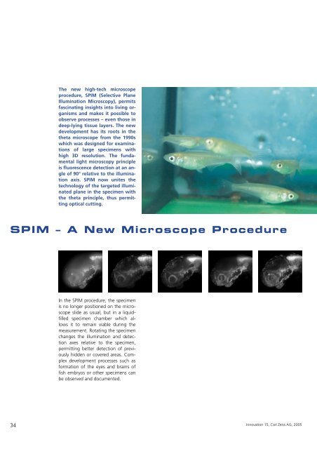
The new high-tech microscope<br />
procedure, SPIM (Selective Plane<br />
Illumination Microscopy), permits<br />
fascinating insights into living organisms<br />
and makes it possible to<br />
observe processes – even those in<br />
deep-lying tissue layers. The new<br />
development has its roots in the<br />
theta microscope from the 1990s<br />
which was designed for examinations<br />
of large specimens with<br />
high 3D resolution. The fundamental<br />
light microscopy principle<br />
is fluorescence detection at an angle<br />
of 90° relative to the illumination<br />
axis. SPIM now unites the<br />
technology of the targeted illuminated<br />
plane in the specimen with<br />
the theta principle, thus permitting<br />
optical cutting.<br />
SPIM – A New Microscope Procedure<br />
34<br />
In the SPIM procedure, the specimen<br />
is no longer positioned on the microscope<br />
slide as usual, but in a liquidfilled<br />
specimen chamber which allows<br />
it to remain viable during the<br />
measurement. Rotating the specimen<br />
changes the illumination and detection<br />
axes relative to the specimen,<br />
permitting better detection of previously<br />
hidden or covered areas. Complex<br />
development processes such as<br />
formation of the eyes and brains of<br />
fish embryos or other specimens can<br />
be observed and documented.<br />
Innovation 15, <strong>Carl</strong> <strong>Zeiss</strong> AG, 2005