Download PDF - Carl Zeiss
Download PDF - Carl Zeiss
Download PDF - Carl Zeiss

Create successful ePaper yourself
Turn your PDF publications into a flip-book with our unique Google optimized e-Paper software.
22<br />
14<br />
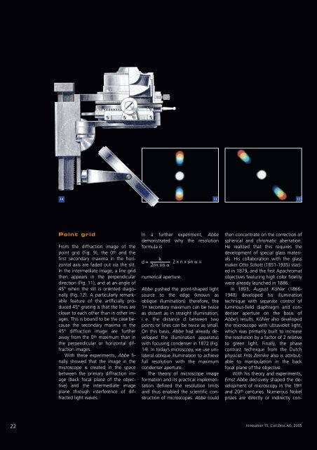
Point grid<br />
From the diffraction image of the<br />
point grid (Fig. 9), the 0th and the<br />
first secondary maxima in the horizontal<br />
axis are faded out via the slit.<br />
In the intermediate image, a line grid<br />
then appears in the perpendicular<br />
direction (Fig. 11), and at an angle of<br />
45° when the slit is oriented diagonally<br />
(Fig. 12). A particularly remarkable<br />
feature of the artificially produced<br />
45° grating is that the lines are<br />
closer to each other than in other images.<br />
This is bound to be the case because<br />
the secondary maxima in the<br />
45° diffraction image are further<br />
away from the 0th maximum than in<br />
the perpendicular or horizontal diffraction<br />
images.<br />
With these experiments, Abbe finally<br />
showed that the image in the<br />
microscope is created in the space<br />
between the primary diffraction image<br />
(back focal plane of the objective)<br />
and the intermediate image<br />
plane through interference of diffracted<br />
light waves.<br />
In a further experiment, Abbe<br />
demonstrated why the resolution<br />
formula is<br />
d=<br />
�<br />
2nx sin �<br />
numerical aperture.<br />
2 x n x sin � =<br />
Abbe pushed the point-shaped light<br />
source to the edge (known as<br />
oblique illumination): therefore, the<br />
1st secondary maximum can be twice<br />
as distant as in straight illumination,<br />
i. e. the distance d between two<br />
points or lines can be twice as small.<br />
On this basis, Abbe had already developed<br />
the illumination apparatus<br />
with focusing condenser in 1872 (Fig.<br />
14). In today’s microscopy, we use unilateral<br />
oblique illumination to achieve<br />
full resolution with the maximum<br />
condenser aperture.<br />
The theory of microscope image<br />
formation and its practical implementation<br />
defined the resolution limits<br />
and thus enabled the scientific construction<br />
of microscopes. Abbe could<br />
11 12<br />
then concentrate on the correction of<br />
spherical and chromatic aberration.<br />
He realized that this requires the<br />
development of special glass materials.<br />
His collaboration with the glass<br />
maker Otto Schott (1851-1935) started<br />
in 1879, and the first Apochromat<br />
objectives featuring high color fidelity<br />
were already launched in 1886.<br />
In 1893, August Köhler (1866-<br />
1948) developed his illumination<br />
technique with separate control of<br />
luminous-field diaphragm and condenser<br />
aperture on the basis of<br />
Abbe’s results. Köhler also developed<br />
the microscope with ultraviolet light,<br />
which was primarily built to increase<br />
the resolution by a factor of 2 relative<br />
to green light. Finally, the phase<br />
contrast technique from the Dutch<br />
physicist Frits Zernike also is attributable<br />
to manipulation in the back<br />
focal plane of the objective.<br />
With his theory and experiments,<br />
Ernst Abbe decisively shaped the development<br />
of microscopy in the 19th<br />
and 20th centuries. Numerous Nobel<br />
prizes are directly or indirectly con-<br />
Innovation 15, <strong>Carl</strong> <strong>Zeiss</strong> AG, 2005