Download PDF - Carl Zeiss
Download PDF - Carl Zeiss
Download PDF - Carl Zeiss

Create successful ePaper yourself
Turn your PDF publications into a flip-book with our unique Google optimized e-Paper software.
From Users<br />
The Zebra Fish as a Model Organism for<br />
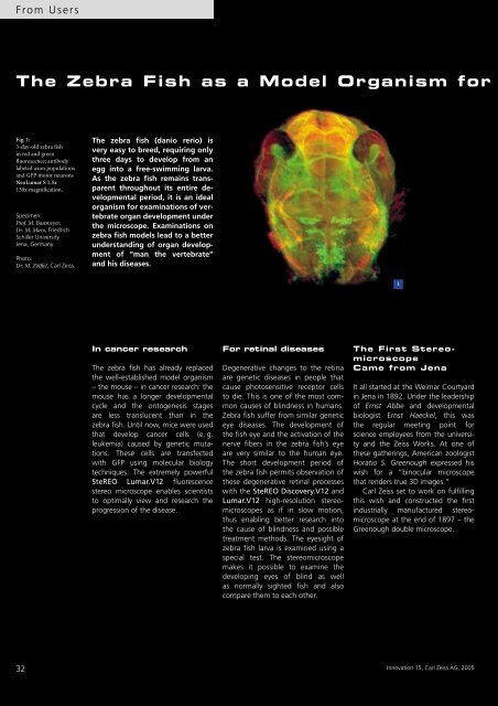
Fig. 1:<br />
3-day-old zebra fish<br />
in red and green<br />
fluorescence: antibody<br />
labeled axon populations<br />
and GFP motor neurons<br />
NeoLumar S 1.5x<br />
150x magnification.<br />
Specimen:<br />
Prof. M. Bastmeyer,<br />
Dr.M.Marx,Friedrich<br />
Schiller University<br />
Jena, Germany.<br />
Photo:<br />
Dr. M. Zölffel, <strong>Carl</strong> <strong>Zeiss</strong>.<br />
32<br />
The zebra fish (danio rerio) is<br />
very easy to breed, requiring only<br />
three days to develop from an<br />
egg into a free-swimming larva.<br />
As the zebra fish remains transparent<br />
throughout its entire developmental<br />
period, it is an ideal<br />
organism for examinations of vertebrate<br />
organ development under<br />
the microscope. Examinations on<br />
zebra fish models lead to a better<br />
understanding of organ development<br />
of “man the vertebrate”<br />
and his diseases.<br />
In cancer research<br />
The zebra fish has already replaced<br />
the well-established model organism<br />
– the mouse – in cancer research: the<br />
mouse has a longer developmental<br />
cycle and the ontogenesis stages<br />
are less translucent than in the<br />
zebra fish. Until now, mice were used<br />
that develop cancer cells (e. g.<br />
leukemia) caused by genetic mutations.<br />
These cells are transfected<br />
with GFP using molecular biology<br />
techniques. The extremely powerful<br />
SteREO Lumar.V12 fluorescence<br />
stereo microscope enables scientists<br />
to optimally view and research the<br />
progression of the disease.<br />
For retinal diseases<br />
Degenerative changes to the retina<br />
are genetic diseases in people that<br />
cause photosensitive receptor cells<br />
to die. This is one of the most common<br />
causes of blindness in humans.<br />
Zebra fish suffer from similar genetic<br />
eye diseases. The development of<br />
the fish eye and the activation of the<br />
nerve fibers in the zebra fish’s eye<br />
are very similar to the human eye.<br />
The short development period of<br />
the zebra fish permits observation of<br />
these degenerative retinal processes<br />
with the SteREO Discovery.V12 and<br />
Lumar.V12 high-resolution stereomicroscopes<br />
as if in slow motion,<br />
thus enabling better research into<br />
the cause of blindness and possible<br />
treatment methods. The eyesight of<br />
zebra fish larva is examined using a<br />
special test. The stereomicroscope<br />
makes it possible to examine the<br />
developing eyes of blind as well<br />
as normally sighted fish and also<br />
compare them to each other.<br />
1<br />
The First Stereomicroscope<br />
Came from Jena<br />
It all started at the Weimar Courtyard<br />
in Jena in 1892. Under the leadership<br />
of Ernst Abbe and developmental<br />
biologist Ernst Haeckel, this was<br />
the regular meeting point for<br />
science employees from the university<br />
and the <strong>Zeiss</strong> Works. At one of<br />
these gatherings, American zoologist<br />
Horatio S. Greenough expressed his<br />
wish for a ”binocular microscope<br />
that renders true 3D images.”<br />
<strong>Carl</strong> <strong>Zeiss</strong> set to work on fulfilling<br />
this wish and constructed the first<br />
industrially manufactured stereomicroscope<br />
at the end of 1897 – the<br />
Greenough double microscope.<br />
Innovation 15, <strong>Carl</strong> <strong>Zeiss</strong> AG, 2005