Download PDF - Carl Zeiss
Download PDF - Carl Zeiss
Download PDF - Carl Zeiss

You also want an ePaper? Increase the reach of your titles
YUMPU automatically turns print PDFs into web optimized ePapers that Google loves.
Developmental Biology<br />
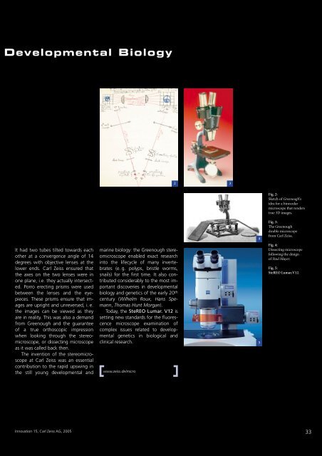
It had two tubes tilted towards each<br />
other at a convergence angle of 14<br />
degrees with objective lenses at the<br />
lower ends. <strong>Carl</strong> <strong>Zeiss</strong> ensured that<br />
the axes on the two lenses were in<br />
one plane, i.e. they actually intersected.<br />
Porro erecting prisms were used<br />
between the lenses and the eyepieces.<br />
These prisms ensure that images<br />
are upright and unreversed, i. e.<br />
the images can be viewed as they<br />
are in reality. This was also a demand<br />
from Greenough and the guarantee<br />
of a true orthoscopic impression<br />
when looking through the stereomicroscope,<br />
or dissecting microscope<br />
as it was called back then.<br />
The invention of the stereomicroscope<br />
at <strong>Carl</strong> <strong>Zeiss</strong> was an essential<br />
contribution to the rapid upswing in<br />
the still young developmental and<br />
Innovation 15, <strong>Carl</strong> <strong>Zeiss</strong> AG, 2005<br />
marine biology: the Greenough stereomicroscope<br />
enabled exact research<br />
into the lifecycle of many invertebrates<br />
(e. g. polyps, bristle worms,<br />
snails) for the first time. It also contributed<br />
considerably to the most important<br />
discoveries in developmental<br />
biology and genetics of the early 20 th<br />
century (Wilhelm Roux, Hans Spemann,<br />
Thomas Hunt Morgan).<br />
Today, the SteREO Lumar. V12 is<br />
setting new standards for the fluorescence<br />
microscope examination of<br />
complex issues related to developmental<br />
genetics in biological and<br />
clinical research.<br />
www.zeiss.de/micro<br />
2 3<br />
4<br />
5<br />
Fig. 2:<br />
Sketch of Greenough’s<br />
idea for a binocular<br />
microscope that renders<br />
true 3D images.<br />
Fig. 3:<br />
The Greenough<br />
double microscope<br />
from <strong>Carl</strong> <strong>Zeiss</strong>.<br />
Fig. 4:<br />
Dissecting microscope<br />
following the design<br />
of Paul Mayer.<br />
Fig. 5:<br />
SteREO Lumar.V12.<br />
33