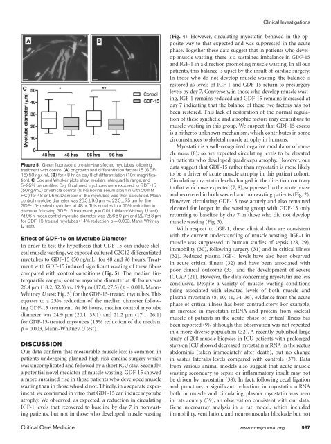
Bloch et alsepsis, showed acute elevation <strong>of</strong> myostatin mRNA, followedby suppression over the next week (40).Our clinical data show the association <strong>of</strong> a more prolongedexposure to GDF-15 with the development <strong>of</strong> muscle wastingfollowing cardiac surgery; therefore, it is possible that thisassociation may represent a novel mechanism for acute musclewasting. GDF-15 was first described as macrophage inhibitorycytokine-1 <strong>and</strong> has hallmark characteristics <strong>of</strong> the TGF-β family<strong>of</strong> proteins (41). It is secreted as a propeptide <strong>and</strong> cleaved t<strong>of</strong>orm a dimeric mature protein <strong>of</strong> 224 amino acids (14). GDF-15 expression increased in response to stress, hypoxia, <strong>and</strong>inflammation in vitro (14, 17) <strong>and</strong> in a variety <strong>of</strong> animal <strong>and</strong>human chronic disease settings (13, 17, 18). All the patients inthis study underwent the significant acute inflammatory insult<strong>of</strong> cardiac surgery <strong>and</strong> as expected circulating GDF-15 roseacutely in all patients.To our knowledge, a direct effect <strong>of</strong> GDF-15 on skeletalmuscle has not previously been demonstrated. GDF-15 inducedsignificant fiber atrophy <strong>of</strong> cultured C2C12 myotubes.This result is consistent with data from cardiac <strong>and</strong> cancer celllines, <strong>and</strong> models in which GDF-15 limited hypertrophy <strong>and</strong>promoted apoptosis (16, 20). Limiting cardiac hypertrophyfor a prolonged period may be beneficial. Mice deficient inGDF-15 show both increased hypertrophy <strong>and</strong> loss <strong>of</strong> cardiacfunction in response to pressure overload compared with wildtypemice (20). Beneficial effects <strong>of</strong> GDF-15 in cardiac tissuealso include the protection <strong>of</strong> the myocardium from infiltration<strong>of</strong> polymorphonuclear leukocytes <strong>and</strong> protection againstmyocardial rupture following infarction (42). However, evenwithin cardiac tissue, GDF-15 is also linked to pathological effects,<strong>and</strong> its role is likely to be bidirectional depending on thesituation <strong>and</strong> environment (14).Similarly, GDF-15 may have a protective role by promotingapoptosis in prostate cancer cells (43, 44); conversely, it mayalso enhance metastatic potential <strong>and</strong> angiogenesis (13). It ispossible that in muscle tissue a similar situation exists suchthat the role <strong>of</strong> GDF-15 may depend on the molecular environment.However, our findings <strong>of</strong> an association between GDF-15 <strong>and</strong> acute muscle wasting following the physiological stress<strong>of</strong> cardiac surgery are consistent with the underst<strong>and</strong>ing thatGDF-15 is released under situations <strong>of</strong> physiological stress (13,14). The ability <strong>of</strong> GDF-15 to cause muscle wasting in culturedmyotubes is consistent with the antihypertrophy/proapoptosisrole described above at least under some conditions. Alsocancer-related cachexia in mice resulting from excess GDF-15expression from transplanted prostate cancer cells can be reversedwith GDF-15 antibodies (19). It was postulated that thiswas due to a central antianorexic effect, <strong>and</strong> reduced food intakewas reversed. However, our data suggest that it is possiblethat GDF-15 also has a direct effect on skeletal muscle, resultingin reduced muscle bulk <strong>and</strong> lean body mass.A weakness <strong>of</strong> our data is that we are unable to demonstratea difference between the two groups on the raw values <strong>of</strong> circulatingfactor’s concentrations alone. This may be due to therelatively small number <strong>of</strong> patients in each group <strong>and</strong> the widerange <strong>of</strong> baseline values. Alternatively, despite the high-risk nature<strong>of</strong> the cardiac surgery that our patients underwent, the overallclinical outcome was very good, in that ICU stay was shortwith a low mortality. Furthermore, although measurable, thesepatients can be considered to only have mild muscle wastingwith clinically insignificant weakness <strong>and</strong> therefore differencesbetween the two groups will be harder to detect. Nevertheless,the validity <strong>of</strong> our approach is supported by the observationthat IGF-1 decreased acutely in all patients <strong>and</strong> remained suppressedin those who developed muscle wasting, consistent withprior observations (21, 32). A larger prospective study involvingmore patients <strong>and</strong> patients with clinically obvious ICUAPwould be desirable as a next step to confirm our findings.It is well established that the use <strong>of</strong> bypass per se may influencecirculating levels <strong>of</strong> inflammatory molecules (45). However,we do not believe this explains the current data becausebypass time was not different in patients who developed wasting<strong>and</strong> those who did not (Table 1). In addition, four patientsin the nonwasting group <strong>and</strong> two in the wasting group did notundergo bypass, <strong>and</strong> these patients had circulating plasma levelsfor the mediators measured <strong>of</strong> the same order <strong>of</strong> magnitudeas the rest <strong>of</strong> the group.A perfect human model <strong>of</strong> ICUAP does not exist. However,we believe our experimental paradigm is <strong>of</strong> interest owing toits prospective nature, the relative homogeneity <strong>of</strong> the patientcohort <strong>and</strong> the uniformity <strong>of</strong> the insult. By contrast, elucidatingmechanisms <strong>of</strong> ICUAP by studying cases is also problematicbecause ICUAP is a heterogeneous disorder that occursin a very mixed cohort <strong>of</strong> patients, <strong>and</strong> the time course <strong>of</strong> thesyndrome is difficult to define. Our model can be criticized forthe fact that patients only develop subclinical muscle weakness<strong>and</strong> therefore it may be difficult to extrapolate findings totrue ICUAP. It is also important to acknowledge that althoughthese patients have all undergone significant physiological insultthe cardiac surgical model may not be generally applicableto ICU patients—in whom, for example, sepsis <strong>and</strong> prolongedstays on critical care are common. However, the strengths <strong>of</strong>the model are that by studying our patients’ blood pr<strong>of</strong>ile frombaseline, we are able to demonstrate prospectively the dynamicpattern <strong>of</strong> change <strong>of</strong> circulating drivers <strong>of</strong> muscle metabolism.Furthermore, by identifying prospectively a specific cohort <strong>of</strong>patients with similar clinical experiences, we are able to controlfor many variables <strong>and</strong> potentially identify specific factors inthe pathophysiology <strong>of</strong> acute muscle wasting in the critically ill.In conclusion, we have shown that even elective high-riskcardiac surgery with a good clinical outcome is commonly associatedwith measurable loss <strong>of</strong> quadriceps muscle mass, <strong>and</strong>our data support the hypothesis that this muscle loss occurs asa result <strong>of</strong> a dynamic imbalance between drivers <strong>of</strong> muscle atrophy<strong>and</strong> hypertrophy. Furthermore, we speculate that GDF-15 may be a clinically relevant cause <strong>of</strong> skeletal muscle wastingin humans; if so GDF-15 antagonism may prove a useful futuretherapeutic approach.REFERENCES1. Latronico N, Bolton CF: Critical illness polyneuropathy <strong>and</strong> myopathy:A major cause <strong>of</strong> muscle weakness <strong>and</strong> paralysis. Lancet Neurol2011; 10:931–941988 www.ccmjournal.org April 2013 • Volume 41 • Number 4