Echocardiographic Evaluation of Pericardial Disease - Casecag.com
Echocardiographic Evaluation of Pericardial Disease - Casecag.com
Echocardiographic Evaluation of Pericardial Disease - Casecag.com

Create successful ePaper yourself
Turn your PDF publications into a flip-book with our unique Google optimized e-Paper software.
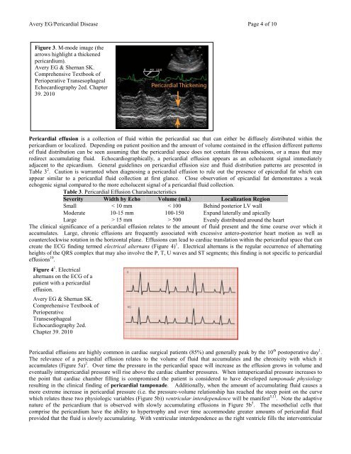
Avery EG/<strong>Pericardial</strong> <strong>Disease</strong> Page 4 <strong>of</strong> 10Figure 3. M-mode image (thearrows highlight a thickenedpericardium).Avery EG & Shernan SK.Comprehensive Textbook <strong>of</strong>Perioperative TransesophagealEchocardiography 2ed. Chapter39. 2010<strong>Pericardial</strong> effusion is a collection <strong>of</strong> fluid within the pericardial sac that can either be diffusely distributed within thepericardium or localized. Depending on patient position and the amount <strong>of</strong> volume contained in the effusion different patterns<strong>of</strong> fluid distribution can be seen assuming that the pericardial space does not contain fibrous adhesions, or a mass that mayredirect accumulating fluid. <strong>Echocardiographic</strong>ally, a pericardial effusion appears as an echolucent signal immediatelyadjacent to the epicardium. General guidelines on pericardial effusion size and fluid distribution patterns are presented inTable 3 2 . Caution is warranted when diagnosing a pericardial effusion to rule out the presence <strong>of</strong> epicardial fat which canappear similar to a pericardial fluid collection at first glance. Close observation <strong>of</strong> epicardial fat demonstrates a weakechogenic signal <strong>com</strong>pared to the more echolucent signal <strong>of</strong> a pericardial fluid collection.Table 3. <strong>Pericardial</strong> Effusion CharacteristicsSeverity Width by Echo Volume (mL) Localization RegionSmall < 10 mm < 100 Behind posterior LV wallModerate 10-15 mm 100-150 Expand laterally and apicallyLarge > 15 mm > 500 Evenly distributed around the heartThe clinical significance <strong>of</strong> a pericardial effusion relates to the amount <strong>of</strong> fluid present and the time course over which itaccumulates. Large, chronic effusions are frequently associated with excessive antero-posterior heart motion as well ascounterclockwise rotation in the horizontal plane. Effusions can lead to cardiac translation within the pericardial space that cancreate the ECG finding termed electrical alternans (Figure 4) 1 . Electrical alternans is the regular occurrence <strong>of</strong> alternatingheights <strong>of</strong> the QRS <strong>com</strong>plex that may also involve the P, T, U waves and ST segments; this finding is not specific to pericardialeffusions 10 .Figure 4 1 . Electricalalternans on the ECG <strong>of</strong> apatient with a pericardialeffusion.Avery EG & Shernan SK.Comprehensive Textbook <strong>of</strong>PerioperativeTransesophagealEchocardiography 2ed.Chapter 39. 2010<strong>Pericardial</strong> effusions are highly <strong>com</strong>mon in cardiac surgical patients (85%) and generally peak by the 10 th postoperative day 1 .The relevance <strong>of</strong> a pericardial effusion relates to the volume <strong>of</strong> fluid that accumulates and the chronicity with which itaccumulates (Figure 5a) 2 . Over time the pressure in the pericardial space will increase as the effusion grows in volume andeventually intrapericardial pressure will rise above the cardiac chamber pressures. When intrapericardial pressure increases tothe point that cardiac chamber filling is <strong>com</strong>promised the patient is considered to have developed tamponade physiologyresulting in the clinical finding <strong>of</strong> pericardial tamponade. Additionally, when the amount <strong>of</strong> accumulating fluid causes amore extreme increase in pericardial pressure (i.e. the pressure-volume relationship has reached the steep point on the curvewhich relates these two physiologic variables (Figure 5b)) ventricular interdependence will be manifest 5,11 . Note the adaptivenature <strong>of</strong> the pericardium that is observed with slowly accumulating effusions in Figure 5b 5 . The mesothelial cells that<strong>com</strong>prise the pericardium have the ability to hypertrophy and over time ac<strong>com</strong>modate greater amounts <strong>of</strong> pericardial fluidprovided that the fluid is slowly accumulating. With ventricular interdependence as the right ventricle fills the interventricular