

JIOMICS
JIOMICS Internacional
JIOMICS Internacional

Create successful ePaper yourself
Turn your PDF publications into a flip-book with our unique Google optimized e-Paper software.
<strong>JIOMICS</strong> | VOL 5 | ISSUE 2 | DECEMBER 2015 | 1-62<br />
JOURNAL OF INTEGRATED OMICS<br />
Journal of Integrated Omics<br />
A METHODOLOGICAL JOURNAL<br />
HTTP://WWW.<strong>JIOMICS</strong>.COM<br />
Special Issue: Proceeding Abstracts of the 4 th International Congress on Analytical Proteomics (ICAP 2015)<br />
Flagellar proteins and their key roles in human diseases: Once neglected,<br />
still underestimated<br />
D. M. Oliveira* 1,2<br />
1<br />
RENORBIO Northeastern Brazil Biotechnology PhD Program. Universidade Estadual do Ceará (UECE). Campus do Itaperi. 60.714.903.<br />
Fortaleza, CE. Brasil. 2 Nutrition and Health Master’s Program. Centro de Ciências da Saúde. UECE. 60.714.903. Fortaleza, CE. Brasil.<br />
*Corresponding author: diana.magalhaes@uece.br<br />
Available Online: 31 December 2015<br />
Abstract<br />
Organelle proteomics, such as the flagellum, has been one of the most suitable approaches used to identify novel components and modifications<br />
of a given cell, tissue, organ or even organism. Information is usually gained at a large scale and is very valuable to further understand<br />
biological processes of the given compartment. In this presentation I will talk about a particular subset of flagellar proteins, which happen to be<br />
amazingly related to protozoa virulence in Leishmaniases. As all eukaryotic flagella, the Leishmania flagellum is a protein nanomachine, but it<br />
has a particular way in being arranged to invade human macrophages. Yes, I just said to invade, rather than to be engulfed or phagocyted by<br />
macrophages. Leishmania flagellar apparatus turns the protozoan into an “infection motor” by means of complex, intricate surface and shallow<br />
subsurface features that provide major biological processes in this trypanosomatid survival and virulence, from motility and cell division, towards<br />
invasion and persistence in host cells. Using conventional proteome assays and computational molecular modeling coupled with atomic<br />
force microscopy (AFM), my research group has studied specific morphological data resulting from protein structural organization of Leishmania<br />
spp. flagellum, gathering information not yet explored neither in a nanostructured nor in a pathogenic approach for this species. AFM<br />
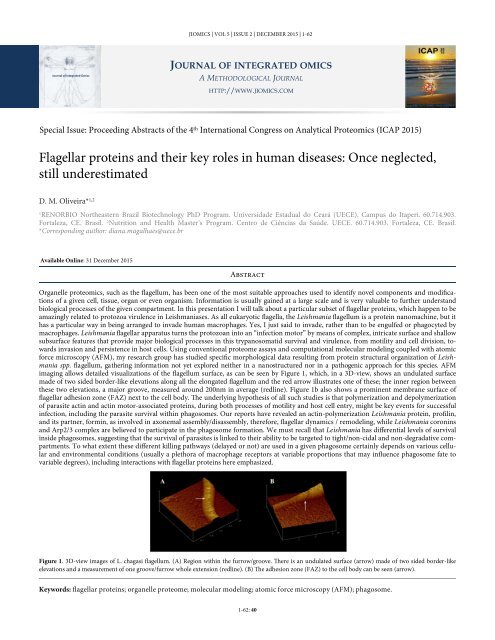
imaging allows detailed visualizations of the flagellum surface, as can be seen by Figure 1, which, in a 3D-view, shows an undulated surface<br />
made of two sided border-like elevations along all the elongated flagellum and the red arrow illustrates one of these; the inner region between<br />
these two elevations, a major groove, measured around 200nm in average (redline). Figure 1b also shows a prominent membrane surface of<br />
flagellar adhesion zone (FAZ) next to the cell body. The underlying hypothesis of all such studies is that polymerization and depolymerization<br />
of parasite actin and actin motor-associated proteins, during both processes of motility and host cell entry, might be key events for successful<br />
infection, including the parasite survival within phagosomes. Our reports have revealed an actin-polymerization Leishmania protein, profilin,<br />
and its partner, formin, as involved in axonemal assembly/disassembly, therefore, flagellar dynamics / remodeling, while Leishmania coronins<br />
and Arp2/3 complex are believed to participate in the phagosome formation. We must recall that Leishmania has differential levels of survival<br />
inside phagosomes, suggesting that the survival of parasites is linked to their ability to be targeted to tight/non-cidal and non-degradative compartments.<br />
To what extent these different killing pathways (delayed or not) are used in a given phagosome certainly depends on various cellular<br />
and environmental conditions (usually a plethora of macrophage receptors at variable proportions that may influence phagosome fate to<br />
variable degrees), including interactions with flagellar proteins here emphasized.<br />
Figure 1. 3D-view images of L. chagasi flagellum. (A) Region within the furrow/groove. There is an undulated surface (arrow) made of two sided border-like<br />
elevations and a measurement of one groove/furrow whole extension (redline). (B) The adhesion zone (FAZ) to the cell body can be seen (arrow).<br />
Keywords: flagellar proteins; organelle proteome; molecular modeling; atomic force microscopy (AFM); phagosome.<br />
1-62: 40