Laboratory news & BioScience March 2017
New Zealand's leading scientific publication for more than 20 years. This bi-monthly magazine provides the latest up to date information on new products and services to a readership which is carefully targeted and updated on a regular basis.
New Zealand's leading scientific publication for more than 20 years. This bi-monthly magazine provides the latest up to date information on new products and services to a readership which is carefully targeted and updated on a regular basis.

Create successful ePaper yourself
Turn your PDF publications into a flip-book with our unique Google optimized e-Paper software.
NEW ZEALAND LABORATORY NEWS | NEW ZEALAND BIOSCIENCE<br />
ISSUE 117 | MARCH <strong>2017</strong><br />
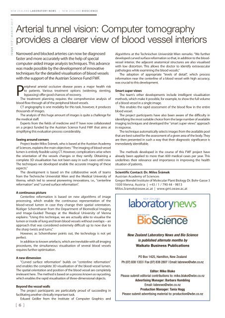
Arterial tunnel vision: Computer tomography<br />
provides a clearer view of blood vessel interiors<br />
Narrowed and blocked arteries can now be diagnosed<br />
faster and more accurately with the help of special<br />
computer-aided image analysis techniques. This advance<br />
was made possible by the development of innovative<br />
techniques for the detailed visualisation of blood vessels<br />
with the support of the Austrian Science Fund FWF.<br />
Peripheral arterial occlusive disease poses a major health risk<br />
to patients. Various treatment options (widening, stenting,<br />
bypassing) offer good chances of recovery.<br />
The treatment planning requires the comprehensive analysis of<br />
blood flow through all of the peripheral blood vessels.<br />
CT angiography is one modality for this task, however, it produces<br />
thousands of images.<br />
The analysis of this huge amount of images is quite a challenge for<br />
the medical staff.<br />
Experts from the fields of medicine and IT have now collaborated<br />
on a project funded by the Austrian Science Fund FWF that aims at<br />
simplifying this evaluation process considerably.<br />
Seeing around corners<br />
Project leader Milos Šrámek, who is based at the Austrian Academy<br />
of Sciences, explains the main objectives: "The imaging of blood-vessel<br />
lumen is entirely feasible using CT. However, complications arise when<br />
the orientation of the vessels changes or they ramify. Obtaining a<br />
complete 3D visualisation has not been easy in such cases until now.<br />
The techniques we developed enable the accurate imaging of these<br />
aspects."<br />
The development is based on the collaborative work of teams<br />
from the Technische Universität Wien and the Medical University of<br />
Vienna, which led to several pioneering innovations, i.e., "centerline<br />
reformation" and "curved surface reformation".<br />
A continuous picture<br />
Centerline reformation is based on new algorithms of image<br />
processing, which enable the continuous representation of the<br />
blood-vessel lumen in case they change their spatial orientation.<br />
Rüdiger Schernthaner from the Department of Biomedical Imaging<br />
and Image-Guided Therapy at the Medical University of Vienna<br />
explains: "Using this technique, we are actually able to visualise the<br />
lumen or inside of lung and brain blood vessels without overlaps – an<br />
approach that was considered extremely difficult up to now due to<br />
the sharp twists and turns."<br />
However, as Schernthaner points out, the technology is not yet<br />
perfect.<br />
In addition to known artefacts, which are inevitable with all imaging<br />
procedures, the simultaneous visualisation of several blood vessels<br />
requires further optimisation.<br />
Algorithms at the Technischen Universität Wien remarks: "We further<br />
developed curved surface reformation so that, in addition to the bloodvessel<br />
interior, the adjacent anatomical structures are also visualised<br />
with low distortion. This allows the doctor to identify extravascular<br />
pathologies while examining the blood vessels."<br />
The adoption of appropriate "levels of detail", which process<br />
information near the centerline of a blood vessel with high accuracy,<br />
was crucial to this development.<br />
Smart super views<br />
The team's other developments include intelligent visualisation<br />
methods, which make it possible, for example, to show the full volume<br />
of a blood vessel in a single image.<br />
This enables the rapid assessment of the blood flow in the entire<br />
blood vessel.<br />
The project participants have also been aware of the difficulty in<br />
identifying the most suitable choice from the large number of available<br />
imaging techniques and developed the "smart super views" approach<br />
in response.<br />
The technique automatically selects images from the available pool<br />
that are best suited for the assessment of a given area of the body. They<br />
are then presented in such a way that their diagnostic significance is<br />
immediately identifiable.<br />
The methods developed in the course of this FWF project have<br />
already been applied to more than 600 medical cases per year. This<br />
underlines their relevance and importance in improving the health<br />
situation of patients.<br />
Scientific Contact: Dr. Milos Šrámek<br />
Austrian Academy of Sciences<br />
Gregor Mendel Institute of Molecular Plant Biology Dr. Bohr-Gasse 3<br />
1030 Vienna, Austria | +43 / 1 / 790 44 - 9813<br />
Milos.Sramek@oeaw.ac.at | www.gmi.oeaw.ac.at<br />
NEW ZEALAND<br />
laboratory<strong>news</strong><br />
New Zealand <strong>Laboratory</strong> News and Bio Science<br />
is published alternate months by<br />
Waikato Business Publications<br />
A new dimension<br />
"Curved surface reformation" builds on "centerline reformation"<br />
and enables the complete 3D visualisation of the blood-vessel lumen.<br />
The spatial orientation and position of the blood vessel are completely<br />
irrelevant here. The method is based on a process known as raycasting,<br />
which enables the rapid visualisation of three-dimensional objects.<br />
Beyond the vessel walls<br />
The project participants are particularly proud of succeeding in<br />
facilitating another clinically important task.<br />
Eduard Gröller from the Institute of Computer Graphics and<br />
[ 6 ]<br />
PO Box 1425, Hamilton, New Zealand<br />
Ph (07) 838 1333 | Fax (07) 838 2807 | Email: lab<strong>news</strong>@wbn.co.nz<br />
Editor: Mike Blake<br />
Please submit editorial contributions to: mike.blake@wbn.co.nz<br />
Advertising Manager: Barbara Hambling<br />
Email: lab<strong>news</strong>@wbn.co.nz<br />
Production Manager: Tania Hogg<br />
Please submit advertising material to: production@wbn.co.nz