EACVI Echocardiography Textbook - sample
Discover the EACVI Textbook of Echocardiography 2nd edition
Discover the EACVI Textbook of Echocardiography 2nd edition

Create successful ePaper yourself
Turn your PDF publications into a flip-book with our unique Google optimized e-Paper software.
12<br />
Chapter 1 general principles of echocardiography<br />
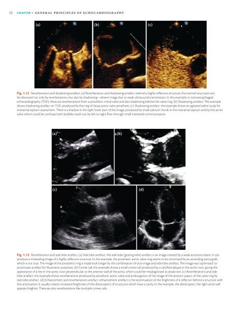
(a) (b) (c)<br />
Fig. 1.12 Reverberation and shadowing artefact. (a) Reverberation and shadowing artefact: behind a highly reflective structure, the normal structures can<br />
be obscured not only by reverberations, but also by shadowing—absent image due to weak ultrasound transmission. In this example, in transoesophageal<br />
echocardiography (TOE), there are reverberations from a prosthetic mitral valve and also shadowing behind the valve ring. (b) Shadowing artefact: The example<br />
shows shadowing artefact on TOE, produced by the ring of tissue aortic valve prosthesis. (c) Shadowing artefact: the example shows an agitated saline study for<br />
interatrial septum assessment. There is a shadow in the right lower part of the image, produced by small calcium chunk in the interatrial septum and by the aortic<br />
valve which could be confused with bubbles wash-out by left-to-right flow through small interatrial communication.<br />
(a)<br />
(b)<br />
(c)<br />
(d)<br />
Fig. 1.13 Reverberation and side lobe artefact. (a) Side lobe artefact: the side lobe (grating lobe) artefact is an image created by a weak accessory beam. It can<br />
produce a misleading image of a highly reflective structure. In the example, the prosthetic aortic valve ring seems to be continued by an ascending aorta graft,<br />
which is not true. The image of the prosthetic ring is made look longer by the combination of true image and side lobe artefact. The image was ‘optimized’ to<br />
accentuate artefact for illustration purposes. (b) Comet tail: the example shows a small comet tail produced by a calcified plaque in the aortic root, giving the<br />
appearance of a line in the aortic root perpendicular to the anterior wall of the aorta, which could be misdiagnosed as dissection. (c) Reverberations and side<br />
lobe artefact: the example shows reverberations produced by prosthetic aortic valve and prolongation of the image of the anterior aspect of the valve ring by<br />
side lobe artefact. (d) Enhancement and reverberations artefact: enhancement artefact is the accentuation of the brightness of a reflector behind a structure with<br />
low attenuation. It usually creates increased brightness of the distal aspect of structures which have a cavity. In the example, the distal aspect the right atrial wall<br />
appears brighter. There are also reverberations like multiple comet tails.