3. FOOD ChEMISTRy & bIOTEChNOLOGy 3.1. Lectures
3. FOOD ChEMISTRy & bIOTEChNOLOGy 3.1. Lectures
3. FOOD ChEMISTRy & bIOTEChNOLOGy 3.1. Lectures

Create successful ePaper yourself
Turn your PDF publications into a flip-book with our unique Google optimized e-Paper software.
Chem. Listy, 102, s265–s1311 (2008) Food Chemistry & Biotechnology<br />
<strong>3.</strong> <strong>FOOD</strong> <strong>ChEMISTRy</strong><br />
& <strong>bIOTEChNOLOGy</strong><br />
<strong>3.</strong>1. <strong>Lectures</strong><br />
L01 NONSACChAROMyCES yEAST IN GRAPE<br />
MuST – ADVANTAGE OR SPOILAGE?<br />
JAROSLAVA KAňUCHOVá PáTKOVá a , EMíLIA<br />
BREIEROVá b and InGRID VAJCZIKOVá<br />
a Institute for viticulture and enology of SARC<br />
Matuškova 25, 831 01 Bratislava, Slovak republic,<br />
b Institute of ChemistrySAS, Dúbravská cesta 9, 845 38 Bratislava,<br />
Slovak republic,<br />
chememi@savba.sk<br />
Introduction<br />
The fresh grape must consists of spontaneous microflora<br />
formed from 90 to 99 % by yeasts. The most important<br />
genus is without doubt Saccharomyces cerevisiae which is<br />
responsible for successive fermentation and good wine quality.<br />
Recently the contribution of non-Saccharomyces yeasts<br />
have been widely discussed as there is not definitive opinion<br />
on their contribution to the wine quality, especially aroma.<br />
Hanseniaspora osmophila and Kloeckera apiculata should<br />
be considered detrimental yeast species, by higher acetic<br />
acid, acetaldehyde, ethyl acetate and acetoin production 1 . To<br />
avoid spoilage it is recommended to inoculate the grape must<br />
by Saccharomyces cerevisiae immediately after pressing.<br />
On the other hand, the apiculate non-Saccharomyces yeast<br />
is a natural indigenous microflora which contributes to the<br />
wine origin. Thus the question wheather to allow the apiculate<br />
microflora to start fermentation or not has not yet been<br />
solved.<br />
The aim of this study work was to determine the aroma<br />
profile of isolated non-Sacaharomyces yeast from the chemical<br />
as well as the sensorial viewpoints. The yeasts of<br />
the genera Rhodotorula, Sporobolomyces, Pichia, Hansenula,<br />
Issat-chenkia, and Torulospora, were tested from the<br />
viewpoint of their contribution to the wine aroma. The results<br />
were than exploited and tested in the real wine-making process.<br />
Experimental<br />
Following yeast strains were isolated from the grape<br />
must and degraded products: Rhodotorula mucilaginosa<br />
(2 strains), Sporobolomyces pararoseus, Pichia membranefaciens,<br />
Pichia anomala (2 strains), Candida intermediata,<br />
Torulospora delbruecki, and Issatchenkia orientalis. For<br />
comparison 3 Saccharomyces cerevisiae strains were also<br />
used. Two of them were isolated from the grape must, the<br />
third was a commercial one (Lallemand).<br />
Consequently the isolated yeast strains were inoculated<br />
to the first cultivation medium: the sterile grape must had<br />
s537<br />
been femented for 10 days at 10 °C under semiaerobic conditions<br />
or 4–6 weeks at 10 °C under anaerobic conditions.<br />
Second cultivation medium: the sterile Vinea drinks<br />
were inoculated by studied yeast strains and cultuivated at<br />
20 °C for 10 days under semiaerobic conditions.<br />
The samples were than sensorially evaluated by a group<br />
of degustators. The same samples were than analysed by gas<br />
chromatography for the aroma compounds production. Each<br />
sample was analysed on the GC MS (Shimadzu QP 2010)<br />
equipment and also on the GC FID equipment (GC 8000 CE<br />
Instruments).<br />
Two methods of sample preparation were done:<br />
The samples (20 ml) were extracted by ether (2 ml), and<br />
centrifuged prior to analysis. The etheric extract was used for<br />
analysis (liquid – liquid extraction). This method was used<br />
for higher alcohols (propanol, isoamylacohol, ethyl ester and<br />
higher alcoholos esters) determination<br />
The samples were extracted by Tenaq (solid phase microextraction)<br />
and than 10 min sampling according to 6 . This<br />
method was used for monoterpenic compounds determination.The<br />
same column and the same conditions were used by<br />
both analysis: Column: DB WAX 30 m, 0.25 × 0.25, temperature<br />
programme: 30 °C, hold 2 min, increase by 4 °C min –1<br />
up to 230 °C, hold 10 min, 1 ml of sample was injected to<br />
injection port at 200 °C, detector temperature 220 °C, carrier<br />
gas: helium, injektion mode: split 1 : 100, flow control mode:<br />
pressure 70 kPa<br />
Results<br />
After inoculation and fermentation of the grape must and<br />
Vinea drink the number of aroma compounds increased significantly,<br />
in both cases and more than 60 compounds were<br />
found. Most of them were recognized as typical fermentation<br />
products, etc. ethanol, izoamylacohol, propanol, etylester of<br />
caproic, caprylic and caprinic acids, ethylacetate, isovaleric<br />
acid, pentylacetate, 2,3-butandiol, furfural, 3-hydroxybutyrate,<br />
methionon, 1,4-butandiol. 2-metyl a 3-metylbutanoic<br />
acid, 2-phenyletylacetate, izoamylacetate, cis 3-hexenylacetate,<br />
etylbenzoate, α-terpineol, etyl isobutyrate, etyl butyrate,<br />
etyl 2-metylbutyrate, etyl isovalerate, isoamyl acetate, ethyl<br />
hexanoate, cis-3-hexenol, ethyl octanoate, furfural, linalool,<br />
ethyl furoate, ethyl decanoate, ethyl benzoate, α-terpineol,<br />
fenylethyl acetate, and geraniol. The increased production of<br />
typicall glycolysis products were also confirmed by several<br />
authors 2,3,4 . Ethyl propionate and propyl acetate, characterized<br />
the sample of the grape must fermented by Kloeckera<br />
apiculata, and 2-propanol and 2-hexanone characterized the<br />
sample of the grape must fermented by Pichia membranefaciens<br />
2 . Rojas 3 studied analysis of non-Saccharomyces yeast<br />
fermentation products and found higher acetate content, especially<br />
2-phenylacetate and isoamylacetate for Pichia yeasts.<br />
Albertazzi 4 also described higher levels of phenylacetate by<br />
other yeast - Pichia pastoris and Kloeckera saturnus (700–<br />
1700 mg dm –3 ). From our results we can confirm incereased<br />
ester production by all studied microorganisms, especially<br />
increased content of ethyl acetate.
Chem. Listy, 102, s265–s1311 (2008) Food Chemistry & Biotechnology<br />
However, we have found that some microorganisms<br />
produced special compounds, which were not recognised<br />
by other yeasts. After the fermentation of the grape must<br />
by R. mucilaginosa and Sp. pararoseus, aroma compounds<br />
significantly increased. Acetate, hexanal, heptanal, octanal,<br />
cyclopentanone, thiazole, decalactone, propyl-3-dimethyl<br />
aminopropyl, nonanone, heptanon, and butanediol were<br />
formed. P. anomala produced especially isoamyl benzyl<br />
ether. The medium fermented by Sporobolomyces was rich in<br />
sabinylacetate, 3,4-hexandion and eicosane. R. mucilaginosa<br />
generated cyklopentanol and, α-cyklogeraniol. S. cerevisiae<br />
produced vericaldehyd and γ-nonalakton.<br />
We have found out that yeasts of the genera Pichia, Rhodotorula,<br />
and Sporobolomyces did not produce the linalool<br />
acetate, contrary to S. cerevisiae.<br />
The differences in the compound production within<br />
the same yeast species were also observed. S. cerevisiae<br />
strain 8 produced caproic aldehdyd, trans-pinocamphon,<br />
dodecanal, 5-methyl-3-heptanon, izo-menthylacetate, contrary<br />
to S. cerevisiae strain 5 which did not produce any of<br />
these compounds. R. mucilaginosa strain 11 produced higher<br />
amounts of 2,3-butandiol, but did not produce any izopen-<br />
Table I<br />
Aroma compounds typically produced by various yeast species<br />
and flavoural characterisation of founded compounds<br />
Yeast strain compound Flavour fragrance<br />
R. mucilaginosa<br />
cyklopentanol,<br />
alfacyklogeraniol<br />
mint aroma<br />
spice flavour<br />
carnation odour<br />
Pichia anomala izoamyacetat banana flavour<br />
Sp. pararoseus sabinylacetate fruity aroma<br />
S. cerevisiae<br />
valericaldehyd,<br />
γ-nonalakton<br />
coffee aroma<br />
coconut odour<br />
Table II<br />
Sensorial evaluation of fermented grape must by various<br />
yeast strains, + positive impression, – negative impression<br />
Anaerobic Semiaerobic<br />
conditions conditions<br />
Yeast strain aroma perc. aroma perc.<br />
C.intermediata yeast + acidic –<br />
R.mucilaginosa 3 socks smelly – acethone –<br />
T.delbruecki pleasant + vanilla +<br />
I.orientalis autolyses – acethone –<br />
Pichia anomala acethone – lime +<br />
P.membranefaciens autolyses – yeasty +<br />
R. mucilaginosa 11 autolyses – honey +<br />
Pichia anomala acethone – honey +<br />
Sp. pararoseus not recognised – yeasty +<br />
S.cerevisiae 5 acidic – ferment +<br />
S.cerevisiae 8 fruity + ferment +<br />
S.cerevisiae 16 honey + ferment +<br />
s538<br />
Table III<br />
Evaluation of Vinea fermented under semiaerobic conditions<br />
Semiaerobic conditions<br />
Vinea<br />
20 °C<br />
Yeast strain<br />
Aroma Percept<br />
C. intermediata acidic +<br />
R. mucilaginosa 3 acethone +<br />
T. delbruecki yeasty +<br />
S. cerevisiae 5 yeastly +<br />
I. orientalis grape must +<br />
S. cerevisiae 8 honey +<br />
P. membranofaciens vinea –<br />
R. mucilaginosa 11 acethone +<br />
P. anomala honey +<br />
Sp. pararoseus acethone +<br />
tylformiate and 3,4-hexandion. These compounds produced<br />
strain Rhodotorula mucilaginosa strain <strong>3.</strong><br />
One of the most important factors in wine proofing is the<br />
sensorial evaluation of wine aroma. It is very difficult to estimate<br />
which from all above mentioned compounds will prevail<br />
over the other ones and wheather the wine could give positive<br />
or negative impression. It is due to the complexity of wine aromas,<br />
the heterogenity of perception and recognition thresholds<br />
for each one compound as well as many interactions occuring<br />
within and after fermentation 5 .<br />
As shown in Table II, under anaerobic conditions only<br />
T. delbruecki from apiculate microflora developed a pleasant<br />
aroma. All the other yeast strains evolved unpleasant, smelly<br />
aroma. However, the situation was radically changed when the<br />
fermentation occured under semi-aerobic conditions. Torulospora<br />
delbruecki produced the pleasant aroma, exactly defined<br />
as vanilla. Also both strains of P. anomala, Pichia membranefaciens,<br />
Sp. pararoseus and one strain of R. mucilaginosa evoked<br />
very pleasant aroma, some of them with honey notes,<br />
some of them fruity or increased fermetative impression.<br />
The results of better sensorial evaluation under semiaerobic<br />
conditions were subsequently tested by real winemaking<br />
fermentation. The problem which usually occurs in<br />
real process is that the present microfloras consists of several<br />
yeast genera which can possitively or negatively contribute<br />
to the wine quality.<br />
The group of degustators confirmed that the wines were<br />
better evaluated when they were fermented under semiaerobic<br />
conditions, with the inocoluation by S. cerevisae not directly<br />
after fermentation, but several hours after grape pressing. The<br />
main advantage of this system is its ability to improve the originality<br />
of the wine.<br />
Conclusions<br />
We have found compounds which were typically produced<br />
by some non-Saccharomyces yeast strains – cyklopentanol,<br />
alfacyklogeraniol for R. mucilaginosa, sabinylacetate,<br />
for Sp. pararoseus, and izoamylbenzylether for P. anomala.
Chem. Listy, 102, s265–s1311 (2008) Food Chemistry & Biotechnology<br />
We can confirm that under anaerobic conditions, most<br />
of the apiculate microflora, except T. delbruecki, negatively<br />
affected wine aroma because they produced higher amounts<br />
of aldehydes – pentanal, hexanal, heptanal, 3,4-hexanedione,<br />
eicosene which caused the buttery and waxy odour.<br />
However under semiaerobic conditions apiculate yeast<br />
species promoted positive aroma perception in products.<br />
Torulospora and Pichia yeast strains increased the fruit and/<br />
or coconut aroma by higher production of sabinyl res. isoamylacetate.<br />
Semiaerobic conditions applied several hours prior<br />
inoculating by S. cerevisiae improve sensorial evaluation<br />
of wine and increase the support of originality and variety<br />
typicity.<br />
However, the fermentation under such conditions is still<br />
very hazardous because oxidative defects or microbiological<br />
defaults of wine could occured.<br />
s539<br />
REFEREnCES<br />
1. Granchi L., Ganucci D., Messini A, Vincenzini M.:<br />
FEMS Yeast Res. 2, 403 (2002).<br />
2. Romano P., Fiore C., Paraggio M., Caruso M., Capece<br />
A.: Int. J. Food Microbiol, 86, 169 (2003).<br />
<strong>3.</strong> Rojas V., Gil J.V., Pinaga F., Manzanares P.: Inter. J.<br />
Food Microbiol. 70, 283 (2001).<br />
4. Albertazzi E, Cardillo R., Servi S.: Biotechnol. Lett. 16,<br />
491 (1994).<br />
5. Ribereau-Gayon P., Glories Y., Dubordieu D., Maujean<br />
A., Handbook of Enology, Ed. Wiley, Chicester, 404<br />
(2000).<br />
6. Kruzlicova D., Mocak J., Hrivnak J.: J. Food nutr. Res.<br />
47, 37 (2008).
Chem. Listy, 102, s265–s1311 (2008) Food Chemistry & Biotechnology<br />
L02 APPROAChES TO MINIMIZATION OF<br />
ACRyLAMIDE LEVEL IN <strong>FOOD</strong>S<br />
ZUZAnA CIESAROVá<br />
VÚP Food Research Institute<br />
Priemyselná 4, 824 75 Bratislava, Slovak Republic,<br />
ciesarova@vup.sk<br />
Introduction<br />
Thermal treatment of foods is a common way for improvement<br />
of digestibility, safety, quality and sensory properties<br />
of many foods which is used for ages. Besides unambiguous<br />
desirable aspects of this treatment some detrimental<br />
effects are still emerging e.g. a loss of nutrition-worthy compounds<br />
and an undesirable generation of contaminants.<br />
In 2002, Swedish researchers have first reported the<br />
formation of acrylamide in foods processed at elevated temperatures<br />
1 . Recent assessment by the Joint FAO/WHO Expert<br />
Committee on Food Additives (JECFA) in 2005 2 confirmed<br />
that a risk cannot be excluded for dietary intake of acrylamide<br />
because it is classified as a probable human carcinogen<br />
by the International Agency for Research on Cancer (IARC) 3 .<br />
In that assessment JECFA concluded that the margin of exposure<br />
for average and high consumers were low for compound<br />
that is genotoxic and carcinogenic and that this may indicate<br />
a human health concern. Therefore the Commission Recommendation<br />
since 2007 announced that “appropriate efforts<br />
to reduce acrylamide concentrations in foodstuffs should<br />
continue” 4 . Moreover, with respect to the last observations<br />
confirming the association between acrylamide intake and<br />
endometrial, ovarian 5 , and breast 6 cancer risk, the concern on<br />
the acrylamide mitigation activity is very urgent.<br />
Occurrence of Acrylamide in Thermally Treated Foods<br />
After the discovery of acrylamide, a lot of studies confirmed<br />
the presence of acrylamide in nearly all fried, baked<br />
and roasted foods. Acrylamide exposure varies depending<br />
upon the population´s eating habits and the way the foods<br />
are processed and prepared. Generally, fried potato products,<br />
ready-to-eat breakfast cereals, baked goods and roasted coffee<br />
are the most important food categories that contribute<br />
most to acrylamide exposure. An average long-term exposure<br />
of acrylamide was estimated of 0.3 to 0.8 μg (kg body<br />
weight) –1 day –1 on the base of the few data which were avail-<br />
able at the FAO/WHO Consultation in 2002 7 . Based on the<br />
reported data, the Committee JECFA in 2005 2 noted that<br />
children may have intakes of acrylamide around two or three<br />
times higher those of adult consumers when expressed on a<br />
body weight basis. It is expected that children and adolescents<br />
have consumption patterns different from adults. Most of the<br />
types of foods in which acrylamide was detected are popular<br />
among children and adolescents, such as French fries, snacks,<br />
biscuits and breads. Moreover, they have a lower average<br />
body weight and, consequently, a higher average food intake<br />
per kilogram body weight than adults. For that, acrylamide<br />
intake by these individuals is considered a concern.<br />
s540<br />
M e c h a n i s m o f A c r y l a m i d e<br />
F o r m a t i o n<br />
Initial results on acrylamide content indicated carbohydrate-rich<br />
foods to generate relatively more acrylamide 1 .<br />
Several researchers have established that the main pathway<br />
of acrylamide formation in foods is linked to the Maillard<br />
reaction and, in particular, the amino acid asparagine 8,9 . The<br />
link of acrylamide to asparagine, which directly provides the<br />
backbone of the acrylamide molecule, has been established by<br />
labelling experiments 9,10 . Study to date clearly shows that the<br />
amino acid asparagine is mainly responsible for acrylamide<br />
formation in heated foods after condensation with reducing<br />
sugars or a carbonyl source. Moreover, the sugar-asparagine<br />
adduct, n-glycosylasparagine, generates high amounts of<br />
acrylamide, suggesting the early Maillard reaction as a major<br />
source of acrylamide 9 . In addition, decarboxylated asparagine<br />
(3-aminopropionamid), when heated can generate acrylamide<br />
in the absence of reducing sugars 10 . A good evidence<br />
supporting the early Maillard rection as a main reaction pathway<br />
involving early decarboxylation of the Schiff base, rearrangement<br />
to the resulting Amadori product, and subsequent<br />
beta-elimination to release acrylamide has been presented 11 .<br />
Factors Affecting Acrylamide Formation in Foods<br />
The resulting acrylamide concentration in foods ultimately<br />
depends on both products and process variables.<br />
Acrylamide formation requires the amino acid asparagine<br />
and a carbonyl compound as the Maillard reaction precursors.<br />
The concentration of acrylamide precursors and temperature<br />
mainly affect the rate of acrylamide formation. It is a fact that<br />
formation and degradation of acrylamide occurs in the same<br />
time during heating at elevated temperatures, it means that<br />
measured acrylamide content of a food is net result of two<br />
consecutive reactions occurred during thermal processing12 .<br />
Based on the current knowledge of the mechanism of<br />
acrylamide formation, many parameters affecting the level<br />
of acrylamide in foods were investigated, e.g. heat intake, the<br />
level and type of saccharides and amino acids, moisture and<br />
water activity, additives, processing steps etc. 13 , and consequently<br />
various ways of acrylamide minimization in foods<br />
have been proposed. Many of them are summarized in a “living”<br />
document “The Acrylamide Toolbox” published by experts<br />
associated in the Confederation of the Food and Drink<br />
Industries of the European Union (CIAA) 14 .<br />
The mitigation approach is divided in two strategies:<br />
Prevention of acrylamide formation through a modification<br />
of food composition (a decline of asparagine<br />
and reducing saccharides contents), processing conditions<br />
(thermal input, pH, moisture), an addition of compounds<br />
suppressing the formation of acrylamide (acids,<br />
enzymes, proteins, antioxidants etc.) and an enhancement<br />
of processing steps (pre-treatment, blanching, fermentation<br />
etc.) 15 .<br />
Facilitation the acrylamide elimination through storage<br />
conditions or the initialization of acrylamide polymerization16,17<br />
•<br />
•<br />
.
Chem. Listy, 102, s265–s1311 (2008) Food Chemistry & Biotechnology<br />
Temperature plays an important role in the formation<br />
and elimination of acrylamide. It is well known that acrylamide<br />
forms in foods that are cooked at high temperatures<br />
(> 120 °C) 8,13,18 . For shorter heating times as in the frying<br />
operation of potato chips or strips, lowering the frying temperature<br />
may significantly reduce the amount of acrylamide<br />
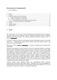
formed (Fig. 1.). The same may not be true for longer heating<br />
periods as in the roasting of coffee beans where extending<br />
the operation may result in a decrease in the amount of<br />
acrylamide persisted in the final product 19 . They may be a<br />
critical temperature/time zone where acrylamide is formed at<br />
a greater rate than it is destroyed, at temperatures outside of<br />
this zone little acrylamide is present.<br />
AA (mg/g Asn)<br />
120<br />
100<br />
80<br />
60<br />
40<br />
20<br />
0<br />
0 10 20 30 40 50<br />
time (min)<br />
180 °C<br />
160 °C<br />
140 °C<br />
130 °C<br />
120 °C<br />
Fig. 1. Amount of acrylamide after heating of equimolar mixture<br />
of glucose and asparagine at different temperatures<br />
The fact that acrylamide is not formed during boiling<br />
indicates that higher temperatures and/or low moisture conditions<br />
are needed for its formation. During heating under<br />
atmospheric conditions, higher temperatures can be reached<br />
only if simultaneous drying takes place, which is the case<br />
in frying, baking and roasting. The loss of water as the food<br />
dries during heating extracts a large amount of the incoming<br />
energy, and hence a bulk of the product is at a temperature<br />
very much lower than that of the heating medium. In this<br />
respect, temperature, time and moisture are key drivers of<br />
acrylamide formation in foods during heating (Fig. 2.). The<br />
moisture content determines the physical state and mobility<br />
of chemical constituents in food matrix. In addition, water<br />
alone affects the chemical route and the mechanistic pathway<br />
for acrylamide formation 13 .<br />
Concerning reducing sugars as carbonyl source, fructose<br />
has been found more effective than glucose in forming acrylamide<br />
(Fig. 2.). Both the chemical reactivity of sugars and<br />
their physical state play an important role in acrylamide formation.<br />
The melting point of fructose and glucose are 126 °C<br />
and 157 °C, respectively 13 . This explains why fructose is<br />
more reactive than glucose on acrylamide formation during<br />
heating. Frying, baking and roasting are simply characterized<br />
as open processes in which heat and mass transfer occur simultaneously.<br />
As the moisture reduces due to evaporation,<br />
sugars initially dissolved in water begin to form a saturated<br />
solution and then crystallize. After crystallization, melting<br />
s541<br />
is required to change their state to liquid, so to make them<br />
chemically reactive. In this respect, reducing sugars having<br />
a lower melting point is expected to form acrylamide earlier<br />
during heating.<br />
AA (mmol/mol Asn)<br />
<strong>3.</strong>0<br />
2.5<br />
2.0<br />
1.5<br />
1.0<br />
0.5<br />
fructose<br />
glucose<br />
0.0<br />
0 10 20 30 40 50 60 70 80<br />
moisture (%)<br />
Fig. 2. Amount of acrylamide after heating of equimolar mixture<br />
of asparagine and glucose/fructose at 180 °C for 20 min with<br />
different addition of water<br />
E n z y m e T r e a t m e n t L e a d i n g t o<br />
A c r y l a m i d e R e d u c t i o n<br />
One of the most effective ways to avoid acrylamide formation<br />
is removing the precursors, particularly amino acid<br />
L-asparagine. L-asparaginase as an enzyme of the hydrolases<br />
group (EC <strong>3.</strong>5.1.1.) selectively hydrolyses the amide bond of<br />
L-asparagine which results in the formation of aspartic acid<br />
and ammonia. Because the acrylamide formation correlates<br />
strongly with a free asparagine concentration, the reduction<br />
of L-asparagine in raw materials leads to the reduced<br />
level of acrylamide in final products 20 . The safety of asparaginase<br />
application is guaranteed by approving of GRAS<br />
status of Aspergillus oryzae asparaginase enzyme from<br />
novozymes A/S 21 , and Aspergillus niger asparaginase enzyme<br />
from DSM 22 , and a positive evaluation from the JECFA<br />
in 2007 23 as well. Moreover, this enzyme is inactivated by<br />
high temperature in the process of proteolysis.<br />
The application of L-asparaginase solution in a simulated<br />
potato matrix resulted in 50 to 90 % reduction of acrylamide<br />
content depending on the conditions (enzyme dose, time and<br />
temperature of incubation). no significant differences in<br />
impacts on L-asparagine conversion into L-aspartic acid in<br />
model samples between bacterial and fungal originated enzymes<br />
were observed. The positive effect of enzyme on the<br />
decrease of acrylamide content was confirmed also after Lasparaginase<br />
application on raw potato mash as well as dehydrated<br />
potato-wheat semi-products (Fig. <strong>3.</strong> and Fig. 4.) 20 .<br />
It is known that each intervention in the technology<br />
can be accompanied with consequences on the quality and<br />
sensory properties of final products which are strongly connected<br />
with the acceptability by consumers. For that reason<br />
the preliminary sensory evaluation of thermally and enzymatically<br />
treated products was done by a panel of trained<br />
judges. They described the main properties important for
Chem. Listy, 102, s265–s1311 (2008) Food Chemistry & Biotechnology<br />
these kinds of products such as darkness, yellowness, appearance,<br />
stickiness, crispness, oiliness, flavour, off-flavour, saltiness,<br />
sweetness and overall acceptability. Changes in colour<br />
were observed in pancakes prepared under different heating<br />
programmes, where darkness and crispness were more intensive<br />
in pancakes prepared at higher temperature of frying.<br />
no differences in evaluated sensory properties mentioned<br />
above were found out in any case of L-asparaginase application<br />
(P = 99 %) that was consider as a great advantage of<br />
the presented effective way of acrylamide reduction in food<br />
products 24 .<br />
AA (ng/g FW) .<br />
700<br />
600<br />
500<br />
400<br />
300<br />
200<br />
100<br />
0<br />
without enzyme<br />
2 U/g FW of enzyme; 10 min/ 37 °C incubation<br />
10 U/g FW of enzyme; 10 min/ 37 °C incubation<br />
Marabel Bellarosa<br />
Fig. <strong>3.</strong> Amount of acrylamide (AA) in raw potatoes (varieties<br />
Marabel and bellarosa) after enzymatic treatment (L-asparaginase<br />
produced by A. oryzae applied at concentration of 2 and<br />
10 u g –1 Fw and incubated at 37 °C for 10 min) and following<br />
heat treatment at 180 °C for 20 min<br />
AA (ug/kg FW) .<br />
800<br />
700<br />
600<br />
500<br />
400<br />
300<br />
200<br />
100<br />
0<br />
without enzyme 1 U/g FW of enzyme, 30 min/ 37°C incubation<br />
175 °C/20 min 180 °C/20 min 200 °C/20 min<br />
Fig. 4. Amount of acrylamide (AA) in pancakes prepared from<br />
potato-wheat powder at different heating temperatures (175 °C,<br />
180 °C and 200 °C) for 20 min with previous enzymatic treatment<br />
(L-asparaginase produced by A. oryzae applied at concentration<br />
of 1 u g –1 Fw and incubated at 37 °C for 30 min)<br />
Conclusions<br />
Since the acrylamide occurrence in foods and its potentiality<br />
to cause detrimental affects on human health attracts<br />
attention in all over the word, the effort to minimize its level<br />
in foods and consequently the human exposure to acrylamide<br />
is extremely advisable. Among many ways of acrylamide reduction<br />
the application of enzyme in order to prevent acry-<br />
s542<br />
lamide formation is feasible and effective without any undesirable<br />
effect on sensory quality of final products. For that<br />
reason, this procedure is protected by the patent application<br />
filed with the Industrial Property Office of the Slovak Republic<br />
under the number 5027-2006.<br />
This work was supported by the Slovak Research and<br />
Development Agency under the contract No. COST-0015-06.<br />
REFEREnCES<br />
1. Tareke E., Rydberg P., Karlsson P., Eriksson S., Tornqvist<br />
M.: J. Agric. Food Chem. 50, 4998 (2002).<br />
2. JECFA 2005 64th meeting Rome, 8-17 February 2005:<br />
http://www.who.int/ipcs/food/jecfa/summaries/summary_report_64_final.pdf<br />
<strong>3.</strong> IARC. Acrylamide. In IARC Monographs on the evaluation<br />
of carcinogen risk to humans: some industrial<br />
chemicals. International Agency for Research on Cancer,<br />
60, p. 389, Lyon, France 1994.<br />
4. Commission Recommendation of 3 May 2007 on the<br />
monitoring of acrylamide levels in food (notified under<br />
document number C(2007) 1873).<br />
5. Hogervorst J. G., Schouten L. J., Konings E. J.,<br />
Goldbohm R. A., Van Den Brandt P. A.: Cancer Epidemiol.<br />
Biomarkers Prevent. 16, 2304 (2008).<br />
6. Olesen P. T., Olsen A., Frandsen H., Frederiksen K.,<br />
Overvad K., Tjonneland A.: Int. J. Canc. 122, 2094<br />
(2008).<br />
7. FAO/WHO (2002): Opinion of the Scientific Committee<br />
on Food on new findings regarding the presence of acrylamide<br />
in food, Brussels, 3 July 2002.<br />
8. Mottram D. S., Wedzicha B. L., Dodson A. T.: nature<br />
419, 448 (2002).<br />
9. Stadler R. H., Blank I., Varga n., Robert F., Hau J.,<br />
Guy P. A., Robert M. C., Riediker S.: nature 419, 449<br />
(2002).<br />
10. Zyzak D. V., Sanders R .A., Stojanovic M., Tallmadge<br />
D. H., Eberhart B. L., Ewald D. K., Gruber D. C., Morsch<br />
T. R., Strothers M. A., Rizzi G. P., Villagran M. D.:<br />
J. Agric. Food Chem. 51, 4782 (2003).<br />
11. Yaylayan V. A., Wnorowski, A., Perez-Locas C.: J. Agric.<br />
Food Chem. 51, 1753 (2003).<br />
12. Biedermann M., Grob K.: Mitt. Geb. Lebensmitt. Hyg.<br />
94, 406 (2003).<br />
1<strong>3.</strong> Ciesarová Z., Kiss E., Kolek, E.: Czech J. Food Sci. 24,<br />
133 (2006).<br />
14. CIAA Acrylamide Toolbox [on-line]. Brussels : Confederation<br />
of the Food and Drink Industries of the European<br />
Union, 11 December 2007.<br />
15. Ciesarová Z.: Chem. Listy 99, 483 (2005).<br />
16. Kolek E., Šimon P., Šimko P.: J. Food Sci. 72, E341<br />
(2007).<br />
17. Kolek E., Šimko P., Šimon P., Gatial, A.: J. Food nutr.<br />
Res. 46, 39 (2007).<br />
19. Senyuva H. Z., Gökmen V.: Food Add. Contam. 22, 214<br />
(2005).
Chem. Listy, 102, s265–s1311 (2008) Food Chemistry & Biotechnology<br />
20. Ciesarová Z., Kiss E., Boegl P.: J. Food nutr. Res. 45,<br />
141 (2006).<br />
21. FDA 2006 Agency Response Letter GRAS notice no.<br />
GRn 000201.<br />
22. FDA 2007 Agency Response Letter GRAS notice no.<br />
GRn 000214.<br />
s543<br />
2<strong>3.</strong> JECFA 2007 68th meeting Geneva, 19-28 June 2007:<br />
http://www.who.int/ipcs/food/jecfa/summaries/summary68.pdf<br />
24. Ciesarová Z., Kukurová K.: Proc. ICC International<br />
Conference Bosphorus 2008, Istanbul 24-26 April 2008,<br />
p. 197.
Chem. Listy, 102, s265–s1311 (2008) Food Chemistry & Biotechnology<br />
L03 SOLID STATE FERMENTATION AS A TOOL<br />
FOR PREPARATION OF bIOPRODuCTS<br />
ENRIChED wITh POLyuNSATuRATED<br />
FATTy ACIDS<br />
MILAn ČERTíK, ZUZAnA ADAMECHOVá and LInDA<br />
néMETH<br />
Department of Biochemical Technology, Faculty of Chemical<br />
and Food Technology, Slovak University of Technology, Radlinského<br />
9, 812 37 Bratislava, Slovak Republic,<br />
milan.certik@stuba.sk<br />
Introduction<br />
Increasing demand for high-value lipids has focused<br />
commercial attention on the provision of suitable biosynthetic<br />
framework for their production. One of the main target for<br />
microbial oil transformation is construction of healthy and<br />
dietary important polyunsaturated fatty acids, such as γ-linolenic<br />
acid (18 : 3 ω-6; GLA), dihomo-γ-linolenic acid (20 : 3<br />
ω-6; DGLA), arachidonic acid (20 : 4 ω-6; AA), eicosapentaenoic<br />
acid (20 : 5 ω-3; EPA) and docosahexaenoic acid (22 : 6<br />
ω-3; DHA). Their applications in biomedical, nutritional and<br />
pharmaceutical fields coupled with their inadequacy from<br />
conventional agricultural and animal sources has looked for<br />
developing suitable biotechnologies to produce these compounds<br />
1 .<br />
Particularly active in the synthesis of PUFAs are species<br />
of fungi belonging to Zygomycetes 2 . Oleaginous fungi<br />
producing PUFA could be economically valuable because<br />
the most of their PUFAs occur in the triacylglycerol fraction<br />
of their lipids. Two basic processes have been developed for<br />
microbial production of PUFAs: submerged and solid state<br />
fermentations 3,4 . However, the principal difficulty that has<br />
been experienced with submerged PUFA-riched oil production<br />
has been in its marketing rather than in developing<br />
the large-scale fermentation and oil extraction process. Therefore,<br />
the association of oleaginous fungi with solid state<br />
fermentations (SSF) has been developed in order to improve<br />
commercial potential of microbial oils and thus to create new<br />
perspectives for the economic competitiveness and market of<br />
microbial polyunsaturated fatty acids (PUFAs). Solid state<br />
fermentation is a process in which microorganisms grow<br />
on a moist solid substrate in the absence of free water 5 . SSF<br />
simulates fermentation reactions occurring in the nature and<br />
allows microbial utilization of raw agro-materials or byproducts<br />
of the agro-food industries. Because some oleaginous<br />
fungi simultaneously decrease anti-nutrient compounds in<br />
the substrates (e.g. phytic acid) and partially hydrolyze substrate<br />
biopolymers, prefermented mass with a high content<br />
of PUFAs may be used as inexpensive food and feed supplement<br />
6 . Thus, SSF might provide the other opportunity to fill<br />
marketing claims in food, feed, pharmaceutical, veterinary<br />
and environmental fields.<br />
This paper deals with effectivity of several lower filamentous<br />
fungi to synthesize various PUFAs during their utilization<br />
of cereals by solid state fermentations.<br />
s544<br />
Experimental<br />
M i c r o o r g a n i s m s<br />
Thamnidium elegans CCF 1456, Cunninghamella echinulata<br />
CCF-103, Cunninghamella elegans CCF-1318, Mortierella<br />
isabelina CCF-14, Mortierella isabelina CCF-1098,<br />
Mortierella alpina CCF 185 were obtained from the Culture<br />
Collection of Fungi (Charles University, Prague, Czech<br />
Republic). The culture was maintained on modified Czapek-<br />
Dox agar slants with yeast extract (2.5 g dm –3 ) at 4 °C.<br />
S u b s t r a t e s a n d C u l t i v a t i o n<br />
C o n d i t i o n s<br />
Depending on the microorganism, various types of substrates<br />
were employed during SSF experiments. Spent malt<br />
grains (SMG) were added to some substrates. Autoclavable<br />
microporous polypropylene bags (160 × 270 mm 2 ) were filled<br />
with 10 g of dry substrate, moistened by the addition of 10 ml<br />
distilled water, soaked for 2 h at laboratory temperature and<br />
sterilized in autoclave (120 kPa, 120 °C, 20 min). In order<br />
to increased yield of PUFAs, sunflower or linseed oils were<br />
added to some substrates. In addition, various amounts of<br />
10% acetone or ethanol solutions of selected plant extracts<br />
were tested with the aim to activate enzymes involved into<br />
PUFA biosynthesis. The substrates were inoculated and<br />
mixed with 2 ml of spore suspension (1–2 × 10 6 spores per<br />
ml). Then each bag was closed with sterile cotton plugs,<br />
inoculated substrate was spread in the bags to obtain substrate<br />
layer of about 1 cm and incubated statically at 25 °C<br />
for 4–6 days (T. elegans, C. echinulata, C. elegans, M. isabellina)<br />
and 10–14 days (M. alpina). Triplicate SSF experiments<br />
for each substrate were prepared to assess reproducibility and<br />
average results are presented.<br />
L i p i d E x t r a c t i o n a n d F a t t y A c i d<br />
A n a l y s i s<br />
Prefermented cereal materials (bioproducts) were gently<br />
dried at 65 °C for 10 h and weighed. Lipids from homogenized<br />
bioproducts were isolated with chloroform/methanol<br />
(2 : 1, v/v) and purified according to Čertík et al. 7 and total<br />
lipids were determined gravimetrically. Fatty acids of total<br />
lipids were analyzed as their methyl esters 8 by gas chromatography<br />
according to Čertík et al 9 . Gas chromatograph (GC-<br />
6890 n, Agilent Technologies) was equipped with a capillary<br />
column DB-23 (60 m × 0.25 mm, film thickness 0.25 μm,<br />
Agilent Technologies) and a FID detector (constant flow,<br />
hydrogen 35 ml min –1 , air 350 ml min –1 , 250 °C). Analyses<br />
were carried out under a temperature gradient (130 °C for<br />
1 min; 130–170 °C at program rate 6.5 °C min –1 ; 170–215°C<br />
at program rate 2.7°C min –1 ; 215 °C for 7 min; 220–240 °C at<br />
program rate 2 °C min –1 ; 240 °C for 2 min) with hydrogen as<br />
a carrier gas (flow 2.1 ml min –1 , velocity 49 cm s –1 , pressure<br />
174 kPa) and a split ratio of 1/50 (inlets: heater 230 °C, total<br />
hydrogen flow 114 ml min –1 , pressure 174 kPa). The fatty<br />
acid methylester peaks were identified by authentic standards<br />
of C 4 –C 24 fatty acid methylesters mixture (Supelco, USA)<br />
and quantified by an internal standard of heptadecanoic acid
Chem. Listy, 102, s265–s1311 (2008) Food Chemistry & Biotechnology<br />
(C17 : 0, Supelco, USA). Fatty acid concentration was evaluated<br />
with ChemStation software B0103 (Agilent technologies,<br />
USA).<br />
Results and Discussion<br />
The extensive research and development of PUFA production<br />
by SSF is basically aimed at improving the economic<br />
competitiveness of that microbial process compared to<br />
plant- and animal-derived oils. Emphasis is put on increasing<br />
the product value, using inexpensive substrates, screening<br />
for more efficient strains and reducing the processing steps.<br />
Therefore, it is necessary to optimize the potential of microorganisms<br />
for transformation of agroindustrial materials and<br />
oil residues into desired metabolites.<br />
Screening of microorganisms has led to selection of<br />
T. elegans, C. echinulata, C. elegans and Mortierella isabellina<br />
as producers of GLA 6,9 and Mortierella alpina as a<br />
producer of DGLA, AA and EPA 10 . Generally, the surface<br />
of substrates was not only covered by the fungal mycelium<br />
during cultivation, but the fungal hyphae also penetrated into<br />
the substrates. Thus, fungal PUFAs were accumulated in the<br />
newly formed bioproduct and their amount depended on the<br />
substrates, microorganisms and cultivation conditions used.<br />
Depending on the microorganism, various types of cereal<br />
substrates were employed during SSF experiments (Table I).<br />
Spent malt grains (SMG) served as an internal support. Substrates<br />
without SMG in most cases led to agglomeration of<br />
substrate particles and created more compact mass which in<br />
turn interfered with microbial respiration and affected substrate<br />
utilization negatively. Presence of SMG improved<br />
bioconversion of linoleic acid from substrates to GLA 9 . Substrates<br />
with internal support not only provided better respiration<br />
and aeration efficiency due to an increased inter-particle<br />
space but also helped to remove the heat generated during<br />
fermentation. It should be noticed that although PUFAs were<br />
synthesized more effectively by SMG addition to substrates,<br />
total PUFAs yield was also dependent on substrate/SMG<br />
ratio. Unbalanced substrate/SMG ratio might provide limited<br />
surface for microbial attack and thus poorer availability of<br />
assimilable compounds (including oils) from substrates.<br />
Growth of fungi on a carbohydrates-containing substrates<br />
resulted after optimization of cultural conditions in constant<br />
lipid yield with the demanded fatty acid profile. Further<br />
improvement of PUFAs formation was achieved by physiological<br />
regulation of the SSF process employing following<br />
steps 10 : a) gradual elevation of carbon/nitrogen ratio with<br />
addition of appropriate carbon source; b) optimization of<br />
water activity, temperature and oxygen availability; c) transformation<br />
of exogenously added oils consisting of precursor<br />
of PUFAs. There is a stock of relatively cheap vegetable<br />
oils containing individual fatty acid precursors and SSF was<br />
applied for microbial utilization of renewable agricultural<br />
oils with the aim to modify their properties for production<br />
of value-added bioproducts with enhanced biological characteristics.<br />
Thus the ability of the strains to utilize exogenous<br />
fatty acids opens new possibilities to prepare PUFAs in high<br />
s545<br />
yield. Moreover, because fungi possess active oil-biotransforming<br />
system, these strains were also tested for their ability<br />
to convert directly oil-rich substrates (corn, sunflower seeds,<br />
linseeds, rapeseeds) to PUFAs.<br />
Table I<br />
Production of γ-linolenic acid (GLA), dihomo-γ-linolenic<br />
acid (DGLA), arachidonic acid (AA) and eicosapentaenoic<br />
acid (EPA) by solid state fermentations of selected fungi utilizing<br />
various cereal substrates. Ratio of susbtrate/SMG was<br />
1 : 3 (w/w)<br />
Strain Substrate PUFA Yield<br />
[g kg –1 BP]<br />
T. elegans oat flakes/SMG GLA 5.9<br />
wheat bran/SMG GLA 5.0<br />
wheat bran/SMG/<br />
GLA 10.0<br />
sunflower oil<br />
crushed corn GLA 10.0<br />
rye bran/SMG GLA 4.2<br />
buckwheat/SMG GLA 4.7<br />
millet/SMG GLA 6.5<br />
amaranth/SMG GLA 4.7<br />
C. echinulata barley GLA 6.1<br />
C. elegans barley GLA 7.0<br />
M. isabellina barley GLA 18.0<br />
M. alpina rice AA 21.4<br />
wheat sprout/SMG AA 36.1<br />
wheat bran/SMG AA 42.3<br />
rye bran/SMG AA 21.9<br />
peeled barley AA 16.2<br />
oat flakes AA 31.2<br />
M. aplina cresed sesame seeds DGLA 21.3<br />
M. alpina peeld barley/linseed EPA/AA 2<strong>3.</strong>4/36.3<br />
oil/SMG<br />
Biosynthesis and profile of fatty acids is controlled by<br />
enzymes involved in lipogenesis, so activation or inhibition<br />
of these metabolic steps is also useful tool for improving carbon<br />
flux to individual PUFAs 11 . For example, bioconversion<br />
of DGLA to AA is catalyzed by ‚∆ 5 ‘ desaturase and inhibition<br />
of this enzyme by crushed sesame seeds was accompanied by<br />
rapid increase of DGLA/AA ratio 11 ). In addition, application<br />
of various plant extracts possessing bioactive compounds<br />
seems to be promising way how to regulate fatty acid biosynthetic<br />
machinery in order to gain bioproduct with high<br />
yield of preferred PUFA. Application of ethanol extracts<br />
from ginger or sweet flag improved GLA yield by 30 % or<br />
25 %, respectively. On the other hand, biosynthesis of GLA<br />
was reduced by 70 % when acetone extract from tansy was<br />
employed to the substrate.<br />
Conclusions<br />
naturally prepared cereal based bioproducts enriched<br />
with PUFAs may be used as an inexpensive food and feed<br />
supplement. Thus, the association of selected microorganisms
Chem. Listy, 102, s265–s1311 (2008) Food Chemistry & Biotechnology<br />
with solid state fermentations has created new perspectives<br />
for the economic competitiveness and market of cereal based<br />
bioproducts containing PUFAs.<br />
The work was supported by grant VEGA No. 1/0747/08<br />
from the Grant Agency of Ministry of Education, Slovak<br />
Republic.<br />
REFEREnCES<br />
1. Gill I., Valivety R.: Trends Biotechnol. 15, 401 (1997).<br />
2. Čertík M., Shimizu S.: J. Biosci. Bioeng. 87, 1 (1999a).<br />
<strong>3.</strong> Čertík M., Shimizu S.: Agro Food Industry Hi-Tech. 10,<br />
26 (1999b).<br />
4. Čertík M., in: Biocatalysis and Bioenergy (Hou C.T.,<br />
Shaw J.-F., ed),p 571-585, Wiley, new York 2008<br />
s546<br />
5. Pandey A.: Biochem. Eng. J. 13, 81 (2003).<br />
6. Slugeň D., Streďanský M., Streďanská S., Čertík M.,<br />
Grego J.: Czech Patent 279043 (1994).<br />
7. Čertík M., Andráši P., Šajbidor J.: J. Am. Oil Chem. Soc.<br />
73, 357 (1996).<br />
8. Christoperson S.W., Glass R.L.: J. Dairy Sci. 52, 1289<br />
(1969).<br />
9. Čertík M., Sláviková L., Masrnová S., Šajbidor J.: Food<br />
Technol. Biotechnol. 44, 75 (2006).<br />
10. Sláviková L., Čertík M.: Chem. Listy, 99, 234 (2005).<br />
11. Čertík, M, Sakuradani, E, Shimizu S.: Trends Biotechnol.<br />
16, 500 (1998).
Chem. Listy, 102, s265–s1311 (2008) Food Chemistry & Biotechnology<br />
L05 PhySIOLOGICAL REGuLATION OF<br />
bIOTEChNOLOGICAL PRODuCTION OF<br />
CAROTENOID PIGMENTS<br />
VLADIMíRA HAnUSOVá a , MARTInA ČARnECKá b ,<br />
AnDREA HALIEnOVá b , MILAn ČERTíK a , EMíLIA<br />
BREIEROVá c and IVAnA MáROVá b<br />
a Department of Biochemical Technology, Faculty of Chemical<br />
and Food Technology, Slovak University of Technology,<br />
Radlinského 9, 812 37 Bratislava, Slovak Republic;<br />
b Faculty of Chemistry, Brno University of Technology, Purkyňova<br />
118, 612 00 Brno, Czech Republic;<br />
c Institute of Chemistry, Slovak Academy of Sciences, Dúbravská<br />
cesta 9, 845 38 Bratislava, Slovak Republic,<br />
milan.certik@stuba.sk<br />
Introduction<br />
Carotenoids represent one of the broadest group of<br />
natural antioxidants (over 600 characterized structurally)<br />
with significant biological effects and numerous of industrial<br />
applications. Because the application of synthetically prepared<br />
carotenoids as food additives has been strictly regulated<br />
in recent years, huge commercial demand for natural carotenoids<br />
has focused attention on developing of suitable biotechnological<br />
techniques for their production.<br />
There are many microorganisms including bacteria,<br />
algae, yeast and fungi, that are able to accumulate several<br />
types of pigments; but only a few of them have been exploited<br />
commercially 1 . From the view of yeasts, a range of species<br />
such as Rhodotorula, Rhodosporidium, Sporidiobolus, Sporobolomyces,<br />
Cystofilobasidium, Kockovaella and Phaffia<br />
have been screened for carotenoids formation. Yeast strains<br />
of Rhodotorula and Sporobolomyces formed β-carotene as<br />
the main pigment together with torulene and torularhodine as<br />
minor carotenoids. In contrast, Phaffia strains accumulated<br />
astaxanthin as a principal carotenoid. Comparative success<br />
in yeast pigment production has led to a flourishing interest<br />
in the development of fermentation processes in commercial<br />
production levels. However, in order to improve the yield of<br />
carotenoid pigments and subsequently decrease the cost of<br />
this biotechnological process, optimizing the culture conditions<br />
including both nutritional and physical factors have<br />
been performed. Factors such as carbon and nitrogen sources,<br />
minerals, vitamins, pH, aeration, temperature, light and<br />
stress showed a major influence on cell growth and yield of<br />
carotenoids.<br />
This paper summarizes our experience with physiological<br />
regulation and scale-up of biotechnological production of<br />
carotenoid pigments by yeasts.<br />
Experimental<br />
M i c r o o r g a n i s m s a n d C u l t i v a t i o n<br />
C o n d i t i o n s<br />
All strains investigated in this study (Sporobolomyces<br />
roseus CCY 19-6-4, S. salmonicolor CCY 19-4-10, Rhodotorula<br />
glutinis CCY 20-2-26, R. glutinis CCY 20-2-31, R. glu-<br />
s547<br />
tinis CCY 20-2-33, R. rubra CCY 20-7-28, R. aurantiaca<br />
CCY 20-9-7 and Phaffia rhodozyma CCY 77-1-1) were<br />
obtained from the Culture Collection of Yeasts (CCY; Institute<br />
of Chemistry, Slovak Academy of Sciences, Bratislava)<br />
and maintained on malt slant agar at 4 °C.<br />
The basic cultivation medium for flasks experiments<br />
for Rhodotorula and Sporobolomyces strains consisted of<br />
(g dm –3 ): glucose – 20; yeast extract – 4.0; (nH 4 ) 2 SO 4 – 10;<br />
KH 2 PO 4 – 1; K 2 HPO 4 . 3H2 O – 0.2; naCl – 0.1; CaCl 2 – 0.1;<br />
MgSO 4 . 7H2 O – 0.5 and 1 ml solution of microelements<br />
[(mg dm –3 ): H 3 BO 4 – 1.25; CuSO 4 . 5H2 O – 0.1; KI – 0.25;<br />
MnSO 4 . 5H2 O – 1; FeCl 3 . 6H2 O – 0.5; (nH 4 ) 2 Mo 7 O 24 . 4H2 O<br />
– 0.5 and ZnSO 4 . 7H2 O – 1]. The basic cultivation medium<br />
for flasks experiments for Phaffia strain consisted of<br />
(g dm –3 ): glucose – 20, yeast autolysate – 2.0, KH 2 PO 4 – 0.4,<br />
(nH 4 ) 2 SO 4 – 2.0, MgSO 4 . 7H2 O – 0.5, CaCl 2 – 0.1, naCl<br />
– 1.0. All strains grew under a non-lethal and maximally tolerated<br />
concentration of ni 2+ , Zn 2+ , Cd 2+ and Se 2+ ions. Also,<br />
stress conditions were induced by addition of various conventrations<br />
of naCl and H 2 O 2 . The cultures were cultivated<br />
in 500 ml flasks containing 250 ml cultivation medium on<br />
a rotary shaker (150 rpm) at 28 °C to early stationary grow<br />
phase. All cultivation experiments were carried out at triplicates<br />
and analyzed individually.<br />
Flasks results were verified in bioreactors and these<br />
scale-up experiments were carried out in 2 L fermentor (B.<br />
Braun Biotech), 20 L (SLF-20) and 100 L (Bio-la-fite) fermentors<br />
with an agitation rate of 250–450 rpm and a temperature<br />
of 20–22 °C. The pH was controlled at pH 5.0 by the<br />
addition of nH 4 OH and the dissolved oxygen concentration<br />
was maintained by supplying sterile air at a flow rate equivalent<br />
to 0.3–0.7 vvm.<br />
P i g m e n t I s o l a t i o n a n d A n a l y s i s<br />
Pigments from homogenized bioproducts were isolated<br />
by organic extraction and analyzed by high-performance<br />
liquid chromatography (HPLC). Analysis was carried out<br />
with an HP 1100 chromatograph (Agilent) equipped with a<br />
UV-VIS detector. Pigments extracts (10 μl) were injected<br />
onto LiChrospher ® 100 RP-18 (5 μm) column (Merck). The<br />
solvent system (the flow rate was 1 ml min –1 ) consisted of<br />
solvent A, acetonitrile/water/formic acid 86 : 10 : 4 (v/v/v),<br />
and B, ethyl acetate/formic acid 96 : 4 (v/v), with a gradient<br />
of 100 % A at 0 min, 100 % B at 20 min, and 100 % A at<br />
30 min.<br />
G e l E l e c t r o p h o p h o r e s i s<br />
1D PAGE-SDS electrophoresis of proteins was carried<br />
out by common procedure using 10% and 12.5% polyacrylamide<br />
gels. Proteins were staining by Coomassie Blue and by<br />
silver staining. For comparison, microfluidic technique using<br />
1D Experion system (BioRad) and P260 chips was used for<br />
yeast protein analysis too. 2D electrophoresis of proteins<br />
was optimized in cooperation with Laboratory of Functional<br />
Genomics and Proteomics, Faculty of Science, Masaryk<br />
University of Brno. 2D gels were obtained from protein pre-
Chem. Listy, 102, s265–s1311 (2008) Food Chemistry & Biotechnology<br />
paratives isolated from lyophilized cells. After optimization<br />
of separation conditions proteomes from stressed R.glutinis<br />
and R.rubra cells were isolated, lyophilized and analyzed.<br />
Quantitative analysis was done using BioRad Laboratories<br />
2D software. Identification of some spots was done using<br />
LC-MS/MS.<br />
Results and Disscussion<br />
To find suitable conditions for carotenoids production,<br />
several pigment forming yeasts have been tested for their<br />
ability to modify carotenoid biosynthesis based on media<br />
composition. Kinetic analysis revealed that yeasts strains<br />
differed from the view of growth rates and pigments formation.<br />
It must be emphasized that overall yield of carotenoids<br />
is directly related to the total biomass yield, thus, to keep both<br />
high growth rates and high flow carbon efficiency to carotenoids<br />
by optimal cultivation conditions is essential in order to<br />
achieve the maximal pigment productivity.<br />
Carotenoid accumulation in cells was significantly activated<br />
when yeasts were treated by selected stress conditions<br />
and their combinations (heavy metals, hydrogen peroxide<br />
and salt). However, such stimulation of pigment biosynthesis<br />
was more effective if stress factors were employed to the<br />
medium in exponential growth phase than from the beginning<br />
of cultivation 2–4 . It may be explained by hypothesizing<br />
a possible activation or inhibition mechanism by metal ions<br />
on specific carotenogenic enzymes, in particular, on specific<br />
enzymes involved in carotenoid biosynthesis, in agreement<br />
with previous studies reporting activation or inhibition by<br />
metal ions in microbial enzymes. The other explanation is<br />
based on observations that presence of heavy metals results<br />
in formation of various active oxygen radicals what, in a turn,<br />
induces generation of protective carotenoid metabolites that<br />
reduce negative behaviour of free radicals 5 .<br />
During environmental stress response many red yeasts<br />
exhibite cross-protective mechanisms. Preincubation of yeast<br />
Table I<br />
Production of carotenoids in flask and fermentation experiments<br />
Strain Pigment Pigment yields<br />
Flasks experiments<br />
S. roseus β-carotene 30 mg dm –3 (by Zn 2+ + H 2 O 2 )<br />
R. glutinis torulene 6 mg dm –3 (by Zn 2+ + H 2 O 2 )<br />
R. glutinis torularhodin 1 mg dm –3 (by Cu 2+ )<br />
P. rhodozyma astaxanthin 20 mg dm –3 (by Se 2+ )<br />
Scale-up fermentations<br />
S. salmonicolor. total carotene 41 mg dm –3 (by NaCl + H 2 O 2 )<br />
R. glutinis β-carotene 35 mg dm –3 (by NaCl + H 2 O 2 )<br />
P. rhodozyma astaxanthin 110 mg dm –3 (by NaCl + NaCl)<br />
s548<br />
culture with low concentration of one stress factor in inoculum<br />
media (e.g. salt, hydrogen peroxide) induces adaptation<br />
pathways resulted in enhanced carotenoid production 6 . Further<br />
addition of higher concentration of either the same or<br />
other stress factor can lead to significant (5–10 times) increase<br />
of β-carotene production in Rhodotorula glutinis and Sporobolomyces<br />
salmonicolor 2 . Simple preincubation of R. glutinis<br />
in presence of 2% naCl in inoculation medium followed by<br />
fermentation in inorganic production medium led to increased<br />
formation of biomass with accumulated carotenoids. This<br />
combined environmental stress using mild stress effect of salt<br />
or hydrogen peroxide could be industrially used for production<br />
of carotene enriched biomass. The results from flasks<br />
experiments are summarized in Table I.<br />
Laboratory flasks experiments were verified in semiscale<br />
conditions (from 2 dm 3 to 100 dm 3 fermentors). It was<br />
confirmend that glucose utilization was followed by increased<br />
pigment biosynthesis. Therefore, fed-batch fermentations<br />
were applied to enhanced carotenoid production. In addition,<br />
combination of aimed environmental condition resulted in<br />
activated accumulation of selected carotenoids (Table I).<br />
Enhancement of carotenoid production by environmental<br />
stress is also associated with changes in expression levels<br />
of various genes. Therefore, molecular changes in yeast cells<br />
on genome, proteome and metabolome level were studied<br />
using PFGE, 2D-GE, LC/MS/MS and EPR techniques. Presence<br />
of stress factors was accompanied by changes in carotenoid<br />
production as well as by alternations in protein levels.<br />
However, further analyses have to be focused on more precise<br />
characterization of proteins those displayed significant<br />
changes under increased biosynthesis of carotenoids.<br />
The work was supported by grant VEGA No. 1/0747/08<br />
and 2/7031/27 from the Grant Agency of Ministry of Education,<br />
Slovak Republic, grant FRVS 2541/G4/2008 of the<br />
Ministry of Education, Youth and Sports of the Czech Republic<br />
and grant IAA400310506 of Grant Agency of the Academy<br />
of Sciences of the Czech Republic.<br />
REFEREnCES<br />
1. Bhosale P.: Appl. Microbiol. Biotechnol. 73, 351<br />
(2004).<br />
2. Márová I., Breierová E., Kočí R., Friedl Z., Slovák B.,<br />
Pokorná J.: Annals of Microbiology 54, 73 (2004).<br />
<strong>3.</strong> Breierová E., Márová I., Čertík M.: Chem. Listy 99, 109<br />
(2005).<br />
4. Čertík M., Masrnová S., Sitkey V., Minárik M., Breierová<br />
E.: Chem. Listy 99, 237 (2005).<br />
5. Rapta P., Polovka M., Zalibera M., Breierová E., Žitňanová<br />
I., Márová I., Čertík M.: Biophys. Chem. 116, 1<br />
(2005).<br />
6. Kočí R., Drábková M., Márová I.: Chem. Listy 99, 297<br />
(2005).
Chem. Listy, 102, s265–s1311 (2008) Food Chemistry & Biotechnology<br />
L06 ENZyMATIC SACChARIDE ACETyLATIONS<br />
IN wATER – COMPARATION OF CATALySTS<br />
VLADIMíR MASTIHUBA a , TATIAnA KRAKOVá b and<br />
MáRIA MASTIHUBOVá a<br />
a Institute of Chemistry, Center for Glycomics, Slovak Academy<br />
of Sciences, Dúbravská cesta 9, 845 38 Bratislava, Slovak<br />
Republic,<br />
b Institute of Molecular Biology, Slovak Academy of Sciences,<br />
Dúbravská cesta 21, 845 51 Bratislava, Slovak Republic,<br />
chemvrma@savba.sk<br />
Introduction<br />
Enzymatic esterification of various hydroxy compounds<br />
is studied for about 40 years. Such reactions are usually performed<br />
in reaction systems comprising controlled amount of<br />
water to prevent product hydrolysis. Followingly, enzymatic<br />
esterification of unprotected saccharides is hampered by their<br />
decreased solubility in low water envíronment.<br />
We had recently described acetyl esterase from Trichoderma<br />
reesei to catalyze acetylations of several carbohydrates<br />
and alcohols in bulk water phase 1 . Acetyl esterases may<br />
be found in several raw commercial preparations of enzymes<br />
dedicated for biomass degradation since acetylation occurs<br />
widely on natural polysaccharides and plant cell wall fragments<br />
2 . Acetyl hydrolyzing activity occurs quite frequently<br />
in lipases (which may be result either of wider acyl specifity<br />
or of protein impurity) and some reports describe also esterase<br />
activity of serum albumins 3 .<br />
Our present work explores performance of several<br />
enzymes/crude enzyme preparations and proteins (Celluclast<br />
1.5 L FG, Ultraflo L, lipases from Candida antarctica, Candida<br />
rugosa, Aspergillus niger and Thermomyces lanuginosus,<br />
bovine serum albumin) in acetylations of glucose (1 a)<br />
and methyl α-D-glucopyranoside (1 b), comparing product<br />
yields and position selectivity of the reaction.<br />
Experimental<br />
Assays of acetyl esterase activity in enzyme preparations<br />
were performed as previously described 1 . Synthetic<br />
reactions were routinely performed by dissolving 1.2 g of<br />
glucose or alpha methyl glucoside in 10 ml mixture of water<br />
and acetyl donor (vinyl acetate or isopropenyl acetate, 4 : 1)<br />
and the reaction was started by adding 1 ml or 300 mg of<br />
the enzyme. Reaction mixture was then stirred at 37 o C for<br />
1–3 days and the course of the reaction was followed by t.l.c.<br />
on silica gel plates (chloroform – methanol, 3 : 1, visualisation<br />
by pouring into 5% sulphuric acid in ethanol and drying<br />
in hot air). Reaction mixture was then concentrated in vacuo,<br />
products separated by chromatography on silicagel column<br />
(chloroform – methanol, 3 : 1) and identified by nMR. For<br />
every reaction, a parallel experiment was executed without<br />
addition of enzyme to check occurence of a spontaneous acetylation.<br />
The spontaneous reactions were tested also separately<br />
in 0.1M acetate and phosphate buffers.<br />
s549<br />
Results<br />
Table I presents level of acetyl hydrolysing activities in<br />
enzyme preparations. Although water insoluble Lipolase 100<br />
was not active in photometric assay, it catalyzed acetylations<br />
of aliphatic alcohols in stired low water reactions (data not<br />
shown).<br />
Table I<br />
Acetyl hydrolysing activity of enzyme preparations<br />
enzyme activity activity<br />
(souce) [U ml –1 ] [U g –1 ]<br />
Celluclast 1.5 L FG<br />
(Trichoderma reesei)<br />
18.4<br />
Ultraflo L<br />
(Humicola insolens)<br />
31.5<br />
novozym 735<br />
(Candida antarctica)<br />
55.6<br />
BSA<br />
(Bos taurus)<br />
4.0<br />
Amano Lipase A<br />
(Aspergillus niger)<br />
219.1<br />
Lipase AYS<br />
(Candida rugosa)<br />
621.1<br />
Lipolyve CC<br />
319.0<br />
(Candida rugosa)<br />
Lipolase 100T<br />
(Thermomyces lanuginosus)<br />
not estimated<br />
Since phosphate buffer catalyzes acetylation of saccharides<br />
by vinyl acetate 4 , we tried to assess the extent of this<br />
process in our reactions (Table II).<br />
Table II<br />
Sponatneous acetylation of glucose by vinyl acetate (VA) and<br />
isopropenyl acetate (IPA) in buffered solutions and water<br />
pH Acetyl donor Monoacetates Diacetates<br />
4.01 VA + –<br />
4.51 VA ++ –<br />
5.02 VA ++ +/–<br />
5.51 VA +++ +<br />
5.72 VA +++ +<br />
6.03 VA +++ +<br />
Water VA – –<br />
Water IPA – –<br />
5.72 IPA +/– –<br />
Increasing pH of the buffer resulted in increased creation<br />
of glucose monoacetates and eventually diacetates when<br />
vinyl acetate was used as acetyl donor. To avoid such unselective<br />
acetylations, all enzymatic reactions were executed<br />
using water instead of the buffer.<br />
Scheme 1 summarizes in general the scale of products<br />
identified in reaction mixtures.
Chem. Listy, 102, s265–s1311 (2008) Food Chemistry & Biotechnology<br />
HO HO<br />
OH<br />
O<br />
OH<br />
R= α/β-H 1a<br />
α-CH3 1b<br />
OR<br />
HO O<br />
HO HO<br />
Yeast lipases gave low yields of pure 3 a,b, thus showing<br />
high specifity for acetylation of primary hydroxyl (Tables<br />
3,4). However, Lipolyve CC was reactive only with the glucoside<br />
1 b and did not acetylate free glucose within 3 days.<br />
On the other side, Amano Lipase A (Aspergillus niger)<br />
as a representative of fungal lipases gave 10% yield of an<br />
equimolar mixture of 2 a, and 3 a. Interestingly, acetylation<br />
O<br />
O<br />
HO HO<br />
O<br />
HO O<br />
O<br />
OH<br />
O<br />
O<br />
OH<br />
2 a,b<br />
O<br />
OH<br />
3 a,b<br />
OH<br />
4 a,b<br />
O<br />
5 a ,b<br />
Scheme 1<br />
Summarization of products created by enzymatic acetylations of<br />
glucose and methyl α-D-glucopyranoside with vinyl acetate or<br />
isoprenyl acetate<br />
Table III<br />
Enzymatic acetylations of glucose by vinyl acetate (VA) and<br />
isopropenyl acetate (IPA) in water<br />
enzyme<br />
acetyl<br />
donor<br />
O<br />
O<br />
O<br />
O<br />
OH<br />
OR<br />
OR<br />
OR<br />
OR<br />
mono-<br />
diacetates reaction<br />
acetates<br />
[%]<br />
time<br />
[%] [days]<br />
Celluclast VA<br />
19.7<br />
2 a : 3 a<br />
12 : 1<br />
0 1<br />
Lipolyve CC IPA 0 0 3<br />
novozym 735 VA<br />
2.0<br />
9.6<br />
3 a<br />
3<br />
not<br />
determined<br />
Amano<br />
Lipase A<br />
IPA<br />
10.0<br />
2 a : 3 a<br />
1:1<br />
4.2<br />
5 a<br />
2<br />
s550<br />
did continue to create only one diacetate (3,6-di-O-acetyl<br />
glucose, 5 a), thus keeping the original specifity for positions<br />
3-O- and 6-O- (Table III). Acetylations of methyl α-Dglucopyranoside<br />
(1 b) with isopropenyl acetate proceeded<br />
with the same positional specifity to produce 2 b and 3 b.<br />
When isopropenyl acetate was replaced by vinyl acetate as<br />
acetyl donor, selectivity increased markedly in favor of 2 b<br />
(Table IV). no reaction was observed when another fungal<br />
lipase – Lipolase 100T – was used as biocatalyst.<br />
Acetylations of 1 b catalyzed by bovine serum albumin<br />
and Ultraflo L (an enzyme cocktail from Humicola insolens)<br />
were unselective, giving mixtures of three monoacetates<br />
(Table IV).<br />
The best results were achieved by use of Celluclast, a<br />
cellulase preparation from Trichoderma reesei RUT C-30<br />
comprising the same acetyl esterase as reported earlier 1 . Acetylations<br />
gave 19.7 % and 10 % of monoacetylated glucose<br />
and methyl glucoside, respectively (Tables III, IV). The selectivity<br />
was exclusively to position 3-O- in acetylation of 1 b<br />
while in acetylation of glucose, almost 8% of 6-O- acetate 3 a<br />
was found in the monoacetate fraction.<br />
Table IV<br />
Enzymatic acetylations of methyl glucoside by vinyl acetate<br />
(VA) and isopropenyl acetate (IPA) in water<br />
enzyme<br />
acetyl<br />
donor<br />
Celluclast IPA<br />
10.0<br />
2 b<br />
Lipolyve CC IPA<br />
2.2<br />
3 b<br />
Lipase AYS IPA<br />
1.9<br />
3 b<br />
Amano<br />
Lipase A<br />
IPA<br />
10.5<br />
2 b : 3 b<br />
2,5 : 1<br />
Amano<br />
Lipase A<br />
VA<br />
6.4<br />
2 b : 3 b<br />
13 : 1<br />
2.1<br />
BSA VA 3 b : 4 b : 2 b<br />
2.0 : 1.3 : 1.0<br />
<strong>3.</strong>9<br />
Ultraflo L IPA 3 b : 4 b : 2 b<br />
4.5 : 2.5 : 1.0<br />
mono-<br />
diacetates<br />
reaction<br />
acetates<br />
[%]<br />
time<br />
[%] [days]<br />
0 1<br />
0 0.9<br />
0 0.16<br />
1.7<br />
5 b<br />
0.7<br />
5 b<br />
Conclusions<br />
Our results proved Celluclast to be the best catalyst for<br />
acetylations of glucose and methyl α-D-glucopyranoside<br />
according to its selectivity, product yields, availability and<br />
price. Since Celluclast has significantly lower acetyl esterase<br />
activity comparing to the other enzymes tested in this study,<br />
the enzyme seems rather unique. The enzyme selectivity to<br />
position 3-O- gives a promiss of its use in two step enzymatic<br />
1<br />
1<br />
0 5<br />
0 1
Chem. Listy, 102, s265–s1311 (2008) Food Chemistry & Biotechnology<br />
esterifications of saccharides, in which acetylation provides<br />
organic solvent-soluble sugar derivatives possessing free primary<br />
hydroxyls.<br />
This work has been supported by a grant from the Slovak<br />
Grant Agency for Science VEGA under the Project No.<br />
2/0145/08.<br />
s551<br />
REFEREnCES<br />
1. Kremnický ľ., Mastihuba V., Côté G. L.: J. Mol. Catal.<br />
B: Enzymatic 30, 229 (2004).<br />
2. de Vries R. P., Visser J.: Microbiol. Mol. Biol. Rev. 65,<br />
497 (2001).<br />
<strong>3.</strong> Sedlacek M., Manoharan I., Boopathy R., Duysen E.<br />
G., Masson P., Lockridge O.: Biochem. Pharmacol. 70,<br />
1673 (2005).<br />
4. Shi C., Zhang J. B., Fu H., Tang J.: Lett. Org. Chem. 3,<br />
932 (2006).
Chem. Listy, 102, s265–s1311 (2008) Food Chemistry & Biotechnology<br />
L07 PREPARATION OF bIODIESEL FROM TALL<br />
OIL<br />
RADOSLAV MIKULáŠIK a , IGOR ŠURInA a ,<br />
SVETOZáR KATUŠČáK a , Ján CVEnGROŠ b and<br />
MARTIn POLOVKA c<br />
Department of Chemical Technology of Wood, Pulp and<br />
Paper, a Institute of Polymer Materials,<br />
b Institute of Physical Chemistry and Chemical Physics, Faculty<br />
of Chemical and Food Technology, Slovak University of<br />
Technology,Radlinského 9, 812 37, Bratislava, Slovakia,<br />
c Food Research Institute, Department of Chemistry and Food<br />
Analysis, Priemyselná 4, 824 75, Bratislava, Slovakia,<br />
radoslav.mikulasik@stuba.sk<br />
Introduction<br />
It has known that after delignification kraft pulping process<br />
almost all extractable dissolved or emulsified substances<br />
of wood pass into black liquor. Evaporation of water from<br />
black liquor causes an increase in sodium salts content. Soaps<br />
are settled on all places, where liquor remains motionless. Tall<br />
soaps flowing on surface are separated in the form of foam<br />
in all tanks and their amount increases with density of black<br />
liquor 1–3 . Tall soap may be incinerated together with higherdensity<br />
black liquor within chemicals recovery or separated<br />
and utilised to high value chemicals. The latter alternative is<br />
more cost effective 4 . Kraft tall soap (TS) may be converted<br />
by acidification to tall oil (TO) and individual components<br />
isolated from it by vacuum distilation.<br />
TO is a dark brown, viscous and odorous liquid 5 containing<br />
mainly higher fatty acids (HFA), resin acids (RA),<br />
sterols and many other extractable substances 6 .<br />
Very interesting is HFA content ranging from 26 to<br />
58 % wt. 7 and depending mainly on the sort of wood. Given<br />
the high amount of wood, which is delignificated in paper<br />
mill in Central-european region, it represents a cheap raw<br />
material for biodiesel production. Biodiesel is exclusively<br />
produced from expensive vegetable oil.<br />
This work is focused on biodiesel production from tall<br />
oil and testing of selected biodiesel properties and their comparison<br />
with En 14214 8 . A method based on esterification<br />
straight by methanol and subsequent vacuum distillation of<br />
methylesters HFA from mixture was chosen. The method<br />
seems to be more advantageous than that based on esterification<br />
of HFA fraction obtained preferentially by vacuum distillation<br />
of TO. namely, boiling point of HFA methylesters is<br />
lower than that of HFA.<br />
Our method is more efficient from the viewpoint of<br />
energy demands when compared with the method realised,<br />
e.g., by AltÂparmak D., Keskin A., Koca A., Gürü M., 9 .<br />
Experimental<br />
M a t e r i a l s<br />
Chemicals: sulphuric acid, 96 % wt., Microchem, SR,<br />
Tall oil was prepared from aqueous solution of tall soap<br />
supplied by MOnDI Packaging Paper Štětí, ČR.<br />
s552<br />
I n s t r u m e n t s , F a c i l i t i e s , M e t h o d s<br />
Gas chromatography and mass spectrometry (GC/MS).<br />
Analysis of tall oil composition was performed by GC/MS<br />
with a Hewlett – Packard 5890A/5790B equipment.<br />
GC conditions: column: PTE–5, 30 m, 0.25 mm ID,<br />
0.25 μm film, carrier gas: helium, flow-rate 1 ml min –1 ,<br />
pressure 50 kPa , purge off: 0 min (split), injection temperature:<br />
275 °C, temperature of detector: 280 °C, temperature<br />
program: 170 °C (3 min), 5 °C min –1 – 270 °C (9 min),<br />
15 °C min –1 – 300 °C (3 min)<br />
MS conditions: mass range: 43–550 amu, EMV: 2400 V,<br />
threshold setting: 1000, SD: 3 min<br />
IR spectroscopy. Infrared spectra of all specimens were<br />
obtained by Digilab Excalibur FTS 3000MX – FTIR method.<br />
Samples were measured by diffusion scatter method (DRIFT)<br />
in KBr powder (5 % wt.).<br />
Vacuum (molecular) distillation. A laboratory molecular<br />
vacuum evaporator MO15 10 was used to isolate methylesters<br />
HFA and also to purify this product.<br />
Other laboratory equipment: furnace, balance, centrifuge,<br />
vacuum rotary evaporator.<br />
TO preparation from TS was carried out at following<br />
conditions:<br />
Table I<br />
Conditions of tall oil preparation from tall soap<br />
Mass TS ~ 1,000 g<br />
Heating time TS 25 → 95 °C ~ 15 min.<br />
H 2 SO 4 concentration 4<strong>3.</strong>6 wt. %<br />
Volume of added H 2 SO 4 solution 1.5 dm 3<br />
Reaction time 200 min. (<strong>3.</strong>3 h)<br />
Agitation intensive (<strong>3.</strong>3 h)<br />
TO was consequently esterified at conditions:<br />
Table II<br />
Conditions of tall oil esterification by CH 3 OH<br />
Conc. sulphuric acid [H 2 SO 4 , 96 % wt.] 5 % wt.<br />
of the mass of HFA in TO<br />
Temperature of reaction mixture 60 °C<br />
Molar ratio of CH 3 OH to HFA in TO 6 : 1<br />
Reaction time 5.5 h<br />
Intensive agitation of reaction mixture 5.5 h<br />
Table III<br />
Properties of tall soap and tall oil<br />
Dry matter [% wt.] 65.56<br />
Ash [% wt.] 11.36<br />
TO in TS [% wt.] 84.00<br />
Acid number of prepared TO<br />
[mg KOH g<br />
154.70<br />
–1 TO]
Chem. Listy, 102, s265–s1311 (2008) Food Chemistry & Biotechnology<br />
Results and Discussion<br />
P r e p a r a t i o n o f T O<br />
TO was prepared by TS acidification by sulphuric acid<br />
at conditions given in Table I. Sulphuric acid was added<br />
in excess. Reaction mixture undergoes vigorous agitation<br />
during the process.<br />
At the beginning, about 1,000 g TS (one batch) was<br />
heated in a 4,000 ml beaker in water bath. Temperature of<br />
water bath was kept in the range of 90–95 °C by adjustable<br />
cooker. After heating of TS at the water bath temperature,<br />
sulphuric acid was added and a blender activated. The blender<br />
ensured intensive agitation during the whole process. At<br />
TO preparation, the following reaction proceeded:<br />
R—COOna + H 3 O + → R—COOH + H 2 O + na +<br />
Scheme 1<br />
Conversion of saponified HFA to acid form<br />
Volume of obtained tall oil after separation of aqueous and<br />
a lignin phase from one batch was 482 ml or 465 g, and the yield<br />
in % wt. was 70.9.<br />
Having concluded the process, phase separation was<br />
performed. TO was separated from aqueous and lignin phases<br />
by separatory. Subsequently, warm water was used to wash<br />
TO, until pH of waste water reached ∼ 6–7 due to necessity<br />
to eliminate H 2 SO 4 residues from esterified oil. Finally, TO<br />
was dehydrated in a vacuum rotary evaporator.<br />
Preparation of tall oil was performed using two apparatus<br />
simultaneously. For totally <strong>3.</strong>3 h, 2 × 482 ml of oil after<br />
separation of aqueous and lignin phase was obtained. This<br />
process was repeated 6 times and a total amount of prepared<br />
oil was ~ 5,784 ml (5,580 g). A total amount of TS used in the<br />
process was ~ 7,867 g (besides moisture).<br />
Average batch yield reached 70.93 % wt. Theoretical<br />
yield was 84.0 % wt., i.e. weight loss was 1<strong>3.</strong>07 % wt. This<br />
amount included the matter remaining on the beaker and funnels<br />
walls owing to a decrease in temperature and increase in<br />
viscosity of tall oil. The inner surface of 4,000 ml beaker and<br />
2,000 ml separatory funnel was significant.<br />
Moreover, not all saponificed HFA and RA were converted<br />
to their acid form by the reaction shown in Scheme 1.<br />
G C / M S A n a l y s i s o f T O<br />
The prepared TO contained 30 % wt. HFA, 20 % wt.<br />
RA, ~ 18.5 % sterols. non-analysed portion represented<br />
~ 44.2 % wt. and contained higher fatty alcohols, hydrocarbons,<br />
lignin and cellulose fragments, non-methylated fatty<br />
and resin acids and inorganic compounds. These substances<br />
cannot be classified into the above three groups. In addition,<br />
these substances could be modified by the preparation process<br />
or its conditions.<br />
s553<br />
Table IV<br />
GC/MS analysis – composition of tall oil<br />
Extractives [% wt.]<br />
Fatty acids 29.8<br />
Resin acids 20.0<br />
Sterols 6.0<br />
Analysed portion 55.8<br />
non-analysed portion 44.2<br />
Total 100.0<br />
E s t e r i f i c a t i o n o f T a l l O i l<br />
– P r e p a r a t i o n o f T a l l O i l<br />
M e t h y l e s t e r s ( T O M E )<br />
The prepared TO, after washing by water and dehydratation<br />
were subjected to esterification at optimal conditions<br />
based on neaves D.E. 11 .<br />
R C<br />
O<br />
O H<br />
R C<br />
+<br />
-<br />
H +<br />
H -<br />
OH<br />
O +<br />
OH<br />
H<br />
OH<br />
R C +<br />
H<br />
- H2O -<br />
H +<br />
O<br />
R C<br />
Mechanism of acid catalysed esterification by methanol<br />
is proposed in Scheme 2. Conditions of esterification are<br />
summarized in Table II.<br />
1,000 g TO with HFA content ~ 30 % wt. i.e. ~ 300 g<br />
was processed. As catalyst, suphuric acid ~ 15 g (8.2 ml at<br />
density 1.835 g cm –3 ) was used. The total amount of methanol<br />
added to the reactor was ~ 204.8 ml (methanol was added<br />
in excess to HFA, in molar ratio 6 : 1)<br />
Average molar mass of fatty acids was estimated as<br />
281.55 g mol –1 . Stemming from the fact that optimal conditions<br />
(time 2–3 h) given by literature concern only the fraction<br />
HFA isolated from TO and not the fraction included in<br />
TO, reaction time was prolonged to 5.5 h to ensure maximum<br />
conversion.<br />
The process was repeated 3 times and total volume of<br />
prepared TOME approached 3,000 ml (2,835 g).<br />
H<br />
O<br />
CH 3<br />
H3C Scheme 2<br />
Mechanism of HFA esterification with methanol in acid media<br />
+<br />
O<br />
O CH 3
Chem. Listy, 102, s265–s1311 (2008) Food Chemistry & Biotechnology<br />
V a c u u m D i s t i l l a t i o n o f T O M E<br />
Data on amounts of obtained distillates (D1, D2), residues<br />
(Z1, Z2) and weight losses (WL) from 1 st and 2 nd distillation<br />
stage are listed in Table V and VI.<br />
Table V<br />
Vacuum Distillation material balance: 1 st stage<br />
TOME D1 Z1 WL<br />
[g] [g] [wt. %] [g] [wt. %] [wt. %]<br />
2362 845 35.8 1419 60.0 4.2<br />
Table VI<br />
Vacuum distillation material balance: 2 nd stage<br />
TOME D2 Z2 WL<br />
[g] [g] [wt. %] [g] [wt. %] [wt. %]<br />
845.3 712 84.2 101.4 12.0 <strong>3.</strong>8<br />
The prepared amount of TOME was heated and then let<br />
settle. Formed water and unreacted methanol were removed<br />
with a vacuum (molecular) evaporator.<br />
Then 2.5 dm 3 TOME was withdrawn from 3 dm 3 volume<br />
of TOME and distillated at the folowing conditions: pressure<br />
10–50 Pa and temperature 135–140 °C. 955 ml (~ 845 g) of<br />
distillate (D1) was obtained which represented ~ 38 % vol.<br />
(~ 36 % wt.). During this distillation D1 was polluted<br />
by RA.<br />
Destillate D1 955 ml (~ 845 g) was consequently distillated<br />
at pressure 2–20 Pa and temperature 130–135 °C.<br />
Under these conditions, we obtained 835 ml (711.8 g) of distillate<br />
D2, i.e. ~ 87 % vol. from D1, (84 % wt.)<br />
The total yield of these processes: from 2,500 ml TOME<br />
(2,363 g) we obtained 835 ml (711.8 g) which represented<br />
3<strong>3.</strong>4 % vol. (30.1 % wt.).<br />
Acid number parameter of D2 (19.610 mg KOH g –1 ) did<br />
not comply with European standard En 14214 for biodiesel<br />
(stipulating less than 0.5 mg KOH g –1 ), D2 had to undergo<br />
further treatment. D2 included some RA. They were neutralised<br />
and precipitated and D2 (tall oil fatty acids methyl esthers<br />
– TOFAME) after this treatment had acid number 0.3 mg<br />
KOH g –1 . This value fully meets En 14214. However, total<br />
volume of the final product decreased from 835 to 630 ml<br />
(566.6 g) which represented – when comparing to the TOME<br />
amount – 25.2 % vol., (~ 24 % wt.).<br />
The final product prepared in such a mode was subjected<br />
to analyses in laboratory of VÚRUP, which was accredited<br />
for analyses of biofuels. Results are assembled in Table VII.<br />
R e s u l t s O b t a i n e d b y F o u r i e r<br />
T r a n s f o r m I n f r a r e d S p e c t r o m e t r y<br />
( F T I R )<br />
FTIR spectra of our product and a sample of soybean<br />
oil methylesters (SOME) were scanned and were found to<br />
s554<br />
comply with En 14214. We observed very good similarity<br />
in absorption peaks of our product and SOME sample. Both<br />
spectra are almost identical. This may mean that both speci-<br />
Fig. 1. Comparison of FTIR spectra: TOFAME and SOME<br />
mens have similar physical and chemical properties.<br />
T e s t R e s u l t s f o r B i o d i e s e l f r o m<br />
Table VII<br />
Values of prepared biodiesel compared with En14214 standard<br />
values<br />
Properties Units Determined En 14214 Uncertainty<br />
value value<br />
Density at<br />
kg m –3 15 °C<br />
Carbon residue<br />
899.3 875–900 0.1 %<br />
(10% distillation wt. %<br />
residue)<br />
Kinematic<br />
< 0.010 max. 0.3 10 %<br />
mm2 s –1 viscosity<br />
at 40 °C<br />
<strong>3.</strong>932 <strong>3.</strong>5–5 1.5 %<br />
Cold filter<br />
plugging point<br />
°C –12<br />
0 to –13<br />
–13 to –20<br />
(winter)<br />
3 °C<br />
Ester content wt. % 95.7 min. 96.5 2.5<br />
Linolenic acid<br />
methyl ester<br />
wt. % 0.7 max. 12 –<br />
Sulphur<br />
mg kg –1 content<br />
1208 max. 10 2 %<br />
A c c r e d i t e d L a b o r a t o r y<br />
Conclusions<br />
Prepared tall oil was esterified by methanol at optimal<br />
conditions in the presence of acid catalyst. Tall oil fatty acids<br />
methyl esters were isolated from TOME mixture by vacuum<br />
distillation.<br />
Total yield of these processes: from 2,500 ml TOME<br />
(2,363 g), 630 ml (566.6 g) resulted which represents
Chem. Listy, 102, s265–s1311 (2008) Food Chemistry & Biotechnology<br />
25.2 % vol. (~ 24 % wt.). Biodiesel prepared in this procedure<br />
complies with En 14214 except of chemicaly bonded<br />
sulphur.<br />
We thank Slovak Grant Agency (Project VEGA<br />
1/0770/08) for its financial support.<br />
REFEREnCES<br />
1. Košík M. , Blažej A.: PHYTOMASS: A Raw Materials<br />
for Chemistry and Biotechnology, Veda-Ellis Horwood<br />
Ltd., Chichester, Great Britain, 199<strong>3.</strong><br />
2. Lužáková V., Vrška M.: Cellul. Chem. Technol. 39, 237<br />
(2005).<br />
<strong>3.</strong> Lužáková V., Marcincinová T., Vrška M.: Chemické<br />
listy 94, 880 (2000).<br />
4. Šutý L.: Výroba a vlastnosti buničín, p.147, ALFA, Bratislava,<br />
1982.<br />
5. Lee S.Y., Hube M. A., Saka Sh.: BioResources 1, 150<br />
(2006).<br />
s555<br />
6. Bokis C. P., Chen Ch. Ch., Orbey H.: Fluid Phase Equilibria<br />
155, 193 (1999).<br />
7. Košík M., Blažej A.: Fytomasa ako chemická surovina,<br />
p. 340, VEDA, Bratislava, 1985.<br />
8. En 14214, EUROPEAn STAnDARD – Automotive<br />
fuels – Fatty acid methyl esters (FAME) for diesel engines<br />
– Requirements and test methods,<br />
http://www.acbiodiesel.net/docs/FAME%20Euro %20st<br />
andard%20draft%20Oct02.pdf<br />
http://www.biopalivo.sk/En%2014214.pdf<br />
9. AltÂparmak D., Keskin A., Koca A., Gürü M.: Bioresour.<br />
Technol. 98, 241 (2007).<br />
10. Cvengroš J.: Chem. Prům. 40, 135 (1990).<br />
11. neaves D. E.: Evaluation of fatty acid fraction derived<br />
from tall oil as a feedstock for biodiesel production,<br />
Master thesis, Faculty of Mississippi State University,<br />
School of Chemical Engineering, May 2007.
Chem. Listy, 102, s265–s1311 (2008) Food Chemistry & Biotechnology<br />
L08 ARTIFICIAL NEuRAL NETwORKS IN <strong>FOOD</strong><br />
ANALySIS<br />
Ján MOCáK a,b , VIERA MRáZOVá a , DáŠA<br />
KRUŽLICOVá b and FILIP KRAIC a<br />
a Department of Chemistry, University of Ss. Cyril & Methodius,<br />
Nám. J. Herdu 2, 917 01 Trnava, Slovakia,<br />
b Institute of Analytical Chemistry, Slovak University of Technology,<br />
Radlinskeho 9, 812 37 Bratislava, Slovakia, jan.<br />
mocak@ucm.sk<br />
Introduction<br />
During the last twenty years the chemists have get<br />
accustomed to the use of computers and consequently to the<br />
exploitation of various complex mathematical and statistical<br />
methods, by which they have been trying to explore multivariate<br />
correlations between the output and input variables<br />
more and more in detail. Input variables of different kind are<br />
often continuous and represent usually the results of instrumental<br />
measurements but also other observations, sometime<br />
discrete or categorical, are important for characterizing the<br />
investigated objects. Such kinds of input variables are for<br />
example the results of sensorial assessment of foodstuffs or<br />
simply some qualitative attributes like odour or colour (agreeable/disagreeable<br />
or clear/yellowish/yellow). The output<br />
variables are the targets of the corresponding study, which<br />
again can be represented by some continous variable or a categorical<br />
one with two or more levels. With the increasing complexity<br />
of analytical measurements, and the analysed sample<br />
itself, it becomes clear that all effects that are of interest cannot<br />
be described by a simple univariate relation but they are<br />
multivariate and often they are not linear. A set of methods,<br />
which allow study of multivariate and non-linear correlations<br />
that have recently found very intensive use among chemists<br />
are the artificial neural networks (Anns for short) 1 .<br />
The Anns are difficult to describe using a simple definition.<br />
Perhaps the closest description would be a comparison<br />
with a black box having multiple input and multiple output<br />
which operates using a large number of mostly parallel connected<br />
simple arithmetic units. The most important thing to<br />
characterize about all Ann methods is that they work best<br />
if they are dealing with non-linear dependence between<br />
the inputs and outputs. They have been applied for various<br />
purposes, e.g. optimisation 2,3 quantification of unresolved<br />
peaks 4,5 , estimation of peak parameters, estimation of model<br />
parameters in the equilibria studies, etc. Pattern recognition<br />
and sample classification is also an important application area<br />
for the Ann 6–8 , which is important and fully applicable in<br />
food chemistry.<br />
The most widespread application areas of implementing<br />
the Anns for solution of foodstuff problems are: (i) wine<br />
characterization and authentification, (ii) edible oil characterization<br />
and authentification, (iii) classification of dairy products<br />
and cheese, (iv) classification of soft drinks and fruit<br />
products, (v) classification of strong drinks. In this paper two<br />
examples are given, which exemplify the application of arti-<br />
s556<br />
ficial neural networks for authentification of varietal wines<br />
and olive oils.<br />
Theory<br />
The theory of the Ann is well described in monographs<br />
9–13 and scientific literature. Therefore only a short<br />
description of the principles needed for understanding the<br />
Ann application will be given here. The use of the Ann for<br />
data and knowledge processing can be characterized by analogy<br />
with biological neurons. The artificial neural network<br />
itself consists of neurons connected into networks. The neurons<br />
are sorted in an input layer, one or more hidden layer(s)<br />
and an output layer. The input neurons accept the input data<br />
characteristic for each observation, the output neurons provide<br />
predicted value or pattern of the studied objects, and<br />
the hidden neurons are interconnected with the neurons of<br />
two adjacent layers but neither receive inputs directly nor<br />
provide the output values directly. In most cases, the Ann<br />
architecture consists of the input layer and two active layers<br />
– one hidden and one output layer. The neurons of any two<br />
adjacent layers are mutually connected and the importance of<br />
each connection is expressed by weights.<br />
The role of the Ann is to transform the input information<br />
into the output one. During the training process the weights<br />
are gradually corrected so as to produce the output values as<br />
close as possible to the desired (or target) values, which are<br />
known for all objects included into training set. The training<br />
procedure requires a pair of vectors, x and d, which together<br />
create a training set. The vector x is the actual input into the<br />
network, and the corresponding target - the desired pre-specified<br />
answer, is the vector d. The propagation of the signal through<br />
the network is determined by the connections between<br />
the neurons and by their associated weights, so these weights<br />
represent the synaptic strengths of the biological neurons. The<br />
goal of the training step is to correct the weights w ij so that<br />
they will give a correct output vector y for the vector x from<br />
the training set. In other words, the output vector y should be<br />
as close as possible to the vector d. After the training process<br />
has been completed successfully, it is hoped that the network,<br />
functioning as a black box, will give correct predictions for<br />
any new object x n , which is not included in the training set.<br />
The hidden x i and the output y i neuron activities are defined<br />
by the relations:<br />
where j = 1, …, p concern neurons x j in the previous<br />
layer which precede the given neuron i. ξ i is the net signal<br />
– the sum of the weighted inputs from the previous layer, υ i is<br />
the bias (offset), w ij is the weight and, finally, t(ξ i ) is transfer<br />
function, expressed in various ways, usually as the threshold<br />
(1)<br />
(2)<br />
(3)
Chem. Listy, 102, s265–s1311 (2008) Food Chemistry & Biotechnology<br />
logic function, sigmoid function or hyperbolic tangent function.<br />
The most common sigmoid function is of the form<br />
where k is a constant. The aim of the neural network<br />
training is to minimize the error E by changing the weights<br />
and offsets<br />
where r is the number of the input-output vector pairs in<br />
the training set, d i is the respective component of the required<br />
output vector and y i is the response to the adequate component<br />
x i of the input vector. The error E is minimized most often by<br />
the steepest descent method or another gradient method. The<br />
described theory is adequate mainly for the multilayer perceptron<br />
algorithms like Back Propagation, Quick Propagation<br />
and Quasi-newton, with some differences in details.<br />
The Ann calculations can be effectively made using<br />
several commercial software packages like Trajan 14 , Statistica<br />
neural networks 15 , SAS JMP 16 and others 17,18 .<br />
Experimental<br />
D e s c r i p t i o n o f W i n e S a m p l e s a n d<br />
I n s t r u m e n t a t i o n<br />
72 wine samples of 6 varieties originated from Small<br />
Carpathian region (Slovakia) and produced in West Slovakia<br />
in 2003 were quantitatively analysed by gas chromatography<br />
using headspace solid-phase microcolumn extraction.<br />
The set of samples contained 11 samples of Frankovka Blue<br />
(code FM), 12 samples of Chardonnay (Ch), 16 samples of<br />
Műller Thurgau (MT), 9 samples of Welsch Riesling (RV),<br />
7 samples of Sauvignon (Sv) and 17 samples of Gruener<br />
Veltliner (VZ). Wine aroma compounds were extracted from<br />
the headspace into a microcolumn; the microcolumn was<br />
then transferred into a modified GC injection port for thermal<br />
desorption and the released compounds were analysed.<br />
Areas of chromatographic peaks of the same retention time<br />
corresponding to the selected 65 volatile aroma compounds<br />
were used in all samples. Wines were characterised by a set<br />
of identified compounds with corresponding relative abundances.<br />
Analyses were carried out on a GC 8000 Top Series,<br />
CE Instruments (Rodano-Milan, Italy) equipped with a modified<br />
split-splitless inlet and flame ionization detector. The<br />
inlet was modified so that it was possible to insert a glass<br />
microcolumn (1 mm i.d., packed with 5.0 mg of 60–80 mesh<br />
Tenax TA). The fused silica capillary column Omegawax<br />
250, 30 m × 0.25 mm × 0.25 μm film thickness (Supelco, Bellefonte,<br />
Pennsylvania, USA) was used. The GC inlet and the<br />
detector temperatures were 250 °C and the initial column<br />
temperature was maintained at 25 °C. The thermal desorption<br />
was performed at 10 kPa pressure for 5 min, then the<br />
pressure was increased to 50 kPa and the column tempera-<br />
(4)<br />
(5)<br />
s557<br />
ture was programmed at a rate of 4 °C min –1 up to 210 °C and<br />
maintained at 210 °C for 10 min. Helium was used as the carrier<br />
gas. A computer program Class-VP 7.2, SP1 (Shimadzu,<br />
Columbia, Maryland, USA) was used for data acquisition.<br />
Analyses of each wine sample were repeated twice.<br />
D e s c r i p t i o n o f D r i n k i n g W a t e r<br />
S a m p l e s a n d I n s t r u m e n t a t i o n<br />
93 water samples containing potable, spring and mineral<br />
waters, originated from Croatia (54 samples), Slovenia (30),<br />
the Czech Republic (6), and France (3) were studied. From<br />
each brand 3 specimens were sampled so that the analyses<br />
were finally made for 15 tap water samples, 51 spring water<br />
samples, of which 12 samples were carbonated, and 27 mineral<br />
water samples, of which 9 samples were carbonated.<br />
Experiments were performed using a high-resolution inductively-coupled<br />
plasma mass spectrometer (ICP MS) Element 2<br />
(Thermo, Bremen, Germany) equipped by autosampler (ASX<br />
510, Cetac Technologies, USA), the sample introduction kit<br />
with a conical nebulizer (Thermo, Bremen, Germany) and a<br />
Scott-type glass spray chamber (Thermo, Bremen, Germany)<br />
for transporting the analytes into the plasma of the ICP MS<br />
unit. The investigated water samples were analysed and<br />
characterized by thirty one continuous variables – nuclide<br />
concentrations determined by the ICP MS measurements:<br />
Ag107, Ag109, Al27, As75, B11, Ba138, Be9, Bi209, Cd111,<br />
Cd114, Co59, Cr52, Cu63, Fe56, Li7, Mn55, Mo95, Mo98,<br />
ni60, Pb208, Sb121, Sb123, Se77, Sn118, Sn120, Sr86, Ti47,<br />
Tl205, U238, V51, and Zn66. The standard solutions and the<br />
blank solutions were prepared by adding of 1 % high purity<br />
nitric acid (Fluka, Steinheim, Switzerland) and 1 % high<br />
purity hydrochloric acid (Merck, Darmstadt, Germany).<br />
Results<br />
C l a s s i f i c a t i o n o f V a r i e t a l W i n e s<br />
For quantitative analysis, based on the integrated peak<br />
area, 65 chromatographic peaks were selected. A complete<br />
assignment of the analysed compounds to the selected peaks<br />
was not necessary in the applied approach, however, for 19<br />
peaks the corresponding species were identified. It is very<br />
important to note that the retention time order for all selected<br />
compounds was the same for all 72 samples and the way of<br />
chromatographic signal evaluation was identical. The obtained<br />
final data matrix suitable for chemometrical processing<br />
contained 72 rows (objects) and 65 columns (variables).<br />
Since the number of variables was too large compared to the<br />
number of objects, selection of the best variables, based on<br />
the F-test, was performed by stepwise feature selection. In<br />
this way, 30 best variables were chosen enabling best discrimination<br />
among the six studied wine varieties. For comparison<br />
purposes, the wine classification was performed not only<br />
using the Anns but also several techniques of discriminant<br />
analysis were implemented.<br />
The classification model was calculated using the training<br />
set of samples containing all samples but one when the<br />
leave-one-out validation was used or without three samples in
Chem. Listy, 102, s265–s1311 (2008) Food Chemistry & Biotechnology<br />
case of using the leave-three-out validation. Thus, the remaining<br />
one sample or three samples were not included in the<br />
training procedure but were used for inspecting the quality<br />
of prediction whether the predicted variety of wine matches<br />
the real wine variety. The results achieved by several classification<br />
techniques and different software are summarized<br />
in Table I. The presented Ann results were achieved after<br />
optimising the neural network; the lowest error was obtained<br />
when using a three layer perceptron with 30 input neurons<br />
(areas of the selected best peaks), 3 hidden neurons and<br />
one six-level output neuron (representing the predicted wine<br />
variety).<br />
All wine samples (100 %) were correctly classified into<br />
six classes by variety when the calculated multidimensional<br />
model is considered – no one sample was allocated to a wrong<br />
class. Considering the validation results, the performance in<br />
leave-one-out validation depend on the applied multivariate<br />
technique. The leave-three-out manual technique can be used<br />
also in the case when the automatic leave-one-out validation<br />
is not enabled for the given method and software. All validation<br />
results shown in Table I are above 90 %, which justifies<br />
very good ways of wine variety prediction enabling to confirm<br />
or reject wine authenticity.<br />
C l a s s i f i c a t i o n o f D r i n k i n g W a t e r<br />
Three classification criteria were used for the classification<br />
of drinking waters: (1) by three types of water – potable,<br />
mineral and spring water, (2) by five types of water – potable,<br />
mineral, mineral carbonated, spring and spring carbonated<br />
water, (3) by three countries of origin – Slovenia, Croatia<br />
and Czechia (the category of French waters was not used due<br />
to a very low number of samples). Before the calculations<br />
new categorical variables were created, which correspond to<br />
the first, second and third classification criterion: WType 3,<br />
WType 5 and Country, resp. In addition, a special categorical<br />
variable Carbon was created in order to mark whether the<br />
sample is carbonated or not (c/n). When using the Anns it is<br />
possible to utilize this variable at the input to provide some<br />
Table I<br />
Success in prediction of wine variety using 72 varietal wines,<br />
30 optimally selected chromatographic peaks, 5 classification<br />
and 2 validation techniques<br />
Method Classif. Leave-1- Leave-3- Software<br />
success out out<br />
LDA<br />
Correct/all<br />
%<br />
69/72<br />
95.8<br />
69/72<br />
95.8<br />
QDA<br />
correct/all<br />
%<br />
66/72<br />
91.7<br />
65/72<br />
90.3<br />
Knn<br />
correct/all<br />
%<br />
72/72<br />
100.0<br />
67/72<br />
9<strong>3.</strong>1<br />
LR<br />
correct/all<br />
%<br />
–<br />
–<br />
69/72<br />
95.8<br />
Ann<br />
correct/all<br />
%<br />
–<br />
–<br />
68/72<br />
94.4<br />
SAS<br />
SAS<br />
SAS<br />
SPSS<br />
JMP<br />
s558<br />
additional information about the sample. It is worth noting<br />
that the discriminant analysis techniques, except logistic<br />
regression, do not permit the use of non continuous input<br />
variables. The mentioned additional information cannot be<br />
of course used when the classification by the second criterion<br />
is used.<br />
Table II shows the classification results for five cases<br />
using categorization by water type into 3 and 5 classes, the<br />
same categorization but with the help of additional categorical<br />
variable Carbon, and finally categorization by 3 countries<br />
of origin. Intelligent Problem Solver is an extremely useful<br />
module of Trajan software facilitating the selection of the<br />
optimal neural network. For the sake of place, Table II exhibits<br />
only five best networks, automatically selected by this<br />
module, but a good possibility is to make a choice among<br />
a larger nuber of networks. Moreover, the networks belonging<br />
to different Ann variants can be examined in this way<br />
(e.g. Radial Base Ann). In Table II, the ordinal number of<br />
the network is marked by no, the number of neurons in individual<br />
layers is marked by I (input), H (hidden), and O (output);<br />
Err. indicates the sum of squares error obtained both<br />
for the training and test sets. The most important results are<br />
Table II<br />
Ann – selection of the best network for 5 different criteria of<br />
water classification using Intelligent Problem Solver of software<br />
Trajan 6.0<br />
MLP networks<br />
Categor<br />
neurons<br />
variables<br />
no I H O<br />
Train.<br />
set<br />
Err.<br />
Test<br />
set<br />
Err.<br />
Success<br />
[%]<br />
1 31 6 1(3) 0.009 <strong>3.</strong>759 77.3<br />
2 31 6 1(3) 0.023 4.706 81.8<br />
Country 3 31 5 1(3) 0.023 2.801 77.3<br />
4 31 6 1(3) 0.017 <strong>3.</strong>464 81.8<br />
5 31 6 1(3) 0.0044 5.239 86.4<br />
1 32 5 1(3) 0.0079 <strong>3.</strong>215 86.4<br />
Country<br />
c/n<br />
2 32<br />
3 32<br />
4 32<br />
6<br />
6<br />
6<br />
1(3)<br />
1(3)<br />
1(3)<br />
0.001<br />
0.100<br />
0.0032<br />
<strong>3.</strong>921<br />
2.242<br />
1.829<br />
81.8<br />
86.4<br />
86.4<br />
5 32 6 1(3) 0.144 0.480 90.9<br />
6 31 6 1(3) 0.0054 0.0026 100.0<br />
7 31 6 1(3) 0.0008 1.602 86.4<br />
WType3 8 31 6 1(3) 0.0002 0.649 95.5<br />
9 31 6 1(3) 0.0010 0.204 95.5<br />
10 31 6 1(3) 0.0000 0.935 86.4<br />
1 32 6 1(3) 1.4 × 10<br />
WType3<br />
–5 2. × 10 –5 100.0<br />
c/n<br />
2 32 6 1(3) 1.1 × 10 –6 5.8 × 10 –6 3 32 5 1(3)<br />
100.0<br />
5.9 × 10 –6 4 32 6 1(3)<br />
0.0029 100.0<br />
1.2 × 10 –9 8.2 × 10 –6 100.0<br />
5 32 6 1(3) 1.5 × 10 –8 9.1 × 10 –3 100.0<br />
1 31 6 1(5) 0.2946 0.744 90.9<br />
2 31 6 1(5) 0.215 0.599 86.4<br />
WType5 3 31 6 1(5) 0.424 1.154 81.8<br />
4 31 6 1(5) 0.197 0.4498 59.1<br />
5 31 6 1(5) 0.480 0.934 90.9
Chem. Listy, 102, s265–s1311 (2008) Food Chemistry & Biotechnology<br />
in the last column, which expresses the ratio between the<br />
numbers of correct classifications to the total number in per<br />
cents. The results are excellent when categorization by the<br />
type of water is concerned. A bit less successful but still very<br />
good is classification by the country of origin, however, some<br />
difficulty here is cause by the fact that drinking waters cannot<br />
be strictly differentiated by the country borders but the geological<br />
factors can be more important.<br />
Conclusions<br />
Theoretical background and practical examples of<br />
exploitation of artificial neural networks were given.<br />
Quantitative results facilitating characterization, classification<br />
and authentification of Slovak varietal wines using<br />
artificial neural networks and other multidimensional chemometrical<br />
techniques were acquired. Six studied wine varieties<br />
were correctly categorized on the basis of the areas of chromatographic<br />
peaks corresponding to the selected 65 volatile<br />
aroma compounds in all samples. In all studied cases the<br />
calculated classification performance was higher than 90 %.<br />
A higher classification performance was achieved when<br />
instead of all peaks 30 optimally selected peaks were used in<br />
the training procedure. Applied approach is suitable mainly<br />
when many analytical data and complex analytical signals<br />
are obtained.The established classification models are fully<br />
applicable for the prediction of the category of an unknown<br />
wine sample.<br />
Using the nuclide concentration determined by the ICP<br />
MS method, five types of drinking water from different European<br />
countries were classified. The classification results corresponding<br />
to the selected classification criteria were very<br />
good – from 90 to 100 % for classification by the water type<br />
and about 85 % for classification by the country of origin.<br />
A detailed study may also enable to find the nuclides, which<br />
concentration level is characteristic for the given kind of the<br />
water sample. Some details concerning implementation of<br />
neural networks were also referred.<br />
s559<br />
The support of this work by the grants VEGA 1/3584/06<br />
and APVV-0057-06 is highly acknowledged.<br />
REFEREnCES<br />
1. Zupan J.: Acta Chim. Sloven. 41, 327 (1994).<br />
2. Havel J., Madden J. E., Haddad P. R.: Chromatographia<br />
49, 481 (1999).<br />
<strong>3.</strong> Havlis J., Madden J. E., Revilla A. L., Havel J.: J. Chromatography<br />
B 755, 185 (2001).<br />
4. Bocaz-Beneventi G., Latorre R., Farková M., Havel J.:<br />
Anal. Chim. Acta 452, 47(2002).<br />
5. Dohnal V., Farková M., Havel J.: Chirality 11, 616<br />
(1999).<br />
6. Fidencio P. H., Ruisanchez I., Poppi R. J.: Analyst 126,<br />
2194 (2001).<br />
7. Sanni O. D., Wagner, M. S., Briggs, D.: Surf. Interface<br />
Anal. 33, 715 (2002).<br />
8. Ball G., Mian S., Holding F., Allibone R.O., Lowe J., Ali<br />
S., Li G., McCardle S., Ellis I.O., Creaser C., Rees R.C.:<br />
Bioinformatics 18, 395 (2002).<br />
9. Gasteiger J., Zupan J.: Angew. Chem. 32, 503 (1993).<br />
10. Devillers J.: Neural Network in QSAR and Drug Design.<br />
Academic Press, San Diego 1996.<br />
11. Kvasnička V., Beňušková L., Pospíchal J., Farkaš I.,<br />
Tiňo P., Kráľ A.: Introduction into the Theory of Neural<br />
Networks (in Slovak). IRIS, Bratislava 1997.<br />
12. Zupan J., Gasteiger J.: Neural Networks in Chemistry<br />
and Drug Design. 2nd Ed., Wiley, Weinheim 1999.<br />
1<strong>3.</strong> Bishop C.M.: Neural Networks for Pattern Recognition.<br />
Oxford Univ. Press, Oxford 2004.<br />
14. Trajan Software: Trajan Neural Network Simulator,<br />
Release 6.0. Trajan Software Ltd., Durham, U.K. 2004.<br />
15. Statistica ver. 7.0: Statsoft, Inc., Tulsa, OK, U.S.A.<br />
2004.<br />
16. JMP ver. 6.0.2: SAS Institute, Cary, nC, U.S.A. 2006.<br />
17. neuroSolutions ver. 5.0.3: neuroDimension, Inc., Gainesville,<br />
FL, U.S.A. 2005.<br />
18. Matlab: Neural Network Toolbox. The MathWorks, Inc.,<br />
natick, MA, U.S.A. 2005.
Chem. Listy, 102, s265–s1311 (2008) Food Chemistry & Biotechnology<br />
L09 DETECTION OF SPICES’ SPICES IRRADIATION by<br />
MODERN SPECTROSCOPIC TEChNIQuES<br />
MARTIn POLOVKA and MILAn SUHAJ<br />
Department of Chemistry and Food Analysis, VÚP Food<br />
Research Institute, Priemyselná 4, P. O. Box 25, 824 75 Bratislava,<br />
Slovak Republic,<br />
polovka@vup.sk<br />
Introduction<br />
Herbs and spices are daily used condiments, frequently<br />
undergoing the microbial contamination (average contamination<br />
by microorganisms and/or their spores can reach up<br />
10 5 –10 8 microorganisms per gram) 1 . Thus γ-irradiation treatment<br />
is accepted as one of the most effective sterilization<br />
technique.<br />
Toxicological and nutritional tests proved the safety<br />
of γ-radiation doses below 10 kGy, which was accepted by<br />
Codex Alimentarius (CA) General Standard for irradiated<br />
foods as the maximum legal/allowed absorbed dose for dried<br />
aromatic herbs, spices and vegetable seasonings sterilisation,<br />
with the exception for cases when the higher dose application<br />
is necessary to achieve a legitimate technological purpose 2 .<br />
In contradiction to CA standards, the limitation of US Food<br />
and Drug Administration (FDA) set the maximum allowed<br />
dose for culinary herbs and spices to 30 kGy 3 .<br />
Besides the positive effects of γ-radiation, its negative<br />
impact on environment or even on human health forces the<br />
food control authorities to develop reliable and sensitive<br />
methods applicable for dosimetric purposes even long time<br />
after the radiation process.<br />
It is well known, that γ-radiation of food samples results<br />
in the formation of free radical species. Thus, Electron Paramagnetic<br />
Resonance (EPR) spectroscopy represents a suitable<br />
tool to investigate the irradiated spice. 5–12 As follows<br />
from several recently published data, the application of EPR<br />
spectroscopy for dosimetric purposes is limited by several<br />
factors, mostly by limited lifetime and thermal stability of<br />
γ-radiation induced radicals. 5–13<br />
Our previous investigations were focused on the monitoring<br />
of radiation-induced changes e.g., in black pepper, oregano,<br />
allspice, ginger, or clove. 5,9–12<br />
The aim of the present study was to monitor the effect<br />
of γ-irradiation on the microbiological quality of powder<br />
samples of ground dry caraway seed (Carum carvi, L.) and<br />
ground dry laurel leaves (Laurus nobilis, L.). The influence<br />
of absorbed dose on the character of formed paramagnetic<br />
structures, as well as their life-time was investigated by means<br />
of EPR spectroscopy. Moreover, antioxidant properties of<br />
individual spice extracts were characterized using both EPR<br />
and UV-VIS spectrophotometer by means of 1,1-diphenyl-<br />
2-picrylhydrazyl ( • DPPH), 2,2’-azinobis (3-ethylbenzothiazoline-6-sulfonic<br />
acid) diammonium salt (ABTS +• ) radicals,<br />
ferric reducing power (FRP) and thiobarbituric acid reactive<br />
substances (TBARS) assays. Total contents of polyphenols<br />
s560<br />
(TPC) in each extract was also monitored and expressed as<br />
Gallic acid equivalent. In addition, multivariate statistical<br />
methods were used for the discrimination of native (non-irradiated)<br />
samples from that exposed to γ-radiation.<br />
Experimental<br />
S a m p l e s C h a r a c t e r i s a t i o n<br />
Samples of ground dry caraway seed (dry matter content,<br />
92.1 %) originating from Austria and laurel leaves (dry<br />
matter content, 92.5 %) from Turkey were provided by Kotanyi,<br />
GmbH, Vienna, Austria. Spice samples were irradiated<br />
using 60 Co source at average doses of 5, 10, 20 and 30 kGy<br />
(dose rate, 2 kGy h –1 ) according to commercial practices at<br />
Artim, Ltd. (Prague, Czech Republic). After the irradiation,<br />
all the samples were stored in closed bags in the darkness at<br />
ambient conditions.<br />
M i c r o b i o l o g i c a l A n a l y s i s<br />
Elementary microbiological analysis of all spice samples<br />
(total counts of microorganisms, presence of coliform bacteria,<br />
yeasts and moulds) was carried out following the relevant<br />
STn ISO standards two times: immediately after the irradiation<br />
and after 6 months of post-irradiation storage. 14–16<br />
E P R E x p e r i m e n t s<br />
EPR experiments with solid samples were performed<br />
identically as previously described elsewhere, using a portable<br />
X-band EPR spectrometer e-scan (Bruker, GmbH, Karlsruhe,<br />
Germany). 5,10–12 Spice sample (100 mg) was placed in<br />
the thin-wall EPR quartz tube (internal diameter, 3 mm) and<br />
cylindrically shaped column was formed (sample column<br />
heights: 2.1 ± 0.2 cm (caraway) and 2.0 ± 0.2 cm (laurel<br />
tree), respectively; and then inserted into the standard rectangular<br />
cavity of EPR spectrometer.<br />
Ethanolic extracts of spice samples were prepared identically<br />
as described in our previously published papers, by<br />
mixing 0.4 g of respective spice sample with 8 ml ethanol of<br />
spectroscopic grade. 5,10,12 Their ability to terminate • DPPH<br />
and ABTS •+ radicals was monitored. 5,10,12,17,18<br />
Experimental EPR spectra were recorded at 298 K. The<br />
response and settings of EPR spectrometers were checked by<br />
means of solid DPPH and Strong pitch standards (Bruker)<br />
daily before the experiments, The obtained spectra were evaluated<br />
using WIn EPR and SimFonia software (Bruker) as<br />
described e.g. in. 5,9–11,19,20<br />
U V - V I S E x p e r i m e n t s<br />
Extracts used in UV-VIS experiments were prepared by<br />
mixing 2.0 g of respective spice sample with 50 ml methanol/<br />
water (80 %, v/v) solvent 9,11 . Double-beam UV-VIS spectrometer<br />
Specord M40 (Carl Zeiss, Jena, Germany) with accessories<br />
was used for the monitoring of antioxidant properties.<br />
All the experiments were carried out in the same square quartz<br />
UV-VIS transparent cells (path length, 1 cm). The monitoring<br />
of antioxidant ability of spices’ extracts was performed
Chem. Listy, 102, s265–s1311 (2008) Food Chemistry & Biotechnology<br />
identically as described in our previous papers, involving the<br />
DPPH, TBARS and FRP assays. Total phenolic compounds<br />
content of extracts was evaluated, as well. 9, 11<br />
M u l t i v a r i a t e S t a t i s t i c a l A n a l y s i s<br />
Canonical discriminant analysis of all results obtained<br />
from UV-VIS experiments was performed using the Unistat ®<br />
software in order to distinguish the native (non-irradiated)<br />
spice samples from that exposed to γ-radiation.<br />
Results<br />
Microbiological analysis performed immediately after<br />
the irradiation process proved, that as a result of γ-irradiation,<br />
the total count of microorganisms in caraway sample irradiated<br />
at dose of 5kGy decreased considerably from 2.8 × 10 4<br />
colony forming unit (CFU) detected in reference sample, to<br />
less than 10 CFU g –1 . The same effect of γ-irradiation on laurel<br />
leaves was achieved using the dose of 10 kGy, still fulfilling<br />
the requirements of international standards on irradiation<br />
3,4 .<br />
Table I<br />
Microbiological analysis of ground caraway seeds (C) and<br />
laurel bay leaves (L) samples, γ-irradiated at doses of <br />
kGy using 60Co-source performed one day after the γ-irradiation<br />
Radiation Total count of<br />
dose microorganisms<br />
Coliform<br />
bacteria<br />
Moulds<br />
[CFU g –1 [kGy] [CFU g<br />
]<br />
–1 ] [CFU g –1 ]<br />
C L C L C L<br />
0 2.8 × 10 4 1.7 × 10 5 1.0 × 10 4 8.6 × 10 3 9.8 × 10 3 5.7 × 10 3<br />
5 < 10 5.7 × 10 3 < 10 5.0 × 10 1 2.5 × 10 2 < 10<br />
10 < 10 < 10 < 10 < 10 < 10 < 10<br />
20 < 10 < 10 < 10 < 10 < 10 < 10<br />
30 < 10 < 10 < 10 < 10 < 10 < 10<br />
As follows from data presented in Table I, the presence of<br />
oliform bacteria as well as of yields and moulds was effectively<br />
suppressed by the irradiation. Analysis performed 6 months of<br />
post-irradiation storage confirmed, that microbial status of both<br />
spices remained practically unchanged.<br />
EPR spectrum of both reference (non-irradiated) samples<br />
represents broad singlet line with unresolved hyperfine splitting,<br />
attributable mostly to Mn 2+ ions, upon which the additional sharp<br />
EPR line (g eff = 2.0022, ∆B pp ~ 1 mT) is superimposed, previously<br />
assigned to stable semiquinone radicals produced by the<br />
oxidation of polyphenolic compounds present in plants. In addition,<br />
the presence of low-intensive EPR singlet line was noticed<br />
in caraway reference sample, attributable to radicals generated<br />
during the grinding process (Table II). 5,9–11<br />
EPR spectra of γ-radiation treated spices showed the formation<br />
of additional paramagnetic structures. As follows from<br />
detail simulation analysis of obtained spectra (Table II), different,<br />
mostly cellulosic and carbohydrate radical structures were<br />
identified.<br />
s561<br />
Table II<br />
Identification of radical structures found in reference and<br />
γ-irradiated samples of ground caraway and laurel leaves<br />
EPR signal g-value Hyperfine ΔB pp [mT]<br />
origin splittings [mT]<br />
Reference samples<br />
Semiquinones g ┴ = 2.0042 – 0.52<br />
g ║ = 2.0030<br />
Carbohydrate I g ┴ = 2.0041 – 0.06<br />
g ║ = 2.0028<br />
γ- irradiated samples<br />
Carbohydrate II g ┴ = 2.0041 A ┴ = 0.7 0.45<br />
g ║ = 2.0033 A ║ = 0.6 (2H)<br />
Carbohydrate III g ┴ = 2.0032 A ┴ = 0.85 0.67<br />
g ║ = 2.0025 A ║ = 0.7 (2H)<br />
Carbohydrate IV g ┴ = 2.0030 A ┴ = 0.45 0.59<br />
g ║ = 2.0038 A ║ = 0.40 (1H)<br />
Cellulosic g ┴ = 2.0029 A ┴ = <strong>3.</strong>00 1.20<br />
g ║ = 2.0014 A ║ = 1.70 (2H)<br />
Integral EPR intensity<br />
8<br />
7<br />
6<br />
5<br />
4<br />
3<br />
2<br />
1<br />
March<br />
April<br />
May<br />
June<br />
0 5 10 15 20 25 30<br />
Radiation dose, kGy<br />
Fig. 1. Dependence of integral EPR intensity of ground caraway<br />
seed on γ-radiation dose measured immediately after<br />
the irradiation (March) and during three month of post irradiation<br />
storage (April-june). EPR spectra were recorded using<br />
0.633 mw microwave power at 298 K<br />
These radicals originate either from cleavage processes<br />
of cellulose matter (laurel leaves) and/or of other polysaccharides<br />
forming the skeleton of plant structures and their cells,<br />
as the cellulosic radicals were not detected in γ-irradiated caraway<br />
samples.<br />
In accord with our previously published papers, the<br />
dose-dependent formation of radical structures’ in γ-irradiated<br />
samples of both spices under study was found (Fig. 1.).
Chem. Listy, 102, s265–s1311 (2008) Food Chemistry & Biotechnology<br />
The obtained dependence can be effectively used as calibration<br />
curve enabling the estimation of previously absorbed<br />
dose. 5,9–11<br />
Relative radical content, a.u.<br />
1.00<br />
0.75<br />
0.50<br />
0.25<br />
0.00<br />
2 34 61 98 128 190<br />
Time after γ-irradiation, day<br />
Semiquinones<br />
Carbohydrates I<br />
Carbohydrates II<br />
Carbohydrates III<br />
Carbohydrates IV<br />
Fig. 2. The dependence of relative radicals’ content on time<br />
after the γ-irradiation obtained from the simulation analysis of<br />
experimental EPR spectra of caraway sample treated at dose of<br />
30 kGy<br />
Relative ABTS •+ concentration, a. u.<br />
1.00<br />
0.75<br />
0.50<br />
0.25<br />
0.00<br />
4 HN 3 OS<br />
CH 2 CH 3<br />
CH 2 CH 3<br />
S<br />
N N<br />
S<br />
3 6 9 12 15 18<br />
Time after ABTS •+ addition, min<br />
SO 3 NH 4<br />
Reference<br />
0 kGy<br />
5 kGy<br />
10 kGy<br />
20 kGy<br />
30 kGy<br />
Fig. <strong>3.</strong> The dependence of relative AbTS •+ concentration on<br />
time after its mixing with caraway ethanolic extracts prepared<br />
from samples exposed to different doses of γ-radiation. Inset<br />
represents the structural formula of AbTS. Extracts were prepared<br />
2 days after the irradiation. Pure ethanol was used as reference<br />
As a result of irreversible decay of radical structures<br />
induced by γ-radiation, the integral EPR spectra intensity<br />
of irradiated samples decreased gradually as is clearly<br />
demonstrated on Fig. 1. and Fig. 2. for caraway samples. On<br />
the contrary, the signal intensity of both reference samples<br />
remains practically unchanged.<br />
s562<br />
Detail evaluation of experimental spectra revealed the<br />
lowest stability of cellulosic radicals in laurel leaves (half life<br />
∼ 10 weeks) followed by carbohydrate radicals, which stability<br />
ranged from 20 up to 60 weeks. These results are in good<br />
agreement with our previously published papers. 5,9–11<br />
Ethanolic extracts of both reference spices revealed significant<br />
ability to terminate • DPPH as well as ABTS •+ radicals.<br />
As demonstrated on Fig. <strong>3.</strong>, it is only slightly influenced<br />
by the absorption of γ-radiation. Results obtained moreover<br />
showed, that radical-scavenging ability of laurel leaves’<br />
extracts is significantly higher than that of caraway; probably<br />
due to the differences in types and concentrations of polyphenolic<br />
compounds.<br />
UV-VIS experiments proved, that radiation treatment<br />
resulted in significantly increased • DPPH radical-scavenging<br />
activity of bay leaves methanolic extracts. In addition, the<br />
TBARS value of caraway extracts, representing a measure of<br />
oxidative products concentration, was slightly increased. As<br />
a result of post-irradiation storage, a minor increase of ferric<br />
reducing power, • DPPH radical-scavenging ability as well as<br />
content of polyphenolic compounds of bay leaves extracts<br />
was noticed, whereas the TBARS values of both, caraway<br />
and bay leaves extracts, was somewhat reduced.<br />
Discriminant analysis of all variables obtained from UV-<br />
VIS experiments was used to differentiate the studied spices<br />
according to the absorbed dose. As depicted on Fig. 4., the<br />
effective discrimination was achieved only when non-irradiated<br />
samples and samples treated by dose of 30 kGy were<br />
compared. Using this approach, 93% correctness of caraway<br />
classification was found, influenced mostly by TBARS values<br />
and TPC content. In the case of bay leaves, 73% correct differentiation<br />
of non-irradiated sample from sample γ-irradiated<br />
at 30 kGy was found.<br />
Fig. 4. Canonical discriminant analysis of non-irradiated<br />
caraway samples (C0) and of samples γ-irradiated at 30 kGy<br />
(C30). Characteristics obtained from uV-VIS experiments<br />
( • DPPh, TbARS, FRP, TPC) immediately after the irradiation<br />
and during the storage of samples at ambient conditions were<br />
used as variables
Chem. Listy, 102, s265–s1311 (2008) Food Chemistry & Biotechnology<br />
Conclusions<br />
It was proved, that γ-irradiation of caraway and bay leaves<br />
samples even at doses of 5 kGy and 10 kGy, respectively,<br />
is an efficient microbiological decontamination tool.<br />
The consideration of γ-radiation impact on spices is a<br />
multi-component problem. As follows from results presented<br />
in this study, EPR spectroscopy represents a valuable dosimetric<br />
tool. In addition, the canonical discriminant analysis<br />
based on UV-VIS characteristics of spices’ extracts can be<br />
effectively used for the differentiation of spices treated at higher<br />
doses of γ-radiation from the unaffected ones.<br />
This work was supported by the Slovak Research and<br />
Development Agency under the contract No. LPP-0136-06<br />
(MP) and by the Research project of Ministry of Agriculture<br />
of the Slovak republic “Development of progressive methods<br />
and practices for continuous quality improvement in the process<br />
of food production and monitoring” No. 08W0301 (MS).<br />
Johann Kotányi GmbH Vienna, Austria, is gratefully acknowledged<br />
for spice samples provision and Dr. Jana Koreňová<br />
from Biocentrum Modra, Slovakia, for microbiological analysis<br />
performance.<br />
REFEREnCES<br />
1. Grecz n., Al-Harity R., Jaw R.: J. Food Saf. 7, 241<br />
(1986).<br />
2. Thayer D. W., Josephson E.S., Brynjolfsson A., Giddings<br />
G.G.: Radiation pasteurization of food. Council for Agricultural<br />
Science and Technology CAST Issue Paper<br />
no. 7. Ames, Iowa, USA 1996.<br />
<strong>3.</strong> CODEX STAn 106-1983: General standard for irradiated<br />
foods.<br />
4. Code of Federal Regulations 21CFR179: Irradiation in<br />
the production, processing and handling of food.<br />
5. Polovka M., Brezová V., Šimko P.: J. Food nutr. Res.<br />
46, 75 (2007).<br />
s563<br />
6. Raffi J., Yordanov n. D., Chabane S., Douifi L., Gancheva<br />
V., Ivanova S.: Spectrochim. Acta A 56, 409<br />
(2000).<br />
7. Yordanov n. D., Aleksieva K.: Radiat. Phys. Chem. 69,<br />
59 (2004).<br />
8. Korkmaz M., Polat M.: Radiat. Phys. Chem. 62, 411<br />
(2001).<br />
9. Suhaj M, Rácová J., Polovka M., Brezová V.: Food<br />
Chem. 97, 696 (2006).<br />
10. Polovka M., Brezová V., Staško A., Mazúr M., Suhaj<br />
M., Šimko P: Radiat. Phys. Chem. 75, 309 (2006).<br />
11. Horváthová J., Suhaj M., Polovka M., Brezová V., Šimko<br />
P.: Czech J. Food Sci. 25, 131 (2007).<br />
12. Sádecká J., Polovka M.: J. Food nutr. Res. 47, 51<br />
(2008).<br />
1<strong>3.</strong> Franco R. W. A., Martin-neto L., Kato M. S. A., Furlan<br />
G. R., Walder J. M. M., Colnago L. A.: J. Food Sci.<br />
Technol. 39, 395 (2004).<br />
14. ISO Standard 4833:2003: Microbiology of food and animal<br />
feeding stuffs – Horizontal method for the enumeration<br />
of microorganisms – Colony-count technique at<br />
30 °C.<br />
15. ISO Standard 4832:2006: Microbiology of food and animal<br />
feeding stuffs – Horizontal method for the enumeration<br />
of coliforms – Colony-count technique.<br />
16. ISO Standard 7954:1987: Microbiology – General guidance<br />
for enumeration of yeasts and moulds – Colony<br />
count technique at 25 °C.<br />
17. Pellegrini n., Simonetti P., Gardana C., Brenna O.,<br />
Brighenti F., Pietta P.: J. Agric. Food Chem. 75, 339<br />
(2001).<br />
18. Staško A., Brezová V., Biskupič S., Rapta P.: J. Food<br />
nutr. Res. 46, 145 (2007).<br />
19. Polovka M.: J. Food nutr. Res. 45, 1 (2006).<br />
20. Polovka M., Brezová V., Staško A.: Biophys. Chem.<br />
106, 39 (2003).
Chem. Listy, 102, s265–s1311 (2008) Food Chemistry & Biotechnology<br />
L10 APPLICATION POTENTIAL OF NOVEL GAS<br />
ChROMATOGRAPhy hIGh ThROuGhPuT<br />
TIME-OF-FLIGhT MASS SPECTROMETERy<br />
SySTEM (TRu TOF) IN <strong>FOOD</strong> AND<br />
ENVIRONMENTAL ANALySIS<br />
JAKUB SCHůREK, JAnA PULKRABOVá and JAnA<br />
HAJŠLOVá<br />
Department of Food Chemistry and Analysis, Faculty of Food<br />
and Biochemical Technology, Institute of Chemical Technology,<br />
Technická 5, Praha 166 28, Czech Republic,<br />
jakub.schurek@vscht.cz<br />
Introduction<br />
In last years, analytical approaches employing gas chromatography<br />
coupled to time-of-flight mass spectrometry<br />
(GC-TOF MS) proved to be a useful tool in assessment of<br />
quality and safety of food 1–4 and also environmental matrices<br />
5 . But only recently, at the end of 2007, new time-of-flight<br />
mass spectrometric (TOF MS) detector specialy designed for<br />
high-throughput of samples, has been introduced. High throughput<br />
is the key to increased profitability of the analyses,<br />
while obtaining faster results and optimization. The need for<br />
selected ion monitoring (SIM) operation and low dynamic<br />
range associated with traditional quadrupoles and ion traps<br />
may take time and money away from laboratory’s bottomline.<br />
The assessed instrument (GC-HT TOF MS) is combining<br />
fast acquisition mass spectrometer (80 Hz) with specific<br />
data-mining algorithms. The aim of this benchtop instrumental<br />
set-up is to achieve the speed and resolution necessary<br />
to accomplish Time-Compressed Chromatography. Using<br />
such detector, sufficient data density is obtained to accurately<br />
characterize even the narrowest GC peaks produced under<br />
conditions of fast chromatography separation. The acquisition<br />
of the full mass spectral information of the sample with<br />
comparable sensitivity as obtained by selected ion monitoring<br />
(SIM) mode with quadrupole or ion trap instruments makes<br />
feasible the application of a deconvolution algorithm obtaining<br />
pure mass spectra even for coeluting compounds and<br />
achieving reliable confirmation. Consequently, trace level<br />
analysis of unknown sample components can be performed.<br />
The schematic view of GC-HT TOF MS is shown in<br />
Figure 1. Within the mass spectrometer source, the filament<br />
continuously generates an electron beam. The GC effluent<br />
is introduced into the source through a heated transfer line.<br />
Electron ionization (EI) occurs as a result of interactions<br />
between an electron beam with an analyte molecule from the<br />
GC effluent. Chemical ionization (CI) occurs as a result of EI<br />
interactions between the electron beam with the CI reagent<br />
gas which creates charged reagent ions that ionize the analyte<br />
molecules from the GC effluent. Ions are pulsed from the orthogonal<br />
accelerator at a nominal frequency of 20 kHz. Each<br />
pulse of ions into the flight tube results in a mass spectrum<br />
which is referred to as a transient. The transients are then<br />
summed to provide mass spectral acquisition rates up to<br />
80 spectra second –1 . The focusing optics are used to direct<br />
s564<br />
ions through the system and ensure a high recovery of signal<br />
at the detector. Deflection optics are used to prevent unwanted<br />
signals, such as ions from a solvent front or unwanted background<br />
ions generated by carrier gas or residual gas, and<br />
extend the life of the detector. The ions are pulsed into the<br />
flight tube with equal kinetic energies (K.E. = 1/2 mv 2 ).<br />
Therefore, ions of varying mass-to-charge ratios will have<br />
different velocities as they move through the flight tube.<br />
Fragment ions with different velocities traveling over the<br />
same fixed distance will have different arrival times at the<br />
end of that distance (velocity = distance/time). Masses are<br />
resolved in time-of-flight mass spectrometers by the time<br />
each mass takes to reach the detector at the end of the flight<br />
path (time = constant x m 1/2 ). For example, a mass of 100 mu<br />
will take approximately 15 microseconds to reach the detector<br />
while a mass of 1,000 mu will take 50 microseconds to<br />
travel the same distance.<br />
In this work we aimed to evaluate new GC-HT TOF<br />
MS instrumentation in analysis of pesticides, pharmaceuticals,<br />
poly-chlorinated biphenyls (PCBs) and poly-brominated<br />
dibenzo ethers (PBDEs). The best GC-MS settings has<br />
been optimized in order to obtaine fast and reliable analytical<br />
methods for routine control of purified extracts of different<br />
food-stuffs, or sediment, and water. Appraisal of mentioned<br />
technique with respect to the cost and time demands was also<br />
done.<br />
Fig. 1. Schematic diagram of GC-hT TOF MS<br />
Experimental<br />
R e a g e n t s a n d M a t e r i a l<br />
Tested compounds (listed in Table II) with purity ranging<br />
from 95 to 99 % were purchased from Dr. Ehrenstorfer<br />
(Augsburg, Germany) in case of pesticides and PCBs.<br />
PBDEs and estrogenic pharmaceuticals were obtained from<br />
Cambridge Isotope Laboratories (CIL, UK). All solvents<br />
used within sample preparation (see Table I) were of analytical<br />
grade (Scharlau, Barcelona, Spain). Working solutions<br />
(concentration 1.25–250 µg dm –3 ) were prepared by series of<br />
dilutions of the stock solutions (10 mg dm –3 ) with appropriate<br />
solvent.
Chem. Listy, 102, s265–s1311 (2008) Food Chemistry & Biotechnology<br />
Table I<br />
Sample preparation and GC-HT TOF MS instrumental set-up<br />
Instrumental<br />
set-up<br />
Pesticides<br />
(baby food)<br />
PCBs<br />
(fat)<br />
Analytes group<br />
PBDEs<br />
(sewage water)<br />
Pharmaceuticals<br />
(river sediment)<br />
Sample<br />
preparation<br />
Ethyl acetate extraction<br />
of baby food followed<br />
by HPGPC clean-up.<br />
Soxhlet extraction of<br />
adipose tissue using<br />
Filtration of water followed<br />
by microextraction in<br />
Hexane:acetone (1 : 1,<br />
v : v) extraction followed<br />
6<br />
hexane:dichlormethane<br />
(1 : 1, v : v) followed by<br />
packed siringe<br />
(MEPS, C-18).<br />
by SPE clean-up.9<br />
8<br />
HPGPC clean-up. 7<br />
Injection: splitless 1 µl,<br />
250 °C<br />
Injection: pulse splitless Injection: PTV, 10 µl, Injection: pulse splitless<br />
1 µl, 250 °C, 1.5 min 50 °C (2 min) 400 °C min–1 GC<br />
Column: forte BPX-5<br />
(40 m × 0.18 mm ×<br />
0.18 μm) Temperature<br />
programming: 70 °C<br />
(1 min), 45 °C min –1<br />
to 300 °C (3 min)<br />
at 50 p.s.i.<br />
Column: forte BPX-5<br />
(30 m × 0.25 mm ×<br />
0.25 µm) Temperature<br />
programming: 80 °C<br />
to 300 °C<br />
Column: DB-XLB<br />
(15 m × 0.25 mm ×<br />
0.1 µm) Temperature<br />
programming: 80 °C<br />
1 µl, 250 °C, 2 min<br />
at 90 p.s.i.<br />
Column: forte BPX-5<br />
(30 m × 0.25 mm ×<br />
0.25 µm) Temperature<br />
programming: 80 °C<br />
(1.5 min) 45° C min–1 (2 min) 50 °C min –1 (2 min) 55 °C min –1<br />
Column flow: 1 ml min –1 to 340 °C (6 min)<br />
Column flow: 1 ml min<br />
to 320 °C (4 min) to 330 °C (5 min)<br />
–1 Column flow: 1.5 ml min –1 Column flow: 2 ml min –1<br />
TOF MS<br />
Detector voltage: 1,750 V<br />
Time of analysis: 10 min<br />
Detector voltage: 1,875 V<br />
Time of analysis: 14 min<br />
Detector voltage: 1,800 V<br />
Time of analysis: 11 min<br />
Detector voltage: 1,875 V<br />
Time of analysis: 12 min<br />
For optimization and validation purposes blank matrix<br />
(pork fat, apple based baby food) was chosen. Sewage water<br />
was obtained from local water treatment facility (Prague) and<br />
sediment was sampled at Podoli site (river Vltava).<br />
S a m p l e A n a l y s i s<br />
Different sample preparation procedures for each analytes<br />
group were applied. 6–9 Their brief overwiev can found<br />
in Table I. The GC-HT TOF MS system consisted of a HP<br />
6890 (Agilent Technologies, Palo Alto, CA, USA) gas chromatograph<br />
with split-splitless injector (or Agilent PTV Inlet)<br />
and LECO TruTOF HT time-of-flight mass spectrometer<br />
(LECO, St Joseph, MI, USA). The detector operated in<br />
electron impact ionisation mode (EI) or positive chemical<br />
ionization mode (CI+). GC-TOF MS was performed using<br />
set-up and conditions summarized also in Table I.<br />
Results<br />
F a s t A n a l y s i s a n d S p e c t r a l<br />
D e c o n v o l u t i o n<br />
To demonstrate the application potential of TruTOF<br />
HT system and ChromaTOF software for detection/identification,<br />
selected contaminats and residues were analyzed<br />
under optimized conditions shown in previous chapter. All of<br />
the analyzed sample has been processed by automated peak<br />
finding and deconvolution function. Within peak finding,<br />
the Chroma TOF software automatically detects peaks at<br />
all acquired single masses (m/z 35–650 in our work) above<br />
certain signal-to-noise level (S/n = 25 was used in the given<br />
cases). After that, deconvolution alghoritm mathematically<br />
separates mass spectra of chromatographically coeluted compounds.<br />
Deconvoluted spectra are then compared to nIST<br />
s565<br />
library and the identification is performed automatically. As<br />
the example Fig. 2. shows the Deconvoluted Total Ion Chromatogram<br />
(DTIC) for matrix matched standard (25 µg dm –3 ).<br />
Due to rapid analysis (fast temperature programming rate<br />
45 °C), several coelutions occured (see Fig. 2.A). Successful<br />
spectral deconvolution followed by positive identification<br />
of overlapping chromatographic peaks have been enabled<br />
by data acquisition rate of 15 Hz offered by the Tru TOF<br />
(see Fig 2.B–C). Althought, within all tested analytes groups<br />
and matrices certain coelutions occured, the quantification<br />
Fig. 2. GC-hT TOF MS analysis of matrixe match standard of<br />
pesticides at 25 µg dm –3 (apple based baby food). (A) Deconvoluted<br />
TIC chromatogram. Measured mass spectrum (upper line)<br />
of Fenthion (b), Chlorpyrifos (C) and Parathion (D) along with<br />
respective NIST library hits (bottom line)
Chem. Listy, 102, s265–s1311 (2008) Food Chemistry & Biotechnology<br />
of target compouds was possible using peak abundancies of<br />
deconvoluted peaks at their particular uniqu masses.<br />
M e t h o d s P e r f o r m a n c e<br />
C h a r a c t e r i s t i c s<br />
Relevant performance characteristics of the analytical<br />
methods (repeatability, LOD/LOQ and linearity) were tested<br />
on spiked samples in three replicates. As long as this work<br />
was mainly focused at the final determinative step of the<br />
whole analytical process, issues associated with sample preparation<br />
(such as recoveries) were not discussed. The results<br />
are summarized in Table II.<br />
Table II<br />
Method performance characteristics of selected analytes in<br />
various food matrices analyzed by GC-HT TOF MS. LOQs<br />
were estimated in purified matrix extracts or in water (for<br />
BPDEs). Repeatability (RSD) was measured at 25 µg dm –3<br />
Compound Linearity LOQ RSD<br />
(R2) [µg dm –3 ] (n =3 , [%])<br />
Pesticides in baby food<br />
HCB 0.9991 2.5 2<br />
DDT 0.9994 5 5<br />
DDE 0.9981 5 7<br />
DDD 0.9980 5 9<br />
lindan 0.9995 2.5 4<br />
endrin 0.9993 5 4<br />
chlorpiryfos 0.9975 5 6<br />
heptachlor 0.9972 5 8<br />
PCBs in pork fat<br />
PCB 28 0.9983 2.5 5<br />
PCB 52 0.9963 5 9<br />
PCB 101 0.9955 5 6<br />
PCB 118 0.9959 5 10<br />
PCB 138 0.9960 5 5<br />
PCB 153 0.9942 10 12<br />
PBDEs in water<br />
BDE-28 0.9987 0.005 2<br />
BDE-47 0.9954 0.001 4<br />
BDE-66 0.9891 0.025 3<br />
BDE-85 0.9930 0.010 4<br />
BDE-99 0.9912 0.010 5<br />
BDE-153 0.9854 0.025 7<br />
BDE-183 0.9900 0.050 12<br />
Pharmaceuticals in sediment<br />
Ethinylestradiol 0.9880 5 6<br />
Dienestrol 0.9947 5 4<br />
Diethylstilbestrol 0.9962 5 3<br />
The limits of detection (LOD), were defined as the<br />
lowest detectable concentration (S/n ≥ 3). The limits of quantification<br />
(LOQ) were estimated as a lowes calibration level<br />
(LCL).<br />
s566<br />
A n a l y s i s o f R e a l - l i f e S a m p l e s<br />
The ability of the presented technique to determine target<br />
analytes in real life samples was tested at various food<br />
extracts (for pesticides, PCBs) and water samples (for PB-<br />
DEs).<br />
As the example, case of PDBEs in sewage water is presented.<br />
It should be noted that the trace level target analysis<br />
as well as analysis of unknown sample components could be<br />
facilitated when working with TOF mass spectrometers due<br />
to their excellent confirmation power. 10 The identification of<br />
BDE-28 is documented in Fig. <strong>3.</strong> Full mass spectrum obtained<br />
as a result of electron ionization (70 eV) was obtained<br />
after automatic spectral deconvolution. This fact proved, that<br />
coupling MEPS with PTV-GC TOF MS would express good<br />
potential for quantification of BFRs at their native concentration<br />
range (10 0 –10 1 ng dm –3 ) in releases of a waste water<br />
treatment plant effluent or sewage water. 11,12 Moreover, such<br />
results showed new possible concepts in rapid water analysis<br />
by employing microextraction in packed sirynge (MEPS)<br />
coupled to PTV-GC TOF MS. This approach minimizes sample<br />
handling and reduces time/cost of measurement.<br />
Fig. <strong>3.</strong> MEPS-GC TOF MS analysis of sewage water sample.<br />
(A) TIC chromatogram of sewage water. (b) Zoomed part of<br />
chromatogram – masses 406 + 408 are displayed.(C) Measured<br />
mass spectrum of natively present bDE-28<br />
Conclusions<br />
This study briefly described a relatively fast separation<br />
for analysis of different microcontaminants in food<br />
and environmental samples. High-speed temperature programming<br />
significantly reduced the overall analysis time<br />
as compared to traditional methods. Detection by TOF MS<br />
gives all of the sensitivity needed in order to identify trace<br />
level components while at the same time providing the data<br />
density needed to define narrow GC peaks and deconvolute<br />
overlapping peaks.<br />
Further tests will be facilitated in order to create ultra fast<br />
multimethod for simultaneous detection of wide spectrum of<br />
contaminants by GC-HT TOF MS. The hyphenation of this
Chem. Listy, 102, s265–s1311 (2008) Food Chemistry & Biotechnology<br />
technique to microextraction sample preparations (MEPS,<br />
SPME) is the possible option to enlarge spectrum of HT TOF<br />
MS applications.<br />
This work was realized with kind support of LECO<br />
Instrumente Plzeň s.r.o.<br />
REFEREnCES<br />
1. Lehotay S.J., Hajslova J.: TrAC 21, 686 (2002).<br />
2. Hajslova J., Zrostlikova J.: J. Chromatogr. A 1000, 181<br />
(2003).<br />
<strong>3.</strong> Schureka J., Portoles T., Hajslova J., Riddellova K.,<br />
Hernandez F.: Anal. Chim. Acta 611, 163 (2008).<br />
4. Agustin M.R.R., Park H., Hong S., Ryu J., Lee K.: J. of<br />
Chromatogr.A 1085, 278 (2005).<br />
5. Ticha J., Hajslova J., Jech M., Honzicek J., Lacina O.,<br />
Kohoutkova J., Kocourek V., Lansky M., Kloutvorova<br />
J., Falta V.: Food Control 19, 247 (2008).<br />
s567<br />
6. Hajslova J., Zrostlikova J.: J. Chromatogr. A 1000, 181<br />
(2003).<br />
7. Suchan P., Pulkrabová J., Hajšlová J., Kocourek V.,<br />
Anal. Chim. Acta 520, 193 (2004).<br />
8. Schurek J., Pulkrabova J., Hajslová J., Lahoutifard n.:<br />
Organohalogen Compd. 2008, 70.<br />
9. Hájková K., Pulkrabová J., Schůrek J., Hajšlová J.,<br />
Poustka J., nápravníková M., Kocourek V.: Anal. Bioanal.<br />
Chem. 387, 1351 (2007).<br />
10. Schurek J., Portoles T., Hajslova J., Riddellova K., Hernandez<br />
F.: Anal. Chim. Acta 611, 1636 (2008).<br />
11. Hale R. C., Guardia M. J. L., Harvey E., Gaylor M. O.,<br />
Mainor T. M.: Chemosphere 64, 181 (2006).<br />
12. north K. D.: California. Environ. Sci. Technol. 38, 4484<br />
(2004).
Chem. Listy, 102, s265–s1311 (2008) Food Chemistry & Biotechnology<br />
L12 INTERACTIONS bETwEEN ORGANIC <strong>FOOD</strong><br />
CONTAMINANTS AND PLASTIC PACKAGING<br />
MATERIALS<br />
P. ŠIMKOa , B. SKLáRŠOVáa , P. ŠIMOnb and<br />
B. DROBnác aFood Research Institute, Priemyselna 4, P. O. Box 25, 824 75<br />
Bratislava,<br />
bInstitute of Physical Chemistry and Chemical Physics, Faculty<br />
of Chemical and Food Technology, Slovak University of<br />
Technology, Radlinského 9, 812 37 Bratislava, Slovakia,<br />
cDepartment of Toxic Organic Pollutants, Research Base of<br />
the Slovak Medical University, Limbova 12, 833 03 Bratislava,<br />
Slovakia,<br />
simko@vup.sk<br />
Introduction<br />
Polychlorinated biphenyls (PCBs) are a group of 209<br />
man-made organic chemicals containing carbon, hydrogen<br />
and differing amounts of chlorine. The commercial production<br />
of PCBs started in 1929 but their use had been banned<br />
or severely restricted in many countries since the 1970s and<br />
80s because of serious risks to human health and the environment.<br />
PCBs have been used in a wide range of products such<br />
as plastics, paints, and adhesives. Since PCBs are resistant<br />
to acids and bases as well as to heat, they have mainly been<br />
used in electric equipment as heat transfer fluids, or lubricants,<br />
respectively. Disposal of waste that contains PCBs in<br />
landfills or incinerators can lead to environmental contamination.<br />
PCBs were first detected in environmental samples<br />
in 1966 1 . PCBs can persist in the environment and accumulate<br />
in animals and along the food-chain. A human organism<br />
may be exposed to PCBs by ingestion of contaminated food<br />
and water, or inhaling contaminated air. PCBs have been<br />
shown to cause cancer and a number of serious non-cancer<br />
health effects in animals, including effects on the immune<br />
system, reproductive system, nervous system, endocrine<br />
system, more frequent infections, and changes of the skin,<br />
particularly rashes and chloracne. Their harmful effects to<br />
man and the environment are well documented in a number<br />
of reviews. 2–7 Therefore, the elimination of PCBs from foods<br />
and the environment is important. Adsorption of organic contaminants<br />
on plastic packaging materials is a promising way<br />
of decreasing levels of organic contaminants in foods. 8–13<br />
For example, on the basis of interactions with plastic package,<br />
the concentrations of polycyclic aromatic hydrocarbons<br />
(PAHs) in a liquid smoke flavour stored in low density<br />
polyethylene (LDPE) bottles were lowered by two orders<br />
during 14 days 8 . The rate-limiting step was diffusion in the<br />
liquid media 14 . PAHs were primarily adsorbed on the LDPE<br />
surface, with migration into the bulk of the polymer, intensifing<br />
the effectiveness of the removal process 15 . This process<br />
is also effective for removal of PAHs from solid media, e.g.<br />
benzo[a]pyrene content in duck meat was lowered by 75 %<br />
after 24 hours of interaction with LDPE 16 . The ability of<br />
polyethylene terephtalate (PET) to decrease PAH concentra-<br />
s568<br />
tions in polar and non-polar liquid media has already been<br />
unambiguously proven 9 . However, the removal was limited<br />
only to surface adsorption which led to equilibrium between<br />
liquid and solid phase. Moreover, this process was also affected<br />
by other compounds (vitamins, synthetic antioxidants,<br />
fytosterols, etc.) present in such liquid media as vegetable<br />
oils 12 . The aim of this work was to study the possibilities of<br />
PCB removal from water and rapeseed oil by adsorption onto<br />
PET and polystyrene (PS) receptacles.<br />
Experimental<br />
P E T R e c e p t a c l e s<br />
In the experiment, pre-bubbled PET receptacles of cylindrical<br />
shape with i.d. of 21.4 mm and height 150 mm were<br />
used. The receptacles were provided by Palma-Tumys. The<br />
company uses them for oil and fruit syrup packaging after<br />
blowing to volume of 2 dm 3 .<br />
P S R e c e p t a c l e s<br />
PS tubes with i.d. of 23 mm and height 120 mm were<br />
supplied by Čechvalab (Bratislava, Slovakia).<br />
P C B<br />
PCB congeners (PCB 28 – 2,4,4 ’ -trichlorbiphenyl;<br />
PCB 52 – 2,2 ’ ,5,5 ’ -tetrachlorbiphenyl; PCB 101 – 2,2 ’ ,4,5,5 ’ -<br />
pentachlorbiphenyl; PCB 103 – 2,2 ’ ,4,5 ’ ,6-pentachlorbiphenyl;<br />
PCB 138 – 2,2 ’ ,3,4,4 ’ ,5 ’ -hexachlorbiphenyl; PCB 153<br />
– 2,2 ’ ,4,4 ’ ,5,5 ’ -hexachlorbiphenyl; PCB 174 – 2,2 ’ ,3,3 ’ ,4,5,6 ’<br />
-heptachlorbiphenyl; PCB 180 – 2,2 ’ ,3,4,4 ’ ,5,5 ’ -heptachlorbiphenyl)<br />
were purchased from LABSERVICE (Spišská nová<br />
Ves, Slovakia) each with the concentration of 100 µg ml –1<br />
in hexane.<br />
S o l v e n t s<br />
Hexane of analytical grade was purchased from Merck,<br />
Darmstadt, Germany. The solvents were rectified just before<br />
use in a distillation apparatus.<br />
O t h e r C h e m i c a l s a n d M a t e r i a l s<br />
Anhydrous na 2 SO 4 , was also purchased from Merck.<br />
E x p e r i m e n t<br />
Distilled water was spiked with six PCB congeners solutions<br />
diluted in acetone to obtain final concentration in water<br />
between 1–7 μg dm –3 . Then the water was heated to 40 °C for<br />
1 hour and occasionally shaken to evaporate residual acetone.<br />
After cooling to 19 °C, the water was sampled for initial PCB<br />
concentration. Then the PET and PS receptacles were filled<br />
with the spiked water and placed into a polystyrene box to<br />
protect them from light and to keep constant temperature of<br />
19.2 °C. The temperature was monitored and recorded by a<br />
thermometer (ThermoScan, BDV, mesto, netherlands). The<br />
samples for analysis were taken after 1; 3; 5; 7; 11; 24; 72 h.<br />
To maintain the same conditions, a new set of receptacles was<br />
taken for each analysis.
Chem. Listy, 102, s265–s1311 (2008) Food Chemistry & Biotechnology<br />
S a m p l e P r e p a r a t i o n<br />
5 ml of water were extracted with hexane four times.<br />
Hexane layers were combined, dried with anhydrous na 2 SO 4<br />
and evaporated until nearly dry. Remaining solvent was evaporated<br />
at room temperature just before analysis. The residuum<br />
was dissolved in a known volume of standard solution<br />
of PCB-103 and analysed by GC.<br />
G C A n a l y s i s<br />
Analysis were performed on gas chromatograph Agilent<br />
Technologies 6890n equipped with electron capture detector<br />
using capillary GC column DB-5, 60 m × 0.25 mm (i.d.),<br />
0.25 µm film thickness (purchased from J&W Scientific, Folsom,<br />
California, USA) using splitless technique with temperature<br />
program as follows: isothermal at 110 °C for 1.5 min.,<br />
then temperature elevation to 200 °C at 30 °C min –1 , hold for<br />
0.2 min, then temperature elevation to 300 °C at 2.5 °C min –1 .<br />
Helium was used as carrier gas with a constant flow rate of<br />
0.8 ml min –1 .<br />
Fig. 1. Changes in PCb concentration in water stored in PET<br />
receptacles (experimentally obtained data compared with data<br />
calculated using the kinetic equation of adsorption 1 )<br />
(♦) – PCB 28 c exp , (░) – PCB 28 c cald , (▲) – PCB 52 c exp , (▒) PCB<br />
52 c cald , (○) –PCB 101 c exp , (— – –) – PCb 101 c cald , (∆) – PCB 138<br />
c exp , (- ) – PCb 138 c cald , (□) – PCB 153 c exp , (─ ─) – PCB 153 c cald ,<br />
(■) – PCB 180 c exp , (──) – PCB 180 c cald<br />
Results<br />
At experiments the concentration of PCBs started to<br />
decrease immediately after filling the receptacles in both systems<br />
studied as seen from Figs. 1 and 2.<br />
The observed dependences of PCBs concentration vs.<br />
time were modelled using the kinetic equation (1), which has<br />
been derived for the diffusion of PAHs in non-stirred liquids<br />
placed into cylindrically shaped receptacles 9 :<br />
, (1)<br />
where c 0 is the initial concentration of PCBs in water, c t is<br />
the concentration of PCBs in the medium at time t, and c ∞<br />
is the concentration of PCBs corresponding to infinite time<br />
(equilibrium), a is the radius of the cylinder, α n are the roots<br />
s569<br />
Fig. 2. Changes in PCb concentration in water stored in PS<br />
receptacles (experimentally obtained data compared with data<br />
calculated using the kinetic equation of adsorption 1 ).<br />
(♦) – PCB 28 c exp , (░) – PCB 28 c cald , (▲) – PCB 52 c exp , (▒) PCB<br />
52 c cald , (○) –PCB 101 c exp , (— – –) – PCb 101 c cald , (∆) – PCB 138<br />
c exp , (-) – PCb 138 c cald , (□) – PCB 153 c exp , (─ ─) – PCB 153 c cald ,<br />
(■) – PCB 180 c exp , (──) – PCB 180 c cald<br />
of the zero-order first-kind Bessel function and D is the diffusion<br />
coefficient of PCB in water. The parameters c ∞ , c 0 – c ∞<br />
and D were identified by the non-linear least squares method<br />
by minimizing the sum of squares of differences between<br />
the PCB concentrations measured experimentally and those<br />
calculated by equation (1). The values of identified parameters<br />
are listed in Skláršová et al. 17 .<br />
The extent of PCBs removal can be characterized by the<br />
distribution coefficient expressed by the formula:<br />
The higher is the value of β, the greater part of PCBs<br />
is removed from water after reaching the equilibrium. The<br />
values of β are listed in Skláršová et al. 17 . The values of β for<br />
PS are much higher than the corresponding values for PET.<br />
This implies a much higher affinity of PCBs to PS in comparison<br />
with PET.<br />
For understanding the sorption process, it is useful to<br />
quantify the ratio of the“total PCBs area” to the contact area<br />
of the plastic receptacles. The surface areas S of the molecules<br />
of the congeners were calculated using the Savol program<br />
(Tripos, St. Louis, Missouri, USA) and are summarised in<br />
Skláršová et al. 17 . The number of PCB molecules adsorbed<br />
on the polymer surfaces was calculated from the differences<br />
between the initial and the equilibrium concentrations of<br />
PCBs in water. The following formula gives C, the coverage<br />
of the plastic surface by the PCB monolayer:<br />
C<br />
. (2)<br />
( − )<br />
N a c c S<br />
A 0 ∞<br />
= , (3)<br />
2M<br />
where M is the molar mass of the PCB, N A is the Avogadro<br />
number. The results showed that the total area composed<br />
from partial PCB areas adsorbed onto PET is equal to 30.2 %
Chem. Listy, 102, s265–s1311 (2008) Food Chemistry & Biotechnology<br />
of the total PET area, while the total area adsorbed onto PS<br />
from water is equal to 50.7 %. These values indicate that the<br />
PCBs removal from water could be classified as monomolecular<br />
adsorption on the plastic surfaces. The values of PCB<br />
diffusion coefficients in water, calculated from equation (1)<br />
for both experimental systems are not very different which<br />
indicates that the same physicochemical processes take place<br />
in the PCB removal process from water. The curves of PCBs<br />
concentration vs. time are subject to measurement uncertainties<br />
that propagate into the values of D.<br />
Conclusions<br />
The results and findings of this work lead to the following<br />
conclusions:<br />
• The PCB concentration in the water filled into PET and<br />
PS receptacles decreases due to adsoption of PCBs onto<br />
the surface of plastics.<br />
• The interaction PCB – plastics can be classified as a<br />
monomolecular adsorption of PCBs on the polymer surface<br />
so that it can be expected that the adsorption equilibrium<br />
will obey the Langmuir isotherm.<br />
• The values of diffusion coefficients obtained enable to<br />
predict the rate of PCB elimination at any time of the<br />
interaction; the values of the distribution coefficients<br />
characterize the extent of the PCBs removal.<br />
• Although both polymers are suitable for PCB removal,<br />
PS is far more effective in comparison with PET.<br />
This work was supported by the Science and Technology<br />
Assistance Agency of Slovak Republic under the contract No.<br />
APVT-27-002204.<br />
REFEREnCES<br />
1. Jensen S.: Report of a new chemical hazard. New Scientist,<br />
32, pp. 116, 1966.<br />
2. IARC Monographs on the evaluation of the carcinogenic<br />
risk of chemicals to humans. Vol. 18. Polychlorinated<br />
biphenyls and polybrominated biphenyls. Lyon : International<br />
Agency for Research on Cancer, 140 p, 1978.<br />
s570<br />
<strong>3.</strong> IARC Monographs on the evaluation of the carcinogenic<br />
risk of chemicals to humans. Supplement 7. Overall<br />
evaluation of carcinogenicity: An updating of IARC<br />
monographs, Vol 1 to 42. Lyon : International Agency<br />
for Research on Cancer, 440 p, 1987.<br />
4. Lorenz H., neumeier G.: Polychlorinated biphenyls<br />
(PCBs). A joint report of the Federal Health Office and<br />
the Federal Environmental Agency. BGA Publication<br />
no.4/8<strong>3.</strong> Munich: mmV Medizin Press, 198<strong>3.</strong><br />
5. Kimbrough R. D.: Annu. Rev. Pharmacool. Toxicol. 27,<br />
87 (1987).<br />
6. Toxicological profiles for polychlorinated biphenyls.<br />
Washington, D.C. : U.S. Department of Health and<br />
Human Services, Agency for Toxic Substances and<br />
Disease Registry, 199<strong>3.</strong><br />
7. Environmental Health Criteria 140. Polychlorinated<br />
biphenyls and Terphenyls. 2 nd ed. Geneva : World Health<br />
Organization, 682 p, 199<strong>3.</strong><br />
8. Šimko P.,Bruncková B.: Food Addit. Contam. 10, 257<br />
(1993).<br />
9. Šimko P.,Šimon, P., Belajová, E.: Eur. Food Res. Technol.<br />
219, 273 (2004).<br />
10. Šimko P., Skláršová B., Šimon P., Belajová E.: Eur. J.<br />
Lipid Sci. Technol. 107, 187 (2005).<br />
11. Šimko P., Skláršová B., Šimon P., Belajová E.: Czech J.<br />
Food Sci. 24, 143 (2006).<br />
12. Skláršová B., Šimko P., Šimon P., Belajová E.: J. Food<br />
nutr. Res. 45, 12 (2006).<br />
1<strong>3.</strong> Šimko P., Kolek E., Skláršová B., Šimon P.: J. Food<br />
nutr. Res. 45, 179 (2006).<br />
14. Šimko P., Šimon P., Khunová V., Bruncková B., Drdák<br />
M.: Food Chem. 50, 65 (1994).<br />
15. Šimko P., Šimon P., Khunová V.: Food Chem. 64, 157<br />
(1999).<br />
16. Chen J., Chen S.: Food Chemistry 90, 461 (2005).<br />
17. Skláršová, B., Šimko, P., Šimon, P., Drobná, B.: J. Food<br />
nutr. Res. 46, 128 (2007).
Chem. Listy, 102, s265–s1311 (2008) Food Chemistry & Biotechnology<br />
L13 ANALySIS OF fusAriuM MyCOTOxINS: A<br />
CRITICAL ASSESSMENT<br />
MILEnA ZACHARIáŠOVá a , JAnA HAJŠLOVá a , JAn<br />
POUSTKA a , MARTA KOSTELAnSKá a , ALEXAnDRA<br />
KRPLOVá a and LUKአVáCLAVíK a<br />
a Institute of Chemical Technology, Prague, Faculty of Food<br />
and Biochemical Technology, Department of Food Chemistry<br />
and Analysis, Technicka 3, 166 28 Prague 6, Czech Republic,<br />
milena.zachariasova@vscht.cz<br />
Introduction<br />
Mycotoxins, secondary metabolites of microscopic filamentary<br />
fungi, are compounds generally considered to be very<br />
toxic. Their occurrence in agricultural commodities represents<br />
a major health concern for humans and animals. One<br />
of the most important fungi genera, that produce mycotoxins,<br />
is Fusarium. Fusarium species are able to produce lots of<br />
structurally different mycotoxins including trichothecenes,<br />
zearalenones and fumonisins. Trichothecenes are a group of<br />
tetracyclic seskviterpene alcohols and can be divided into two<br />
groups: type A and type B (they differ from each others in the<br />
existence of ketone at C8 in trichothecene type B molecule).<br />
The main representatives of type A trichothecenes include<br />
e.g. T-2 toxin, HT-2 toxin, diacetoscirpenol, 15-acetoscirpenol<br />
and neosolaniol. The most important naturally occurring<br />
type B trichothecenes is deoxynivalenol representing the<br />
most abundant trichothecene found in cereals, followed by<br />
15 acetyldeoxynivalenol, 3-acetyldeoxynivalenol, nivalenol<br />
and fusarenone-X. Other mycotoxins belonging to the genus<br />
Fusarium are fumonisins (esters of 20-carbone backbone and<br />
two tricarballylic acids) and zearalenone (substituted lacton<br />
of resorcyclic acid). 1–3 Beside free mycotoxins, the existence<br />
of mycotoxin conjugates, toxins bound to more polar compounds<br />
like glucose, which originate as a result of mycotoxins<br />
metabolism by plants, was proved 4,5 . Deoxynivalenol-3-glucoside<br />
(DOn-3-Glc) was found to be dominating 6 .<br />
Because of relatively high incidence of mycotoxins,<br />
there is an urgent need for effective monitoring of these compounds<br />
in foodstuffs. Over the past 15 years, a great progress<br />
in mycotoxins analysis area has been made. Sampling contributes<br />
to the largest variability in the analysis of mycotoxins ,<br />
thus, obtaining of a representative sample represents a crucial<br />
step. Sampling strategies of agricultural commodities within<br />
mycotoxin analysis are defined by the European legislation<br />
EC/401/2006. 7–9<br />
There are various criteria typically considered when<br />
analytical method is developed. not only the overall cost, but<br />
also other factors such as speed of the analysis, level of technical<br />
skills of an analyst, and the type of results provided by<br />
the method (qualitative or also quantitative) need to be balanced<br />
and the relative importance of each criterion evaluated.<br />
Especially for multidetection methods, when a large number<br />
of mycotoxins possessing a wide range of physicochemical<br />
properties considered, a compromise between the factors<br />
mentioned above is usually needed. 8–10<br />
s571<br />
The first step in multimycotoxin analysis includes the<br />
isolation of particular analytes. The extraction should perform<br />
a compromise between the solvent strength required for<br />
the transfer of analytes from the matrix to the solution and the<br />
compatibility of solvent with further analytical process. An<br />
acetonitrile – water (84 : 16, v/v) mixture is commonly used<br />
providing sufficient recoveries, and simultaneously minimises<br />
number of co-extracted matrix compounds. Additionally,<br />
this solvent mixture represents an azeotropic composition<br />
and thereby its evaporation within further sample preparation<br />
procedure is facilitated11 .<br />
Once the sample extract is obtained, the clean-up step<br />
should be performed to remove impurities potentially interfering<br />
during the determination step. Beside this, the cleanup<br />
procedure helps to concentrate the mycotoxins prior to<br />
their analysis; especially immunoaffinity columns based on<br />
specific immunochemical reaction of analyte and antibody<br />
present in the cartridge are very convenient for this purpose.<br />
The specificity of immunoaffinity clean-up for only one target<br />
mycotoxin or class of mycotoxins can be in some respect<br />
a limiting factor, particularly in the case of multimycotoxin<br />
analysis9,12 . However, multimycotoxin immunoaffinity<br />
clean-up cartridges specific to a wide range of analytes are<br />
already available13,14 . Additionally, common SPE approaches<br />
representing by e.g. MycoSep columns are also widely used.<br />
The main advantage of MycoSep clean-up is the speed of<br />
procedure: no time-consuming rinsing steps as in the case of<br />
immunoaffinity and/or other SPE clean-up are required. 15–17<br />
The final step of the analytical procedure is the determinative<br />
step. nowadays, reference methods for mycotoxins<br />
determination involve almost exclusively liquid chromatography<br />
coupled to tandem mass spectrometry (LC-MS/<br />
MS), which provide both selectivity and sensitivity of the<br />
detection. 7–9 There are no limitations in sample preparation<br />
such as derivatisation step, which is needed within<br />
the gas chromatographic approaches GC-ECD or GC-MS<br />
(Note: derivatisation of hydroxyl groups in order to attain<br />
the volatility is typically performed using heptafluorobutylimidazole<br />
or trimethylsilylether). 18–20 A challenging option<br />
in the field of LC-MS analysis is the use of high-resolution<br />
system LC-TOFMS, which provides a feasible tool for non<br />
target masked mycotoxin screening.<br />
Beside classical confirmatory chromatographic methods<br />
mentioned above, there are also methods based on immunological<br />
assays. Especially ELISA as rapid quantitative tools<br />
for mycotoxin analysis is very suitable. However, possible<br />
overestimation of results caused by cross-reactions with<br />
compounds similar to target analytes could be a limitation of<br />
this approach.<br />
7– 9,17<br />
In the following text, three different analytical approaches<br />
are discussed. Firstly, performance characteristics of<br />
GC-ECD and LC-MS/MS methods are compared. Secondly,<br />
results obtained by means of two commercial ELISA assays<br />
and reference LC-MS/MS method are introduced. A proof of<br />
ELISA capability in terms of its cross-reaction potential is<br />
presented.
Chem. Listy, 102, s265–s1311 (2008) Food Chemistry & Biotechnology<br />
Experimental<br />
M a t e r i a l s a n d M e t h o d s<br />
Standards of mycotoxins nivalenol (nIV), deoxynivalenol<br />
(DOn), deoxynivalenole-3-glucoside (DOn-3-Glc),<br />
3-acetyldeoxynivalenol (3-ADOn), 15-acetyldeoxynivalenol<br />
(15-ADOn), fusarenone-X (fus-X), T-2 toxin (T-2),<br />
HT-2 toxin (HT-2) and zearalenone (ZEA) were purchased<br />
from Biopure (Tulln, Austria). A Synergi Hydro RP column<br />
(Phenomenex, Torrance, CA, USA), a liquid chromatograph<br />
HP1100 Binary Series LC system (Agilent Technologies,<br />
Palo Alto, CA, USA) coupled to an ion trap mass analyser<br />
LCQ Deca (Finnigan, San Jose, CA, USA) were employed<br />
for LC separation and MS/MS detection. For GC–ECD<br />
method, a gas chromatograf HP 5890 Series II with an<br />
electron capture detector ( 63 ni) and a capillary column HP-<br />
35 (30 m × 0,25 mm I.D. × 0,25 μm phase) with (35%-fenyl)methylpolysiloxan<br />
stationary phase (Agilent Technologies,<br />
Palo Alto, CA, USA) were used. For ELISA analysis, two<br />
commercial available DOn kits, i.e. Ridascreen ® DOn (R-<br />
Biopharm, Darmstadt, Germany) and AgraQuant ® DOn<br />
Assay 0.25/5.0 Test Kit (Romer Labs, Tulln, Austria), were<br />
purchased.<br />
gC-eCD method: An amount of 10 g of homogenised<br />
ground sample was extracted by shaking with 100 mL of an<br />
acetonitrile–water mixture (84 : 16, v/v) for one hour and<br />
extract was filtered (Filtrak no. 390, VEB Freiberger, Berlin,<br />
Germany). The crude extract was passed through MycoSep<br />
#225 and four millilitres of cleaned extract was evaporated.<br />
Residue after evaporation was redissolved in methanol,<br />
transferred into the derivatisation vial, and after methanol<br />
evaporation using a gentle stream of nitrogen, derivatisation<br />
for 20 min at 60 °C using 100 μl trifluoracetanhydride with<br />
addition of 10 mg naHCO 3 was performed. Further, 500 μl<br />
of isooctane, 1 ml of deionised water and 0.5 g of anhydrous<br />
sodium sulphate was added followed by removing of 300 µl<br />
of the organic layer and addition of 200 μl of isooctane to the<br />
vial for GC–ECD analysis.<br />
lC-Ms/Ms method: An amount of 12.5 g of homogenised<br />
ground sample was extracted by shaking with 50 mL<br />
of an acetonitrile–water mixture (84 : 16, v/v) for one hour<br />
and the crude extract was filtered (Filtrak no. 390, VEB Freiberger,<br />
Berlin, Germany). Eight millilitres of filtered extract<br />
were passed through the MycoSep #226 column and four<br />
millilitres of filtered extract were evaporated to dryness and<br />
dissolved in 1 mL of a water–methanol mixture (50 : 50, v/v).<br />
Finally, the sample was passed through a 0.2 µm microfilter<br />
(Alltech, Deerfield, IL, USA) prior to analysis.<br />
elisA: In case of immunochemical assays, both sample<br />
preparation and the ELISA analysis itself were carried<br />
out strictly according to manufacturer recommendations. An<br />
amount of 20 g of ground sample was extracted with 100 ml<br />
of deionised water and vigorously shaken for 3 minutes. Further,<br />
the extract was filtered and an appropriate aliquot was<br />
placed into the microwell.<br />
s572<br />
Fig. 1. Correlation of the results of wheat samples obtained by<br />
LC–MS/MS and GC–ECD methods for a) NIV b) DON.<br />
Fig. 2. DON content in reference materials measured by LC–<br />
MS/MS and two types of commercial ELISA kits.<br />
G C - E C D v s . L C - M S / M S<br />
Using the previously used GC–ECD method, nIV,<br />
DOn, FUS-X, 3-ADOn, 15-ADOn, HT-2 and T-2 were<br />
analysed within a single run. Recoveries 68–97% and relative<br />
standard deviations (RSDs) ≤ 16 % were obtained for<br />
all of analytes analysed. As regards LC–MS/MS method,<br />
the scope of target analytes has been extended for ZEA and<br />
“masked” mycotoxin DOn-3-Glc. Recoveries of LC-MS/MS
Chem. Listy, 102, s265–s1311 (2008) Food Chemistry & Biotechnology<br />
ranged between 76–95 % with exception of nIV and DOn-<br />
3-Glc, where recoveries were somewhat lower (approx.<br />
65 and 43 %, respectively). The low recoveries of nIV and<br />
DOn-3-Glc can be contributed to their partial adsorption<br />
onto the polar MycoSep #226 column cartridge. In spite of<br />
somewhat recoveries for nIV and DOn-3-Glc, the RSDs of<br />
LC-MS/MS method were ≤ 10%.<br />
The detection limits (LODs) were 2–10 times lower for<br />
LC-MS/MS compared to GC-ECD. In addition, for T-2, the<br />
LOD decreased even less (from 130 to 1 ng g –1 ) by employing<br />
LC-MS/MS procedure.<br />
With respect to our effort to increase the DOn-3-Glc<br />
recovery, the LC-MS/MS analytical procedure was performed<br />
without MycoSep clean-up step. Although the LODs<br />
of LC-MS/MS method were approximately 5 times higher<br />
compared to LC-MS/MS method with MycoSep #226<br />
clean-up. , the recovery of nIV and DOn-3-Glc increased to<br />
78 and 89 %, respectively. Since a higher amount of matrix<br />
components is introduced to the MS system (causing the ionisation<br />
suppression in ESI ion source), it is important to use<br />
for quantification purposes the matrix-matched standards to<br />
avoid quantification errors.<br />
For comparison purposes, the data obtained by GC-ECD<br />
(with MycoSep #225 clean-up) and LC–MS/MS methods<br />
(with MycoSep #226 clean-up), a set of 25 naturally contaminated<br />
wheat samples has been analysed. With regard to<br />
low incidence of other mycotoxins than DOn and nIV in<br />
the investigated samples, correlation of results was carried<br />
out for these two analytes only (see Fig. 1.). As shown in<br />
Fig. 1.a, GC-ECD method provided systematically underestimated<br />
results for nIV compared to LC-MS/MS. This phenomenon<br />
can be explained by decomposition of nIV during<br />
derivatisation 18 . For DOn, acceptable correlation between<br />
GC-ECD and LC-MS/MS results was achieved (except of<br />
five outliers). Discrepancies between the results presumably<br />
lies also in the crucial derivatisation step 21,22 .<br />
E L I S A v s . L C - M S / M S<br />
Once the immunochemical assay was performed, comparison<br />
with results obtained by LC-MS/MS was accomplished.<br />
We should note that LC-MS/MS analyses were<br />
performed without MycoSep clean-up to avoid the discrimination<br />
of DOn-3-Glc caused by its trapping on MycoSep cartridges).<br />
As shown in Fig. 2., the results obtained by ELISA<br />
(Ridascreen) are comparable to that of LC-MS/MS (Note:<br />
total DOn content calculated from DOn-3-Glc and acetylated<br />
DOns contribution is considered). Whereas AgraQuant kit<br />
provided overestimated results compared to LC-MS/MS<br />
(overestimation 49, 46 and 69 % for if total DOn obtained<br />
by LC-MS/MS is considered 100 %). This trend could be<br />
explained by cross-reaction of kit antibodies with matrix<br />
components and/or with other DOn conjugates. Similar phenomenon<br />
was observed during the analysis of beer samples<br />
by AgraQuant ELISA kit (overestimation compared to total<br />
DOn determined by LC-MS/MS was up to 600 %) 23 .<br />
s573<br />
Conclusions<br />
Different analytical strategies for sample preparation and<br />
instrumental analysis for the determination of mycotoxins<br />
were investigated. The choice of method used depends on<br />
various factors such as overall cost, detection level required,<br />
complexity of matrix and available instrumentation. Obtained<br />
results differed by method used, which was demonstrated<br />
especially for DOn and partially also for nIV. For GC-ECD,<br />
derivatisation step was shown to the most critical part of<br />
the method; losses of analytes were demonstrated compare<br />
to LC-MS/MS approach. As regards ELISA assays (determination<br />
of DOn), they tended to overestimate the results<br />
compare to LC-MS/MS method. We hypothesis that this can<br />
be partially explained by the cross-reactivity to DOn conjugates<br />
(acetylated DOns and DOn-3-Glc shown in our recent<br />
study) 23 , however further research is needed to fully explain<br />
this phenomenon.<br />
This study was undertaken within the project MSM<br />
6046137305 supported by the Ministry of Education, Youth<br />
and Sport of the Czech Republic.<br />
REFEREnCES<br />
1. Moss M. O., Thrane U.: Tox. Lett. 153, 23 (2004).<br />
2. Cumagun C. J. R. Rabenstein F., Miedaner T.: Plant<br />
Pathol. 53, 446 (2004).<br />
<strong>3.</strong> Yu K., Yuan X., Huang B.: Siliao Yanjiu, 2006, 35.<br />
4. Berthiller F., Lemmens M., Werner U., Krska R., Hauser<br />
M. T., Adam G., Schuhmacher R.: Mycotoxin Res. 23,<br />
68 (2007).<br />
5. Berthiller F., Werner U., Adam G., Krska R., Lemmens<br />
M., Sulyok M., Hauser M. T., Schuhmacher R.: Ernaehrung<br />
30, 477 (2006).<br />
6. Berthiller F., Krska R., Dall’Asta C., Lemmens M.,<br />
Adam G., Schuhmacher R.: Mycotoxin Res. 21, 205<br />
(2005).<br />
7. Krska R., Molinelli A.: Anal. Bioanal. Chem. 387, 145<br />
(2007).<br />
8. Pittet A.: Mitteilungen aus Lebensmitteluntersuchung<br />
und Hygiene 96, 424 (2005).<br />
9. Krska R., Schubert-Ullrich P., Molinelli A., Sulyok M.,<br />
MacDonald S., Crews C.: Food Addit. Contam. 25, 152<br />
(2008).<br />
10. Miller J. D.: Food Addit. Contam. 25, 219 (2008).<br />
11. Sulyok M., Krska R., Schuhmacher R.: Anal. Bioanal.<br />
Chem. 389, 1505 (2007).<br />
12. Barricelli M., Schmidt K., Boerner B.: Deutsche Lebensmittel-Rundschau<br />
104, 22 (2008).<br />
1<strong>3.</strong> Lattanzio V. M. T., Solfrizzo M., Powers S., Visconti A.:<br />
Rapid. Commun. Mass Spectrom. 21, 3253 (2007).<br />
14. MacDonald S., XII IUPAC Symposium on Mycotoxins<br />
and Phytotoxins, Istanbul, 21-25 May 2007 (Poster<br />
no. 1438).<br />
15. Liang Y., Liu L., Zhang Ch.: Zhongguo Liangyou<br />
Xuebao 21, 160 (2006).
Chem. Listy, 102, s265–s1311 (2008) Food Chemistry & Biotechnology<br />
16. Pussemier L., Pierard J. Y., Anselme M., Tangni E. K.,<br />
Motte J. C., Larondelle Y.: Food Addit. Contam. 23,<br />
1208 (2006).<br />
17. Krska R.: J. Chromatogr. A 815, 49 (1998).<br />
18. Croteau S. M., Prelusky D. B., Trenholm H. L.: J. Agric.<br />
Food Chem. 42, 928 (1994).<br />
19. Razzazi-Fazeli E., Rabus B., Cecon B., Bohm J.: J.<br />
Chromatogr. A 968, 129 (2002).<br />
20. Langseth W., Rundberget T.: J. Chromatogr. A 815, 103<br />
(1998).<br />
s574<br />
21. Welzig E., Drs E., Josephs R. D., Schothorst R. C.,<br />
van Egmond H. P., Pettersson H., Chan D., Krska R.:<br />
Mycotoxin Res. 21, 224 (2005).<br />
22. Krska R., Welzig E., Drs E., Josephs R. D., Schothorst<br />
R. C., van Egmond H. P., Pettersson H., Chan D.,<br />
MacDonald S.: J. AOAC Int. 89, 1573 (2006).<br />
2<strong>3.</strong> Zachariasova M., Hajslova J., Kostelanska M., Poustka<br />
J., Krplova A., Cuhra P., Hochel I.: Anal. Chim. Acta<br />
(paper submitted).
Chem. Listy, 102, s265–s1311 (2008) Food Chemistry & Biotechnology<br />
<strong>3.</strong>2. Posters<br />
P01 DETERMINATION OF β-CAROTENE IN THE<br />
GELATIN CAPSuLE<br />
ZUZAnA KOLAJOVá a , BAYAnMUnKH<br />
ALTAnGEREL a , DAnIELA KRAMářOVá a , OTAKAR<br />
ROP a and IGnáC HOZA a<br />
a Department of Food Engineering Faculty of Engineering,<br />
Faculty of Technology, TBU in Zlin, Czech Republic,<br />
bayanmunkh_mn@yahoo.com<br />
Introduction<br />
β-carotene is the most well known of the carotenoids,<br />
a phytonutrients family that represents one most of the widespread<br />
groups of naturally occurring pigments. Steenbock<br />
suggested that the plant pigment carotene was responsible for<br />
the vitamin A activity and that carotene could not be vitamin<br />
A, it may be converted metobolitically to the actual vitamin in<br />
the 1919 1 . β-carotene consists of a chain of 40 carbon atoms,<br />
with conjugated double bonds and a ring structure at each<br />
end of the chain. Depending on the positions of the molecular<br />
groups attached to the carbon chain, naturally occurring beta<br />
carotene may be: cis-trans and all trans isomers. The most<br />
common carotenoid in all green plants is β-carotene. Animal<br />
and our body cannot synthesis β-carotene and they depend on<br />
feed for their supply. There are many studies showing β-carotene<br />
can show role of antioxidant potency. Also several studies<br />
have been focused on the content of β-carotene in food<br />
products, food supplements and biological fluids. 2–4<br />
One of the vitamin supplement package products is<br />
gelatin capsule. Gelatin capsule has a very rapid dissolution<br />
property, usually 90 seconds in water at 37 °C. So, this protein<br />
is a very useful raw material for emulsifier in food, pharmaceutical<br />
and others products. Previous studies show that<br />
gelatin is used as a carrier, coating or separating agent for<br />
other substances. Supplemental intake of β-carotene probably<br />
should not exceed 3–15 mg per day. β-carotene supplements<br />
are found on the Generally Recognized as Safe (GRAS) list<br />
issued by the Food and Drug Administration.<br />
The purpose of this study was to find new method for<br />
determination of β-carotene and isolation process, of course.<br />
Experimental<br />
High-performance liquid chromatography is generally<br />
accepted as the modern method of choice to separate, identify,<br />
and quantify caroteniods. Specially, it combined with<br />
ECD, UV-VIS and fluorescence detector are the common<br />
method for determination and identification of β-carotene in<br />
vegetables and other samples.<br />
C h e m i c a l s a n d M a t e r i a l s<br />
β-carotene was obtained from personal prepared tablet<br />
materials (at TBU in Zlin). Methanol (purity: 99.8 %), acetonitrile<br />
(purity: 99.9 %) were obtained from Merck KGa<br />
(Darmstadt, Germany). Phosphoric acid (purity: 85 %) was<br />
s575<br />
obtained from Chemapol (Prague, Czech Republic). All solvents<br />
were of HPLC grade. Deionized water, purified by Aqua<br />
osmotic system (Aquaosmotic, Tisnov, Czech Republic) was<br />
used for solve sample preparation stages. Hexan (purity:<br />
99.9 %) was obtained from Penta (Prague, Czech Republic),<br />
was used in extraction of sample. Ethanol was obtained from<br />
Chemapol (Prague, Czech Republic), used to solve sample<br />
after evaporation.<br />
E q u i p m e n t a n d C h r o m a t o g r a p h i c<br />
C o n d i t i o n s<br />
HPLC separation was performed using HPLC with ECD<br />
detection (Coulochem III, ESA). Potentials of the cells for<br />
detector were 500 and 600 mV.<br />
β-carotene was separated on the Supelcosil LC18 DB<br />
(250 × 4.6 mm,5 μm) with a mobile phase of methanol/acetonitrile/phosphoric<br />
acid (70 : 29.5 : 0.5, v/v/v) at a flow rate of<br />
1.1 ml min –1 . The separation was carried out ambient temperature.<br />
In this experiment we used isocratic elution.<br />
S a m p l e P r e p a r a t i o n<br />
Filling from gelatin capsule was dissolved into 10 ml<br />
of deionized water and after that extracted using 10 ml of<br />
n-Hexan solution and extraction process was repeat six times.<br />
After extraction, sample solution was evaporated to dryness<br />
in rotary evaporator at 35–40 °C. After evaporation, the residue<br />
was dissolved in 10 ml of ethanol and was filtered trough<br />
0.45 μm filter (nylon) before injected into column. Sample<br />
(20 μl) was injected into a column that has been equilibrated<br />
with solvent mixture of mobile phase.<br />
Results<br />
The total carotenoid content of the tested gelatin capsule<br />
of vitamin supplement (β-carotene, extract from blueberries)<br />
was 6 mg (Table I).<br />
Table I<br />
β-carotene content in the gelatin capsules<br />
name Weight β-Carotene Extraction of<br />
[g] [mg] blueberries [mg]<br />
0.3156 6 150<br />
Fig. 1. Calibration curve of β-Carotene
Chem. Listy, 102, s265–s1311 (2008) Food Chemistry & Biotechnology<br />
Standard curve of β-carotene are shown in Fig. 1. We<br />
prepared standard solution from 25 µg ml –1 to 400 µg ml –1 for<br />
construction of a standard curve in our study.<br />
We were interested to quantification of real value of<br />
β-Carotene in oil matrix gelatin capsule vitamin supplement.<br />
We were conducting extraction process at water bath at 30 °C<br />
for 2 min and at ambient temperature. The samples were<br />
completely extracted 6 times after 30 minute extraction using<br />
n-hexane.<br />
Fig. 2. Chromatograms of a gelatin capsule vitamin supplements<br />
Peak 3 were positively identified like β-carotene. Each<br />
sample was measured five times (Table II).<br />
Result of our study shows 5.83 ± 0.02 mg per capsule<br />
β-carotene in examined samples.<br />
s576<br />
Table II<br />
Content of β-carotene in a gelatin capsule vitamin supplement<br />
Sample Average peak area Content of β-caroten<br />
[mV s –1 ] in tablet [mg]<br />
1 52.00 5.804<br />
2 51.25 5.700<br />
3 5<strong>3.</strong>30 5.986<br />
4 52.45 5.867<br />
5 5<strong>3.</strong>40 5.999<br />
Average 52.48 5.83 ± 0.020<br />
Conclusions<br />
This study shows that the gelatin capsule vitamin supplement<br />
analyzed here contains 5.83 ± 0.02 mg of β-carotene.<br />
Producer declares 6.00 mg in each capsule. Must be checked<br />
it, if producer has a right vehicle for mixture of β-carotene<br />
and blueberry extract, because we have verified our method<br />
with standard addition. Finally we can conclude that HPLC<br />
with ECD method have shown to be acceptable for determination<br />
and quantification of β-carotene in a gelatin capsule<br />
vitamin supplements.<br />
REFEREnCES<br />
1. Gerald F.: Jr. “The vitamins” fundamental aspects in<br />
nutrition and health 1999, 2<strong>3.</strong><br />
2. Hiroshi Iwase.: J. Anal. Chem. Act. 463, 21 (2002).<br />
<strong>3.</strong> Olives Barba A. I., Camara Hurtado M, Sanchez Mata<br />
M. C., Fernandez Ruiz V., Lopez Saenz de Tejada M.: J.<br />
Food Chem. 95, 328 (2006).<br />
4. Ferruzzi M., Mario G.: J. Anal. Biochem. 256, 74<br />
(1998).
Chem. Listy, 102, s265–s1311 (2008) Food Chemistry & Biotechnology<br />
P02 DETERMINATION OF β-CAROTENE<br />
IN TOMATO by hIGh PREFORMANCE<br />
LIQuID ChROMATOGRAPhy wITh<br />
ELECTROChEMICAL DETECTOR<br />
PETRA VOJTíŠKOVá a , BAYAnMUnKH<br />
ALTAnGEREL a , DAnIELA KRAMářOVá a , OTAKAR<br />
ROP a and IGnáC HOZA a<br />
a Department of food Engineering, Faculty of Technology,<br />
T.Bata University in Zlin, Czech Republic,<br />
bayanmunkh_mn@yahoo.com<br />
Introduction<br />
Commonly studied carotenoid in the human diet is<br />
β-carotene because of its antioxidative and provitamin A<br />
activities. Provitamin A carotenoids, particularly β-carotene<br />
in fruits and vegetables, are the major source of vitamin A<br />
for its deficiency is a serious health problem in many developing<br />
countries. The abundant sources of β-carotene are<br />
sweet potatoes, carrots, spinach, tomato and other vegetables<br />
and fruits. Since tomatoes are major use for human dietary<br />
in many countries, it is becoming a prevention of deficiency<br />
of antioxidant vitamins. Many studies have been focused on<br />
antioxidant vitamins content in tomatoes. Abdulnabi et al. 1<br />
investigated the antioxidative vitamin (vitamin E, vitamin C<br />
and β-carotene) content in tomatoes cultivated in Hungary,<br />
using HPLC. This study suggests that the highest values of<br />
β-carotene were found in Gitana, Katinka and Delfino cultivars<br />
(<strong>3.</strong>13–<strong>3.</strong>79 μg g –1 ) and the lowest levels of β-carotene<br />
were in Tampo and Selma cultivars. Therefore, β-carotene<br />
occurs in tomatoes and various tomato products in amount of<br />
0.23–2.83 mg 100 g –1 (ref. 2 ).<br />
The objective of this work was to estimate, using modern<br />
analytical techniques (ESA Coulochem III Multi-Electrode<br />
Detector), and the β-carotene content of tomatoes.<br />
Experimental<br />
High performance liquid chromatography (HPLC) is<br />
the most commonly used method for the separation, quantitation,<br />
and identification of carotenoids found in vegetables<br />
samples.<br />
S a m p l e P r e p a r a t i o n<br />
Initially a 10 g sample of tomato was placed in a 50 ml<br />
flask and mixed with 26 ml of extraction solvent (acetone<br />
: hexane, 50 : 50, v/v). The mixture was shaken in a water<br />
bath at 35 °C for 20 min. The upper phase was collected and<br />
poured into a 50 ml flask. The lower phase was extracted<br />
again with same solvent and shaken for 30 min. The upper<br />
phase was also collected and poured into the same flask.<br />
After filtration through Filtrak no.390 filter paper. The filtrate<br />
was poured into a 250 ml flask and evaporated under<br />
vacuum at 30 °C and residues were redissolved in 5 ml of<br />
ethanol. The solution was filtrated through a 0.45 μm nylon<br />
filter and 20 μl was injected into HPLC system. HPLC separation<br />
was performed using ESA HPLC with ECD detection<br />
s577<br />
(Coulochem III) which is equipped with analytical column<br />
Supercosil LC18-DB (250 × 4.6mm, particle size 5 μm).<br />
Separation took place at 30 °C, flow rate of mobile phase<br />
(methanol : acetonitrile : phosphoric acid) was 1.0 ml min –1 .<br />
Potentials of the cells for detector were 500 and 600 mV.<br />
β-carotene standard solution concentrations ranging<br />
from 25 to 400 μl ml –1 were prepared for the standard curve.<br />
I d e n t i f i c a t i o n a n d Q u a n t i f i c a t i o n<br />
o f β - c a r o t e n e i n T o m a t o<br />
The identification of β-carotene was carried out by the<br />
retention time. The equation from the calibration curve was<br />
used for the calculation of the amount of β-carotene in tomatoes.<br />
The regression equation and correlation coefficient (R 2 )<br />
were obtained using Microsoft Excel 2003 software to calculate<br />
the quantity of β-carotene in tomatoes.<br />
Results<br />
Typical chromatogram depicting the separation of a βcarotene<br />
standard solution is shown in Fig. 1. We were interested<br />
in checking chromatographic condition and retention<br />
time of β-carotene using a selected method in this investigation.<br />
Fig. 1. Chromatogram of standard β-carotene<br />
The predominant peak at approximately <strong>3.</strong>3 min is<br />
β-carotene. This value is used for identification of β-carotene<br />
in samples. The calibration curve was measured with the<br />
Fig. 2. Calibration curve of β-Carotene
Chem. Listy, 102, s265–s1311 (2008) Food Chemistry & Biotechnology<br />
standard of β-carotene that was dissolved in ethanol. Fig. 2.<br />
shows calibration curve of a standard β-carotene.<br />
The regression equation was obtained from the calibration<br />
curve of β-carotene, y = 0.3026 x + 10.353 (R 2 = 0.9904).<br />
This studied range was appropriate to calculate concentration<br />
of β-carotene in tomatoes.<br />
The small peak showing the β-carotene on Fig. <strong>3.</strong> (very<br />
closely at 2.912 min) has not been identified, because it is an<br />
admissive peak area that matches β-carotene’s peak in chromatograms.<br />
Fig. <strong>3.</strong> Chromatograms from tomato extract<br />
However, this does not pose any problem; usually tomatoes<br />
do not contain high concentration of β-carotene. Result<br />
of this chromatogram suggests the presence of other organic<br />
compounds in tomato. Many study indicated that lycopene<br />
and/or other common cartenoids’s levels are higher than βcarotene<br />
in tomatoes.<br />
Table I shows the results obtained from the analysis of<br />
β-carotene in different samples. The first sample was immediately<br />
extracted with extraction solution after purchased<br />
from supermarket. The second sample was stored 2 weeks in<br />
a refrigerator before extraction.<br />
The higher value of β-carotene (1.197 mg 100 g –1 ) was<br />
obtained with the first sample. The values obtained in this<br />
s578<br />
Table I<br />
Tomato concentration of β-carotene<br />
Sample Weight Peak area Amount of β-carotene<br />
[g] [mV s –1 ] [mg 100 g –1 of fresh mass]<br />
1 10.0707 14.0 1.197<br />
2 10.1726 1.6 0.024<br />
study are very close to those reported by Olives Barba et al. 3 ,<br />
who reported a range of 0.6–1.2 mg per 100 g for β-carotene<br />
in 4 tomato varieties.<br />
Bender (1993) has summarised the contradictory results<br />
of published studies designed to estimate the magnitude of<br />
vitamin loss during frozen storage of vegetables and fruits.<br />
Thus, we presumed that the content of β-carotene was reduced<br />
in second sample during storage period.<br />
Conclusions<br />
The present study shows that the measured levels of<br />
β-carotene (1.197 mg 100 g –1 ) were low compared to the<br />
recommended intake. European epidemiological studies<br />
revealed 6–10 mg β-carotene as the recommended daily<br />
intake to provide good health and a reduction in risk of diseases<br />
(Lachance, 1998; national Research Council, 1989).<br />
Accordingly, one serving 100 g tested tomato contributes<br />
11.97–19.95 % of the recommended daily intake of β-carotene.<br />
REFEREnCES<br />
1. Abdulnabi A. Abushita, Emhemed, A, Hedshi, Hussien<br />
G, Daood, Reter A. Biacs: J. Food Chem. 60, 207<br />
(1997).<br />
2. Barenska M., Schutze W., Schulz H.: J. Anal. Chem. 15,<br />
78 (2006).<br />
<strong>3.</strong> Olives Barba. A. I., Camara Hurtado M., Sanchez Mata<br />
M. C., Fernandez RuizV., Lopez Saenz de Tejada M.: J.<br />
Food Chem. 95, 328 (2006).
Chem. Listy, 102, s265–s1311 (2008) Food Chemistry & Biotechnology<br />
P03 PROTECTIVE EFFECT OF LyCOPENE FROM<br />
TOMATOES POwDER IN OxIDATIVE STRESS<br />
INDuCED by ADMINISTRATION OF L-<br />
ThyROxIN IN RATS<br />
SAnDA AnDREI, ADELA JOAnTA, ADELA PInTEA<br />
and M. MERCA SARLEA<br />
University of Agricultural Sciences and Veterinary Medicine,<br />
Faculty of Veterinary Medicine,Department of Biochemistry<br />
3–5 Mănăştur Street, Cluj-Napoca, Romania,<br />
sandrei@usamvcluj.ro<br />
Introduction<br />
Accumulating evidence has suggested that the hyperthyroidism<br />
is associated with increases in radical oxygen<br />
species (ROS) production and lipid peroxidation products in<br />
some tissues of rats. The response of the antioxidant systems<br />
is unclear. The changes in the levels of antioxidant enzymes<br />
in various tissues were found to be imbalanced and often<br />
opposite 1 . Lycopene, the main carotenoids in tomatos, has<br />
been shown to be a potent antioxidant in vitro and in vivo.<br />
Recent interest in lycopene is due to the finding of an inverse<br />
association between dietary lycopene and risk of some types<br />
of cancer and cardiovascular disease 2 .<br />
There are few literature data, which present studies regarding<br />
the effect of nonenzymatic antioxidants administration<br />
on oxidative stress induced by thyroid hyper function. In the<br />
present work, effects of the ingestion of tomatoes powder,<br />
rich in lycopene, on the activities of antioxidant enzymes<br />
(superoxid dismutase – SOD, catalase, and glutathion peroxidase),<br />
the oxidation of lipids and on fatty acids composition<br />
on liver, thyroid and myocardium were examined in rats<br />
intraperitonial injected with L-thyroxin.<br />
Experimental<br />
E x p e r i m e n t a l P r o c e d u r e<br />
Experiments were performed on three groups of male<br />
white Wistar rats: control (C – nontreated animals, normal<br />
diet); H group (treated animals with thyroxin and normal<br />
diet) and HT group (animals treated with thyroxin and tomatoes<br />
powder enriched diet). We induced hyperthyroidism<br />
by intraperitoneal L-thyroxine administration, 10 µg (100 g<br />
bodyweight) –1 , daily for a week. Daily dietary for group HT<br />
diet was supplemented with a natural product, tomato powder,<br />
obtained by Kunding Food Company. These powders contain<br />
62.5 µg lycopene (1 g powder) –1 . Each rats from HT group<br />
received 25 µg lycopene (100 g wet) –1 day –1 corresponding<br />
to 0.4 g tomato powder (100 g wet) –1 day –1 during 7 days.<br />
After 8 days the animals were sacrificed and thyroid, liver<br />
and myocardium tissue were collected.<br />
B i o c h e m i c a l D e t e r m i n a t i o n s<br />
Protein extracts was obtained using potassium phosphate<br />
buffer (pH = 7.35). The activities of SOD, catalase,<br />
glutathion peroxidase and lipids oxidation were estimated in<br />
this protein extract using photometric methods 3 .<br />
s579<br />
Lipids extracts from different tissues was obtained with<br />
methanol and chloroform, based on Folch procedure The<br />
fatty acids analysis was made on total lipids extracts using<br />
gas-chromatography (GC) 4 .<br />
Results<br />
In all biological samples we can observed an increase of<br />
antioxidant enzymes activities in H – rats compared with control.<br />
In the case of rats treated with diet rich tomatoes powder<br />
it can be observed a diminution of antioxidant activities probably<br />
due the antioxidant properties of lycopene (Table I).<br />
The increase of antioxidant enzymes activity in thyroidian<br />
tissue could be explained due to stimulation effect of thyroid<br />
hormones on protein shynthesis.<br />
Table I<br />
Antioxidant enzymes activity<br />
Glutathone Superoxid<br />
Tissue/ Catalase<br />
peroxidase dismutase<br />
Experimental ncat<br />
groups [g wet tissue –1 U GPx U SOD<br />
]<br />
[g wet tissue –1 ] [g wet tissue –1 ]<br />
Liver<br />
C 5068.79 62.93 225.69<br />
H 8499.99 40.30 24<strong>3.</strong>61<br />
HT 5891.08<br />
Thyroid<br />
61.64 206.23<br />
C 410.10 4.66 58.87<br />
H 79<strong>3.</strong>33 7.66 8<strong>3.</strong>99<br />
HT 424.99 5.16 100.96<br />
Myocardium<br />
C 717.64 20.18 5<strong>3.</strong>84<br />
H 835.08 28.55 11.33<br />
HT 690.62 2<strong>3.</strong>67 95.14<br />
Lipid peroxides levels were significantly increased compared<br />
with control group. Oral administration of lycopene<br />
resulted in a significant reduction of lipid peroxides levels<br />
in HT-group compared with rats treated only with thyroxin<br />
(Table II).<br />
Table II<br />
Lipids peroxides level (MDA μmoles g wet tissue –1 )<br />
Experimental<br />
groups<br />
Liver Thyroid Myocardium<br />
C 1239.44 4.98 912.11<br />
H 1750.40 12.91 1201.92<br />
HT 1294.74 7.96 988.24<br />
The results of the analysis of fatty – saturated fatty acids<br />
(SFA), monounsaturated fatty acids (MUFA) and polyunsaturated<br />
fatty acids (PUFA) – are showed in detail by Table III.<br />
In thyroid gland, compare with the control, we can observed<br />
a decrease of total fatty acids content in experimental<br />
group treated with L-thyroxin. The determination of content
Chem. Listy, 102, s265–s1311 (2008) Food Chemistry & Biotechnology<br />
in PUFA and the ratio between UFA and SFA are the other<br />
methods that can be used in order to determinate the level of<br />
lipids peroxidation process. These ratio decrease in T group<br />
(so in this case appear a rise of lipids peroxidation level) but<br />
increase in group supplemented with lycopene.<br />
Table III<br />
Fatty acids contents in total lipids extract [mg g –1 wet<br />
tissue]<br />
Tissue/FA C group H group HT group<br />
Liver<br />
SFA 1.56 <strong>3.</strong>59 10.07<br />
MUFA 1.03 <strong>3.</strong>52 5.81<br />
PUFA 1.36 5.70 18.15<br />
Thyroid<br />
SFA 11.91 2.26 16.14<br />
MUFA 5.85 1.20 7.95<br />
PUFA 16.13 2.88 26.34<br />
Myocardium<br />
SFA 4.02 2.23 5.32<br />
MUFA 2.69 2.26 4.33<br />
PUFA 4.35 <strong>3.</strong>08 8.68<br />
Thyroid hormones stimulated the biosynthesis of FA<br />
in liver 5 . In the case of rats treated with L-thyroxin the FA<br />
content is higher compare with the control. The highest level<br />
apear in the case of rats from HT group because the administration<br />
of L-thyroxin as an effect the stimulation of FA synthesis<br />
and lycopene protect these acids against oxidation.<br />
s580<br />
In myocardium fatty acids content decrease in T group<br />
compared with the control because in this tissue the lypogenic<br />
activities of thyroid hormones is lower compared with<br />
other tissue.<br />
Conclusions<br />
Tomatoes powder reduces the burden of oxidative stress<br />
in hyperthyroidism and led to reduction in lipid peroxidation<br />
and to increase in antioxidant level.<br />
The administration of L-thyroxin has an effect a decrease<br />
of FA content in thyroid gland and myocardium. The content<br />
of FA in hepatic tissue prelevated from rats treated with<br />
L-thyroxin is higher compared with the control because this<br />
thyroid hormone stimulate the synthesis of these acids.<br />
In all analyzed tissue the tomatoes powder administration<br />
has an effect an increase of FA content.<br />
REFEREnCES<br />
1. Joanta A., Filip A., Andrei S.: Acta Physiol. Hung., 93,<br />
347 (2006).<br />
2. Andrei S., Pintea A., Catoi C.: Bulletin USAMV-MV<br />
Cluj, 60, 24 (2003).<br />
<strong>3.</strong> Andrei S., Pintea A., Bunea A.: Bulletin USAMV-MV<br />
Cluj, 64, 32 (2007).<br />
4. Andrei S., .Pintea A., Catoi C.: Bulletin USAMV-MV<br />
Cluj, 62, 17 (2005).<br />
5. Blennemann B., Moon Y., Freake H: Endocrinology,<br />
130, 637 (1992).
Chem. Listy, 102, s265–s1311 (2008) Food Chemistry & Biotechnology<br />
P04 ThERMOPhILIC bACTERIA APPLICATION<br />
TO whEy bIODEGRADATION<br />
LIBOR BABáK and RADKA BURDYCHOVá<br />
Institute of Food Science and Biotechnology, Faculty of Chemistry,<br />
Brno University of Technology<br />
Purkyňova 118, 612 00 Brno, Czech Republic,<br />
babak@fch.vutbr.cz<br />
Introduction<br />
In former times the whey was thought as valueless waste<br />
of dairy industry rising in cheese, cottage and casein production.<br />
Its application was in feed only. In these days, the whey<br />
meaning inceases simultaneously with new knowledges of<br />
nutrition, with sepative methods development, envirtonment<br />
protection endeavour and rapid progress of food and pharmaceutical<br />
industry. Environmental aspect is purity rivers maintenance<br />
and it means the prohibition of waste-water disposal<br />
(the whey too). It is possible to make the whey „liquidation“<br />
by the help of microbial systems. Several microbial systems<br />
were used for these purposes but they are still pure described.<br />
Testing of new microbial systems for the degradation ot the<br />
whey is in progress. In this work, the testing of degradation of<br />
the whey medium using the mixture of thermophilic bacteria<br />
is described.<br />
Experimental<br />
Experiments were practised with thermophilic aerobic<br />
culture, the mixture of bacteria of the genus Bacilus and the<br />
genus Thermus (sludge from waste treatment plant Bystřice<br />
pod Hostýnem). The whey from cheese production (type<br />
ermine) was used as the medium. Casein (0.1 mol dm –3<br />
H 2 SO 4 ) and α,β-�lactoglobulins (20 min at 85 °C) were taken<br />
out before cultivations. The whey was centrifuged and pH<br />
value was adjusted. All cultivations were practised in the<br />
laboratory fermentor BIOSTAT B (B. Braun Biotech.) with<br />
working volume 2 dm 3 .<br />
Fig. 1. batch cultivation progress in longer time (312 hours)<br />
s581<br />
Cultivation conditions: multiple turbo-stirrer 250 min –1 ,<br />
aeration 10 dm 3 min –1 , pH adjusted on value 6.5, medium<br />
temperature 60 °C, vessel isolation Mirelon. The batch was<br />
selected as the cultivation method. The cultivation time was<br />
66 h (1 st experiments) and 312 h (2 nd experiments). The process<br />
characteristics were determined direct by fermentor system<br />
(dissolved oxygen concentration) or by the help of samples<br />
taking and their analyses (biomass – turbidimeter, COD<br />
– spectrofotometer at 600 nm, lactose – HPLC after microfiltration,<br />
100 % lactose ≈ 46 g.dm –3 . The statistical analyse<br />
was realized in the relevancy level 0.05.<br />
Results<br />
The experimental results are summarized in Fig. 1. & 2.<br />
Fig. 2. batch cultivation progress in shorter time (66 hours)<br />
•<br />
•<br />
•<br />
•<br />
•<br />
•<br />
Hence it follows:<br />
Lactose was not used as primary substrate source<br />
(Fig. 2.), consequently the exponential growth<br />
phase in time 3–43 hours (specific growth rate<br />
µ = 0.05 ± 0.01 h –1 ) is a result of simpler whey compound<br />
utilization probably, for example lactate.<br />
Lactose metabolisation was combined with the 2 nd<br />
growth phase in time 120–260 hours (specific growth<br />
rate µ = 0.010 ± 0.006 h –1 ) (Fig. 1.).<br />
The biomass accrument was very slow. It was probably<br />
an inhibition effect of concentrated substrate, especially<br />
in the 1 st cultivation period (Fig. 1., Fig 2.). Total biomass<br />
concentration after 312 hours: (<strong>3.</strong>3 ± 0.3) g dm –3 at<br />
productivity 0.01 g.dm –3 h –1 .<br />
The oxygen limitation can contibute to slow biomass<br />
growth (primarily in the 1 st growth phase).<br />
Maximum COD elimination was in the 1 st half of every<br />
exponetial growth phase: 15 ± 3 % after the 1 st growth<br />
phase and ± 4 % after the 2 nd growth phase.<br />
The oxygen transfer coefficient was determined too,<br />
k L a = (270 ± 21) h –1 .
Chem. Listy, 102, s265–s1311 (2008) Food Chemistry & Biotechnology<br />
Conclusions<br />
It was achieved total COD elimination 62 % after 312<br />
hours of the batch cultivation with thermophilic culture on<br />
waste whey. The biodegradation process rate is limited of<br />
lower biomass concentration and oxygen insufficiency. The<br />
correspondence between the time of exponential bacteria<br />
growth and the main COD elimination period was demonstrated.<br />
In conclusion, the mixture of termophilic bacteria<br />
population has significant whey biodegradation abilities in<br />
view of its application to dairy wastes.<br />
REFEREnCES<br />
1. Babak L., Rychtera M.: J. Biotechnol. 118, S184<br />
(2005).<br />
2. Brock T. D.: Thermophiles: General, Molecular and<br />
Applied Mikrobiology, Wiley, new York 1986.<br />
<strong>3.</strong> Drbohlav J.: Mlékařské Listy 41, 4 (1990).<br />
s582<br />
4. Jeppsson U.: Ph. D. Thesis, Lund Inst. of Technology<br />
1996.<br />
5. Kaštánek F.: Bioinženýrství, Academia, Praha 2001.<br />
6. Kittner Z.: Průmyslové vody, jejich úprava a čištění,<br />
VUT, Brno 1980.<br />
7. Kristjansson J. K.: Thermophilic Bakteria, CRC Press<br />
Inc., new York 1992.<br />
8. La Para T. M., Alleman J. E.: Wat. Res. 33, 895 (1999).<br />
9. LaPara T. M. et al.: J. Environ. Eng. 126, 739 (2000).<br />
10. Lopez Zavala, M. A. et al.: Wat. Res. 38, 5 (2004).<br />
11. Mullin W. J., Emmons, D. B.: Food Res. Int. 30, 2<br />
(1997).<br />
12. Rychtera M., Páca J.: Bioinženýrství kvasných procesů,<br />
SnTL, Praha 1985.<br />
1<strong>3.</strong> Vismara R.: Wat. Res. 4, 441 (1985).
Chem. Listy, 102, s265–s1311 (2008) Food Chemistry & Biotechnology<br />
P05 GROwTh CuRVES OF MIxED<br />
ThERMOPhILIC bACTERIA<br />
LIBOR BABáK, RADKA BURDYCHOVá and<br />
VLASTIMIL DOHnAL<br />
Institute of Food Science and Biotechnology, Faculty of<br />
Chemistry, Brno University of Technology, Purkyňova 118,<br />
612 00 Brno, Czech Republic,<br />
babak@fch.vutbr.cz<br />
Introduction<br />
Temperature is one of the most important enviromental<br />
factors controlling the activities and evolution of organisms<br />
and is one of the easiest variables to measure. not all temperatures<br />
are equally suitable for the growth and reproduction<br />
of living organisms. Most of animals, plants and eucaryotic<br />
microorganisms are not able to exist at temperatures above<br />
50 °C. It contrast to this fact, some procaryotic microorganisms<br />
can grow at temperatures above 60 °C commonly. We<br />
call them thermophiles. The most thermophilic microorganisms<br />
live in the thermal zone of the Earth. Their industrial use<br />
is various, from food industry to waste-water treatment. The<br />
important knowladge for any application of these microorganisms<br />
is their basic growth characteristics. The aim of this<br />
work was testing of growth of the mixture of thermophilic<br />
bacteria with possible potential to be used in waste-water industry.<br />
Experimental<br />
Experiments were carried out using the mixture of thermophilic<br />
bacteria of the genus Bacilus and genus Thermus<br />
(sludge from waste treatment plant Bystřice pod Hostýnem).<br />
The substrates composition: C-source 4 g dm –3 , MgSO 4 .7H 2 O,<br />
1. g dm –3 , (nH 4 ) 2 SO 4 0.3 g dm –3 , KH 2 PO 4 7 g dm –3 , yeast extract<br />
2.4 g dm –3 , Peptone 8.5 g dm –3 . Glucose, lactose, sacharose<br />
and maltose were tested as the main source of carbon in<br />
succesive steps. Inoculum was prepared in the same conditions<br />
as tested cultivations. Inoculation rate: 1 : 10. Cultivations<br />
were practised 50 hours in two different systems – in<br />
1.5 ml vials (anaerobic conditions) and in 1.5 dm 3 laboratory<br />
fermentor (aerobic conditions).<br />
The vials, with magnetic bar and tightly screw capped,<br />
were placed into autosampler plate thermostated at 60 °C.<br />
Flow injection analysis (FIA) was used for measurement of<br />
microorganism quantity. The amount of 10 µl of the sample<br />
was injected into water stream (0.2 ml min –1 ) and the absorbance<br />
at 600 nm was measured. The area under the curve of<br />
chromatogram was used for microorganism quantification.<br />
The sample was drawn and analysed each 5 minutes for<br />
50 hours. The HPLC system HP 1100 (Agilent Technologies,<br />
Palo Alto, USA) consisted of vacuum degasser unit<br />
(model G1322A), quaternary pump (G1311A), autosampler<br />
(G1313A) and quadrupole mass spectrometer (G1946VL)<br />
with electrospray ionization was used. The ChemStation software<br />
(Rev. A 10.02) controlled the chromatographic system<br />
and it was used for chromatogram evaluation. The vials with<br />
s583<br />
cultivation media were placed in thermostated plate KEVA<br />
(Ing. Pavel Krásenský, Brno, Czech Republic) and stirred using<br />
magnetic stirrer.<br />
The cultivations in bioreactor were practised in a laboratory<br />
batch fermentor BIOSTAT B (B. Braun Biotech.) Cultivation<br />
conditions: multiple turbo-stirrer 250 min –1 , aeration<br />
10 dm3 min –1 , pH adjusted on value 6.5, medium temperature<br />
60 °C, vessel isolation Mirelon. Biomass concentration was<br />
determined by the help of samples taking and their analyses<br />
in turbidimeter.<br />
Results<br />
The experimental results are summarized in Fig. 1. & 2.<br />
Fig. 1. Growth curves on different substrates into vial<br />
(10,000 mAu.s ~ 1 g dm –3 (biomass concentration)<br />
Fig. 2. Growth curves on different substrates in fermentor<br />
•<br />
Hence it follows (Fig. 1.):<br />
Sacharose was not utilised by mixed thermophilic bacteria<br />
in anaerobic system only. Enzyme decomposed sacha-
Chem. Listy, 102, s265–s1311 (2008) Food Chemistry & Biotechnology<br />
•<br />
•<br />
•<br />
•<br />
•<br />
•<br />
•<br />
•<br />
•<br />
rose (invertase) was probably not formed (Fig. 1.).<br />
In both systems, maximal cells growth was achieved on<br />
lactose (Fig. 1., 2.). Total biomass concentration after<br />
50 hours: 1.7 ± 0,2 g dm –3 in vials and 2.7 ± 0,4 g dm –3<br />
in the fermentor.<br />
Total biomass concentration on others substrates was as<br />
follows (Fig. 1., 2.): glucose 0.6 ± 0.2 g dm –3 (vials) and<br />
1.9 ± 0.5 g dm –3 (fermentor), maltose 0.3 ± 0.1 g dm –3<br />
(vials) and 1.4 ± 0.3 g dm –3 (fermentor), sacharose<br />
0,1 g dm –3 (vials) and 1.3 ± 0.2 g dm –3 (fermentor).<br />
The difference between total biomass concentration in<br />
the vials and in the fermentor is statistically significant<br />
on statistic level 0.05. It follows from these results that<br />
tested culture has mainly aerobic character.<br />
The biggest biomass productivity was reached in cultivation<br />
in fermentor, on lactose: 0.054 ± 0.009 g dm –3 h –1 .<br />
Lag-phase period (Fig. 1., Fig 2.) was the shortest in cultivations<br />
in the aerobic fermentor sytems (~ 3h on all<br />
substrates). In anaerobic vials was this phase very long:<br />
on maltose ~ 8h, on lactose (~ 25h). (Sacharose is not<br />
comparised).<br />
Specific growth rates: glucose 0.19 ± 0.05 h –1 (vials)<br />
and 0.10 ± 0.05 h –1 , maltose: 0,05 ± 0.01 h –1 (vials) and<br />
0.06 ± 0.02 h –1 (fermentor), sacharose 0.06 ± 0.02 h –1<br />
(fermentor only). The difference between specific<br />
growth rates in the vials and in the fermentor is not statistically<br />
significant on statistic level 0.05.<br />
It is possible to differ two exponential growth phases<br />
(I: 3–15 h., specific growth rate 0.25 ± 0.03 h –1 and<br />
II: 22–38 h. specific growth rate 0.12 ± 0.02 h –1 ) in the<br />
bioreactor cultivation on lactose. Lactose served as the<br />
main carbon source in the first phase of cultivation, in<br />
the second phase lactose was consumed immedately and<br />
the growth proceeds on organics acids by metabolic way<br />
generated (Fig. 2.).<br />
In vials on lactose was observed different situation, there<br />
was one exponential growth phase with specific growth<br />
rate: 0.31 ± 0.01 h –1 .(Fig. 1.).<br />
It follows from our experiments that the biggest growth<br />
rate was achieved in bacteria cultivation on lactose (in<br />
both systems – in the fermentor and in the vials).<br />
s584<br />
Conclusions<br />
The dramatically differences between growth rate and<br />
total biomass concentration in cultivations on lactose and<br />
others substrates were detected. Lactose was the most suitable<br />
substrate in growth aspect in both aerobic and anaerobic<br />
conditions. With regard to better growth results in the fermentor<br />
cultivations, the main population of the mixture of<br />
bacteria tested is aerobic.<br />
Maltose and glucose were utilised by thermophilic microorganisms<br />
too, but growth was slower. Sacharose was not<br />
utilised in anarobic vials, thermophilic bacteria was not able<br />
to decompose this substrate.<br />
In conclusion, this work described growth and growth<br />
characteristics of tested microorganisms and contributed to<br />
better knowledge of thermophilic mixed bacteria cultures.<br />
REFEREnCES<br />
1. Andrade C.M.M.C. et al.: Rev. Microbiol. 30, 4 (1999).<br />
2. Babak L., Rychtera M.: J. Biotechnol. 118, 184 (2005).<br />
<strong>3.</strong> Brock T. D.: Thermophiles: General, Molecular and Ap-<br />
plied Microbiology, Wiley, new York 1986.<br />
4. Brock T. D.: Thermophilic microorganisms and life at<br />
high temperatures, Springer-Verlag, new York 1978.<br />
5. Frank R. et al.: Thermophiles: Biology and Technology<br />
at high temperature, CRC Press Inc., new York, 2007.<br />
6. Jeppsson U.: Ph. D. Thesis, Lund Inst. of Technology<br />
1996.<br />
7. Kaštánek F.: Bioinženýrství, Academia, Praha 2001.<br />
8. Kristjansson J. K.: Thermophilic Bacteria, CRC Press<br />
Inc., new York 1992.<br />
9. Lopez Zavala, M. A. et al.: Wat. Res. 38, 5 (2004).<br />
10. Pozmogova I.n.: Mikrobiologiia 44, 3 (1975).<br />
11. Reysenbach A-L. et al.: Thermophiles: Biodiversity,<br />
Ecology and Evolution, Springer, new York, 2001.
Chem. Listy, 102, s265–s1311 (2008) Food Chemistry & Biotechnology<br />
P06 MONITORING OF ACRyLAMIDE LEVELS<br />
uSING LC-MS/MS: MALTS AND bEERS<br />
VEROnIKA BARTáČKOVá, LUKአVáCLAVíK,<br />
KATEřInA RIDDELOVá, VLADIMíR KOCOUREK and<br />
JAnA HAJŠLOVá<br />
Institute of Chemical Technology Prague,<br />
Department of Food Chemistry and Analysis,<br />
Technicka 5, Prague 6 – Dejvice, 166 28,<br />
veronika.bartackova@vscht.cz<br />
Introduction<br />
Acrylamide (AA), a hazardous chemical, is classified as<br />
a probable human carcinogen (IARC, 1994) 1 . AA is formed<br />
during Maillard reaction in starch-rich foods. Its occurrence<br />
in human diet was for the first time reported by Swedish<br />
scientists in 2002 2 .<br />
Alike many other heat processed foods prepared from<br />
cereals, malts may contain AA, originated from sugar and<br />
asparagin contained in barley. Besides of other factors, AA<br />
levels depend on the temperature and the time employed for<br />
treatment.<br />
In our study, various types of malts, commonly used as<br />
additives (colorants, aroma donors etc.) in bakery and/or in<br />
brewing industry were examined for AA levels using LC-<br />
MS/MS.<br />
In the next phase of our experiments the transfer of this<br />
processing contaminant into the beer was studied. The analytical<br />
procedure used for the analysis consists of cleanup step<br />
employing solid-phase extraction (SPE) followed by liquid<br />
chromatography coupled to tandem mass spectrometry (LC–<br />
MS/MS). A wide range of bottled beers obtained at the Czech<br />
retail market was examined within this monitoring study.<br />
Experimental<br />
C h e m i c h a l s a n d M a t e r i a l s<br />
AA (purity 99,5 %) was from Sigma/Aldrich/Fluka<br />
(Switzerland). 13 C3 -AA (isotopic purity ≥ 99 %) was<br />
purchased from CIL (USA). Magnesium sulphate was from<br />
Sigma/Aldrich/Fluka (Switzerland). Sodium chloride was<br />
from Penta (Czech Republic). Aluminium oxide (basic) was<br />
from Merck (Germany). All organic solvents were of HPLC<br />
grade quality.<br />
Calibration standard solutions were prepared in water by<br />
diluting stock standard solutions in concentration range<br />
1–250 ng ml –1 with fixed amount of 13 C3 –AA –<br />
100 ng ml –1 .<br />
Isolute Multimode ® (6 ml, 500 mg) and Isolute EnV+ ®<br />
(6 ml, 500 mg) SPE cartridges were purchased from IST<br />
(U.K.).<br />
S a m p l e s<br />
Czech special roasted malts (4 from barley and 1 from<br />
wheat) were employed for the study. In addition to the malt<br />
samples, intermediates collected during processing (that was<br />
s585<br />
conducted under different time and temperature conditions)<br />
were examined.<br />
The German set of samples included 12 malts (brew,<br />
special, caramel, wheat and rye), used in beer industry.<br />
Thirty beer samples representing various typical brands<br />
were purchased from the Czech retail market.<br />
M a l t S a m p l e P r e p a r a t i o n<br />
The key step of this method is the transfer of AA from<br />
the primary aqueous extract into acetonitrile forced by added<br />
salts (MgSO 4 and naCl); separation of aqueous and organic<br />
phases is induced 3 . Most matrix interferences are then removed<br />
from organic phase by dispersive solid phase extraction<br />
(MgSO 4 and basic Al 2 O 3 are used for this purpose). Acetonitrile<br />
is evaporated under a gentle stream of nitrogen and<br />
solvent is exchanged to water.<br />
B e e r S a m p l e P r e p a r a t i o n<br />
After decarbonization the beer sample undergoes cleanup<br />
with two subsequent SPE cartridges. Final extract is in<br />
60% methanol in water (v/v solution). The methanol is evaporated<br />
by a gentle stream of nitrogen 4 .<br />
A n a l y s i s o f A A b y L C - M S / M S<br />
LC-MS/MS measurements were accomplished with the<br />
use of a separation module Alliance 2695 (Waters, USA)<br />
interfaced to a Quattro Premier XE mass spectrometer<br />
(Micromass, UK). Chromatographic separation was carried<br />
out using an Atlantis T3 column (3 mm × 150 mm × 3 μm).<br />
Isocratic elution of AA was achieved with mobile phase composed<br />
of acetonitrile and water (3 : 97, v/v).<br />
The mass spectrometer, equipped with pneumatically<br />
assisted electrospray interface (ESI), was operated in a positive<br />
ionization mode.<br />
LOD´s and LOQ´s for malt samples were 10 µg kg –1 and<br />
30 µg kg –1 , respectively. LOD´s and LOQ´s for beer samples<br />
were 2 µg dm –3 and 5 µg dm –3 , respectively.<br />
Results<br />
AA levels in malt corresponded to the temperatures<br />
employed for their production. In dark malts AA content was<br />
relatively high, clearly due to advanced Maillard reaction<br />
induced by heating process.<br />
Table I<br />
Czech malts<br />
Malt AA (μg kg –1 )<br />
Semi-finished light caramel 35<br />
Light caramel 37<br />
Semi-finished dark caramel 404<br />
Dark caramel 243<br />
Coloured 111<br />
Czech 25<br />
Wheat < 10
Chem. Listy, 102, s265–s1311 (2008) Food Chemistry & Biotechnology<br />
The overview of this processing contaminant in various<br />
Czech and German malts is summarized in Table I and II.<br />
The AA levels as high as 900 µg kg –1 were found in German<br />
caramel rye malt used for the production of the speciality<br />
beers. Generally, only low AA levels were found in pale<br />
malts.<br />
Table II<br />
German malts<br />
Malt AA (μg kg –1 )<br />
Rye < 30<br />
Chocolate wheat 49<br />
Wheat malt < 10<br />
Munich dark 150<br />
Red caramel 81<br />
Caramel rye 909<br />
Light caramel < 30<br />
Caramel aromatic 409<br />
Melanoidin 105<br />
Smoked < 30<br />
Pilsner < 30<br />
Munich 67<br />
Some transfer of AA into beer was shown by analysis of<br />
beers collected at the Czech detail market (Table III). About<br />
30 % of examined samples contained levels above limit of<br />
quantification of the LC-MS/MS method. The range of AA<br />
level was between 5–10 µg dm –3 . The measured mean concetratin<br />
was 7 µg dm –3 . Two samples of non-alkoholic beer<br />
contained AA. In the 1 of 2 semi-dark beers was determined<br />
AA. Only 2 from the all (16) light beers were positive for AA.<br />
The most percentage of positive samples were in the group of<br />
dark beers. 4 from 5 dark beers contained acrylamide in mean<br />
level 9 µg dm –3 . no AA was detected in two special beers.<br />
Conclusions<br />
The levels of AA in malts vary in a wide range. Generally,<br />
the highest level of this proccesing contaminant occurred<br />
in caramel rye malt used for production of special beers.<br />
The examination of semi-finished malts indicates<br />
possible degradation of AA during roasting of malts. After<br />
reaching a maximum, the levels successively decrease.<br />
Predictably, the highest levels of AA were found in dark<br />
beers. In a few of light beer samples was AA detected too.<br />
s586<br />
Table III<br />
Czech beers<br />
Beer AA µg dm –3<br />
Light 6<br />
Light, non-alcoholic 6<br />
Light < 5<br />
Light, lager < 5<br />
Special lager < 2<br />
Light < 2<br />
Light, lager < 2<br />
Light, low sugar content < 2<br />
Dark, lager 10<br />
Light < 2<br />
Light, lager < 5<br />
Light, lager < 5<br />
Light, lager < 5<br />
Light, non-alcoholic < 2<br />
Light < 5<br />
Light, lager < 5<br />
24% Special, dark < 2<br />
Light, non-alcoholic 6<br />
16% Special, < 2<br />
Dark, lager 10<br />
Light, lager < 5<br />
Dark, lager 8<br />
Light, lager < 2<br />
Light < 5<br />
Dark, lager < 5<br />
Semi-dark, lager < 5<br />
Semi-dark, lager 5<br />
Light < 2<br />
Light, lager 7<br />
Dark, lager 8<br />
This study was carried out with support from TRU-<br />
E<strong>FOOD</strong> project – Traditional United Europe Food (<strong>FOOD</strong>-<br />
CZ-2006-016264).<br />
REFEREnCES<br />
1. IARC: IARC Monographs on the Evaluation of Carcinogenic<br />
Risks to humans. Lyon, France, 1994.<br />
2. Report from Swedish Scientific Expert Comittee: Acrylamide<br />
in food – Mechanisms of formation and influencing<br />
factors during heating of foods, June 30 2002.<br />
<strong>3.</strong> Mastovska K., Lehotay S. J.: J. Agric. Food Chem. 54,<br />
7001 (2006).<br />
4. Rosén J., Hellenäs K.-E.: Analyst 127, 880 (2002).
Chem. Listy, 102, s265–s1311 (2008) Food Chemistry & Biotechnology<br />
P07 CONTENT OF POLyCyCLIC AROMATIC<br />
hyDROCARbONS IN hONEyS FROM ThE<br />
CZECh REPubLIC<br />
PETRA BATELKOVá, IVAnA BORKOVCOVá, KLáRA<br />
BARTáKOVá and LEnKA VORLOVá<br />
University of Veterinary and Pharmaceutical Science Brno<br />
Palackého 1/3, 612 42 Brno,<br />
pbatelkova@vfu.cz<br />
Introduction<br />
Polycyclic aromatic hydrocarbons (PAHs) belong to the<br />
family of persistent organic pollutants having many properties<br />
with a negative impact on human organism (carcinogenicity,<br />
mutagenicity).<br />
In addition to the inhaled air the main exposition source<br />
of polycyclic aromatic hydrocarbons for the most of population<br />
are foodstuffs. PAHs were found in many foodstuffs both<br />
of animal origin (mainly as a result of culinary treatment) and<br />
plant origin (due to atmospheric deposition and growing in<br />
the contaminated soil). The data about PAHs content in honey<br />
are rare 1 . In the study dealing with the PAHs content in honey<br />
considerably high concentrations are reported 2 , especially<br />
for the honeydew honey, not only for those PAHs with 3–4<br />
aromatic rings (acenaphten 187 μg kg –1 , fluoren 163 μg kg –1 ,<br />
phenanthren 625 μg kg –1 , anthracen 635 μg kg –1 ) predominantly<br />
occurring in foodstuffs ant materials of plant origin 3 ,<br />
but also for the toxic PAHs (benzo-k-fluoranthen 58 μg kg –1 ,<br />
benzo-a-pyren 126 μg kg –1 ).<br />
The aim of this study was to estimate content of PAHs<br />
in Czech honeys whose high quality is known (absence of<br />
antibiotics, minimal content of hydroxymethylfurfural). The<br />
content of 15 priority pollutants according to the US EPA was<br />
monitored, namely naphtalen (nAPT), acenaphten (ACEn-<br />
APT), fluoren (FLU), anthracen (AnT), fluoranthen (FLT),<br />
pyren (PY), benzo-a-anthracen (BaA), chrysen (CHRY)<br />
benzo-b-fluoranthen (BbF), benzo-k-fluoranthen (BkF),<br />
benzo-a-pyren (BaP), dibenzo-a,h-anthracen (DBahA),<br />
benzo-g,h,i-perylen (BghiPE) and indeno-1,2,3-cd-pyren<br />
(IPY).<br />
Experimental<br />
I n s t r u m e n t s<br />
The Waters 2695 Alliance chromatographic system<br />
was equipped with the Waters 2475 fluorescence detector.<br />
A column PAH C18 (250 mm × 4.6 mm I.D. 5 µm (Waters,<br />
Germany) was used. The column was held at 30 °C with<br />
a column heater. Mobile phase A: water, B: acetonitrile,<br />
flow 1.4 ml min –1 , linear gradient were used. PAHs were<br />
detected by the fluorescent detector using an excitation and<br />
emission wavelength program (λ exc ranged from 232 nm to<br />
300 nm and λ em from 330 nm to 500 nm). The HPLC system<br />
was controlled and the data were processed by Waters<br />
Empower 2 software.<br />
s587<br />
C h e m i c a l s a n d R e a g e n t s<br />
Acetonitrile (Merck KGaA), dichloromethane (Merck<br />
KGaA) and hexane (Scharlau), the PAHs standard mixture<br />
(PAH mix 9, Dr. Ehrenstorfer), sodium sulphate anhydrous,<br />
sodium chloride, HPLC water. All solvents were HPLC grade<br />
or for residual analysis, other chemicals p.a. quality at least.<br />
S a m p l e s<br />
10 samples of honey obtained directly from Czech beekeepers<br />
and 10 samples of honey from shops with the Czech<br />
Republic declared as a country of origin were analysed. Each<br />
sample was analysed in at least duplicates, blank samples<br />
in each series were performed.<br />
P r o c e d u r e<br />
Three kinds of procedures of sample preparation were<br />
used.<br />
The first procedure: 10 g sample of honey was dissolved<br />
in 100 ml of deionised water, 1 g naCl and 10 ml hexane<br />
added and thoroughly shaken for 30 min. By means of separatory<br />
adapter an aliquot of organic layer was taken and evaporated<br />
to dryness. The residue was dissolved in 1 ml of acetonitrile,<br />
filtered through nylon membrane filter (0.45 μm)<br />
and analysed by HPLC.<br />
The second procedure: 10 g of honey sample was mixed<br />
with anhydrous sodium sulphate, added 40 ml of dichloromethane<br />
and extracted by means of ultrasonic bath and Ultra<br />
Turrax. After filtration the solvent was evaporated and next<br />
process was the same as in the first procedure.<br />
The third procedure: 10 g of sample was dissolved<br />
in 100 ml of deionised water and extracted by means of solid<br />
phase of SPE cartridges. The next process was as in the first<br />
procedure.<br />
Results<br />
Fig. 1. represents typical chromatogram obtained with<br />
honey sample. The average concentrations of each PAHs<br />
ranged from 0,02 to 2.22 μg kg –1 (Table I). Recoveries,<br />
which were calculated by using observed and spiked concentrations<br />
for PAHs, ranged from 60 to 90 %. Limit of quanti-<br />
Table I<br />
Concentrations of PAHs [μg.kg –1 ] in samples of honey<br />
honey from nAPT ACEnAP FLU PHE<br />
bee-keepers 0.29 0.20 0.66 0.32<br />
shops 0.82 0.45 0.49 2.22<br />
AnT FLT PY BaA<br />
bee-keepers 0.07 0.02 0.15 0.55<br />
shops 0.07 0.07 0.05 0.16<br />
CHRY BbF BkF BaPY<br />
bee-keepers 0.15 0.04 0.08 0.05<br />
shops 0.02 0.02 0.02 0.02<br />
DBahA BghiPE IPY<br />
bee-keepers 0.26 0.10 0.07<br />
shops 0.09 0.0 0.02
Chem. Listy, 102, s265–s1311 (2008) Food Chemistry & Biotechnology<br />
EU<br />
8 ,00<br />
7 ,00<br />
6 ,00<br />
5 ,00<br />
4 ,00<br />
3 ,00<br />
2 ,00<br />
1 ,00<br />
0 ,00<br />
fication, which was obtained from blank experiments, was<br />
0.01 μg kg –1 . RSD ranged from 0.01 to 20 %. These results<br />
were similar for each type of procedures.<br />
Conclusions<br />
The content of polycyclic aromatic hydrocarbons in<br />
analysed samples were very low, they ranged between 0.02<br />
to 2.22 μg kg –1 for individual PAHs. These values are usually<br />
found in uncontaminated foodstuffs. In conclusion we<br />
can say that Czech honeys proved their traditional safety and<br />
high quality.<br />
NAPT - 7,306<br />
ACENAPT - 10,413<br />
FLU - 11,013<br />
PHE - 12,045<br />
2 ,00 4,00 6,00 8,00 10 ,00 12,0 0 14 ,00<br />
M inutes<br />
1 6,00 18 ,00 2 0,00 22,0 0 2 4,00 26,0 0<br />
Fig. 1. 1. Chromatogram of PAhs PAHs in in honey from shops. The Conditions: conditions the of Waters analysis 2695 are shown Alliance in Section chromatographic 2<br />
system with the<br />
ANT - 13,247<br />
s588<br />
FLT - 14,249<br />
PY - 14,989<br />
BaA - 17,722<br />
CHRY - 18,515<br />
BbF - 20,174<br />
This work has been supported by grant MSM6215712402<br />
“Veterinary aspect of food safety and quality” of the Ministry<br />
of Education, Youth and Sports of the Czech Republic.<br />
REFEREnCES<br />
1. Albero B., Sanchez-Brunete C., Tadeo J. L.: J. AOAC<br />
Int. 86, 576 (2003).<br />
2. Dobrinas S., Birghila S., Coatu V.: J. Food Compost.<br />
Anal. 27, 71 (2008).<br />
<strong>3.</strong> EC (European Commission) 2002: Opinion of the Scientific<br />
Committee on Food on the risks to human health<br />
of Polycyclic Aromatic Hydrocarbons in food. http://<br />
ec.europa.eu/food/fs/sc/scf/out153_en.pdf. Date of<br />
download 20.5.2008.<br />
BkF - 21,383<br />
BaPY - 22,126<br />
DBahA - 23,678<br />
BghiPE - 24,006<br />
IPY - 24,994
Chem. Listy, 102, s265–s1311 (2008) Food Chemistry & Biotechnology<br />
P08 QuANTITATIVE DETERMINATION OF<br />
SuLFONAMIDE RESIDuES IN ChICKEN<br />
MEAT by A NEw SOLID PhASE ExTRACTION<br />
AND hPLC-uV FOR DETECTION<br />
COnSTAnTIn BELE a , OCTAVIAn nEGREA a , ADELA<br />
PInTEA a , FRAnCISC VASILE DULF a , DAn LUPU b and<br />
ALEXAnDRU R. BIRIS b<br />
a University of Agricultural Sciences and Veterinary Medicine,<br />
R- 400372 Cluj-Napoca, Romania,<br />
b National Institute for Research and Development of Isotopic<br />
and Molecular Technologies, R-400293 Cluj-Napoca,<br />
Romania,<br />
cbele2002@yahoo.com<br />
Introduction<br />
Sulfonamides (SAs) are a group of synthetic antibiotics<br />
that have been used in food-producing animals for therapeutic<br />
and prophylactic purposes 1 . There is a risk of occurrence<br />
of unwanted residues in animal products if these antimicrobials<br />
have been improperly administered or if the proper<br />
withdrawal period has not been observed. To safeguard<br />
human health, the European Community has adopted for SAs<br />
safe maximum residue limits (MRLs) at the total level of<br />
100 μg kg –1 in food of animal origin 2 .<br />
Many methods such as LC and LC-MS, GC and GC-<br />
MS, ELISA, biosensor immunoassay (BIA) and high performance<br />
capillary electrophoresis (HPCE) have been developed<br />
for the determination of SA residues in food 3 . Solid phase<br />
extraction (SPE) was used as clean-up or enrichment method<br />
for SAs in tissues. The main sorbents used for the extraction<br />
of SAs are C18, aluminium oxide, strong cation-exchange 4 .<br />
Multi-walled carbon nanotubes (MWCnTs) are a novel<br />
carbon material used as an effective solid phase adsorbent for<br />
organic compounds (including SAs) 5 .<br />
In this paper, a sensitive method was developed for the<br />
determination of six SAs in chicken meat using MWCnTs<br />
and aluminium oxide as solid phase adsorbents followed by<br />
HPLC with UV detector.<br />
Experimental<br />
M a t e r i a l s a n d R e a g e n t s<br />
Chicken muscle tissues were purchased from local food<br />
market and deep frozen until analysis. Organic solvents such<br />
as acetonitrile, acetic acid and 1- propanol were all pesticide<br />
residue grade , commercially available from Merck . Anhydrous<br />
sodium sulfate was analytical grade (Bucarest, Romania).<br />
Deionized and redistilled water was prepared from Milli-Q<br />
Plus (Millipore). Sodium acetate trihydrate (Merck) was used<br />
as buffer for HPLC mobile phase. Sulfadiazin (SDZ), sulfamerazine<br />
(SMR), sulfapyridine (SPY), sulfisoxazole (SIO),<br />
sulfamethoxazole (SMO) and sulfadimethoxine (SDM) were<br />
purchased from Sigma.<br />
MWCnTs were purchased from Institute for Research<br />
and Development of Isotopic and Molecular Technologies<br />
Cluj-napoca, Romania.<br />
s589<br />
Standard stock solutions were prepared by accurately<br />
dissolving approximately 10 mg of SAs in 10 ml of acetonitrile<br />
LC grade and stored at 4 °C. Working standards were<br />
prepared weekly by appropriate dilution in acetate buffer at<br />
pH 4.5.<br />
C h r o m a t o g r a f i c S y s t e m a n d<br />
C o n d i t i o n s<br />
All experiments were carried out by using Shimadzu VP<br />
Series liquid chromatograph equipped with a UV-VIS detector.<br />
The chromatographic separation was accomplished in 30 min<br />
with gradient elution on a C 18 (250 mm × 4.6 mm, 5 μm)<br />
analytical column (Alltima) with a mobile phase 0.01M acetate<br />
buffer pH 4.5 (A) and acetonitrile (B). Flow 1 ml min –1<br />
was used for the separation of analytes at the following program:<br />
20 % B to 50 % within 22 min, back to 20 % in 3 min,<br />
equilibration for 5 min. The injection volume was 20μl and<br />
the detection of SAs was conducted at 266 nm.<br />
S a m p l e P r e p a r a t i o n a n d S a m p l e<br />
C l e a n - u p<br />
Ten grams of minced chicken tissue was placed into a<br />
50 ml polypropylene tube. 20 ml acetonitrile and 5 grams<br />
baked anhydrous sodium sulfate was then added. The sample<br />
was homogenized with an Ultraturax for about 1 min.,<br />
and then centrifuged at 5,000 rpm for 5 min.The residue was<br />
extracted by sonication with 20 ml acetonitrile and then centrifuged<br />
at 5000 rpm for 5 min.The extracts were combined<br />
and the solvent was concentrated to 5 ml.The solution was<br />
passed through the Alumina n SPE cartridge preconditioned<br />
by 10 ml acetonitrile.The analytes were eluted with 5 ml acetonitrile<br />
: water (80 : 20, v/v).The eluent and loading solvents<br />
were combined and transferred to concentration bottle.Then<br />
5 ml 1-propanol was added and the solution was evaporated<br />
to near dryness. The residue was dissolved by ultrasonication<br />
for 1 min with 30 ml acetate buffer (pH 5). A second SPE<br />
cartridge (200 mg MWCnTs) was utilized to remove potential<br />
interferences. The SPE was conditioned with acetonitrile<br />
(5 ml) and water (5 ml) prior to loading the sample. The<br />
analytes were eluted with a mixture of 2 ml acetate buffer<br />
(pH 4.5) and 4 ml acetonitrile. The eluate was evaporated to<br />
1 ml under nitrogen stream in a 40 °C block heater and filtered<br />
through a 0.45 μm disposable syringe filter.<br />
Results<br />
The present procedure uses two SPE cartridges for<br />
clean-up because acetonitrile extracted a lot of endogenous<br />
compounds from meat sample.The first one, Sep-Pak Alumina<br />
n, is a polar sorbent SPE cartridge. The second SPE<br />
cartridge (MWCnTs) is a non-polar sorbent and was utilized<br />
to further cleanse the extract.<br />
The calibration graphs obtained by plotting peak area<br />
versus drug concentration in 0.1–10 μg ml –1 range are<br />
reported in Table I.
Chem. Listy, 102, s265–s1311 (2008) Food Chemistry & Biotechnology<br />
Table I<br />
Retention time (t R ) and linearity of SAs<br />
Sulfonamide t R [min] b a a b<br />
SDA 7.51 127301 –3582<br />
SPY 8.82 108274 –24580<br />
SMR 9.65 121645 –9957<br />
SIO 12.88 97992 2987<br />
SMO 1<strong>3.</strong>37 112048 18902<br />
SDM 20.79 83942 –14029<br />
b a : Slope<br />
a b : Intercept<br />
The linear correlation coefficient (r 2 ) are all above<br />
0.999.<br />
Chromatograms of a blank sample and of a spiked sample<br />
of chicken muscle are shown in Fig. 1. and 2.<br />
Fig. 1. The chromatogram of blank chicken muscle<br />
Table II<br />
Recoveries of SAs in chicken meat tissue (n = 3)<br />
Sulfonamides Spiked [μg kg –1 ] Recovery [%]<br />
SDA<br />
SPY<br />
SMR<br />
SIO<br />
SMO<br />
SDM<br />
50 78. ± 2.3<br />
100 80.4 ± 4.7<br />
50 72.6 ± 6.8<br />
100 75.3 ± 7.2<br />
50 65.6 ± 4.3<br />
100 67.9 ± 2.5<br />
50 77.8 ± 1.2<br />
100 79.4 ± <strong>3.</strong>4<br />
50 74.7 ± 5.6<br />
100 76.5 ± 6.4<br />
50 76.3 ± 7.6<br />
100 78.7 ± 6.9<br />
s590<br />
Fig. 2. The chromatogram of the spiked sample with 50 μg kg –1<br />
of the following sulfonamides: 1:SDA, 2: SPy, 3: SMR, 4: SIO,<br />
5: SMO, 6:SDM<br />
Under this extraction and pre-treatment procedure , the<br />
sulfonamide fortified chicken sample did not induce peaks<br />
interfering with SAs peaks. The recoveries of analytes were<br />
evaluated by spiking 0.5 and 1 μg of each standard analyte to<br />
10.0 g tissues. The results are listed in Table II.<br />
Good recoveries ranging from 65.6 to 80.4 % were<br />
determined, which indicated that sample preparation procedure<br />
was suitable for the analysis of SAs in chicken meat<br />
sample.<br />
As an application, 30 samples of commercial chicken<br />
meat purchased from local food market were analysed using<br />
the present method. no residue sulfonamide was detected in<br />
all meat sample. All HPLC chromatograms were free from<br />
interferences.<br />
Conclusion<br />
In this study , an analytical method to determine six SAs<br />
in chicken meat was developed utilizing two SPE cartridges<br />
for sample clean-up and pre-concentration of analytical components<br />
prior to HPLC analysis. The newly developed sample<br />
pre-treatment procedure effectively removed the potential<br />
matrix interferences from endogenous compounds of meat.<br />
Average recoveries of analytes from spiked meat ranging<br />
from 65.6 to 80.4 % of six SAs were determined.<br />
This work has been supported by the Natio-nal Authority<br />
for Scientific Research of Romania, CEEX project no.<br />
62/2006.<br />
REFEREnCES<br />
1. Kim D. S., Park M. S.: Yonsei Med. J. 39, 595 (1998).<br />
2. Establishment of Maximum Residue Levels of Veterinary<br />
Medical products in foodstuffs of animal origin,<br />
Council Regulation no. 2377/90 of EEC.
Chem. Listy, 102, s265–s1311 (2008) Food Chemistry & Biotechnology<br />
<strong>3.</strong> Shao B., Dong D., Wu Y., Hu J., Meng J., Tu X., Xu S.:<br />
Anal. Chim. Acta 546, 174 (2005).<br />
4. Kishida K., Furusawa n.: J. Chromatogr. A 937, 49<br />
(2001).<br />
5. Fang G. Z., He J. Z., Wang S.: J. Chromatogr. A 1127, 12<br />
(2006).<br />
s591
Chem. Listy, 102, s265–s1311 (2008) Food Chemistry & Biotechnology<br />
P09 MONITORING OF SENSORIALLy ACTIVE<br />
SuLPhuR SubSTANCES IN MALT AND bEER<br />
REnATA MIKULíKOVá, SYLVIE BěLáKOVá,<br />
ZDEněK SVOBODA and SIMOnA MACUCHOVá<br />
Research Institute of Brewing and Malting, Plc., Malting<br />
Institute Brno, Mostecká 7, 614 00 Brno,<br />
mikulikova@brno.beerresearch.cz<br />
Introduction<br />
Recently, a great deal of research effort has been devoted<br />
to sensorially active substances affecting beer quality. Quality<br />
of brewing materials, unhopped wort and hopped wort<br />
production technology, fermentation technology, beer maturation<br />
and beer ageing contribute significantly to analytical<br />
and sensorial beer characteristics.<br />
Heterocyclic and sulphur compounds, some of them with<br />
high sensorial activity even in extremely low concentrations,<br />
belong to sensorially active substances affecting beer quality<br />
principally 1 . Trace amounts of these compounds commonly<br />
detectable in food contribute to their flavor, therefore this effect<br />
can be generally assessed as favourable. In malt and beer,<br />
however, it is true to a limited extend only and the presence<br />
of heterocyclic and sulphur substances is evaluated rather<br />
negatively 2 . Sulphur compounds get into beer either with initial<br />
materials (malt, hop) or they are formed in the course<br />
of chemical or enzymatical reaction during the respective<br />
production phases (mashing, brewing, fermentation, ageing).<br />
Content of sulphur compounds in barley and hop depends not<br />
only on a variety but on a growing locality, course of weather<br />
and growing technology employed as well. In malt, content<br />
of sulphur substances depends first of all on malting technology<br />
and possible contamination with undesirable microorganisms<br />
3 . Most of the sulphur compounds present in barley,<br />
malt and beer are non-volatile substances (amino acids, proteins,<br />
inorganic sulphates). These substances do not directly<br />
account for unfavourable beer flavours and odours but under<br />
certain conditions they may be important precursors of sensorially<br />
active substances. These substances are, in majority of<br />
cases, volatile and their amount is usually less than 1 % of the<br />
total amount of the sulphur containing substances in beer, i.e.<br />
actual amounts of substances responsible for sulphur odours<br />
are extremely low 4 .<br />
Experimental<br />
S e n s o r i c a l l y A c t i v e S u l p h u r<br />
S u b s t a n c e<br />
Following volatile sulphur substances were monitored:<br />
dimethylsulphide [75-18-3], dimethyl disulphide [624-92-0],<br />
dimethyltrisulphide [3658-80-8], carbondisulphide [75-15-0],<br />
ethylsulphide [75-08-1], diethyldisulphide [111-81-6], methionol<br />
[505-10-2], 3-methylthiophen [616-44-4], ethylthioacetate<br />
[625-60-5], 2-methyl-1-buthanthiol [1878-18-8].<br />
s592<br />
S e l e c t i o n o f t h e A n a l y z e d S a m p l e s<br />
Sensorially active sulphur substances were determined<br />
in beers bought in retail stores. Six pale dispensed beers, 4<br />
dark, 6 lager and 3 non alcoholic beers were selected for the<br />
analyses.<br />
The analyzed malt samples were produced from different<br />
barley varieties (Bojos, Jersay, Malz, Prestige, Tolar,<br />
Xanadu), from 3 growing stations (Branišovice, Věrovany,<br />
Vysoká).<br />
D e t e r m i n a t i o n o f V o l a t i l e S u l p h u r<br />
S u b s t a n c e s<br />
Direct analysis of sulphur sensorially active substances<br />
is not easily applicable regarding their very low concentrations<br />
(µg kg –1 ,dm –3 – ng kg –1 ,dm –3 ) in the analyzed matrixes<br />
(malt, beer) 5,6 . Prior to the analysis, the analytes must be<br />
extracted from the matrix and concentrated. The HS-SPME<br />
method was used for extraction and concentration 7,8 . The<br />
CAR/PDMS fibre was chosen as the optimum fibre for this<br />
technique. The gas chromatographic analysis with flame photometric<br />
detection was employed to determinate sensorially<br />
active sulphur substances.<br />
HS-SPME/ GC/ FPD conditions:<br />
Sample: 2 ml of beer (vial 4 ml)<br />
6 ml of malt extract (vial 50 ml)<br />
Sample temperature: 45 °C, 10 min<br />
SPME fiber: 85 μm CAR/PDMS<br />
Extraction: HS-SPME, 30 min, 45 °C<br />
Desorption: 3 min, 250 °C<br />
Chromatographic conditions:<br />
Capillary column: GS-Gaspro (30 m × 0.32 mm)<br />
Thermal program: from 40 °C to 235 °C<br />
Flow of carrying gas He: 1.5 ml min –1<br />
PTV injector: 250 °C, splitless (3 min)<br />
FPD detector: T detector – 150 °C,<br />
T base – 250 °C<br />
Results and Discussion<br />
Two types of fibres (PEG, CAR/PDMS) were tested for<br />
the HS-SPME analysis. Fig. 1 shows higher affinity to the<br />
CAR/PDMS fiber for the analytes studied. This fibre was chosen<br />
for the analysis of sulphur substances, sorption time was<br />
optimized at 45 °C. Optimum sorption time was 30 minutes.<br />
Validation parameters for the relevant analytes are given<br />
in Table I.<br />
Carbon disulphide, methionol, dimethylsulphide,<br />
3-methylthiophen and diethyldisulphide were detected in<br />
beers. Table II shows results of beer sample analyses.<br />
In malt samples only dimethylsulphide was determined.<br />
Results of malt analyses are given in Fig. 2.
Chem. Listy, 102, s265–s1311 (2008) Food Chemistry & Biotechnology<br />
Table I<br />
Validation parameters of the analyzed sulphur substances<br />
Analyte<br />
Conclusions<br />
The HS-SPME/GC/FPD method was optimized and<br />
validated for the assay of sulphur volatile substances in malt<br />
and beer. This method determined following volatile sulphur<br />
substances in the beer sample: carbon disulphide, carbon<br />
disulphide, and dimethylsulphide in the sample of malt.<br />
Range of calibration<br />
R2 LOQ [μg dm –3 extended relative<br />
]<br />
concentrations [μg dm –3 ] uncertainty [%]<br />
carbon disulphide 0.04–0.15 0.9809 0.01 22<br />
methionol 2.4–10.0 0.9308 0.5 25<br />
ethylsulphide 0.15–0.70 0.9845 0.01 30<br />
dimethylsulphide 0.10–0.50 0.9898 0.005 3<br />
3-methylthiophen 0.05–0.20 0.9945 0.001 4<br />
dimethyldisulphide 0.03–0.15 0.9918 0.01 13<br />
2-methyl -1- buthanthiol 0.10–0.55 0.9768 0.05 29<br />
diethyldisulphide 0.2–0.8 0.9782 0.05 28<br />
dimethyltrisulphide 0.03–0.15 0.9956 0.005 25<br />
ethylthioacetate 0.03–0.15 0.9867 0.01 15<br />
Table II<br />
Contents of sulphur substances in beer<br />
[μg dm –3 ] Pale dispensed beers (n = 6) Pale lager beer (n = 6) Dark beer (n = 4) nonalcoholic beer (n = 3)<br />
carbon disulphide 0.02–0.05 < 0.01 0.05 < 0.01<br />
methionol 4.30–6.20 2.0–<strong>3.</strong>34 2.03–4.01 < 0.5<br />
ethylsulphide < 0.01 < 0.01 < 0.01 < 0.01<br />
dimethylsulphide 0.85–1.45 0.29–0.67 0.9–1.29 1.0<br />
3-methylthiophen 0.10–0.16 < 0.001 0.18 < 0.001<br />
dimethyldisulphide < 0.01 < 0.01 < 0.01 < 0.01<br />
2-methyl -1-buthanthiol < 0.05 < 0.05 < 0.05 < 0.05<br />
diethyldisulphide 0.08–0.20 0.05–0.25 0.12–0.19 0.36<br />
dimethyltrisulphide < 0.005 < 0.005 < 0.005 < 0.005<br />
ethylthioacetate < 0.01 < 0.01 < 0.01 < 0.01<br />
Fig. 1. Comparison of suitability of SPME fibres for the analysis<br />
of sulphur substances<br />
s593<br />
Fig. 2. DMS content in the analyzed malts<br />
This work has been supported by project MSM<br />
6019369701 of Czech Ministry of Education.
Chem. Listy, 102, s265–s1311 (2008) Food Chemistry & Biotechnology<br />
REFEREnCES<br />
1. Soltoft M.: Brygmesteren, 2, 18 (1988).<br />
2. White F. H., Parsons R.: European Brewery Convention<br />
Proceedings of the 15th Congress, nice, 1975, p. 721-<br />
734.<br />
<strong>3.</strong> Anness B. J., Bamforth C. W.: J Inst. Brew., 88, 244<br />
(1982).<br />
4. Werkhoff P., Guntert M., Krammer G., Sommer H.,<br />
Kaulen J.: J. Argic. Food Chem., 46, 1076 (1998).<br />
s594<br />
5. Mestres M., Busto O., Guasch J.: J. Chromatogr. A, 773,<br />
261 (1997).<br />
6. Pawliszyn J.: Solid phase Microextraction: theory and<br />
practice. 1997.<br />
7. Hill P. G., Smith R.M.: J. Chromatogr. A, 872, 2003<br />
(2000).<br />
8. Scarlata Ch. J., Ebeler S. E.: J. Agric. Food Chem., 47,<br />
2505 (1999).
Chem. Listy, 102, s265–s1311 (2008) Food Chemistry & Biotechnology<br />
P10 MONITORING OF FERuLIC ACID CONTENT<br />
DuRING MALT PRODuCTION<br />
SYLVIE BěLáKOVá, REnATA MIKULíKOVá,<br />
ZDEněK SVOBODA and SIMOnA MACUCHOVá<br />
Research Institute of Brewing and Malting, Malting Institute<br />
Brno, Mostecká 7, 614 00 Brno, Czech Republic,<br />
belakova@brno.beerresearch.cz<br />
Introduction<br />
Antioxidant activity of ferulic acid plays an important<br />
role in a biochemical process of beer production. Currently,<br />
causes of wort haze have been investigated and relationships<br />
between wort haze, filterability and beer quality have been<br />
studied. Information on ferulic acid may help elucidate causes<br />
of haze formation 1 .<br />
Ferulic acid (4-hydroxy-3-methoxycinnamic acid)<br />
belongs to phenolic acids derived from cinnamic acid. It is a<br />
natural antioxidants and inhibitors of enzymatic browning. In<br />
plant cell walls it protects cells from the effect of pathogens,<br />
controls expansibility of cells, growth and affects biological<br />
digestibility. Ferulic acid is frequently a part of fibre where it<br />
is bound by ester linkage to hemicelluloses.<br />
Generally, ferulic acid plays a principal role in breakdown<br />
of barley caryopsis cell walls. High content of ferulic<br />
acid in walls can serve as a protection against premature<br />
hydrolysis. The presence of ferulic acid protects cell walls<br />
against microbial attack. It stops lipid peroxidation and is effective<br />
at suppression of hydroxyl radicals 2,3 .<br />
The aim of our project was to determine the dependence<br />
of ferulic acid content on a barley variety and malting technology.<br />
Experimental<br />
Ferulic acid was determined in samples of eight barley<br />
varieties (Jersey, Maltz, Bojos, Radegast, Sebastian, Prestige,<br />
Tolar, and Xanadu). Barleys were from harvest 2007 and they<br />
came from two different growing localities – Krásné Údolí<br />
(forage area, worse growing conditions for spring malting<br />
barley) and Věrovany (sugar-beet area, good growing condi-<br />
s595<br />
tions for spring malting barley). Malts were prepared in the<br />
micro malting plant of the Malting Institute of the RIBM in<br />
Brno using the method with short steep and sucking off CO 2 .<br />
Wort was made by a so-called congress method, i.e. standard<br />
mashing procedure with finely ground malt.<br />
Alcalic hydrolysis with 2 M of natrium hydroxide was<br />
used to release ferulic acid from compact homogenized samples.<br />
After pH adjustment, the extract was purified using the<br />
SPE (Solid Phase Extraction).<br />
The purified extract was microfiltered and transferred to<br />
a vial. In liquid samples extraction with alcalic hydrolysis<br />
was omitted, following procedure remains the same.<br />
The samples were then analyzed using gradient elution<br />
on the UPLC WATERS ACQUITY liquid chromatograph<br />
equipped with WATERS 2996 PDA detector. Mobile phase<br />
consisted of 0.05M phosphate buffer (pH <strong>3.</strong>5) and acetonitrile.<br />
Column used was ACQUITY UPLC BEH C18 1.7 µm<br />
2.1 × 50 mm.<br />
Results<br />
Content of total ferulic acid was determined in a series<br />
barley – malt – wort. Results are presented in the Table I.<br />
Conclusions<br />
The results of measurement prove the dependence of<br />
content of total ferulic acid on a variety and growing locality<br />
in the series barley – malt – wort.<br />
Concentration of total ferulic acid in barley samples<br />
varied in the range of 650–950 mg kg –1 in dependence on a<br />
variety and growing locality.<br />
Concentration of total ferulic acid in malt samples varied<br />
in the range of 900–1,740 mg kg –1 in dependence on a variety<br />
and growing locality.<br />
Concentration of total ferulic acid in wort samples varied<br />
in the range of 6–9 mg dm –3 in dependence on a variety and<br />
growing locality.<br />
Results were obtained in the framework of solution of<br />
projects: VZ MSM 6019369701 and MŠM VC 1M0570<br />
Table I<br />
Content of total ferulic acid in a barley – malt – wort chain from the localities Krásné Údolí and Věrovany<br />
Krásné údolí Věrovany<br />
Barley Malt Wort Barley Malt Wort<br />
[mg kg –1 ] [mg kg –1 ] [mg dm –3 ] [mg kg –1 ] [mg kg –1 ] [mg dm –3 ]<br />
Jersey 746.1 1,012.6 7.99 825.1 1,537.0 8.09<br />
Maltz 69<strong>3.</strong>7 1,044.6 7.42 80<strong>3.</strong>1 1,61<strong>3.</strong>2 8.01<br />
Bojos 672.9 970.5 6.14 682.0 1,392.1 5.52<br />
Radegast 806.7 1,067.0 6.05 844.1 1,361.6 5.91<br />
Sebastian 712.2 948.7 8.87 832.9 1,737.4 6.96<br />
Prestige 73<strong>3.</strong>5 1,11<strong>3.</strong>9 7.90 911.0 1,428.6 7.23<br />
Tolar 734.3 1,15<strong>3.</strong>7 6.11 825.4 1,56<strong>3.</strong>2 5.57<br />
Xanadu 648.8 905.9 6.44 692.4 1,26<strong>3.</strong>0 8.45
Chem. Listy, 102, s265–s1311 (2008) Food Chemistry & Biotechnology<br />
REFEREnCES<br />
1. Kanauchi M., Bamforth C. W.: J.Inst Brew. 108, 73<br />
(2002).<br />
2. Sun R. S., Sun X. F., Wang S. Q., Zhu W., Wang X. Z.:<br />
Industrial Crops and Products 15, 179 (2002).<br />
<strong>3.</strong> Lee I. Y., Volm T. G., Rosazza J. P. n.: Enzyme Microb.<br />
Technol. 23, 261 (1998).<br />
s596
Chem. Listy, 102, s265–s1311 (2008) Food Chemistry & Biotechnology<br />
P11 CONTENT OF STRObILuRIN FuNGICIDES IN<br />
bARLEy, MALT AND bEER<br />
REnATA MIKULíKOVá, SYLVIE BěLáKOVá,<br />
ZDEněK SVOBODA and SIMOnA MACUCHOVá<br />
Research Institute of Brewing and Malting, Plc., Malting<br />
Institute Brno, Mostecká 7, 614 00 Brno<br />
mikulikova@brno.beerresearch.cz<br />
Introduction<br />
Strobilurin pesticides represent an important group of<br />
fungicides with a broad efficiency spectrum. These fungicides<br />
began to be used commercially in 1996 and they were<br />
produced by the companies Syngenta (azoxystrobin) and<br />
BASF (kresoxim-methyl) 1 .<br />
natural strobilurins (Strobilurus and Oudemansiella)<br />
were isolated from wood-rotting bracket fungi (Basidiomycetes).<br />
These wood decay fungi produce fungicidially<br />
acting substances in defense against lower fungi. The name<br />
strobilurine comes from Strobilurus tanacetus, one of the first<br />
fungi from which they were isolated (strobilurin A) 1 .<br />
Strobilurins represent an important group of efficient<br />
fungicides and together with oxazoline-diones (famoxadone)<br />
and imidazolinones (fenamidone) they are Qo inhibitors<br />
(Quinone outside Inhibitors). Their action is based on inhibition<br />
of mitochondrial respiration, they bind Qo instead of<br />
cytochrom b, which is a part of cytochromal complex bc1<br />
localized in the inner mitochondrial membrane of fungi.<br />
Binding blocks the transfer of electrons between cytochrom<br />
b and cytochrom c1, inhibiting thus production of ATP and<br />
disrupting the energy cycle of fungi. Originally, strobilurins<br />
are natural substances produced by higher fungi, e.g. Strobilurus<br />
tanacetus (strobilurin A), Oudimansiella mucida<br />
(oudemansin A). Chemically they belong to methoxyacrylates<br />
(e.g. azoxystrobin and picoxystrobin), methoxycarbamates<br />
(pyraclostrobin), oximino acetates (kresoxim – methyl,<br />
trifloxystrobin), ozimino-acetamides (dimoxystrobin, etc.),<br />
and dihydro-dioxazinones (fluoxastrobin). Their characters,<br />
including efficiency spectrum, are very variable. They affect<br />
mainly preventively, but also curatively and some even have<br />
eradicative effects. All of them act in contact, some of them<br />
in depth or translaminarily or systemically (azoxystrobin<br />
and picoxystrobin). They move acropetally in a plant. Their<br />
efficacy is very wide, they are active against sac fungi (Ascomycetes),<br />
bracket fungi, imperfect fungi and oomycetes.<br />
They are highly imperiled by the origin of resistance (cross<br />
resistance within the QoI fungicides) 1,2 .<br />
Resistance was proved in more pathogenic fungi. In<br />
cereals, powdery mildew (Blumeria graminis f.sp. tritici,<br />
Blumeria graminis f.sp. hordei), septoria blotch (Mycosphaerella<br />
graminicola), net blotch (Pyrenophora teres),<br />
helminthosporioses in wheat (Pyrenophora tritici – repentis),<br />
and in the other crops, e.g. in apple, scab (Venturia<br />
inaequalis), grapevine downy mildew (Plasmopara viticola),<br />
and grapevine powdery mildew (Erysiphe necator). When<br />
applying strobilurins, observance of all provisions is highly<br />
s597<br />
recommended to avoid the development of resistance 1,2,3 .<br />
Following fungicides are used against fungal diseases<br />
of barley in the Czech Republic azoxystrobin (Amistar),<br />
picoxystrobin (Acanto, Acanto Prima), trifloxystrobin (Sfera<br />
267.5 EC), fluoxystrobin (Fandango 200 EC), and kresoximmethyl<br />
(Juwel, Juwel Top). Azoxystrobin (Ortiva) is registered<br />
against fungal diseases of hop in the CR.<br />
Considering the increase in the extent of strobilurin use<br />
in the protection of barley and hop and their stability in a<br />
plant, it is necessary to introduce monitoring of their residues,<br />
intermediaries and final product.<br />
Experimental<br />
25 samples of beer, 50 samples of malt and 50 samples<br />
of barley were analyzed.<br />
60 ml of methanol/acetone mixture was added to 20 g of<br />
ground matrix. Disintegration of the mixture was performed<br />
in an ultrasound bath for 30 minutes. The disintegrated<br />
mixture was centrifuged at 6,500 rev min –1 for 15 min at<br />
10–15 °C. The obtained solutions were transferred to boiling<br />
flasks (250 ml) and evaporated to dryness on a vacuum evaporator.<br />
The obtained dry residue was dissolved with 20 ml<br />
of distilled water in the ultrasound bath for 1 minute. Mixture<br />
was purified through the SPE column EnVICarbII/PSA.<br />
pH of the beer sample (100 ml) is adjusted to pH 6 by<br />
adding naOH solution and the sample is then purified through<br />
the LiChrolut column.<br />
The analyses of samples were performed on a gas chromatograph<br />
(Trace GC Ultra, Thermo Finnigan) connected<br />
to a mass detector (Trace DSQ, Thermo Finnigan). To separate<br />
the analyzed substances a DB5-MS capillary column<br />
(30 m × 0.25 mm i.d., 0.25 mm) with following thermal program<br />
was used: initial temperature 70 °C for 1 min., increase<br />
in temperature 10 °C min –1 to 280 °C, maintained for 5 min.<br />
Table I<br />
Barley, malt (SPE-EnviCarbII/PSA)<br />
SPE<br />
recovery<br />
[%]<br />
LOQ RSD<br />
[mg kg –1 ] [%]<br />
azoxystrobin 83–88 0.003 8–12<br />
kresoxym - methyl 80–84 0.002 5–7<br />
picoxystrobin 85–90 0.002 5–7<br />
trifloxistrobin 82–87 0.002 7–10<br />
Table II<br />
Beer (SPE-LiChrolut)<br />
SPE<br />
recovery<br />
[%]<br />
LOQ RSD<br />
[mg kg –1 ] [%]<br />
azoxystrobin 88–90 0.003 < 8<br />
kresoxym – methyl 87–90 0.002 < 5<br />
picoxystrobin 90–95 0.002 < 5<br />
trifloxistrobin 87–91 0.002 < 5
Chem. Listy, 102, s265–s1311 (2008) Food Chemistry & Biotechnology<br />
Table III<br />
Content of strobilurin residues in the analyzed samples<br />
Programmed flow of carrying gas He was from 1.5 ml min –1<br />
to 3 ml min –1 . Temperature of PTV injector 280 °C, splitlless<br />
regime for 0.8 min. Temperature in the transfer line between<br />
GC and MSD was 200 °C. Mass spectrometer was adjusted<br />
in SCAn (50–450 m/z) and SIM (Selected Ion Monitoring)<br />
mod (EI+ – positive electron ionisation) and selected values<br />
(m/z) for relevant analytes were as follows:<br />
•<br />
•<br />
•<br />
•<br />
number of analyzed<br />
samples<br />
azoxystrobin – 344, 388 (m/z)<br />
trifloxistrobin – 116, 131, 222 (m/z)<br />
picoxystrobin – 145, 335 (m/z)<br />
kresoxym – methyl – 116, 131, 222 (m/z)<br />
Results<br />
The method for determination of strobilurins in barley,<br />
malt and beer was optimized. Validation parameters for the<br />
azoxystrobin kresoxym – methyl picoxystrobin trifloxistrobin<br />
barley 50 < LOQ < LOQ < LOQ < LOQ<br />
malt 50 < LOQ < LOQ < LOQ < LOQ<br />
beer 25 < LOQ < LOQ < LOQ < LOQ<br />
s598<br />
individual matrices and SPE columns are given in Table I and<br />
Table II. Contents of strobilurins in all the analyzed samples<br />
were below the limit of quantification (Table III).<br />
This work has been supported by project MSM<br />
6019369701 of Czech Ministry of Education.<br />
REFEREnCES<br />
1. Bartlett D. W., Clough J. M., Godwin J. R., Hall A. A.,<br />
Hamer M., Parr-Dobrzanski B.: Pest. Manag. Sci. 58,<br />
649 (2002).<br />
2. Beautement K., Clough J. M., de fraine P. J., Godfrey C.<br />
R. A.: Pestic. Sci. 31, 499 (1991).<br />
<strong>3.</strong> de Melo Abreu S., Correia M., Hebbert P., Santos L.,<br />
Alves A.: Food. Addit. Contam. 22, 549 (2005).
Chem. Listy, 102, s265–s1311 (2008) Food Chemistry & Biotechnology<br />
P12 ThE EFFECT OF FERMENTATION<br />
CONDITIONS ON ThE yEAST AROMA<br />
PROFILE<br />
JAROSLAVA KAňUCHOVá PáTKOVá a , EMíLIA<br />
BREIEROVá b , KORnéLIA nEMCOVá a and InGRID<br />
VAJCZIKOVá c<br />
a Institute for viticulture and enology of SARC<br />
Matuškova 25, 831 01 Bratislava, Slovak republic,<br />
b Institute of Chemistry SAS, Dubravska cesta 9, 845 38 Bratislava,<br />
Slovak republic,<br />
chememi@savba.sk<br />
Introduction<br />
Several factors influence the aroma profile during the<br />
alcoholic fermentation of the grape must. One of the widely<br />
discussed topics is the using of apiculate microflora and its<br />
s599<br />
contribution to the aroma of wine. Several authors have studied<br />
1–3 the behaviour of various yeast genera – Kloeckera,<br />
Pichia, Bretanomyces and their contribution to the wine<br />
aroma, considering the apiculate microflora as spoilage. Our<br />
previous results 4 confirmed the role of apiculate microflora<br />
in the wine aroma production under semiaerobic conditions<br />
and support of the original character of wine by apiculate<br />
microflora.<br />
The aim of this work was to determine the effect of<br />
changes in external conditions – oxygen, temperature or substrate<br />
composition on the sensorial expression of genera Rhodotorula,<br />
Sporobolomyces, Pichia, Hansenula Issat-chenkia,<br />
and Torulospora under the winemaking process. All above<br />
mentioned factors influence positively or negatively the final<br />
wine product. Multivariate analysis of variance was used to<br />
determine the overall weight of each one factor.<br />
Table I<br />
Sensorial evaluation of fermented grape must by various yeast strains under various fermentation conditions<br />
Anaerobic conditions Anaerobic conditions<br />
grape must synthetic medium<br />
Yeast strain<br />
10°C<br />
aroma percept aroma<br />
25°C<br />
percept<br />
10°C<br />
aroma percept<br />
25°C<br />
aroma percept<br />
C. intermediata yeast + sulphuric – grape + acetic –<br />
R.mucilaginosa socks smelly – yeast + yeast + socks smelly –<br />
T.delbruecki yeast + acetic – sweety + bread –<br />
S. cerevisiae acidic – fruity + sulphuric – sulphuric –<br />
I. orientalis autolyses – acethone – yeast – not recognised –<br />
Pichia anomala acethone – acidic – fruity + acethone –<br />
S.cerevisiae yeast – sulphuric – fruity + acethone –<br />
P. membranofaciens autolyses – sulphuric – socks smelly – not recognised –<br />
R.mucilaginosa autolyses – acidic – corny + sweet –<br />
P. anomala acethone – fruity– apple + candy floss + not recognised –<br />
Sp.pararoseus not recognised + fruity + socks smelly – acethone –<br />
S.cerevisiae acidic – sulphuric – rubber smell – autolyses –<br />
S.cerevisiae fruity + sulphuric – acidic + autolyses –<br />
Semiaerobic conditions Semiaerobic conditions<br />
grape must synthetic medium<br />
Yeast strain<br />
10°C<br />
aroma percept aroma<br />
25°C<br />
percept<br />
10°C<br />
aroma percept<br />
25°C<br />
aroma percept<br />
C. intermediata acidic – fruity + floral + fruity +<br />
R. mucilaginosa acethone – fruity + frowstilly – vanilla +<br />
T.delbruecki vanilla + vanilla + honey + fruity +<br />
S. cerevisiae autolyses – grape + sulphuric – acetic –<br />
I. orientalis acethone – fruity + autolyses – autolyses –<br />
P. anomala honey + fruity + fruity + fruity +<br />
S. cerevisiae honey + acetic – yeast + floral +<br />
P. membranofaciens yeast + acetic – medical – yeast +<br />
R.. mucilaginosa honey + fruity + sulphuric – not recognise –<br />
P. anomala honey + fruity – sweety + yeast +<br />
S. pararoseus yeast + fruity + sweety + not recognise +<br />
S. cerevisiae yeast + yeast + sweety + not recognised +<br />
S. cerevisiae yeast + fruity – not recognised + not recognised +<br />
S. cerevisiae yeast + fruity – autolyses – not recognised +
Chem. Listy, 102, s265–s1311 (2008) Food Chemistry & Biotechnology<br />
Experimental<br />
Following yeast strains were isolated from the grape<br />
must: Rhodotorula mucilaginosa (2 strains), Sporobolomyces<br />
pararoseus, Pichia membranefaciens, Pichia anomala (2<br />
strains), Candida intermediata, Torulospora delbruecki, and<br />
Issatchenkia orientalis. For comparison 4 Saccharomyces<br />
cerevisiae yeast strains were also used. Two of them were<br />
isolated from the grape must; two of them were commercial<br />
active dry yeast strains (Lallemand).<br />
Fermentation media: grape must or semisynthetic<br />
medium (10 g dm –3 glucose, 5 g dm –3 (nH4 ) 2SO4 , 2 g dm –3<br />
KH2PO4 , 1 g dm –3 MgSO .<br />
4 7H2O, 0.1 g dm –3 CaCl .<br />
2 2H2O, 0.1 g dm –3 naCl, 3 g dm –3 yeast autolysate, were used. The<br />
fermentation processed under semiaerobic and anaerobic<br />
conditions at the temperature 10 and 25 °C, respectively.<br />
At the end of fermentation process the samples were<br />
sensorially evaluated by a group of degustators.<br />
The identical samples were subsequently analysed by<br />
gas chromatography for the aroma compounds production.<br />
Each sample was analysed on the GC MS (Shimadzu QP<br />
2010) equipment and also on the GC FID equipment (GC<br />
8000 CE Instruments).<br />
Two methods of sample preparation were done:<br />
Samples (20 ml) were extracted by ether (2 ml), and centrifuged<br />
prior to analysis. The etheric extract was used<br />
for analysis. (liquid – liquid extraction). This method<br />
was used for higher alcohols (propanol, isoamylacohol,<br />
ethyl esterand esters determination.<br />
Samples were extracted by Tenaq (solid phase microextraction)<br />
and than 10 min sampled in the GC according<br />
to5 •<br />
•<br />
.<br />
The same column and the same conditions were used by<br />
both analysis: Column: DB WAX 30 m, 0.25 × 0.25, temperature<br />
programme: 30 °C, hold 2 min, increase by 4 °C min –1<br />
up to 230 °C, hold 10 min, 1 ml of sample was injected to<br />
injection port at 200 °C, detector temperature 220 °C, carrier<br />
gas: helium, injektion mode: split 1 : 100, flow control mode:<br />
pressure 70 kPa<br />
Results<br />
MAnOVA test was used to explain the variation in the<br />
aroma compounds produced by various yeast strains under<br />
aerobic/anaerobic conditions. The aerobic conditions influ-<br />
s600<br />
enced the variance in several compounds like ethylacetate,<br />
izoamylacete, and propanol.<br />
The other important factor was the temperature. When<br />
the temperature increased more compounds were produced<br />
by all yeast strains, but the effect of higher temperature was<br />
negative. From the statistical view point the less important<br />
factor was evaluated by the media composition, interpreting<br />
only 5 % of data variance.<br />
Table I shows the sensorial evaluation of fermented<br />
grape must by various yeast strains. There were differences in<br />
the specific aroma in several cases. For example Rhodotorula<br />
mucilaginosa was recognized as fruity aroma in fermenting<br />
grape must, and as vanillic aroma fermenting the synthetic<br />
media under semiaerobic conditions, however socks smelly<br />
and yeasty under anaerobic ones. Similar results were obtained<br />
by Torulospora delbruecki, fermenting semiaerobically<br />
grape must, however “only” honey and fruity aroma fermenting<br />
synthetic medium.<br />
Conclusions<br />
Using statistical data, we confirmed that the aroma compounds<br />
production is strongly influenced by the presence of<br />
oxygen. The temperature affected the production of aroma<br />
compounds to the lower extend and the media composition<br />
had the minor impact.<br />
The changes in the sensorial evaluation of aroma are<br />
more intensive and important than changes in the chemical<br />
composition.<br />
Under defined conditions the apiculate microflora can<br />
produce aromas which directly contribute to the variety or<br />
origin character and highly improve them.<br />
REFEREnCES<br />
1. Granchi L., Ganucci D., Messini A, Vincenzini M.:<br />
FEMS Yeast Res. 2, 403 (2002).<br />
2. Romano P., Fiore C., Paraggio M., Caruso M., Capece<br />
A.: Int. J. Food Microbiol. 86, 169 (2003).<br />
<strong>3.</strong> Rojas V., Gil J.V., Pinaga F., Manzanares P.: Inter. J.<br />
Food Microbiol. 70, 283 (2001).<br />
4. Pátková-Kaňuchová J., nemcová K., Vajcziková I., Breierová<br />
E.: In proceedings from 36 th Annual Konference<br />
on Yeasts, (2008).<br />
5. Kruzlicova D., Mocak J., Hrivnak J.: J. Food nutr.. Res..<br />
47, 37 (2008).
Chem. Listy, 102, s265–s1311 (2008) Food Chemistry & Biotechnology<br />
P13 bIOGENIC AMINES REDuCTION by<br />
PRObIOTIC l. CAsei DuRING RIPENING OF<br />
FERMENTED SAuSAGES<br />
RADKA BURDYCHOVá, VLASTIMIL DOHnAL and<br />
PETRA HOFERKOVá<br />
Department of Food Technology, Mendel University of Agriculture<br />
and Forestry in Brno, Zemedelska 1, 613 00 Brno,<br />
Czech Republic,<br />
burdycho@node.mendelu.cz<br />
Introduction<br />
Fermentation technology is one of the oldest food technology<br />
applications. The use of starter cultures in the field of<br />
meat fermentation has been reviewed several times 1–3 . Starter<br />
cultures have been used in the sausage production to reduce<br />
fermentation time, ensure low residual nitrate and nitrite<br />
contents in the end product and standardize the organoleptic<br />
characteristics 2 . Starter microorganisms can also improve<br />
safety and stability of the product, extending the shelf life<br />
and provides diversity resulting in new sensory properties.<br />
Metabolic activities of the microorganisms used as starters<br />
are essential for the desirable changes determining the particular<br />
characteristics of the fermented meat product. Lactic<br />
acid bacteria represent the most important group of starter<br />
microorganisms as they are well adapted to the meat environment<br />
and are involved in all changes occuring during fermentation<br />
and ripening 3 .<br />
In the last years, the adding of probiotics into fermented<br />
sausages became more and more popular. It has confronted<br />
new challenge in the area of functional food. Probiotics are<br />
defined by the FAO/WHO as live microorganisms which<br />
when administered in adequate amounts confer a health benefit<br />
on the host. When ingested in sufficient numbers regularly,<br />
probiotics are believed to play an important role in the<br />
control of host intestinal microbiota and maintenance of its<br />
normal state 4 . The presence of adequate number of live probiotic<br />
cells in fermented sausages in the time of consumption<br />
represents the first challenge for the development of such<br />
probiotic product, taking the recommended dose of at least<br />
10 8 cells a day 1 .<br />
Biogenic amines are organic molecules which occur in<br />
wide variety foods, mainly fermented foods like meat and<br />
dairy products 5–8 . They are formed mainly by decarboxylation<br />
of amino acids or by amination and transamination<br />
of aldehydes and ketones 9 . When high amounts of biogenic<br />
amines are consumed or when normal methabolic pathways<br />
of amine catabolism are inhibited, various negative effects<br />
like hypertension or hypotension, nausea, headache, cardiac<br />
palpitation or other complications can occur 10 . When applied<br />
into fermented foods, some probiotic were described to decrease<br />
biogenic amines formation (Sacco, Italy). To evaluate<br />
this theory, this study was carried out.<br />
s601<br />
Experimental<br />
The aim of this work was to study the effect of probiotic<br />
bacteria L. casei in the mixture with two different starter cultures<br />
(1 – Staphylococcus carnosus, Lactobacillus curvatus;<br />
2 – Pediococcus acidilactici) on biogenic amine formation<br />
during fermentation, ripening and storage of typical Czech<br />
fermented sausages „Herkules“. Other parameters like pH,<br />
total counts of microorganisms, LAB, counts of probiotic<br />
L. casei and the amounts of biogenic amines tyramine, histamine,<br />
putrescine and kadaverine were determined.<br />
B a c t e r i a l C u l t u r e s<br />
Probiotic culture L. casei 431 (Sacco, Italy) was obtained<br />
in lyofilized form. Two different starter cultures<br />
(i) Lactobacillus curvatus, Staphylococcus carnosus and<br />
(ii) Pediococcus acidilactici were used for production of fermented<br />
sausages.<br />
S a u s a g e s P r o d u c t i o n<br />
Two batches of sausages called „Herkules“ were made<br />
using as follows: mixture of frozen pork and pork fat (80 %),<br />
beef (20 %), sugars (0.6 % dextrose), 2.5 % nitrate salt (with<br />
0.9 % nitrite), E 250, E 316 and spices were added according<br />
to traditional recepture. Furthermore, 0.025 % starter<br />
(1 and 2) and the same amount of probiotic bacteria (L. casei)<br />
were added. Two control batches (starters 1 and 2) with no<br />
probiotic bacteria were included. The sausages were smoked<br />
for 6 days at 26–24 °C and processed in climate chamber<br />
in accordance with a traditional four weeks programme<br />
(11–13 °C, 75 % relative humidity). After fermentation, sausages<br />
were ripened at 15 °C for further 21 days.<br />
p H M e a s u r e m e n t<br />
pH was measured directly from the sausages as the mean<br />
value of three measurements in the day of production and<br />
after 3, 5, 7, 14, 21, 28 and 49 days of fermentation and ripening.<br />
M i c r o b i o l o g y A n a l y s i s o f S a u s a g e s<br />
Three sausages from each batch were analyzed at the day<br />
of production and after 14, 28 and 49 days after production<br />
to observe probiotic counts. Ten grams of each sample were<br />
homogenized in 90 ml of Ringer solution (noack, France) using<br />
Stomacher. 1 ml of appropriate dilution was plated on:<br />
(i) Plate count agar (PCA, noack, France) to determine total<br />
microbial count; (ii) MRS agar (Merck, Germany) to count<br />
lactic acid bacteria (LAB); (iii) MRS agar (noack, France)<br />
containing 112 mg dm –3 moxalactam (Sigma-Aldrich, Germany)<br />
for selective enumeration of L. casei. Plates for enumeration<br />
of total microbial count were incubated at 30 °C for<br />
72 hours. All other plates were incubated anaerobically for<br />
3 days at 37 °C.<br />
B i o g e n i c A m i n e s D e t e r m i n a t i o n<br />
The analytical determinations of biogenic amines were<br />
performed in triplicate. The amount of 10 g of each sausage
Chem. Listy, 102, s265–s1311 (2008) Food Chemistry & Biotechnology<br />
sample was weighted into the plastic test tube. A volume<br />
of 0.5 ml of an internal standard, 1,7-diaminoheptane, with<br />
concentration 1 mg ml –1 was added to the sample and it was<br />
extracted with 20 ml of 5% trichloroacetic acid for two minutes<br />
using a disintegrator Heidolph Diax 900 (Heidolph Instruments,<br />
Germany). Suspension was then centrifuged for<br />
10 minutes (3,000 rpm, 4 °C). The supernatant was filtered<br />
through paper filter (Filtrak no. 390) and the solid residue<br />
was extracted for the second time (using method described<br />
above). Both extracts were collected into one tube, filled in<br />
50 ml with 5% trichloroacetic acid and filtered through the<br />
nylon membrane filter (13 mm, 0.45 μm, Chromatography<br />
research Supplies, Edison, USA). An aliquot 1 ml of an extract<br />
was mixed with 0.5 ml of saturated sodium carbonate<br />
solution (Sigma-Aldrich, s.r.o, Prague, Czech Republic) and<br />
derivatized with 1 ml of acetonic solution of dansyl chloride<br />
(5 mg ml –1 ) for 1 hour at 40 °C. The excess of unreacted<br />
derivatisation agent was removed by reaction with 250 μl<br />
10mM ammonia solution. Derivatized biogenic amines were<br />
then extracted into diethyl ether (3 × 1 ml); etheric extract<br />
was evaporated to dryness and dissolved in 500 μl of acetonitrile.<br />
The same procedure was applied on standard solution<br />
of biogenic amines (Sigma-Aldrich). Biogenic amines<br />
were determined as described in Burdychova and Komprda 11<br />
and Burdychova and Dohnal 12 by high performance liquid<br />
chromatography with UV detection at 254 nm. The formation<br />
of biogenic amines by starter and probiotic cultures was<br />
screened as described by Burdychova and Dohnal 12 .<br />
Results<br />
M i c r o b i a l C o u n t s<br />
A number of health benefits have been claimed for probiotic<br />
bacteria such as Lactobacillus casei. Because of the<br />
potential health benefits, this organism is increasingly incorporated<br />
into fermented foods. However, studies have shown<br />
low viability of this probiotic in market preparations 13 .<br />
In both bathes with probiotic bacteria, L. casei was detected<br />
at concentration of 10 4 CFU g –1 during ripening period<br />
and stayed at this level during the whole storage period. The<br />
changes in probiotic flora during ripening and storage of sausages<br />
are shown in Fig. 1. Initial LAB counts in probiotic<br />
batches were significantly higher than in the control samples<br />
without probiotics (p < 0.05), due to the inoculation of starter<br />
strains. During first 14 days of ripening LAB numbers<br />
reached levels up to 10 7 CFU g –1 in all batches; thereafter,<br />
during next 14 days of ripening, the counts of LAB significantly<br />
decreased in all batches to the level of 10 6 CFU g –1 .<br />
The total microbial count reached levels up to 10 8 CFU g –1<br />
in all batches during first 14 days of ripening and their counts<br />
gradually decreased to the level of 10 7 CFU g –1 during next<br />
14 days of ripening and 21 days of storage (data not shown).<br />
To provide health benefits, the suggested concentration<br />
for probiotic bacteria is 10 6 CFU g –1 of a product 13 and daily<br />
consummation of 100 g of such product is recommended.<br />
However, the minimal dose is depended on several factors<br />
such as individual person, probiotic strain and type of food<br />
s602<br />
product 14 . On the other hand, dry sausages are products,<br />
which may be suitable carriers for probiotics into the human<br />
gastrointestinal tract. The question still is, if those types of<br />
meat products can be called functional foods and consumed<br />
daily.<br />
Fig. 1. Changes of probiotic l. casei during ripening (0–28)<br />
and storage (28–49) of sausages „Herkules“, ▲ starter + probiotic<br />
l. casei, ♦ starter 2 + probiotic l. casei<br />
B i o g e n i c A m i n e s F o r m a t i o n<br />
Biogenic amines are formed by the microbial decarboxylation<br />
of free amino acids in food 15 and they are generally<br />
present in dry sausages as reviewed by Maijala 16 and eerola<br />
et al. 17 As described by Buckenhüskes 18 , the absence of biogenic<br />
amines formation should be a selection criterion for<br />
strains used as meat starter cultures. In our study was confirmed<br />
that neither starter nor probiotic cultures formed biogenic<br />
amines (results not shown). All sausages were analyzed<br />
for biogenic amines by HPLC. The concentrations of BA in<br />
the raw material were low but during ripening, fermentation<br />
and storage period a sharp rise occurred in both control and<br />
probiotic batches. The only BA which were at low levels<br />
during the whole ripening and storage periods was histamine,<br />
which concentrations varied from 0.5 to 2.5 mg kg –1 in all<br />
batches (data not shown). nout 19 pointed out that histamine<br />
contents should be in the range of 50–100 mg kg –1 in sausages<br />
processed according to „Good Manufacturing Practice“, the<br />
amount of histamine measured in this study is in agreement<br />
with this rule.<br />
Putrescine was the main amine formed in both control<br />
batches, followed by cadaverine and tyramine. The concentration<br />
of putrescine increased from 1.9 to 101.2 mg kg –1<br />
(control batch 1) and from 4.6 to 350.5 mg kg –1 (control<br />
batch 2). The amount of cadaverine increased from 2.0 to<br />
39.9 mg kg –1 (control batch 1) and from 11.7 to 81.6 mg kg –1<br />
(control batch 2). The level of tyramine increased from 6.3 to<br />
204.4 mg kg –1 (control batch 1) and from 10.5 to 300.4 mg kg –1<br />
(control batch 2), respectively. In this study, putrescine, cadaverine<br />
and tyramine accumulation was significantly inhibited<br />
in probiotic sausages when compared with controls without<br />
probiotic L. casei culture. Putrescine concentration increased<br />
gradually during ripening and storage period, reaching levels<br />
5<strong>3.</strong>7 mg kg –1 (probiotic batch 1) and 16<strong>3.</strong>5 mg kg –1 (probio-
Chem. Listy, 102, s265–s1311 (2008) Food Chemistry & Biotechnology<br />
Fig. 2. Changes of bA during ripening (0–28) and storage<br />
(28–49) of sausages „herkules“. (A) tyramine concentrations,<br />
(b) putrescine concentrations, (C) cadaverine concentrations.<br />
▲ starter 1, ♦ starter 1 + probiotic l. casei (control batch 1)<br />
tic batch 2) and at the end of storage. Cadaverine amounts<br />
increased gradually during ripening period, during storage<br />
period level down to 1<strong>3.</strong>2 mg kg –1 (probiotic batch 1) and<br />
46.3 mg kg –1 (probiotic batch 2). The levels of tyramine fol-<br />
s603<br />
Fig. <strong>3.</strong> Changes of bA during ripening (0–28) and storage<br />
(28–49) of sausages „herkules“. (A) tyramine concentrations,<br />
(b) putrescine concentrations, (C) cadaverine concentrations.<br />
▲ starter 2, ♦ starter + probiotic l. casei (control batch 2)<br />
lowed the same trend reaching concentrations 144.6 mg kg –1<br />
(probiotic batch 1) and 137.5 mg kg –1 (probiotic batch 2) at<br />
the end of storage period. The results are shown in Fig. 2.<br />
and Fig. <strong>3.</strong>
Chem. Listy, 102, s265–s1311 (2008) Food Chemistry & Biotechnology<br />
The allowable maximum level of tyramine in foods<br />
is 100–800 mg kg –1 and 1,080 mg kg –1 is toxic 20 . The tyramine<br />
contents were lower than these values in all cases.<br />
nout 19 concluded that for „Good Manufacturing Practice“,<br />
the amount of tyramine should be in the range of<br />
100–800 mg kg –1 . I was described 21–23 that with occurrence<br />
and accumulation of biogenic amines putrescine and cadaverine<br />
largely contributes the contaminant microbial population<br />
(such as enterobacteria). Probiotic L. casei has evidently<br />
inhibitory effect on the growth of this bacteria or formed<br />
metabolites which influence the production of BA.<br />
p H<br />
pH profiles of control and probiotic batches were almost<br />
the same (data not shown). During the first 3 days of ripening,<br />
the pH of all sausages decreased (P < 0.05) to about 4.1–4.5<br />
due to production of lactic acid and other organic acids by<br />
lactic acid bacteria as described by Lücke3 . After that, pH<br />
increased (P < 0.05) due to decomposition of acids to become<br />
almost constant. Some authors have reported that the main<br />
reason for low levels of biogenic amines is the low pH during<br />
r ipening period6 . In this study, no correlation between pH<br />
and biogenic amines formation was found.<br />
Conclusions<br />
Some authors published that the inoculation of competitive<br />
and decarboxylase-negative starter cultures has been<br />
shown to be a useful tool to inhibit spontaneous aminogenic<br />
microflora and thus considerably reduce aminogenesis 24,22 .<br />
It seems that the lowering of BA concentrations by probiotic<br />
Lactobacillus casei described in this study is in agreement<br />
with these statements. Evidently, L. casei 01 (Sacco, Italy)<br />
was well adapted to meat fermentation and could have inhibitory<br />
effect on microorganisms producing BA.<br />
REFEREnCES<br />
1. Hammes W. P., Hertel C.: Meat Sci. 49, 125 (1998).<br />
2. Hugas M., Monfort J. M.: Food Chem. 59, 547 (1997).<br />
<strong>3.</strong> Lücke F. K. Meat Sci. 56, 105 (2000).<br />
s604<br />
4. Fuller R., in: Probiotics 2: Applications and practical<br />
aspects (Fuller R., ed.), chapter I, p.1. Chapman� Hall,<br />
new York 1997.<br />
5. Santos M. H. S.: Int. J. Food Microbiol. 29, 213 (1996).<br />
6. Maijala R., Eerola S.: Meat Sci. 35, 387 (1993).<br />
7. Stratton J. E., Hutkins R. W., Pylor S. L.: J.. Food Prot.<br />
54, 460 (1991).<br />
8. Vitova E. Loupancova B., Zemanova J., Stoudkova<br />
H., Brezina P., Babak L.: Czech. J. Food Sci. 24, 268<br />
(2006).<br />
9. Maijala R., nurmi E., Fischer A.: Meat Sci. 39, 9<br />
(1995).<br />
10. Rawles D. D., Flick G. J., Martin R. E.: Adv. Food nutr.<br />
Res. 39, 329 (1996).<br />
11. Burdychova R., Komprda T.: FEMS Mikrobiol. Lett.<br />
276, 149 (2007).<br />
12. Burdychova R., Dohnal V.: Chem. listy 101, 907<br />
(2007).<br />
1<strong>3.</strong> Shah n. P.: J. Dairy Sci. 83, 894 (2000).<br />
14. Perdigon G., Maldonado-Galdeano C., Valdez C., Medici<br />
M.: Eur J Clin nutr, 56, 21 (2002).<br />
15. Askar A., Treptow, H.:<br />
16. Maijala R.: Academic dissertation. Helsinki, Finland,<br />
1994.<br />
17. Eerola S., Hinkkanen R., Lindfors E., Hirvi T.: J. AOAC<br />
Int. 76, 575 (1993).<br />
18. Buckenhüskes H.: FEMS Microbiol. Rev. 12, 253<br />
(1993).<br />
19. nout M. J. R.: Food Res. Int. 27, 291 (1994).<br />
20. Shalaby A. R. Food Res. Int. 29, 675 (1996).<br />
21. Bover-Cid S., Holzapfel W. H.: Int. J. Food Mikrobiol.<br />
53, 33 (1999).<br />
22. Bover-Cid S., Izquierdo-Pulido M., Vidal-Carou M. C.:<br />
J. Food Prot. 64, 367 (2001).<br />
2<strong>3.</strong> Halász A., Bárách A., Simon-Sarkadi L., Holzapfel W.:<br />
Trends Food Sci. Technol. 5, 42 (1994).<br />
24. Bover-Cid S., Hugas M., Izquierdo-Pulido M., Vidal-<br />
Carou M. C.: J. Food Prot. 63, 237 (2000).
Chem. Listy, 102, s265–s1311 (2008) Food Chemistry & Biotechnology<br />
P14 TESTING OF DIFFERENT sACHArOMyCes<br />
SPECIES FOR ThE AbILITy TO SORb<br />
DEOxyNIVALENOL<br />
RADKA BURDYCHOVá, VLASTIMIL DOHnAL and<br />
DAnA VRánOVá<br />
Mendel University of Agriculture and Forestry in Brno<br />
Zemedelska 1, 613 00 Brno, Czech Republic,<br />
burdycho@node.mendelu.cz<br />
Introduction<br />
Mycotoxin contamination of food and feed is a high<br />
risk to human and animal health 2 . The majority of the causal<br />
organisms are producers of mycotoxins such as highly toxic<br />
trichothecenes 4 . Four types of trichothecenes are described,<br />
type A and type B, which differ in the presence or absence of<br />
a keto group at C-8 of the trichothecene skeleton, type C with<br />
additional epoxydic group and macrocyclic type D. The most<br />
common trichothecene in cereals is type B trichothecene deoxynivalenol<br />
(DOn).<br />
Deoxynivalenol, also called vomitoxin, is produced by<br />
Fusarium fungi, such as F. graminearum or F. culmorum. It<br />
has negative effect on animal growth and health. DOn inhibits<br />
the synthesis of DnA, RnA and proteins at the ribosomal<br />
level. High doses causes the vomiting in pigs, lower<br />
concentrations in the diet reduces feed intake and animal<br />
growth 5 . Different physical and chemical methods have been<br />
recommended for detoxification of mycotoxin-contaminated<br />
food and feed. nevertheless, only a few of them have been<br />
accepted for practical use. Thermal degradation of trichothecenes<br />
is not so effective, because they are relatively stable<br />
and they decomposes at 210 °C within 30–40 min 10 . From<br />
physical methods are the most frequently used feed additives<br />
on sorbent basis, such as bentonite or active charcoil. The<br />
disadvantages of adsorbents are their relatively high dosage<br />
and sorption of biologically active compounds, e.g. vitamins<br />
or trace metals. In addition, the sorbents can bind to<br />
only a limited group of mycotoxins and in some cases does<br />
not provide required effect 7 . Biological decontamination of<br />
mycotoxins using microorganisms is one of the well-known<br />
strategies for the management of mycotoxins in food and<br />
feed. Biological decontamination of mycotoxins by different<br />
microorganisms was reviewed several times. 1,8,11,15 There<br />
are two ways of action – sorption on cell walls or enzymatic<br />
degradation, for example using epoxydase. De-epoxylated<br />
form is less toxic than original DOn. Among the different<br />
potential decontaminating microorganisms, the genus Saccharomyces<br />
represent the unique group, which is widely used<br />
in food fermentation and preservation. The aim of this study<br />
was screening of ability of different Saccharomyces species<br />
to remove deoxynivalenol from liquid medium.<br />
s605<br />
Experimental<br />
M i c r o b i a l C u l t u r e s a n d C u l t u r e<br />
C o n d i t i o n s<br />
All cultures were obtained from Culture Collection of<br />
Yeast (Bratislava, Slovakia). The cultures were Saccharomyces<br />
cerevisiae 20 1 ; isolated from loaf of hornbeam, Saccharomyces<br />
bayanus 21-31-12; isolated from mushrooms,<br />
Saccharomyces paradoxus 2 isolated from needles of spruce,<br />
Saccharomyces paradoxus 21-53-2 isolated from soil and<br />
Saccharomyces paradoxus isolated from the loaf of locust.<br />
All cultures were cultivated in Plate Count Broth (PCB;<br />
Merck, Germany) at 30 °C for 48 hours and then subcultured<br />
by transfering 4 ml of the culture to the cultivation test tube.<br />
Three replicates per sample of inoculum were used at each<br />
measuring. On the base of O. D. (600 nm) values that have<br />
been taken during the cultivation of yeasts with deoxinyvalenol<br />
the analysis of their counts has been done.<br />
P r e p a r a t i o n o f Y e a s t C u l t u r e s f o r<br />
T e s t i n g o f D O n S o r b t i o n<br />
All used chemicals were analytical or gradient grade.<br />
Standard of deoxynivalenol (DOn) was obtained from<br />
Sigma-Aldrich, s.r.o. (Czech Republic). The stock solution<br />
of DOn was prepared by dissolution of 1 mg of DOn<br />
in 5 ml of acetonitrile to give solution with concentration<br />
0.2 mg ml –1 . The solution was stored at –18 °C. Working<br />
solutions for calibration curve measurement were prepared<br />
by dilution of stock solution.<br />
The amount of 0.5 μg of DOn was transferred to test<br />
tube and evaporated to dryness. In the next step, 4 ml of cultivation<br />
media (PCB) with yeasts were added to the test tube<br />
and the sample was cultivated in thermostated box at 30 °C<br />
for 4 hours. The concentration of free DOn was measured at<br />
the beginning of cultivation and after 4 hours of cultivation.<br />
Prior the DOn determination it was necessary to remove<br />
yeasts from culture medium using ultrafiltration through<br />
polytetrafluoroethylene membrane filter (SMI-LabHut Ltd.,<br />
UK) with pore size 0.20 μm. After this step, the filtrate was<br />
diluted with acetonitrile (in ratio 16 : 84). next, the clean-up<br />
with MycoSep ® 225 Trich was applied. Briefly, 5 ml of solution<br />
was transferred to the glass tube and pushed through the<br />
MycoSep ® 225 Trich column. 2 ml of this eluate were evaporated<br />
to dryness and redissolved in 400 μl of HPLC mobile<br />
phase. Mobile phase consisted of 1mM formic acid/acetonitrile<br />
(90 : 10, v:v) with flow rate 1 ml min –1 .<br />
C h r o m a t o g r a p h i c D e t e r m i n a t i o n o f<br />
D O n<br />
The HPLC system HP 1100 (Agilent Technologies,<br />
Palo Alto, USA) consisted of vacuum degasser unit<br />
(model G1322A), quaternary pump (G1311A), autosampler<br />
(G1313A) and quadrupole mass spectrometer (G1946VL)<br />
with electrospray ionization was used. The ChemStation software<br />
(Rev. A 10.02) controllig chromatographic system and<br />
was used for chromatogram evaluation.
Chem. Listy, 102, s265–s1311 (2008) Food Chemistry & Biotechnology<br />
Fig. 1. Elution of DON in mobile phase<br />
Fig. 2. Elution of DON in culture medium (Plate Count broth)<br />
Analytical determination of DOn was performed on<br />
reverse-phase chromatographic column LUnA (250 × 4.6 mm,<br />
particle size 5 μm; Phenomenex, USA). DOn was detected on<br />
mass spectrometric detector (MS) as positively charged ions:<br />
[DOn + H] + , m/z = 297 and [DOn + na] + , m/z = 319). The<br />
time of analysis was 15 min, DOn was eluted at 10.9 minutes.<br />
The separation was performed at laboratory temperature.<br />
The calibration curve was measured using standard addition<br />
method. The appropriate amount of DOn was added into<br />
pure cultivation media solution and processed in the same<br />
way as the real samples.<br />
Results<br />
Knowledge on interaction of yeasts with mycotoxins<br />
goes back more than three decades 6,12 .<br />
The yeast of the genus Saccharomyces were described<br />
as capable to bind different types of mycotoxins, like<br />
aflatoxins 14 , ochratoxin A 13 , T2 toxin and zearalonon 9 .<br />
s606<br />
In this study, isolates of yeasts belonging to different<br />
species of the genus Saccharomyces were tested for deoxinyvalenol<br />
binding.<br />
First of all, the detection of DOn in culture medium was<br />
tested. It was observed that the detector signal is much lower<br />
when analyzed DOn in culture medium (PCB) than when<br />
analyzed it in pure mobile phase (see Fig. 1. and 2.).<br />
The reason of this phenomenon could be the lowering of<br />
deoxynivalenol inonization caused by interfering compounds<br />
in culture medium.<br />
The detector signal of initial concentration of deoxynivalenol<br />
in culture medium was taken as 100 % for futher<br />
calculations.<br />
In all culture batches there was observed decrease in the<br />
concentration of DOn, which was measured using HPLC.<br />
Of the 5 Saccharomyces isolates, all of them were able<br />
to sorbe DOn.
Chem. Listy, 102, s265–s1311 (2008) Food Chemistry & Biotechnology<br />
The percentages of lowering of DOn concetrations after<br />
cultivation with all Saccharomyces isolates are shown in<br />
Table I.<br />
Table I<br />
Lowering of DOn concentration [%] in culture media with<br />
different Saccharomyses strains<br />
Yeast strains<br />
Incubation time [hours]<br />
0 4 8<br />
Concentration [%]<br />
S. cerevisiae 67.71 71.12 78.15<br />
S. bayanus 67.75 68.07 72.05<br />
S. paradoxus 66.04 67.92 75.65<br />
S. paradoxus 5.83 12.67 18.95<br />
S. paradoxus 57.77 62.61 76.25<br />
The maximum sorbtion of deoxinyvalenol was observed<br />
in all yeasts strains immediately after DOn addition to culture<br />
medium with cultured yeasts.<br />
The next cultivation did not significantly influenced further<br />
lowering of DOn concentration.<br />
Most of the yeast strains bound more than 70 % (w/w) of<br />
deoxinyvalenol. The only isolate from soil (Saccharomyces<br />
paradoxus 21-53-2) bound less than 20 % (w/w) of the added<br />
toxin in PCB.<br />
It follows from Table II that the most significant decrease<br />
of live yeast cells was detected at isolate of S. bayanus<br />
(isolated from muschrooms).<br />
This strain was the most sensitive to deoxynivalenol.<br />
On the other hand, the most resistant to deoxynivalol was<br />
S. paradoxus (isolated from the loaf of locust).<br />
From results obtained in this study it is clear that the<br />
ability to sorb deoxynivalenol by several strains of the genus<br />
Saccharomyces was demonstrated. Commertially, the yeast<br />
Tabulka II<br />
The yeasts concentration in cultivation media after addition<br />
of DOn<br />
Yeast strains<br />
Incubation time (hours)<br />
0 4 8<br />
Yeast concentration [CFU ml –1 ]<br />
S. cerevisiae 1.4 × 10 7 1.3 × 10 6 1.1 × 10 5<br />
S. bayanus 1.9 × 10 7 1.6 × 10 5 1.5 × 10 4<br />
S. paradoxus 1.7 × 10 6 1.6 × 10 5 1.3 × 10 5<br />
S. paradoxus 9.9 × 10 4 4.0 × 10 4 5.5 × 10 3<br />
S. paradoxus 4.5 × 10 5 1.5 × 10 5 1.1 × 10 5<br />
s607<br />
cell walls are applied as feed additives (Mycosorb). Their<br />
detoxifying activity is based on complex formation between<br />
glucomannan and mycotoxins.<br />
Conclusions<br />
It seems that, according to results of experiments realized<br />
till present time, microorganisms are the main living<br />
organisms applicable of mycotoxin biodegradation. Further<br />
screening of microorganisms for their ability to sorbe deoxynivalenol<br />
may lead to detection of more efficient and better<br />
applicable yeasts and bacteria.<br />
REFEREnCES<br />
1. Bata A., Lasztity R.: Trends Food Sci. Technol. 10, 223<br />
(1999).<br />
2. Bennett J. W., Klich M.: Clin. Microbiol. Rev. 16, 497<br />
(2003).<br />
<strong>3.</strong> Bhatnagar D., Lillehoj E. B., Bennet J. W.: In J. E. Smith<br />
and R. S. Henderson (Eds.) Mycotoxins and animal<br />
foods, p. 816, MA: CRC Press, Boston, 1991.<br />
4. Bottalio A., Perrone G.: Eur. J. Plant Pathol. 108, 611<br />
(2002).<br />
5. Canady R. A., Coker R. D., Egan S. K., Krska R., Kuiper-Goodman<br />
T., Olsen M., Pestka J., Resnik S., Schlatter<br />
J.: Deoxynivalenol (JECFA 47, 2001). http://www.<br />
inchem.org/documents/jecfa/jecmono/v47je05.htm.<br />
6. Ciegler A., Lillehoj E. B., Peterson R. E., Hall H. H.:<br />
Appl. Microbiol. 14, 934 (1966).<br />
7. Danicke S., Valenta H., Doll S., Ganter M., Flachowsky<br />
G.: Anim. Feed Sci. Technol 114, 141 (2004).<br />
8. Dohnal V., Ježková A., Jun D., Kuča K.: Curr. Drug<br />
Metab. 9, 77 (2008).<br />
9. Freimund S., Sauter M., Rys P.: J. Environ. Sci. Health<br />
B38, 243 (2003).<br />
10. Kamimura H. (1989) Removal of mycotoxins during<br />
food processing. In: natori, S., Hashimoto, K. & Ueno,<br />
Y., eds, Mycotoxins and Phycotoxins ’88,p. 169, Amsterdam:<br />
Elsevier Science Publisher.<br />
11. Karlovsky P.: nat. Toxins 7, 1 (1999).<br />
12. Mann R.: Zeitschrift für Lebensmittel Forschung 163,<br />
39 (1977).<br />
1<strong>3.</strong> Raju M. V., L. n. and Devegowda G.: Br. Poultry Sci.<br />
41, 640 (2000).<br />
14. Shetty P. H. and Jespersen L.: Trends Food Sci. Technol.<br />
17, 48 (2006).<br />
15. Styriak I., Concova E.: Vet. Hum. Toxicol. 44, 358<br />
(2002).
Chem. Listy, 102, s265–s1311 (2008) Food Chemistry & Biotechnology<br />
P15 SEPARATION OF COMPLEx<br />
OLIGOSACChARIDES FROM wORT AND<br />
bEER uSING hPLC<br />
JAnA CABáLKOVá and JAnETTE BOBáľOVá<br />
Institute of Analytical Chemistry of the AS CR, v. v. i., Veveří<br />
97, 602 00 Brno, Czech Republic,<br />
cabalkova@iach.cz<br />
Introduction<br />
Oligosaccharides (OS) including maltooligosacharides<br />
(MOS) are essential non-volatile compounds in fermentation<br />
processes appeared in a wide range of food products and<br />
beverage production involving beer. Specifically, monitoring<br />
of changes in carbohydrate pattern before, during, and after<br />
the malting is of great fundamental and practical significance<br />
for brewing technologies and the sensory characteristics of<br />
the beer. 1−3<br />
The intent of the study was to optimize analytical conditions<br />
for separation and quantification of the OS in the congress<br />
wort, hopped worth, green beer, and lager beer.<br />
Experimental<br />
M a t e r i a l s a n d M e t h o d s<br />
Standards of MOS (Sigma-Aldrich, St. Louis, USA)<br />
with degree of polymerization (DP) from maltotriose (MA3)<br />
up to maltoheptaose (MA7) were used for the primal identification<br />
and column calibration. Separation was achieved<br />
on 1100 Series HPLC system equipped with refractive index<br />
(RI) detector (Agilent Technologies) on Prevail Carbohydrate<br />
ES analytical column (250 × 4.6 mm, 5 μm; Alltech) using<br />
Fig. 1. Separation of congress wort (full line), hopped worth<br />
(dotted line), green beer (dashed line) of the same variety and<br />
lager beer (broken line) using hPLC. Detail of separation in<br />
the low-weight range (A) and higher oligosaccharides (b). Peak<br />
numbers correspond to the DP<br />
s608<br />
a mixture of acetonitrile (ACn)/water 3 : 2 (v/v) with a flow<br />
rate of 1 ml min –1 and 10 μl injection loop.<br />
S a m p l e s<br />
Eleven defined varieties of wort samples (congress wort,<br />
hopped worth, and green beer) were harvested during the year<br />
2006 in different regions all around the Europe. For comparison<br />
one sample of home-made lager beer with 5.0 % (v/v)<br />
of ethanol was used. Samples were sonicated for 10 minutes,<br />
centrifuged and filtered with 0.22 μm PVDF Millipore filter<br />
(Millipore, Bedford, USA) before analysis.<br />
Results<br />
Standards of non-derivatized MOS from MA3 up to<br />
MA7 were separated using different ratio of ACn/water to<br />
find optimal separation conditions. The ratio of 3 : 2 (v/v)<br />
enabled simultaneous and sufficiently selective separation<br />
in 20 minutes duration. Then, calibration dependencies were<br />
determined. Standard solutions of MOS in the concentration<br />
range of 0.25–10.0 mg ml –1 for MA3, 0.25–5.0 mg ml –1 for<br />
MA4, and 0.2–2.0 mg ml –1 for MA5, MA6, and MA7 were<br />
prepared by dissolving in mixture of ACn/water 1 : 1 (v/v).<br />
Triple injections were used for each standard solution and<br />
the peak areas were plotted against the corresponding concentration.<br />
The concentration dependencies were linear with<br />
correlation coefficients about 0.99 for all standards.<br />
After optimization, samples of eleven different barley<br />
varieties of congress wort, hopped worth, green beer, and<br />
the lager beer were separated using HPLC according to their<br />
DP. Elution time below 45 minutes for all wort samples was<br />
observed (Fig. 1.).<br />
Individual peaks in chromatogram (Fig. 1.) were labeled<br />
by DP in their increasing order. In general, the retention times<br />
Fig. 2. Differences in concentration profiles of OS in the congress<br />
wort prepared from variety Maltasia (full line) and Quench<br />
(dashed line) using hPLC. Detail of separation for low-weight<br />
OS. Peak numbers same as in Fig. 1.
Chem. Listy, 102, s265–s1311 (2008) Food Chemistry & Biotechnology<br />
Fig. <strong>3.</strong> Concentration profile of individual OS in the congress<br />
wort (A) and green beer (b) for different barley varieties. Symbols:<br />
DP 3 (vertical lines), DP 4 (white), DP 5 (black), DP 6 (horizontal<br />
lines), and DP 7 (transversal lines).<br />
of eluted OS with DP < 4 corresponded exactly to the retention<br />
times of standards of MA3, MA4 respectively, while<br />
small shifts in the retention times for OS with DP > 4 compared<br />
to the standards of MOS were observed. It seems that OS<br />
with DP < 4 are mostly consisted of linear α-(1 → 4)-linked<br />
glucose chains, while higher OS can be substituted by maltose/maltotriose<br />
residues of α-(1 → 6)-linked chains to form<br />
hardly fermentable saccharides so called dextrins. To identify<br />
the actual chain position, combination of HPLC with mass<br />
spectrometry has to be applied.<br />
Beside the quality differences, noticeable changes<br />
in the quantity of OS for multiple DP were observed. There<br />
was high concentration of mono-, di-, and trisaccharides<br />
s609<br />
in the congress wort and hopped worth, while green beer<br />
and beer revealed significant decrease of these saccharides<br />
as expected after fermentation process. Especially maltose<br />
– the most abundant saccharide in the congress wort – was<br />
mostly fermented, so its concentration significantly decreased<br />
in the green beer (Fig. 1.A). In comparison, the concentration<br />
of OS with DP > 4 did not show any significant<br />
changes neither in the wort nor in the beer samples. Their<br />
concentration remained on the same levels in all wort samples<br />
(Fig. 1.B).<br />
Beside concentration changes in the wort samples of the<br />
same variety, significant changes in the content of low-weight<br />
saccharides (up to DP 4) in the congress wort of different barley<br />
varieties were found (Fig. 2.).<br />
Obtained calibration data were used for quantification<br />
of OS corresponded to those with DP 3 to DP 7. The content<br />
of individual OS from different barley varieties is shown in<br />
Fig. <strong>3.</strong><br />
The highest concentration of MA3 and MA4 was found<br />
in the Bojos variety, while Maltasia revealed the lowest concentration.<br />
Observed changes in the saccharide concentration<br />
can serve as the indicator of the quality and the usefulness of<br />
individual barley varieties; therefore, is essential in the development<br />
and production of new sort of beer.<br />
Conclusions<br />
ACn/water 3 : 2 (v/v) with flow rate of 1 ml min –1 provided<br />
for good separation of oligosaccharides up to tetradecasaccharides<br />
in 45 minutes duration. Significant changes<br />
in the quantity of low oligosaccharides in eleven different<br />
wort samples were found. The changes in the concentration<br />
and pattern of OS are important knowledge for development<br />
of the new brewing technologies.<br />
Work was supported by the Ministry of Education, Youth<br />
and Sports of theCzech Republic, project No. 2B06037 and<br />
Institutional Research Plan AV0Z40310501.<br />
REFEREnCES<br />
1. de Keukeleire D.: Quim. nova 23, 108 (2000).<br />
2. Vinogradov E., Bock K.: Carbohydr. Res. 309, 57<br />
(1998).<br />
<strong>3.</strong> nogueira L. C., Silva F., Ferreira I. M. P. L. V. O., Trugo<br />
L. C.: J. Chromatogr. A 1065, 207 (2005).
Chem. Listy, 102, s265–s1311 (2008) Food Chemistry & Biotechnology<br />
P17 ExTENSION OF ASPARAGINASE<br />
APPLICATION TO ACRyLAMIDE<br />
MINIMIZATION FROM POTATO TO CEREAL<br />
PRODuCTS<br />
ZUZAnA CIESAROVáa , KRISTínA KUKUROVáa ,<br />
ALEnA BEDnáRIKOVáa and FRAnCISCO J.<br />
MORALESb aVÚP Food Research Institute, Priemyselná 4, 824 75 Bratislava,<br />
Slovak Republic,<br />
bCSIC Consejo Superior De Investigaciones Cientificas, Instituto<br />
Del Frio, José Antonio Novais 10, E-28040 Madrid, Spain,<br />
ciesarova@vup.sk<br />
Introduction<br />
Acrylamide is a suspected human carcinogen, formed in<br />
fried and baked carbohydrate-rich foodstuffs such as potatoes<br />
and cereals. The free amino acid asparagine and reducing<br />
sugars are considered as the main precurosrs 1 .<br />
A number of raw material pre-treatments were<br />
investigated which could mitigate acrylamide formation. Unfortunately,<br />
most of them may also have an impact on the<br />
product sensory quality. For example, the acidification may<br />
result in a sour product taste, the addition of amino acids<br />
may generate unpleasant off-flavours upon heating or in the<br />
case of calcium chloride addition, product texture might be<br />
improved, but on the other hand it causes a bitter aftertaste 2 .<br />
However, the application of L-asparaginase enzyme before<br />
heat treatment results in a sufficient decrease of acrylamide<br />
amount with no undesirable impact on sensory quality of<br />
final products 3 .<br />
The aim of this work was an extension of enzymatic way<br />
of acrylamide elimination in potato products 3 to cereal of rosquillas<br />
type in collaboration with CSIC Institute del Frio in<br />
Spain. In addition, the impact on the sensory quality of products<br />
prepared with L-asparaginase was evaluated.<br />
Experimental<br />
L-asparaginase (novozymes, Denmark) produced by<br />
Aspergillus oryzae was applied in simulated potato and cereal<br />
matrices and food products on potato and cereal base such as<br />
potato pancakes and typical Spanish cereal product named<br />
rosquillas (Spanish doughnuts), respectively. Following ana-<br />
lytical parameters were determined:<br />
•<br />
•<br />
•<br />
Saccharides using HPLC/RI according to ref. 4<br />
Amino acids using LC/MS/MS according to ref. 5<br />
Acrylamide using LC/MS/MS according to ref. 6<br />
The content of main acrylamide precursors (monosaccharides<br />
and amino acid asparagine) were analysed in raw<br />
material. The conversion of asparagine to aspartic acid and<br />
final acrylamide content after heat treatment was observed.<br />
In potato pancakes the preliminary sensory evaluation<br />
was done using a dual method. An appearance, colour, texture<br />
and selected descriptors of taste and aroma were deter-<br />
s610<br />
mined using 0–5 point scale. Five trained assessors payed an<br />
extraordinary attention to offlavour detection.<br />
P o t a t o M a t r i x<br />
Before the enzyme application in food products, the<br />
appropriate conditions of L-asparaginase incubation (time<br />
and temperature) were tested in simulated matrices. Potato<br />
matrix consisted of the main acrylamide precursors (asparagine<br />
and glucose), potato starch and water in typical proportions:<br />
asparagine and glucose were used in equimolar ratio and<br />
content of water was 80 %. Potato starch was dried before<br />
the application to matrix in the ratio of 1 : 0.2 with mixture of<br />
asparagine and glucose.<br />
C e r e a l M a t r i x<br />
Cereal matrix simulated Spanish cereal products recipe<br />
for rosquillas preparation and was composed of asparagine,<br />
glucose, fructose, wheat starch and water. Glucose and fructose<br />
were used in equimolar ratio and in 1 : 4 ratio with starch<br />
in a final mixture. Potency of 3 levels of asparagine (0.1, 0.5<br />
and 1.0 %) were compared. Content of water in cereal matrix<br />
was 50 %.<br />
A s p a r a g i n a s e A p p l i c a t i o n<br />
L-asparaginase was applied in concentrations of 2 and<br />
10 U g –1 to simulated potato matrix or 0.1, 0.5 and 1.0 U g –1<br />
to simulated cereal matrix. The enzyme was incubated at 20,<br />
37, 50, 60 and 70 °C for 5, 10, 20, 30 and 60 min in thermostat<br />
(Fried Electric, Haifa, Israel). Subsequently L-asparaginase<br />
was applied to different potato varieties (Marabel and Bellarosa)<br />
purchased from local Slovak market in concentrations<br />
of 1 and 2 U g –1 , respectively, to potato pancakes (potatocereal<br />
food product) in concentration of 1 U g –1 , and to cereal<br />
product rosquillas in concentrations of 100 and 500 U kg –1 of<br />
flour. L-asparaginase in food samples was incubated at 37 °C<br />
for 10 and 15 min, respectively.<br />
T h e r m a l T r e a t m e n t<br />
For potato matrix 0.2 g of glucose and asparagine<br />
mixture, 1 g of potato starch was weighed into the vial and<br />
4 ml of water (control sample) or 4 ml of enzyme solution<br />
with required concetration was added and in next step, after<br />
enzyme incubation, treated at 180 °C for 20 min in thermoblock<br />
(Liebisch Labortechnik, Bielefeld, Germany).<br />
For cereal matrix 0.24 g of glucose and fructose mixture<br />
and 0.76 g of wheat starch was weighed into the vial, 0.76 ml<br />
of asparagine solution (concentration of 0.1, 0.5 and 1.0 g in<br />
per 100 ml) in control sample or 0.38 ml of asparagine solution<br />
(concentration 0.2, 1.0 and 2.0 g per 100 ml) and 0.38 ml<br />
of enzyme solution with demanded concentration was added.<br />
Vials were incubated and heated at 190 °C for 15 min in thermoblock.<br />
Then, samples were cooled to room temperature and<br />
final content of acrylamide was determined.
Chem. Listy, 102, s265–s1311 (2008) Food Chemistry & Biotechnology<br />
P o t a t o P a n c a k e s P r e p a r a t i o n<br />
Potato-cereal powder for pancakes preparation was<br />
purchased from local market (Bramborák, Amylon, Czech<br />
Republic). Dough was prepared with water (control sample)<br />
and L-asparaginase solution (1 U g –1 ). 4 pancakes with<br />
weight of 35 ± 1 g were fried on teflon pan (Tescoma, Czech<br />
Republic) in one run in microwave MW 800 HW 25 (Orava,<br />
Slovak Republic) using convection programme (170, 185 and<br />
200 °C), 10 min for each side of pancake.<br />
acrylamide (mg.g -1 FW)<br />
5<br />
4<br />
3<br />
2<br />
1<br />
0<br />
0 10 20 30 40 50 60 70<br />
time (min)<br />
20 °C<br />
37 °C<br />
50 °C<br />
60 °C<br />
70 °C<br />
Fig. 1. L-asparaginase efficiency to acrylamide mitigation in<br />
dependence on the incubation temperature (20, 37, 50, 60 and<br />
70 °C) in a model system<br />
amino acids (ppb)<br />
5,0<br />
4,5<br />
4,0<br />
3,5<br />
3,0<br />
2,5<br />
2,0<br />
1,5<br />
1,0<br />
0,5<br />
0,0<br />
0 20 40 60 80 100 120 140<br />
time of incubation (min)<br />
Asn Bellarosa 2 U/g Asn Marabel 2 U/g<br />
Asn Bellarosa 10 U/g Asn Marabel 10 U/g<br />
Asp Bellarosa 2 U/g Asp Marabel 2 U/g<br />
Asp Bellarosa 10 U/g Asp Marabel 10 U/g<br />
Fig. 2. Conversion of asparagine to aspartic acid by L-asparaginase<br />
application (2 and 10 u g –1 , 37 °C) in 2 varieties of potatoes<br />
(Marabel, bellarosa)<br />
C e r e a l P r o d u c t s P r e p a r a t i o n<br />
Typical Spanish cereal products rosquillas were preapared<br />
from dough according to three different and simplified<br />
recipes consisted of flour, sugar or equimolar mixture of glucose<br />
and fructose, in water (flour : sugar : water was in 4 : 1 : 2<br />
ratio). In one recipe also sodium bicarbonate was added.<br />
From the dough samples of 10.0 ± 0.5 g weight and stick<br />
shape were formed and fried in sunflower oil in a fryer (Taurus,<br />
Spain). 4 samples in one run and also in duplicate were<br />
prepared for each temperature (180 and 200 °C), time (4, 6,<br />
8 min) for each recipe (control and two levels of L-asparaginase:<br />
100 and U kg –1 of flour). The dough with enzyme was<br />
incubated at 37 °C for 15 min in thermostat (Memmert, Germany).<br />
s611<br />
Results<br />
The content of asparagine in potato varies in the range<br />
from 2.3 to 39.4 mg g –1 of dry weight 7 that was comprised<br />
in a simulated potato matrix construction. Using different<br />
concentration of enzyme (2, 10 and 20 U g –1 ), time and temperature<br />
of enzyme incubation (at 20, 37, 50, 60 and 70 °C<br />
for 5–60 min) the efficiency of L-asparaginase activity was<br />
compared. It was found out that temperature of 37 °C was<br />
the most suitable for sufficient acrylamide content elimination<br />
(Fig. 1.). In the case of potato matrix the time of enzyme<br />
incubation was reckoned as a limiting factor. After 30 min at<br />
37 °C acrylamide reduction in final heated samples in levels<br />
of 8, 25 and 46 % was achieved and 91 % using 60 min of<br />
enzyme incubation.<br />
The content of asparagine in the used potato samples<br />
was determined on 4.42 mg g –1 of fresh weight for variety<br />
Marabel and <strong>3.</strong>52 mg g –1 of fresh weight for variety Bellarosa.<br />
Two levels of L-asparaginase (2 and 10 U g –1 ) were<br />
applied and incubated at 37 °C. In potatoes the conversion<br />
of asparagine to aspartic acid was observed to be very fast<br />
(Fig. 2.) resulting in a sufficient acrylamide elimination<br />
(98–99 % in Marabel and 84–86 % in Bellarosa) after 10 min<br />
of enzyme incubation.<br />
The content of asparagine in potato pancakes was<br />
2.2 mg g –1 of powder. Using 1 U g –1 of L-asparaginase and<br />
15 min of enzyme incubation at 37 °C, the 86% reduction of<br />
acrylamide content in a final product was achieved. no differences<br />
in sensory properties caused by enzyme were observed<br />
in the appearance (colour, texture) as well as in the taste<br />
and the aroma.<br />
The asparaginase application in the simulated cereal matrix<br />
resulted in 18, 90 and 96% reduction of acrylamide using<br />
enzyme concentration of 0.1, 0.5 and 1.0 U g –1 , respectively,<br />
which was followed by enzyme treatment of cereal products.<br />
This time the determination of final acrylamide reduction in<br />
these products is in progress.<br />
Conclusions<br />
L-asparaginase application is proved to be an effective<br />
way of acrylamide reduction in potato and cereal products. In<br />
the presented study the 80–98 % of acrylamide elimination<br />
was achieved in dependence on matrix composition. Unambiguously,<br />
the distinctive advantage of L-asparaginase application<br />
is the avoiding undesirable changes in organoleptic<br />
properties of final products.<br />
This work was supported by the Slovak Research and<br />
Development Agency under the contract No. COST-0015-06.<br />
REFEREnCES<br />
1. Stadler R. H. et al.: J. Agric. Food Chem. 52, 5550<br />
(2004).<br />
2. Mestdagh F. et al.: Food Chem. 106, 914 (2008).<br />
<strong>3.</strong> Ciesarová Z., Kiss E., Boegl P.: J. Food nutr. Res. 45,<br />
141 (2006).
Chem. Listy, 102, s265–s1311 (2008) Food Chemistry & Biotechnology<br />
4. Ozcan S., Senyuva H. Z.: J. Chromatogr. A 1135, 179<br />
(2006).<br />
5. Wilson A. M. et. al.: J. Food Sci. 46, 300 (1981).<br />
6. Ciesarová Z., Kiss E., Kolek E.: Czech J. Food Sci. 24,<br />
133 (2006).<br />
7. Tayemans D. et al.: Crit. Rev. Food Sci. nutr. 44, 323<br />
(2004).<br />
s612
Chem. Listy, 102, s265–s1311 (2008) Food Chemistry & Biotechnology<br />
P18 AMINO ACIDS PROFILE OF SELECTED<br />
whOLEGRAINS IMPORTANT TO<br />
ACRyLAMIDE FORMATION IN CEREALbASED<br />
PRODuCTS<br />
ZUZAnA CIESAROVá a , KRISTínA KUKUROVá a ,<br />
ALEnA BEDnáRIKOVá a , PETER HOZLáR b and<br />
ľUBOMíR RUCKSCHLOSS b<br />
a VÚP Food Research Institute, Priemyselná 4, 824 75 Bratislava,<br />
Slovak Republic,<br />
b Research and Breeding Station at Vígľaš-Pstruša, 962 12<br />
Detva, Slovak Republic,<br />
ciesarova@vup.sk<br />
Introduction<br />
Acrylamide as a suspected carcinogen attracts great<br />
attention due to its widespread occurrence in many staple<br />
foods of daily usage 1 as well as due to the recommendation of<br />
the European Commission since 2005 to minimize its level 2 .<br />
It is known that acrylamide arises from naturally occurred<br />
compounds in plants such as reducing saccharides and amino<br />
acids during the commonly used process of heat treatment<br />
of foods 3,4 . Acrylamide is preferably formed from amino<br />
acid L-asparagine, only in a less extent from aspartic acid,<br />
glutamine, and glutamic acid in the presence of saccharides<br />
during baking 5 . It is well established that the amount of reducing<br />
sugars is more important than free asparagine for the<br />
formation of acrylamide in potato-based products 6 . However,<br />
in cereal foods including bread, the main determinant of acrylamide<br />
formation during baking is free asparagine amount in<br />
raw material and ingredients, in particular cereal flour.<br />
The concentration of free asparagine have been studied<br />
in different commercial milling fractions of wheat and rye 7 .<br />
Whole grain flours showed higher amounts of asparagine (for<br />
wheat and rye 0.5 g kg –1 and 1.1 g kg –1 , respectively) versus<br />
sifted flours. Of the wheat fractions, wheat germs was<br />
reported to have the highest level of asparagine (4.9 g kg –1 ).<br />
However, agronomical factors (e.g. crop variety, climatic<br />
conditions fertilizer regimes) may significantly impact the<br />
amount of free asparagine in cereal crops 8 and consequently<br />
the concentration of acrylamide in the final product. Regional<br />
differences may account for levels that vary by more than<br />
fivefold, and today the scientific data that may explain this<br />
variability is lacking.<br />
Based on this knowledge it can be said that in the cereal<br />
sector the main way of acrylamide reduction is through<br />
amino acids control responsible for acrylamide formation.<br />
From the point of view of acrylamide formation, the selected<br />
10 sorts of wholegrains appointed for human usage in bread<br />
and cereal breakfast production which are bred in Slovakia<br />
as well as 6 kinds of wheat flours from different milles in<br />
Slovakia were assorted according to profile of amino acids<br />
asparagine, aspartic acid, glutamine, and glutamic acid.<br />
s613<br />
Experimental<br />
R a w M a t e r i a l s<br />
Wheat grains of 5 varieties (PS-3/05, PS-11, PS-9/06,<br />
PS-27/06, PS-51/06) and oat grains of 5 registrated varieties<br />
(Vendelin, Valentin, Zvolen, Atego, Detvan) were obtained<br />
from the Research and Breeding Station at Vígľaš-Pstruša.<br />
Wheat flours originated from Slovak milles (PMD Bratislava,<br />
Kolárovo, Sládkovičovo, Šurany) were purchased from local<br />
markets.<br />
R e a g e n t s<br />
Asparagine (Asn) standard (99.5%) was supplied by<br />
Fluka (Steinheim, Germany) and aspartic acid (Asp), glutamic<br />
acid (Glu), glutamine (Gln) standards (99%) were<br />
supplied by Merck (Darmstadt, Germany). D3-glutamic<br />
acid (d3-Glu) standard (97%) was supplied by Cambridge<br />
Isotope Laboratories (Andover, USA). Acetic acid (glacial)<br />
was HPLC reagent grade and obtained from Fisher Scientific<br />
(Loughborough, UK). Perfluorooctanoic acid (PFOA) (96%)<br />
and HPLC gradient grade acetonitrile were obtained from<br />
Sigma-Aldrich (Steinheim, Germany). Deionized water from<br />
a PURITE Select system (Oxon, UK) was used for preparation<br />
of amino acid and ion-pairing reagent solution.<br />
I n s t r u m e n t a t i o n<br />
The LC/ESI-MS-MS apparatus for quantification of<br />
4 free amino acids were performed by Agilent 1200 HPLC<br />
system (Waldbronn, Germany) consisting of a binary pump,<br />
an autosampler and a temperature controlled column oven,<br />
coupled to an Agilent 6410 Triple Quad detector equipped<br />
with ESI interface. The analytical separation was performed<br />
on a Purospher ® STAR RP-8ec column (150 mm × 4.6 mm,<br />
3 µm) using isocratic mixture of 100 ml of acetonitrile and<br />
900 ml of aqueous solution of PFOA (0.05mM) at a flow rate<br />
0.5 ml min –1 at room temperature. All parameters of the ESI-<br />
MS-MS system were based on in-source generation of the<br />
protonated molecular ions of the 4 amino acids measured and<br />
the internal standard (d3-Glu), as well as collision-induced<br />
production of amino acid-specific fragment ions for multiple<br />
reaction monitoring (MRM) experiments.<br />
S a m p l e P r e p a r a t i o n<br />
Stock solution of amino acids 1,000 µg ml –1 were prepared<br />
by dissolving 25 mg of each in 25 ml of deionized water.<br />
Working standards were prepared by diluting the stock solution<br />
of amino acids to concentrations of 0.05–2.00 µg ml –1 .<br />
Each working standard solutions consist of 0.5 µg ml –1 of<br />
internal standard (d3-Glu). Finely ground or homogenized<br />
sample (1 g) was extracted by 10 ml of 0.2mM acetic acid<br />
and after mixing in a vortex mixer for 2 min the mixture was<br />
centrifuged at 5,000 rpm for 10 min at –5 °C and filtered through<br />
0.45 µm nylon syringe filter prior to LC/MS analysis 9 .<br />
Results<br />
In this study, 5 varieties of wheat grains, and 5 varieties<br />
of oat grains as well as 6 kinds of wheat flours were used
Chem. Listy, 102, s265–s1311 (2008) Food Chemistry & Biotechnology<br />
for determination of amino acids profile. Since particularly<br />
free amino acid L-asparagine (Asn) and in a less extent also<br />
L-aspartic acid (Asp), glutamine (Gln) and glutamic acid (Glu)<br />
are responsible for the formation of acrylamide in cereal products<br />
during baking, the amino acids mentioned above were<br />
determined in the samples of wheat flours (Table I), wheat<br />
grains (Table II), and oat grains (Table III).<br />
Table I<br />
The amount of asparagine, aspartic acid, glutamine and glutamic<br />
acid in the samples of wheat flours<br />
Wheat flours<br />
Asn Asp Gln Glu<br />
[mg kg –1 ] [mg kg –1 ] [mg kg –1 ] [mg kg –1 ]<br />
A 78.51 130.86 25.01 45.53<br />
B 115.22 154.14 39.12 77.82<br />
C 56.14 106.85 22.28 51.68<br />
D 92.56 13<strong>3.</strong>03 32.38 67.73<br />
E 88.37 117.11 24.62 56.20<br />
F 81.75 121.87 31.03 59.59<br />
Table II<br />
The amount of asparagine, aspartic acid, glutamine and glutamic<br />
acid in the samples of wheat grains<br />
Wheat grains Asn Asp Gln Glu<br />
[mg kg –1 ] [mg kg –1 ] [mg kg –1 ] [mg kg –1 ]<br />
PS-51/06 221.71 79.46 24.74 76.41<br />
PS-11 228.88 218.43 44.08 126.72<br />
PS-27/06 290.86 181.67 121.25 187.29<br />
PS-9/06 222.07 137.94 51.61 142.20<br />
PS-3/05 188.04 117.02 32.76 11<strong>3.</strong>39<br />
Table III<br />
The amount of asparagine, aspartic acid, glutamine and glutamic<br />
acid in the samples of oat grains<br />
Oat grains Asn Asp Gln Glu<br />
[mg kg –1 ] [mg kg –1 ] [mg kg –1 ] [mg kg –1 ]<br />
Detvan 792.24 268.85 396.91 468.79<br />
Vendelin 567.55 144.62 211.15 261.89<br />
Valentin 546.22 156.83 240.65 269.36<br />
Zvolen 720.76 160.05 312.94 285.28<br />
Atego 522.36 201.01 199.48 305.56<br />
It is evident that in the wheat flour there is the aspartic<br />
acid which has the highest portion among determined<br />
amino acids. The amount of free L-asparagine varies between<br />
78.51 mg kg –1 and 115.22 mg kg –1 . On the other hand, in the<br />
s614<br />
whole grains of wheat and oat the free L-asparagine occupies<br />
the highest ratio of free amino acids. Moreover, the oat grains<br />
contain approx. two or three fold higher amount of L-asparagine<br />
comparing to wheat grains. It is important from the point<br />
of view of the next usage of these grains at their heat treatment<br />
in the process of production of bakery ware or breakfast<br />
cereals. Furthermore, there were observed some differences<br />
between the amino acid profile in the similar kinds of wheat<br />
flour. These differences should be considered at the choice of<br />
flour which should be used for production of high heat treated<br />
products.<br />
Conclusions<br />
Human exposure of acrylamide from cereal products<br />
represents approx. one half of daily exposure of this contaminant.<br />
The determination of amino acids in flours as well<br />
as in the whole grains which are responsible for acrylamide<br />
formation after heat treatment could be helpful at the choice<br />
of suitable flour and cereal variety with the lowest asparagine<br />
amount and could decrease of human exposure of acrylamide<br />
from cereal products.<br />
This work was supported by the Slovak Research and<br />
Development Agency under the contract No. COST-0015-06.<br />
REFEREnCES<br />
1. Tareke E., Rydberg P., Karlsson P., Eriksson S., Tornqvist<br />
M.: J. Agric. Food Chem. 50, 4998 (2002).<br />
2. JECFA 2005 64th meeting Rome, 8-17 February 2005:<br />
http://www.who.int/ipcs/food/jecfa/summaries/summary_report_64_final.pdf<br />
<strong>3.</strong> Mottram D. S., Wedzicha B. L., Dodson A. T.: nature<br />
419, 448 (2002).<br />
4. Stadler R. H., Blank I., Varga n., Robert F., Hau J.,<br />
Guy P. A., Robert M. C., Riediker S.: nature 419, 449<br />
(2002).<br />
5. Yaylayan V. A., Wnorowski, A., Perez-Locas C.: J. Agric.<br />
Food Chem. 51, 1753 (2003).<br />
6. Amrein T. M., Bachman S., noti A., Biedermann M.,<br />
Barbosa M. F., Biedermann-Brem S., Grob K., Keiser<br />
A., Realini P., Escher F., Amado R.: J. Agric. Food<br />
Chem. 51, 5556 (2003).<br />
7. Frederiksson H., Tallving J., Rosen J., Aman P.: Cereal<br />
Chem. 81, 650 (2004).<br />
8. Taeymans D., Wood L., Ashby P., Blank I., Studer A.,<br />
Stadler R. H., Gonde P., van Eijck P., Lalljie S., Limgnert<br />
H., Lindblom M., Matissek R., Muller D., Tallmadge D.,<br />
O´Brien J., Thompson S., Silvani D., Whitmore T.: Crit.<br />
Rev. Food Sci. & nutr. 44, 323 (2004).<br />
9. Ozcan S., Senyuva H. Z.: J. Chromatogr. A 1135, 179<br />
(2006).
Chem. Listy, 102, s265–s1311 (2008) Food Chemistry & Biotechnology<br />
P19 ChARACTERIZATION OF bETA-CAROTENE<br />
ENRIChED bIOMASS PRODuCTION by RED<br />
yEASTS<br />
MARTInA ČARnECKá a , AnDREA HáROnIKOVá a ,<br />
TEREZIE DVOřáKOVá a , AnDREA HALIEnOVá a ,<br />
IVAnA MáROVá a and EMíLIA BREIEROVá b<br />
a Brno University of Technology, Faculty of Chemistry,<br />
Department of Food Chemistry and Biotechnology, Purkyňova<br />
118, 612 00 Brno, Czech Republic,<br />
b Institute of Chemistry, Slovak Academy of Sciences, Dúbravská<br />
cesta 9, 845 38 Bratislava, Slovak Republic,<br />
carnecka@fch.vutbr.cz<br />
Introduction<br />
During the last decades fast progress has been made<br />
within the field of biochemistry of carotenoid biosynthesis<br />
in bacteria, fungi, and plants. Although more than 600 different<br />
carotenoids have been identified in nature, only a few<br />
are used industrially. There are many yeast strains able to<br />
produce different carotenoids. However, the heterobasidiomycetous<br />
yeast Xanthophyllomyces dendrorhous, are the<br />
only microbial systems with commercial potentials for the<br />
production of astaxanthin. Several genes involved in the astaxanthin<br />
biosynthetic pathway of X. dendrorhous have been<br />
cloned and characterized recently. Analysis of DnA in other<br />
red yeast is very complicated and only several genes were<br />
described yet.<br />
There are many yeast strains able to produce different<br />
carotenoids, mainly as a part of stress response. In this work<br />
three different red yeast strains (Sporobolomyces roseus,<br />
Rhodotorula glutinis, Rhodotorula rubra) were enrolled into<br />
a comparative study. To increase the yield of carotenoid pigments<br />
at improved biomass production, several types of exogenous<br />
as well as nutrition stress were tested. Each strain was<br />
cultivated at optimal growth conditions and in medium with<br />
modified carbon and nitrogen sources. Synthetic media with<br />
addition of complex substrates (e.g. yeast extract) and vitamin<br />
mixtures as well as some waste materials (whey, potato<br />
extract) were used as nutrient source. Some types of exogenous<br />
stress – peroxide, salt were applied too. The production<br />
of carotene-enriched biomass was carried out in flasks as well<br />
as in laboratory fermentor. Changes in yeast cells on metabolome<br />
level were studied using LC/MS techniques to carotenoid<br />
analysis.<br />
Materials and Methods<br />
S t r a i n s<br />
In the study following red yeast strains were tested: Rhodotorula<br />
glutinis CCY 20-2-26, Sporobolomyces roseus CCY<br />
19-4-8; Rhodotorula rubra CCY 20-7-31; Phaffia rhodozyma<br />
CCY 77-1-1, Sporobolomyces salmonicolor CCY 19-4-10.<br />
C u l t i v a t i o n<br />
Red yeasts were cultivated in a simple glucose medium<br />
aerobically at 28 °C. Physilogical stress was induced by nutri-<br />
s615<br />
tion components (C and n source) and by addition of 5 mM<br />
peroxide and 2% and/or 5 % naCl. Stress cultivations with S.<br />
salmonicolor, R. glutinis and P. rhodozyma were realized in<br />
flasks as well as in 2 L-laboratory fermentor (B.Braun Biotech).<br />
Three series of cultivations were realized with each<br />
strain. Two-step inoculation was done. All strains were firstly<br />
inoculated into a medium containing yeast autolysate (7 g),<br />
(nH 4 ) 2 SO 4 (5 g), glucose (40 g), KH 2 PO 4 (5 g), MgSO 4<br />
(0.34 g) per liter (InO I) and cultivated at 28 °C for 24 hours<br />
at permanent shaking and lighting. Second inoculum (InO II)<br />
was prepared similarly, in 1 st series was used the same<br />
medium as InO I, in 2 nd series lyophilized whey was added<br />
(7 g dm –3 ) and in 3 rd series potato extract (7 g dm –3 ) was<br />
added into InO II. Cultivation in InO II undergo at 28 °C<br />
for 24 hours at permanent shaking and lighting. Production<br />
media contained (nH 4 ) 2 SO 4 (5 g), glucose (40 g), KH 2 PO 4<br />
(5 g), MgSO 4 (0.34 g) per liter. Several waste substrates were<br />
added and cultivation was done for 80 hours at 28 °C under<br />
permanent lighting and shaking. Production media were pre-<br />
pared according to following scheme:<br />
•<br />
•<br />
•<br />
1 st series: InO I --- InO II --- production: 1 – control,<br />
2 – 5 mM peroxide, 3 – 2% naCl, 4 – 5% naCl,<br />
5 – lyophillized whey non-processed (7 g dm –3 ), 6<br />
– lyophillized whey processed by deproteination agent<br />
(7 g dm –3 ), 7 – liquid whey (250 ml dm –3 ), 8 – potato<br />
extract (Hi Media; 7 g dm –3 )<br />
2 nd series: InO I --- InO II (whey, 7 g dm –3 ) --- production:<br />
1 – control, 2 – 5 mM peroxide, 3 – 2% naCl,<br />
4 – 5% naCl, 5 – lyophillized whey non-processed<br />
(7 g dm –3 ), 6 – lyophillized whey processed by deproteination<br />
agent (7 g dm –3 ), 7 – liquid whey (250 ml dm –3 )<br />
3 rd series: InO I --- InO II (potato extract 7 g dm –3 ) --<br />
- production: 1 – control, 2 – 5 mM peroxide, 3 – 2%<br />
naCl, 4 – 5% naCl, 5 – potato extract (7 g dm –3 ).<br />
A n a l y z e d P a r a m e t e r s<br />
In all samples biomass and carotenoid content were evaluated.<br />
Biomass was determined gravimetrically. Levels of<br />
carotenoids – lycopene and beta-carotene were analyzed using<br />
HPLC/VIS (450 nm) and verified by HPLC/MS. Ergosterol<br />
and phytoene were analyzed by RP-HPLC (280 nm).<br />
Results<br />
In this work the growth of some red yeasts on waste substrates<br />
and subsequent effect of these substrates on beta-carotene<br />
production was studied. It was observed that addition<br />
of non-processed or deproteined whey or potato extract to<br />
media can increase beta-carotene production.<br />
In Rhodotorula glutinis addition of deproteined whey<br />
into production medium led to <strong>3.</strong>5 × increased production of<br />
beta-carotene without changes in biomass. non-processed<br />
whey or potato extract added to production media led to<br />
about 3 × increase of beta-carotene production but it was<br />
accompanied by lost in biomass. The highest yield was reached<br />
after addition of lyophillized whey to InO II as well as
Chem. Listy, 102, s265–s1311 (2008) Food Chemistry & Biotechnology<br />
Table I<br />
Production of biomass and beta-carotene by stressed red yeasts in laboratory flasks<br />
R. glutinis biomass R. glutinis Beta-carotene S. roseus biomass S. roseus Beta-carotene<br />
control 11.1 0.18 11.5 0.16<br />
2 mM peroxide/InO 10.6 0.17 10.6 0.26<br />
2% salt/ InO 11.8 0.38 11.6 0.05<br />
2% salt/ InO/<br />
10.1 0.56 11.7 0.17<br />
5 mM peroxide/prod.<br />
2 mM peroxide/InO/<br />
5 mM peroxide/prod.<br />
to production media. Liquid whey exhibited negative effect.<br />
Also potato extract added into InO II led to increased betacarotene<br />
production, while biomass yield was lower.<br />
Rhodotorula rubra is poor producer of carotenoids<br />
when compared with other strains. However, cultivation in<br />
presence of potato extract in InO II combined with salt stress<br />
in production medium led to the highest biomass as well as<br />
beta-carotene production observed yet.<br />
Sporobolomyces roseus exhibited substantial changes<br />
in biomass : carotene ratio dependent on whey addition. Substantial<br />
biomass decrease in presence of lyophillized whey<br />
in InO II (under 5 g dm –3 ) was accompanied by very high<br />
beta-carotene yield.<br />
In flasks combined stress led to induction of carotenoid<br />
production in all studied strains; 3 × increase of beta-carotene<br />
(R.glutinis) was obtained. Addition of stress factor into inoculation<br />
medium induced slight increase of biomass production<br />
(salt - R.glutinis) and beta-carotene production (R. glutinis).<br />
Conclusions<br />
Changes in medium composition can lead to substantial<br />
changes in biomass as well as carotenoid production. Waste<br />
10.0 0.29 10.0 0.29<br />
s616<br />
substrates can be used as medium component, which can in<br />
particular strains and conditions induce carotenoid as well<br />
as biomass production. Thus, waste substrates could be used<br />
industrially for carotenoid-rich biomass production.<br />
Predominantly strain Rhodotorula glutinis CCY 20-2-26<br />
can be used for industrial production of carotenoid-rich biomass<br />
using processed waste substrates and/or mild physiological<br />
stress.<br />
This work has been supported by project IAA400310506<br />
of Grant Agency of the Academy of Sciences of the Czech<br />
Republic.<br />
REFEREnCES<br />
1. Breierová E., Márová I., Čertík M.: Chem. Listy 99, 109<br />
(2005).<br />
2. Schmidt-Dannert C., Umeno D., Arnold F. H.: natur.<br />
Biotechnol. 18, 750 (2000).<br />
<strong>3.</strong> Lee P. C., Momen A. Z. R., Mijts B. n., Schmidt-Dannert<br />
C.: Chem. Biol. 10, 453 (2003).<br />
4. Marova I., Breierova E., Koci R., Friedl Z., Slovak B.,<br />
Pokorna J.: Ann. Microbiol. 54, 73 (2004).
Chem. Listy, 102, s265–s1311 (2008) Food Chemistry & Biotechnology<br />
P21 bIOGENIC AMINES IN ChEESES AND huMAN<br />
hEALTh<br />
ZUZAnA DIČáKOVá and eVA DUDRIKOVá<br />
University of Veterinary Medicine, Komenského 73, 041 81<br />
Košice, Slovak Republic,<br />
dicakova@uvm.sk<br />
Introduction<br />
Food safety is an increasingly important public health<br />
issue. Governments all over the world are intensifying their<br />
efforts to improve food safety. These efforts are in response to<br />
an increasing number of food safety problems and rising consumer<br />
concerns 1 . Like of other processed foods, also cheeses<br />
result from a manufacturing process involving the selection<br />
of raw material (e.g. milk, lactic acid bacteria, rennet), preparatory<br />
treatments such as milk pasteurization, renneting, drainage,<br />
salting, the ripening operation proper, preservation (if<br />
needed), packaging and storage. Last but not least, treatment<br />
of the food by the consumer influences its condition. Thus, an<br />
integral approach should be taken when assign the implications<br />
of individual risk factors for the safety of the consumer 2 .<br />
Cheeses are among those high-protein-containing foodstuffs<br />
in which enzymatic and microbial activities cause the<br />
formation of biogenic amines from amino acid decarboxylation<br />
(Innocente et al., 2007). Codex Alimentarius of the Slovak<br />
Republic stated the maximum limit only for the tyramine<br />
in hard cheeses, in which the concentration of tyramine must<br />
not exceed value of 200 mg kg –1 .<br />
The aim of this study was to determine cadaverine<br />
(CAD), histamine (HIS), tyramine (TYR) and putrescine<br />
(PUT) in some commercial cheeses commonly consumed<br />
in Slovakia using ion-exchange chromatography. Chemical<br />
analyses were carried out in parallel with determination of<br />
sensory assessment of cheeses and their microbiological quality<br />
with the respect of Enterobacteriaceae.<br />
Experimental<br />
In total 31 samples of five cheese typologies were taken<br />
from supermarkets and retail shops: 5 high cooked cheeses,<br />
like Emmental, 8 low cooked cheeses, like Edam, 9 mozzarella<br />
like cheeses, 5 mould cheeses, and 4 sour natural<br />
ripened cheeses containing yeasts and Brevibacterium linens.<br />
All products were sampled during the correct their shelf life.<br />
Samples were stored at 4 ° C until to analysis.<br />
B i o g e n i c A m i n e s A n a l y s i s<br />
The five biogenic amines (BA) studied were: cadaverine<br />
(CAD), histamine (HIS), tyramine (TYR) and putrescine<br />
(PUT). Ion exchange chromatography with amino acid analyzer<br />
(AAA 339 – T Microtechna, Czech Republic) was used<br />
for both qualitative and quantitative studies of biogenic amines<br />
in cheeses 3 .<br />
Each sample (100–200 g) was homogenised and mixed<br />
thoroughly with a Moulinex blender on a day of collection or<br />
purchase. A 10 g sample was extracted with 5% trichloracetic<br />
s617<br />
acid, centrifuged and filtrated through a membrane filter with<br />
a pore size of 0.45 μm. All the samples were prepared and<br />
analysed in duplicates.<br />
M i c r o b i o l o g i c a l A n a l y s i s<br />
All cheese samples were subjected to microbiological<br />
routine analysis: Enterobacteriaceae, E. coli, Staphylococcus<br />
aureus, yeasts and moulds. The cultured selective agar plates<br />
(blood agar, VRBG Agar, Endo Agar, Baird-Parker Agar;<br />
Oxoid, UK) were used. Petri dishes were inoculated with<br />
0.1 ml of diluting sample and cultivated in aerobic conditions<br />
24–48 h at 37 °C and 25 °C for five days, respectively. Pure<br />
cultures were identified at species by routine microscopical,<br />
cultivation and biochemical methods. Micromycetes were<br />
identified macroscopically and microscopically, using the<br />
method according to St-Germain and Summerbell 4 .<br />
S e n s o r y E v a l u a t i o n<br />
Six volunteers (all women) were evaluated attributes<br />
related to the flavour and mount feel of all type of cheeses<br />
used in the experiment. The descriptor chosen were: colour,<br />
consistency, mouth couting, cheese flavour intensity, sourness,<br />
bitterness, off-flavour, and overall acceptability.<br />
Results<br />
The mean value of biogenic amine sum (CAD + H<br />
IS + TYR + PUT) concentration in mg kg –1 varied from<br />
9.82 in cheese with moulds to 1,092.33 in sour natural<br />
ripened cheeses. In both groups of hard cheese the mean<br />
value of the sum biogenic amine concentration was very<br />
similar (175.05 mg kg –1 in high cooked cheeses versus<br />
198.32 mg kg –1 in low cooked cheeses). The prevailing amine<br />
in all cheeses included into the experiment was tyramine, following<br />
by histamine, although HIS was not detected in any<br />
case of mozzarella-like cheeses evaluated.<br />
Enterobacteriaceae count ranged from<br />
1.12 × 10 3 CFU g –1 (high cooked cheese samples) to<br />
21 × 10 3 CFU g –1 in cheese with mould. Totally, Enterobacteriaceae<br />
were isolated in 19 (61.29 %) of 31 evaluated cheese<br />
samples in this study.<br />
According to the sensory assessment, 87.01 % of evaluated<br />
cheese had characteristic organoleptic properties<br />
depending on the kind of cheese. In four of 31 evaluated cheeses,<br />
the cheese samples did not show excellent sensory qua-<br />
Table I<br />
Concentration of BA in low cooked cheeses and Enterobacteriaceae<br />
count (Ent) (CFU × 10 3 g –1 )<br />
Col. stats HIS TYR PUT CAD Sum Ent<br />
[mg kg –1 ]<br />
Mean 8.5 134.8 1<strong>3.</strong>2 18.0 17<strong>3.</strong>5 8.2<br />
sd 10.6 128.6 16.5 25.0 138.9 12.1<br />
Minimum 1.0 1.30 0.0 0.0 8.00 0.0<br />
Maximum 3<strong>3.</strong>2 305.1 39.9 64.8 324.0 32.5<br />
Median 4.5 111.7 <strong>3.</strong>9 8.2 17<strong>3.</strong>5 1.0
Chem. Listy, 102, s265–s1311 (2008) Food Chemistry & Biotechnology<br />
lity mainly in the flavour. In one of did not accepted cheeses<br />
from sensory viewpoint, the cheese sample from high cooked<br />
smoked and grate cheese, the original packing of cheese from<br />
the hypermarket we detected large defects in sensory cheese<br />
characteristic.<br />
Table II<br />
Concentration of BA in high cooked cheeses and Enterobacteriaceae<br />
count (Ent) (CFU × 10 3 g –1 )<br />
Col. stats<br />
HIS TYR PUT CAD Sum Ent<br />
[mg kg –1 ]<br />
Mean 24.7 150.7 1<strong>3.</strong>5 9.3 196.6 1.1<br />
sd 28.9 180.6 18.4 12.1 168.5 1.5<br />
Minimum 1.0 1.30 0.0 0.0 62.5 0.0<br />
Maximum 67.7 432.6 4<strong>3.</strong>2 29.7 464.0 <strong>3.</strong>1<br />
Median 12.8 102.6 4.9 4.9 174.0 0.0<br />
Table III<br />
Concentration of BA in mould cheeses and Enterobacteriaceae<br />
count (Ent) (CFU × 10 3 g –1 )<br />
Col. stats HIS TYR PUT CAD Sum Ent<br />
[mg kg –1 ]<br />
Mean 2.4 1.6 0.0 0.35 <strong>3.</strong>7 21.1<br />
sd 1.7 2.5 0.0 0.7 <strong>3.</strong>5 46.3<br />
Minimum 1.0 0.0 0.0 0.0 1.0 0.0<br />
Maximum 4.9 5.3 0.0 1.4 9.0 104.0<br />
Median 1.9 0.6 0.0 0.0 2.5 0.6<br />
Table IV<br />
Concentration of BA in mozzarella like cheeses and Enterobacteriaceae<br />
count (Ent) (CFU × 10 3 g –1 ) (*p < 0.05)<br />
Col. stats<br />
HIS TYR PUT CAD Sum Ent<br />
[mg kg –1 ]<br />
Mean 0.0 1.2 <strong>3.</strong>7 2.0 6.7* 1.2*<br />
sd 0.0 2.6 5.8 4.3 11.5 1.5<br />
Minimum 0.0 0.0 0.0 0.0 0.0 0.0<br />
Maximum 0.0 8.0 14.1 12.6 34.0 4.0<br />
Median 0.0 0.0 0.0 0.0 0.0 0.5<br />
Table V<br />
Concentration of BA in sour natural ripened cheeses and Enterobacteriaceae<br />
count (Ent) (CFU × 10 3 g –1 ) (*p < 0.05)<br />
Col. stats<br />
HIS TYR PUT CAD Sum Ent<br />
[mg kg –1 ]<br />
Mean 66.2 249.2 207.4 67<strong>3.</strong>2 1,194.0* 2.0*<br />
sd 51.2 169.2 194.3 462.8 78<strong>3.</strong>4 4.1<br />
Minimum 14.2 8<strong>3.</strong>8 18.2 190.9 44<strong>3.</strong>0 0.0<br />
Maximum 152.1 454.2 526.5 1,408.0 2446.0 8.2<br />
Median 168.1 151.4 65<strong>3.</strong>9 907.0 2446.0 0.0<br />
s618<br />
Cheese consisted of the gross crust of dark brown<br />
colour with a rare presence of cheese curd of golden – yellow<br />
colour. The consistency of cheese was dry and hard, odour<br />
expressive smoked and taste was biter and tarry (sum of<br />
BA = 466.3 mg kg –1 , Enterobacteriaceae were not isolated).<br />
According to the sensory assessment, 87.01 % of evaluated<br />
cheese had characteristic organoleptic properties<br />
depending on the kind of cheese. In four of 31 evaluated<br />
cheeses, the cheese samples did not show excellent sensory<br />
quality mainly in the flavour. In one of did not accepted<br />
cheeses from sensory viewpoint, the cheese sample from<br />
high cooked smoked and grate cheese, the original packing<br />
of cheese from the hypermarket we detected large defects in<br />
sensory cheese characteristic. Cheese consisted of the gross<br />
crust of dark brown colour with a rare presence of cheese<br />
curd of golden – yellow colour. The consistency of cheese<br />
was dry and hard, odour expressive smoked and taste was<br />
biter and tarry (sum of BA = 466.3 mg kg –1 , Enterobacteriaceae<br />
were not isolated).<br />
Conclusions<br />
From a “good manufacturing practice” point of view,<br />
total of 100–200 mg kg –1 of biogenic amines in fermented<br />
foods are regarded as acceptable 2 . The major BA producers in<br />
foods are except some lactic acid bacteria which do not produce<br />
significant levels of BA, the undesirable bacteria mostly<br />
Enterobacteriaceae and Enterococcus sp.5 According to our<br />
results, the count of Enterobacteriaceae and sum of biogenic<br />
amines was statistically significant only in mozzarella like<br />
cheeses and in sour natural ripened cheeses (p < 0.05).<br />
From a health point of view the sum BA as well as<br />
the concentration of individual biogenic amines should not<br />
to increase some certain levels. According to Spanier et al.<br />
(1991) the sum of BA (HIS+TYR+PUT+CAD) 900 mg g –1<br />
in cheese is recommended as the highest acceptable concentration.<br />
This limit was exceeded in sour natural ripened cheeses<br />
in which the sum of BA was determined in a value of<br />
1,194 ± 78<strong>3.</strong>4 mg kg –1 of cheese sample.<br />
This work has been supported by grant VEGA 1/3493/06<br />
and KEGA 3/5082/07.<br />
REFEREnCES<br />
1. WHO: Food safety and foodborne illness. Fact sheet no.<br />
237, 2007.<br />
2. nout M. J. R.: Food Chem., 101, 1285 (2007).<br />
<strong>3.</strong> Innocente n., Biasutti M., Padovese M., Moret S.:<br />
Determination of biogenic amines in cheese using HPLC<br />
technique and direct derivatization of acid extract.<br />
4. St-Germain G., Summerbell R.: Identifying filamentous<br />
fungi: A clinical laboratory handbook. Belmont California,<br />
USA, 1996.<br />
5. Spanier M. C., Bruin T. J. F., van Roode B. A. S. W.:<br />
nutr. Technol. Anal. Saf. VI. 15, 213 (1991).
Chem. Listy, 102, s265–s1311 (2008) Food Chemistry & Biotechnology<br />
P22 PhyTOSTEROL AND FATTy ACID PROFILE<br />
OF FOuR EDIbLE OILS PROCESSED IN<br />
ROMANIA<br />
FRAnCISC VASILE DULF a , CARMEn SOCAIU a ,<br />
COnSTAnTIn BELE a , ADELA PInTEA a and MIHAELA<br />
UnGURESAn b<br />
a University of Agricultural Sciences and Veterinary Medicine,<br />
3–5 Mãnãstur Str., 400372, Cluj-Napoca, Romania,<br />
b Technical University of Cluj-Napoca, Chemical Department,<br />
103–105 Muncii Bvd., 400641, Cluj-Napoca, Romania,<br />
francisc_dulf@yahoo.com<br />
Introduction<br />
Triglycerides represent the principal component (95–<br />
98 %) of most edible oils obtained from seeds and fruits.<br />
Small amounts of sterols, free fatty acids, hydrocarbons,<br />
tocopherols, phospholipids and triterpenic acids are also present.<br />
Fatty acids occur predominantly as esters of glycerol,<br />
i.e., triacylglycerol, in natural fats of animal and plant origin.<br />
Two distinct families of essential fatty acids exist in the<br />
s619<br />
human body: the ω-3 family (derived from α-linolenic acid)<br />
and ω-6 family (derived from linoleic acid) 1 .<br />
Phytosterols are a group of natural compounds found in<br />
all plants and in food products of plant origin. In vegetable<br />
oils, sterols are found primarily as free and esterified forms 2 .<br />
Four edible oils: walnuts, sesame, peanut and poppy<br />
seed oil, processed by minimal technologies (cold pressing)<br />
in Romania were investigated. The goal of the present study<br />
was the identification and quantification of fatty acids as<br />
FAMEs (fatty acid methyl esters), in the total lipid extracts<br />
(TLE) and in the esterified sterol fractions (ESF).Total, free<br />
and esterified sterols were also quantified as trimethylsilyl<br />
ether derivatives (TMS), using gas-chromatography (GC)<br />
with flame ionization detector (FID).<br />
Experimental<br />
S a m p l e P r e p a r a t i o n<br />
The FAMEs were prepared by transesterification of<br />
the oil samples and the esterified sterol fractions by sodium<br />
methoxide catalysis1 .<br />
Saponification, extraction, purification and preparation<br />
of TMS derivatives of the total, free and esterified sterols<br />
were made according to Phillips et al. 3 .<br />
Fig. 1. The fatty acid compositions [weight %] in total lipid extracts (TLE) and esterified sterol fractions (ESF) of: (a) walnut,<br />
(b) sesame, (c) peanut and (d) poppysed oils
Chem. Listy, 102, s265–s1311 (2008) Food Chemistry & Biotechnology<br />
G C A n a l y s i s<br />
GC conditions (for FAMEs): gas chromatograph SHI-<br />
MADZU GC-17-A equipped with FID detector and capillary<br />
column Alltech ATWAX (30 m × 0.25 mm × 0.25 µm)<br />
with temperature programme 150 o C held for 5 min, ramp<br />
4 o C min –1 up to 235 o C, held for 5 min. The flow rate of the<br />
carrier gas helium was 1.8 ml min –1 (split ratio 1 : 20). The<br />
injector and the FID temperature was 260 o C. The identification<br />
of FAMEs was based on retention times and comparison<br />
with those of standard FAMEs.<br />
GC conditions (for derivatized sterols): GC-FID apparatus<br />
was the same. An RTX-5 fused silica capillary column<br />
(30 m × 0.25 mm × 0.25 µm) was used. The temperature programme<br />
was: 200 o C held for 5 min, ramp 10 o C min –1 up to<br />
300 o C (held 20 min); injector temperature 280 o C; detector<br />
temperature 300 o C. The flow rate of the carrier gas helium<br />
was 0.58 ml min –1 (split ratio 1 : 17). Identification of sterols<br />
was based on comparison of their relative retention times<br />
(RR t to β-sitosterol) with data from the literature 4, 5 . A mixture<br />
of sterol standards was studied in the same conditions<br />
and the retention times (R t ) were used to assist the peak identification.<br />
The sterol concentrations were calculated using the<br />
area of the internal standard peak.<br />
All extractions and GC-FID runs were performed in triplicate.<br />
The mean values and standard deviations were calculated.<br />
Results<br />
The fatty acid and sterol compositions reported in this<br />
study are in good agreement with those reported in the literature.<br />
3,4,6,7<br />
Table I<br />
Total sterol concentrations after direct saponification of the<br />
oils (mg 100g –1 )<br />
Sterols Walnut Sesame Peanut Poppyseed RRt<br />
1.<br />
1.01<br />
± 0.12<br />
n.d. n.d. n.d. 0.80<br />
2. n.d. n.d. n.d. n.d. 0.83<br />
<strong>3.</strong><br />
1<strong>3.</strong>10<br />
± 0.74<br />
71.00<br />
± 0.32<br />
2<strong>3.</strong>90<br />
± 0.34<br />
41.18<br />
± 0.25<br />
0.90<br />
4.<br />
0.60<br />
± 0.07<br />
1.11<br />
± 0.11<br />
1.53<br />
± 0.06<br />
4.09<br />
± 0.11<br />
0.91<br />
5.<br />
0.42<br />
± 0.05<br />
34.10<br />
± 0.63<br />
14.60<br />
± 0.32<br />
11.44<br />
± 0.57<br />
0.94<br />
6.<br />
147.00<br />
± 1.03<br />
345.00<br />
± 0.66<br />
114.00<br />
± 0.86<br />
187.40<br />
± 0.56<br />
1.00<br />
7.<br />
7.15<br />
± 0.23<br />
2.71<br />
± 0.15<br />
2.46<br />
± 0.10<br />
2.33<br />
± 0.08<br />
1.02<br />
8.<br />
28.00<br />
± 0.94<br />
50.70<br />
± 0.50<br />
15.30<br />
± 0.49<br />
2<strong>3.</strong>29<br />
± 0.70<br />
1.03<br />
Total<br />
197.28<br />
± <strong>3.</strong>18<br />
504.62<br />
± 2.37<br />
171.79<br />
± 2.17<br />
269.73<br />
± 2.27<br />
s620<br />
Fig. 2. Free and esterified sterol concentrations in four edible<br />
oils: (a) walnut, (b) sesame, (c) peanut and (d) poppysed oil<br />
F a t t y A c i d C o m p o s i t i o n<br />
Fig. 1. illustrates the fatty acids compositions of edible<br />
oils used in this study. Three fatty acids were predominant in<br />
the analyzed oils (TLE) and theirs subfractions (ESF): linoleic<br />
acid (37.27–71.41 %), oleic (11.58–40.36 %) and palmitic<br />
acid (6.54–24.62 %).The unsaturated fatty acids/saturated
Chem. Listy, 102, s265–s1311 (2008) Food Chemistry & Biotechnology<br />
fatty acids ratios varied from <strong>3.</strong>96 to 10.62 for the total lipid<br />
extracts. For the studied ESF’s, these ratios varied from 2.64<br />
to <strong>3.</strong>44.<br />
The polyunsaturated fatty acids were the most abundant<br />
out of the unsaturated fatty acids of TLE’s, in all studied oils,<br />
except for peanut oil. The polyunsaturated/monounsaturated<br />
fatty acids ratios ranged from 0.92 to 4.49 for the analyzed<br />
TLE’s and from 1.83 to <strong>3.</strong>06 for ESF’s, respectively.<br />
S t e r o l C o m p o s i t i o n<br />
The predominant phytosterols in all four samples were<br />
sitosterol, campesterol and ∆5-avenasterol (see Table I).The<br />
proportion of free and esterified sterols varied widely among<br />
samples (see Fig. 2.). In all oils except sesame oil, most of the<br />
β-sitosterol was esterified.<br />
Conclusions<br />
The determination of fatty acids and sterols’ composition<br />
is essential for the analytical assessment of the quality,<br />
s621<br />
origin, extraction method, refining procedure and possible<br />
adulteration of the vegetable oils.<br />
REFEREnCES<br />
1. Christie W. W.: Gas Chromatography and Lipids. The<br />
Oily Press Ltd., Glasgow 1989.<br />
2. Abidi S. L.: J. Chromatogr. A 935, 173 (2001).<br />
<strong>3.</strong> Phillips K. M., Ruggio D. M., Toivo J.I., Swank M.A.,<br />
Simpkins A. H.: J. Food Comp. Anal. 15, 123 (2002).<br />
4. Phillips K. M., Ruggio D. M., Ashraf-Khorassani M.: J.<br />
Agric. Food Chem. 53, 9436 (2005).<br />
5. Yang B., Karlsson R. M., Oksman P. H., Kallio H.P.: J.<br />
Agric. Food Chem. 49, 5620 (2001).<br />
6. Dubois V., Breton S., Linder M., Fanni J., Parmentier<br />
M.: Eur. J. Lipid Sci. Tehnol. 109, 710 (2007).<br />
7. Beardsell D., Francis J., Ridley D.: J. Food Lipids 9, 1<br />
(2002).
Chem. Listy, 102, s265–s1311 (2008) Food Chemistry & Biotechnology<br />
P23 ANTIOxIDANT ACTIVITy OF FLAVANOLS<br />
FROM GRAPE SEED ExTRACTS<br />
RUGInA DUMITRITA, SIMOnA VICAS, CARMEn<br />
MOMEU and CARMEn SOCACIU<br />
Univeristy of Agricultural Sciences and Veterinary Medicine,<br />
Cluj-Napoca, Calea Manastur Street, No. 3–5, Romania,<br />
oliviapreda@gmail.com<br />
Introduction<br />
natural antioxidants, particularly from fruits and vegetables,<br />
have gained increased interest among consumers and<br />
scientific community, due to their lower risk for cardiovascular<br />
diseases and cancer demonstrated by many studies 6 .<br />
Grape seeds are a rich source of flavanols, having monomers<br />
such as catechin, epicatechin, epicatechin-3-o gallate<br />
and dimers, trimers, teramers. Extracts from four varieties of<br />
Romanian grapes: Merlot, Mustoasa, Feteasca, and Chasla<br />
were first analyzed by HPLC (high performarce liquid chromatography)<br />
regarding their flavonol content.<br />
The aim of this research was to evaluate the efficiency<br />
of ORAC, DPPH and ABTS radicals and to estimate the antioxidative<br />
capacity of grape seed extracts. The antioxidative<br />
activity of the extracts was determined through:<br />
•<br />
•<br />
•<br />
Oxygen radical absorbance capacity assay (ORAC)<br />
2,2’-diphenyl-1-pichrylhydrazyl assay (DPPH)<br />
Trolox equivalent antioxidant capacity assay (TEAC)<br />
Total phenolic compounds content was determinated<br />
colorimetrically using Folin-Ciocalteu reagent.<br />
Material and Methods<br />
P r e p a r a t i o n o f G r a p e S e e d<br />
e x t r a c t s<br />
Seeds of the Vitis vinifera grape were obtain from<br />
Recas area (Bihor, RO). Grape seed were first grinded until<br />
a powder was obtained, which was afterwards deoiled with<br />
hexan (1 part of powder to 10 parts of hexane w/v). The solid<br />
residue was kept under the hood in the dark to evaporate the<br />
hexane. Seed extracts were prepared for analysis by mixing<br />
the powder with metanol/water/acetic acid (70 : 29.05 : 0.5,<br />
v/v/v) for a ratio of 1 part powder to 10 parts solvent (w/v).<br />
The mixture was sonicated for 15 min and shaken for 30 min<br />
at 4,000 rpm. The extract was concentrated in vacuum rotary<br />
evaporator at 40 °C. Volume of the concentrate was then<br />
s622<br />
adjusted to obtain a concentration of 1 g solids ml –1 by adding<br />
a predetermined volume of methanol 7 .<br />
O R A C A s s a y<br />
ORAC assay measures antioxidant inhibition of peroxyl<br />
radical induced oxidation, reflecting classical radical chain<br />
breakage of antioxidant activity, by hydrogen atom transfer.<br />
The ORAC assay was performed as described by Ou et. al. 4 .<br />
D P P H A s s a y<br />
DPPH assay is based on the measurement of the reduction<br />
ability of antioxidants toward DPPH •+ . The kinetics<br />
of 400 µl grape seed extract in 2.8 ml of DPPH (80 µM<br />
in etanol) were registered in 30 min by monitoring DPPH<br />
disappearance at 515 nm.<br />
T E A C A s s a y<br />
TEAC assay asseses the capacity of a compound to scavenge<br />
ABTS radical (ABTS •+ ). Intensely colored radical cation<br />
ABTS •+ is formed by peroxyl radical oxidation of ABTS.<br />
The antioxidant ability is measured as the ability of test compounds<br />
to decrease the color formation. Using the method<br />
of Arnao et. al. (2002) 1 the interaction between the antioxidants<br />
and the ABTS ·+ was monitored spectrophotometricaly<br />
at 734 nm.<br />
F o l i n - C i o c a l t e u A s s a y<br />
In Folin-Ciocalteu assay 250 µl extract were mixed with<br />
1.25 ml of Folin-Ciocalteu reagent and 1.9 ml of sodium<br />
carbonate respectively and allowed to react for 2 hours.<br />
The absorption was measured with a Biotek Synergy HT<br />
spectrophotometer. The total phenolic compounds content<br />
was expressed as gallic acid equivalents (GAE mg g –1 dry<br />
weight DW).<br />
Results and Discussion<br />
The four grape seed extracts were analyzed by HPLC-<br />
UV, Merlot variety having 80 % of flavanols content, Feteasca<br />
50 %, Mustoasa 40 %, and the poorest flavanol content was<br />
for Chasla, only 20 % (data not shown). Table I and Fig. 1.<br />
shows the value obtained for the antioxidant capacity assays.<br />
ORAC, TEAC and DPPH values are expressed in µM Trolox<br />
g –1 DW. The antioxidant activity using TEAC and ORAC<br />
ranged from 86 to 280, and respectively from 100–183 µM<br />
Trolox g –1 DW. From the four grape seed extracts, Merlot and<br />
Table I<br />
Antioxidant activity of grape seed extracts as determined by ORAC, DPPH, TEAC and Folin-Ciocalteu assay<br />
Sample name µM Trolox g–1 DW mg GAE 100 mg –1 DW<br />
ORAC DPPH TEAC Folin-Ciocalteu<br />
Merlot 18<strong>3.</strong>73 ± 2.4 1,140 ± 0.08 280.3 ± 1.8 651.2 ± 0.02<br />
Mustoasa 12<strong>3.</strong>1 ± 0.2 424 ± 0.09 129.6 ± 0.6 467.5 ± 0.04<br />
Feteasca 155.3 ± 2.1 656.1 ± 1.3 202.1 ± 0.6 645.2 ± 0.01<br />
Chasla 100.7 ± 1.8 67.2 ± 1.1 86.18 ± <strong>3.</strong>1 42<strong>3.</strong>2 ± 0.01
Chem. Listy, 102, s265–s1311 (2008) Food Chemistry & Biotechnology<br />
Chasla varieties have the highest (280.3 ± 1.8), and respectively<br />
the lowest (86.18 ± <strong>3.</strong>1) TEAC values. Chasla have also<br />
the lowest ORAC value (100.7 ± 1.8).<br />
The antioxidant activities determined by TEAC and<br />
ORAC showed a very good correlation (r = 0.99). A good<br />
correlation was obtained also for ORAC and DPPH assay<br />
(r = 0.97). Both assays are based on Trolox (a water soluble<br />
derivative of vitamin E) equivalents, although the ORAC<br />
assay represents a hydrogen atom transfer reaction mechanism<br />
and the TEAC assay represents a single electron transfer-based<br />
method 5 . Ou et al. (2002) reported no correlation<br />
between the FRAP and ORAC techniques among most of 927<br />
freeze and dried vegetable samples, whereas these methods<br />
reveald a hight correlation in blueberry fruit (Connor et al,<br />
2002) 3,4 . Similarly, Awika et al, 2003 observed high correlation<br />
between ABTS, DPPH, ORAC among sorghum and its<br />
products 2 . DPPH assay values shows significant differences<br />
between the flavanol richest Merlot sample (1,140 ± 0.08) and<br />
the flavanol poorest Chasla sample (67.2 ± 1.1) respectively.<br />
The amounts of total phenolic compounds were of the same<br />
order of magnitude among the studied grape seed varieties<br />
from 423 mg GAE 100 g –1 DW to 651 mg GAE 100 g –1 DW<br />
material (Table I). This is most probably due to the presence<br />
of the same type of phenolic compounds in the samples 5 .<br />
Conclusions<br />
The flavanols contribute to the antioxidant activity of<br />
grape seed extracts. Ranking of antioxidant capacity was higly<br />
consistent across the different methods used, obtaining<br />
the same following ranking from the varities of the grapes<br />
seed extracts tested:<br />
Merlot> Feteasca> Mustoasa > Chasla<br />
This study confirms also the correlation between ORAC,<br />
DPPH and TEAC assays, their values being proportional with<br />
phenolic compounds contents.<br />
This work has been supported by CNCSIS TD, 2006–<br />
2008, Romanian Research Project.<br />
s623<br />
Fig. 1. The antioxidant activity of grape seed extracts determined<br />
by TEAC, ORAC, DPPh assays<br />
REFEREnCES<br />
1. Arnao M. B., Cano A., Alcolea J. F., Acosta M.: Phytochem.<br />
Anal. 12, 138 (2001).<br />
2. Awika J. M., Rooney L. W., Wu X., Prior R. L., Cisneros-Zevallos<br />
L.: J Agric Food. Chem. 51, 6657 (2003).<br />
<strong>3.</strong> Connor A. M., Luby J. J., Hancock J. F., Berkheimer S.,<br />
Hanson E. J.: J. Agric. Food. Chem. 50, 893 (2002).<br />
4. Ou B., Huang D., Hampsch-Woodill M., Flanagan J. A.,<br />
Deemer E. K.: J. Agric. Food. Chem. 50, 3122 (2002).<br />
5. Prior R. L., Cao G.: Proc. Soc. Exp. Biol. Med. 220, 255<br />
(1999).<br />
6. Thaipong K.: J. Agric. Food. Comp. Anal. 19, 659,<br />
(2006).<br />
7. Yilmaz Y., Toledo R. T.: J. Agric. Food. Chem. 52, 255<br />
(2004).
Chem. Listy, 102, s265–s1311 (2008) Food Chemistry & Biotechnology<br />
P24 INFLuENCE OF LONG-TERM STORAGE<br />
CONDITIONS ON ANTIOxIDANT AND<br />
OThER ACTIVE COMPONENT CONTENT IN<br />
SEVERAL SORTS OF APPLES<br />
KATEřInA DUROňOVá a , KATEřInA PAřILOVá a ,<br />
AnDREA HALIEnOVá a , JITKA DVOřáKOVá a ,<br />
RADKA KOČí a , JAn GOLIአb and IVAnA MáROVá a<br />
a Institute of Food Science and Biotechnology, Brno University<br />
of Technology, Purkyňova 118, 612 00 Brno, Czech<br />
Republic,<br />
b Department of Post-Harvest Technology of Horicultural<br />
Products, Valtická 337 691 44 Lednice, Czech Republic,<br />
katka.duronova@centrum.cz<br />
Introduction<br />
Apples are one of the most common sources of natural<br />
antioxidants in Czech population. This work was focused on<br />
study of changes of antioxidant content, enzyme activities,<br />
and protein composition in several local sorts of apples stored<br />
for a long time under controlled atmosphere with reduced<br />
oxygen content.<br />
To qualitative as well as quantitative analysis of individual<br />
low-molecular weight antioxidants RP-HPLC/UV-VIS<br />
and LC/MS were used. Activites of superoxide dismutase,<br />
peroxidase and polyphenoloxidase were measured spectrophotometrically,<br />
antioxidant activity was measured by Randox<br />
kit. Proteins were analyzed by 1D microfluidic system<br />
Experion, saccharides by HPLC/RI. Except long-term storage<br />
conditions also influence of some other commonly used<br />
technological processes were tested (freezing, drying).<br />
Material and Methods<br />
P l a n t M a t e r i a l<br />
Apples of three cultivars (Idared, Golden Delicious and<br />
Jonagored) were harvested and stored under Regular Atmosphere<br />
(RA) and controlled atmosphere-Fluctuated Anaerobiosis<br />
(FAn) for 158 days at 1 °C.<br />
T o t a l P h e n o l i c , F l a v o n o i d a n d<br />
A n t i o x i d a n t C a p a c i t y A s s a y s<br />
Total solube phenolics were analyzed colometrically<br />
with Folin Ciocalteu reagent using photometric detection<br />
(750 nm) and results were expressed as mg gallic acid per<br />
1 g of apple.<br />
Total flavonoid content was analyzed colometrically<br />
with nanO 2 + AlCl 3 using photometric detection (510 nm).<br />
Results were expressed as mg catechin per gram of apple tissue.<br />
Total antioxidant capacity was measured by Randox kit.<br />
This colorimetric method use radical ABTS˙ + (2,2’-Azinobis(3-ethyl-2,3-dihydroBenzoThiazol-6-Sulphonate))and<br />
photometric detection (600 nm). Results were expressed as<br />
mmol Trolox (6-hydroxy-2,5,7,8-tetramethylchroman-2-carboxylic<br />
acid) per liter of raw apple juice.<br />
s624<br />
I n d i v i d u a l A n t i o x i d a n t s A s s a y s<br />
Individual flavonoids were analyzed using RP- HPLC<br />
method with the extrenal standarts ((–)catechin, catechin<br />
gallate, chlorogenic acid, epicatechin, morin, phlorizin, quercetin,<br />
rutin). Spectrophotometric detection (UV-VIS) after<br />
their extraction was used. In these assays two types of extrac-<br />
tion were used:<br />
•<br />
•<br />
water extraction<br />
extraction by mixture of 1% HCl + ethylacetate<br />
Samples (20 μl) were injected into the RP-18 column<br />
(Biospher PSI 200 C18, 7 μm, 150 mm × 4.6 mm). Mobile<br />
phases were methanol/water (55 : 45) for water extraction<br />
and methanol/acetonitrile/water with 1% phosporic acid<br />
(20 : 30 : 50) for organic extration. The flow rate was maintained<br />
at 0.75 ml min –1 , analysis was performed at 30 °C.<br />
Carotenoids (beta-carotene, lycopene, luteine) were<br />
analyzed by RP-HPLC with spectrophotometric detection,<br />
organic extraction (acetone + diethylether) was used for pigment<br />
isolation. Samples (20 μl) were injected into the RP-18<br />
column (Hypersil C18, 5 μm, 250 mm × 4.6 mm). As mobile<br />
phase for isocratic elution methanol was used. The flow rate<br />
was 1.1 ml min –1 , analysis was done at 45 °C.<br />
A s c o r b i c A c i d A s s a y<br />
Ascorbic acid was determined using RP-HPLC on<br />
Hypersil APS-2, nH 2 , 5 μm, 150 mm × 4.6 mm column. Samples<br />
were stabilized by 2% HPO 3 , 20 μl was injected. Mobile<br />
phase was natrium acetate/acetonitrile (95/5). Analysis was<br />
performed at flow rate 0.6 ml min –1 and 30 °C.<br />
Microtitration method with 2,6-dichlorindofenol was<br />
used as comparative method too. The end point of titration<br />
was determined by pink colour.<br />
Results were expressed as mg of ascorbic acid per kg<br />
of apples.<br />
S u r f a c e M i c r o f l o r a A s s a y<br />
natural microflora was determined using Evirocheck ®<br />
kit (Merck). Two types of kit were used:<br />
•<br />
•<br />
Contact TVC – Total Viable Counts<br />
Contact YM(R) – Yeasts and Mould<br />
These tests can be used for analysis of liquid material<br />
as well as for surface testing. Results were expressed as<br />
KBE cm –2 or cfu cm –2 . Artificial injection of Gloeosporium<br />
and Penicillium moulds was done for comparison of effect<br />
of surface infection on apple quality. Infected apples were<br />
stored in darkness and cold (6 °C). After 4–8 weeks surface<br />
changes were observed and surface microscopy of apples was<br />
done too.<br />
S e n s o r y A n a l y s i s<br />
A group of 21 respondents were enrolled into orientation<br />
sensory study. They tested several apple varieties and evaluated<br />
basic sensory parameters. Apple variety preferences<br />
and apple intake was studied too. The group of respondents<br />
was divided into two age-different groups:
Chem. Listy, 102, s265–s1311 (2008) Food Chemistry & Biotechnology<br />
•<br />
•<br />
seniors: total 13, age 68.5 ± 7.16; 10 F/3 M,<br />
juniors: total 8, age 27.13 ± <strong>3.</strong>63; 6 F/2 M.<br />
Results<br />
Antioxidant levels in apples are relatively low. Differences<br />
were observed according to fruit colour (variety) – high<br />
content was found mainly in yellow sorts (Golden Delicious).<br />
Freezing led to very low changes of antioxidant levels. Controlled<br />
drying caused concentration of all antioxidants including<br />
ascorbate.<br />
Apple surface natural microflora contained mostly<br />
moulds and yeasts. Artificial infection of long-term stored<br />
apples (158 days) was more intensive in apples stored under<br />
normal atmosphere than under controlled storage conditions<br />
(fluctuated anaerobiosis).<br />
Total phenolic and flavonoid levels differed according<br />
to apple sort and colour. A part of phenolics formed by flavonoids<br />
was in range of 30–60 %. Ascorbate levels detected in<br />
apples were in range 12–51 mg kg –1 . In Idared apples about<br />
4 × higer content of ascorbate was found when compared<br />
with Jonagored apple. Concentrations of individual flavonoids<br />
(chlorogenic acid, morin, quercetin, rutin) carotenoids<br />
(lycopene, beta-carotene) and catechins ((–)catechin, catechin<br />
gallate, epicatechin) differed slighty according to apple sort.<br />
These substances exhibited significant contribution to final<br />
colour of apple fruits. Their content in apples was relatively<br />
low. TAS levels were related to the content of phenolics and<br />
flavonoids, no direct correlation was found. Antimutagenicity<br />
corresponded to TAS.<br />
Freezing is very suitable procedure for long-term conservation<br />
without significant changes of active substance<br />
content. After drying under regulated conditions (12 hours,<br />
50 °C) the highest decrease was observed in ascorbate level<br />
(62 %), TAS level decreased by 42 %, flavonoids and phenolics<br />
content decreased by 40–50 %. Regulated drying has<br />
probably no significant negative effect on biological activity<br />
of apples with regard to amount of dried fruit intake.<br />
Long-term storage (158 days) led to some increase in<br />
levels of total as well as individual antioxidants in all studied<br />
sorts. The highest level of all antioxidants was found<br />
in Idared variety, which is characteristic by the lowest longterm<br />
stability. Levels of most of antioxidants in apples stored<br />
under modified atmosphere were also lower than under RA.<br />
It seems that great group low molecular weight as well as<br />
s625<br />
high molecular weight antioxidants is mobilized in overriped<br />
or damaged fruits, the higest response was observed in Idared<br />
apples. Simultaneously, this variety was very sensitive to artificial<br />
mould infection, fruit damage after artificial infection<br />
was in this sort the highest from all studied apple varieties.<br />
Thus, some unbalance in apple antioxidant status could be<br />
accompanied with increased sensitivity to mould infection.<br />
In stored apples some antioxidant enzymes were analyzed<br />
too. The highest activity exhibited catalase (CAT) followed<br />
by superoxide dismutase (SOD). Level of polyphenoloxidase<br />
(PPO) was very low. Activities of CAT and PPO<br />
were lower in apples stored under regular than under modified<br />
atmosphere, while SOD activity was higher in apples<br />
stored under FAn. In red varieties the antioxidant enzymes<br />
exhibited higher values than in the green ones. Mainly CAT<br />
and PPO are mobilized on overriped and/or slightly damaged<br />
apples.<br />
Sensory analysis results obtained in two gorups of agedifferent<br />
subjects showed that in both groups no significant<br />
differences in evaluation taste, aroma, colour and texture in<br />
4 sorts of apples were found. The lowest degree of consumer<br />
acceptance was found in Idared apple, while Jonagold was<br />
the most prefferd sort from analyzed apples. In general, red<br />
apple sorts were better accepted than green sorts. Both group<br />
preffered fresh apples than dried fruits. Apple intake in senior<br />
group (1 apple daily, on average) was about 2 × higher than<br />
in junior group.<br />
Conclusions<br />
In general, apples contain a large number of different<br />
low-molecular weight as well as enzyme antioxidants, but<br />
levels of individual derivatives are relatively low. However,<br />
high intake of apples confirmed by questionnaire analysis can<br />
lead to cumulative effect. Thus, apples are one of the most<br />
important sources of natural antioxidants in Czech population<br />
(average intake in population is about 600 g per week).<br />
At present, more than 90 % of produced apples are<br />
being stored and the losses range from 5 % to 25 %. Study of<br />
modern storage technologies and their influence of fuit quality<br />
and consumer acceptability is very important problem.<br />
This work has been partially supported by project<br />
QH81056 of the Ministry of Agriculture of the Czech<br />
Republic.
Chem. Listy, 102, s265–s1311 (2008) Food Chemistry & Biotechnology<br />
P25 CONTENT OF POLyPhENOLS AND<br />
ANTIRADICAL ACTIVITy OF bEE POLLEN<br />
KATARínA FATRCOVá-ŠRAMKOVáa , JAnKA<br />
nôŽKOVáb , MAGDA MáRIáSSYOVác , MIROSLAVA<br />
KAČánIOVád and eVA DUDRIKOVáe aDepartment of Human Nutrition, Slovak University of<br />
Agriculture in Nitra (SUA), Tr. A. Hlinku 2, 949 76 Nitra,<br />
bInstitute of Biodiversity Conservation and Biosafety, SUA<br />
Nitra,<br />
cFood Research Institute, Centre in Modra,<br />
dDepartment of Microbiology, SUA Nitra,<br />
eUniversity of Veterinary Medicine in Košice, Slovak<br />
Republic,<br />
katarina.sramkova@uniag.sk<br />
Introduction<br />
Recently, many investigations have been concerned with<br />
antioxidant properties of different nutritional products. Bee<br />
gathered pollen is regarded as valuable special food and is<br />
used also in apitherapy 1 . This beehive product also has several<br />
useful pharmacological properties, such as antibiotic, antineoplastic,<br />
antidiarrhoeatic and as an antioxidant agent 2 . The<br />
antioxidant activity of honeybee-collected pollen has been<br />
recognized as a free radical scavenger and as a lipid peroxidation<br />
inhibitor 2,3 . This activity has been associated with the<br />
phenolic pollen content 2 .<br />
The aim of the study was to measure content of polyphenols<br />
and the antiradical activity of dried bee pollen.<br />
Materials and Methods<br />
The pollen loads were collected by 20 honey bee colonies<br />
(Apis mellifera) settled in hives with bottom-fitted pollen<br />
traps, from different areas of Slovakia, during the season<br />
2007.<br />
The fresh bee pollen was stored at –18 °C for approximately<br />
half of year, with moisture 20 %, until analysed.<br />
The dried pollen samples were dried (moisture 9–11 %).<br />
The moisture was tested by thermo-gravimetric analyzer.<br />
The pollen loads for analysis were taken from the following<br />
plant species: Papaver somniferum L., Brassica napus subsp.<br />
napus L., Helianthus annuus L.<br />
The pollen samples (10 g) were milled, homogenized<br />
and diluted in 100 ml 90% ethanol. The ethanol extracts of<br />
pollen were stored at 5 ºC for further analysis.<br />
The modified method by Brand-Williams was used 4,5 .<br />
Antiradical activity of various bee pollen samples was determined<br />
using the free DPPH • radical. Absorbance at 515.6 nm<br />
was measured at different time intervals using Shimadzu 1601<br />
UV/VIS spectrophotometer (UV-1601, Shimadzu, Tokyo,<br />
Japan) until the reaction reached a plateau. The absorbance of<br />
the 2,2-diphenyl-1-pikrylhydrazyle radical (DPPH • ) without<br />
an antioxidant (i.e. the control), was measured first. The percent<br />
of inhibition of the DPPH • radical by the sample was<br />
then calculated according to the formula:<br />
s626<br />
% inhib = [(A C0 – A At )/A C0 ] . 100, (1)<br />
where A C0 is the absorbance of the control at t = 0 minute,<br />
A At is the absorbance of the antioxidant at time t minutes,<br />
% inhib equals percentage of free DPPH • radicals.<br />
Total polyphenols content was quantified according to<br />
the Folin-Ciocalteau spectrophotometric method using tanin<br />
as reference standard 6 . Results were expressed as milligrams<br />
of tanins equivalent per kilogram of pollen and were presented<br />
as the mean of triplicate analyses. All values of antioxidant<br />
and antiradical activity are expressed as mean ± standard<br />
deviation.<br />
Results and Discussion<br />
The antiradical activity was in the particular samples<br />
in range from 48.83 to 86.12 % (average 71.39 ± 16.45 %).<br />
Antiradical activity as determined by the DPPH radical scavenging<br />
method decreased in the order: Brassica napus ><br />
Papaver somniferum > Helianthus annuus. In samples of bee<br />
pollen was the polyphenol content in the range from 76<strong>3.</strong>67<br />
to 1377.67 mg kg –1 (average 1026.67 ± 258.31 mg kg –1 ).<br />
Antiradical activity increased in the same order than content<br />
of polyphenols (Table I). In present investigations, great variability<br />
regarding content of polyphenols as well as antiradical<br />
activity in 3 pollens was found.<br />
Table I<br />
Antiradical activity and content of polyphenols in pollen<br />
Pollen<br />
DPPH Polyphenols<br />
[% of inhibition] [mg kg –1 ]<br />
H. annuus 48.43 ± 0.29 76<strong>3.</strong>67 ± 5.56<br />
P. somniferum 79.61 ± 0.45 938.67 ± <strong>3.</strong>09<br />
B. napus 86.12 ± 0.48 1377.67 ± <strong>3.</strong>68<br />
Almaraz-Abarca et al. 7 reported that antioxidant activities<br />
were different for each species and were not clearly<br />
associated to the flavonol content in pollen. Pollen from different<br />
botanical origin had different antioxidant capacity. The<br />
flavonol and phenolic acid composition, rather than the concentration,<br />
could be the determinant factor.<br />
Great variability of phenolic contents was observed in<br />
the pollen of 12 plant species. Great differences in the radical-scavenging<br />
activity (RSA) were observed and were not<br />
correlated with the content of phenolic compounds. The pollen<br />
species can be divided into three groups: those of high<br />
(61–91.3 %), medium (2<strong>3.</strong>5–29.6 %), and low RSA<br />
(8.6–16 %). In some of them a very high RSA corresponded<br />
to high levels of total phenols, phenylpropanoids and flavonols.<br />
8<br />
In the present investigations very high and high antiradical<br />
activity in the case of Brassica napus and Papaver<br />
somniferum, respectively, were manifested by very high and<br />
high content of polyphenols. The above results are partially<br />
in agreement with the Leja et al. 8 reports. Helianthus annuus<br />
pollen integrated to second group with medium RSA reported
Chem. Listy, 102, s265–s1311 (2008) Food Chemistry & Biotechnology<br />
by Leja et al. (2007). none of investigated pollens from our<br />
research is being possible to integrate to third category with<br />
the low RSA. High levels of phenolic constituents are often<br />
accompanied by high antioxidative capacity of pollen; however,<br />
according to reports of Campos et al. 9,10 , no direct correlation<br />
between flavonoids and radical-scavenging activity<br />
was found.<br />
Conclusions<br />
Further studies of the antioxidant properties (include<br />
antiradical activity) and the antioxidant components of bee<br />
pollen from different botanical origins are required, especially<br />
identification and quantification of individual antioxidants<br />
contained in pollen.<br />
This work has been supported by Science and Technology<br />
Assistance Agency under the contract No. APVT-20-<br />
026704.<br />
s627<br />
REFEREnCES<br />
1. Bogdanov S.: Apiacta 38, 334 (2004).<br />
2. Campos R. M. G.: Thesis. School of Pharmacy. Universidade<br />
de Coimbra. Portugal. 318 pp. (1997).<br />
<strong>3.</strong> Campos M. G. et al.: Polyphenols 94, 415 (1994).<br />
4. Brand-Williams W. et al.: Lebensm. Wiss. u. Technol.<br />
28, 25 (1995).<br />
5. Sánchez-Moreno C. et al.: J. Sci. Food Agric. 76, 270<br />
(1998).<br />
6. Singleton V. L. et al.: Meth. Enzymol., 299, 152 (1999).<br />
7. Almaraz-Abarca n. et al.: Interciencia 29, 574 (2004).<br />
8. Leja M. et al.: Food Chem. 100, 237 (2007).<br />
9. Campos M. G. et al.: J. Agric. Food Chem. 51, 742<br />
(2003).<br />
10. Campos M. G. et al.: Free-radical scavenging properties<br />
of bee-pollens – the non-involvement of flavonoids?<br />
Polyphenols Communications 2000, September 11-15.<br />
Freising-Weihenstephan Germany.
Chem. Listy, 102, s265–s1311 (2008) Food Chemistry & Biotechnology<br />
P26 REDuCTION POwER, POLyPhENOLS<br />
CONTENT AND ANTIMICRObIAL ACTIVITy<br />
OF hONEy<br />
KATARínA FATRCOVá-ŠRAMKOVáa , JAnKA<br />
nôŽKOVáb , MAGDA MáRIáSSYOVác , MIROSLAVA<br />
KAČánIOVád and eVA DUDRIKOVáe aDepartment of Human Nutrition, Slovak University of Agriculture<br />
in Nitra (SUA), Tr. A. Hlinku 2, 949 76 Nitra,<br />
bInstitute of Biodiversity Conservation and Biosafety, SUA<br />
Nitra,<br />
cFood Research Institute, Centre in Modra,<br />
dDepartment of Microbiology, SUA Nitra,<br />
eUniversity of Veterinary Medicine in Košice, Slovak Republic,<br />
katarina.sramkova@uniag.sk<br />
Introduction<br />
Recent views propose honey not only as a health-promoting<br />
dietary supplement, but shed light on its antioxidant<br />
properties. Honey has been reported to have antifungal activity,<br />
but not many species of fungi have been tested 1 . Superficial<br />
fungal infections are amongst the most difficult diseases<br />
to successfully treat, antibiotics which successfully combat<br />
bacterial diseases being largely ineffective against fungi.<br />
So a treatment which has both antifungal and antibacterial<br />
activities would be most beneficial. Therefore the effectiveness<br />
of honey against the dermatophyte species which<br />
most frequently cause superficial mycoses (tineas such as<br />
ringworm and athletes foot) was investigated 2 . The aim of<br />
the study was to measure the reduction power, content of<br />
polyphenols and antimicrobial activity of honey.<br />
Materials and Methods<br />
Two honey samples were obtained directly from beekeepers<br />
during the 2007 harvest, from different locations across<br />
Slovak Republic. The floral origin of the samples was specified<br />
by microscopic analyses of pollen grains at Institute of<br />
beekeeping in Liptovsky Hradok. Honeys were derived from<br />
different plant species namely Castanea sativa Mill. and Brassica<br />
napus subsp. napus L. Honey samples were stored at<br />
4 °C in the dark until analysed.<br />
Reduction power was evaluated by the method of Prietto<br />
et al. 3 . This method is established on reduction of Mo (VI)<br />
to Mo (V) with an effect of reduction parts in the presence<br />
of phosphor under formation of green phosphomolybdenum<br />
complex. Solution absorbance of reducing sample was<br />
measured at 705 nm (UV-1601, Shimadzu, Tokyo, Japan)<br />
toward black experiment (distilled water). Reduction power<br />
of compounds (RP AA ) expressed as quantity of ascorbic acid<br />
necessary to achieve the same effect in µg ml –1 was calculated<br />
using the equation (1).<br />
Total polyphenols content was quantified according to<br />
the Folin-Ciocalteau spectrophotometric method using tanin<br />
as reference standard 4 .<br />
s628<br />
RP AA = (A 705 nm – 0.0011)/0.00236 (1)<br />
The potential antimicrobial activity of selected honey<br />
samples against Alternaria infectoria, Scopulariopsis brevicaulis,<br />
Trichophyton ajelloi and Saccharomyces cerevisiae<br />
was studied using the agar well diffusion method. The strains<br />
of fungi were maintained on Czapek-Dox agar (CDA, HiMedia).<br />
Honey solutions were prepared in three fractions: 50, 25<br />
and 10 % (by mass per volume). Experimental results were<br />
expressed as means ± standard deviation. All tests were performed<br />
in triplicate.<br />
Results and Discussion<br />
Reduction power of chestnut honey compounds<br />
was higher (4,08<strong>3.</strong>67 ± 4.50 µg ml –1 ) than of rape honey<br />
(3,618.33 ± <strong>3.</strong>30 µg ml –1 ). Comparison of polyphenols<br />
content also refers on higher values in chestnut<br />
honey than in rape honey (65.33 ± <strong>3.</strong>86 mg kg –1 versus<br />
41.00 ± 2.16 mg kg –1 ). The value of polyphenols for chestnut<br />
honey was approximately 1.6-fold higher than that for rape<br />
honey.<br />
Bertoncelj et al. 5 reported that total phenolic content<br />
(determined by the modified Folin-Ciocalteu method), antioxidant<br />
activity and colour parameters differ widely among<br />
7 different Slovenian honey types. In the case of chestnut<br />
honey the phenol content was 199.9 ± 34.1 mg kg –1 , i.e. 3fold<br />
more than in our present study. The results obtained from<br />
the study by Beretta et al. 6 showed that the total phenol content<br />
in chestnut honey was 211.2 mg kg –1 , i.e. <strong>3.</strong>2-fold more<br />
than in our present study.<br />
Vela et al. 7 observed that the phenolic compounds are<br />
partly responsible for the antioxidant effects of honey, but<br />
obviously there are other factors involved.<br />
Table I<br />
Antimicrobial effect of honey samples<br />
Honey concentration [%]<br />
10 25 50<br />
Castanea sativa Mill.<br />
A. infectoria 11.46 ± 0.45 15.83 ± 0.99 18.21 ± 0.40<br />
S. brevicaulis 11.70 ± 0.67 14.40 ± 0.99 18.75 ± 0.25<br />
T. ajelloi 14.55 ± 1.12 18.11 ± 0.39 21.75 ± 0.35<br />
S. cerevisiae 8.15 ± 0.18 10.32 ± 0.30 12.84 ± 0.93<br />
Brassica napus subsp. napus L.<br />
A. infectoria 10.94 ± 1.59 15.77 ± 0.12 17.22 ± 0.35<br />
S. brevicaulis 9.93 ± 0.09 12.36 ± 0.44 17.37 ± 0.43<br />
T. ajelloi 1<strong>3.</strong>77s628 ± 1.01 17.38 ± 0.83 20.99 ± 0.52<br />
S. cerevisiae 7.51 ± 0.47 11.83 ± 0.89 14.48 ± 0.86<br />
Antifungal activities of the two honey samples with<br />
different concentration against fungi Alternaria infectoria,<br />
Scopulariopsis brevicaulis, Trichophyton ajelloi and Saccharomyces<br />
cerevisiae strains are presented in Table I. The<br />
obtained results characterize honey samples as a product with<br />
a broad antimicrobial effect.
Chem. Listy, 102, s265–s1311 (2008) Food Chemistry & Biotechnology<br />
A comparative method of adding honey to culture media<br />
was used to evaluate the action of starch on the antifungal<br />
activity of honey 8 . Brady et al. 9 studied antifungal activity<br />
of honey to dermatophytes. The results of this investigation<br />
show that the common dermatophytes are sensitive to the<br />
antimicrobial activity of honey, indicating that clinical evaluation<br />
of honey in the treatment of tineas is warranted.<br />
Conclusions<br />
Phenolic compounds appear to be responsible for the<br />
antioxidant activity of honey. Further studies are necessary to<br />
clarify the antioxidant effect of honey.<br />
This work has been supported by Science and Technology<br />
Assistance Agency under the contract No. APVT-20-<br />
026704.<br />
s629<br />
REFEREnCES<br />
1. Prietto P. et al.: Anal. Biochem., 269, 337 (1999).<br />
2. Singleton V. L. et al.: Methods Enzymol. 299, 152<br />
(1999).<br />
<strong>3.</strong> Brady n. F. et al.: Pharm. Sci. 2, 471 (1996).<br />
4. Boukraâ L., Bouchegrane, S.: Rev. Iberoam. Micol. 31,<br />
309 (2007).<br />
5. Rademaker M.: n. Z. Med. J. 106, 14 (1993).<br />
6. Bertoncelj J. et al.: Food Chem. 105, 822 (2007).<br />
7. Beretta G. et al.: Anal. Chim. Acta 533, 185 (2005).<br />
8. Vela L. et al.: J. Sci. Food Agric. 87, 1069 (2007).
Chem. Listy, 102, s265–s1311 (2008) Food Chemistry & Biotechnology<br />
P27 COMPARISON OF PECTATE hyDROLASES<br />
FROM PARSLEy ROOT CELLS<br />
DAnA FLODROVá a,b , EVA STRATILOVá b , SOňA<br />
GARAJOVá b and JIřInA OMELKOVá a<br />
a Brno University of Technology, Faculty of Chemistry, Purkyňova<br />
118, 612 00 Brno,<br />
b Institute of Chemistry, Slovak Academy of Sciences, Dúbravská<br />
cesta 9, 845 38 Bratislava, Slovakia,<br />
flodrova@fch.vutbr.cz<br />
Introduction<br />
Plant pectate hydrolases are in general supposed to be<br />
bound on primary cell wall where they cause the homogalacturonan<br />
degradation. Polygalacturonases can be classified<br />
into two groups depending on action pattern; enzymes randomly<br />
cleaving substrate (polygalacturonases, EC <strong>3.</strong>2.1.15)<br />
and enzymes terminally cleaving substrate (exopolygalacturonases,<br />
EC <strong>3.</strong>2.1.67) 1 . The biological function, structure as<br />
well as gene-expression of polygalacturonases have been studied<br />
in detail, while the research of exopolygalacturonases<br />
(exoPGs) is still on its beginning. ExoPGs have not yet been<br />
fully characterized in terms of developmental roles but could<br />
clearly have significant involvement in cell expansion processes.<br />
These enzymes are supposed to play a key role in the<br />
turnover of biologically active oligogalacturonates as signalling<br />
molecules affecting plant growth and development.<br />
Plant exoPGs were supposed to prefer only polymeric<br />
substrate and the ability for cleaving substrate with lower<br />
degree of polymerization (DP) was strictly attributed to<br />
enzymes produced by microorganisms. First description of<br />
plant enzyme preferring oligogalacturonates (oligogalactu-<br />
Scheme 1<br />
s630<br />
ronate hydrolase, OGH) appeared in 2005 when an enzyme<br />
from carrot roots was described 2 .<br />
Experimental<br />
P u r i f i c a t i o n o f E x o p e c t a t e<br />
H y d r o l a s e s<br />
In this work, exopectate hydrolases were isolated from<br />
parsley root juice and pulp extracts. The pulp protein mixture<br />
presents a very heterogeneous material what requires<br />
more complicated purification process. Accordingly, two different<br />
purification pathways were developed using different<br />
separation methods involving gel-permeation, affinity chromatography,<br />
chromatofocusing as well as preparative IEF<br />
(Scheme 1.).<br />
C h a r a c t e r i z a t i o n o f E x o p e c t a t e<br />
H y d r o l a s e s<br />
Activities of pectate hydrolases, utilizing substrates with<br />
various DP and different pHs, were determined by Somogyi<br />
assay 3 . The zymogram technique after IEF 4 with a colourless<br />
d-galacturonan DP 10 followed by staining of non cleaved<br />
substrate with ruthenim red was used for determination of<br />
pI. Molecular mass analysis of native and deglycosylated<br />
enzymes was done using SDS-PAGE with silver or Commasie<br />
Blue staining method for band visualization. Presence of<br />
Fig. 1. The ph optima of pectate hydrolases in parsley roots;<br />
enzyme activity on: • – 0,5% sodium pectate, ○ – 1 µmol ml –1<br />
pentagalacturonate, A – enzymes from parsley juice, b – enzymes<br />
from parsley pulp<br />
A<br />
B
Chem. Listy, 102, s265–s1311 (2008) Food Chemistry & Biotechnology<br />
these enzyme forms in individual cell structures was determined<br />
using the differential centrifugation method 5 .<br />
Results<br />
Only pectate hydrolases with terminal action pattern<br />
on substrate were found in parsley roots cells 6,7 . ExoPG and<br />
two types of OGH were isolated, partially purified, further<br />
characterized and in-between compared. The comparison of<br />
their molecular masses, isoelectric points (Table I), temperature<br />
optima, thermal stability and also action pattern showed<br />
very similar results. On the other hand there were observed<br />
differences in their pH optima (Fig. 1., Table I) and substrate<br />
specificity in respect to DP of substrate (Fig. 2., Table I). In<br />
addition, the individual enzyme forms occurred in different<br />
cell structures as is partially shown (Fig. <strong>3.</strong>).<br />
Fig. 2. The initial rates of pectate hydrolases from parsley<br />
roots on substrates with various DP: A – enzymes isolated from<br />
parsley juice with ph optimum <strong>3.</strong>6 – �, 4.2 – �, 4.6 – � and 5.2<br />
– �, b – enzyme found in parsley pulp with ph optimum 4.7<br />
Table I<br />
Characterization of pectate hydrolases from parsley root<br />
cells<br />
Enzyme<br />
DP of<br />
pH<br />
optimum preferred<br />
substrate<br />
A<br />
B<br />
M r pI Occurrence<br />
OGH6a <strong>3.</strong>6 6 55.3 5.45 organelles<br />
OGH6b 4.2 6 55.3 5.35 plasts, walls<br />
OGH6c 4.6 6 55.3 5.60 intracel. comp.<br />
OGH10 4.7 10 5<strong>3.</strong>5 5.30 wall<br />
exoPG 5.2 PGA 55.3 5.55 wall, ER<br />
s631<br />
Fig. <strong>3.</strong> Abundance of individual exopectate hydrolase activities<br />
in different cell component: A – cell wall, b – cytosol<br />
ExoPG preferring the polymeric substrate has its pH<br />
optimum at 5.2. In contrast, the other types of pectate hydrolases,<br />
OGHs, favour oligomeric substrates, hexagalacturonate<br />
(OGH6) and decagalacturonate (OGH10) respectively.<br />
OGH10 has its pH optimum at 4.7.<br />
OGH6 includes three isoforms with pH optima at <strong>3.</strong>6,<br />
4.2 and 4.6 (Table I). All forms showed temperature optima<br />
between 60–70 °C and were 100% stable at 55 °C (all<br />
unbound forms) and 50 °C (OGH10) respectively.<br />
Conclusions<br />
Five forms of pectate hydrolases purified from parsley<br />
root cells were described 6,7 . The main difference between<br />
these enzymes is mainly the substrate preference in relation<br />
to the chain length. It is accompanied by the sharp decrease<br />
of pH optima with decrease in DP of preferred substrate.<br />
The relationship between pH decrease in primary cell wall<br />
during auxin activation of proton pump bound on plasmatic<br />
membrane 8 and DP decrease in linear parts of pectin molecule<br />
side chains 9 regulated by pectate hydrolases with terminal<br />
action pattern can be indicated.<br />
One enzyme only (OGH10), with preference to decameric<br />
substrate, was strictly bound to the cell wall. next three<br />
forms, with preference to hexagalacturonate, were found in<br />
other cell structures. Only one typical form of exoPG with<br />
preference to polymeric substrate was identified; partially<br />
bound to the cell wall and partially free in the cytosol.<br />
A<br />
B
Chem. Listy, 102, s265–s1311 (2008) Food Chemistry & Biotechnology<br />
The OGH10 enzyme with unique substrate preference<br />
to decamer could also be important in determining the halflive<br />
of oligogalacturonic acids in the infected plant tissue.<br />
It could be the endogenous signals regulating aspect of programmed<br />
developmental events or responses to “invanders”.<br />
Therefore, this enzyme and other pectate hydrolases may be<br />
of importance to the plant from several perspectives.<br />
This research was supported by grants 2/6133/26,<br />
II/2/2005 and MSM 021630501<br />
REFEREnCES<br />
1. Rexová-Benková ľ., Markovič O.: Adv. Carbohydr.<br />
Chem. 33, 323 (1976).<br />
2. Stratilová E., Dzúrová M., Malovíková A., Omelková J.:<br />
Z. naturforsch 60c, 899 (2005).<br />
s632<br />
<strong>3.</strong> Somogyi M. J.: Biol. Chem. 195, 19 (1952).<br />
4. Radola B. J.: Electrophoresis 1, 43 (1980).<br />
5. Garajová S., Flodrová D., Ait-Mohand F., Farkaš V.,<br />
Stratilová E.: Biologia 63, 313 (2008).<br />
6. Flodrová D., Dzúrová M., Lišková D., Ait Mohand F.,<br />
Mislovičová D., Malovíková A., Voburka Z., Omelková<br />
J., Stratilová E.: Z. naturforsch. 62, 382 (2007).<br />
7. Flodrová D., Garajová,S., Malovíková A., Mislovičová<br />
D., Omelková J., Stratilová E.: Biologia, submitted<br />
8. Rayle D.L., Clealand R. E.: Plant Physiol. 99, 1271<br />
(1992).<br />
9. Vincken J. P., Schols H. A.,Oomen R. J., McCann M.<br />
C., Ulvskov P., Voragen A. G. J., Visser R. G. F.: Plant<br />
Physiol. 132, 1781 (2003).
Chem. Listy, 102, s265–s1311 (2008) Food Chemistry & Biotechnology<br />
P28 REACTION MEChANISM OF xyLOGLuCAN<br />
ENDOTRANSGLyCOSyLASE (xET) FROM<br />
PeTrOseliNuM CrisPuM ROOTS<br />
SOňA GARAJOVá, DAnA FLODROVá, VLADIMíR<br />
FARKAŠ and eVA STRATILOVá<br />
Institute of Chemistry, Slovak Academy of Sciences, Dúbravská<br />
cesta 9, 845 38 Bratislava, Slovakia<br />
chemsong@savba.sk<br />
Introduction<br />
Plant xyloglucan endotransglycosylases (XETs, EC<br />
2.4.1.207) catalyze the random cleavage of β-1,4-polyglucose<br />
backbone of the donor molecule (xyloglucan, XG) and<br />
the transfer of part of XG carrying the newly created reducing<br />
end to hydroxyl group at C-4 on the non-reducing end of<br />
another XG molecule or XG-derived oligosaccharide (acceptors).<br />
The process of transglycosylation can be described by<br />
Bi Bi reaction mechanism. Generally, there are two possible<br />
models of Bi Bi reaction mechanism: Pin-Pong and Sequential.<br />
In the Ping-Pong mechanism, the cleavage and transfer<br />
of part of polyglycan chain to the acceptor run in two steps.<br />
The first product (represented by the part of polyglycan chain<br />
carrying the original reducing end) is released from the stable<br />
enzyme-substrate complex before the acceptor substrate can<br />
bind. In contrast to the Ping-Pong mechanism, the sequential<br />
mechanism is observed like one step reaction, where a ternary<br />
complex of enzyme with the both substrates is formed.<br />
The products are released at the same time.<br />
The main XET form of parsley roots with the isoelectric<br />
point 4.6 has a broad pH optimum in the region of its stability<br />
(pH 4.5–9.0) with one maximum in acidic (pH 5.8) and<br />
the second one in alkalic (pH 8.8) region 1 . The kinetic analysis<br />
at these two pH optima was performed using radioactive<br />
alditols of XG octasaccharide (XLXGol + XXLGol) as<br />
an acceptor substrate as well as in dependence on the degree<br />
of polymerization (DP) of reducing xyloglucan oligosaccharides<br />
(XGOs). XGOs with DP 7, 8 and 9 were used.<br />
The mechanism of BiBi reaction was suggested from the<br />
nonlinear regression of kinetic data.<br />
Experimental<br />
E x t r a c t i o n o f X T H f r o m P a r s l e y<br />
R o o t s<br />
XET from parsley roots (Petroselinum crispum cv.<br />
Olomoucká dlouhá) was isolated and partially purified as<br />
described previously 1 .<br />
S u b s t r a t e s<br />
Tamarind seed xyloglucan used in this study was from<br />
Dainippon Pharmaceutical Co., Ltd, Osaka, Japan.<br />
XGOs with DP 7–9 were prepared by digestion of tamarind<br />
xyloglucan with Trichoderma cellulase 2 . They were<br />
purified on Biogel P2 column and further fractionated by<br />
preparative HPLC on TSK Gel Amide column (Tosoh) as<br />
described 3 . XG octasaccharide was converted to the corre-<br />
s633<br />
sponding 1-deoxy-1-aminoalditols (glycamines) by reductive<br />
amination 4 . Radioactive alditol of XG octasaccharide<br />
or reducing XGOs with DP 7, 8 and 9 were prepared by their<br />
reduction with na-borotritide [ 3 H]naBH 4 (ICn Radiochemicals,<br />
San Diego, CA) as described earlier 5 .<br />
K i n e t i c P a r a m e t e r s<br />
Standard XET assays were performed at 29 °C utilizing<br />
the radiometric method according to Fry et al. The initial<br />
rates were determined at pH 5.8 and 8.8 using five different<br />
concentrations of xyloglucan (0.5–5 g dm –3 at pH 5.8 and<br />
0.5–1.5 g dm –3 at pH 8.8, respectively) and of radioactive<br />
alditols of XG octasaccharide (0.6–<strong>3.</strong>1 µM). Further experiments<br />
were all performed at pH 5.8 using the same XG concentration<br />
range as described previously for this pH. Reducing<br />
XGOs with DP 7, 8 and 9 were used in the range of<br />
5–200 µM. The incorporation of each [1– 3 H]-labelled XGOs<br />
was measured by scintillation counting (Liquid Scintillation<br />
Analyzer Tri-Carb 2800TR, PerkinElmer, Illinois, USA) as<br />
described previously (Sulová et al., 1995). The K M values<br />
were calculated from nonlinear regression using Origin 6.0.<br />
Results<br />
XET as a member of glycoside hydrolase family GH16<br />
utilize a double displacement/retaining mechanism of transfer.<br />
This mechanism involves the formation of a covalent<br />
enzyme-substrate intermediate 6 , what is characteristic for<br />
1/v (nmol -1 .min)<br />
1/v (nmol -1 .min)<br />
75000<br />
50000<br />
25000<br />
150000<br />
100000<br />
50000<br />
0<br />
0 0.5 1 1.5 2 2.5<br />
1/XG (g -1 .L)<br />
0<br />
0 0.5 1 1.5 2 2.5<br />
1/XG (g -1 .L)<br />
Fig. 1. Plot of 1/v = f(1/XG) at different concentrations of mixture<br />
of XLXGol + XXLGol: 0.86 µM (▲), 1 µM (�), 1.24 µM (�),<br />
1.54 µM (◊), 2.1 µM (�) and <strong>3.</strong>1 µM (Δ). Michaelis parameters<br />
were determined at ph 5.8 (A) and 8.8 (b), respectively<br />
A<br />
B
Chem. Listy, 102, s265–s1311 (2008) Food Chemistry & Biotechnology<br />
retaining glycanase operating by Ping-Pong mechanism. This<br />
is supported by the most of works published to this date 3,7,8 .<br />
On the other hand, the kinetic data of XET isolated from suspension-culture<br />
poplar cells are consistent with the sequential<br />
mechanism 9 .<br />
1/v (nmol -1 .min)<br />
1/v (nmol-1.min)<br />
1/v (nmol -1 .min)<br />
750<br />
500<br />
250<br />
0<br />
0 0.5 1 1.5 2 2.5<br />
1800<br />
1200<br />
600<br />
1200<br />
1/XG (g -1 .L)<br />
0<br />
0 0.5 1 1.5 2 2.5<br />
800<br />
400<br />
1/XG (g -1 .L)<br />
0<br />
0 0.5 1 1.5 2 2.5<br />
1/XG (g -1 .L)<br />
Fig. 2. Plot of 1/v = f(1/xG) at different concentrations of<br />
nonasaccharide (A), octasaccharide (b) and heptasaccharide<br />
(C): 5 µM (�), 10 µM (♦), 20 µM (▲), 30 µM (�), 50 µM (�)<br />
and 200 µM (◊)<br />
Kinetic analysis was performed at both pH optima of<br />
XET (Figure 1 A, B) using radioactive alditols of XG octasaccharide<br />
(XLXGol + XXLGol).<br />
While K M values for mixture of XLXGol + XXLGol<br />
were very similar at both pH optima (123 µM at pH 5.8<br />
and 137 µM at pH 8.8, respectively) for XG they differed<br />
(0.565 g dm –3 at pH 5.8 and 2.42 g dm –3 at pH 8.8, respectively).<br />
A strong inhibition with higher concentrations of XG<br />
was observed especially at pH 8.8 where the linearity of reaction<br />
in dependence on XG concentration was limited. From<br />
A<br />
B<br />
C<br />
s634<br />
Table I<br />
Kinetic parameters calculated from nonlinear regression<br />
Acceptor Parameter values ± S.D.<br />
substrate KMXG [g dm –3 ] KMXGO [µmol dm –3 ]<br />
nonasaccharide 0.854 ± 0.21 42.199 ± 6.546<br />
octasaccharide 0.589 ± 0.22 92.396 ± 10.120<br />
heptasaccharide 0.755 ± 0.08 117.612 ± 5.588<br />
this reason the further study of kinetic parameters was carried<br />
out only at acidic pH using reducing XGOs.<br />
The Lineweaver-Burk plots for XET showed parallel<br />
lines at higher concentrations of reducing XGOs with DP 7, 8<br />
and 9 as acceptor substrates (Fig. 2. A, B, C).<br />
The kinetic parameters K M were determined by measuring<br />
initial transfer rates of reducing XGOs with DP 7, 8<br />
and 9 into XG (Table I). The data concluded in Table I show<br />
that the K M values for acceptor substrates increase with their<br />
decreasing DP. On the other hand the lowest K M value for<br />
donor substrate was calculated for octasaccharide as an<br />
acceptor.<br />
Conclusions<br />
The kinetic study analyzed at both pH optima of XET (pH<br />
5.8 and 8.8, respectively) indicate that the affinity of enzyme<br />
to acceptor substrate (mixture of XLXGol + XXLGol) is<br />
independent on pH unlike the K M values for XG, where significant<br />
differences can be seen.<br />
The relationship between the K M values and DP of<br />
acceptor substrates shows the decrease of enzyme affinity to<br />
reducing XGOs with their decreasing DP. As a consequence,<br />
the best acceptor substrate seems to be the nonasaccharide.<br />
The comparison of K M values for octasaccharides indicates<br />
that parsley XET has a higher affinity for reducing<br />
oligosaccharide than to its alditol.<br />
In all cases excepting the kinetic analysis using low concentrations<br />
of XGOs, the Lineweaver-Burk plots for XET<br />
showed parallel lines. These results indicated that the enzyme<br />
catalyzed the reaction utilizing a Ping-Pong (Bi Bi) mechanism<br />
rather than a sequential one. Hence, the non-parallel<br />
lines at lower concentrations of XGOs can be explained by<br />
the stronger influence of side reactions like interpolymeric<br />
transglycosylation and inhibitions at such concentrations of<br />
acceptor substrate.<br />
This research was supported by the Slovak Grant Agencies<br />
VEGA No. 2/6133/26, APVV No. LPP/0177/06 and by<br />
grant No. II/2/2005 from the Slovak Academy of Sciences to<br />
Centre of Excellence GLYCOBIOS.<br />
REFEREnCES<br />
1. Garajová S., Flodrová D. Ait-Mohand F., Farkaš V.,<br />
Stratilová E.: Biologia 63, 313 (2008).<br />
2. Sulová Z., Lednická M., Farkaš V.: Anal. Biochem. 229,<br />
80 (1995).
Chem. Listy, 102, s265–s1311 (2008) Food Chemistry & Biotechnology<br />
<strong>3.</strong> Baran R., Sulová Z., Stratilová E., Farkaš V.: Gen. Physiol.<br />
Biophys. 19, 427 (2000).<br />
4. Fry S. C.: Plant J. 11, 1141 (1997).<br />
5. Sulová Z., Takáčová M., Steele n. M., Fry S. C., Farkaš<br />
V.: Biochem. J. 330, 1475 (1998).<br />
6. Sinnott M. L.: Chem. Rev. 90, 1171 (1990).<br />
7. nakamura A., Haga K., Yamane K.: FEBS Lett. 337, 66<br />
(1994).<br />
s635<br />
8. Saura-Valls M., Faure R., Ragas S., Piens K., Brumer H.,<br />
Teeri T. T., Cottaz S., Driguez H., Planas A.: Biochem. J.<br />
395, 99 (2006).<br />
9. Takeda T., Mitsuishi Y., Sakai F., Hayashi T.: Biosci.<br />
Biotech. Biochem. 60, 1950 (1996).
Chem. Listy, 102, s265–s1311 (2008) Food Chemistry & Biotechnology<br />
P29 MuLTIELEMENTAL PROFILING AS<br />
FINGERPRINT OF wINES by SIZE-<br />
ExCLuSION COuPLED TO uV AND ICP-MS<br />
TAMARA GARCíA-BARRERA and JOSé LUIS GóMEZ-<br />
ARIZA<br />
Departamento de Química y Ciencia de los Materiales,<br />
Facultad de Ciencias Experimentales, Campus de El Carmen,<br />
Avda. de Fuerzas Armadas S/N, 21007-Huelva, Spain,<br />
tamara@dqcm.uhu.es<br />
Introduction<br />
A number of studies describes the levels of metals in wines<br />
1,2 and organic compounds with high complexation capacity<br />
with them like anthocyanins 3 , phenolic compounds 4,5 ,<br />
and so on. Correlation of metals and organic compound has<br />
been pointed out but it can not be probed since the measurements<br />
were performed off-line. Only in the case of Pb, the<br />
size exclusion chromatography coupled to an inductively<br />
coupled plasma mass spectrometry (SEC-ICP-MS) was used<br />
to determine biomolecular complexes 6 . However, the multielemental<br />
profiling of wines by SEC-ICP-MS has not been<br />
performed until now. This approach has been successfully<br />
applied to the fractionation of several elements in nuts 7,8 ,<br />
soybean flour 9 , and organs from Mus musculus 10 . Multielemental<br />
fractionation studies have also been performed by<br />
SEC-ICP-MS in premature human 11,12 and whey milk 13 . The<br />
aim of this work is to determine the metal-binding molecules<br />
profiles in red and white wines from different grape varieties<br />
as a preliminary step to find key compounds that can be<br />
used as fingerprint as well as to know all the metallospecies<br />
present in wines. The analytical methodology to obtain the<br />
distribution patterns of these elements was based on SEC online<br />
coupled to UV and ICP-MS.<br />
Experimental<br />
S a m p l e s<br />
Samples of red and white wines were purchased in a<br />
local supermarket. All the samples were stored al 4 °C until<br />
analysis. Table I shows the list of wines analyzed and the<br />
assigned codes.<br />
P r o c e d u r e s<br />
The SEC was carried out in a 26/70 XK column packed<br />
with Sephadex LH-20 (hydroxypropylated dextran beads<br />
crosslinked to yield a polysaccharide network) all from<br />
Amersham Biosciences (Uppsala, Sweden). An AKTA-Prime<br />
system (pump and UV detector at 254 nm) (Amersham) was<br />
used as the eluent delivery system, equipped with a 200 µl<br />
sample loop. ICP-MS Agilent, Model 7500 ce (Agilent Technologies,<br />
Tokyo, Japan). The instrumental operating conditions<br />
are given in Table II. Samples were two-fold diluted with<br />
mobile phase and 200 µl was injected in the SEC-UV-ICP-<br />
MS system.The mobile phase for SEC was daily prepared<br />
with a pH 4 methanol-buffer (50 : 50, v/v) solution.<br />
s636<br />
Table I<br />
Analysed samples<br />
Sample Origin Grape Variety Comments<br />
1 nAVARRA Tempranillo, Garnacha Red<br />
2 nAVARRA not determined Red<br />
3 JUMILLA Monastrell Red<br />
4 nAVARRA Garnacha Red<br />
5 RIOJA Tempranillo,<br />
Garnacha, Graciano<br />
Red<br />
6 JUMILLA Monastrell, Tempranillo Red<br />
7 PEnEDéS Tempranillo,<br />
Garnacha, Carieña<br />
Red<br />
8 RIOJA Tempranillo, Garnacha Red<br />
9 VALDEPEÑAS Tempranillo Rosé<br />
10 CAMPO DE BORJA<br />
Tempranillo, Garnacha Red<br />
11 PEnEDéS Garnacha, Cariñena Red<br />
12 LA MAnCHA Tempranillo Red<br />
13<br />
RIBERA<br />
DEL DUERO<br />
not determined Red<br />
14 JUMILLA Monastrell Red<br />
15 RIOJA Tempranillo, Garnacha Red<br />
16 CATALUÑA Macabeo White<br />
17<br />
COnDADO<br />
DE HUELVA<br />
Zalema White<br />
18<br />
COnDADO<br />
DE HUELVA<br />
Zalema, Palomino White<br />
19 PEnEDéS Chardonnay, Parellada White<br />
Table II<br />
Instrumental operating conditions for SEC-UV-ICP-MS<br />
SEC-UV<br />
Column Sephadex LH-20<br />
Exclusión limit 5,000 Da<br />
Mobile phase Methanol-water (pH 4.0)<br />
Flow rate 2 ml min –1<br />
Injection volumen 200 µl<br />
UV-visible wavelength 254 nm<br />
ICP-MS<br />
Forward power 1,500 W<br />
Carrier gas flow rate 0.8 dm 3 min –1<br />
Make up gas 0.1 dm 3 min –1<br />
S/C temperature –2 °C<br />
Sampling depth 8 mm<br />
Sampling and skimmer cones Pt<br />
Dwell Time 0.1 s per isotope<br />
55 Mn, 60 ni, 63 Cu,<br />
Isotopes monitored 66 Zn, 75 As, 208 Pb, 53 Cr,<br />
57 Fe, 59 Co, 82 Se, 111 Cd<br />
Results<br />
The fractionation profiles of the elements in wines are<br />
shown in Figs. 1.–4. Only the most interesting profiles are<br />
discussed in this paper.
Chem. Listy, 102, s265–s1311 (2008) Food Chemistry & Biotechnology<br />
M a n g a n e s e F r a c t i o n a t i o n P r o f i l e s<br />
Fig. 1 shows the chromatographic profiles of 55 Mn in the<br />
studied samples. The typical molecular mass distribution pattern<br />
of this element is the presence of only one highly abundant<br />
fraction between 2,126–7,000 Da. This fact indicates<br />
that manganese in wines is mainly associated with polysaccharides,<br />
peptides, proanthocyanidins or low molecular mass<br />
proteins. Condensed tannins and anthocyanins are the most<br />
important metal ligands, since these species have numerous<br />
coordination sites capable of binding metal cations. In addition,<br />
the molecular mass indicates that the organic compound<br />
is not an anthocyanin. It has been reported that cyanidin-3-<br />
O-glucoside anthocyanin with two – OH groups in the ortho<br />
position can complex Mn at the ratio 2 : 1 ref. 4 , but the chromatograms<br />
shows the absence of Mn in the retention time of<br />
this anthocyanin or the abundance is too low.<br />
Fig. 1. Molecular mass distribution of manganese species in<br />
wines<br />
Lead Fractionation Profiles<br />
Fig. 2. shows the superimposed chromatographic profiles<br />
of 208 Pb in wines. Lead is mainly bound with high<br />
molecular mass compounds as can be concluded from the<br />
co-elution with Metallothionein I (7,000 Da). All the samples<br />
present around the same abundance of this Pb-containg<br />
fraction. It is remarkable the presence of two peaks in this<br />
molecular mass region in sample 5. Sample 10 is the only<br />
one that presents a Pb-containing fraction of about 2,126 Da.<br />
In several samples 1,4,11,17,18 , a low abundant fraction with<br />
a molecular mass from 1,325 to 2,126 Da, have also been<br />
detected. Experiments with untreated wine passed through<br />
a minicolumn packed with polyurethane foam modified by<br />
2-(2-benzothiazo-lylazo)-p-cresol indicated that Pb(II) is<br />
strongly associated to other constituents, possibly bound with<br />
pectic polysaccharides and/or other related high-molecular-<br />
s637<br />
Fig. 2. Molecular mass distribution of lead species in wines<br />
weight natural organic species 14 . As previously reported 6 ,<br />
lead in wine can be bound with a structurally complex pectic<br />
polysaccharide (rhamnogalacturonan II-RG II) that present<br />
the ability to form dimmers cross-linked by 1 : 2 borate diol<br />
esters (dRG II). RG-II is a major polysaccharide of wine and<br />
is mainly present as a dimmer, although the monomeric form<br />
can also be detected. The molecular mass of dRG II is about<br />
10 kDa, so the presence of B and Pb in this region is a marker<br />
of the presence of the complex. For this reason 11B was<br />
monitored together with 208 Pb and they were detected in all<br />
the samples in the fraction of about 7,000 Da. Fig. 3 shows<br />
the presence of both elements in the most abundant fraction<br />
of lead in wines (about 7,000 Da) in sample 1.<br />
Fig. <strong>3.</strong> Coelution of Pb and b in sample 1<br />
A r s e n i c F r a c t i o n a t i o n P r o f i l e s<br />
The molecular mass fractionation profiles of arsenic<br />
(Fig. 4.) are very different to those previously considered for<br />
other elements. Although the abundance of As-containing<br />
peaks is low and not all the samples present this element<br />
above the detection limits, we can find that arsenic is present<br />
in two fractions, one of low molecular mass (526–1,355 Da)<br />
and other of about 2,126 Da. It has been reported (15) that<br />
the inorganic arsenic, As (III) (arsenite) is the major arsenic<br />
specie in wines but the organic species such as dimethylarsinic<br />
acid (DMAA) and monomethylarsonic acid (MMAA),<br />
are under the detection limits of hidride generation-atomic<br />
spectroscopy fluorescence (HG-AFS). Other papers report<br />
that, in most of wines, DMAA is the most abundant specie,<br />
but the total inorganic aresenic fraction is considerable 16 .<br />
Some arsenosugars have also been determined in wines with<br />
a molecular mass from 326 to 478 Da 17 . However, the association<br />
of As in wines with an organic compound of high<br />
molecular mass (2,126 Da) have not been reported until<br />
now.
Chem. Listy, 102, s265–s1311 (2008) Food Chemistry & Biotechnology<br />
Fig. 4. Molecular mass distribution of arsenic species in wines<br />
Conclusions<br />
The identification of chemical forms of elements in wine<br />
enables their toxicity and bioavailability to be estimated. The<br />
use of ICP-MS allows the multielement speciation in wines<br />
in only one chromatographic run, which constitutes a reliable<br />
technique with high throughput. The possibility to screen<br />
many elements in a given chromatographic peak from a sample<br />
is critical since there are no errors associated with different<br />
measurements. On the other hand, food samples are<br />
complex and the selectivity of the ICP-MS avoids the tedious<br />
sample preparation. Therefore, ICP-MS is an excellent guide<br />
for further studies of the fraction of interest like reversed<br />
phase coupled to ICP-MS, refractive index and UV detectors.<br />
The simultaneous determination of elements, carbohydrates,<br />
polysaccharides and polyphenols will be allowed. Finally,<br />
complexes will be identified by organic mass spectrometry.<br />
This work was supported by the projects CTM2006-<br />
08960-C02-01 from the Ministerio de Educación y Ciencia.<br />
REFEREnCES<br />
1. Jos, A.; Moreno, I.; González, A.G.; Repetto, G.;<br />
Cameán, A.M.: Talanta 63, 377 (2004).<br />
s638<br />
2. Serapinas, P.; Venskutonis, P.R.; Aninkevicius, V.;<br />
Ezerinskis, Z.; Galdikas, A.; Juzikiene, V.: Food Chem.<br />
107, 1652 (2008).<br />
<strong>3.</strong> Gómez-Ariza, J.L.; García-Barrera, T.; Lorenzo, F.:<br />
Anal. Chim. Acta 570, 101, (2006).<br />
4. Esperanza, I.; Salinas, I.; Caballero, I.; Santamaría, C.;<br />
Calvo, I.; García-Minac J.M.; Fernandez J.M.: Anal.<br />
Chim. Acta 524, 215 (2004).<br />
5. Karadjova, I.; Izgi, B.; Gucer, S.: Spectrochim. Acta Part<br />
B 57, 581 (2002).<br />
6. Szpunar, J.; Pellerin, P.; Makarov, A.; Doco, T.; Williams,<br />
P.; Medina, B.; Lobinski, R.: J. Anal. At. Spectrom.<br />
13, 749 (1998).<br />
7. Wuilloud, R.G.; Kannamaumarath, S.S.; Caruso, J.A.:<br />
Anal. Bioanal. Chem. 379, 495 (2004).<br />
8. Gómez-Ariza, J.L.; Arias-Borrego, A.; García-Barrera,<br />
T.: J. Chromatogr. A 1121, 191 (2006).<br />
9. Fingerová, H.; Koplíc, R.: Fresenius J. Anal. Chem. 363,<br />
545 (1999).<br />
10. González-Fernández, M.; García-Barrera, T.; Arias-Borrego,<br />
A.; Bonilla-Valverde, D.; López-Barea, J.; Pueyo,<br />
C.; Gómez-Ariza, J.L.: Anal. Bioanal. Chem. 390, 17<br />
(2008).<br />
11. de la Flor St Remy, R.R.; Fernández Sánchez, M.L.;<br />
López Sastre, J.B.; Sanz-Medel, A.: J. Anal. At.<br />
Spectrom. 19, 1104 (2004).<br />
12. Michalke, B.; Schramel, P.: J. Anal. At. Spectrom. 19,<br />
121 (2004).<br />
1<strong>3.</strong> Rivero-Martino, F.A.; Fernández-Sánchez, M.L.; Sanz-<br />
Medel, A.: J. Anal. At. Spectrom. 17, 1271 (2002).<br />
14. Pyrzynska, K.: Crit. Rev. Anal. Chem. 34, 69 (2004).<br />
15. Karadjova, I.B.; Lampugnani, L.; Onor, M.; D’Ulivo, A.;<br />
Tsalev, D.L.: Spectrochim. Acta Part B 60, 816 (2005).<br />
16. Herce-Pagliai, C.; Moreno, I.; González, A.G.; Repetto,<br />
M.; Cameán, A.M.: Food Addit. Contam. 19, 542<br />
(2002).<br />
17. Wangkarn, S.; Pergantis, S.A.: J. Anal. At. Spectrom. 15,<br />
627 (2000).
Chem. Listy, 102, s265–s1311 (2008) Food Chemistry & Biotechnology<br />
P30 SOLATION OF Cu-METALLOPROTEINS<br />
IN Mus MusCulus bRAIN ExTRACTS by<br />
REVERSE PhASE-hPLC COuPLED TO ICP-MS<br />
M. GOnZáLEZ-FERnánDEZ, T. GARCíA-BARRERA,<br />
J. LóPEZ-BAREA, C. PUEYO and J. L. GóMEZ-ARIZA<br />
Dpto. Química y Ciencias de los Materiales. Faculatad de<br />
Ciencias Experimentales. Universidad de Huelva, Campus<br />
de El Carmen, 21007 (Huelva), Spain,<br />
macarena.gonzalez@dqcm.uhu.es<br />
Introduction<br />
Orthogonal chromatography systems coupled to ICP-<br />
MS and organic mass spectrometry has been widely used for<br />
metal-biomolecules characterization in biological samples<br />
connected with in environmental issues. The use of these<br />
analytical approaches provides very interesting information<br />
to deep insight on toxicological concern of metals in the<br />
environment. The use of heteroatoms can be used as markers<br />
that simplify the traditional proteomic approaches 1,2 .<br />
In a previous study, several organs of Mus musculus<br />
mice were extracted and analyzed by size exclusion chromatography<br />
coupled to UV and inductively coupled plasma<br />
mass spectrometry (SEC-UV-ICP-MS) 3 . Some differences<br />
were found in the molecular mass distribution patterns of elements<br />
in the studied organs as in the case of a Cu-containing<br />
fraction that was only present in the brain. This fraction was<br />
collected, lyophilized and separated by reversed-phase (RP)<br />
high performance liquid chromatography (HPLC) following<br />
the Cu with the ICP-MS.<br />
2D-PAGE study of the extracts was performed in<br />
parallel, for the identification of overall proteins, after tryptic<br />
digestion of the spots. Comparison of metallomics and proteomics<br />
results was performed and correlated with Mus musculus<br />
genome information from data base.<br />
Experimental<br />
S t a n d a r d S o l u t i o n s a n d R e a g e n t s<br />
Methanol (Teknokroma, Barcelona, Spain) was of LC<br />
gradient grade. Double de-ionized water (18.2 MΩ CM)<br />
obtained from a Milli-Q water system (Millipore, Bedford,<br />
MA, USA) was used throughout. All the reagents used were<br />
of the highest available purity.<br />
The buffer solution was prepared by dissolving 100 mM<br />
of ammonium acetate (Merck, Darmstadt, Germany) in water<br />
containing 1 % (v/v) methanol (buffer A) and in methanol<br />
(buffer B), adjusting the pH to 7.4. Suprapur acetic acid (100%<br />
m/m) and ammonia (25% m/m) used for pH adjustment of<br />
the mobile phases were purcharsed from Merck 4 .<br />
I n s t r u m e n t a t i o n<br />
HPLC was performed with an Agilent 1100 Series<br />
(Waldbronn, Germany). Reversed-phase HPLC was performed<br />
with a Spherisorb ODS 2 column (250 mm × 4.6<br />
mm, 5 μm particle size: type PEEK) (Waters, Massachusetts,<br />
USA). The reversed-phase HPLC column was directly con-<br />
s639<br />
nected to the nebulizer of the ICP-MS instrument via PEEK<br />
tubing.<br />
Elemental detection was performed using a model<br />
Agilent 7500ce ICP-MS instrument (Waldbronn, Germany)<br />
Table I<br />
Instrumental conditions for (RP-HPLC) and (ICP-MS)<br />
Instrumental operating conditions<br />
Reverse phase-HPLC conditions<br />
Columns Spherisorb ODS 2 (250 mm × 4.6 mm, 5 μm)<br />
Mobile phase Concentration gradient methanol (pH 7.4)<br />
A: 100 mM nH 4 Ac in 1% methanol<br />
B: 100 mM nH 4 Ac in methanol<br />
Time [min] Buffer [%]<br />
0 0<br />
5 30<br />
15 30<br />
25 0<br />
Flow rate 0.4 ml min -1<br />
Injection volume 50 μl<br />
ICP-MS conditions<br />
Forward power 1,500 W<br />
Plasma gas flow rate 15.0 dm –3 min –1<br />
Auxiliary gas flow rate 1.00 dm –3 min –1<br />
Carrier gas flow rate 0.86 dm –3 min –1<br />
Sampling depth 6.5 mm<br />
Sampling and skimmer cones Platinum<br />
Dwell time 0.3 s per isotope<br />
Isotopes monitored 63 Cu<br />
P r o c e d u r e s<br />
Animals and sample preparation<br />
Mus musculus (inbred BALB/c strain) mice were from<br />
Charles River Laboratory (Spain). Mice of 7 weeks of age<br />
were fed ad libitum with feed conventional pellets. This feed<br />
contained 11.9 % moisture, 16.1 % crude protein, <strong>3.</strong>1 %<br />
crude oil, 60 % n-free extract (including starch, sugars,<br />
crude fibre, etc.) and 5.1 % total minerals. Cu concentrations<br />
included in the feed were as follows (the limits recommended<br />
for this element is indicated in parentheses): 17 mg kg –1<br />
Cu (10–35 mg kg –1 ).<br />
Mice were individually killed by cervical dislocation<br />
and dissected. Individual organs were excised, weighed in<br />
Eppendorf vials, cleaned with 0.9% naCl solution, frozen in<br />
liquid n 2 and stored at –80 °C until they were used for extract<br />
preparation. Mice were handled according to the norms stipulated<br />
by the European Community. The investigation was performed<br />
after approval by the Ethical Committee of the University<br />
of Córdoba (Spain). Entire organs of each type (lungs,<br />
livers, spleens, kidneys, brains, testicles, hearts and muscles)<br />
from 20 different animals were pooled. The weight of the<br />
brain in the pool was <strong>3.</strong>265 g. After that a solution (3 ml g –1 )<br />
was added containing the following: 50 mM Tris-HCl buffer<br />
solution at pH 8, 1 mM DTT, 1 mM PMSF and protease inhibitors<br />
(100 μl ml –1 ). Later, benzonase was added (500 U ml –1 )
Chem. Listy, 102, s265–s1311 (2008) Food Chemistry & Biotechnology<br />
to the extracts, they were incubated for 30 min at room temperature<br />
and, finally, were centrifuged at 80,000 rpm for 1 h.<br />
Extracts were stored at –80 °C until analysis.<br />
Results<br />
The profiles of different elements, such as Cu, Pb, Zn, ni,<br />
As, Mn were obtained with two SEC: High molecular mass<br />
(HMM) for a separation range of 3–70 kDa and low molecular<br />
mass (LMM) for a separation range below 10 kDa ref. 3 .<br />
This work focuses the profile of copper from different<br />
organs with the LMM column. In this figure, a differential<br />
peak between 2,126 to 7,000 Da was observed in the brain<br />
extract.<br />
Fig. 1. Molecular size distribution patterns of Cu obtained by<br />
SEC-ICP-MS with the LMw column<br />
For this reason, the copper fraction from brain was isolated<br />
for a preliminary study and was used a second chromatographic<br />
separation to purify the fraction previously mentioned<br />
by using (RP)-HPLC and ICP-MS.<br />
In Fig. 2., we can observe the two peaks obtained.<br />
The mass balance corresponding to both chromatography<br />
separation of this element was performed. A good recovery<br />
is obtained which de notes an acceptable isolation of the<br />
Cu molecules. For SEC the recovery of cooper from the total<br />
injected was 44.40 % and for (RP)-HPLC 76.5 %. The first<br />
peak contains 44.43 % of cooper from the total recovery for<br />
(RP)-HPLC and the second one 32.07 %. The total concentrations<br />
of cooper have been reported elsewhere 3 .<br />
Conclusions<br />
Previous works report the molecular size distribution<br />
patterns of elements in Mus musculus mice. In spite of Mus<br />
musculus is well known since a great number of proteomic<br />
s640<br />
Fig. 2. Elution profiles of Cu by (RP)-HPCL-ICPMS from the<br />
brain extract<br />
and genomic studies have been performed (i.e., the genome is<br />
completely known), the metallome has not been reported until<br />
now. The most interesting fraction is that from Cu in brain<br />
extract which is not present in any other organ. This fraction<br />
has been latterly purified by RP-HPLC-ICP-MS. The pure<br />
extracts from RP-HPLC present only two peaks that can be<br />
related to Cu-biomolecules. Future works will be focused<br />
in the identification of these copper-compounds by organic<br />
mass spectrometry.<br />
The ICP-MS constitutes a powerful technique to obtain<br />
the patterns of many elements in complex biological tissues<br />
and it is an excellent guide for further studies of the fraction<br />
of interest like further separations by orthogonal techniques,<br />
molecular mass-spectrometric elucidations of the metallospecies<br />
involved and so on.<br />
This work has been supported by the projects CTM2006-<br />
08960-C01/02 and BFU2005-02896 from the Ministerio de<br />
Educación y Ciencia and by projects FQM-348 and RNM-<br />
523 from the Consejería de Innovación, Ciencia y Empresa<br />
(Junta de Andalucía). M.G.-F. thanks the Ministerio de Educación<br />
y Ciencia for a predoctoral scholarship.<br />
REFEREnCES<br />
1. Gómez-Ariza, J.L.; García-Barrera, T.; Lorenzo, F.;<br />
Bernal, V.; Villegas, M. J.; Oliveira, V.: Anal Chim Acta<br />
524, 15 (2004).<br />
2. Szpunar, J.: Anal Bioanal Chem 378, 54 (2004).<br />
<strong>3.</strong> González-Fernández, M.; García-Barrera, T.; López-<br />
Barea, J.; Puedo, C.; Gómez-Ariza, J.L.: Anal. Bioanal.<br />
Chem. 390, 17 (2008).<br />
4. Goenaga-Infante, H.; Cuyckens, F.; Van Campenhout,<br />
K.; Blust, R.; Claeys, M.; Van Vaeck, L.; Adams, F.: J.<br />
Anal. At. Spectrom. 19, 156 (2004).
Chem. Listy, 102, s265–s1311 (2008) Food Chemistry & Biotechnology<br />
P31 ANALySIS OF FLAVOR COMPOuNDS IN<br />
FRuITS by GC wITh TwO DIMENSIONAL<br />
DETECTION by FID AND MS<br />
R. JARA BIEDMA, T. GARCíA-BARRERA and<br />
J. L. GóMEZ-ARIZA<br />
Dpto. de Química y CC.MM. Facultad de Ciencias Experimentales.<br />
Universidad de Huelva. Campus de El Carmen,<br />
21007 Huelva, Spain,<br />
tamara@dqcm.uhu.es<br />
Introduction<br />
The fruit juice industry has become one of the world´s<br />
major agricultural businesses with world trade in fruit juices<br />
annually exceeding 10 billion $ 1 . A key characteristic of<br />
this product is the flavour. 2–7 Sweet orange (Citrus sinensis)<br />
typical aroma is attributed to alcohols, hydrocarbons, esters<br />
and aldehydes considering solely the number of compounds<br />
involved. Among these compounds, citral, limonene, linalool,<br />
α-pinene, ethyl butanoate, acetaldehyde and octanal have<br />
been identified as most contributing to orange flavour, and<br />
can be used in orange juice authentication. In addition, some<br />
orange juice off-flavours can be developed such as α-terpineol<br />
that is a well known off-flavour compound present in<br />
stored citrus products formed from d-limonene or linalool 8 .<br />
In this work, a method based on liquid-liquid extraction<br />
with ethyl acetate and later preconcentration has been optimised<br />
for the extraction of about twenty flavour compounds<br />
in orange juice. The method is complemented by a chromatographic<br />
separation (GC) for the identification of different<br />
compounds based on the retention times obtained by gas<br />
chromatography. 4–9 with flame ionization detector (GC-FID)<br />
and the mass spectra of each analyte (GC-MS). Quantification<br />
data was obtained with both detectors. The levels of the<br />
flavour compounds have been used to enhance the quality<br />
of oranges for industrial juice production and authentication<br />
purposes.<br />
Experimental<br />
M a t e r i a l s<br />
The gas chromatograph used for this study is an Agilent<br />
6890n.<br />
•<br />
•<br />
•<br />
•<br />
Chromatographic parameters:<br />
Column: HP-5MS. 30 m in length 0.25 mm ID. 0.25 μm<br />
film.<br />
Stationary phase (5% phenyl)-methylpolysiloxane<br />
Injector Temperature: 250 °C<br />
Oven Programme (Table I):<br />
Table I<br />
Oven program for the chromatographic separation<br />
Temperature [°C] Rate [°C min –1 ] Hold [min] Total [min]<br />
50 5 5<br />
225 3 10 7<strong>3.</strong>33<br />
s641<br />
•<br />
•<br />
•<br />
•<br />
Detector temperature: 300 °C.<br />
Column flow rate: 1 ml min –1<br />
Carrier gas: Helium<br />
Injection volume: 5 μl<br />
A n a l y t i c a l M e t h o d s<br />
Sample preparation<br />
An aliquot of 30 ml of juice, previously tempered and<br />
homogenized by manual or mechanical shaking, was measured<br />
using a test tube of 50 ml Class A and transferred using a<br />
funnel tapered to a 100 ml separating funnel, provided with<br />
a Teflon key.<br />
After that, 20 ml of juice was extracted with a mixture<br />
of ethyl ether: methanol: ethyl acetate (18.5 : 1 : 0.5) and centrifuged<br />
to 5 °C and 4000 rpm for 5 minutes. Supernatant was<br />
withdrawn and passed to a 100 ml bottle with screw cap. The<br />
juice was extracted again with 20 ml of pentane and centrifuge<br />
to 5 °C and 4,000 rpm for 5 minutes. Supernatant was<br />
withdrawn and passed to 100 ml container together with the<br />
previous organic phase.<br />
The third extraction was carried out with 20 ml of dichloromethane:<br />
methanol (19 : 1) mixture and centrifuged to 5 °C<br />
and 4,000 rpm for 5 minutes. The contents of the centrifuge<br />
tube were transferred carefully to a 100 ml separating conical<br />
funnel. After few minutes the organic phase (bottom) was<br />
separated in a vial. Anhidrous sodium sulphate was added<br />
and the content was stirred and centrifuged under the same<br />
conditions above described.<br />
The total of all three phases was passed to a functional<br />
round-bottoned flask for evaporation with a rotary evaporator,<br />
keeping the bath temperature at 70 °C to eliminate the<br />
most volatile solvents (all of them with boiling point lower<br />
than 70 °C except ethyl acetate). The extract was transferred<br />
to an eppendorf tube and the internal standard (α-ionone)<br />
was added to achieve a final concentration 50 mg dm –3 . The<br />
extract was concentrated to a final volume of 1 ml.<br />
Chromatographic analysis<br />
5 μl of the extract with the internal standard were injected<br />
in the gas chromatograph with a FID detector. For the inequi-<br />
Fig. 1. Chromatogram of a sample of orange juice using GC-<br />
FID: 1. hexanal + ethyl butyrate, 2. Cis-3-hexen-1-ol, <strong>3.</strong> hexanol,<br />
4. α-Pinene, 5. (-) β-Pinene, 6. Ehyll hexanoate, 7. Limonene, 8.<br />
γ-Terpinene, 9. L-linalool, 10. (+)Terpinen-4-ol, 11. S-α-terpineol,<br />
12. Decanal, 1<strong>3.</strong> Neral, 14. Carvone, 15. Geranial, 16. Dodecanal,<br />
17. α-ionone (internal standard), 18. β-ionone, 19. Valencene
Chem. Listy, 102, s265–s1311 (2008) Food Chemistry & Biotechnology<br />
vocal identification of some compounds in the sample, the<br />
mass spectrometer detector was used.<br />
Results<br />
C a l i b r a t i o n a n d D e t e c t i o n L i m i t s<br />
Fig. 1 shows the typical chromatogram of a sample<br />
of orange juice. As we can see, 19 compounds responsible<br />
of the flavour were identified depending on their retention<br />
times using standards.<br />
P r e s e n c e o f V o l a t i l e C o m p o u n d s i n<br />
J u i c e s S t u d i e d<br />
Fig. 2 shows the concentration of minority compouns<br />
present in the analysed oranges juices.<br />
Fig. 3 shows the major flavour compounds found in<br />
the analysed samples. As we can see, the concentration of<br />
limonene is very high (with concentrations up to 72 ppm)<br />
and lower concentrations of S-α-Terpineol, a compound that<br />
comes from the degradation of limonene and is responsible of<br />
unpleasant odors at concentrations greater than 2 ppm.<br />
Fig. 2. Three-dimensional bar chart that represents the concentration<br />
of the compounds responsible for the aroma minority<br />
ten samples of orange juice<br />
Conclusions<br />
• It has been optimized a fast, accurate and sensible methodology<br />
for the quantification of volatile compounds<br />
responsible for the flavour of the orange juice.<br />
• It has developed a procedure for extraction of these<br />
compounds with recoveries around 80%.<br />
s642<br />
Fig. <strong>3.</strong> Three-dimensional bar chart that represents the concentration<br />
of the compounds responsible majority responsible<br />
for the scent of ten samples of orange juice<br />
•<br />
•<br />
The methodology developed is transferable to the food<br />
industry and can contribute to the development of a new<br />
products of enhanced quality<br />
The method allows monitoring the process of maturation<br />
of the orange (Citrus sinensis) by assessing the concentrations<br />
of aromas.<br />
REFEREnCES<br />
1. Robards, K.; Antolovich, M.: Analyst 120, 1 (1995).<br />
2. Mussinan C. J.; Morello M. J.; Flavour Analysis, Developments<br />
in Isolation and Characterization, American<br />
Chemical Society, Washington, DC, 1998.<br />
<strong>3.</strong> Hinterholzer, A.; Schieberle, P.: Flavour Fragr. J. 13, 49<br />
(1998).<br />
4. Högnadottir, A.; Rouseff, R. L. J.: Chromatogr. A 998,<br />
201 (2003).<br />
5. Brat, P.; Rega, B.; Reynes, M.; Brillouett, J.M. : J. Agric.<br />
Food Chem.1, 3442 (2003).<br />
6. Buettner, A.; Schieberle, P.: J. Agric. Food Chem. 49,<br />
2387 (2003).<br />
7. Porto, C. D.; Pizzale, L.; Bravin, M.; Conte, L.S.: Flavour<br />
Fragr. J. 18, 66 (2003).<br />
8. Gómez-Ariza, J. L.; García-Barrera, T.; Lorenzo, F.: J.<br />
Chromatogr. A 1047, 313 (2004).<br />
9. naim M.; Rouseff, R.L.; Zehavi, U.; Schutz, O.; Halvera-Toledo,<br />
E. in: Mussinan, C. J.; Morello, M. J.<br />
(Eds.): Flavour Análisis, Developments in Isolation<br />
and Characterization, American Chemical Society,<br />
Washington, p. 30, 1998.
Chem. Listy, 102, s265–s1311 (2008) Food Chemistry & Biotechnology<br />
P32 CONTENT OF CADMIuM IN SELECTED MEAT<br />
PRODuCTS SuPPLIED ON ThE SLOVAK<br />
MARKET<br />
JOZEF GOLIAn, BRAnISLAV ŠIŠKA, ROBERT<br />
TOMAn and MICHAL BOŠIAK<br />
Department of Food Hygiene and Safety, Faculty of Biotechnology<br />
and Food Sciences, Slovak Agricultural University in<br />
Nitra,Trieda A. Hlinku 2, 949 76 Nitra<br />
Jozef.Golian.AF@uniag.sk<br />
Introduction<br />
One of the most important aspects of environmental pollution<br />
to humans is that a significant intake of heavy metals<br />
occurs through diet. Food-producing animals are a rich source<br />
of food, but they are also a source of various contaminants.<br />
Heavy metals are natural components of the environment, but<br />
in recent years industrial, agricultural and zootechnic development<br />
has been responsible for the diffusion of these substances<br />
in the environment 1 . The toxicity and metabolism of<br />
metals may be influenced by number of factors, which vary<br />
in importance for different metals. Many factors, such as sex,<br />
age, metal interaction and diet, can affect the absorption and<br />
accumulation of these metals 2 . The cadmium levels in meat<br />
from muscle are of the order of 0.01 mg kg –1 for animal at<br />
slaughter, although they may be higher in older animals.<br />
Cadmium concentrations in liver, and particularly, kidney<br />
are substantially higher than that in muscle. In livers of calves,<br />
pigs and poultry, Cadmium levels range from 0.02 to<br />
0.2 mg kg –1 , these levels may also be higher in older animals.<br />
Cadmium levels found in kidneys from calves and pork pigs<br />
from 0.05 to 0.5 mg kg –1 , while the concentration in ox kidneys<br />
may approach 1 mg kg –1 . Horse kidney and liver may<br />
have cadmium concentrations exceeding 10 mg kg –1 (ref 3 ).<br />
The levels of cadmium and other elements were determined<br />
in beef and pork imported to Sweden from six different countries.<br />
All results for cadmium were below the detection limits<br />
of 0.001 mg kg –1 fresh weight 4 .<br />
Experimental<br />
In this study we analysed 360 samples of meat products<br />
– sausages, hams and frankfurters. The 90 of samples<br />
descended from Slovak Republic, 90 of samples from Czech<br />
Republic, 90 of samples from Austria and 90 of samples from<br />
Germany. The samples were collected of the Slovak market<br />
in 2007.<br />
Cadmium in food samples was determined by pressure<br />
mineralisation in the combination with atomic absorption<br />
spectrophotometry. The samples were separately packed in<br />
polyethylene bags and stored at –20 o C until the analysis.<br />
Samples were weighed in glass digestion tubes on a precision<br />
balance. Prior to analyses the samples were homogenised and<br />
then 1.5 g of sample was taken for pressure decomposition.<br />
A 10 ml high purity nitric acid (HnO 3 ) added to the tube and<br />
enclosed into pressure cooker, and stationed into pre-heat<br />
drier. The pressure decomposition insisted 3 hour at 150 o C.<br />
s643<br />
The measurements of cadmium were performed on an atomic<br />
absorption spectrophotometer VARIAn AA-175.<br />
Results<br />
We evaluated the content of cadmium in three kinds of<br />
the meat products: sausages, hams and frankfurters, which<br />
descended from different producers. The basic variance-statistical<br />
characteristics of cadmium in meat products are showed<br />
in Table I. The highest mean value of cadmium in sausages<br />
(0.0589 mg kg –1 ) was found at products supplied from Austria.<br />
The lowest mean value of cadmium (0.0525 mg kg –1 )<br />
was found in Slovak sausages. The highest mean value of<br />
cadmium (0.0583 mg kg –1 ) was found in Slovak hams, and<br />
the lowest average value of cadmium (0.046 mg kg –1 ) was detected<br />
in Austria hams. The highest mean value of cadmium<br />
(0.0679 mg kg –1 ) was found in Slovak frankfurters and lowest<br />
value of cadmium (0.0513 mg kg –1 ) was found in Austria<br />
frankfurters. Variability content of cadmium in individual<br />
meat products was very different and varied from 69.32 % to<br />
111.03 %. At sausages was the highest variability (111.03 %)<br />
at products made in Slovak republic, the lowest variability<br />
content of cadmium (70.28 %) was found in products made in<br />
Austria. At hams was the highest variability (96.77 %) of the<br />
content of cadmium at products made in the Slovak Republic,<br />
and the lowest variability at hams (69.32 %) was at products<br />
made in Germany. At frankfurters was found the highest variability<br />
of cadmium from Austria (107.5 %) and the lowest<br />
variability of cadmium was at frankfurters of Czech Republic<br />
(77.99 %). The number of samples for individual meat products<br />
and countries, which suitable respectively unsuitable<br />
applications of Food Codex Slovak Republic are shown in<br />
Table II. The number of unsuitable samples in the each group<br />
of meat products, ranged from 2 to 6. From total number 360<br />
of samples were an excess of the allowable limit in 57 cases.<br />
Content of selected chemical element in the followed meat<br />
products was different. The origin of cadmium in the meat<br />
products is from raw material (meat and organs of animals),<br />
additive material (condiment) and water. Due to this, it has to<br />
be stated, that differences in the content of chemical elements<br />
are of reason. Meat from different animals, of different conditions<br />
and breeding was used for products. They were fed with<br />
different feed. Therefore content of selected metal in their<br />
muscle is different. Meat has been minced and mixed together,<br />
therefore content of cadmium in the meat products is<br />
dependent on the level of mince or homogenisation respectively.<br />
Different occurrence of studied chemical element in the<br />
meat products is dependent on the level of meat and organs<br />
contaminant.<br />
Many of previous reports has followed the content of<br />
chemical elements in the muscle, kidney and liver. From the<br />
point of view of human exposure it is necessary to follow the<br />
content of toxic substance in meat products, as all slaughtered<br />
animals are not examined individually. Therefore monitoring<br />
of distribution and occurrence of chemical elements in<br />
the meat products is necessary.
Chem. Listy, 102, s265–s1311 (2008) Food Chemistry & Biotechnology<br />
The occurrence of cadmium in meat and meat products<br />
studied other authors; referred to toxic effects of cadmium 5,6,7 .<br />
The obtained results content of cadmium in meat products are<br />
comparing with the results of other authors 8,9 .<br />
Table I<br />
Basic variance-statistical characteristics of cadmium in meat<br />
products<br />
Parameters x S. D* C. V **<br />
x min<br />
x max<br />
[mg kg –1 ] [mg kg –1 ] [%]<br />
Slovak republic<br />
Sausages 0.0525 0.0583 111.03 0.004 0.241<br />
Hams 0.0583 0.0564 96.77 0.005 0.217<br />
Frankfurters 0.0679 0.0636 9<strong>3.</strong>67 0.008 0.247<br />
Czech republic<br />
Sausages 0.0544 0.0518 95.35 0.002 0.168<br />
Hams 0.0534 0.0427 79.83 0.004 0.146<br />
Frankfurters 0.0547 0.0427 77.99 0.001 0.174<br />
Austria<br />
Sausages 0.0589 0.043 7<strong>3.</strong>0 0.006 0.147<br />
Hams 0.046 0.0424 92.18 0.002 0.156<br />
Frankfurters 0.0513 0.0552 107.5 0.002 0.241<br />
Germany<br />
Sausages 0.034 0.0375 70.28 0.002 0.141<br />
Hams 0.047 0.0353 69.32 0.006 0.144<br />
Frankfurters 0.0562 0.0459 81.63 0.004 0.212<br />
Table II<br />
numbers of samples suitable and unsuitable by Food Codex<br />
in the Slovak Republic<br />
Products<br />
Suitable<br />
samples<br />
Slovak republic<br />
Unsuitable<br />
samples<br />
Sausages 24 6<br />
Hams 25 5<br />
Frankfurters 24<br />
Czech republic<br />
6<br />
Sausages 25 5<br />
Hams 25 5<br />
Frankfurters 25<br />
Austria<br />
5<br />
Sausages 24 6<br />
Hams 26 4<br />
Frankfurters 26<br />
Germany<br />
4<br />
Sausages 27 3<br />
Hams 28 2<br />
Frankfurters 24 6<br />
s644<br />
Conclusions<br />
We found, that meat product were contaminated by<br />
cadmium. From evaluated 360 samples (salami, hams<br />
and sausages) was 15.8 % (57 of samples) evaluated as<br />
over-limit in comparison with the highest allowable value<br />
(0.1 mg kg –1 ) by Food Codex Slovak Republic. The highest<br />
values of cadmium were detected in sausages<br />
made in the Slovak Republic (0.247 mg kg –1 respectively<br />
0.224 mg kg –1 ). As primary source of cadmium in meat products<br />
can be regarding liver, kidney and meat. The high variability<br />
content of cadmium is given mainly thereby, that products<br />
descended from different producers.<br />
This research is supported by a grant from Scientific<br />
Grant Agency of Ministry of Education of the Slovak Republic<br />
and Slovak Academy of Sciences, no. of project: VEGA<br />
1/3475/06.<br />
REFEREnCES<br />
1. Miranda M., Lopéz M. A., Castillo C., Hernández J.,<br />
Benedito J. L.: Vet. Hum. Toxicol. 42, 265 (2000).<br />
2. Goyer R. A., in: Metal toxicology (Goyer R. A., Klaassen<br />
C. D., Waalkes M. P.ed.). Academic Press, London<br />
1995.<br />
<strong>3.</strong> Satarug S., Haswell-Elkins M. R., Moore R. M.: Brit. J.<br />
nutr. 84, 79 (2000).<br />
4. Jorhem L., Sundstrom B., Engman J., Astrand-Yates C.,<br />
Olsson I.: Food Add. Contam. 13, 737 (1996).<br />
5. Miranda M., Lopéz M. A., Castillo C., Hernández<br />
J., Benedito J. L.: Eur. Food Res. Technol. 212, 426<br />
(2001).<br />
6. DPIE/nFA. Sources of cadmium in agricultural products.<br />
(Rayment G. E.). Queesland Department of natural<br />
Resources, Brisbane 1995.<br />
7. Ysart G., Miller P., Croasdale M., Crews H., Robb P.,<br />
Baxter M., De L´Argy Ch., Harrison n.: Food Add.<br />
Contam. 17, 755 (2000).<br />
8. Sola S., Barrio T., Martin A.: Food Add. Conatam. 15,<br />
580 (1998).<br />
9. Lopez A. M., Benedito J. L., Miranda M., Castillo C.,<br />
Hernandez J., Shore R. F.: Food Add. Contam. 17, 477<br />
(2000).
Chem. Listy, 102, s265–s1311 (2008) Food Chemistry & Biotechnology<br />
P33 ChIRAL SPECIATION OF IODINE IN<br />
ThyDOID hORMONES by uSING hPLC/MS/<br />
MS AND hPLC-ICP-MS<br />
ELISA GOMEZ DE LAS HERAS, JOSE LUIS GóMEZ-<br />
ARIZA and TAMARA GARCíA-BARRERA<br />
Universidad de Huelva, Departamento de Química y Ciencia<br />
de los Materiales, Facultad de Ciencias Experimentales,<br />
Campus de El Carmen, 21007-Huelv, Spain<br />
pippinheroe@hotmail.com<br />
Introduction<br />
Iodine is a key-element in the thyroid functions as active<br />
centre of the thyroid hormones, which activity depend on the<br />
number of iodine atoms into the molecule. These hormones<br />
are involved in the regulation of different important biological<br />
processes.<br />
Thyroxine (T 4 or 3,5,3’,5’-tetraiodothyronine) is the<br />
major hormone produced and secreted in the follicular cells<br />
of the thyroid gland by the protein thyroglobulin (Tg). Other<br />
compounds homologues have also been subsequently characterized.<br />
In addition, thyroid hormones have enantiomeric forms<br />
which affect their biological activities 1,2 . Therefore, these<br />
species have to be determined separately to know their real<br />
metabolic behaviour and action on living organisms.<br />
There are a number of problems associated with the<br />
analysis of thyroid hormones. The use of liquid chromatography/mass<br />
spectrometry presents several advantages over<br />
the currently used radio-immunoassay (RIA) and gas chromatography-mass<br />
spectrometry methods. The use of a UV<br />
detector to monitoring chromatographic separations allows<br />
a good sensitivity. There is an increasing interest for the use<br />
of HPLC coupled to MS, which increases the selectivity<br />
with acceptable sensitivity that is advisable for the complex<br />
biological matrices. However, the most sensitive analytical<br />
approach for iodinated hormones is HPLC-ICP-MS, which<br />
has been proposed by Michalke and other authors 3 .<br />
In the present study an analytical method for eight thyroid-related<br />
compounds has been performed. The coupling<br />
of HPLC-UV(PDA)-MS allows the identification and quantification<br />
of all the compounds and HPLC-ICP-MS the analysis<br />
of very low level of these hormones. A procedure for<br />
the analysis of these substances in human serum samples has<br />
been optimized and method reliability validated by recovery<br />
experiments.<br />
Experimental<br />
All the calibrants: L-thyronine – T 0 ; 3,5-diiodo-L-thyronine<br />
– L-T 2 ; 3,3’,5-triiodo-L-thyronine – L-T 3 ; 3,3’,5’-triiodo-L-thyronine<br />
– L-r-T 3 ; L-Thyroxine – LT 4 ; D-thyroxine<br />
– D-T4; 3,5-diiodo-L-tyrosine – DIT; 3-iodo-L-tyrosine<br />
– MIT were obtained from Sigma-Aldrich Chemie (Steinheim,<br />
Germany). Stock solutions were prepared at a concentration<br />
of 1 mg ml –1 in a mixture of MeOH and 10 nM naOH<br />
(1 : 1 (v/v)).<br />
s645<br />
C h r o m a t o g r a p h i c C o n d i t i o n s a n d<br />
I n s t r u m e t a t i o n<br />
A SpectraSYSTEM P4000 (HPLC) coupled in series<br />
with a SpectraSYSTEM UV6000LP photo diode array detector<br />
(PDA) and a LCQ Advantage mass spectrometer from<br />
Thermo-Finnigan (San José, CA, USA) with electrospray<br />
ionization (ESI) as ion source was used with a CHIRAL-<br />
PAK ® Qn-AX column. The mobile phase was 3% acetic<br />
acid in 40 % acetonitrile and 60 % water mixture; flow rate<br />
0.7 ml min –1 .<br />
H P L C - I C P - M S C o u p l i n g<br />
An ICP-MS (HP 7600e, Hewlett-Packard, USA) was<br />
used with the same chromatographic conditions of previous<br />
paragraph. Iodine was determined at m/z 127. The operational<br />
conditions were the followings: forward power 1500 W,<br />
plasma gas flow rate 15.0 ml min –1 , auxiliary gas flow rate<br />
1.0 ml min –1 , carrier gas flow rate 0.6 ml min –1 , optional gas<br />
(O 2 ) 6%, sampling depth 6.5 mm, sampling and skimmer<br />
cones of platinum. The dwell time was set to 0.3 s.<br />
H o r m o n e s E x t r a c t i o n f r o m S e r u m<br />
A ClinChek ® -Control serum (RECIPE, Chemicals+<br />
Instruments GmbH, Munich, Germany) serum was used for<br />
hormone extraction. A volume of sample between 300 and<br />
450 µl of serum was treated with a triple volume of 1% formic<br />
acid in acetonitrile. The mixture was vortexing for 5 min<br />
and centrifugated at 3,000 rpm for 10 min. The supernatant<br />
was filtered by 0.2 µm before to inject into the chromatographic<br />
column.<br />
Results<br />
The use of the in series coupling HPLC-PDA (UV-vis)-<br />
MS intents to combine the sensitivity of UV detector with the<br />
PDA system and the selectivity of MS. However, the very<br />
low levels of these hormones in healthy and hypothyroid people<br />
require more sensitive detection that can be reached with<br />
HPLC-ICP-MS.<br />
A chiral separation of these hormones was performed<br />
using a tert-butyl carbamoylated quinine stationary phase.<br />
Fig. 1. Tert-butyl carbamoylated quinine stationary phase<br />
Several solvent mixtures were tested as mobile phases.<br />
The optimum separation was obtained with 3 % of acetic in<br />
acetonitrile:water (40:60) at a flow rate 0.7 ml min –1 .<br />
In Fig. 2 is shown a typical chromatogram obtained<br />
with the HPLC-(PDA)UV system for 20 mg dm –3 of each<br />
compound. The different compounds are well resolved but
Chem. Listy, 102, s265–s1311 (2008) Food Chemistry & Biotechnology<br />
overlapping of T0 and L-MIT was observed. For this reason<br />
MS/MS mode detection was tested for selective extraction<br />
of the ion [M + H] + of To (m/z 274.1) against L-MIT (m/z<br />
308.1) – Fig <strong>3.</strong><br />
A similar chromatogram was obtained with HPLC-ICP-<br />
MS, but limits of detection decreased drastically and the mixtures<br />
of hormones were traced at 5 µg dm –3 (Fig. 4).<br />
Fig. 2. Chromatogram of thyroid hormones with hPLC-<br />
(PDA)uV<br />
Fig. <strong>3.</strong> Chromatogram of thyroid hormones with hPLC-MS/MS<br />
M e t h o d s P e r f o r m a n c e<br />
The method based on HPLC-(PDA)UV present a linear<br />
range between 1 and 25 mg dm –3 , with detection limits<br />
between 0.04 to 0.1 mg dm –3 .<br />
For the HPLC-ICP-MS method the linear range was<br />
obtained between the detection limit and 100 µg dm –3 , and<br />
detection limits between 0.15 and 2.0 µg dm –3 , depending of<br />
compound considered.<br />
s646<br />
Fig. 4. Chromatogram of thyroid hormones with hPLC-ICP-MS<br />
H o r m o n e s E x t r a c t i o n f r o m S e r u m<br />
Proteins present in the serum were eliminated by precipitation<br />
with 1 % of formic acid in acetonitrile.<br />
The final approach were appplied to a control human<br />
serum (ClinChek ® -Control serum), usually used in the hospital<br />
for quality control, with good recoveries.<br />
C o n c l u s i o n s<br />
The combination of HPLC-PDA-UV-MS and HPLC-<br />
ICP-MS allows identification and cuantification of thyroid<br />
hormones in human serum. The use t-BuCQn colunm allows<br />
chiral speciation of these hormones. Proteins elimination<br />
from serum is necessary for the analysis of thyroid hormones<br />
in human serum. The method is suitable for the analysis of<br />
these hormones in control serum samples. Further studies are<br />
necessary to check the applicability of the approach to real<br />
human serum.<br />
This work has been supported by the Grant CTM2006-<br />
08960-C02-01(Ministerio de Educación y Ciencia-Spain<br />
and project FQM-348 from the Consejería de Innovación,<br />
Ciencia y Empresa (Junta de Andalucía)<br />
REFEREnCES<br />
1. Gorman C. A., Jiang n. S., Elldfson R. D., Elveback L.<br />
R.: J.Clin. Endocrinol. Metab. 49, 1 (1979).<br />
2. Eisdorfer I. B., Post A., J. Pharm. Sci. 56, 1092 (1967).<br />
<strong>3.</strong> Simon R., Tietge J. E., Michalke B., Degitz S., Schramm<br />
K. W.: Anal. Bioanal. Chem. 372, 481 (2002).
Chem. Listy, 102, s265–s1311 (2008) Food Chemistry & Biotechnology<br />
P35 COMPARISON OF PROTEOME AND<br />
METAbOLOME ChANGES IN STRESSED<br />
yEAST STRAINS rHODOTOrulA gluTiNis<br />
AND rHODOTOrulA rubrA<br />
AnDREA HALIEnOVá a , IVAnA MáROVá a ,<br />
ZBYněK ZDRáHAL b , HAnA KOnEČná b , MARTInA<br />
ČARnECKá a , KATEřInA PAřILOVá a and EMíLIA<br />
BREIEROVá c<br />
a Department of Food Chemistry and Biotechnology, Faculty<br />
of Chemistry, Brno University of Technology, Purkyňova 118,<br />
612 00 Brno, Czech republic,<br />
b Laboratory of Functional Genomics and Proteomics, Faculty<br />
of Science, Masaryk University of Brno, Czech Republic,<br />
c Institute of Chemistry, Slovak Academy of Sciences, Dúbravská<br />
cesta 9, 845 38 Bratislava, Slovak Republic,<br />
halienova@fch.vutbr.cz<br />
Introduction<br />
Exogenous stress and other environmental factors can<br />
induce many changes in cell composition. Some of these<br />
changes involved in stress response may lead to over- or<br />
underexpression of cell proteins. Identification of metabolic<br />
markers characteristic for certain events provides important<br />
insight into the metabolism control. Additionally, this information<br />
could be used to the biotechnological production of<br />
some industrially significant metabolites.<br />
Carotenogenic yeasts produce high amount of lipidic<br />
compounds. Carotenoids are membrane-bound lipid-soluble<br />
pigments, which can act as effective antioxidants and scavenge<br />
singlet oxygen. In red yeasts they probably act as adaptive<br />
and/or protecive mechanism against exogenous oxidative<br />
stress and UV-irradiation. Carotenoids are produced by a specific<br />
branch of common isoprenoid pathway and accumulated<br />
in particular cell organelles.<br />
In this work, protein and metabolic profiles of salt- and<br />
peroxide- stressed carotenogenic yeasts of the genus Rhodotorula<br />
were analyzed. Yeast cells Rhodotorula glutinis CCY<br />
20-2-26 and Rhodotorula rubra CCY 20-7-29 were cultivated<br />
in glucose medium in presence of 2–5% naCl and 2–5 mM<br />
hydrogen peroxide. In yeast cells carotenoids and ergosterol<br />
as specific stress metabolites were measured using HPLC/<br />
DAD. Proteins were separated by 1D and 2D electrophoresis.<br />
Some spots were identified by LC/MS/MS.<br />
Materials and Methods<br />
S t r a i n s<br />
Industrial yeasts Rhodotorula glutinis CCY 20-2-26<br />
and Rhodotorula rubra CCY 20-7-29 were used as tested<br />
strains. As a comparative strain Saccharomyces cerevisiae<br />
CCY 21-4-88 was used. Red yeasts were cultivated on glucose<br />
medium aerobically at 28 °C. Exogenous stress was<br />
induced by 2–5 mM peroxide and 2–5% naCl.<br />
s647<br />
C e l l F r a c t i n a t i o n<br />
Protein fractions of red yeast cells were obtained by<br />
gradually separation using combination of chemical (naOH<br />
and detergents) and mechanical (glass beads; 100 µm) lysis.<br />
Protein fraction for 2D analysis was isolated mainly from<br />
lyophilized cells.<br />
C a r o t e n o i d A n a l y s i s<br />
Levels of carotenoids – lycopene, beta-carotene, torulen<br />
and phytoene were analyzed using HPLC/MS. Ergosterol<br />
was analyzed by RP-HPLC (280 nm).<br />
1 D G e l E l e c t r o p h o r e s i s<br />
1D PAGE-SDS electrophoresis of proteins was carried<br />
out by common procedure using 10% and 12.5% polyacrylamide<br />
gels. Proteins were staining by Coomassie Blue and by<br />
silver staining. For comparison, microfluidic technique using<br />
1D Experion system (BioRad) and P 260 chips was used for<br />
yeast protein analysis too.<br />
2 D G e l E l e c t r o p h o r e s i s a n d L C -<br />
M S / M S<br />
2D electrophoresis of proteins was optimized in cooperation<br />
with Laboratory of Functional Genomics and Proteomics,<br />
Faculty of Science, Masaryk University of Brno.<br />
2D gels were obtained using protein preparatives isolated<br />
from lyophilized cells. After optimization of separation conditions<br />
proteomes from stressed R. glutinis and R. rubra cells<br />
were isolated, lyophilized and analyzed. Quantitative analysis<br />
was done using BioRad Laboratories 2D software. Identification<br />
of some spots was done using LC-MS/MS.<br />
Results<br />
Proteins from red yeast species were isolated with cell<br />
lysis combined with mechanical disintegration and protein<br />
profiles were obtained by 1D and 2D electrophoresis. Meta-<br />
Fig. 1. 1D protein profiles – stressed r. rubra (NaOh);<br />
lane 1 – standard 6, 2 – proteome from lyofilized cells, 3 – proteome<br />
from non-lyofilized cells, 4 – proteome 2% NaCl, lyo,<br />
5 – 2 % NaCl, non-lyo, 6–7 – 5 % NaCl, 8–9 – 2 mM hydrogen<br />
peroxide, 10–11 – 5 mM peroxide; 12, 13 – standards 4, 5
Chem. Listy, 102, s265–s1311 (2008) Food Chemistry & Biotechnology<br />
bolome changes connected with proteome changes were<br />
observed. Presence of exogenous stress factor led to overproduction<br />
of carotenoids.<br />
Fig. 2. 1D protein profiles – stressed r. glutinis; lane 1 – standard<br />
6, 2 – proteome isolated by NaOh, 3 – proteome isolated<br />
by SDS, 4 – proteome 2% NaCl, NaOh, 5 – 2 % NaCl, SDS,<br />
6–7 – 5 % NaCl, 8–9 – 2 mM hydrogen peroxide, 10–11 – 5 mM<br />
peroxide; 12, 13 – standards 4, 5<br />
a b<br />
Fig. <strong>3.</strong> Protein profiles of rhodotorula glutinis a) control b) peroxide<br />
stress<br />
a b<br />
Fig. 4. Protein profiles of rhodotorula rubra a) control<br />
b) peroxide stress<br />
s648<br />
Conclusions<br />
In this work proteome changes in two Rhodotorula strains<br />
grown under salt and peroxide stress was studied. Isolation<br />
of proteins from red yeast cells is extremely complicated<br />
by unusual character of cell wall. Combination of several<br />
glycosidases and glucuronidases was the most common procedure.<br />
However, this method can lead to high interference of<br />
lytic enzymes during electrophoretic separation. In this work<br />
isolation of proteome using detergent SDS or alcalic lysis<br />
(naOH) combined by mechanical disintegration by glass<br />
beads was optimized.<br />
1D electrophoresis PAGE-SDS exhibited relatively<br />
good separation of proteins. Better results were obtained by<br />
alcaline lysis.<br />
2D electrophoresis was done using stressed yeasts Rhodotorula<br />
glutinis and Rhodotorula rubra. Proteomic profiles<br />
differed according to strain and stress type.<br />
In R. glutinis proteome more spots were evaluated.<br />
Significant 2D proteome changes were observed mainly in<br />
peroxide stress. Relatively well-balanced response to stress<br />
influence was observed in presence of 2 % salt; it seems that<br />
R.glutinis cells could exhibit long-term adaptation to this<br />
stress factor.<br />
In R. rubra proteome less protein spots were detected<br />
and changes in stress conditions were not such intensive as<br />
in R.glutinis.<br />
Proteome changes corresponded to some metabolome<br />
changes in Rhodotorula cells. Presence of some exogenous<br />
stress factor led to important overproduction of beta-carotene<br />
and ergosterol. Both these industrially significant metabolites<br />
are induced by the same way through the common isoprenoid<br />
pathway. In presence of salt well-balanced adaptation<br />
mechanisms were probably activated and, thus, salt stress<br />
could be used industrially to enhanced production of betacarotene<br />
enriched biomass. R. rubra produced substantially<br />
lower amount of beta-carotene, this production was quite<br />
independent on stress factor.<br />
Further experiments are needed for detailed characterization<br />
of molecular changes connected with exogenous stress<br />
and for identification of proteins involved in stress response<br />
of red yeasts.<br />
This work was supported by projects MSM 0021630501<br />
of the Czech Ministry of Education, Youth and Sport and<br />
IAA400310506 of Grant Agency of the Academy of Sciences<br />
of the Czech Republic.<br />
REFEREnCES<br />
1. Halienova A., Marova I., Carnecka M., Konečná<br />
H., Hanusova V., Hezinova V.: J. Biotechnol. 131,<br />
202 (2007).
Chem. Listy, 102, s265–s1311 (2008) Food Chemistry & Biotechnology<br />
P36 (1→3)(1→4)-β-D-GLuCAN: VARIABILITY AND<br />
FACTORS AFFECTING ITS CONTENT IN OAT<br />
GRAIN<br />
MICHAELA HAVRLEnTOVá, MILAn ČERTíK and Ján<br />
KRAIC<br />
Agricultural Biotechnologies Department, Slovak Agricultural<br />
Research Center – Research Institute of Plant Production<br />
Piešťany, Bratislavská cesta 122, 921 68 Piešťany, Slovak<br />
Republic,<br />
havrlentova@vurv.sk<br />
Introduction<br />
β-D-glucan is major polysaccharide constituent of cell<br />
walls in the Poales, an order that includes cereals as well as<br />
other grass species. It is a linear and partially water-soluble<br />
polysaccharide that consists only of glucose linked through<br />
both β-(1→3)- and β-(1→4)-glykosidic linkages in various<br />
ratios. β-D-glucan plays an important role in cell wall architecture<br />
and plant development 1 . It has a positive impact on<br />
human health in terms of lowering cholesterol and blood glucose<br />
levels 2 , increasing immunity against infection and positive<br />
influencing of bowel function 3 .<br />
Heritability estimates for β-D-glucan content have ranged<br />
from 0.27 to 0.58(ref. 4 ). β-D-glucan content is affected<br />
by environmental factors. Genotype and the environment are<br />
significant sources of variation for its content; the ranking of<br />
genotypes is generally consistent over the environments 5 .<br />
Experimental<br />
Mature grains of 99 oat genotypes were milled and<br />
passed through 0.5 mm sieve. The level of β-D-glucan was<br />
determined using Mixed-linkage Beta-glucan assay procedure,<br />
Megazyme (Ireland) 6 . Samples were suspended and<br />
dissolved in a 0.02M sodium phosphate buffer (pH 6.5),<br />
incubated with purified lichenase enzyme, and an aliquot of<br />
filtrate was reacted with purified β-glucosidase enzyme. The<br />
glucose product was assayed using an oxidase/peroxidase<br />
reagent. The measurement of the amount of β-D-glucan was<br />
performed in three parallel assays and expressed on a dry<br />
weight basis.<br />
Results and Discussion<br />
The β-D-glucan content ranged from 1.73 to 5.70 %,<br />
with <strong>3.</strong>44 % as a mean value. The result is consistent with<br />
the determined values of β-D-glucan content in the set of<br />
European oat genotypes (<strong>3.</strong>90 %, <strong>3.</strong>64 %, respectively) 7,8 .<br />
Our results show, likewise the literature 8 , that naked genotypes<br />
dispose of higher levels of this metabolite (4.38 %<br />
on average) in comparison with the hulled oat, where the<br />
mean content was <strong>3.</strong>26 % (Table I). The majority (90 %) of<br />
naked genotypes contain higher level of β-D-glucan than is<br />
the average in analysed oat set. The increased value of β-<br />
D-glucan is connected with the presence of gene nud for a<br />
hulless type of grain 9 . naked oats are the feed that provide<br />
the largest welfare benefits 10 and their demand as a food<br />
crop is on a world-wide scale increasing 11 . Genotypes Terra,<br />
s649<br />
Avenuda, SV-5, Arnold, neon, Izak, Salomon, Saul, Bandicoot,<br />
and ábel with the highest level (> 4.00 %) observed can<br />
be therefore used as a convenient material in breeding and<br />
food industry.<br />
In our work a relation between the oat glume colour (yellow,<br />
white, black) and the content of β-D-glucan was found.<br />
Our results indicate that the black genotypes show the lowest<br />
standard deviations in the content of evaluated metabolite.<br />
The degree variance of β-D-glucan level expressed as a coefficient<br />
of variation displayed the highest value in white hulled<br />
oat, next in yellow, and the lowest values were calculated<br />
for the genotypes with black glume colour (Table I). Because<br />
higher value of variation coefficient directly correlates with<br />
lower variance of metabolite biosynthesis within a set, we<br />
could state, that black hulled oat with low variation coefficient<br />
are characterized by markedly stable biosynthesis of<br />
β-D-glucan in the comparison with yellow and white types.<br />
Similarly, biosynthesis of other biochemical parameters (proteins,<br />
fatty acids, and lipids) also shows to by more stabile in<br />
black hulled oat 12 .<br />
Analysis of variance suggested that oat morphological<br />
and biochemical parameters depend on the environment and<br />
the genotype variously 13,14 . Test weight, groat percentage and<br />
weight, protein, and β-D-glucan content were about equally<br />
influenced by the environment and genotype 13 , whereas oat<br />
lipids, total starch, and amylose content were more strongly<br />
influenced by genotype.<br />
The content of monitored parameter according to the<br />
year of genotype registration is documented on the Fig. 1.<br />
The mean value of β-D-glucan in oats with the breeding date<br />
before the year 1960 was 2.53 %, moreover a stabilisation<br />
was observed during the period of years 1961–2000. At the<br />
beginning of the 21 st century a gradually raising of β-Dglucan<br />
content in oat seeds was detected.<br />
Table I<br />
Mean values of the β-D-glucan content in naked and hulled<br />
oat and in individual groups according to the glume colour<br />
Group of Mean value Standard Variation<br />
oat [%] deviation coefficient<br />
naked 4.38 *** 0.96 22.03<br />
Hulled <strong>3.</strong>26 0.60 18.34<br />
Yellow <strong>3.</strong>28 0.62 18.97<br />
White <strong>3.</strong>46 0.71 20.55<br />
Black <strong>3.</strong>31 0.46 1<strong>3.</strong>89 ***<br />
*** significant at P ≤ 0.001<br />
Conclusions<br />
naked genotypes dispose of higher β-D-glucan content<br />
in comparison with the hulled. Genotypes Terra, Avenuda,<br />
SV-5, Arnold, neon, Izak, Salomon, Saul, Bandicoot, and<br />
ábel with the highest level of monitored health beneficial<br />
polysaccharide can be used in breeding programs and seed<br />
processing in food industry as its donors. Genotypes with<br />
black colour of the glumes account significantly lower stan-
Chem. Listy, 102, s265–s1311 (2008) Food Chemistry & Biotechnology<br />
dard deviation and variation coefficients in the content of<br />
β-D-glucan, what indicates markedly stable biosynthetic<br />
mechanism of the studied metabolite.<br />
3,2<br />
3,1<br />
3<br />
2,9<br />
2,8<br />
2,7<br />
2,6<br />
2,5<br />
1951-60 1961-70 1971-80 1981-90 1991-94 1995-99 2000 2001 2002<br />
years<br />
present<br />
s650<br />
2. Kerckhoffs D. A. J. M., Hornstra G., Mensink R. P.: Am.<br />
J. Clin. nutr. 78, 221 (2003).<br />
<strong>3.</strong> Mälkki Y., Virtanen E.: Lebensmittel-Wissenschaft<br />
Technol. 34, 337 (2001).<br />
4. Kibite S., Edney M. J.: Can. J. Plant Sci. 78, 245<br />
(1998).<br />
5. Peterson D. M., Wesenberg D. M., Burrup D. E.: Crop<br />
Sci. 35, 965 (1995).<br />
6. McCleary B. V.: http://secure.megazyme.com/ downloads/en/data/K-BGLU.pdf<br />
7. Redaelli R., Sgrulleta D., DeStefanis E.: Cereal Res.<br />
Comm. 31, 185 (2003).<br />
8. Grausgruber H., Scheiblauer J., Schönlechner R., Ruckenbauer<br />
P., Berghofer E.: Genetic variation for plant<br />
breeding, Proceedings of the 17th EUCARPIA General<br />
Congress, Tulln, 8–11 Sept. 2004, Proceedings Book<br />
(Vollmann J., Grausgruber H., Ruckenbauer P., eds.,) p.<br />
Fig. 1. Mean content of β-D-glucan [%] according to the bree-<br />
Figure ding date<br />
1: Mean content of β-D-glucan (%) according to the breeding date. 2<strong>3.</strong><br />
9. Swanson J. S.: J. Cereal Sci. 22, 157 (1995).<br />
The work was supported by National Project of Research 10. Brennan J. P., Singh R. P.: Econ. Res. Rep. 3, 1 (2000).<br />
and Development No. 2003 SP 27/028 0E 02/028 0E 02 from 11. Stevens E. J., Armstrong K. W., Bezar H. J., Griffin W.<br />
Ministry of Agriculture and grant VEGA No. 1/0747/08 from B., Hampton J. G.: www.fao.org/docrep/008/y5765e/<br />
the Grant Agency of Ministry of Education, Slovak Republic. y5765e0i.htm<br />
The authors are grateful to Peter Hozlár for providing the 12. Čertík M., Havrlentová M., Ješko D., Bieliková M.,<br />
plant material.<br />
Hozlár P., Kraic J.: Agric. 54, 2 (2008).<br />
1<strong>3.</strong> Doehlert D. C., McMullen M. S., Hammond J. J.: Crop<br />
REFEREnCES<br />
Sci. 41, 1066 (2001).<br />
1. Buckeridge M. S., Rayon C., Urbanowicz B., Tiné M. A. 14. Rhymer C., Ames n., Malcolmson I., Brown D., Duguid<br />
S., Carpita n. C.: Cereal Chem. 81, 115 (2004).<br />
S.: Cereal Chem. 82, 197 (2005).
Chem. Listy, 102, s265–s1311 (2008) Food Chemistry & Biotechnology<br />
P37 ExTRACTION AND DETERMINATION OF<br />
FLAVONOIDS IN PLANT MATERIALS<br />
BARBORA HOHnOVá a,b , PAVEL KARáSEK b and<br />
MILEnA VESPALCOVá a<br />
a Faculty of Chemistry, Brno University of Technology, Purkyňova<br />
118, Brno 612 00,<br />
b Institute of Analytical Chemistry of the ASCR, v.v.i.,Veveří 97,<br />
Brno 602 00,<br />
hohnova@fch.vutbr.cz<br />
Introduction<br />
Flavonoids are one of the largest groups of secondary<br />
metabolites and they are responsible for coloration of plants.<br />
They are found in fruits, vegetables, beverages like tea,<br />
wine, beer, and large amount of flavonoids was also found in<br />
several medicinal plants. Flavonoids play important role in<br />
human diet because of their antioxidant properties, estrogenic<br />
action and wide range of antimicrobial and pharmaceutical<br />
activities 1 .<br />
Extraction of flavonoids from the samples is usually performed<br />
by maceration with aqueous methanol 2 , solid-phase<br />
extraction 3 , sonication 4 , and supercritical fluid extraction<br />
(SFE) 5 . High-performance liquid chromatography (HPLC) 6 ,<br />
capillary electrophoresis (CE) 7 , capillary electrochromatography<br />
(CEC) 8 or gas chromatography (GC) 9 are commonly<br />
used for their determination and quantification.<br />
Since rapid and efficient extraction technique prior to<br />
chromatographic analysis is nowadays of primary interest, the<br />
liquid extraction at high temeperature and pressure – Pressurized<br />
fluid extraction (PFE), was introduced. PFE offers several<br />
advantages over conventional extraction procedures. The use<br />
of higher temperature increases the diffusion rate, solubility<br />
and mass transfer of the compounds and decreases the viscosity<br />
and surfance tension of the extraction solvent. These<br />
changes improve the contact of analytes with the solvent and<br />
enhance extraction, which can be achieved more rapidly with<br />
less solvent consumption as compared with conventional<br />
extraction methods. The elevated pressure not only maintains<br />
the extraction solvents in liquid state, but also improves the<br />
contact of solvent with analytes trapped in matrix pores. The<br />
absence of light and air significantly reduces degradation and<br />
oxidation of compounds during extraction 10 .<br />
In the last years, pressurized fluid extraction has been<br />
succesfully applied to the extraction of flavonoids from plant<br />
materials and foods. This application of PFE as powerful<br />
sample preparation step was recently reviewed by Mendiola<br />
et al. 11<br />
Recently, intense attention has been paid to Stevia rebaudiana<br />
Bertoni (Asteraceae) because of content of low-caloric<br />
high intensity sweet diterpene glycosides (50–400 times<br />
sweeter than sucrose) 12 . Besides sweet glycosides also flavonoids<br />
have been isolated from Stevia. Rajbhandari et al.<br />
determined that Stevia contains apigenin, luteolin, quercetin,<br />
kaempferol and quercitrin. The flavonoids were extracted by<br />
s651<br />
classical maceration and their structures were determined by<br />
standard methods of uv, nmr, and ms spectroscopy 13 .<br />
In this study, we focused on optimalization of liquid<br />
chromatograpfic conditions after extraction step for rapid<br />
and succesful determination and quantification of quercetin,<br />
luteolin, apigenin and kaempferol contained in Stevia leaves.<br />
Experimental<br />
C h e m i c a l s a n d S t a n d a r d S o l u t i o n s<br />
Stevia leaves were obtained from Ukraine. Acetonitrile,<br />
methanol and formic acid, all HPLC-grade, were purchased<br />
from Riedel-de Haën (Prague, Czech Republic). Water was<br />
purified with a reverse osmosis system Ultra Clear UV<br />
(Barsbüttel, Germany).<br />
Flavonoid standards quercetin, kaempferol, luteolin and<br />
apigenin were obtained from Sigma-Aldrich (Prague, Czech<br />
Republic). Stock solution of all flavonoids (1 g dm –3 each)<br />
was prepared in methanol and stored in the fridge at 5 ˚C.<br />
P F E o f S t e v i a R e b a u d i a n a L e a v e s<br />
A static PFE of Stevia leaves was performed using a<br />
onePSE extractor (Applied Separations, Allentown, PA,<br />
USA). A portion (1 g) of leaves was placed into 22 ml extraction<br />
cell that contained inert material (glass beads (570–<br />
700 µm)) at the bottom of the cell. The PFE parameters were<br />
set as follows, temperature 60–120 ºC, pressure 15 MPa,<br />
extraction time 3 × 5 minutes, rinsing time 20 s, and nitrogen<br />
purge time 90 s after each cycle and 120 s after extraction<br />
run. After PFE run, the extract was cooled to 5 ºC and stored<br />
in the fridge until HPLC analysis.<br />
C h r o m a t o g r a p h i c C o n d i t i o n s<br />
HPLC apparatus was eqCipped with LC 1150 pump<br />
(GBC Scientific Equipment Pty Ltd, Dandenong, Australia),<br />
injection valve with 20-µl sample loop (Ecom, Praha, Czech<br />
Republic), column oven LCO 101 (Ecom, Praha, Czech<br />
Republic) and Linear UVIS-206 Multiple Wavelength detector<br />
(Linear Instruments, Fremont, CA). The wavelength was<br />
set at 360 nm. Mobile phase was composed of acetonitrile<br />
(A) and water (B) adjusted at pH 2.3 by formic acid. Several<br />
reversed-phase columns were tested: 1) 4.6 mm i.d., 150 mm<br />
long Symmetry C18 5 µm stationary phase (Waters, USA);<br />
2) <strong>3.</strong>0 mm i.d., 150 mm long Xbridge C18 <strong>3.</strong>5 µm stationary<br />
phase (Waters, USA); 3) <strong>3.</strong>0 mm i.d., 150 mm long Atlantis<br />
T3 C18 3 µm stationary phase (Waters, USA); 4) <strong>3.</strong>0 mm<br />
i.d., 150 mm long Xterra RP C18 <strong>3.</strong>5 µm stationary phase<br />
(Waters, USA); and 5) 4.6 mm i.d., 250 mm long Supelcosil<br />
LC-18 5 µm stationary phase (Supelco, USA). Gradient<br />
programme was employed at flow rate 0.5 ml min –1 : 0 min<br />
10 % A; 20 min 70 % A. When Xterra C18 column was used,<br />
the flow rate was set to 0.3 ml min –1 because of high backpressure.<br />
LC systems were connected to PC and controlled<br />
by Clarity software (DataApex, Czech Republic).
Chem. Listy, 102, s265–s1311 (2008) Food Chemistry & Biotechnology<br />
Results<br />
Since luteolin, quercetin, apigenin and kaempferol have<br />
very similar structures (Fig. 1.), the main point of view of<br />
this study was to find optimal analytical conditions for their<br />
efficient separation.<br />
Several single columns with different column lengths<br />
and different sorbent parameters were tested. The gradient<br />
of water/acetonitrile mixture was used as mobile phase. The<br />
replacement of acetonitrile with methanol as mobile phase<br />
component was also investigated, but the separations didn’t<br />
show satisfactory results. After modification of mobile phase<br />
compositions by formic acid at pH = 2.3, significant peak<br />
shape improvement of the observed compounds was noticed.<br />
In all experiments, the flow rate of mobile phase was<br />
0.5 ml min –1 , only when Xterra C18 column was tested, the<br />
flow rate was set to 0.3 ml min –1 because of the high column<br />
backpressure. The experiments were performed with flavonoid<br />
standard solution (c = 30 µg ml –1 ). Injected amount of<br />
analyte was 20 µl.<br />
Fig. 1. Chemical structures of analyzed flavonoids<br />
The comparison of column efficiency for separation of<br />
the above compounds is shown in Fig. 2. First, two hybrid<br />
C18 columns with the same column parameters were compared:<br />
(a) <strong>3.</strong>0 mm i.d., XBridge 150 mm long column, <strong>3.</strong>5 µm<br />
particle size, (b) <strong>3.</strong>0 mm i.d., Xterra 150 mm long column,<br />
<strong>3.</strong>5 µm particle size. Both separations showed only three<br />
peak. In separation (a) very nice separation of apigenin and<br />
kaempferol was observed, while in separation (b) the separation<br />
of apigenin and kaempferol was slightly poor. However,<br />
the column performance of both columns was too poor to<br />
separate luteolin and quercetin, the peak coelution was observed.<br />
nevertheless, neither the utilization of longer column,<br />
(c) 4.6 mm i.d., Supelcosil C-18 250 mm long column, 5 µm<br />
particle size, didn’t show the improvement of luteolin and<br />
quercetin separation and also the separation of kaempferol<br />
and apigenin wasn’t very acceptable. Further, (d) <strong>3.</strong>0 mm<br />
i.d., Atlantis T3 C18 150 mm long column, <strong>3.</strong>0 µm particle<br />
size was tested. The column separated not only apigenin<br />
and kaempferol but also luteolin and quercetin. However,<br />
although the column proved to separate single flavonoids, the<br />
peak shapes didn’t match our ideas. The efficient separation<br />
of observed compounds was achieved by (e) 4.6 mm i.d.,<br />
Symmetry C18 150 mm long column 5 µm stationary phase.<br />
The kaempferol and apigenin were separated to baseline and<br />
separation of luteolin and quercetin was sufficient for succesful<br />
determination and quantification of both compounds<br />
present in the sample.<br />
s652<br />
Fig. 2. hPLC chromatogram of comparison column separation<br />
of flavonoid standards. (1) quercetin, (2) luteolin, (3) apigenin,<br />
(4) kaempferol. (a) xbridge C18 column (<strong>3.</strong>0 × 150 mm length),<br />
(b) xterra C18 column (<strong>3.</strong>0 × 150 mm length), (c) Supelcosil<br />
C18 column (4.6 × 250 mm length), (d) Atlantis T3 C18 column<br />
(<strong>3.</strong>0 × 150 mm length), (e) Symmetry C18 column (4.6 × 150 mm<br />
length). For chromatographic conditions see the experimental<br />
part<br />
Conclusion<br />
The data presented in this report indicate that there<br />
are wide variations in the effectiveness of different reversed-phase<br />
HPLC columns when used to analyse flavonoids.<br />
High efficiency separation was obtained with Symmetry C18<br />
column. Gradient elution with acetonitrile in water adjusted<br />
to pH 2.3 by formic acid facilitated the separation of luteolin,<br />
quercetin, kaempferol and apigenin. In the future, this procedure<br />
will be used for quantitative analysis of Stevia flavono-
Chem. Listy, 102, s265–s1311 (2008) Food Chemistry & Biotechnology<br />
ids after PFE and appropriate pre-cleaning step of the extracts<br />
prior to HPLC analysis.<br />
The financial support of this work by the Czech Science<br />
Foundation (Project No. GA203/08/1536) and by the Academy<br />
of Sciences of the Czech Republic (Institutional Research<br />
Plan No. AV0Z40310501) is gratefully acknowledged.<br />
REFEREnCES<br />
1. Carlo G., Mascolo n., Azzo A. A., Capasso F.: Life Sci.<br />
65, 337 (1999).<br />
2. Molnár-Perl I., Füzfai Z.: J. Chromatogr. A 1073, 201<br />
(2005).<br />
<strong>3.</strong> Michalkiewicz A., Biesaga M., Pyrzynska K.: J. Chromatogr.<br />
A 1187, 18 (2008).<br />
4. Ogawa A., Arai H., Tanizawa H., Miyahara T., Toyooka<br />
T.: Anal. Chim. Acta 383, 221 (1999).<br />
5. Lin M. C., Tsai M. J., Wen K. C.: J. Chromatogr. A 830,<br />
387 (1999).<br />
s653<br />
6. Fang F., Li J. M., Pan Q. H., Huang W. D.: Food Chem.<br />
101, 428 (2007).<br />
7. Lee B. L., Ong C. n.: J. Chromatogr. A 881, 439<br />
(2000).<br />
8. Rodriquez-Delgado M. A., Pérez M. L., Corbella R.,<br />
Gonzalez G., Garcia Montelongo F. J.: J. Chromatogr. A<br />
871, 427 (2000).<br />
9. Deng F., Zito S. W.: J. Chromatogr. A 986, 121 (2003).<br />
10. Richter B. E., Jones B. A., Ezzell J. L., Porter n. L.:<br />
Anal. Chem. 68, 1033 (1996).<br />
11. Mendiola J. A., Herrero M., Cifuentes A., Ibañez E: J.<br />
Chromatogr. A 1152, 234 (2007).<br />
12. Brandle J. E., Starratt A. n., Gijzen M.:Can. J. Plant Sci.<br />
78, 527 (1998).<br />
1<strong>3.</strong> Rejbhandari A., Roberts M. F.: J. nat. Prod. 46, 194<br />
(1983).
Chem. Listy, 102, s265–s1311 (2008) Food Chemistry & Biotechnology<br />
P39 TriCHOsPOrON CuTANeuM: CELL<br />
ADhESION ON CELLOPhANE SuRFACE<br />
JITKA HRDInOVá, TEREZA KRULIKOVSKá,<br />
VLADIMíR JIRKů, OLGA SCHREIBEROVá, ALEnA<br />
ČEJKOVá and JAn MASáK<br />
Institute of Chemical Technology Prague,<br />
Technická 5, 166 28 Praha 6 – Dejvice, Czech Republic,<br />
jitka.hrdinova@vscht.cz<br />
Introduction<br />
Recently we have faced the problem of increasing<br />
amount of solid cellulose wastes that come mainly from agriculture<br />
activities, food industry as well as from municipal<br />
waste 1,2 .<br />
Cellulose is considered to be a solid nontoxic pollutant;<br />
however, its recalcitrant nature causes many difficulties in<br />
removal of cellulosic waste. The microbial degradation is one<br />
of the possibilities how it could be solved.<br />
Colonization of solid material by microbial cells is<br />
the crucial step in their biodegradation. Biofilm formation<br />
by cellulolytic microorganisms is not investigated enough.<br />
Therefore, establishing more efficient arrangement for technological<br />
solubilization of solid cellulosic wastes could be<br />
achieved using cellulolytic biofilms, formed by direct colonization<br />
of these wastes by cell populations. In this association,<br />
cellophane was chosen as a representative of solid cellulosic<br />
substrates (carrier) and the yeast Trichosporon cutaneum as a<br />
relatively little researched cellulolytic strain.<br />
Experimental<br />
The yeast Trichosporon cutaneum CCY 30-4-5 was<br />
obtained from Department of Genetics and Microbiology,<br />
Faculty of Science, Charles University, Czech Republic.<br />
Inoculum was grown in complex medium and after<br />
2 days it was replaced in minimal medium supplemented<br />
with 1% cellulosic substrates as a sole source of carbon. Sigmacell<br />
Type 101, carboxymethylcellulose – CMC, hydroxyethylcellulose<br />
– HEC, cellophane and filter paper was used<br />
as the representatives of these substrates. Minimal medium<br />
composition in g dm –3 : KH 2 PO 4 – 1.7; na 2 HPO 4 . 7H2 O<br />
– 0.75; (nH 4 ) 2 SO 4 – 5.0; MgSO 4 . 7H2 O – 0.02; CaCl 2 – 0.02;<br />
FeSO 4 . 7H2 O – 0.001; MnSO 4 . H2 O – 0.001, pH 5.8. The<br />
temperature was maintained at 28 °C. Cultures were harvested<br />
during the exponential phase; at 48–72 hr. Separated<br />
and rinsed cells were used for experiments.<br />
F C 8 1 F l o w C e l l D e s c r i p t i o n<br />
The FC 81 Flow Cell in Fig. 1. is a flat plate flow cell<br />
designed for use with transmitted light microscopes and it<br />
was used for observation of biofilm formation by yeast Trichosporon<br />
cutaneum. The capability of the yeast cells to<br />
colonize cellophane as well as the effect of shear stress was<br />
investigated.<br />
s654<br />
•<br />
•<br />
•<br />
•<br />
Experiment conditions:<br />
Cell suspension – OD 400 nm 0.1<br />
Temperature – 22 °C<br />
Flow rate – 2-20 ml min –1<br />
Time period – 2 hr.<br />
Fig. 1. Photograph of the FC 81 Flow Cell (biosurface Technologies,<br />
Corp., uSA)<br />
Experiments were carried out as shown by Fig. 2.;<br />
Fig. <strong>3.</strong> illustrates an emplacement of cellophane stripe in the<br />
flow cell.<br />
H y d r o p h o b i t y o f Y e a s t C e l l s<br />
The yeast populations were prepared in cultivation<br />
medium supplemented with different types of cellulosic substrates.<br />
Hydrophobity of yeast cell surface was determined<br />
using MATH method 3 .<br />
M i c r o s c o p y a n d I m a g e A n a l y s i s<br />
The colonization of cellophane was observed with<br />
transmitted light microscopy and analysis of the images was<br />
accomplished with Lucia (Laboratory Imaging, Ltd., UK).<br />
The areal parameters of objects (colonies, cells) such as<br />
length, width and colonized area were measured with image<br />
analysis4. The observation area of the flow cell was divided<br />
into three fields (inflow, middle, and outflow). Ten images<br />
were taken in every field.<br />
•<br />
•<br />
•<br />
Microscope – nIKOn Eclipse E400, Plan Fluor objective,<br />
10 ×/0.30, Ph1 DLL, ∞/0.17 WD 16.0 (Japan)<br />
Filter – 45 mm, nCB11<br />
Camera and software – Canon PowerShot A620, Zoom-<br />
Browser EX 5.5
Chem. Listy, 102, s265–s1311 (2008) Food Chemistry & Biotechnology<br />
3<br />
1 – Aeration<br />
2 – Stock bottle with mineral medium<br />
3 – Pump<br />
1 - Aeration<br />
4 – Flow Cell FC 81<br />
2 - Stock bottle with mineral medium<br />
Fig. 3 - 2. Pump Scheme of the FC 81 Flow Cell connection during the<br />
experiment 4 - Flow cell FC 81<br />
5<br />
5<br />
1<br />
2 3<br />
1 – Cover Glass<br />
1 - Cover 2 – Slide glass<br />
4 – Space for medium flow<br />
2 - Slide 3 – Cellophane 5 – Parts of the flow cell<br />
3 - Cellophane<br />
Fig. <strong>3.</strong> Cross-section of the FC 81 Flow Cell<br />
4 - Space for medium flow<br />
5 - Parts of the flow cell<br />
Results<br />
C h a r a c t e r o f C e l l E n v e l o p e s o f<br />
T r i c h o s p o r o n C u t a n e u m<br />
The results obtained by MATH method are summarized<br />
in Table I. The cell envelopes of the population Trichosporon<br />
cutaneum grown in the presence of filter paper had the most<br />
4<br />
4<br />
2<br />
Table II<br />
Influence of shear stress on the cellophane colonization and object morphology changes<br />
5<br />
5<br />
1<br />
s655<br />
Table I<br />
Hydrophobity of cell envelopes in dependence on using different<br />
types of cellulosic substrates as sole of carbon<br />
Cellulosic substrate Cell hydrofobity [%]<br />
filter paper 8<strong>3.</strong>8<br />
cellophane 51.8<br />
carboxymethylcellulose 76.4<br />
hydroxyethylcellulose 79.3<br />
Sigmacell Type 101 9.6<br />
Fig. 4. Cells of Trichosporon cutaneum attached on cellophane<br />
(phase contrast)<br />
hydrophobic character (8<strong>3.</strong>8 %), whereas the population cultivated<br />
on Sigmacell had very hydrophilic cell surfaces. The<br />
hydrophobity of cellophane stripes (95.6 %) was determined<br />
at Department of Polymers, ICT Prague. We wanted to choose<br />
cells with similar surface characters as the cellophane<br />
stripes. Filter paper could not be used for biomass preparation<br />
because of low biomass concentration. For this reason<br />
HEC was used for following experiments.<br />
I n f l u e n c e o f S h e a r S t r e s s o n<br />
C e l l o p h a n e C o l o n i z a t i o n<br />
Low flow rates of medium are less favourable for<br />
the cellophane colonization (Table II). Also small areas<br />
Flow rate [ml min –1 ] Colonized area [%] Objects in field Area [μm 2 ] Lenght [μm] Width [μm] Elongation*<br />
2 4.86 12 180.98 27.92 5.94 1.76<br />
4 4.42 12 176.64 24.00 6.33 1.81<br />
6 16.07 27 194.27 29.56 5.40 1.76<br />
8 8.49 40 80.42 15.02 5.01 1.69<br />
10 4.25 21 97.32 15.69 5.50 1.74<br />
15 2.38 15 86.32 15.79 5.25 1.97<br />
20 6.08 24 117.22 20.90 5.65 2.18
Chem. Listy, 102, s265–s1311 (2008) Food Chemistry & Biotechnology<br />
(on an average 4 %) were colonized as long as the rates are higher<br />
than 8 ml min –1 (probably the influence of shear stress).<br />
8–16 % area was colonized using the flow rates between 6<br />
and 8 ml min –1 . The increase of shear stress had not significant<br />
influence on morphological parameters of objects<br />
– width and elongation with exception of length and area of<br />
objects.<br />
Conclusions<br />
The capacity of Trichosporon cutaneum cells to colonize<br />
cellophane (used as a carrier and the source of carbon)<br />
was proved (Fig. 4.). FC 81 Flow Cell appears to be suitable<br />
apparatus for microscopy study of biofilm formation. Image<br />
analysis is favourable tool for monitoring and evaluation of<br />
biofilm formation.<br />
The flow rate 10 ml min –1 was appropriate for study of<br />
biofilm formation. However, for the purpose of cellophane<br />
degradation more colonized areas are required (6 ml min –1 ).<br />
s656<br />
The effect of shear stress is one of required information<br />
needed for optimalization of these processes. nutrient starvation,<br />
cell suspension density, medium composition etc. are<br />
factors that could probably affect biofilm formation.<br />
This work has been supported by project Eureka E !<br />
3654 Biopols.<br />
REFEREnCES<br />
1. Lynd R. L., Weimer J. P., Zyl W. H., Pretorius I. S.:<br />
Microbiol. Mol. Biol. Rev. 66, 505 (2002).<br />
2. Šulák M., Šmogrovičová D.: Chem. listy 102, 108<br />
(2008).<br />
<strong>3.</strong> Rosenberg M. D., Gutnick E.: Fems Microbiol. Lett. 9,<br />
29 (1980).<br />
4. Yang X., Beienal H.,: J Microbiol Methods 39, 109<br />
(2000).
Chem. Listy, 102, s265–s1311 (2008) Food Chemistry & Biotechnology<br />
P40 SEASONAL ChANGES OF RubISCO<br />
ACTIVITy AND ITS CONTENT IN NORwAy<br />
SPRuCE ExPOSED TO AMbIENT AND<br />
ELEVATED CO 2 CONCENTRATIONS<br />
MIROSLAV HRSTKA a , LUCIE ZACHOVá a , OTMAR<br />
URBAn b and MARTInA KOŠVAnCOVá b<br />
a Department of Food Chemistry and Biotechnology, Faculty<br />
of Chemistry, Brno University of Technology, Purkyňova 118,<br />
612 00 Brno, Czech Republic,<br />
b Institute of Systems Biology and Ecology AS CR, Poříčí 3b,<br />
603 00 Brno, Czech Republic,<br />
htka@fch.vutbr.cz<br />
Introduction<br />
Ribulose-1,5-bisphosphate carboxylase/oxygenase,<br />
Rubisco (EC 4.1.1.39) is the most abundant protein on the<br />
Earth. This enzyme catalyses carboxylation of D-ribulose-<br />
1,5-bisphosphate (RuBP), the first step of the Calvin cycle<br />
in competition with oxygenation of RuBP that leads to the<br />
photorespiratory pathway. Rubisco is a key enzyme of photosynthesis<br />
regulation1 . It must be reversibly activated with<br />
CO2 and Mg2+ before catalysis can occur. The activation in<br />
vivo must be facilitated by the presence of a second protein,<br />
Rubisco activase2 .<br />
Rubisco activity depends on the amount of enzyme and<br />
the activation state of its active sites and changes within<br />
several minutes depending on temperature, RuBP supply,<br />
irradiance, CO2 concentration, inorganic phosphate content,<br />
and presence of inhibitors in the active site3 . Rubisco content<br />
varies over the time scale of hours and days in dependence on<br />
specific saccharide contents (e.g. glucose, sucrose). Increased<br />
contents of these sugars lead, via a hexokinase-related signal,<br />
to the repression of Rubisco gene expression and subsequent<br />
decrease in the content of Rubisco protein4,5 . Decrease<br />
in Rubisco amount may be also the result of non-selective<br />
decrease in leaf nitrogen content6,7 . Many authors8–10 present<br />
that Rubisco content reaches a maximum soon after full<br />
expansion of the leaf. Then Rubisco is gradually degraded<br />
and its nitrogen is translocated into growing organs during<br />
senescence.<br />
Short-term exposure of higher plants to elevated CO2 concentrations usually increases photosynthetic CO2 uptake.<br />
Long-term growth in elevated CO2 usually leads to decrease<br />
of content and activity of Rubisco. This biochemical<br />
adjustment, which is often termed acclimation or down-regulation,<br />
reduces photosynthetic capacity11 .<br />
In this work we measured seasonal changes of Rubisco<br />
initial and total activities in vitro as well as Rubisco content<br />
in norway spruce needles to answer following quesions:<br />
• Is there a decrease of Rubisco activity and content in<br />
norway spruce during the growing season?<br />
• Is there a down-regulation of Rubisco activity and content<br />
in norway spruce cultivated in elevated CO2 concentration?<br />
s657<br />
Experimental<br />
M a t e r i a l s<br />
The experiment was conducted in 2007 at the experimental<br />
site Bílý Kříž in Beskydy Mts. Five-year old seedlings<br />
of norway spruce (Picea abies [L.] Karst.) were grown<br />
under ambient (A = 375 µmol (CO 2 ) mol –1 ) and elevated<br />
(E = 700 µmol (CO 2 ) mol –1 ) CO 2 concentrations using the<br />
glass domes with adjustable windows. Last year needles were<br />
sampled between 11 a.m. and 3 p.m. at the following dates:<br />
15th May, 23th July and 10th October. About 0.06 g of needles<br />
were weighed and the projected area of these needles was<br />
estimated according 12 . needles were inserted into microtube<br />
Eppendorf and put into liquid nitrogen.<br />
M e t h o d s<br />
Rubisco activity assay. needles from one microtube<br />
were homogenized in a chilled mortar with 0.02 g inert sand<br />
and 5 ml extraction buffer composed of: 50 mM HEPES,<br />
5 mM na 2 EDTA, 5 mM dithiothreitol (DTT), and 1 % insoluble<br />
polyvinylpolypyrrolidone, all at pH 8.0. The homogenate<br />
was centrifuged at 10,000 × g for 30 s and an aliquot of<br />
the supernatant was used immediately for spectrophotometric<br />
Rubisco activity assay, based on the continuous measurement<br />
of 3-phosphoglycerate-dependent nADH oxidation in<br />
a coupled enzyme system 13 . The initial Rubisco activity was<br />
determined by adding 20 μl of extract to 100 μl of activation<br />
solution which contained 25 mM KHCO 3 and 20 mM MgCl 2<br />
and 850 μl of assay solution composed of: 50 mM HEPES<br />
(pH 8.0), 25 mM KHCO 3 , 20 mM MgCl 2 , 5 mM na 2 EDTA,<br />
5 mM DTT, <strong>3.</strong>5 mM ATP, 0.35 mM nADH, <strong>3.</strong>5 mM phosphocreatine,<br />
80 nkat glyceraldehyde-3-phosphate dehydrogenase,<br />
80 nkat 3-phosphoglyceric phosphokinase, and<br />
80 nkat creatine phosphokinase. The reaction was started by<br />
the addition 30 μl of ribose-5-phosphate (R5P) at final concentration<br />
0.4 mM and the changes in absorbance at 340 nm<br />
were immediately measured at 25 ºC using Helios γ (Spectronic<br />
Unicam, UK) spectrophotometer.<br />
The total activity was measured after 15 min incubating<br />
20 μl of the extract with 100 μl of activation solution. 850 μl<br />
of assay solution were added, the reaction was again started<br />
by adding 30 μl of R5P at final concentration 0.4 mM and the<br />
changes in absorbance at 340 nm were measured.<br />
Rubisco content determination. needles from one<br />
microtube were homogenized in a chilled mortar with 0.02 g<br />
inert sand and 2 ml extraction buffer containing 62 mM Tris,<br />
2% (w/v) sodium dodecyl sulphate (SDS), 65 mM DTT, and<br />
10% (v/v) glycerol, all at pH 6.8. The homogenate was centrifuged<br />
at 10,000 × g for 2 min, 0.5 ml of the supernatant<br />
were added to 0.5 ml sample buffer composed of 3% (w/v)<br />
Tris, 5% 2-mercaptoethanol, 10% (w/v) SDS, 20% (v/v) glycerol,<br />
and 0.2% (w/v) bromophenol blue and the mixture was<br />
incubated 5 min at 100 ºC.<br />
Rubisco content was determined by SDS-PAGE with a<br />
Mini-PROTEAn 3 system (Bio-Rad). Resolving gels contained<br />
10 % (w/v) acrylamide, 0.27 % (w/v) n,n´-methy-
Chem. Listy, 102, s265–s1311 (2008) Food Chemistry & Biotechnology<br />
lene-bis-acrylamide, 0.37M Tris-HCl (pH 8.8) 0.1 % (w/v)<br />
SDS, 0.04 % (v/v) n,n,n’,n’-tetramethylethylene-diamine<br />
(TEMED), and 0.1 % (w/v) ammonium persulphate. Stacking<br />
gels contained 5 % (w/v) acrylamide, 0.13 % (w/v)<br />
n,n´-methylene-bis-acrylamide, 0.19M Tris, 0.02 % (v/v)<br />
TEMED, and 0.1 % (w/v) ammonium persulphate.<br />
A 5 μl of sample solution were loaded on polyacrylamide<br />
gel. Electrophoresis ran 60 min at the constant voltage<br />
140 V. The large subunit of Rubisco was detected by staining<br />
with Coomassie brilliant blue and its identity was confirmed<br />
by co-electrophoresis of Rubisco standard (Sigma-Aldrich)<br />
(Fig.1). Quantification of individual bands was performed on<br />
HP Scanjet 5590P with programme Advanced Image Data<br />
Analyzer, ver. <strong>3.</strong>2<strong>3.</strong>001 (Raytest, Germany).<br />
Results<br />
Fig. 1. A representative SDS-PAGE gel with separated proteins<br />
from Norway spruce needles (six left columns) and Rubisco<br />
standard 1 mg ml –1 (the first column from the right). The upper<br />
arrow indicates the large subunit of Rubisco protein and the<br />
lower arrow indicates the small one<br />
Both initial and total activities in E were lesser than<br />
those in A with the exception of autumn. It is in good accordance<br />
with data of other authors 14–16 . Maximum of activities<br />
we measured in May and then they gradually decreased.<br />
Rubisco initial activity in A decreased about 57 % while in<br />
E dropped about 41 % during the growing season. Total activity<br />
decreased about 66 % in A and about 44 % in E. Rubisco<br />
content dropped about 55 % in A and about 50 % in E. The<br />
sharp diminution of Rubisco content in E in July could be<br />
caused by enormous drought in the first half of summer in<br />
combination with the phenomenon that elevated CO 2 concentrations<br />
accelerate the senescence of leaves 6,8,10 . Both initial<br />
and total activities as well as content decreased less in E than<br />
in A (Fig. 2.). We hypothesised that down-regulation in E – if<br />
there was any – proceeded in the first year of needles life.<br />
s658<br />
Fig. 2. Rubisco activity and content in Norway spruce, A –<br />
ambient [CO 2 ], E – elevated [CO 2 ], n = 10<br />
Conclusions<br />
We confirmed a decrease both Rubisco initial and total<br />
activities as well as Rubisco content in norway spruce during<br />
the growing season. According to our measurements, there is<br />
not down-regulation of Rubisco activity and its content in last<br />
year needles in norway spruce during the growing season.<br />
The work forms a part of the research supported by<br />
grants No. 522/06/0930 (GA CR) and by the Research Intention<br />
of ISBE AS CR AV0Z60870520.
Chem. Listy, 102, s265–s1311 (2008) Food Chemistry & Biotechnology<br />
REFEREnCES<br />
1. Farquhar G. D., von Caemmerer S., Berry J. A.: Planta<br />
149, 78 (1980).<br />
2. Portis A. R. Jr.: Biochim. Biophys. Acta 1015, 15<br />
(1990).<br />
<strong>3.</strong> von Caemmerer S., Quick P.W.: Rubisco:Physiology in<br />
vivo. – In: Leegood R.C., Sharkey T.D., von Caemmerer<br />
S.: Photosynthesis: Physiology and Metabolism. Kluwer<br />
Acad. Publ., Dordrecht (2000).<br />
4. Drake B. G., Gonzales-Meler M.A., Long S.P.: Annu.<br />
Rev. Plant Physiol. Plant. Mol. Biol. 48, 609 (1997).<br />
5. Moore B. D., Cheng S. H., et al.: Plant Cell Environ. 22,<br />
567 (1999).<br />
6. Makino A., Harada M., et al.: Plant Physiol. 115, 199<br />
(1997).<br />
s659<br />
7. Curtis P. S. Vogel C. S., at al.: Ecol. Appl. 10, 3 (2000).<br />
8. Suzuki Y., Makino A., Mae T.: Plant Cell Environ. 24,<br />
1353 (2001).<br />
9. nie G. Y, Long S. P., et al.: Plant Cell Environ. 18, 855<br />
(1995).<br />
10. Makino A.: Soil Sci. Plant nutr. 49, 319 (2003).<br />
11. Urban O.: Photosynthetica, 41, 9 (2003).<br />
12. Kalina J., Slovák V.: Ekológia 23, 163 (2004).<br />
1<strong>3.</strong> Lilley R. M., Walker D. A.: Biochim. Biophys. Acta 358,<br />
226 (1974).<br />
14. Griffin K. L., Tissue D. T., et al.: Plant Cell Environ. 23,<br />
1089 (2000).<br />
15. Myers D. A., Thomas R. B., DeLucia E. H., et al.: Plant<br />
Cell Environ. 22, 473 (1999).<br />
16. Rogers A., Ellsworth D. S.: Plant Cell Environ. 25, 851<br />
(2002).
Chem. Listy, 102, s265–s1311 (2008) Food Chemistry & Biotechnology<br />
P41 LIPID DEGRADATION AND ITS APPLICATION<br />
TO LIPID-CONTAINING wASTEwATER<br />
KATEřInA ILLKOVá, JIřInA OMELKOVá and<br />
BOHUMILA VLČKOVá<br />
Institute of Food Chemistry and Biotechnology, Faculty of<br />
Chemistry, Brno University of Technology, Purkynova 118,<br />
612 00 Brno, Czech Republic,<br />
xcillkova@fch.vutbr.cz<br />
Introduction<br />
Food industry and restaurants produce each year about<br />
15 % of the total wastewater and 50 % of the organic pollution.<br />
Character and composition of wastewater depends on<br />
manufacturing technology and current raw material. Wastewater<br />
from food industry and restaurants contain lipids.<br />
These lipids are present in the wastewater. They are difficult<br />
to remove and degrade, because they are difficult to dissolve<br />
in the water 1 .<br />
For facilitation of production unit and treatment wastewater<br />
fats and oils, enzymatic preparations (EP), which are<br />
used, they are accelerated decomposition of organic material<br />
lipids matter. These EP mix of lipases, proteases, amylases<br />
and cellulases, some of them contain suitable combination<br />
of bacteria, and they contain detergents and surfactants, as<br />
well.<br />
In this study was evaluated 5 EP for their ability to degraded<br />
lipids in the wastewater. It was made isolation of lipiddegrading<br />
bacteria and the proof of lipolytic bacteria. They<br />
investigated their lipolytic activities at agar plates by aerobic<br />
and anaerobic conditions. Further ability of lipid degradation<br />
and fatty acids utilization at submerged cultivation and chemical<br />
oxygen demand was followed.<br />
Experimental<br />
Enzymatic preparations are mixtures of enzymes and<br />
bacteriological cultures. These preparations were inoculated<br />
agar plates with tributyrin agar (tributyrin agar base). Plates<br />
were incubed for 7 days at 27 °C for aerobic and anaerobic<br />
conditions. The growth colonies were used for determination<br />
of the lipolytic activity.<br />
Samples for determination of lipid degradation and utilization<br />
fatty acids were taken from submerged cultivation<br />
The medium for submerged cultivation contained per liter<br />
distilled water 1.12 g K 2 HPO 4 ; 0.48 g KH 2 PO 4 ; 5 g naCl;<br />
0.1 g MgSO 4 . 7H2 O; 2 g (nH 4 ) 2 SO 4 and 100 µl EDTA.<br />
Medium was autoclaved at 121 °C for 20 min and then the<br />
medium was supplemented with 1 ml olive oil, as the natural<br />
substrate. In the end, the medium was inoculated by enzymatic<br />
preparations. Medium were incubated under aerobic<br />
conditions at 27 °C and agitated at 160 rev min –1 for 14 days<br />
in Erlenmeyer flasks in a shaker. The % free fatty acids were<br />
determined as an indication of the olive oil degradation by<br />
the tested enzymatic preparations 1 .<br />
s660<br />
S p i r i t B l u e A g a r ( D e t e c t i o n o f<br />
L i p o l y t i c A c t i v i t y )<br />
Microoragnisms was grown on spirit blue agar plates,<br />
to which tributyrin and tween 80 were added as a lipase substrate<br />
in ratio 1 : 1. Plates were incubated at 27 °C. Lipolytic<br />
activity was identified the plates as a transparent halo around<br />
the colonies after 7 days of incubation 2 .<br />
E n z y m e a s s a y<br />
The activity was determined by using culture in 0, 65<br />
ml 0, 05 M phosphate buffer (pH 7.2) a 0.1 ml 0.025M pnPlaurate<br />
in ethanol. The hydrolytic reaction was carried out<br />
at 37 °C for 30 min, after which 0.25 ml 0.1M na 2 CO 3 was<br />
added. The mixture was centrifuged and the activity determined<br />
at 420 nm. One unit of lipase activity is defined as<br />
the amount of enzyme which liberates 1 µg p-nitrophenol<br />
from p-nP-laurate, as a substrate in 30 min under assay condition<br />
3,4 .<br />
D e t e r m i n a t i o n L i p i d D e g r a d a t i o n<br />
a n d f a t t y A c i d s U t i l i z a t i o n<br />
From each Erlenmeyer flasks 20 ml culture medium<br />
was aseptically drawn and transferred to a separating funnel,<br />
where it was mixed with 20 ml of hexane. The mixture was<br />
agitated for 2 min and the upper layer was put to the clean<br />
and weight beaker. The lower layer was re-extracted by a<br />
fresh 20 ml of hexane and the upper layer after the extraction<br />
was collected to the beaker again. The extract in the beaker<br />
was evaporated by heating at 100 °C. Then the dry extracted<br />
lipids were weight and dissolved in 50 ml of alcohol in the<br />
presence of phenolphethalene indicator. The solution was titrated<br />
with 0.1M KOH until the developing of pink color. The<br />
same procedure was repeated within 2, 6, 8, 10 and 14 days.<br />
The free fatty acids % in the sample was indicated lipid<br />
degradation and fatty acids utilization, which was calculated<br />
according to this equation:<br />
%free fatty acids<br />
V . c . M . 100<br />
KOH KOH<br />
= , (1)<br />
1000. m<br />
where V KOH is the volume of 0.1M KOH at the end<br />
point, c KOH actual concentration of the 0.1M KOH, M is the<br />
molecular weight of the oleic acid and m is the weight of the<br />
dry extract 1 .<br />
C h e m i c a l O x y g e n D e m a n d ( C O D )<br />
Chemical oxygen demand (COD) is a measure of the<br />
capacity of the water to consume oxygen during the decomposition<br />
of organic matter and the oxidation of inorganic<br />
chemicals such as ammonia and nitrite. COD measurements<br />
are commonly made with samples of wastewater or natural<br />
water, contaminated by domestic or industrial waste.<br />
COD was determined by the standard potassium dichromate<br />
method, descriebed in the Methods for the Examination<br />
of Waters and Associated Materials (2007).
Chem. Listy, 102, s265–s1311 (2008) Food Chemistry & Biotechnology<br />
At 80 ml of water, 2 ml of the oil and 50 µl enzymatic<br />
preparations was added in to the separatory funnel. One<br />
of the separatory funnel was used as a control. The volume<br />
of the separatory funnel was mixed and within 20 min the<br />
sample of the water phase was taken to determine chemical<br />
oxygen demand (COD).<br />
Results<br />
S p i r i t B l u e A g a r ( D e t e c t i o n o f<br />
L i p o l y t i c A c t i v i t y )<br />
The lipolytic microorganisms metabolized the lipid in<br />
the medium and form colonies with halos indicating lipolysis.<br />
It was found out, that in all tested microorganism preparations<br />
were presented and formed colonies with halos indicated<br />
lipolysis.<br />
Fig. 1. bacterial colonies with halos indicating lipolysis<br />
L i p o l y t i c A c t i v i t y<br />
Cultivation on agar plates. The highest value of the<br />
lipolytic activity was determined at EP 1, the lowest lipolytic<br />
activity was determined at EP <strong>3.</strong><br />
The highest calculated value was found in supernatant<br />
of EP 1 by anaerobic conditions, value of the activity was<br />
a = 9. 16 × 10 –3 mmol min –1 , the lowest value was in sediment<br />
of EP 3 by aerobic conditions, (a = 1 × 10 –4 mmol min –1 ).<br />
Lipolytic activity was detected at all five enzymatic preparations.<br />
Table I<br />
Lipolytic activity in 5 of enzymatic preparations (expression<br />
a × 10 –3 mmol min –1 )<br />
eP aerobe anaerobe<br />
supernatant sediment supernatant sediment<br />
1 7.11 1.96 8.79 2.95<br />
2 6.59 1.40 8.51 2.86<br />
3 5.04 0.10 7.25 1.77<br />
4 5.44 1.19 7.42 2.48<br />
5 8.52 2.32 9.16 <strong>3.</strong>28<br />
s661<br />
Fig. 2. Comparsion values of the lipolytic activity for the individual<br />
enzymatic preparations<br />
Submers cultivation. The highest activity was measured<br />
on the 10 th day of the cultivation. The decrease of activity<br />
was registered on the 10 th day. The highest activity had the<br />
enzymatic preparation 5 and the lowest had the enzymatic<br />
preparation <strong>3.</strong><br />
Table II<br />
Relativic activity a rel [%] in enzymatic preparations during<br />
submers cultivation<br />
eP<br />
[days]<br />
1 2 3 4 5<br />
2 62.97 5<strong>3.</strong>09 46.06 50.32 7<strong>3.</strong>56<br />
6 8<strong>3.</strong>26 74.35 66.98 68.69 85.46<br />
8 84.73 78.74 75.54 77.82 94.15<br />
10 100.00 100.00 100.00 100.00 100.00<br />
14 75.52 68.73 62.23 6<strong>3.</strong>50 90.00<br />
3 . 3 L i p i d D e g r a d a t i o n a n d F a t t y<br />
A c i d s U t i l i z a t i o n<br />
Results of the determination demonstrate at Table III<br />
and at Fig. <strong>3.</strong><br />
Table III<br />
% free fatty acids of EP during submers cultivation<br />
EP1 EP2 EP3 EP4 EP5 days<br />
9.69 10.07 14.87 <strong>3.</strong>68 <strong>3.</strong>72 2<br />
1<strong>3.</strong>04 37.74 19.02 4.75 25.43 6<br />
48.91 48.42 3<strong>3.</strong>28 7.13 39.02 8<br />
66.57 56.5 40.75 37.66 48.04 10<br />
20.1 22.83 26.49 20.38 26.15 14
Chem. Listy, 102, s265–s1311 (2008) Food Chemistry & Biotechnology<br />
Fig. <strong>3.</strong> Comparsion individual EP during 14 day’s submerged<br />
of cultivation<br />
D e t e r m i n a t i o n o f C O D<br />
Results of determination of COD show Table IV, the<br />
best values were reached at the EP 1 and EP 5.<br />
Table IV<br />
Chemical oxygen demand<br />
EP 1 2 3 4 5 control<br />
sample<br />
c<br />
[mg O2 dm<br />
61 89 143 94 64 31<br />
–3 ]<br />
s662<br />
Conclusions<br />
There were tested 5 commercial preparations with aim to<br />
find out, if they have lipolytic activity and if they have ability to<br />
degrade fats. Samples were cultivated at the agar plates and submerged<br />
cultivation in Erlenmeyer flasks. The comparison results<br />
of lipolytic activities of both cultivations were followed. The<br />
submerged cultivation, after 2 days of EP demonstrated approximately<br />
the same value of activities as cultivation on agar<br />
plates after 7 days.<br />
All tested EP had ability to degraded olive oil and released<br />
fatty acids. The best degrading ability had EP 1, the<br />
lowest ability EP 4.<br />
It was made a laboratory verification of function 5 EP<br />
for liquefaction and decomposition of fats and oils in the<br />
wastewater. On the basis obtained by dates, it might be choosen<br />
the best EP for suggestion technology. Biotechnology<br />
effects were made out for EP 1 and 5, the lowest efficiency<br />
was made out by EP 4.<br />
This work has been supported by Ministry of Education,<br />
Youth and Sports under project MSM 021630501.<br />
REFEREnCES<br />
1. El-Bestawy E., El-Masry M. H., El-Adl n. E.: World J.<br />
Microbiol. Biotechnol. 21, 815 (2005).<br />
2. Costas M., Deive F. J., Longo M. A.: Process Biochem.<br />
39, 2109 (2004).<br />
<strong>3.</strong> nawani n., Dosanjh n. S., Kaur J.: Biotechnol. Lett 20,<br />
997 (1998).<br />
4. Sigurgísladóttir S., Konráösdóttir M., Jónsson A.,<br />
Kristjánsson J. K., Matthiasson E.: Biotechnol. Lett 15,<br />
361.(1993).
Chem. Listy, 102, s265–s1311 (2008) Food Chemistry & Biotechnology<br />
P42 GAS ChROMATOGRAPhy-MASS<br />
SPECTROMETRy ChARACTERIZATION OF<br />
SEA buCKThORN<br />
AnDREEA IORDACHE, RADU MInEA, MOnICA<br />
CULEA and CAMELIA LEHEnE<br />
Babes-Bolyai University, Str. Kogalniceanu, nr.1, 3400 Cluj-<br />
Napoca, Romania,<br />
mculea@phys.ubbcluj.ro<br />
Introduction<br />
Besides naturally growing plants, sea buckthorn (Hippophae<br />
rhamnoides) hybrids of different subspecies are cultivated<br />
in order to prevent desertification, since it forms strong<br />
root and it is resistant to extreme temperature, drought and<br />
poor soil. There is a long tradition in Asia for using sea buckthorn<br />
berries in folk medicine. It was demonstrated that the<br />
oils extracted from pulp and seeds have regenerating, antiinflammatory,<br />
anti-ulcerogenic, hepatoprotective, cytoprotective<br />
properties. 1–4 Sea buckthorn berries are exceptionally<br />
rich in both lipophilic and hydrophilic bioactive compounds<br />
and represent an important resource for pharmacy and food<br />
industry. The pulp oil has a special composition with a very<br />
high content of unusual palmitoleic acid, up to 54 % of total<br />
fatty acids, while the seeds oil is rich in linoleic and α-linolenic<br />
acids. Other important compounds of sea buckthorn oil<br />
are sterols, tocopherols and tocotrienols and their content<br />
depend on the berries origin, growth condition and oil processing<br />
method.<br />
The aim of this study was to develop a sensitive analytical<br />
method for determination of fatty acids, vitamin F and<br />
E and sterols in sea buckthorn oil, by gas chromatography/<br />
mass spectrometry (GC/MS). The method was applied for<br />
testing the lipophilic compounds after the treatment at different<br />
kGy doses with accelerated electrons.<br />
Experimental<br />
M a t e r i a l s a n d M e t h o d s<br />
Standards of fatty acids methyl esters and acetyl chloride<br />
were from Fluka (Buchs, Switzerland), α-tocopherol<br />
standard was from Sigma-Aldrich (Munich, Germany). All<br />
other chemicals were from Comchim (Bucharest). The oil<br />
and plant samples were from Hofigal (Bucuresti).<br />
G C - M S a n d H P L C<br />
A Trace DSQ ThermoFinnigan quadrupol mass spectrometer<br />
coupled with a Trace GC was used. The methyl esthers<br />
of the fatty acids were separated on a Rtx-5MS capillary<br />
column, 30 m × 0.25 mm, 0.25 µm film thickness, using<br />
a temperature program from 50 °C (1 min) to 310 °C, at<br />
8 °C min –1 ; then kept 8 minutes at 310 °C. The conditions<br />
used for GC-MS were: transfer line temperature 250 °C,<br />
injector temperature 200 °C; ion source temperature 250 °C;<br />
Splitter: 10 : 1. Electron energy was 70 eV and emission current,<br />
100 µA.<br />
s663<br />
E x t r a c t i o n a n d D e r i v a t i z a t i o n<br />
Fatty acid methyl esters (FAME) were obtained by esterification<br />
reaction of known quantities of oil with 100 µl 3M<br />
HCl methanol at 100 °C for 60 min followed by evaporation<br />
to dryness under n 2 stream 0.1 mg of the internal standard<br />
C11 : 1 was added at 20 µl oil. Then the sample was solved<br />
in 100 µl DCM or ethyl acetate, and 3 µl were injected into<br />
GC-MS. Vitamin E and sterols were also determined in the<br />
different samples.<br />
Fig. 1. FAME, vitamin E and sterols separation in sea buckthorn<br />
oil<br />
H P L C A n a l y s i s o f α - t o c o p h e r o l<br />
The sea buckthorn oil samples were injected into HPLC<br />
for vitamin E determination. The sea buckthorn oil was saponified<br />
with 60% KOH in ethanol, in the presence of ethanol<br />
and pyrogallol, at 75 °C for 45 min. The sample mixture<br />
Table I<br />
The compounds identified in sea buckthorn oil<br />
Compounds RT [min] area [%]<br />
Methyl myristate (C14 : 0) 19.85 0.79<br />
methyl palmitoleate(C16 : 1) 22.36 30.68<br />
methyl palmitate(C16 : 0) 22.63 24.53<br />
methyl oleate +linoleat<br />
(C18 : 1 + C18 : 2)<br />
24.73 37.18<br />
methyl stearate (C18 : 0) 24.89 0.63<br />
C20 : 1 26.83 0.55<br />
methyl arachinate (C20 : 0) 27.01 0.3<br />
C22 : 1 28.93 0.86<br />
behenic acid methyl ester (C22 : 0) 29.03 0.22<br />
C24 : 1 30.57 0.41<br />
methyl lignocerate (C24 : 0) 30.89 0.14<br />
C26 : 0 32.31 0.26<br />
vitamin E 34.47 0.6<br />
sitosterol 36.36 2.13<br />
lupeol 37.41 0.42<br />
urs-12-en-28-ol 40.8 0.31
Chem. Listy, 102, s265–s1311 (2008) Food Chemistry & Biotechnology<br />
was extracted with hexane: ethyl acetate (9 : 1). The organic<br />
layer was washed with 5% sodium chloride solution and then<br />
evaporated to dry and dissolved in methanol. HPLC system<br />
contained LC20 AT Shimadzu pumps and a Waters 990<br />
PDA detector. A Supelco Discovery C18 (25 cm × 4.6 mm,<br />
5 μm particle diameters) HPLC column was used. The<br />
mobile phase consisted in methanol: water (98 : 2) and the<br />
chromatogram was monitored at 292 nm. Tocopherol identification<br />
was based on the retention time of α-tocopherol<br />
standard (Sigma) and UV-VIS spectra. Quantitative analysis<br />
was done using a calibration curve with α-tocopherol<br />
(0.01–0.2 mg ml –1 ), r = 0.989.<br />
Results<br />
The results obtained for the sea buckthorn oil by GC-MS<br />
are presented in Table I. The results obtained for sea buckthorn<br />
vitamin E at different doses are shown in Table II.<br />
Table II<br />
Vitamin E determination in sea buckthorn oil by HPLC at<br />
different doses [kGy]<br />
Dose [kGy] α-tocopherol [mg 100 ml oil –1 ]<br />
0 57.75<br />
0.03 50.52<br />
0.07 29.1<br />
0.38 26.8<br />
0.72 25.9<br />
1.05 25.8<br />
Fig. 2. The ratio between unsaturated and saturated fatty acids<br />
evolution with doses [kGy] in sea buckthorn oil<br />
Vitamin E decreased two times at 1.05 kGy treatment<br />
(Table II). In sea buckthorn oil, at the highest dose applied,<br />
the ratio between unsaturated and saturated fatty acids has<br />
decreased at half value of control (Fig. 2.). Also the sterols<br />
and vitamin E values have decreased with irradiation more<br />
than two times, as presented in Fig. 4. Similar results have<br />
been obtained in juice of the fruits. In sea buckthorn leaves<br />
the changes are not significant (Fig. <strong>3.</strong> and Fig. 5.).<br />
s664<br />
Fig. <strong>3.</strong> The ratio between unsaturated and saturated fatty acids<br />
evolution with doses [kGy] in sea buckthorn leaves<br />
Fig. 4. Vitamin E and sitosterol [% weight] versus applied<br />
doses (GC-MS determination)<br />
Fig. 5. E vitamin and sterols [area %] with doses [kGy] in sea<br />
buckthorn leaves<br />
Conclusions<br />
The GC-MS and HPLC methods developed showed significant<br />
changes of antioxidants as vitamins E, F and sterols<br />
with doses after the treatment of sea buckthorn oil, juice but<br />
not in leaves at different kGy doses with accelerated electrons.<br />
The treatment for decontamination needs to be optimized to<br />
avoid loses of its important nutrients.<br />
This work has been supported by the Romanian Research<br />
Foundation (CEEX, project number 761/2006).<br />
REFEREnCES<br />
1. Roussi A.: Annales Botanici Fennici 8, 177 (1971).<br />
2. Geetha S., Sai Ram M., Singh V., Ilavazhagan, G.,<br />
Sawhney, R.C.: J. Ethnopharmacol. 79, 373 (2002).<br />
<strong>3.</strong> Eccleston C., Yang B., Tahvonen R., Kalli, H., Rimbach<br />
G. H., Minihan A. M.: J. nutr. Biochem. 13, 346 (2002).<br />
4. Yang B., Kallio H.: Trends Food Sci. Technol. 13, 160<br />
(2002).
Chem. Listy, 102, s265–s1311 (2008) Food Chemistry & Biotechnology<br />
P43 RAPID AuThENTIFICATION OF NATuRAL<br />
juICES by GC-MS<br />
AnDREEA IORDACHE, MOnICA CULEA and OnUC<br />
COZAR<br />
Babes-Bolyai University, Str. Kogalniceanu, nr.1, 3400 Cluj-<br />
Napoca, Romania<br />
mculea@phys.ubbcluj.ro<br />
Introduction<br />
A simple and rapid GC-MS method for the detection of<br />
adulteration of natural fruit juices is presented. The method<br />
consists of the analysis of sterol patterns and it is a useful<br />
rapid approach for the fruit juices control by GC-MS 1 .<br />
A GC-MS analytical method is described for some<br />
natural juices analysis. The fingerprint of sterols was used to<br />
characterize the natural juice.<br />
Experimental<br />
A rapid liquid-liquid extraction method was used. The<br />
fruit juices were extracted by using petrol ether as a solvent.<br />
The fruit juice extracts were separated on a capillary column,<br />
and identified with a mass spectrometer.<br />
A p p a r a t u s<br />
The sterols were separated on a Rtx-5MS capillary<br />
column, 15 m × 0.25 mm, 0.25 µm film thickness, in a temperature<br />
program from 50 °C for 1 min, then ramped at<br />
15 °C min –1 to 300 °C and held for 15 min. Identification of<br />
sterols and their patterns were used for juice characterization.<br />
A Trace DSQ ThermoFinnigan quadrupol mass spectrometer<br />
in the EI mode coupled with a Trace GC was used.<br />
The following conditions were used: ion source temperature<br />
250 °C, injector temperature 200 °C, transfer line temperature<br />
250 °C, splitter 10 : 1, electron energy 70 eV and emission<br />
current 100 µA.<br />
E x t r a c t i o n P r o c e d u r e<br />
Ethanol, fruit juice and petrol ether 2 : 2.5 : 0.4, v/v/v,<br />
were mixed in a cap vial for 2 minutes. Then the mixture was<br />
centrifugated 2 minutes. 1μl was injected twice into the GC.<br />
Ethanol was used for pectine and emultion agents precipitation.<br />
Results<br />
Different compounds such as volatile aroma compounds,<br />
fatty acids and sterols have been identified into the cromatograms<br />
of the extracts of orange (Fig. 1.), grapefruit and<br />
pineapple juices. A nIST library was used for compounds<br />
identification from the mass spectra obtained after gas chromatographic<br />
separation. The sterols identified from the fruits<br />
juices were the following: cholesterol, campesterol, ergostanol,<br />
stigmasterol, beta-sitosterol, the main sterol, isofusterol<br />
and citrostadienol. Limonen was the main volatile compound<br />
extracted from citrus fruits.<br />
s665<br />
Fig. 1. Compounds separation from orange extract<br />
Fig. 2. presents the separation of sterols from the fruit<br />
juice extract (orange). The fingerprint chromatograms of<br />
different fruits differ in the concentration of this sterols, but<br />
there are also few variation of the sterols identified in orange,<br />
grapefruit and pineapple juices In pineapple was identified<br />
stigmastenone at the retention time of citrostadienol<br />
(alpha 1 sitosterol).<br />
Fig. 2. Separation of sterols from orange extract<br />
Table I<br />
Sterols identified in orange juice extract<br />
Tr [min] Compound M m/z<br />
11.3 Valencene 204 161<br />
14.6 nooklactone 218 147<br />
25.3 Campesterol 400 400<br />
25.4 Ergostanol 402 233<br />
25.5 beta-stigmasterol 412 412<br />
25.9 beta-sitosterol 414 414<br />
25.97 isofucosterol 412 314<br />
26.8 citrostadienol 426 285<br />
Table I presents the compounds identified into the chromatogram<br />
of the orange juice extract.<br />
Table II presents the important ions of the compounds<br />
identified in juices. The ratio of different peaks area from
Chem. Listy, 102, s265–s1311 (2008) Food Chemistry & Biotechnology<br />
Fig. <strong>3.</strong> Comparison of sterol profiles from pineapple, orange and grapefruit extracts in a faster temperature program<br />
Table II<br />
Relative intensity of the important ions of the sterols identified<br />
in orange juice extract<br />
Compound Base peak, Important ions<br />
m/z m/z, rel. intensity [%]<br />
Valencene 161 91, 93 (65); 105, 107 (60),<br />
133, 204 (50); 147, 189 (37)<br />
nooklactone 147 79, 121 (84); 91, 133 (65),<br />
105, 175, 203(56)<br />
Campesterol 400 315, 382, 367, 289 (50),<br />
145, 213, 255 (30)<br />
Ergostanol 233 424(99), 203,<br />
205 (20), 397 (30)<br />
beta-stigmasterol 412 255, 271, 300 (50), 351 (33)<br />
beta-sitosterol 414 329, 396 (50), 303, 381 (40)<br />
isofucosterol 314 281 (34), 229 (26), 315(25)<br />
citrostadienol 285 328, 286, 267 (35)<br />
s666<br />
the mass spectrum can be used for a better evaluation of the<br />
differences among juices, the base peak but also some other<br />
important ions could be used for authenticity evaluation of<br />
the natural juice, as adulteration of grapefruit more expensive<br />
juice with orange juice.<br />
The sterol profile is a useful approach for confirming the<br />
presence of juices of orange, grapefruit, pineapple and passion<br />
fruit in compounded beverages and for detecting of adulteration<br />
of fruit juices.<br />
Conclusions<br />
The developed method is very useful for fruit juices<br />
authentification. The method can be used in the control of<br />
adulteration of juices in the food industry. Sensitivity of the<br />
method could be 100 times improved in the SIM mode.<br />
This work has been supported by Romanian Scientific<br />
Research Agency (GRANT, project number 1311).<br />
REFEREnCES<br />
1. ng L.-K., Hupe M.: J. Sci. Food Agric. 76, 617 (1998).<br />
2. Ronards K., Antolovich M.: Analyst 120, 1 (1995).
Chem. Listy, 102, s265–s1311 (2008) Food Chemistry & Biotechnology<br />
P44 COMPARATIVE ExTRACTION METhODS OF<br />
SOME bIOLOGIC ACTIVE COMPOuNDS IN<br />
hERbS<br />
AnDREEA IORDACHE, MOnICA CULEA and OnUC<br />
COZAR<br />
Babes-Bolyai University, Str. Kogalniceanu, nr.1, 3400 Cluj-<br />
Napoca, Romania,<br />
mculea@phys.ubbcluj.ro<br />
Introduction<br />
Many studies are made for the optimization of the experimental<br />
design approach for obtaining the best recoveries,<br />
low solvent consumption and reduced extraction times. In the<br />
last years the number of procedures using extraction of organic<br />
compounds from different matrices has increased 1,2 .<br />
The aim of the present work is to present a comparison<br />
between some extraction methods, for qualitative characterization<br />
of flavors extracted from herb plants of different<br />
sources.<br />
Experimental<br />
A liquid-liquid extraction method (LLE), compared with<br />
two different solid-phase extraction methods (SPE- on 300 mg<br />
RP-18, C-18 silica bonded and 300 mg TCS), a microwave<br />
one (MWE) and an ultrasonic extraction one (USE) were<br />
compared using an aroma compounds standard mixture. The<br />
analytical method chosen were gas chromatography and gas<br />
chromatography – mass spectrometry (GC-MS).<br />
M a t e r i a l s a n d M e t h o d s<br />
A Hewlett Packard GC 5890 couplet with a MS engine<br />
5989B in the EI mode was used for compounds identification.<br />
The GC was equipped with a HP-5MS capillary column<br />
30 m × 0.25 mm diameter, 0,25 µm film thickness, in the temperature<br />
program: 50 °C for 2 min, then increased to 250 °C<br />
with a rate of 8 °C min –1 , helium flow rate 1 ml min –1 . Deactivation<br />
by treating the injector glass liner with 5% dimethyldichlorosilane<br />
in toluene was very important for a better sensitivity.<br />
The GC/MS interface line and the ion source were<br />
maintained to 200 °C, and quadrupol analyser at 100 °C.<br />
Electron energy was 70 eV and electron emission 300 µA.<br />
Methanol, methylen chloride, hexane, ethyl acetate, acetone<br />
were purchased from Comchim (Bucharest, Romania).<br />
The standards were from Fluka, Sueden: 1. 3-Hepten-2-one,<br />
used as external standard (ES), 2. 1,8-cineol (eucalyptol) <strong>3.</strong><br />
linalool 4. geraniol 5. alpha-terpinyl-acetate 6. geranyl acetate<br />
7. amyl salicylate 8. myristic acid (C14 : 0) 9. palmitic acid<br />
(C16: 0) 10. Stearic acid (C18 : 0) . FAME were from Polyscience<br />
Corporation, Evantson, Illinois, USA. The standards,<br />
100 µl each, formed the mixture M, except of the external<br />
standard, 3-hepten-2-one, added separately before extraction.<br />
Cartridges of 300 mg RP-18 and TSC were obtained from<br />
Merck. A stock solution was obtained by diluting 100 µl of<br />
each compound. Working standard was prepared by diluting<br />
the stock solution to obtain a concentration of <strong>3.</strong>3 % vol.<br />
s667<br />
L L e e x t r a c t i o n<br />
A mixture of three solvents (S) was prepared: ethyl acetate:<br />
hexane: methylen chloride (5 : 1 : 1, v/v/v). The LLE<br />
extraction procedure was: 30 µl mixture M in 1 ml solution<br />
distilled water: ethanol (1 : 1, v/v), or 1 ml hydroalcoholic<br />
flavour extract), 1 ml distilled water and 0.33 ml solvent<br />
S, (3 : 3 : 1, v/v/v) were mixed 1.5 minutes and then centrifugated<br />
2 minutes. 1 µl 3-hepten-2-one, was added to the<br />
supernatant and then 1 µl was injected two times by using the<br />
autosampler injector.<br />
S P E E x t r a c t i o n<br />
The solid phase was conditioned with 3 ml methanol<br />
and 3 ml distilled water. After sample application, washing<br />
and drying 10 minutes at vacuum, the sample was eluted with<br />
3 × 0.3 ml solvent. The solvent was solvent S in the case of<br />
RP-18 cartridges and chloroform-acetone (1 : 1, v/v), in the<br />
case of TSC cartridges. After adding of 1 µl of the external<br />
standard to the eluate, 1 µl was injected by using the<br />
autosampler injector. Each sample was injected twice.<br />
M W e e x t r a c t i o n<br />
The microwave extraction procedure was performed<br />
at 2,45 GHz for 4 sec, to a temperature of 60 °C, in a screw<br />
cap vessel. 30 µl mixture S was added to 1 ml hydroalcoholic<br />
solution, 1 ml distilled water and 0.33 ml solvent S were placed<br />
in the microwave funnel and extracted. Then 1 µl 3-hepten-2-one<br />
(ES), was added to the supernatant and analyzed<br />
twice.<br />
U S E E x t r a c t i o n<br />
The ultrasonic extraction procedure was performed<br />
1min, at a temperature of 60 °C. The ultrasonic probe was<br />
placed in the vessel containing 30 µl mixture S in 1 ml hydroalcoholic<br />
solution, 1 ml distilled water and 0.33 ml solvent<br />
S. After extraction, 1 µl 3-hepten-2-one (ES) was added to<br />
the supernatant and analyzed.<br />
Table I<br />
Recovery [%] obtained by different procedures<br />
Component LLE SPE SPE MW US<br />
heptenone<br />
1,8-cineole<br />
(eucalyptol)<br />
98.97 81.47 68.54 100.84 99.75<br />
linalool 96.67 86.96 72.07 100.98 100.50<br />
geraniol 94.31 87.54 77.91 111.48 112.12<br />
alfa-terpenyl<br />
acetate<br />
97.57 86.13 7<strong>3.</strong>52 102.26 100.37<br />
geranyl<br />
acetate<br />
96.28 8<strong>3.</strong>14 72.75 102.57 99.86<br />
amyl<br />
slicilate<br />
97.23 85.72 7<strong>3.</strong>99 120.91 118.03<br />
C14 : 0 100.82 86.24 75.07 98.02 95.04<br />
C16 : 0 97.14 86.73 75.87 96.91 92.95<br />
C18 : 0 96.93 85.54 74.68 95.74 92.07
Chem. Listy, 102, s265–s1311 (2008) Food Chemistry & Biotechnology<br />
Results<br />
The average values resulted from two or three extraction<br />
procedures and two injections of each extract. The relative<br />
standard deviation values were bellow 3 % for each extraction<br />
procedure. The mean values for the recoveries found for<br />
each aroma compound studied are presented for different<br />
extraction modes in Table I. The best values for recovery<br />
were found for MWE (103 %) and USE (101 %), followed<br />
by SPE (85.5 %) on RP-18 and LLE (97.3 %).<br />
Table II<br />
LLE precision characterization for vermut extracts<br />
Compound mean SD RSD [%]<br />
eucalyptol 8.86 0.14 1.58<br />
fenchone 2.34 0.05 2.35<br />
linalool 30.65 0.31 1.02<br />
terpinen 2.96 0.06 2.16<br />
geraniol 2.12 0.05 2.35<br />
cinnamaldehyde 0.56 0.05 8.89<br />
trans-anethole 4.2 0.09 2.22<br />
thymol 1.55 0.06 4<br />
alpha-terpenyl acetate 1.74 0.07 4.06<br />
isobornyl isovalerate 2.07 0.14 6.76<br />
eugenol 2.9 0.11 <strong>3.</strong>73<br />
miristicin 1.14 0.05 4.8<br />
elemicin 1.08 0.03 <strong>3.</strong>16<br />
herniarine 0.93 0.05 5.24<br />
bisabolol oxid A 1.04 0.03 <strong>3.</strong>17<br />
myristic acid 1.24 0.07 5.7<br />
ethyl miristate 1.39 0.14 9.78<br />
lactone 0.78 0.05 6.37<br />
Separation chromatogram is presented in Fig. 1. The<br />
LLE method (3 : 3 : 1, v/v/v) was used for studies on vermut,<br />
bitter and herbs aroma compounds. The relative standard<br />
deviation values found for four different extraction procedures<br />
injected twice were bellow 5 % in the case of vermut and<br />
also for bitter aroma compounds (n = 4).<br />
Fig. 1. Separation chromatogram for the standard mixture<br />
s668<br />
Tables II and III present the precision obtained for different<br />
aroma compounds extracted by LLE from vermut and<br />
respectively from bitter (n = 4). The identification of the<br />
compounds was obtained by GC/MS.<br />
Fig. 2. Recovery comparison for different extraction methods<br />
for the standard mixture<br />
Table III<br />
LLE precision characterization for bitter extracts<br />
Compound mean SD RSD [%]<br />
eucalyptol 0.34 0.001 0.29<br />
fenchone 2.51 0.03 1.05<br />
trans-sabinene<br />
hydrate<br />
1.23 0.04 2.93<br />
p-anis aldehyde 1.35 0.15 10.76<br />
cinammaldehyde 1.61 0.05 2.79<br />
trans anethol 8.87 0.42 4.68<br />
eugenol 71.79 0.71 0.98<br />
coumarin 0.86 0.05 6.15<br />
miristicin 1.11 0.01 1.04<br />
Conclusions<br />
This work presents a comparison of different extraction<br />
methods for the characterization of some aroma compounds.<br />
The methods are suitable for the determination<br />
of trace amounts of aroma compounds in plant extract.
Chem. Listy, 102, s265–s1311 (2008) Food Chemistry & Biotechnology<br />
Good recovery mean values were obtained by LLE (97 %)<br />
using an appropriate mixture of solvents comparable with the<br />
recoveries of the other extraction methods as SPE (86 %),<br />
MWE (103 %) and USE (101 %). The characterization of bitter<br />
and vermut aroma by LLE method showed R.S.D. lower<br />
than 5 %. The comparison of flavours of herbs from different<br />
sources showed qualitative and quantitative differences.<br />
s669<br />
This work has been supported by the Romanian Scientific<br />
Research Agency (Grant ID_501 number).<br />
REFEREnCES<br />
1. Gherman C., Culea M., Cozar O.: Talanta 53, 253<br />
(2000).<br />
2. Hu, S. G., Li L., He X. W.: J Chromatogr A. 1062, 31<br />
(2005).
Chem. Listy, 102, s265–s1311 (2008) Food Chemistry & Biotechnology<br />
P45 bIOREMEDIATION OF bOTTON SEDIMENTS<br />
uSING bACillus MegATeriuM AND<br />
bACillus Cereus<br />
KATARínA JABLOnOVSKá and IVETA ŠTYRIAKOVá<br />
Department of Biotechnology, Institute of Geotechnice of<br />
Slovac Academy of Sciences, Watsonova 45, 043 53 Kosice,<br />
Slovak Republic<br />
jablonov@saske.sk<br />
Introduction<br />
The water reservoir Ružín I. lies in an area, which for<br />
several centuries has been known for mining and metallurgical<br />
activities. The bottom sediments are contaminated<br />
with heavy metals, in the concrete with Hg, Cu, Mn, Zn, ni<br />
and Cd, which were washed away into the water reservoir<br />
from locations of former mining activities (Cicmanova et al.,<br />
2003). Enhanced amounts of heavy metals preclude of direct<br />
utilization of sediments in agricultural and building industry<br />
and also in ground shaping (Brehuv, J., 2000).<br />
The availing of bioleaching techniques on mobilization<br />
of heavy metals from sediments seems to be an appropriate<br />
manner of retreatment. In this attempt, besides indigenous<br />
microflora bacterial species Bacillus megaterium and Bacillus<br />
cereus was used. These bacterial species were izolate<br />
from the soil matrix of sampling place Hornád-inlet. Because<br />
heavy metals are increasingly found in microbial habitats due<br />
to natural and industrial processes, microbes have envolved<br />
several mechanisms to tolerate the presence of heavy metals<br />
(by either efflux, complexation, or reduction of metal ions) or<br />
to use them as terminal electron acceptors in anaerobic respiration<br />
(Senenska-Pobel S. et al, 1998). The objectives of this<br />
work were to determine the differences of influence Bacillus<br />
cereus and Bacillus megaterium on leachibility ni and Cd<br />
ions from polluted sediments.<br />
Experimental<br />
Sediment samples were obtained from the botton of<br />
water reservoir Ružín I in Slovakia at differend depths (20<br />
and 40 cm). Heavy metals composition is given in Table I.<br />
Biological leaching of the sample material was carried<br />
out in conical flasks with 30 g sediment and 600 ml Ashby´s<br />
medium. The Ashby´s medium contained (per liter) 0.2 g<br />
urea, 1 g K 2 HPO 4 , 0.075 g naCl. Glucosse (2 g dm –3 ) was<br />
added as the organic substrate. The flasks were inoculated<br />
with Bacillus megaterium and Bacillus cereus cultures, originally<br />
isolated from the sediment of water reservoir Ružín I.<br />
in Slovakia. The two strains were purified and followed by<br />
streak plating on nutrient agar cultures. The isolates were<br />
identified with the BBL Crystal Identification System (Becton<br />
Dickinson, USA).<br />
The flasks were incubated under static conditions for<br />
6 months at 25 °C in the dark. Appropriate abiotic controls<br />
were included in these experiments. Changes in the chemical<br />
composition of solid and liquid phases were measured by<br />
atomic absorption spectrometry (Varian AA240-Z with GTA-<br />
s670<br />
120 a AA240-FS). The particle size distribution was measured<br />
by the laser radiation scattering using a model 22 Laser-<br />
Particle – Sizer Analysette (Fritsch GmbH, Idar-Oberstein,<br />
Germany).<br />
Results<br />
The ni and Cd concentrations of sediments taken from<br />
20 and 40 cm depth layers are shown in Table I. The amount<br />
of investigated heavy metals overrun the limit values given<br />
by Metodical Instruction ministry of Environment – Slovak<br />
Republic no. 491/2002. The established values give information<br />
about the potential risk of ni and Cd releasing and about<br />
surrounding and outlying contamination ecosystems.<br />
Table I<br />
The results of surface layer analysis of sediments from 20 cm<br />
depth and their comparison with Metodical Instruction Ministry<br />
of Environment – Slovak Republic no. 491/2002<br />
Element ni Cd<br />
Depth [mg kg –1 ]<br />
20 cm 87 1.2<br />
40 cm 86 0.9<br />
MIME-SR MPC 44 12.0<br />
no. 491/2002 TV 35 0.8<br />
Table II<br />
Effectivity of bioleaching process with application of Bacillus<br />
megaterium and Bacillus cereus<br />
element<br />
Before BL<br />
[mg kg –1 ]<br />
After BL<br />
Bacillus<br />
megaterium<br />
[mg kg –1 ]<br />
After BL<br />
Bacillus cereus<br />
[mg kg –1 ]<br />
ni 86.0 79.0 70.0<br />
Cd 1.2 0.8 1.0<br />
Fig.1. depicts the kinetic of the bioleaching process of<br />
ni from sediment sample taken from the depth 20 cm. ni<br />
concentration was observed during bioleaching in soluble<br />
form. The curves present the differences between the effectivity<br />
of bioleaching processes in two bacterial systems,<br />
Bacillus megaterium and Bacillus cereus. The above mentioned<br />
processes took 54 days. The maximum concentration<br />
of eluted ni was achieved in the 21 th day (B. megaterium)<br />
and by B. cereus in the 14 th day. The maximum concentration<br />
ni in B. cereus system achieved 1,247.3 ng ml –1 and in B.<br />
megaterium system 1,087.5 ng ml –1 . The results refer to the<br />
higher effectivity in the case of application of the bacterial<br />
strain Bacillus cereus.<br />
Fig. 2. presents the kinetic curves of the bioleaching<br />
process of Cd from the sediment sample (20 cm depth).<br />
The Cd leaching processes were carried out under the same<br />
conditions as in the case of the ni leaching. In the presence<br />
of Bacillus megaterium an increased concentration<br />
of Cd was detected. The medium in the 14th day contained
Chem. Listy, 102, s265–s1311 (2008) Food Chemistry & Biotechnology<br />
Fig. 1. basic kinetic of the bioleaching Ni, sediment depth<br />
– 20 cm, bM – bacillus megaterium, bC – bacillus cereus<br />
14.2 ng ml –1 Cd. These experiments give a considerable evidence<br />
about the differences of metabolic activity of two bacterial<br />
strains, Bacillus cereus and Bacillus megaterium. The<br />
results show more effective application of Bacillus megaterium,<br />
because of the shorter time needed to Cd releasing<br />
into the solution. The concentration of Cd in the medium is<br />
approximately 3 times higher as in the case of obtained from<br />
the depth 40 cm were proved through the same experimental<br />
processes as the sediment from the 20 cm, but the behaviour<br />
and the results of bioleaching ni and Cd were in both cases<br />
the same (Data not showed).<br />
Fig. 2. basic kinetic of the bioleaching Cd, sediment depth<br />
– 20 cm, bM – bacillus megaterium, bC – bacillus cereus<br />
pH of bioleaching system has a direct relationship to the<br />
mobility of metals relating to their solubility. The concentration<br />
of cations in sediment solution usually increase greatly<br />
under a low pH condition (Wu et al., 2006). The decrease<br />
of pH values was achieved through bacterial production of<br />
acetic acid, butyric acid, pyruvic acid, lactic acid and formic<br />
acid (Styriakkova et al, 99). Fig. 5 presented the decreasing<br />
of pH values in both B. cereus and B. megaterium bioleaching<br />
s671<br />
Fig. <strong>3.</strong> The decrease of ph in course of bioleaching. bM – bioleaching<br />
system with the application of bacillus megaterium<br />
inoculum bC – bioleaching system with the application of bacillus<br />
cereus inoculum<br />
systems. The measurements indicate a markedly change in<br />
the pH solution during the first 20 days from pH 7 to 4.6<br />
(B. cereus) and 4,9 (B. megaterium). In the next experiment a<br />
relatively constant level pH values are shown.<br />
Conclusions<br />
Our study demonstrated that Bacillus spp. related to the<br />
species Bacillus megaterium and Bacillus cereus, presented<br />
and isolated from heavy metals contaminated sediments,<br />
are able to release ni and Cd ions from polluted sediments.<br />
This kind of heterotrophyc microorganisms may pose positive<br />
impact on the availability and mobility of ni and Cd and<br />
other heavy metals in sediments from water reservoir Ružín<br />
I . In this study Cd ions seems to be easier leached through<br />
Bacillus megaterium strain than Bacillus cereus according to<br />
their metabolic activity and adaptability to the Cd polluted<br />
environment. The ni ions was easier leached with application<br />
of Bacillus cerus strain. In general, the bacteria innoculation<br />
enhanced the leachability of ni and Cd. The possible mechanisms<br />
may also be responsible for the integrated effects of<br />
lowering pH through bacteria metabolism and production of<br />
organic acids.<br />
This work has been supported by VEGA Agency supporting<br />
the projects No-2/0049/08.<br />
REFEREnCES<br />
1. Cicmanova S., Lučivjanska V.: Ekototox. Char. Rizikik.<br />
Obl., MŽP SR and ŠGÚDŠ (2003).<br />
2. Brehuv J.: Act. Mont. Slov. 5, 3006, (2000).<br />
<strong>3.</strong> Selenska – Pobell S., Panak P., Miteva V., Boudakov I.,<br />
Bernard G., nitsche I.: FEMS Micr.Ecol. 29, 66 (1998).<br />
4. Wu S. C., Luo Y. M., Cheby K. C., Won M. H.: Env.<br />
Pollut. 144, 772 (2006).<br />
5. Štyriakova I., Štyriak I., Kušnierova M.: Biohydr.<br />
Environ. the Env. IBS ,99, 595, (1999).
Chem. Listy, 102, s265–s1311 (2008) Food Chemistry & Biotechnology<br />
P46 bIOSORbENTS PREPARATION FOR hEAVy<br />
METALS REMOVING FROM wATERS<br />
JAnA JEnČáROVá and ALEnA LUPTáKOVá<br />
Institute of Geotechnics of Slovak Academy of Sciences<br />
Watsonova 45, 043 53 Kosice, Slovakia,<br />
jencarova@saske.sk<br />
Introduction<br />
Heavy metals are very important pollutants in waters.<br />
They are non-degradable and therefore continue to accumulate<br />
in water bodies. Because of their toxic properties and the<br />
tendency for bioaccumulation in the food chain, it is imperative<br />
to take effective precautions and to reduce the concentration<br />
levels of heavy metals in waters 1 .<br />
Current methods in order to remove the toxic metal ions<br />
from wastewater are: chemical precipitation, ion exchange,<br />
extraction, adsorption, reverse osmosis techniques 2 .<br />
Adsorption is understood to involve the interphase<br />
accumulation or concentration of substances at a surface or<br />
interface. Absorption is a process in which the molecules or<br />
atoms of one phase interpenetrate nearly uniformly among<br />
those of another phase to form a solution with it 3 . A specific<br />
type of sorption based on the used solid phase that is derived<br />
from various types of biomaterials or biomass is biosorption<br />
4 .<br />
Microbial biomass is frequently used for the bioremediation<br />
of heavy metals polluted waters as the biomass capable<br />
absorb, adsorb and in some cases precipitate the metals out<br />
of solution 5 .<br />
Sulphate reducing bacteria (SRB) are genera of dissimilatory<br />
bacteria, which utilise sulphate as a terminal electron<br />
acceptor and perform dissimilatory reduction of sulphate ions<br />
(SO 4 2– ) to hydrogen sulphide (H2 S) gas. The hydrogen sulphide<br />
produced by the SRB, as part of their metabolic processes<br />
is capable to react with metal cations to form stable<br />
sulphide precipitates 6 .<br />
Microbial iron sulphide is well known as an adsorbent<br />
for the treatment of metallic ion polluted wastewater 5 .<br />
Material and Methods<br />
C u l t i v a t i n g M e d i u m<br />
For sulphate-reducing bacteria cultivation was modified<br />
nutrient medium DSM-63 (ref. 7 ) with the addition of<br />
Fe 2 (SO 4 ) 3 . 9H2 O used. This medium creates optimal conditions<br />
for SRB growth genera Desulfovibrio and Desulfotomaculum.<br />
S u l p h a t e - R e d u c i n g B a c t e r i a<br />
Bacteria were isolated from a mixed culture obtained<br />
from potable mineral water collected at Gajdovka spring<br />
(Košice, Slovak republic) with pH 7.5 and H 2 S odour.<br />
S o r b e n t s P r e p a r a t i o n<br />
The iron sulphides preparation was realized in the bioreactor<br />
filled with 500 ml of modified nutrient medium DSM-<br />
s672<br />
63 and inoculated with 100 ml of a culture of SRB during<br />
21 days at 30 °C under anaerobic conditions. These conditions<br />
were generated by introducing an inert gas (n 2 ) and chemically<br />
with sodium thioglycollate. The pH of the medium<br />
was adjusted to the value 6.8 with sodium hydroxide.<br />
Preparation was realized under 2 different modes. During<br />
first mode the bioreactor worked at 30 °C under anaerobic<br />
conditions without addition of fresh nutrient medium during<br />
21 days. During second process of preparation the bioreactor<br />
worked 4 days in batch mode and then 3 days in continuous<br />
mode (i.e. fresh medium was supplied into reactor for more<br />
intensive SRB growth and iron sulphides production). We<br />
realized 3 periods (21 days).<br />
M o d e l S o l u t i o n s<br />
Model solutions were prepared by dissolving<br />
ZnSO 4 . 7H2 O and CuSO 4 . 5H2 O analytical grade salts. For<br />
sorption experiments were solutions with contain of Zn 2+ and<br />
Cu 2+ 100 mg dm –3 used.<br />
S o r p t i o n E x p e r i m e n t s<br />
Batch sorption experiments were realized in Erlenmeyer<br />
flasks filled by 100 ml of model solution of Zn 2+ or Cu 2+ and<br />
the sorbent. The sorbent dose was 0.1 g. For the best contact<br />
between sorbent and model solution were samples agitated by<br />
mechanical laboratory shaker at 250 oscillations per minute.<br />
Sampling was conducted during 90 minutes. The concentration<br />
of zinc and copper was determined by atomic absorption<br />
spectrometry.<br />
Results and Discussion<br />
The process of the iron sulphides preparation by SRB<br />
was successful in both modes, demonstrated by black precipitates<br />
in Fig. 1. and the sensorial detection of H 2 S odour.<br />
Fig. 1. bacterially produced iron sulphides<br />
Sorption of Cu and Zn cations from model solutions by<br />
biogenic sorbents prepared in batch-continuous mode was<br />
studied. Fig. 2. shows removal of Zn 2+ and Cu 2+ during 90<br />
minutes. All copper ions were removed. In the same time<br />
only 50 % of zinc ions were removed.
Chem. Listy, 102, s265–s1311 (2008) Food Chemistry & Biotechnology<br />
Fig. 2. Removal of copper and zinc ions from model solutions.<br />
c – concentration of copper and zinc ions, �copper, � zinc,<br />
t – time<br />
In Fig. <strong>3.</strong> are present quantities of copper and zinc ions<br />
that iron sulphides were able to capture from 100 ml of solution<br />
in calculation on 1 g weights of dry the sorbent.<br />
Fig. <strong>3.</strong> Sorption of copper and zinc ions from model solutions.<br />
q – sorption, �copper,� zinc, t – time<br />
Fig. 4. shows sorption of zinc ions by iron sulphides prepared<br />
in batch-continuous mode (BCM) and in discontinuous<br />
mode (DM) from model solutions. We can see, that “batchcontinuous<br />
sorbent”, which production process is possible to<br />
Fig. 4. Removal of zinc ions using sorbent prepared in 2 different<br />
modes. c – concentration of zinc ions, � batch-continuous<br />
sorbent, � discontinuous sorbent, t – time<br />
s673<br />
see in Fig. 5., was able to remove more ions in the same times<br />
than “discontinuous sorbent”.<br />
Fig. 5. bioreactor during continuous mode<br />
In Fig. 6. are present quantities of zinc ions that sorbents<br />
captured from solutions in calculation on 1 g weights of dry<br />
the sorbent.<br />
Fig. 6. Sorption of zinc ions using sorbents prepared in 2 different<br />
modes. q – sorption � batch-continuous sorbent � discontinuous<br />
sorbent, t – time<br />
Conclusions<br />
This work was oriented on preparation of iron sulphides<br />
sorbents by sulphate-reducing bacteria for the heavy metals<br />
removing from waters and realization sorption experiments.<br />
From results it is clear that sorpion of copper ions was higher<br />
than sorption of zinc ions after 90 minutes of experiments.<br />
We can also conclude that different mode of iron sulphides<br />
preparation reflects in sortpive capability of sorbent, because<br />
batch-continuous prepared sorbent was more effective than<br />
discontinous prepared.<br />
This work was supported by the Slovak Research and<br />
Development Agency under the contract No. APVV-51-<br />
027705 and Slovak Grant Agency VEGA under the Project<br />
2/0075/08.
Chem. Listy, 102, s265–s1311 (2008) Food Chemistry & Biotechnology<br />
REFEREnCES<br />
1. Ozverdi A., Erdem M.: J. Hazard. Mater. B137, 626<br />
(2006).<br />
2. Erdem M., Ozverdi A.: Sep. Purif. Technol. 51, 240<br />
(2006).<br />
<strong>3.</strong> Volesky B.: Sorption and Biosorption. BV-Sorbex, Inc.,<br />
St.Lambert, Quebec 2004.<br />
4. Kaduková J., Virčíková E.: Minerálne biotechnológie<br />
III- biosorpcia kovov z roztokov. ES VŠB TU Ostrava<br />
200<strong>3.</strong><br />
s674<br />
5. Marius M. S., James P. A. B., Bahaj A. S., Smallman D.<br />
J.: J. Magn. Magn. Mater. 293, 567 (2005).<br />
6. Marius M. S., James P. A. B., Bahaj A. S., Smallman D.<br />
J.: J. Phys.: Conference Series 17, 65 (2005).<br />
7. Bergey´s Manual of Determinative Bacteriology. Willians<br />
& Wilkins, USA, Baltimore 1989.
Chem. Listy, 102, s265–s1311 (2008) Food Chemistry & Biotechnology<br />
P47 GENOTyPE VARIAbILITy OF FATTy ACIDS<br />
IN CEREAL GRAINS<br />
DALIBOR JEŠKO a and MILAn ČERTíK b<br />
a Slovak Agricultural Research Centre – Research Institute<br />
of Plant Production Piešťany, Bratislavská 122, 921 68<br />
Piešťany, Slovak Republic,<br />
b Department of Biochemical Technology, Faculty of Chemical<br />
and Food Technology, Slovak University of Technology,<br />
Radlinského 9, 812 37 Bratislava, Slovak Republic,<br />
jeskod@vurv.sk<br />
Introduction<br />
Cereals are important part of human and animal nutrition.<br />
From the view of lipids, cereal grains are quite low in<br />
fats (averaging <strong>3.</strong>6 %) that consist of mainly palmitic, oleic<br />
and linoleic acids. Oat (Avena spp) includes many species<br />
of which Avena sativa L is the most commonly cultivated 1 .<br />
It is mainly grown for feed use, but due to its high nutritional<br />
value, much interest is now focused on expanding its food<br />
applications. Among cereal grains, oat has the best protein<br />
quality and quantity and is a valuable source of minerals and<br />
vitamins, especially thiamine and pantothenic acid 2 . Moreover,<br />
the lipid content of oat is high with very good balance<br />
between polyunsaturated and saturated fatty acids 3,4 and,<br />
therefore it is important crop from the nutritional point of<br />
view 5 . Significant levels of other lipid compounds, such as<br />
phospholipids, glycolipids, and phytosterols, may also find<br />
application in various pharmaceutical, medical, food, and<br />
industrial fields.<br />
Buckwheat is annual melliferous crop. For many years<br />
cultivation of buckwheat declined, but recent interest in old,<br />
traditional foods have led to resurgence in its cultivation.<br />
Buckwheat belongs to pseudo-cereals together with amaranth<br />
and millet. These grains have been important food crops in<br />
various parts of the world and have potential for much greater<br />
and more widespread use 6 .<br />
Because lipids are essential compounds of cereal grains,<br />
this study was focused on genotype variability of fatty acid<br />
composition in following cereal grains: oat, millet, amaranth<br />
and buckwheat. The research in this field could significantly<br />
help in cereal classification.<br />
Experimental<br />
Oil content and fatty acid composition in following<br />
cereal grains have been determined in this study: oat (85 varieties),<br />
millet (29 varieties), amaranth (20 varieties) and buckwheat<br />
(10 varieties).<br />
Total lipids were extracted from oat grains 2-times by<br />
100 ml chloroform/methanol (2 : 1, v/v) for 60 min at laboratory<br />
temperature with occasional stirring. After extraction the<br />
mixture was filtered and 0.97% KCl (1.2-fold of total extract<br />
volume) was added. The mixture was stirred vigorously for<br />
1 min and centrifuged (5 min, 3,000 g) to effect phase separation.<br />
The chloroform – lipid containing layer was filtered<br />
through anhydrous na 2 SO 4 and evaporated under vacuum 7 .<br />
s675<br />
Table I<br />
Average amounts (AV), standard deviation (SD) and variation<br />
coefficient (VC) of lipid content and fatty acids in studied<br />
cereal grains<br />
Cereals<br />
(number)<br />
Lipids Fatty acids [%]<br />
[%] C16 : 0 C18 : 0 C18 : 1 C18 : 2 C18 : 3<br />
Oat<br />
(79)<br />
AV 4.5<br />
SD 1.2<br />
VK 26.9<br />
16.0<br />
0.8<br />
4.9<br />
1.7<br />
0.4<br />
22.2<br />
37.8<br />
2.5<br />
6.6<br />
38.5<br />
2.2<br />
5.6<br />
1.6<br />
0.4<br />
21.9<br />
Millet<br />
(29)<br />
AV <strong>3.</strong>8<br />
SD 0.57<br />
VK 14.9<br />
7.5 1.9 21.3 6<strong>3.</strong>6 1.2<br />
0.35 0.63 1.83 2.65 0.25<br />
4.6 3<strong>3.</strong>5 8.6 4.2 20.7<br />
AV 2.3 1<strong>3.</strong>3<br />
Buckwheat<br />
SD 0.54 0.52<br />
(10)<br />
VK 2<strong>3.</strong>5 <strong>3.</strong>9<br />
2.1 36.3 35.2<br />
0.07 0.97 0.53<br />
<strong>3.</strong>4 2.7 1.5<br />
1.9<br />
0.07<br />
<strong>3.</strong>5<br />
AV 5.2 17.1 2.7 2<strong>3.</strong>6 4<strong>3.</strong>0 0.7<br />
Amaranth<br />
SD 0.79 2.50 0.29 4.34 6.34 0.11<br />
(10)<br />
VK 15.1 14.6 10.8 18.4 14.8 15.3<br />
Fatty acids of total lipids were analyzed as their methyl<br />
esters by gas chromatography (GC-6890 n, Agilent Technologies)<br />
using a capillary column DB-23 (60 m × 0.25 mm,<br />
film thickness 0.25 μm, Agilent Technologies) and a FID detector<br />
(constant flow, hydrogen 35 ml min –1 , air 350 ml min –<br />
1 , 250 °C) under a temperature gradient (130 °C for 1 min;<br />
130–170 °C at program rate 6.5 °C min –1 ; 170–215 °C at<br />
program rate 2.7 °C min –1 ; 215 °C for 7 min; 220–240 °C at<br />
program rate 2 °C min –1 ) with hydrogen as carrier gas (flow<br />
2.1 ml min –1 , velocity 49 cm s –1 , pressure 174 kPa) and a<br />
split ratio of 1/50 (Inlets: heater 230 °C, total hydrogen flow<br />
114 ml min –1 , pressure 174 kPa) 8 . The fatty acid methylester<br />
peaks were identified by authentic standards of C 4 –C 24 fatty<br />
acid methylesters mixture (Supelco, USA) and evaluated<br />
by ChemStation 10.1 (Agilent Technologies). The variation<br />
coefficient (VC) of individual fatty acids was calculated<br />
from the equation: VC = (δ n-1 /average value of selected fatty<br />
acid) × 100, where δ n-1 is a decisive deviation.<br />
Results<br />
C18:2c-n6 [%]<br />
70<br />
65<br />
60<br />
15 20 25<br />
C18:1c-n9 [%]<br />
Fig. 1. Relationship between oleic and linoleic acid in millet<br />
lipids
Chem. Listy, 102, s265–s1311 (2008) Food Chemistry & Biotechnology<br />
C18 [%]<br />
C18:2c-n6 [%]<br />
85<br />
83<br />
81<br />
79<br />
77<br />
45<br />
40<br />
35<br />
30<br />
12 14 16 18<br />
C16 [% ]<br />
30 35 40 45<br />
C18:1c-n9 [%]<br />
Fig. 2. Relationship between main fatty acids in oat lipids<br />
Lipid amounts in investigated cereals varied between<br />
1.5 to 8.2 %. Linoleic (C18 : 2c-n6), oleic (C18 : 1c-n9) and<br />
palmitic (C16 : 0) acids were the major fatty acids whereas<br />
α-linolenic acid (C18 : 3c-n3) and stearic acid (C18 : 0) were<br />
minor fatty acids (Table I).<br />
Correlation between levels of oleic and linoleic acids<br />
C18:1c-n9/C18:2c-n6<br />
0.9<br />
0.7<br />
0.5<br />
0.3<br />
5 6 7 8 9<br />
Squalene<br />
Fig. <strong>3.</strong> Relationship between C18 : 1c-n9/C18 : 2c-n6 ratios and<br />
squalene amounts in amaranthus grains<br />
indicated that activity of ∆ 12 -desaturase might be controlled<br />
genotypically (Fig. 1.). The results indicate that elevation<br />
amount of total C16 fatty acids is related with loss of amount<br />
total C18 fatty acids in oat lipids. Also decrease of oleic acid<br />
amount is accompanied with higher linoleic and α-linolenic<br />
acid amounts. Accumulation of linoleic acid amount is<br />
<strong>3.</strong>5<br />
<strong>3.</strong>0<br />
2.5<br />
2.0<br />
1.5<br />
1.0<br />
C18:3c-n3 [%]<br />
s676<br />
C16:0 [%]<br />
C18:3c-n3 [%]<br />
18<br />
16<br />
14<br />
12<br />
10<br />
<strong>3.</strong>5<br />
<strong>3.</strong>0<br />
2.5<br />
2.0<br />
1.5<br />
1.0<br />
30 35 40 45<br />
C18:1c-n9 [%]<br />
30 35 40 45<br />
C18:2c-n6 [%]<br />
related with accumulation of α-linolenic acid amount as well<br />
(Fig. 2.). Variations in individual fatty acid levels also demonstrated<br />
deviation in biosynthesis stability of these compounds<br />
in selected cereal genotypes. Moreover, relationship between<br />
C18 : 1c-n9/C18 : 2c-n6 ratios and squalene amounts were<br />
found in amaranthus varieties (Fig. <strong>3.</strong>).<br />
Conclusions<br />
Although fatty acid composition in all varieties of individual<br />
cereal type was similar, statistical analysis and calculation<br />
of various ratios among fatty acids clearly showed that<br />
fatty acid profile can be used for more precise description<br />
of single cereral genotypes. Oats containing lower lipids<br />
amounts were characterized by increased fatty acid unsaturation<br />
due to elevated concentrations of both linoleic and<br />
α-linolenic acids and reduced levels of oleic acid. Fatty acid<br />
composition together with calculated fatty acid parameters<br />
offers not only valuable taxonomic and physiologic information<br />
for classification of various cereal varieties, but also may<br />
be used by farmers and producers for aimed applications of<br />
cereals from the view of desired lipid attributes.<br />
This work has been supported by National Project of<br />
Research and Development No. 2006 UO 27/091 05 01/091<br />
05 11 from Ministry of Agriculture, Slovak Republic and by<br />
grant VEGA No. 1/0747/08 from the Grant Agency of Ministry<br />
of Education, Slovak Republic.
Chem. Listy, 102, s265–s1311 (2008) Food Chemistry & Biotechnology<br />
REFEREnCES<br />
1. Schrickel D. J.: Oats production, value, and use, In Oats<br />
- Chemistry and Technology. Ed by Webster F. H., American<br />
Association of Cereal Chemists Inc, St Paul, Minnesota,<br />
USA, 1 (1986).<br />
2. Lockhart H. B., Hurt H. D.: Nutrition of oats In Oats<br />
- Chemistry and Technology. Ed by Webster F. H., American<br />
Association of Cereal Chemists Inc, St Paul, Minnesota,<br />
USA, 297 (1986).<br />
<strong>3.</strong> Cordain L.: World Rev. nutr. 84, 19 (1999).<br />
s677<br />
4. Molteberg E. L., Vogt G., nilsson A., Frolich W.: Cereal<br />
Chem. 72, 88 (1995).<br />
5. Zhou M., Robards K., Holmes M. G., Helliwell S.: J.<br />
Am.. Oil Chem..Soc.. 76, 159 (1999).<br />
6. Belton P. S., Taylor J. R. n.: Pseudocereals and less common<br />
cereals. Springer, 269 (2002).<br />
7. Čertík M., Andráši P., Šajbidor J.: J. Am. Oil Chem. Soc.<br />
73, 357 (1996).<br />
8. Čertík M., Sláviková L., Masrnová S., Šajbidor J.: Food<br />
Technol. Biotechnol. 44, 75 (2006).
Chem. Listy, 102, s265–s1311 (2008) Food Chemistry & Biotechnology<br />
P48 PRELIMINARy STuDIES ON ThE<br />
ANTIFuNGAL ACTIVITy AND<br />
COMPOSITION OF ThE hExANE ExTRACT<br />
OF ThE bRAZILIAN CHeNOPODiuM<br />
AMbrOsiOiDes L.<br />
GULAB nEWAnDRAM JHAM, CAROLInA<br />
MARAnGOn JARDIM, OnKAR DEV DHInGRA and<br />
MARCELO MOREIRA FREIRE<br />
Federal University of Viçosa (Universidade Federal de<br />
Viçosa), Department of Chemistry, Minas Gerais, 36571-<br />
000, Brazil,<br />
gulab@ufv.br<br />
Introduction<br />
Extracts of several edible botanicals are reported to have<br />
antifungal activity. However, little work has been carried out<br />
to manage fungal deterioration of stored products by plant<br />
derived bioactive compounds.<br />
Epazote (Chenopodium ambrosioides L.) is an herb<br />
native to South America and although there are a few reports<br />
on fungicidal properties of its dichloromethane extracts 1,2 , no<br />
studies have been conducted on its chemical compositions.<br />
In this study, we report our preliminary results on antifungal<br />
activity of the crude hexane extract hexane extract (HE) of<br />
the Brazilian epazote against four fungi (Aspergillus flavus,<br />
A. glaucus, A. niger, A. ochraceous) and identification of<br />
volatiles in the hexane extract by gas chromatography (GC)<br />
and gas chromatography combined with mass spectrometry<br />
(GC-MS).<br />
Experimental<br />
P l a n t M a t e r i a l a n d<br />
H y d r o d i s t i l l a t i o n<br />
The epazote leaves were harvested from shrubs in Viçosa,<br />
Minas Gerais, Brazil, and extracted with hexane (200 ml) for<br />
12 h. The organic phase was collected, dried over anhydrous<br />
sodium sulfate; hexane was evaporated in a rotatory evaporator<br />
at 30 °C under reduced pressure, weighed and evaluated.<br />
A n t i f u n g a l A c t i v i t y<br />
The antifungal activity of the hexane extract was tested<br />
on potato-dextrose agar (PDA) with use of poison food assay.<br />
Percent growth inhibition was calculated by dividing radial<br />
growth in the treatment plates by growth in the control plates<br />
and multiplying by 100. The data were analyzed by AnOVA<br />
and the means compared by the Tukey test (p = 0.05).<br />
I d e n t i f i c a t i o n o f C o m p o u n d s<br />
The volatiles in the crude hexane extract were identified<br />
by GC using RI (Kováts retention index) and GC-MS. The<br />
peaks were first identified by GC-MS library system based on<br />
similarity indexes (SI). RI was obtained for most GC peaks.<br />
The final identification was based on the best SI and RI fits<br />
s678<br />
Results and Discussion<br />
Depending upon the fungus an inhibition of 25–100 %<br />
was obtained (Fig. 1.). In this study, at concentration of 0.3 %,<br />
100% inhibition of four important post-harvest fungi was<br />
obtained. The minimum concentration of 0.1 % was reported<br />
for complete inhibition of Rhizoctonia solani 1 .<br />
Fig. 1. Percent radial growth inhibited by crude Chenopodium<br />
ambrosioides hexane extract after 6-day incubation (25 °C) at concentrations<br />
of 0.3 %, 0.2 and 0.1 %. Mean of three replications.<br />
For each concentration the histograms of different fungi, headed<br />
by the same letter do not differ at (p = 0.05). The bars represent<br />
the standard deviation within the treatment. A.f. = Aspergillus<br />
flavus, A.g. = A. glaucus, A.n = A. niger, A.o = A. ochraceous<br />
Table I<br />
Identification, based on gas chromatography (RIs-Kováts<br />
retention indexes) and gas chromatography-mass spectrometry<br />
and % composition of the crude hexane extract of the<br />
Brazilian Chenopodium ambrosioides<br />
Crude hexane extract<br />
Peak no KI % Compound Structure<br />
1 1019 11.2 α-terpinene<br />
2 1026 6.0 p-cymene<br />
3 1031 0.4 benzyl alcohol<br />
4 1247 54.0 (Z)-ascaridole<br />
5 1287 2.3 carvacrol<br />
6 1305 17.3 (E)-ascaridole<br />
Other identified 8.8 – –<br />
compounds<br />
The composition of the hexanic extract has not been<br />
reported in the literature. About 91.8 % of the volatiles in the<br />
hexane extract were identified by GC and GC-MS (Table I,
Chem. Listy, 102, s265–s1311 (2008) Food Chemistry & Biotechnology<br />
Fig. 2.). In adddition, other non-volatiles such as mono, di<br />
and triacylglycerides etc., which would not be dectected<br />
under our GC conditions were presumably also present.<br />
The % composition and antifungal activities were similar<br />
to those reported for the C. ambrosioides essential oil<br />
(EO) reported by us 3 .<br />
Fig. 2. Reconstructed gas chromatogram obtained on analyses<br />
of crude Chenopodium ambrosioides hexane extract<br />
s679<br />
Conclusions<br />
The antifungal activities of the C. ambrosioides essential<br />
oil 3 and hexane extract were very high and were attributed to<br />
terpene-like compounds. We are now conducting studies to<br />
isolate the active fraction of the hexane extract as well as<br />
extend our studies to other common storage fungus.<br />
Acknowledgements This work was supported by the Brazilian<br />
govt agencies (CAPES- Coordenação de Aperfeiçoamento<br />
de Pessoal de Nível Superior, CNPq-Conselho Nacional<br />
de Pesquisa and FAPEMIG- Fundação de Amparo de<br />
Pesquisa do Estado de Minas Gerais).<br />
REFEREnCES<br />
1. Arispuro I. V., Bernal S. A. Tellez M. A. M., nieblas M.<br />
O.: Rev. Mex. Fitopatolo. 15, 91 (1997).<br />
2. Dubey n. K., Kishore n., Srivastava O. P., Dikshit A.,<br />
Singh S. K.: Plant Soil 72, 91 (1983).<br />
<strong>3.</strong> Jardim C. M., Jham G. n. Dhingra O. D., Freire M. M.<br />
J.: Chem. Ecol. (2008, in press).
Chem. Listy, 102, s265–s1311 (2008) Food Chemistry & Biotechnology<br />
P49 EVALuATION OF ANTIOxIDANT AND<br />
ANTIMICRObIAL ACTIVITIES OF NATuRAL<br />
hONEyS<br />
MIROSLAVA KAČánIOVá, MARTInA FIKSELOVá,<br />
SOňA FELŠÖCIOVá, VLADIMíRA KňAZOVICKá,<br />
KATARínA FATRCOVá-ŠRAMKOVá, JAnKA<br />
nôŽKOVá and IVETA UBREŽIOVá<br />
Faculty of Biotechnology and Food Sciences, Slovak University<br />
of Agriculture, Tr. A.Hlinku 2, Nitra, 949 76 Slovakia,<br />
miroslava.kacaniova@uniag.sk<br />
Introduction<br />
In general, the antioxidant capacity of honey appeared to<br />
be a result of the compounds including phenolics, peptides,<br />
organic acids, enzymes and other phytochemicals (Gheldof et<br />
al., 2002). The phenolic compounds contribute significantly<br />
to the antioxidant capacity in many studies. Reports on honey<br />
as antifungal agent were comprehensively reviewed by Molan<br />
(1992). The “inhibine number” is the degree of dilution to<br />
which a honey retains its antibacterial activity, representing a<br />
sequential dilution of honey in steps of 5 %, from 25 to 5 %.<br />
Allen et al., (1991) found many honeys with low activity and<br />
36 % of samples with activity near or below detectable levels.<br />
In this study, antioxidant effects (by DPPH method) as well<br />
as the antifungal effects of natural honeys on five species of<br />
fungi collected, were tested.<br />
Experimental<br />
Honey was extracted from the hives of bees and kept at<br />
4 °C away from direct light before use. Samples were derived<br />
from different plant species of Acacia (2 samples), Brassica<br />
(1 sample), Thyme (1 sample) and the rest of samples (5 samples)<br />
were of floral origin. Samples originated from different<br />
places of Slovakia and one sample came from Rodos<br />
A n t i o x i d a n t A c t i v i t y<br />
A method was adapted according to Sanches Moreno<br />
et al. (1998) and Brand-Wiliams et al. (1995). Honey (10 g)<br />
was dissolved in 100 ml of ethanol (90%), and 0.1 ml of<br />
solution was taken to <strong>3.</strong>9 ml of DPPH methanolic solution<br />
(25 mg dm −3 ) and mixed. Absorbance at 515 nm was measured<br />
at different time intervals. The blank reference cuvette<br />
contained DPPH methanolic solution. Results were expressed<br />
as % inhibition.<br />
A n t i f u g a l A c t i v i t y<br />
All samples were prepared aseptically and handled protected<br />
from direct sunlight. Honey solutions were prepared in<br />
three fractions: 50, 25 and 10 % (by mass per volume). Five<br />
species of fungi were isolated from wheat samples collected<br />
from different locations in Slovakia: Penicillium crustosum,<br />
P. expansum, P. griseofulvum P. raistrickii and P. verrucosum<br />
by the agar well diffusion method. All fungal isolates were<br />
identified microscopically, and samples of each fungus were<br />
deposited in the fungal collection bank. The strains of fungi<br />
s680<br />
were maintained on Czapek yeast extract agar (CYA, HiMedia).<br />
All plates were incubated at 25 °C in the dark and inhibition<br />
zones were measured after 3 days. The statistical processing<br />
of the data obtained was implemented by means with<br />
SAS software.<br />
Results<br />
In term of antioxidant activity of honeys the best results<br />
were found at thyme honey from Rodos (11.84 %) and floral<br />
honeys from different places of Lučenec (10.09 %, 10.19 %).<br />
The lowest antiradical activity was determined at Acacia<br />
honey (7.615 %). Kücük et al. (2007) studied three different<br />
Turkish honeys and found high levels of polyphenolics and<br />
high antioxidant activity in three antioxidant assays. Radical<br />
scavenging assay was the highest at chestnut honey, then followed<br />
heterofloral honey and the least at rododendron one.<br />
Table I<br />
Antioxidant activity [%] of honey samples<br />
Honey/Place of origin Inhibition [%] ± SD<br />
Acacia/nitra 8.53 ± 0.08<br />
Brassica/nitra 9.89 ± 0.33<br />
Floral/Smrečany 8.39 ± 0.11<br />
Thyme/Rodos 11.84 ± 0.35<br />
Floral/nitra 8.98 ± 0.24<br />
Floral/Hlohovec 9.96 ± 0.40<br />
Floral/Lučenec 10.19 ± 0.53<br />
Acacia/Lučenec 7.62 ± 0.56<br />
Floral/Lučenec 10.09 ± 0.34<br />
All 9.50 ± 1.25<br />
Al-Mamary et al. (2002) showed a linear positive relationship<br />
with the extract concentration. The antioxidant activity<br />
of diluted honey samples increased with increasing the<br />
levels (50 μl, 100 μl, 200 μl) of honey samples. The total<br />
antioxidant activities of diluted samples varied from −6.48 %<br />
(prooxidant activity) to 65.44 % inhibition. In our case determined<br />
antioxidant activity (Table I) was low, varied from<br />
7.62 % (Acacia/Lučenec) to 11.84 % (Thyme/Rodos).<br />
The results obtained characterize honey samples as<br />
a product with a broad antimicrobial effect. The strongest<br />
antifungal effect was shown by honey samples of 50 % concentration<br />
against Penicillium raistricki strains. The least<br />
antifungal effect was shown by honey samples of 10 % concentration<br />
against to Penicillium expansum strains. no inhibitory<br />
activity was detected with any of the seven species<br />
with the pasture honey at any concentration up to the highest<br />
tested 50 % (v/v). Maughrabi (2003) found that wild honey<br />
was effective against Alternaria solani and Phytophthora<br />
infestanns, also against Rhizoctonia solani and Fusarium<br />
oxysporum, and to a less extent against Stemphylium solani<br />
and Colletotrichum sp.
Chem. Listy, 102, s265–s1311 (2008) Food Chemistry & Biotechnology<br />
Table II<br />
Antimicrobial effect of honey samples against Penicillium<br />
strains<br />
Microscopic Honey concentration [%]<br />
fungi 10 25 50<br />
Penicillium<br />
11.67 ± <strong>3.</strong>00 22.22 ± 8.38<br />
crustosum<br />
28.67 ± 8.11<br />
Penicillium<br />
expansum<br />
7.22 ± 5.63 12.22 ± 4.79 24.22 ± 16.54<br />
Penicillium<br />
10.78 ± 1.92 15.11 ± <strong>3.</strong>62<br />
griseofulvum<br />
21.56 ± 5.53<br />
Penicillium<br />
raistrickii<br />
8.67 ± 2.87 16.22 ± 5.07 3<strong>3.</strong>11 ± 14.59<br />
Penicillium<br />
verrucosum<br />
7.67 ± 1.94 12.78 ± <strong>3.</strong>67 2<strong>3.</strong>44 ± 9.84<br />
Conclusions<br />
Selected honeys showed low scavenging ability to DPPH<br />
radical, but can be classified as a product with a broad antimicrobial<br />
effect. The strongest antifungal effect was shown<br />
by honey samples of 50% concentration against Penicillium<br />
raistricki strains.<br />
s681<br />
This work has been supported by Science and Technology<br />
Assistance Agency under the contract No. APVT-20-<br />
026704.<br />
REFEREnCES<br />
1. Al-Mamary M., Al-Meeri A., Al-Habori M.: nutr. Res.<br />
22 (9), 1041 (2002).<br />
2. Allen K. L., Molan P. C., Reid G. M.: J. Pharm. Pharmacol.<br />
43, 817 (1991).<br />
<strong>3.</strong> Brady n. F., Molan P. C., Harfoot C. G.: Pharm. Sci. 2,<br />
471 (1996).<br />
4. Brand-Williams W., Cuvelier M. E., Berset C.: Lebensm.<br />
– Wiss. U.- Technol. 28, 25 (1995).<br />
5. Gheldof n., Wang X. H., Engeseth n. J.: J. Agric.Food<br />
Chem., 50 (21), 5870 (2002).<br />
6. Kücük M., Kolay S., Karaoglu S.,et al.: Food Chem.,<br />
100, 2526 (2007).<br />
7. Molan P.C.: Bee World 73, 5 (1992).<br />
8. Molan P.C.: Bee World 73, 59 (1992).<br />
9. Mughrabi K. I. A.: Phytopathol. Mediterr. 42, 280<br />
(2003).<br />
10. Sánchés-Moreno C., Larruri A., Saura-Calixto F.: J. Sci.<br />
Fd Agric. 270 (1998).
Chem. Listy, 102, s265–s1311 (2008) Food Chemistry & Biotechnology<br />
P50 COMbINED TEChNIQuE LC/MS IN<br />
ANALySES OF ANTIOxIDANT COMPOuNDS<br />
RADKA KOČí, MARIE TRČKOVá, LUKአMüLLER<br />
and IVAnA MáROVá<br />
Department of Food Chemistry and Biotechnology<br />
Faculty of Chemistry,Brno University of Technology<br />
Purkyňova 118, 612 00 Brno, Czech Republic,<br />
koci-r@fch.vutbr.cz<br />
Introduction<br />
Antioxidants constitute a large group of plant and microbial<br />
secondary metabolites (for exemple carotenoids, phenolics,<br />
oligopeptides, saccharidic derivates etc.) with major<br />
interest because of their anti-inflammatory effects and of their<br />
potential anti-atherogenic properities 1,2 . Many foods are good<br />
sources of antioxidants: vegetables, legumes, whole-grain<br />
cereals, tea extracts, honey etc. The best source of these physiologically<br />
active compounds with rapid effects on human<br />
health is represented by some sorts of fruit 1 . Especially citrus<br />
fruit contain high-levels of phenolics and/or ascorbic acid 3 .<br />
Widely consumed apples are rich on polyphenols too with the<br />
most abundant chlorogenic acid 4 .<br />
Phenolics in human plant nurtur play important roles in<br />
sensory properties of food, because of its influence on colour<br />
and/or flavour. Moreover major part of phenolic substances<br />
occure as physiologicaly active components with very hight<br />
antioxidative capability as well as effective protection against<br />
cancer growth and/or other degenerative diseases 3,4 .<br />
Analysis of phenolic substances could be realised using<br />
many different instrumental methods starting with TLC and<br />
ending with GC (some special applications). In the present<br />
the most preffered technique in separation of non-volatile<br />
compounds, such as phenolics are, is modern instrumental<br />
method LC/MS combining perfect separation with universal<br />
detection of individual sample components 5 .<br />
Experimental<br />
Presented work was focused on characterisation of several<br />
substances (phenolics, carotenoids) with antioxidant activity<br />
containned in biologic materials and on their distribution<br />
in individual sample fractions (juice, pulp, peel).<br />
Antioxidants were isolated (i) by ethylacetate-ether<br />
(phenolics) and/or aceton-ether (carotenoids) extraction, (ii)<br />
by SPE method with amide-2 (phenolics) and/or C18 AR<br />
columns (carotenoids).<br />
Separation and detection of phenolics were optimalizated<br />
using followed conditions: Restek C18 Ultra aqueous column<br />
heated on 30 °C, gradient elution using 1% acetic acid:<br />
acetonitril in range 60-45 : 40-55 with flow of mobil phase<br />
0.4 ml min –1 , UV-VIS detection (280 nm and/or 370 nm),<br />
MS tune file on chlorogenic acid (negative ion mode).<br />
Identification of carotenoids was gone on column<br />
Polaris C18 A heated on 45 °C, using isocratic elution with<br />
LC/MS methanol (flow 0.25 ml min –1 ), UV-VIS detection<br />
s682<br />
(450 nm) and/or MS detection (tune file on β-carotene in<br />
positive ion mode).<br />
Standard compounds as well as food samples were determined<br />
by MS full scan and/or MS/MS full scan mode.<br />
Results<br />
Standards of antioxidants were analysed using off-line<br />
and/or on-line HPLC/UV-VIS/ESI-MS. However reported<br />
results were obtained by on-line LC/UV-VIS/MS, because of<br />
low response of off-line method.<br />
Table I<br />
Precursors (MS full scan) and fragments (MS 2 full scan) of<br />
standards<br />
Compound m/z [M-H]– or [M] +• Fragments (m/z)<br />
β-carotene 537 –<br />
epicatechin 289 217, 245, 271<br />
catechin 289 227, 245, 271<br />
quercetin<br />
301<br />
107, 121, 151,<br />
239, 257, 273<br />
morin 301 229, 257, 273<br />
fisetin 285 163, 213, 229<br />
kaempferol 285 151, 229, 257<br />
acid gallic 169 –<br />
acid chlorogenic 353 191, 309, 339<br />
myricetin 317 179, 245, 289, 299<br />
naringenin 271 –<br />
rutin 609 301<br />
procyanidin 577 –<br />
In Table I determined parent ions (MS full scan) and/or<br />
typical fragments obtained by tandem mass spectrometry are<br />
resumed. MS full scan spectra of chosen phenolic standards<br />
are demonstrated on Fig. 1. and Fig. 2.<br />
Fig. 1. MS full scan spectrum of rutin<br />
Fig. 2. MS full scan spectrum of acid chlorogenic
Chem. Listy, 102, s265–s1311 (2008) Food Chemistry & Biotechnology<br />
Compounds in food samples are identified in MS spectra<br />
according to m/z values of determined standards (Table I).<br />
Phenolic substances were analysed with higher sensitivity<br />
than carotenoids, because of its moderate polarity – general<br />
condition of electrosprey use 3,5 .<br />
Fig. <strong>3.</strong> shows an exemple of chromatogram of several<br />
phenolics contained in orange.<br />
Fig. <strong>3.</strong> Phenolics in orange juice: naringanin, catechin, quercetin,<br />
kaempferol, rutin, acid chlorogenic and procyanidin<br />
Table II<br />
Antioxidants in food samples<br />
Sample<br />
Total phenolics β-carotene<br />
[mg 100 g –1 ] [μg 100 g –1 ]<br />
orange 572 ± 13 264.3 ± 1.2<br />
lemon 388 ± 12 218 ± 8<br />
tangerine 148 ± 13 720 ± 25<br />
grapefruit 172 ± 6 714 ± 36<br />
apple 2181 ± 43 593 ± 70<br />
carrot 1352 ± 46 5289 ± 123<br />
tomato 1<strong>3.</strong>4 ± 0.8 14.1 ± 2.1<br />
sweet pepper 4.37 ± 0.03 4660 ± 27<br />
onion 16.3 ± 1.0 40.1 ± 1.9<br />
garlic 1.32 ± 0.04 –<br />
potato 379 ± 16 2077 ± 127<br />
gourd – seeds 1898 ± 28 –<br />
sunflower-seeds 517 ± 10 –<br />
Fruit samples were observed as the best and complex<br />
sources of phenolics (Table II). Amount of total phenolics<br />
was highest in apple, the mostly consummed fruit. But considerable<br />
yield was observed in orange as well as in sunflowerseeds.<br />
Very important food source of phenolics are also carrot<br />
and/or gourd-seeds.<br />
Antioxidant capacity of carrot is increaced by highlevel<br />
of β-carotene (carotenoids). Important concentration of<br />
s683<br />
carotenoids was observed also in sweet pepper and/or potato<br />
(Table II).<br />
Studied samples contained rutin and/or acid chlorogenic<br />
as principal phenolics, their distribution in individual fractions<br />
of food is demonstrated on Fig. 4 and Fig. 5.<br />
General part of phenolic substances is presented in fruit<br />
and/or vegetable juice, while higher amount of selected phenolics<br />
was observed in pulp.<br />
Fig. 4. Occurrence of rutin and acid chlorogenic in juice<br />
Fig. 5. Presence of rutin and acid chlorogenic in pulp<br />
Peel was analysed in apple and potato only (data not<br />
show). This fraction was characteristic by higher amount of<br />
chlorogenic acid, while other analysed phenolics were presented<br />
in very lower concentration and/or was not determined.<br />
Conclusions<br />
Combined instrumental technique LC/ESI-MS is more<br />
suitable for analysis of phenolics than in identification of<br />
carotenoids. Carotenoid are lipid-soluble pigments, so ionisation<br />
process with ESI source is insufficient. More important<br />
method for carotnoid analysis is LC/APCI-MS.<br />
Analysis of compound distribution is surely influenced
Chem. Listy, 102, s265–s1311 (2008) Food Chemistry & Biotechnology<br />
by extraction method, however results demonstrate that fruit<br />
and/or vegetable should be used as whole product. The one<br />
fraction consummation (for exemple fruit juice only) reduces<br />
conciderable antioxidant yields contained in pulp as well as<br />
in peel of fruit and/or vegetable sources.<br />
REFEREnCES<br />
1. Abad-Garcia B., Berrueta, L. A., Berrueta, L. A., Crespo-Ferrer,<br />
I., Gallo, B., Vicente, F.: J. Chromatogr. A<br />
1154, 87 (2007).<br />
s684<br />
2. Li H., Tyndale S. T., Heath D. D., Letcher R. J.: J. Chromatogr.<br />
B 816, 49 (2005).<br />
<strong>3.</strong> Bilbao M.D.M., Andres-Lacueva C., Jauregui O.,<br />
Lamuela-Raventos R.M.: Food Chem. 101, 1742<br />
(2007).<br />
4. Oszmiański J., Wolniak M., Wojdylo A., Wawer I.: Food<br />
Chem. 107, 1473 (2008).<br />
5. de Rijke E., Out P., niessen W.M.A., Arbese F., Gooijer<br />
C., Brinkman U.A.Th.: J. Chromatogr. A 1112, 31<br />
(2006).
Chem. Listy, 102, s265–s1311 (2008) Food Chemistry & Biotechnology<br />
P51 INFLuENCE OF ChEMICAL TREATMENT ON<br />
FuSARIuM TOxINS IN bARLEy hARVESTED<br />
2005–2007<br />
JAnA KOHOUTKOVá, KATEřInA LAnCOVá,<br />
ALEXAnDRA KRPLOVá, OnDřEJ LACInA, JAnA<br />
HAJŠLOVá, JAnA MARKOVá and JAROSLAVA<br />
EHREnBERGEROVá<br />
Department of Food Chemistry and Analysis, Institute of<br />
Chemical Technology, Technická 5, 166 28 Prague 6<br />
Jana.Kohoutkova@vscht.cz<br />
Introduction<br />
Mycotoxins are toxic secondary metabolites produced<br />
by certain toxigenic microscopic fungi in various crops used<br />
as raw materials for the production of foodstuffs and fodder.<br />
Fusarium toxins have been shown to cause a variety of<br />
toxic effects (both acute and chronic) in humans and animals<br />
(vomiting, dermal irritation, haemorragic lesions, weight<br />
reduction, etc.).<br />
The main objective of this study was to investigate the<br />
influence of key agricultural practices (resistance of barley<br />
cultivars, chemical treatment use, pre-crop and different<br />
growing locality) on Fusarium mycotoxins in barley. Field<br />
trials were realized in cooperation with the Agricultural<br />
Research Institute Kroměříž (harvest 2005, 2006 and 2007).<br />
For the determination of the most important Fusarium<br />
mycotoxins, the high-performance liquid chromatography<br />
coupled to tandem mass spectrometry (LC–MS/MS) was<br />
used. The trueness of the generated data was successfully<br />
demonstrated in proficiency test (Food Analysis Performance<br />
Assessment Scheme, FAPAS ® ) organized by Central Science<br />
Laboratory (CSL, York, UK).<br />
Experimental<br />
M y c o t o x i n s<br />
Characterisation of analytical method: isolation<br />
of the most important Fusarium mycotoxins from sample<br />
was carried out using an acetonitrile/water mixture for<br />
one hour by a shaker. Extract was analysed by LC–MS/MS<br />
(LCQ Deca, Finnigen, USA), on column Synergi Hydro RP<br />
(150 mm ×3 mm × 4 μm) after clean-up by solid phase extraction<br />
employing the MycoSep ��226<br />
® cartridges.<br />
Target mycotoxins:<br />
• trichothecenes type B – deoxynivalenol (DOn), nivalenol<br />
(nIV), fusarenon-X (Fus-X), sum of 13-acetyldeoxynivalenol<br />
and 15-acetyldeoxynivalenol (expressed as<br />
ADOn)<br />
• trichothecenes type A – HT-2 toxin (HT-2), T-2 toxin<br />
(T-2)<br />
• zearalenone (ZOn)<br />
P e s t i c i d e s<br />
Characterisation of analytical method: Isolation of<br />
target pesticides from sample was carried out using a acetonitrile<br />
after addition 1% formic acid. Extract was analysed by<br />
s685<br />
LC–MS/MS (Quattro Premier XE, Waters, USA) on column<br />
Discovery C18 (100 mm × 3 mm × 5 μm).<br />
Table I<br />
Used pesticides<br />
G r o w i n g L o c a l i t y<br />
harvest 2005:<br />
•<br />
•<br />
•<br />
•<br />
•<br />
•<br />
•<br />
•<br />
•<br />
•<br />
•<br />
•<br />
Active compound Type Product<br />
Amidosulfuron Herbicid Secator<br />
Mefenpyr-diethyl Herbicid Secator<br />
Iodosulfuron-methyl-sodium Herbicid Secator<br />
Fenpropimorph Fungicid Cerelux<br />
Flusilazole Fungicid Cerelux<br />
MCPA Herbicid Optica trio<br />
Mecoprop Herbicid Optica trio<br />
Tebuconazole Fungicid Falcon<br />
Triadimenol Fungicid Falcon<br />
Spiroxamin Fungicid Falcon<br />
Žabčice<br />
chemical treatment (pesticide)<br />
no chemical treatment (pesticide)<br />
Kroměříž<br />
no chemical treatment (pesticide)<br />
harvest 2006:<br />
Kroměříž<br />
no chemical treatment (pesticide)<br />
Branišovice<br />
chemical treatment (pesticide)<br />
no chemical treatment (pesticide)<br />
Uherský Ostroh<br />
chemical treatment (pesticide)<br />
no chemical treatment (pesticide)<br />
harvest 2007:<br />
Žabčice<br />
chemical treatment (pesticide)<br />
no chemical treatment (pesticide)<br />
Kroměříž<br />
chemical treatment (pesticide)<br />
no chemical treatment (pesticide)<br />
e x a m i n e d S a m p l e s<br />
Varieties of barley: Amulet, Bojos, Jersey, Malz,<br />
Prestige, Sebastián, Tolar and other<br />
harvest 2005:<br />
6 barley samples<br />
30 malt samples prepared from tested barley
Chem. Listy, 102, s265–s1311 (2008) Food Chemistry & Biotechnology<br />
Results<br />
Table II<br />
harvest 2006:<br />
40 barley samples<br />
harvest 2007:<br />
36 barley samples<br />
Positive samples:<br />
Toxin [%]<br />
Harvest 2005<br />
DOn (major mycotoxin) 100.0<br />
Barley HT-2 toxin 86.0<br />
nIV 28.0<br />
T-2 toxin 17.0<br />
DOn (majority mycotoxin) 100.0<br />
ZOn 1<strong>3.</strong>3<br />
Malt HT-2 toxin 2<strong>3.</strong>3<br />
T-2 toxin 10.0<br />
sum ADOn 10.0<br />
Harvest 2006<br />
DOn (major mycotoxin) 100.0<br />
Barley nIV 3<strong>3.</strong>0<br />
T-2 toxin 65.0<br />
HT-2 toxin 22.5<br />
Harvest 2007<br />
DOn (major mycotoxin) 94.0<br />
nIV 92.0<br />
Barley HT-2 toxin 86.0<br />
T-2 toxin 58.0<br />
Fig. 1. Changes of DON concentrations after malting process<br />
Conclusions<br />
• Influence of pesticide treatment has not been documented.<br />
Highest and lowest incidence of DOn were detected<br />
at “no - chemical treatment” samples.<br />
s686<br />
Fig. 2. Influence of barley variety on trichothecene levels<br />
Fig. <strong>3.</strong> Influence of chemical treatment on production of fusarium<br />
toxins in barley samples<br />
•<br />
•<br />
The differences among localities were significant, that<br />
could be caused by different weather conditions and different<br />
spectrums of trichothecenes indicated a contamination<br />
of barley by various fusarium fungi.<br />
Influence of barley variety was significant. Highest incidences<br />
of mycotoxins were detected at Amulet and Merlin<br />
varieties.<br />
This study was carried out within the project RC No.<br />
1M0570 and the project MSM 6046137305 granted by the<br />
Ministry of Education, Youth and Sports of the Czech Republic.<br />
REFEREnCES<br />
1. Obst A., Lepschy J., Beck R., Bauer G., Bechtel A.:<br />
Mycotoxin Res., 16A, 16 (2000).<br />
2. Šíp V., Chrpová J., Leišová L., Sýkorová S., Kučera<br />
L., Ovesná J.: Czech Genet. J. Genet. Plant Breeding<br />
(2007).
Chem. Listy, 102, s265–s1311 (2008) Food Chemistry & Biotechnology<br />
P52 MONITORING OF ACRyLAMIDE IN <strong>FOOD</strong>S<br />
IN ThE CZECh REPubLIC<br />
JAnA KOLářOVá, IREnA řEHůřKOVá and JIří<br />
RUPRICH<br />
National Institute of Public Health – Centre for Hygiene of<br />
Food Chains, Palackeho 3a, 612 42 Brno, Czech Republic,<br />
kolarova@chpr.szu.cz<br />
Introduction<br />
Acrylamide is a substance that is produced, first of all<br />
(but not exclusively), in starch foods as a result of high temperature<br />
(> 120 °C) used during culinary processes, particularly<br />
during baking, grilling, frying or microwaving. Acrylamide<br />
can cause cancer in animals and experts assume that<br />
acrylamide can probably cause cancer also in humans 1,2 .<br />
The main mechanism of acrylamide formation in food<br />
is expected via a reaction between the amino acid asparagine<br />
and reducing sugars, e.g. glucose, under high temperature 1,3 .<br />
Methodology<br />
Since 2004, content of acrylamide has been monitored<br />
within the framework of “The Project on Dietary Exposure of<br />
the Czech Population to Selected Chemical Substances”carried<br />
out by the Centre for the Hygiene of Food Chains in Brno<br />
with the objective to describe the dietary exposure of the<br />
Czech population to chemical substances.<br />
Sample collection is designed to cover current diet composition<br />
and also various regions in the country. Important<br />
feature of the monitoring project is a fact that samples are<br />
culinary treated so that they could be analysed in the same<br />
stage as they are consumed 4 .<br />
The determination of acrylamide is conducted in starchrich<br />
foods but also othercomm. commodities, where acrylamide<br />
was previously detected 5 (for example olives, coffee<br />
and so on) within the range of so-called “Food basket for the<br />
Czech population”. Individual commodities were purchased<br />
four times per year.at a retail market of twelve towns in<br />
the CR 6 .<br />
The acrylamide is isolated from a matrix by the extraction<br />
to 100 ml demineralised warm water (60 °C). After 20 min.<br />
mixing the sample is centrifuged at 11,000 rpm for 10 min.<br />
7 ml of supernatant is then transferred into a stoppered flask<br />
containing 2 g anhydrous potassium bromide. Derivatization<br />
(conversion of acrylamide into 2,3-dibromopropionamide)<br />
is a next step. 2.5 ml saturated solution bromine (100 ml<br />
H 2 O + 3 ml bromine) were added into stoppered flask with<br />
sample. A product of bromination (over night reaction) was<br />
extracted with 2.5 ml ethylacetate and converted into stable<br />
2-bromopropenamide by dehydrobromination with triethylamine.<br />
The ethylacetate extract is filtered and analysed by gas<br />
chromatography (HP 5890) coupled with mass spectrometry<br />
(HP 5972) employing 13 C 3 acrylamide as the internal standard.<br />
The m/z 149 and 151 were used in the method of quantification<br />
(SIM) of 2-bromopropenamide and the m/z 152 and<br />
s687<br />
154 for labelled 2-bromopropenamide. The ions separated by<br />
two mass units are due to the contribution of the two isotopes<br />
of bromine. The ratio of 79Br to 81Br is 1 : 1, which enables<br />
us to choose between the above m/z in the process of quantification.<br />
The m/z value, which isn‘t influenced by interference,<br />
is chosen.<br />
The method was validated and accredited according to<br />
CSn En ISO/IEC 17025 (Czech Accreditation Institute). The<br />
quality control was implemented by participating in proficiency<br />
testing organised by Food Analysis Performance Assessment<br />
Scheme (FAPAS).<br />
In addition, FAPAS samples of known acrylamide content<br />
were used as reference materials for internal testing.<br />
Results and Discussion<br />
In total 803 various samples were analysed during<br />
2005–2007. The highest content of acrylamide was found in<br />
potato crisps (range 268–3,817 µg kg –1 ), french-fries (range<br />
< 15–705 µg kg –1 ), honey gingerbread (73–429 µg kg –1 ),<br />
cocoa powder (56–567 µg kg –1 ). Relatively high acrylamide<br />
levels were detected in spices (147–1,076 µg kg –1 ).<br />
Table I<br />
Acrylamide content in food samples (2005–2007)<br />
Commodity n µg kg –1<br />
Potato crisps 36 267.88–3817.48<br />
French fries 33 < 15–704.65<br />
Ground paprika 12 147.39–1075.98<br />
Pepper 12 182.06–456.48<br />
Biscuits 36 50.19–1337.24<br />
Savoury biscuits 24 68.70–708.38<br />
Gingerbread 24 7<strong>3.</strong>36–429.18<br />
Cocoa powder 24 55.63–566.91<br />
Wafers 36 22.00–420.94<br />
Plain chocolate 12 61.11–246.86<br />
Cornflakes 24 < 15–299.48<br />
Cake 24 15.59–190.24<br />
Muesli 24 < 15–195.36<br />
Flaky pastry 12 22.54–146.93<br />
Wheat-rye bread 24 19.84–8<strong>3.</strong>58<br />
Rye bread 36 < 15–92.67<br />
Cream cake 26 < 15–97.51<br />
Wholemeal bread 36 < 15–87.09<br />
Wholemeal rolls 36 < 15–98.19<br />
Pizza 12 < 15–66.29<br />
Chocolate sweets 36 < 15–8<strong>3.</strong>01<br />
Chocolate bars 36 < 15–12<strong>3.</strong>22<br />
Wheat rolls 24 17.78–70.26<br />
French loaf 24 16.17–5<strong>3.</strong>67<br />
Milk chocolate 12 28.19–6<strong>3.</strong>49<br />
Coffee (infusion) 12 4.42–21.81<br />
Peanuts 12 < 15–48.97<br />
Sponge biscuits 24 < 15–34.79<br />
Cocoa instant drink 12 < 15–20.84
Chem. Listy, 102, s265–s1311 (2008) Food Chemistry & Biotechnology<br />
Concentration in peanuts, chocolate sweets, bread, rolls,<br />
sponge biscuits ranged from 15 µg kg –1 (LOQ = 15 µg kg –1 )<br />
to 100 µg kg –1 . The results in Table I summarize results of<br />
work at the Centre for the Hygiene of Food Chains during<br />
2005–2007.<br />
Fig. 1. Acrylamide content in wholemeal bread<br />
Fig. 2. Acrylamide content in potato crisps<br />
The variation of acrylamide levels were recorded<br />
between individual samples of same commodities. It can be<br />
s688<br />
explained by fluctuation of acrylamide precursors (asparagine<br />
and sugars) in raw materials (potatoes, cereales – .),<br />
technology, storage conditions, etc. . For example Fig. 1. and<br />
Fig. 2. document variability of results for wholemeal breads<br />
and potato crisps.<br />
Conclusion<br />
The results will be used for both a point and probabilistic<br />
assessment of dietary exposure doses and consequently<br />
for health risk characterization. For more precise estimate of<br />
exposure doses more acrylamide concentration results are<br />
still required.<br />
REFEREnCES<br />
1. Eriksson S.: Acrylamide in food products: Identification,<br />
formation, and analytical methodology, doctoral thesis.<br />
Department of Environmental Chemistry, Stockholm<br />
University, Sweden 2005.<br />
2. Acrylamide : withdraw: www.acrylamide-food.org<br />
<strong>3.</strong> Acrylamide : withdraw: http://www.ciaa.be/documents/<br />
others/biscuits-En-final.pdf.<br />
4. Ruprich J.: The Health Risk Assessment of Dietary<br />
Exposure to Selected Chemical Substances in year 2002.<br />
national Institute of Public Health, Prague, 200<strong>3.</strong><br />
5. Kolářová J., Hradilová J., řehůřková I., Ruprich J.:<br />
Český Těšín, 55 2006.<br />
6. Ruprich J., Dofková M., Kopřiva V., Resová D., řehůřková<br />
I.: Spotřební koš potravin pro ČR. SZÚ Praha, 289<br />
p., 2000.<br />
7. Kolářová J., řehůřková I., Ruprich J.: Konference XXX-<br />
VII. Lenfeldovy a Hoklovy dny, Brno, Sborník přednášek,<br />
p. 90. 2007.
Chem. Listy, 102, s265–s1311 (2008) Food Chemistry & Biotechnology<br />
P53 TRANS FATTy ACIDS IN DIET OF ThE CZECh<br />
POPuLATION DuRING yEAR 2007<br />
JAnA HRADILOVá, IREnA řEHůřKOVá, JIří<br />
RUPRICH, SYLVA SALáKOVá and DAGMAR<br />
PEČInKOVá<br />
National Institute of Public Health, Centre for the Hygiene of<br />
Food Chains, Palackého 3a, 612 42 Brno, Czech Republic,<br />
hradilova@chpr.szu.cz<br />
Introduction<br />
The topic “trans fatty acid” recently appears as a problem<br />
of growing interest. This is especially due to the relationship<br />
to negative health impact of these fatty acids.<br />
Unsaturated fatty acids can have the same chemical formula<br />
but different chemical and consequently also physiological<br />
properties due to different geometrical configuration.<br />
The double bond of the unsaturated fatty acid can be in either<br />
cis or trans configuration. In nature most unsaturated fatty<br />
acids are in a cis configuration. This means that the hydrogen<br />
atoms are on the same side of the two carbons of the double<br />
bond. In the trans configuration, the hydrogen atoms are on<br />
the opposite side of the two double bond carbons.<br />
Trans fatty acids (TFA) are formed in technological and<br />
microbiological processes by isomerization of cis double<br />
bonds to trans double bonds. Small amounts of trans fat are<br />
produced in the rumen of ruminants and then found in dairy<br />
and beef fat. Trans fats are predominately and not-intentionally<br />
produced commercially in large quantities through a<br />
process called partial hydrogenation used to protect foods<br />
from spoilage. The goal of partial hydrogenation is to add<br />
hydrogen atoms to unsaturated fats, making them more saturated.<br />
These more saturated fats have a higher melting point,<br />
which makes them attractive for baking and extends their<br />
shelf-life. These more stable fats are used in margarines and<br />
shortenings. Most of the trans fats are found in foods made<br />
with or fried in partially hydrogenated oils (margarines, highfat<br />
baked goods, especially doughnuts, cookies, pastries and<br />
crackers). The trans fat content of these foods may be as high<br />
as 45–50 % of the fat.<br />
High intakes of TFA may have an influence on total cholesterol.<br />
Trans fats increase levels of LDL (so-called “bad”<br />
cholesterol) and also lower levels of HDL (so-called “good”<br />
cholesterol). TFA increase the risk of coronary heart disease<br />
and of other chronic health problem (cancer, diabetes, obesity,<br />
interfility).<br />
Several countries and the World Health Organization<br />
(WHO) have taken regulatory initiatives on the intake of<br />
TFA levels and proposed revisions to the criteria for nutrient<br />
content claims. The United States Food and Drug Administration<br />
(FDA) issued a regulation requiring manufacturers to<br />
list trans fat on the nutrition Facts panel of foods and some<br />
dietary supplements. With this rule, consumers will have<br />
more information to make healthier food choices that could<br />
lower their consumption of trans fat as a part of heart-health<br />
diet. The WHO has tried to balance public health goals with<br />
s689<br />
a practical level of trans fat consumption, recommending<br />
in 2003 that trans fat be limited to less than 1 % of overall<br />
energy intake. 1–7<br />
The important input highlighting problems and questions<br />
around TFA in the Czech diet was an opinion of the<br />
Scientific Committee on Foods (VVP) 8,9 . This opinion was<br />
requested by the Czech Ministry of Health as a reaction on<br />
the open letter written by the group of Czech researchers criticizing<br />
content of TFA in some foods (shortening fat, cakes<br />
and biscuits, where TFA is main element – 50 % of all fatty<br />
acids content) placed on a market and requesting obligatory<br />
labeling similar to that used in the USA or in Denmark. The<br />
VVP repeated some ideas of EFSA and measures which can<br />
help to reduction the amount of TFA in foods. The VVP also<br />
recommended to analyse food samples representing current<br />
Czech diet to estimate main dietary sources and their influence<br />
on the total TFA intake 8,9 .<br />
The national Institute of Public Health in Prague, Centre<br />
for the Hygiene of Food Chains in Brno, is responsible to<br />
organize and perform longitudinal monitoring project named<br />
“Dietary Exposure to Selected Chemical Substances”. In the<br />
framework of this project fatty acids have been monitored<br />
since 2005 10 .<br />
Material and Methods<br />
Fatty acids were determined in food samples representing<br />
so-called food basket of the Czech population. 268 various<br />
food samples were collected for analyses of FA in 2007<br />
(food samples are collected in 12 places which create 4 regions<br />
in the Czech Republic). Samples were culinary treated so<br />
that they could be analysed in the same stage as they are consumed.<br />
The result of the preanalytical treatment is a homogenous<br />
sample of cooked foods, which is then analysed 11 . One<br />
sample (mixture of samples from 3 places) represents composite<br />
sample for each region (A, B, C, D).<br />
The analytical procedure consists of the three consequent<br />
steps - extraction of the triacylglycerols from the matrix,<br />
saponification and ester interchange with methanol and finally<br />
analyse methylesters of fatty acid using the GC – FID method.<br />
After culinary treatment and pre-homogenisation the samples<br />
were homogenized and extracted with mixture of petroleum<br />
ether/acetone (ratio 2 : 1) as a solvent by homogenizator (high<br />
frequency of revolutions) or by using of a hot solvent. Triacylglycerols<br />
of the extracted fat were saponified and ester<br />
interchanged with methanol. Methylesters of fatty acid were<br />
shaken out with hexan, dried, filtrated and then were analysed<br />
by gas chromatograph (TRACE GC) with flame ionization<br />
detection, split/splitless injection and capillary column (SP<br />
2560, 100 m × 0,25 mm, thickness of film 0,2 µm). Standard<br />
Supelco 37 Component FAME Mix was used.<br />
The method was validated using EffiValidation <strong>3.</strong>0 software.<br />
The accuracy of the method was confirmed by analysing<br />
CRMs. Analytic quality control was implemented by<br />
participating in proficiency testing organised within Food<br />
Analysis Performance Assessment Scheme (FAPAS) of the<br />
Central Science Laboratory York (UK). Used method is
Chem. Listy, 102, s265–s1311 (2008) Food Chemistry & Biotechnology<br />
Table I<br />
Results of the monitoring of fat and TFA in 2007 in food samples collected in 4 regions (n = 4)<br />
name of sample Fat [%] TFA [%]<br />
Gingerbread 8.2–16.6 24.8–32.51<br />
Wafers 26.6–30.9 0.23–12.14<br />
Chocolate sweets 16.1–31.3 0.25–14.34<br />
Biscuits 14.8–20.1 0.85–5.73<br />
French fries 4.9–6.7 0.77–1.12<br />
Potato crisps 29.3–36.7 0.93–1.10<br />
Wheat-rye bread 0.2–0.3 0.14–0.45<br />
Rye bread 0.6–2.1 0.27–0.34<br />
Wholemeal bread 1.5–5.7 0.21–0.33<br />
Wheat rolls 2.6–<strong>3.</strong>4 0.09–1.73<br />
Sponge biscuits <strong>3.</strong>0–10.6 0.34–4.26<br />
Flour 0.7–0.7 0.08–0.12<br />
Milk 0.8–1.1 1.95–2.41<br />
Condensed milk 7.4–10.5 1.73–2.25<br />
Infant milk formula 15.1–21.9 0.74–1.05<br />
Butter 81.3–82.4 1.94–2.20<br />
Butter spread 28.2–32.6 2.0–9.87<br />
Margarines 41.2–59.3 0.13–1.15<br />
Milk pudding 1.4–1.7 0.97–1.43<br />
Whole milk yoghurt 4.9–5.6 1.65–1.78<br />
Curd dessert 1.0–4.4 1.76–1.98<br />
Whipping cream 24.8–31.3 1.40–1.70<br />
Cream 10.2–11.2 1.09–1.50<br />
Sour cream 11.5–14.0 2.03–2.27<br />
Ice cream 5.7–8.0 1.24–7.13<br />
Cream dessert 11.3–15.3 1.94–2.24<br />
Processed cheese 15.0–19.5 1.90–4.15<br />
Blue cheese 25.2–39.0 1.64–1.81<br />
Camembert cheese 18.7–24.1 1.54–2.0<br />
Hard cheese Edam 14.0–21.0 1.51–2.87<br />
Eggs 7.6–9.4 0.24–0.47<br />
accredited according to En ISO IEC 17025 by the Czech<br />
Accreditation Institute.<br />
Results and Discussion<br />
Fatty acids were determined in food samples representing<br />
current diet consumed by the Czech population. The highest<br />
concentrations of TFA have been found in gingerbread,<br />
wafers, chocolate sweets and biscuits. Relatively high concentrations<br />
of TFA were also determined in butter spread,<br />
sour cream and ice cream. Very low concentrations of TFA<br />
have been found in poultry offal, pork liver, marinated fish,<br />
sea fish, flour and cocoa. In the Table I the name of sample,<br />
the ranges of content of fat and TFA are shown.<br />
In total 268 individual food samples were analysed in<br />
2007 (food samples are collected in 12 places which create<br />
4 regions A, B, C, D) in the Czech Republic. In most of them<br />
the content of TFA did not exceed concentration 1 % TFA in<br />
fat (in 65 % of samples). The TFA concentration > 10 % in fat<br />
was observed in 2 % of samples. (Fig. 1.).<br />
s690<br />
name of sample Fat [%] TFA [%]<br />
Cocoa 2.2–<strong>3.</strong>3 not detected<br />
Semolina 0.2–0.4 0.07–1.11<br />
Soya products 1.4–2.2 0.05–0.05<br />
Smoked fish 15.8–2<strong>3.</strong>0 0.17–0.20<br />
Freshwater fish 11.1–16.5 0.49–0.66<br />
Canned fish 15.2–18.8 0.09–0.92<br />
Sea fish 0.29–0.29 0.07–0.07<br />
Marinated fish 4.2–12.6 0.06–0.07<br />
Poultry offal <strong>3.</strong>3–7.9 0.09–0.18<br />
Pork liver <strong>3.</strong>5–5.1 0.10–0.14<br />
Sausages 14.8–22.2 0.14–0.24<br />
Black and white pudding 12.8–24.7 0.19–0.21<br />
Beef 10.7–14.2 1.46–2.70<br />
Rabbit <strong>3.</strong>0–7.9 0.24–0.30<br />
Turkey 1.1–1.6 0.38–0.47<br />
Chicken 12.3–20.0 0.22–0.24<br />
Minced meat 22.5–29.8 0.76–1.14<br />
Hen 7.7–11.4 0.13–0.23<br />
Smoked meat 10.1–19.8 0.22–0.89<br />
Pork 9.7–1<strong>3.</strong>3 0.17–0.22<br />
Pork flank 22.2–35.8 0.15–0.22<br />
Frankfurters 14.7–38.9 0.51–1.0<br />
Liver sausage 19.6–28.6 0.17–0.21<br />
Cooked salami 14.7–2<strong>3.</strong>2 0.13–0.59<br />
Dry salami 25.1–48.9 0.21–0.32<br />
Bacon 45.7–62.1 0.15–0.22<br />
Poultry specialities 6.8–17.2 0.14–0.25<br />
Knackwurst 31.0–34.4 0.26–0.41<br />
Pork ham 2.9–4.3 0.10–0.60<br />
Head cheese 17.0–24.7 0.30–0.44<br />
Lard 86.2–97.9 0.15–0.18<br />
Fig. 1. The content of the TFA as % of fat in samples collected<br />
in 2007<br />
In Fig. 2. an example of the comparison four kinds of<br />
sweet food samples from 4 regions (A, B, C, D) is shown.<br />
In this samples were measured the highest concentrations<br />
of TFA; it is possible to see differences between individual<br />
measures.
Chem. Listy, 102, s265–s1311 (2008) Food Chemistry & Biotechnology<br />
Fig. 2. The content of the TFA in samples with the highest measured<br />
concentrations of TFA in 2007 from 4 regions<br />
As an example the average content of the TFA in meat<br />
(pork, hen, chicken, turkey, rabbit and beef) samples collected<br />
in 2007 from 4 regions is shown in Fig. <strong>3.</strong><br />
Fig. <strong>3.</strong> The content of the TFA in selected meat samples collected<br />
in 4 regions<br />
Conclusions<br />
The topic “trans fatty acid” has importance for public<br />
health protection and promotion.<br />
s691<br />
The content of TFA in the samples representing food<br />
basket for the Czech population was monitored in the year<br />
2007. The highest concentrations of TFA were found in gingerbread,<br />
wafers and biscuits. Very low concentrations of<br />
TFA were found in pork liver, sea fish, flour and cocoa.<br />
Ongoing monitoring program can cumulate new data<br />
which could be used for point and also probabilistic assessment<br />
of usual intake of FA/TFA. Results can considerably<br />
contribute to open public discussion focused on this important<br />
area of human nutrition.<br />
REFEREnCES<br />
1. Fatty Acids, http://www.cyberlipid.org/fa/acid0001.htm,<br />
loaded May 7, 2008.<br />
2. Brát J.: Potravinářská rev. 3, 23 (2005).<br />
<strong>3.</strong> Trans fat, http://en.wikipedia.org/wiki/Trans_fat, loaded<br />
May 7, 2008.<br />
4. Trans Fatty Acids and the Heart, http://heartdisease.<br />
about.com/cs/cholesterol/a/Transfat.htm, loaded<br />
May 7, 2008.<br />
5. Trans Fatty Acids, http://www.americanheart.org/presenter.jhtml?identifier=3030450,<br />
loaded 7.5.2008.<br />
6. World Health Organization, http://www.who.int, loaded<br />
May 7, 2008.<br />
7. Steinhart H., Rickert R., Winkler K.: Eur. J. Med. Res. 8,<br />
358 (2003). Review.<br />
8. Trans-mastné kyseliny v potravinách, http://www.chpr.<br />
szu.cz/vedvybor/dokumenty/stanoviska/stan_2004_11_<br />
deklas_TFA_info1rev.pdf, loaded September 25, 2006.<br />
9. Vědecký výbor pro potraviny, http://www.chpr.szu.cz<br />
loaded September 25, 2006.<br />
10. Ruprich J.: The Health Risk Assessment of Dietary<br />
Exposure to Selected Chemical Substances in year 2002.<br />
national Institute of Public Health, Prague 200<strong>3.</strong><br />
11. Ruprich J.: Food basket for CR: Exposure Factors – CR<br />
1997. national Institute of Public Health, Prague 2000.
Chem. Listy, 102, s265–s1311 (2008) Food Chemistry & Biotechnology<br />
P54 DIETARy ExPOSuRE MONITORING OF<br />
PERSISTENT ORGANIC POLLuTANTS FOR<br />
ThE CZECh POPuLATION<br />
SYLVA SALáKOVá, JAnA KOLářOVá, IREnA<br />
řEHůřKOVá and JIří RUPRICH<br />
National Institute of Public Health – Centre for Hygiene of<br />
Food Chains, Palackého 3a, 612 42 Brno, Czech Republic,<br />
salakova@chpr.szu.cz<br />
Introduction<br />
Persistent organic pollutants (POPs) are organic compounds<br />
that are resistant to environmental degradation through<br />
chemical, biological and photolytic processes. They<br />
are highly persistent, lipophilic and bioaccumulative industrial<br />
contaminants. POPs accumulate in fat of organisms.<br />
Some of them are classified as possible human carcinogens.<br />
Therefore POPs have been controlled as contaminants in the<br />
Czech environment for many years.<br />
The Centre for the Hygiene of Food Chains in Brno<br />
guarantees “the Project on Dietary Exposure to Selected<br />
Chemical Substances” whose objective is to describe dietary<br />
exposure to selected chemical substances for the Czech population.<br />
In the framework of this project 26 OCPs – p,p´-DDD,<br />
o,p´-DDD, p,p´-DDT, o,p´-DDT, p,p´-DDE , o,p´-DDE,<br />
endosulfan I + II, endosulfan sulfate, HCH(alpha-, beta-,<br />
gamma-, delta-,), aldrin, endrin and its metabolite endrinketone,<br />
dieldrin, methoxychlor, heptachlor and its metabolites<br />
heptachlorepoxide (A + B), HCB, alpha and gamma chlordane,<br />
mirex and the seven most significant indicators of<br />
PCBs (28, 52, 101, 118, 153, 180) have been monitored since<br />
1994. Cis- and trans- chlordane, oxychlordane and mirex<br />
were quantified since 2002.<br />
Experimental<br />
PCBs and OCPs were determined in samples which<br />
represent more than 95 % of composition (by weight) of<br />
current diet for the Czech population. First of all the samples<br />
bought on the Czech market were subjected to culinary<br />
treatment so that they could be analyzed under the same<br />
conditions as they are consumed. The result of the preanalytical<br />
treatment was a homogenous sample, which was then<br />
analyzed. The analytical procedure consisted of: (i) isolation<br />
(extraction) of the analytes from the matrix, (ii) removal of<br />
co-extracted compounds of the matrix and (iii) analysis using<br />
the GC method. The procedure was optimized as a multiresidual<br />
analysis for determination of polychlorinated biphenyls<br />
(PCBs) and organo-chlorine pesticides (OCPs).<br />
S t a n d a r d s<br />
We used a single standard solution of PCBs and OCPs<br />
(concentration 10 ng µl –1 , Dr. Ehrenstorfer) for preparation of<br />
calibration standard. Internal standards (PBB1, PCB 209 and<br />
2,4,5,6-Tetrachloro-m-xylene, concentration 10 ng µl –1 , Dr.<br />
Ehrenstorfer) were used to determine the extent of recovery<br />
of the analytical procedure.<br />
s692<br />
S a m p l e P r e p a r a t i o n<br />
The amount of the food samples for the analysis was<br />
50–200 g. Samples were ground with anhydrous sodium<br />
sulphate and spiked with PBB1, PCB 209 and 2,4,5,6-Tetrachloro-m-xylene<br />
as recovery standards. The first step of the<br />
preparation is the extraction. The type of extraction depends<br />
on the matrix of sample. The fat samples were extracted on<br />
automatic extraction device (Buchi Extraction System B-811)<br />
by hot solvents, non-fat samples were extracted at high speeds<br />
homogenizator (Polytron PT 3100) and the liquid samples<br />
were extracted in the liquid/liquid system. The mixture of<br />
petroleum ether/acetone (ratio 2 : 1) or dichloromethane was<br />
used as a solvent for liquid samples. The extracts were evaporated<br />
to constant weight in a rotary evaporator and the<br />
lipids were determined gravimetrically. The sample extracts<br />
were purified by a gel permeation chromatography (GPC) on<br />
column “Envirogel GPC cleanup column” fy Waters and by<br />
column chromatography on Florisil.<br />
G C A n a l y s i s<br />
The cleaned samples were analyzed by gas chromatograph<br />
(GC-Hewlett-Packard 5890) with two column system<br />
using different stationary phases and ECD detectors. For analyses<br />
were used following conditions:<br />
column: DB 5 (30 m × 0.25 mm × 0.25 µm)<br />
column: DB 17(30m × 0.25 mm × 0.25 µm)<br />
initial temperature: 90 °C<br />
initial time: 2 min<br />
temperature rate 1: 15 °C min –1<br />
final temperature: 180 °C<br />
temperature rate 2: 3 °C min –1<br />
final temperature: 220 °C<br />
temperature rate 3: 1 °C min –1<br />
final temperature: 250 °C, final time 0.67 min<br />
run time: 52 min<br />
The method for the determination of POPs is accredited<br />
by the Czech Accreditation Institute by the CSn En ISO/IEC<br />
17025 Standard. Quality control of the results was conducted<br />
by the means of testing materials (TM) and certified reference<br />
materials (CRM). Certified reference material for PCBs was<br />
BCR 350 (mackerel oil) and certified reference material for<br />
OCPs was BCR 598 (cod liver oil) and BCR 430 (pork fat).<br />
Limits of quantification, depending on the type of the matrix,<br />
ranged between 0.002 and 0.05 µg kg –1 .<br />
Results<br />
Every year from 1994 to 2007 the content of 37 POPs<br />
was determined in the samples of foodstuffs. 110 samples<br />
of foodstuffs are analyzed per year. The highest content of<br />
POPs was observed in fishes, meat products, butter and milk<br />
products.<br />
The results showed that the most abundant of all the<br />
measured analytes were p,p´-DDE and PCB 138, 153,180.<br />
The highest concentrations of these analytes were repeatedly
Chem. Listy, 102, s265–s1311 (2008) Food Chemistry & Biotechnology<br />
found in freshwater fish and fish products. Their occurrence<br />
was also observed in meat. A common source of them is also<br />
milk fat. Selection of the highest sums of analytical findings<br />
in 2007 (n = 110) is shown in the Fig. 1. and Fig. 2. The nonfat<br />
samples are logically less contaminated groups concerning<br />
these analytes. The data were used for assessment of<br />
dietary exposure of the population of the Czech Republic<br />
(Fig. <strong>3.</strong>, Fig. 4.).<br />
Conclusions<br />
As the PCBs can accumulate in animal tissues, the foods<br />
of animal origin are one of the most significant sources of<br />
exposure. Higher exposure doses can be particularly expected<br />
in persons with high intake of animal fats. Hence, the decrease<br />
in consumption of animal fat can significantly contribute<br />
to lowering exposure doses.<br />
Fig. 1. Selection of foods with the highest concentrations of<br />
PCbs (sum of PCb 28, 52, 101, 118, 138, 153, 180) in 2007<br />
Fig. 2. Selection of foods with the highest concentrations of<br />
sum of DDT + DDE + DDD in 2007<br />
The trends in population exposure to DDT isomers and<br />
its analogues (DDD, DDE) and PCBs have been investigated<br />
since 1994. The found results expressed as the dietary exposure<br />
for the average Czech population (considering food<br />
consumption and the culinary factor) did not even reach the<br />
acceptable daily intake for any monitored POPs. The found<br />
exposition doses of individual POPs probably do not present<br />
a serious health risk for the average population of the Czech<br />
Republic. Detailed information about the monitoring can<br />
be found in publications of the national Institute of Public<br />
s693<br />
Fig. <strong>3.</strong> The trend of exposure doses for a sum of 7 PCbs (28, 52,<br />
101, 118, 138, 153, 180) since 1996 to 2007<br />
Fig. 4. The trend of exposure to sum DDT (= DDT + DDE + DDD)<br />
since 1996 to 2007<br />
Health in Prague, describing human dietary exposure in the<br />
Czech Republic.<br />
REFEREnCES<br />
1. Ruprich J.: The Health Risk Assessment of Dietary<br />
Exposure to Selected Chemical Substances in year 2002.<br />
national Institute of Public Health, Prague 2003<br />
2. Ruprich J.: Food basket for CR: Exposure Factors – CR<br />
1994 . natinal Institute of Public Health, Prague 1997<br />
<strong>3.</strong> Ruprich J., Dofková M., KOPřIVA V., Resová D.,<br />
řehůřková I.: Spotřební koš potravin pro ČR. Praha:<br />
SZU Praha, 2000.<br />
4. Ruprich J., et al.: Zdravotní důsledky zátěže lidského<br />
organizmu cizorodými látkami z potravinových řetězců<br />
v roce 2005: Státní zdravotní ústav Praha, 2006. hppt://<br />
www.chpr.szu.cz/monitor/tds05c/tds05c.htm<br />
5. řehůřková I.: Cent. Eur. J. Publ. Health 10, 174 (2002).
Chem. Listy, 102, s265–s1311 (2008) Food Chemistry & Biotechnology<br />
P55 OCCuRRENCE OF FuSARIuM MyCOTOxINS<br />
AND ThEIR CONjuGATED FORMS IN<br />
CEREAL-bASED <strong>FOOD</strong>S<br />
M. KOSTELAnSKá, J. HAJŠLOVá, M.<br />
ZACHARIáŠOVá, A. KRPLOVá, J. POUSTKA and<br />
J. KOHOUTKOVá<br />
Institute of Chemical Technology Prague,<br />
Technická 3, 166 28 Prague 6 – Dejvice, Czech Republic,<br />
marta.kostelanska@vscht.cz<br />
Introduction<br />
Cereal-based foodstuff such as bread and beer belong<br />
in many countries to the most important items in markets´<br />
basket. Trichothecene mycotoxins, the toxic secondary<br />
metabolites produced by species of Fusarium genus, are the<br />
common “natural” contaminats of cereals worldwide. Furthermore,<br />
these toxins are relatively stable, surviving household/industrial<br />
processing, thus transferred into the final<br />
food products 1,2,3 .<br />
Deoxynivalenol (DOn) is one of the most abundant<br />
Fusarium mycotoxin. Recently, it has been shown, that besides<br />
the native form, DOn also occures in cereals conjugated<br />
to glucose 1 . This “masked” DOn-3-Glucoside (DOn-3-Glc)<br />
is produced by metabolism of living plants and therefore, the<br />
consumers are exposed to both these mycotoxins that occur<br />
in food. The recent studies have shown that, alike the DOn,<br />
“masked” forms can be transmitted thoughout the processing<br />
to food products 3 . With regard to the above mentioned, the<br />
special attention should be paid to control maximum limits<br />
established for toxins in unprocessed grains as well as food<br />
products.<br />
Several studies concern with mycotoxin contamination<br />
of cereal-based commodities such as bread or beer, have been<br />
published up to now 4,5 , however, only very limited information<br />
is known about the occurrence of conjugated forms of<br />
trichothecenes, especially DOn in processed cereal-based<br />
foodstuff. The current study concern with analysis of DOn-<br />
3-Glc in bread and beer is aiming at fillig the gap in this area.<br />
The aim of study was to determine: (i) the influence of baking<br />
technological times on levels of mycotoxins in bread and (ii)<br />
the average contamination of commercially available beers.<br />
Experimental<br />
C h e m i c a l s a n d R e a g e n t s<br />
Pure crystalline standards of analysed mycotoxins<br />
deoxynivalenol (DOn), DOn-3-Glucoside (DOn-3-Glc),<br />
3-acetylDOn and 15-acetylDOn (ADOns), nivalenol (nIV),<br />
fusarenon-X (Fus-X) HT-2 toxin (HT-2), T-2 toxin (T-2) and<br />
zearalenone (ZOn) were purchased from Biopure (Austria).<br />
Organic solvents in HPLC grade used for LC-MS/MS analysis<br />
were products of Sigma-Aldrich (Germany). Cellite<br />
for beer samples purification was obtained from Sigma-Aldrich<br />
as well. Ultra-pure water was produced by Milli-Q systém.<br />
Working composite standards stock solution prepared<br />
s694<br />
in acetonitrile at concentration 1,000 ng ml –1 was stored at<br />
4 °C. Matrix-matched standards used for analysis were prepared<br />
by procedure described below (see sample preparation)<br />
at concentration range 1–1,000 ng ml –1 .<br />
S a m p l e s<br />
Bread samples (n = 9) were prepared in laboratory scale<br />
from whole-wheat flour, which was ground from naturaraly<br />
infected wheat grains. The loaves of bread were prepared in<br />
3 variants differing in times of proofing, fermentation and<br />
baking (variant 1: 40 min. proofing and fermentation, variant<br />
2: 45 min. proof. + ferment., variant 3: 50 min. proof. + ferment).<br />
For the dough-making process flour (300 g), yeasts<br />
(5.4 g), salt (4.5 g), saccharose (6 g), vegetable oil (4.5 g) and<br />
distilled water (150–165 ml) were used. Immediately after<br />
farinographic kneading the dough was placed into the laboratory<br />
thermostat for 40 or 50 min. After the proofing the dough<br />
was divided into three pieces that were allowed to ferment<br />
for 40 or 50 min. The bread loaves were baked in an electric<br />
laboratory oven at 240 °C for 15 or 20 min.<br />
23 light and 7 dark beer samples, which were collected<br />
in Czech retail market in the year 2008. All of beers analysed<br />
within this monitoring study were derived only from barley<br />
malts and contained 4–6 volume % of alcohol.<br />
S a m p l e P r e p a r a t i o n<br />
Whole-wheat flour and bread samples after the drying<br />
were processed as follows: 12.5 g of homogenised samples<br />
were extracted with 50 ml of acetonitril-water mixture<br />
(84 : 16, v/v) for 30 min using an automatic laboratory shaker<br />
(IKA Laboratortechnik, Germany). Crude extract was then<br />
filtered (Filtrak no.390, VEB Freiberger, Germany) and 4 ml<br />
aliquot were evaporated to dryness, the residue was transfered<br />
into 1 ml of mobile phase in HPLC grade, consisted<br />
of water-methanol (1 : 1, v/v) and passed through a 0.2 μm<br />
microfilter (Alltech, USA).<br />
The extraction step of beer samples was carried out by<br />
analogous procedure. To 16 ml of degassed beer were added<br />
<strong>3.</strong>2 g of Cellite and 84 ml of acetonitrile. This heterogenous<br />
mixture was shaken for 30 min and then filtered. 5 ml<br />
of extract were evaporated to dryness and residue again redissolved<br />
in mixture of methanol-water (1 : 1, v/v).<br />
L C - M S / M S M y c o t o x i n s A n a l y s i s<br />
Separation and quantification of target analytes were<br />
carried out by means of procedure described in detail in our<br />
previous study 2 . Briefly, HPLC separation (HP 1100 LC system,<br />
Agilent Technologies, USA) with reversed phase hyphenated<br />
to tandem mass spectrometer (Finnigan LCQ Deca,<br />
USA) were used for chromatographic separation of analytes.<br />
Mobile phase consisted of methanol and water acidified with<br />
ammonium acetate. Further MS/MS identification and quantification<br />
of analytes was carried out by ion trap analyzer with<br />
APCI ion source in both positive and negative mode.
Chem. Listy, 102, s265–s1311 (2008) Food Chemistry & Biotechnology<br />
Q u a n t i f i c a t i o n a n d Q u a l i t y<br />
A s s u a r a n c e<br />
The sample analysis was carried out in three repetitive.<br />
Matrix-matched standards of samples were prepared as<br />
described above with the only one difference, residue after<br />
crude extract evaporation was dissolved in toxins standard<br />
solution (methanol: water, 1 : 1, v/v). By means of described<br />
analytical method the limits of detection were achieved<br />
in range 0.5–5 ng ml –1 for measured mycotoxins.<br />
Results<br />
The levels of Fusarium toxins found in wheat grains,<br />
whole-wheat flour and breads prepared by three alternative<br />
procedures are summarized in Table I. Out of 8 monitored<br />
target mycotoxins only nIV, DOn and its conjugates ADOns<br />
and DOn-3-Glc were present at levels exceeded method detection<br />
limits. no Fus-X, HT-2, T-2 and ZOn were found in<br />
examined samples. It should be noted, that conjugated DOn<br />
was contained in experimental wheat at relatively high level,<br />
in this particular case the DOn-3-Glc: DOn ratio value was<br />
approximately 3 : 10, while in previous studies the ratio was<br />
reported to be 1 : 10 ref. 2 .<br />
Within the baking experiments decrease of both DOn<br />
and DOn-3-Glc occurred (up to 33 % and 49 %, respectively),<br />
ADOns and nIV decreased below the limits of detection.<br />
Although no significant differences in mycotoxins<br />
levels were obtained in bread loaves prepared under various<br />
experimental conditions, some indirect correlation between<br />
DOn and DOn-3-Glc concentrations could be seen. The<br />
longer was fermentation and proofing phase the lower was<br />
DOn decrease, while decline DOn-3-Glc was higher. These<br />
results indicate a potential breakdown of DOn-3-Glc during<br />
bread-making yielding in free DOn. At the same time other<br />
unknown transformations of these mycotoxins take place<br />
resulting in overall lower trichothecenes levels in bread in<br />
comparison with wheat taken into the process.<br />
Table I<br />
Levels of mycotoxins in wheat, flour and bread variations<br />
Sample<br />
Mycotoxins [μg kg–1 ]<br />
DOn DOn-3-Glc ADOns nIV<br />
Wheat 469 162 73 47<br />
Flour 337 102 23 15<br />
Bread-var.1 263 29 < 5 < 5<br />
Bread-var.2 278 25 < 5 < 5<br />
Bread-var.3 292 22 < 5 < 5<br />
The data obtained in the second part of experiments concerned<br />
with beers are presented in Table II. The majority of<br />
beers contained detectable levels of DOn, ADOns and DOn-<br />
3-Glc. Occurrence of DOn-3-Glc was shown in all of light<br />
s695<br />
Table II<br />
Levels of mycotoxins in beers<br />
Beer Mycotoxin Incidence Mean Range<br />
[μg dm –3 ] [%] [μg dm –3 ] [μg dm –3 ]<br />
DOn 82 12.9 1.5–3<strong>3.</strong>7<br />
Light DOn–3–Glc 100 16.3 <strong>3.</strong>2–38.0<br />
ADOns 69 8.1 <strong>3.</strong>9–27.8<br />
DOn 84 9.3 1.0–29.5<br />
Dark DOn–3–Glc 92 11.0 2.5–40.2<br />
ADOns 71 8.8 5.1–19.8<br />
beer samples. Interestingly, its mean levels exceeded DOn<br />
levels in light as well as dark beers. Although bioavailability<br />
of DOn-3-Glc has not been documented until now, guestion<br />
arises whether this “masked” DOn does not increase the<br />
healt risk associated with trichothecenes dietary intake. The<br />
relative ratio of DOn-3-Glc and DOn in beers ranged from<br />
0.8 to 1.<strong>3.</strong><br />
Conclusions<br />
The results obtained in this study can be summarized as<br />
follows:<br />
(i) Some decrease of Fusarium mycotoxins including<br />
DOn and its major conjugates ADOns and DOn-3-Glc occurred<br />
during baking.<br />
(ii) no significant impact of the tested technological<br />
times (proofing and fermentation) was observed.<br />
(iii) Extensive contamination of beers obtained at Czech<br />
market demonstrated. The levels of DOn, ADOns and DOn-<br />
3-Glc in light and dark beers were comparable, the later compound<br />
was dominating.<br />
This study was carried out within the scope of research<br />
projects NPV II, 2B08049 supported by the ministry of Education,<br />
Youth and Sports of the Czech Republic.<br />
REFEREnCES<br />
1. Berthiller F, Dall’Asta Ch, Schuhmacher R, Lemmens<br />
M, Adam G, Krska R.: J. Agric. Food Chem. 53, 3421<br />
(2005).<br />
2. Lancova K., Hajslova J., Kostelanska M., Kohoutkova<br />
J., nedelnik J., Moravcova H., Vanova M.: Food Addit.<br />
Contam. 25, 650 (2008).<br />
<strong>3.</strong> Lancova K., Hajslova J., Poustka J., Krplova A., Zachariasova<br />
M., Dostalek P., Saschambula L.: Food Addit.<br />
Contam. 25, 732 (2008).<br />
4. Schollenberger M., Suchy S., Terry Jara H., Drochner<br />
W., Miller H.M.: Mycopathologia 147, 49 (1999).<br />
5. Papadopolou-Bouraoui A., Vrabcheva T., Valzacchi<br />
S., Stroka J., Anklam E.: Food Addit. Contam. 21, 607<br />
(2004).
Chem. Listy, 102, s265–s1311 (2008) Food Chemistry & Biotechnology<br />
P56 LIGNANS IN FLAxSEED – LC-MS/MS<br />
DETERMINATION<br />
AnnA KRAJČOVá a , VěRA SCHULZOVá a , JAnA<br />
HAJŠLOVá a and MARIE BJELKOVá b<br />
a Department of Food Chemistry and Analysis, Faculty of<br />
Food and Biochemical Technology, ICT Prague, Technická 5,<br />
166 28 Prague 6, Czech Republic,<br />
b Agritec Plant Research s. r. o., Šumperk<br />
anna.krajcova@vscht.cz<br />
Introduction<br />
Lignans constitute a group of important biologically<br />
active plant phenols, structually characterized by the coupling<br />
of two phenylpropanoid units by a bond between β-positions<br />
in propane side chains. Lignans were identified in trees<br />
already in the 19th century. Currently, wide occurrence of lignans<br />
in various vascular plants has been well documented1 .<br />
The interest in lignans and other phytoestrogens has<br />
grown in the recent years because of their putative beneficial<br />
health effects2 . They have been suggested to induce a various<br />
effects, such as antioxidant, antitumor, estrogenic, and<br />
antiestrogenic activities, and the protection against coronary<br />
heart disease3 .<br />
The reachest dietary source of lignans are flaxseeds,<br />
with glycosides of secoisolariciresinol (SECO) (Fig. 1.) and<br />
matairesinol (MAT) (Fig. 2.) as the major representatives of<br />
this group2 . After ingestion, these lignans are deglycosylated<br />
and partly converted to the mammalian enterodiol and enterolactone<br />
by colonic bacteria. These forms are considered as<br />
the biologically active substances responsible for the benefical<br />
effects in humans4 .<br />
In the presented study, a quantitative method (LC MS/<br />
MS) for determination of the plant lignans, secoisolariciresinol<br />
and matairesinol, in flaxseed has been developed. The<br />
study of variation in SECO and MAT content was based on<br />
examination of various cultivars of flaxseed.<br />
HO OH<br />
HO OH<br />
HO<br />
OH<br />
HO<br />
OH<br />
MeO<br />
MeO<br />
Figure 1: SECO<br />
Fig. 1. Figure SECO 1: SECO<br />
HO<br />
HO<br />
MeO<br />
MeO<br />
Fig. 2. MAT<br />
Figure 2: MAT<br />
Figure 2: MAT<br />
O<br />
O<br />
O<br />
O<br />
OMe<br />
OMe<br />
OH<br />
OH<br />
OMe<br />
OMe<br />
s696<br />
Experimental<br />
C h e m i c a l s<br />
The lignan standards, secoisolariciresinol and mataresinol<br />
were purchased from Sigma Aldrich (Germany). The<br />
β-glucuronidase/sulfatase was purchased from Sigma-Aldrich<br />
(Germany).<br />
Ethylacetate, cyclohexane, and acetic acid glacial were<br />
purchased from Sigma-Aldrich (Germany). Sodium hydroxide<br />
was purchased from Penta Chrudim (Czech republic).<br />
The deionised water was prepared using Milli-Q water system<br />
(Millipore, USA).<br />
S a m p l e s<br />
The flaxseeds were obtained from Agritec Plant Research<br />
s.r.o. (Šumperk, Czech republic). The levels of SECO and<br />
MAT were monitored in different cultivars. Cultivars AGT,<br />
Alaska, Astral, Atalante, Bajkal, Baladin, Flanders, Lola,<br />
Oural, Recital represented oil cultivars. Cultivar Venica<br />
represented fibre flax.<br />
S a m p l e P r e t r e a m e n t<br />
Defated flaxseeds were incubated overnight with enzyme<br />
of Helix pomatia β glucuronidase/sulfatase in sodium acetate<br />
buffer at 37 °C. Subsequently the enzymatic hydrolysates<br />
were extracted with ethylacetate/cyclohexane. After<br />
removing the solvent, the residue was transfered into mobile<br />
phase.<br />
A n a l y s i s<br />
LC-MS/MS method was employed for examination of<br />
extracts. The system consisted of Alliance chromatography<br />
separation Module 2695 (Waters, USA) equipped with Discovery<br />
C 18 column (Supelco, Germany) (50 mm × <strong>3.</strong>0 mm<br />
i.d., 5 µm), and Quattro Premier XE (Waters, UK) mass<br />
spectrometer detector employing an electrospray ionization<br />
source operating in negative mode.<br />
M e t h o d V a l i d a t i o n<br />
Analytes were quantified by the standard addition<br />
method to compensate the influence of the co-isolated matrix<br />
on the effectivity of the ionisation process. Matrix standards<br />
could not be used due to unavailability of blank flaxseeds.<br />
Repeated measurements were carried out to get performance<br />
characteristic of the method.<br />
Results<br />
Optimization of extraction and hydrolysis. The release<br />
of aglycones (SECO, MAT) from their glycosidic forms prior<br />
to analysis is necessary. Enzymatic hydrolysis was tested,<br />
and optimized. β glucuronidase/sulfatase (Helix pomatia)<br />
in sodium acetate buffer was used for hydrolysis of defated<br />
ground flaxseeds. Subsequent extraction of target analytes<br />
was carried out with ethylacetate/cyclohexane solvent mixture.<br />
Chromatografic and detection conditions. The LC MS/<br />
MS method earlier described by Milder at al. (2004) was
Chem. Listy, 102, s265–s1311 (2008) Food Chemistry & Biotechnology<br />
tested in the first stage. However, due to relatively high limits<br />
of detection, the composition of mobile phase was modified<br />
using acetic acid to enhance MS ionisation.<br />
Performance characteristics. The quantification was<br />
carried out using the standard addition method. The crude<br />
flaxseed extracts were spiked with analytes to increase the<br />
analytical signal by a factor of 1.5 to <strong>3.</strong><br />
The limits of detection (LOD) were determined by five<br />
repetitive analysis of standard solutions, the found values<br />
were for SECO 0.020 mg kg –1 and for MAT 0.025 mg kg –1 .<br />
The repeatability was determined by six times analysis<br />
of flaxseed extracts and was expressed as relative standard<br />
deviation (RSD, %), the value for MAT was <strong>3.</strong>2 % and for<br />
SECO 4.7 %.<br />
Table I<br />
Average levels of SECO and MAT in different cultivars of<br />
flaxseed<br />
Cultivar MAT [mg kg –1 ] SECO [mg kg –1 ]<br />
AGT 13 1157<br />
Alaska 22 2317<br />
Astral 14 1955<br />
Atalante 14 1317<br />
Bajkal 13 1172<br />
Baladin 11 1197<br />
Flanders 14 1193<br />
Lola 12 1150<br />
Oural 11 716<br />
Recital 12 1030<br />
Venica 16 682<br />
s697<br />
Concentration of lignans in flaxseeds. The mean levels<br />
of lignans are shown in Table I. Their amount in various flaxseed<br />
samples ranged for SECO from 219 (Venica) to 2610<br />
(Alaska) mg kg –1 , and for MAT from 5 (AGT 984) to 22<br />
(Alaska) mg kg –1 with average levels 1031 mg kg –1 of SECO<br />
and 13 mg kg –1 of MAT. The levels of SECO were significatly<br />
lower in fibre flax cultivar Venica (220–860 mg kg –1 )<br />
compared to linseed oil cultivars (280–2600 mg kg –1 ). The<br />
differences in MAT content were comparable in both types<br />
of linseeds.<br />
Conclusions<br />
LC-MS/MS method for determination of plant lignans,<br />
SECO and MAT, in flaxseed has been developed and validated.<br />
Flaxseeds were shown to be a rich dietary source of<br />
lignans. The dominating phytoestrogens representing this<br />
biologically active compounds were SECO and MAT. The<br />
highest lignans content was in cultivar Alaska.<br />
This study was carried out within the projects NPVII<br />
2B06087 and MSM 6046137305 supported by the Ministry of<br />
Education, Youth and Sports of the Czech Republic.<br />
REFEREnCES<br />
1. Willför S. M., Smeds A. I., Holmbom B. R.: J.: Chromatogr.<br />
A. 64, 1112 (2006).<br />
2. Sicilia T., niemeyer H. B., Honig D. M., Metzler M.: J.<br />
Agric. Food. Chem. 51, 1181 (2003).<br />
<strong>3.</strong> Smeds A. I., Eklund P. C., Sjohölm R. E., Willför S. M.,<br />
nishibe S., Deyama T., Holmbon B. R.: J. Agric. Food.<br />
Chem. 55, 1337 (2007).<br />
4. Charlet S., Bensaddek L., Raynaud S., Gillet, F.,<br />
Mesnard F., Fliniaux M.-A: Plant Physiol. Biochem. 40,<br />
225 (2002).
Chem. Listy, 102, s265–s1311 (2008) Food Chemistry & Biotechnology<br />
P57 ThE OCCuRENCE OF PhThALIC ACID<br />
ESTERS IN SELECTED FEEDSTuFFS<br />
LEnKA KRáTKá, ALŽBETA JAROŠOVá, JAnA<br />
MALYSZOVá, VLASTA STAnCOVá and JIří<br />
HARAZIM<br />
Mendelova zemědělská a lesnická univerzita v Brně<br />
Zemědělská 1, 613 00 Brno,<br />
krtaska@seznam.cz<br />
Introduction<br />
Phthalate acid esters (PAEs) are used in large quantities as<br />
softeners in many plastic products, paint, glue, putty, pharmaceutical<br />
products and cosmetics. 1–3 Significant migration<br />
into the environment is demonstrated during their production,<br />
manufacture, use and disposal. 4–5 Phthalates are not chemically<br />
bound to these products and are therefore continuously<br />
released into the air or through leaching into liquids. 6–11<br />
Humans can be exposed to vinyl plasticizers through ingestion,<br />
inhalation, direct injection, or by skin contact. Exposures<br />
to phthalates range from milligrams to micrograms 12 . The<br />
occurrence of di-(2-ethylhexyl) phthalate (DEHP) ranks over<br />
50 % of total PAEs. Butyl-benzyl phthalate (BBP) and di-nbutyl<br />
phthalate (DBP) are common occurrence 13 .<br />
Experimental<br />
The aim of this investigation was to determine the occurance<br />
and content of phthalate acid esters (DBP and DEHP)<br />
in feedstuffs and oils.<br />
M a t e r i a l<br />
Samples of feedstuffs, premixes and feed additives for<br />
livestocks were collected by qualified expert from ÚKZUZ<br />
(Central Agricultural Control and Testing Institute) in years<br />
2005 (n = 73) and 2006 (n = 59). These samples were collected<br />
from received feeds, dispatch stores, reception stores<br />
and multicomponent scales and stocked in polyethylene bags<br />
and glass bottles. Samples of oils A, B, C (n = 23) were collected<br />
during technological production processing in 2007.<br />
Oils A were stored in glass bottles, B and C in plastic barrels.<br />
M e t h o d s<br />
Proved methods were used for PAEs determination in<br />
feedstuffs 14 . Samples were stored in refrigerator after sampling<br />
and transporting.<br />
Before starting the analyses, glass and laboratory materials<br />
were washed and rinsed with acetone. Samples were sufficiently<br />
homogenized, extracted by organic solvents mixture<br />
hexan:acetone (1 : 1). PAEs were separated from co-extracts<br />
by GPC (gel permeation chromatography) in a Bio-beads S-<br />
X3 filling. After GPC, the eluate was cleaned with hydrated<br />
sulphuric acid. PAEs were determinated by HPLC and UV<br />
detection at 224 nm in a Cogent e-Column, C 18, using acetonitrile:water<br />
(9 : 1) as the mobile phase. The detection limit<br />
for PAEs in feeds is 0.03 mg kg –1 .<br />
s698<br />
Table I<br />
Concentrations of DBP, DEHP and ΣDBP + DEHP in feedstuffs<br />
in 2005<br />
Sample<br />
DBP DEHP ΣDBP + DEHP<br />
[mg kg –1 ] [mg kg –1 ] [mg kg –1 ]<br />
Wheat 0.91 <strong>3.</strong>15 4.06<br />
Soybean oil 110.96 20.46 131.42<br />
natrium chloride 0.12 0.79 0.91<br />
Vitamin K3 0.30 0.22 0.52<br />
Vitamin D3 0.09 0.99 1.08<br />
L-lysin < 0.03 < 0.03 < 0.03<br />
Vegetable<br />
soybean oil<br />
19.89 8.60 28.49<br />
Alimet-methionine 38.36 < 0.03 38.36<br />
Animal fat 47.29 11.58 58.87<br />
Animal fat 26.22 15.33 41.55<br />
Folic acid 0.07 1.46 1.53<br />
Corn 1.02 1.01 2.03<br />
Results<br />
Detected concentrations of DBP, DEHP and<br />
ΣDBP + DEHP in the first year of study (2005) are digested<br />
in Table I. The highest concentration of ΣDBP + DEHP<br />
was found in soybean oil (131.42 mg kg –1 ) and animal fat<br />
(58.87 mg kg –1 ). The lowest concentration was measured<br />
for L-lysin (< 0.03 mg kg –1 ). In 2006 (n = 59) levels of<br />
ΣDBP + DEHP ranged from 0.04 mg kg –1 (Wheat – start of<br />
processing) to 32.4 mg kg –1 (Rapeseed oil – end. plastic).<br />
A list of results in 2006 is given in Table II. PAEs in oils<br />
are listed in Table III. The highest concentrations of PAEs<br />
were measured in oils C (ΣDBP + DEHP = 61.55 mg kg –1 ),<br />
whereas lowest levels were found in oils A (5.14 mg kg –1<br />
ΣDBP + DEHP).<br />
Table II<br />
Concentrations of DBP, DEHP and ΣDBP + DEHP in feedstuffs<br />
in 2006<br />
Sample<br />
DBP DEHP ΣDBP + DEHP<br />
[mg kg –1 ] [mg kg –1 ] [mg kg –1 ]<br />
Vitamin D3 0.08 0.28 0.36<br />
Vitamin K3 < 0.03 0.19 0.19<br />
Biotin 0.21 1.68 1.89<br />
nicotinic acid 0.04 2.98 <strong>3.</strong>02<br />
L-lysin 0.05 1.71 1.76<br />
Vitamin E 0.62 1.53 2.15<br />
Wheat - start of<br />
processing<br />
< 0.03 0.04 0.04<br />
Rapeseed oil – start 2.43 18.55 20.98<br />
Rapeseed oil – end 4.68 17.19 21.87<br />
Rapeseed oil – end<br />
plastic<br />
26.63 5.77 32.4<br />
Alimet 1.29 <strong>3.</strong>23 4.52<br />
Mycocarb 0.05 0.04 0.09
Chem. Listy, 102, s265–s1311 (2008) Food Chemistry & Biotechnology<br />
Table III<br />
Concentration of DBP, DEHP and ΣDBP+DEHP in oils<br />
Sample<br />
DBP DEHP ΣDBP + DEHP<br />
[mg kg –1 ] [mg kg –1 ] [mg kg –1 ]<br />
Oil A 1.11 5.71 6.82<br />
Oil C 2.13 37.38 39.51<br />
Conclusions<br />
Samples of feedstuffs for livestock (complementary<br />
feeds, premixes, feed additives and raw materials) were collected<br />
in years 2005 and 2006, oils in 2007. Various levels<br />
of contamination were determinated in feedstuffs. Feed additives,<br />
premixes and complementary feeds as well are poor<br />
in phthalates. Raw materials have higher amounts of PAEs,<br />
but the highest concentrations were measured in materials<br />
with high fat content. Because of phthalate´s lipophilic<br />
nature, they are released mainly into feedstuffs containing<br />
fat 15 . Phthalic acid esters determinated in this investigation<br />
are similar to RASZYK et al. (1998) 16 , where was DBP level<br />
in feed additives 0.207 mg kg –1 and DEHP 0.216 mg kg –1 .<br />
Oils before technological production processing (in plastic<br />
barrels) contained lower levels of PAEs than after processing,<br />
so this study confirmed, that PAEs can easily migrate from<br />
platics to fat.<br />
This study was carried out within the scope of project<br />
NAZV 234/2202/MZ260041, supported by the Department of<br />
Agriculture, Res. & Dev., Czech Republic.<br />
REFEREnCES<br />
1. Agency for Toxic Substances and Disease Registry.<br />
2001. Toxicological Profile for Di-n-butyl Phthalate.<br />
Atlanta, GA: Agency for Toxic Substances and Disease<br />
Registry.<br />
s699<br />
2. Agency for Toxic Substances and Disease Registry.<br />
2002. Toxicological Profile for Di(2-ethylhexyl) Phthalate<br />
(DEHP). Atlanta, GA: Agency for Toxic Substances<br />
and Disease Registry.<br />
<strong>3.</strong> Schettler T.: Int. J. Androl. 29, 134 (2006).<br />
4. Polo M., Llompart M., Garcia-Jares C., Cela R.: J. Chromatogr.<br />
A 63, 1072 (2005).<br />
5. Staples C. A., Peterson D. R., Parkerton T. F., Adams W.<br />
J.: Chemosphere 35, 667 (1997).<br />
6. Afshari A., Gunnarsen L., Clausen P. A., Hansen V.:<br />
Indoor Air. 14,120 (2004).<br />
7. Fujii M., Sinohara n., Lim A., Otake T., Kumagai K.,<br />
Yanagisawa Y.: Atmos. Environ. 37, 5495 (2003).<br />
8. Elsisi A. E., Carter D. E., Sipes I. G.: Fundam. Appl.<br />
Toxicol.. 12, 70 (1989).<br />
9. McKee R. H., El-Hawari M., Stoltz M., Pallas F., Lington<br />
A. W.: J. Appl. Toxicol. 22, 293 (2002).<br />
10. Wormuth M., Scheringer M., Vollenweider M., Hungerbuhler<br />
K.: Risk. Anal. 26, 803 (2006).<br />
11. Schettler T.: Int. J. Androl. 29:134, 181 (2006).<br />
12. http://www.reason.org/peg2.html<br />
1<strong>3.</strong> Lopez-Jimenez F. J., Rubio S., Perez-Bendito D.: Anal.<br />
chim. acta 142, 551 (2005).<br />
14. Jarošová A. Habilitační práce. MZLU. Brno, 2004,<br />
p.137<br />
15. Tsumura Y., Ishimitsu S., namakura Z., Yoshii K., Kaihara<br />
A., Tonogai Y.: J. Food Hyg. Soc. Jap. 42, 128<br />
(2001).<br />
16. Raszyk J., Gajdůšková V., Jarošová A., Salava J., Palac<br />
J.: Veterinární Medicína – Czech 43, 93 (1998).
Chem. Listy, 102, s265–s1311 (2008) Food Chemistry & Biotechnology<br />
P58 NEw APPROACh IN ANALySIS OF FuSARIuM<br />
MyCOTOxINS AND ThEIR “MASKED”<br />
FORMS: IMMuNOAFFINITy COLuMN<br />
CLEAN-uP<br />
ALEXAnDRA MALACHOVá (KRPLOVá), MARTA<br />
KOSTELAnSKá, MILEnA ZACHARIáŠOVá, JAnA<br />
KOHOUTKOVá and JAnA HAJŠLOVá<br />
Institute of Chemical Technology Prague,<br />
Technická 5, 166 28 Prague 6 – Dejvice, Czech Republic,<br />
alexandra.malachova@vscht.cz<br />
Introduction<br />
Mycotoxins are classified as toxic, low molecular weight<br />
secondary metabolites of microscopic filamentous fungi.<br />
Fusarium toxins are the most common contaminants in cereals.<br />
Recently several research study have been focused on<br />
masked deoxynivalenol and other mycotoxins of these group.<br />
The hyphothesis on existence of “masked” mycotoxins was<br />
postulated as soon as in mid-1980s. Mycotoxicosis cases<br />
were observed in farm animals although the laboratory examination<br />
of a feeding stuff did not indicate high levels of<br />
toxins. It was concluded that hydrolysis of precursor toxins in<br />
digestive tracts of animals occurs. Follow up research showed<br />
that mycotoxins can be partly metabolized by living plants<br />
as a result of detoxification process. “Masked” mycotoxins<br />
escape routine analysis for several reasons. These substances<br />
are more polar than the precursor toxins, they are difficult to<br />
extract with the common organic solvents or get lost in the<br />
clean-up procces. In addition, standards of these substances<br />
are not commercially available. Currently deoxynivalenol-3glucoside<br />
(D3G) representing the “masked” form of the most<br />
common mycotoxin deoxynivalenol (DOn) is available.<br />
The most common method in mycotoxin analysis is<br />
liquid chromatography coupled with tandem mass spectrometry<br />
(LC-MS/MS). To reduce interferences due to matrix<br />
coextracts various clean-up procedures are involved in sample<br />
processing step.<br />
This study assessed the afficacy of several immunoaffinity<br />
columns (IACs) designed either for single fusarium toxins<br />
(DOnPREP ® , EASI EXTRACT HT-2�T-2 ® , EASI EXTRACT<br />
ZOn ® ) and DZT Multi Myco IACs for multimycotoxin analysis.<br />
Based on our recent research we investigated occurence<br />
of this phenomenon that documented cross reactivity of D3G<br />
in DOn dedicated ELISA kits in the above IACs.<br />
MycoSep ® 226 AflaZon� columns clean-up based on<br />
adsorbtion chromatography usage were used for crude extract<br />
processing.<br />
Experimental<br />
C h e m i c a l s<br />
Standards of analysed mycotoxins DOn, D3G, HT-2<br />
toxin (HT-2), T-2 toxin (T-2) and zearalenone (ZOn) were<br />
purchased from Biopure (Austria).<br />
Calibration standard solutions were prepared in the LC<br />
mobile phase methanol:water (50 : 50, v/v) by diluting of stock<br />
s700<br />
standard solutions in concentration range 5–1000 ng ml –1 .<br />
While standards in solvent were employed for calibration<br />
in case of purified samples, matrix-matched standards were<br />
used for unpurified extract.<br />
S a m p l e s<br />
naturaly contaminated maize containing several fusarium<br />
toxins (see Table I) was used for our experiments.<br />
Table I<br />
Contents of trichothecene mycotoxins<br />
Trichothecene<br />
Mean s.d.<br />
[µg kg –1 ] [µg kg –1 ]<br />
deoxynivalenol 620 21<br />
deoxynivalenol-3-glucoside 100 26<br />
HT-2 toxin 27 18<br />
T-2 toxin 52 15<br />
zearalenone 462 23<br />
I m m u n o a f f i n i t y C o l u m n s a n d<br />
M y c o S e p<br />
Immunoaffinity columns DOnPREP ® , EASI EXTRACT<br />
HT-2T-2 ® , EASI EXTRACT ZOn ® and DZT Multi Myco<br />
IACs were purchased from R-Biopharm Rhône Ltd (Germany).<br />
DOnPREP ® columns contain a highly specific monoclonal<br />
antibodies purifying and concentrating DOn from a<br />
sample extract. EASI EXTRACT HT-2�T-2 ® columns are<br />
made for determination of HT-2 and T-2 and EASI EXTRACT<br />
ZOn ® are specified for ZOn analysis. DZT Multi Myco<br />
IACs are designed as multi-mycotoxins columns. MycoSep ®<br />
226 AflaZon� columns (Romer Labs ® , Austria) are designed<br />
for complex matrices with more interference such as gluten,<br />
meal feed and processed food.<br />
S a m p l e P r e p a r a t i o n ( E x t r a c t i o n<br />
a n d C l e a n - u p )<br />
For preparation of purified extract column application<br />
instructions recommended by producers were followed.<br />
Extraction processes and all subsequented steps – diluting,<br />
passing through column and eluting are summarized in<br />
Table II.<br />
In addition, the acetonitrile:water (84 : 16, v/v) crude<br />
extract was prepared for LC-MS/MS analyses. This procedure<br />
is accredited to according ČSn En ISO/IEC 17025:2005 for<br />
direct LC-MS/MS analysis and accuracy of generated data is<br />
regularly documented through proficiency tests (FAPAS ® ).<br />
A n a l y s i s o f M y c o t o x i n s U s i n g L C -<br />
M S / M S<br />
High performance liquid chromatograph (HP1100<br />
binary series LC system, Agilent Technologies, USA)<br />
coupled with tandem mass spetrometer (Finnigan LCQ<br />
Deca, USA) were employed. Chromatographic separation of
Chem. Listy, 102, s265–s1311 (2008) Food Chemistry & Biotechnology<br />
Tabulka II<br />
Procedures employed for sample processing (according to producer recommendation)<br />
Step of sample Immunoaffinity columns SPE columns<br />
preparation DOnREP ® Easi exctract HT-2 & T-2 ® Easi exctract ZOn ® DZT Multi MyCo MycoSep ® 226<br />
extraction<br />
solution<br />
dilution crude<br />
extract<br />
deionised water<br />
no<br />
methanol : water<br />
(90 : 10, v/v)<br />
phosphate buffered<br />
saline<br />
acetonitrile : water<br />
(75 : 25, v/v)<br />
phosphate buffered<br />
saline<br />
methanol : water acetonitrile : water<br />
(75 : 25, v/v) (84 : 16, v/v)<br />
phosphate buffered<br />
no<br />
saline (PBS)<br />
mLs of extract<br />
passed through<br />
comumn<br />
2 ml extract 25 ml diluted extract<br />
25 ml diluted<br />
extract<br />
25 ml diluted<br />
exctract<br />
8 ml of extract<br />
washing<br />
5 ml deionised<br />
water<br />
20 ml deionised water 20 ml PBS 10 ml deionised water no<br />
elution<br />
3 × 1.5 ml<br />
methanol<br />
3 × 1.5 ml methanol<br />
3 × 1.5 ml<br />
methanol<br />
3 × 1.5 ml<br />
methanol<br />
no<br />
samples components was carried out in a reverse phase system<br />
using the column with polar endcapping (Synergi Hydro<br />
RP, 150 mm × 3mm × 4 μm).<br />
MS-detector was operated in atmospheric pressure chemical<br />
ionization (APCI) mode, selective negative ions were<br />
aquired for DOn, D3G and ZOn, while for HT-2 and T-2<br />
positive ions were monitored.<br />
Results<br />
The recoveries of fusarium toxins obtained within validation<br />
process employing spiked samples (200 μg kg –1 ) for<br />
repeated analysis procedures (n = 3) characterized in Table II<br />
were summarized in Table III. The cross reactivity value for<br />
D3G is higher than recovery for DOn in DOnPREP ® . Similarly<br />
D3G crossreacted in DZT Multi Myco IACs, nevertheless,<br />
it was fairly lower than aprox. 40 %. These DZT Multi<br />
Myco IACs provided good recoveries for HT-2 and T-2 even<br />
higher than in EASI EXTRACT HT-2�T-2 ® IACs dedicated<br />
Table III<br />
Recoveries of mycotoxins for various columns<br />
Columns Analyte Recovery [%]<br />
DOnPREP ® DOn<br />
D3G�<br />
76<br />
103<br />
EASI EXTR. HT-2T-2 ® HT-2<br />
T-2<br />
90<br />
73<br />
EASI EXTR. ZOn ® ZOn 66<br />
DOn 79<br />
D3G� 40<br />
DZT Multi MyCo HT-2 102<br />
T-2 106<br />
ZOn 94<br />
MycoSep ® DOn 94<br />
D3G� 40<br />
226 HT-2 92<br />
T-2 84<br />
ZOn 98<br />
s701<br />
these two toxins. Similarly, higher recovery was achieved<br />
in DZT Multi Myco IACs than in EASI EXTRACT ZOn ®<br />
IACs.<br />
The levels of fusarium toxins determined in analysis<br />
of contaminated maize corrected to recoveries are shown in<br />
Table IV. When taking the results in Table I obtained by direct<br />
LC-MS/MS as a reference (= 100 %), some more pronounced<br />
differences for results were seen. The most pronounced are<br />
higher concentration determined for HT-2 and T-2 by DZT<br />
Multi Myco IACs and ZOn by EASI EXTRACT ZOn ®<br />
IACs. On the other hand, underestimation of D3G content<br />
accords when it is used IACs clean-up step. We can see good<br />
agreement of generated data for most of tested approaches.<br />
Conclusions<br />
The usage of immunoaffinity columns represent the<br />
challenging approach in selective pre-concentration of target<br />
Table IV<br />
Data of different extraction and clean up process corrected to<br />
reference values noted in Table I<br />
Columns Analyte %<br />
DOnPREP ® DOn<br />
D3G<br />
86<br />
44<br />
EASI EXTR. HT-2T-2 ® HT-2<br />
T-2<br />
93<br />
125<br />
EASI EXTR. ZOn ® ZOn 151<br />
DZT Multi MyCo<br />
MycoSep ® 226<br />
DOn 97<br />
D3G 35<br />
HT-2 152<br />
T-2 131<br />
ZOn 101<br />
DOn 98<br />
D3G 25<br />
HT-2 104<br />
T-2 110<br />
ZOn 124
Chem. Listy, 102, s265–s1311 (2008) Food Chemistry & Biotechnology<br />
mycotoxins, moreover, isolation of same metabolite of parcial<br />
toxins is possible depending on cross reactivity of particular<br />
antibodies. Thanks to removing most of interferences<br />
with ionization process (resulting in signal suppression) and<br />
on this account, a lower detection limits can be obtained in<br />
LC-MS/MS. In addition, the improvement of method performance<br />
characteristics, the usage of standards in net solvent is<br />
possible for calibration. On the other hand, the cost of analysis<br />
is fairly increased by using immunoaffinity columns. Therefore,<br />
for analysis of sample containing relatively high levels<br />
of mycotoxins such as maize used in this study, the direct<br />
analysis of crude extract is better option. Moreover, more<br />
accurate quantification of D3G is obtained in this extract. As<br />
far as, usage of IACs is prefered to obtained high sensitivity<br />
s702<br />
of analysis then DZT Multi MyCo IACs are recommended<br />
for reliable pre-concentration of trichothecenes and ZOn.<br />
This study was carried out with support from<br />
MYKOTOXINY project (2B08049) – National program of<br />
research II.<br />
REFEREnCES<br />
1. Berthiller F., Dall’Asta Ch., Schuhmacher R., Lemmens<br />
M., Adam G., Krska R.: J. Agric. Food Chem. 53, 3421<br />
(2005).<br />
2. Lattanzio V. M. T., Solfrizzo M., Powers S., Visconti A.:<br />
Rapid Commun. Mass Spectrom. 21, 3253 (2007).
Chem. Listy, 102, s265–s1311 (2008) Food Chemistry & Biotechnology<br />
P59 ThE EFFECT OF CELL ADhESION ON<br />
POLLuTANT bIODEGRADATION<br />
TEREZA KRULIKOVSKá, OLGA SCHREIBEROVá,<br />
JITKA HRDInOVá, JAn MASáK, ALEnA ČEJKOVá<br />
and VLADIMíR JIRKů<br />
Institute of Chemical Technology Prague, Department of<br />
Fermentation Chemistry and Bioengineering<br />
Technicka 5, 166 28 Prague, Czech Republic,<br />
tereza.krulikovska@vscht.cz<br />
Introduction<br />
The unicellular microorganisms naturally form multicellular<br />
communities called biofilm. Bacterial biofilms are generally<br />
described as surface associated community consisting of<br />
microcolonies surrounded by a matrix of exopolymers (EPS)<br />
with varied composition. The characteristics of biofilm which<br />
are in focus of present research are the ability to withstand<br />
and degrade high concentration of various toxic substances.<br />
Phenol and catechol are toxic and persistent pollutants<br />
of the environment. They enter the environment from a number<br />
of industrial sources, namely from the production of pesticides,<br />
herbicides and many others. The method of removal<br />
by bacterial population is one of the possible solutions 1 .<br />
For our experiments gram-positive Rhodococcus erythropolis<br />
was chosen as a bacterium with a broad degradation<br />
potential, owning to its highly versatile metabolism.<br />
Experimental<br />
Rhodococcus erythropolis CCM 2595 was cultivated in<br />
BSM 2 medium at 28 °C. Carbon source was added (phenol or<br />
catechol 0.7 g dm –3 ) after sterilisation.<br />
Phenol and catechol concentration in media were determined<br />
by HPLC: Watrex column 250 × 4 mm, nucleosil<br />
100 C18, acetonitrile/deionized water (40/60), 1.0 ml min –1 ,<br />
UV detection 254 nm.<br />
Changes in biomass concentration were measured by<br />
spectrophotometric method at 595 nm as a total protein concentration<br />
3 .<br />
The cell surface hydrophobicity was assayed using a<br />
procedure according to Rosenberg 4 – BATH test. Polymer<br />
material surface hydrophobicity was measured by a contact<br />
angle measurement method 5 .<br />
For EPS composition analysis the biomass was removed<br />
from carrier by ultrasound and 2% EDTA addition. Saccharides<br />
were assayed according to Dubois 6 , proteins according<br />
to Bradford 3 .<br />
Rate of adhesion during initial period of cultivation was<br />
determined by fluorescent microscopy (Microscope nikon<br />
Eclipse E400). After staining of biofilm by SYTO 13 the part<br />
of colonized area was measured by the method of image analysis<br />
(LUCIA, Laboratory Imaging Ltd., CZ).<br />
s703<br />
Results<br />
S u s p e n d e d C e l l s<br />
Experiments were carried out in shaken flasks (120 rpm)<br />
in medium with phenol or catechol as a sole carbon source.<br />
The concentration of 0.7 dm l –3 of the pollutant was chosen<br />
as a stressed concentration according to previous experiments<br />
(data not shown). The changes of biomass concentration<br />
during cultivation were expressed as a total protein<br />
concentration. The results are presented in the Fig. 1. The<br />
biomass concentrations at the end of both cultivations were<br />
13 mg dm –3 . The phenol (0.7 g dm –3 ) was totally degraded after<br />
49.5 h, catechol (0.7 g dm –3 ) after 119.4 h.<br />
Experiments have shown that catechol biodegradation<br />
is affected by day light. During cultivation catechol was<br />
decomposed to compounds which interacted with glass. The<br />
repeated use of glass vessels was impossible.<br />
Fig. 1. rhodococcus erythopolis suspended cells growth and<br />
pollutant biodegradation. The cells were cultivated in shaken<br />
flasks in BSM medium at 28 °C. The phenol or catechol was used<br />
as sole carbon source (concentration 0.7 g dm –3 ). The phenol/catechol<br />
and biomass (expressed as a total protein) concentration<br />
were monitored.<br />
B i o f i l m<br />
The cell adhesion on solid materials is significantly<br />
affected by cell envelope composition and by hydrophobic/<br />
hydrophilic interactions between cells and carrier materials.<br />
The hydrophobicity and variance in EPS composition were<br />
studied.<br />
Table I<br />
Carrier materials hydrophobocity<br />
Material hydrophobicity Contact angle<br />
[°]<br />
Glass (microscopic slide) 26.4 ± 6.6<br />
Silicone 97.0 ± <strong>3.</strong>6
Chem. Listy, 102, s265–s1311 (2008) Food Chemistry & Biotechnology<br />
The material hydrophobicity was determined by contact<br />
angle measurement. The results are summarised in the<br />
Table I. If value of contact angle is less than 90 ° the material<br />
is considered to be hydrophilic, if contact angle is greater<br />
than 90 ° the material is hydrophobic.<br />
The cell hydrophobicity was determined by the BATH<br />
test. It was found that Rhodococcus erythropolis belongs to<br />
microorganisms with hydrophobic cell envelope character;<br />
the determined hydrophobicity is 89.0 ± 6.3 %.<br />
The quantity of proteins and saccharides in EPS is<br />
shown in the Table II. Both proteins and saccharides amount<br />
is approximately two times higher in biofilm EPS from silicon<br />
carriers.<br />
Table II<br />
The concentration of saccharides and proteins in EPS<br />
EPS composition<br />
silicon glass<br />
Saccharides 4.9 2.7<br />
Proteins 4.8 2.5<br />
Rate of adhesion during initial period of cultivation was<br />
determined by the method of image analysis. Results have<br />
shown (Table III) that silicone is colonized more rapidly<br />
and better that glass. Small colonies were visible on silicone<br />
materials immediately after beginning of cultivation. Their<br />
area increased in time.<br />
Table III<br />
Portion of carrier occupied area by biofilm<br />
Image analysis<br />
1h 24h<br />
colonized area [%] 0.0 0.2<br />
glass number of objects in the<br />
measured area<br />
0 46<br />
colonized area [%] 12.5 47.9<br />
silicone number of objects in<br />
measured area<br />
1,394 1,103<br />
The biofilm experiments were carried out in glass<br />
columns (Ø = 5 cm, volume 220 ml). The silicone and glass<br />
tubes (external diameter 0.5 cm, 1.0 cm length) were chosen<br />
as carriers. The biofilm growth was the first stage of<br />
the process. The cells were cultivated in columns in mineral<br />
medium (BSM) with phenol (concentration 0.3 g dm –3 ,<br />
changed at regular intervals) for 3 weeks at 20 °C. After this<br />
period the stabilized biofilm was used for phenol degradation.<br />
The results are summarised in the Fig. 2. The biofilm<br />
concentration on glass (silicone) tubes was 4.1 (8.7) g dm –3 ,<br />
respectively. The concentration of suspended cells in medium<br />
s704<br />
was lower in column with silicon tubes than in column with<br />
glass ones. The phenol was totally degraded in column with<br />
glass/silicone after 65 h/90 h, respectively.<br />
Fig. 2. Phenol biodegradation by biofilm of rhodococcus<br />
erythopolis. The cultivation was carried out in glass columns<br />
with two types of carrier (glass and silicone tubes, external diameter<br />
0.5 cm, length 1 cm). The biomass was measured as total<br />
protein concentration at 595 nm<br />
The comparison of the biodegradation rates of the pollutants<br />
are summarised in the Table IV. Results indicate that<br />
catechol has a lower degradation rate than phenol. The highest<br />
biodegradation rate is reached in biofilm system with<br />
glass carriers.<br />
Table IV<br />
Comparison of biodegradation rate of the pollutants by suspended<br />
and sessile cells<br />
Biodegradation rate<br />
Suspended<br />
cells<br />
Biofilm<br />
Phenol Catechol<br />
0.7 g dm –3 0.7 g dm –3<br />
<strong>3.</strong>89 1.61<br />
glass silicon not tested in<br />
12.01 4.09 glass collums<br />
Conclusions<br />
It was found out that Rhodococcus erythroposlis cells<br />
better colonize silicone carriers to glass ones. Although the<br />
biomass concentration on glass carriers was two times lower<br />
the biodegradation rate was three times higher than in system<br />
with silicone. This indicates that only a part of biofilm is<br />
metabolically active. Results confirmed the hypothesis of higher<br />
biodegradation velocity of pollutants in biofilm system.
Chem. Listy, 102, s265–s1311 (2008) Food Chemistry & Biotechnology<br />
This work was supported by the Ministry of Education,<br />
Youth and Sports, Czech Republic, projects: MSM6046137305<br />
and AROMAGEN – 2B08062.<br />
REFEREnCES<br />
1. Melenova I., Demnerova K.: Chem. Listy 98, 908<br />
(2004).<br />
2. Masak J., Cejkova A., Jirku V.: J. Microbiol. Methods<br />
30, 133 (1997).<br />
s705<br />
<strong>3.</strong> Bradford M. :Anal. Biochem. 72, 248 (1976).<br />
4. Rosenberg M.: FEMS Microbiol. Lett. 22, 289 (1984).<br />
5. Masak J., Cejkova A., Jirku V., Kotrba D., Hron P.,<br />
Siglova M.: Environ. Int. 31,197 (2005).<br />
6. Dubois M, Gilles KA, Hamilton JK, Rebers PA, Smith<br />
F: Anal. Chem. 28, 350 (1956).
Chem. Listy, 102, s265–s1311 (2008) Food Chemistry & Biotechnology<br />
P60 CATEGORIZATION OF OLIVE OILS<br />
DáŠA KRUŽLICOVá a , Ján MOCáK a,b and ERnST<br />
LAnKMAYR c<br />
a Institute of Analytical Chemistry, Slovak University of Technology,<br />
Radlinskeho 9, 812 37 Bratislava, Slovakia,<br />
b Department of Chemistry, University of Ss. Cyril & Methodius,<br />
Nám. J. Herdu 2, 917 01Trnava, Slovakia,<br />
c Institute for Analytical Chemistry, University of Technology<br />
Graz, A-8010 Graz, Austria,<br />
dasa.kruzlicova@stuba.sk<br />
Introduction<br />
Olive oil is an important food component, which enjoys<br />
special and increasing popularity in many countries not only<br />
due to its delicate taste but also because of its nutrition value.<br />
Depending on regional conditions a variety of oils are produced<br />
in different quality.<br />
Olive oil has several favourable health effects related to:<br />
reducing the content of adversely acting blood LDL cholesterol<br />
causing risk of cardiovascular diseases, decreasing the blood<br />
pressure, glucose content in blood, and increasing absorption of<br />
vitamins A, D, E, and K. The beneficial health effects of olive<br />
oil are connected to high contents of mono-unsaturated fatty<br />
acids and antioxidative substances.<br />
Chemical analysis of olive oil is cumbersome since it<br />
consists of a complex mixture of chemical compounds and<br />
due to enormous matrix effect 1 . In this study, olive oil samples<br />
of different oil types were characterized by absorbances<br />
in their UV-VIS spectra and by sensorial assessment. Using<br />
a new chemometrical approach 2 , based on the absorbances<br />
at the most informative wavelengths, neither the standard<br />
materials nor assignment of chemical compounds were<br />
necessary so that it is cheaper, requires less laboratory work<br />
but demands more computations.<br />
Experimental<br />
O l i v e O i l S a m p l e s a n d T h e i r<br />
C h a r a c t e r i s t i c s<br />
193 olive oil samples of Greek origin were studied<br />
belonging to five different oil types coded by M (31 samples),<br />
K (37 samples), E (13 samples), n (94 samples), and T (18<br />
samples); these codes were used instead of commercial brand<br />
on demand. The sensorial assessment of the oils was made in<br />
a ten-point scale. Absorbances were measured at 2001 wavelengths<br />
in the range 200–700 nm. Acidity, the peroxide value,<br />
and traditional measurements K232 and K270 were used as<br />
further characteristics of the oil quality.<br />
I n s t r u m e n t a t i o n a n d<br />
C h e m o m e t r i c a l T e c h n i q u e s<br />
UV-Vis spectra of olive oil samples were measured by<br />
spectrophotometer Varian, Cary 50 Conc (Varian, Vic., Australia)<br />
and software Cary Win UV was used for data acquisition<br />
and processing. Absorption spectra of diluted (1 : 300, v/v)<br />
olive oil were measured in isooctane (spectroscopy grade);<br />
s706<br />
then they were digitized using a 0.25 nm step and saved to<br />
the PC.<br />
For classification purposes new categorical variables<br />
Sens, Variety and Location were used denoting sensorial<br />
quality, oil type and geographical origin of the sample, resp.<br />
Linear discriminant analysis, quadratic discriminant analysis,<br />
logistic regression, the K-th nearest neighbour method and<br />
artificial neural networks were utilized as the classification<br />
techniques. The classification performance was evaluated<br />
for: (i) the training set used for computing the classification<br />
model, (ii) the test set created by the individual samples<br />
excluded from the training set by the “leave-one-out” principle<br />
3 .<br />
Results<br />
C a t e g o r i e s o f O l i v e O i l s<br />
According to sensorial characteristics, the collected<br />
samples were categorized into three classes using categorical<br />
variable Sens: the highest quality oils (6.5–9.0 points,<br />
denoted as “best”), the medium quality samples (<strong>3.</strong>5–6.4,<br />
“good”), and not acceptable quality (1.0–<strong>3.</strong>4, “worst”).<br />
Two further categorization principles were applied: (i)<br />
the type of olive oil using categorical variable Variety - five<br />
categories M, K, E, n, and T, (ii) the geographic locality,<br />
Peloponnese, Central Greece, and Crete, using the three-class<br />
categorical variable Location.<br />
C l a s s i f i c a t i o n o f O l i v e O i l s b y<br />
D i f f e r e n t C r i t e r i a<br />
Linear discriminant analysis (LDA) and other classification<br />
techniques need an optimal reduction of the original<br />
number of variables eliminating unimportant ones. For this<br />
purpose the stepwise variable selection was used, by which<br />
60 optimal wavelengths were selected in the case of the oil<br />
type categorization, 37 wavelengths were selected when sensorial<br />
quality was categorized, and 60 wavelengths when categorizing<br />
the oils by geographical origin.<br />
Fig. 1. 3D LDA plot of 5 different varieties of olive oils
Chem. Listy, 102, s265–s1311 (2008) Food Chemistry & Biotechnology<br />
Display of the olive oils discrimination by the oil type<br />
(Variety) in 3D space of first three discriminant functions is<br />
shown in Fig. 1. Analogical 2D displays in Fig. 2. and Fig. <strong>3.</strong><br />
depict the oils classification by sensorial quality and geographic<br />
origin. A very good separation of the oil classes is evident<br />
in all three studied cases.<br />
Fig. 2. 2D LDA plot of three olive oil classes differing by the<br />
sensorial quality<br />
Fig. <strong>3.</strong> 2D LDA plot of three olive oil classes differing by geographical<br />
origin<br />
Quadratic discriminant analysis (QDA), logistic regression<br />
(LR) and K-th nearest neighbour method (Knn) are<br />
further frequently used classification techniques 4 . They were<br />
used together with LDA and artificial neural networks (Ann) 5<br />
for a complex classification of olive oils. A very good separation<br />
of the olive oil samples according to two selected criteria<br />
is shown in Table I and Table II.<br />
s707<br />
Table I<br />
Results of classification of 187 olive oils by variety<br />
Method Classification Model Leave-1-out Software<br />
success<br />
LDA<br />
number of correct<br />
%<br />
186<br />
99.5<br />
177<br />
94.7<br />
QDA<br />
number of correct<br />
%<br />
187<br />
100.0<br />
131<br />
70.1<br />
Knn<br />
number of correct<br />
%<br />
186<br />
99.5<br />
185<br />
98.7<br />
LR<br />
number of correct<br />
%<br />
187<br />
100.0<br />
–<br />
–<br />
SPSS<br />
SAS<br />
SAS<br />
SPSS<br />
The effectivity of classification using different classification<br />
techniques and software is here expressed by the ratio<br />
of the successfully categorized objects to all objects in per<br />
cents. The most important are the results of cross-validation<br />
achieved here by leave-one-out method. They show the effectivity<br />
of prediction new objects (olive oils) not involved<br />
in the training procedure; the best techniques are close to<br />
100 %.<br />
Table II<br />
Results of classification of 93 olive oils by sensorial quality<br />
Method Classification Model Leave-1-out Software<br />
success<br />
LDA<br />
number of correct<br />
%<br />
87<br />
95.6<br />
81<br />
89.0<br />
QDA<br />
number of correct<br />
%<br />
91<br />
100.0<br />
60<br />
65.9<br />
Knn<br />
number of correct<br />
%<br />
85<br />
9<strong>3.</strong>4<br />
80<br />
87.9<br />
LR<br />
number of correct<br />
%<br />
91<br />
100.0<br />
–<br />
–<br />
Ann<br />
number of correct<br />
%<br />
91<br />
100.0<br />
–<br />
–<br />
SPSS<br />
SAS<br />
SAS<br />
SPSS<br />
JMP<br />
Classification of the oil samples by geographic origin<br />
was performed only by the LDA technique, which categorized<br />
98.4 % of samples by leave-one-out technique in a correct<br />
way (consistent with Fig. <strong>3.</strong>).<br />
Conclusions<br />
Classification of olive oils by variety, sensorial quality<br />
and geographic location was successfully performed by several<br />
chemometrical techniques using modern commercial software.<br />
The classification effectivity was in best cases close to<br />
100 %. A speciality and novelty of the applied approach is<br />
processing of spectral data without the need to utilize standards<br />
representing the most important components of olive<br />
oils. Therefore a very complex quantitative multicomponent<br />
analysis is not necessary and the expenses for it and for the<br />
standard materials are saved.
Chem. Listy, 102, s265–s1311 (2008) Food Chemistry & Biotechnology<br />
The support of this work by the grants VEGA 1/3584/06<br />
and APVV-0057-06 is highly acknowledged.<br />
REFEREnCES<br />
1. Kruzlicova D., Mocak J., Lankmayr E.: Proc. YISAC<br />
2006. Zagreb University, Zagreb 2006, p. 4<strong>3.</strong><br />
2. Lankmayr E., Mocak J., Serdt K., Balla B., Wenzl T.,<br />
Bandoniene D., Gfrerer M., Wagner S.: J. Biochem.<br />
Bioph. Methods 61, 95 (2004).<br />
s708<br />
<strong>3.</strong> Vandeginste B. G. M., Massart D. L., Buydens L. M. C.,<br />
de Jong S., Lewi P. J., Smeyers-Verbeke J.: Handbook<br />
of Chemometrics and Qualimetrics: Part B. Elsevier,<br />
Amsterdam 1998.<br />
4. Khattree R., naik D. n.: Multivariate data reduction<br />
and discrimination. SAS Institute, Cary, n.C. 2000.<br />
5. Zupan J., Gasteiger J.: neural networks in Chemistry<br />
and Drug Design. 2nd Ed. Wiley-VCH, Weinheim<br />
1999.
Chem. Listy, 102, s265–s1311 (2008) Food Chemistry & Biotechnology<br />
P61 ThE PROTEOMIC ANALySIS OF bARLEy<br />
ALbuMINS AND GLObuLINS<br />
MARKéTA LAŠTOVIČKOVá and JAnETTE<br />
BOBáľOVá<br />
Institute of Analytical Chemistry of the ASCR, v.v.i., 602 00<br />
Brno, Veveří 97, Czech Republic,<br />
lastovickova@iach.cz<br />
Introduction<br />
The proteins and glycoproteins play many important<br />
roles not only in nature, but also in technologies (e.g. food<br />
chemistry, medical technologies).<br />
The characterization of glycoproteins is not an easy task<br />
and several techniques have to be used for the resolving of<br />
their total chemical composition. Today, methods including<br />
gel electrophoresis, LC, and MS together with the database<br />
searches have been increasingly used. MS has become a widely<br />
used method in protein analysis after the invention of<br />
MALDI and electrospray ionization techniques 1 .<br />
Proteomics is increasingly used to address questions<br />
of development, physiology and quality of crop plants 2,3 .<br />
Modern analytical techniques are used for the characterization<br />
of cereal seed composition in order to increase basic<br />
knowledge about these plants for economical and nutritional<br />
aspects. One of the most important crops is barley (Hordeum<br />
Vulgare), which is used mainly for malting and animal feed 4 .<br />
The production of (glyco)proteins in barley seed is of particular<br />
interest, because this approach can be utilized to produce<br />
barley lines with improved properties, such as malting quality<br />
due to expression of thermotolerant enzymes 5 .<br />
Experimental<br />
e x t r a c t i o n o f P r o t e i n s<br />
The grains of malting barley (cultivar Jersey) were<br />
obtained from Research Institute of Brewing and Malting<br />
(Brno, Czech Republic). 250 mg of flour was mixed with<br />
2 ml of extraction reagent (TRIS buffer – 0.1 M, pH 7.8 containing<br />
0.5 M naCl; 1 mM MnCl 2 ; 1 mM CaCl 2 ) and shaken<br />
2 h. The suspension was centrifuged (20 min; 14,000 rpm).<br />
The supernatant containing the proteins was filtered using<br />
0.45 µm microfilter (Millipore) and used for the following<br />
experiments.<br />
A f f i n i t y C h r o m a t o g r a p h y ( A C )<br />
A glycoprotein enriched fraction was obtained by Concanavalin<br />
A (Con A) lectin chromatography (Calbiochem).<br />
4 ml of TRIS extract was applied on the column. Purified<br />
glycoproteins were released by 500 mM glucose.<br />
1 D - G E<br />
The proteins were separated by 12.5 % SDS gels. SDS-<br />
PAGE separations were performed on 16 × 14 cm gels (OWL<br />
SEPARATIOn SYSTEMS).The visualization was carried<br />
out by Coomassie Brilliant Blue R-250 (CBB).<br />
s709<br />
I n - G e l T r y p s i n D i g e s t i o n a n d M a s s<br />
S p e c t r o m e t r y<br />
The particular barley proteins and glycoproteins separated<br />
by 1-D GE were subjected to in-gel trypsin digestion 6 .<br />
Peptides, obtained by trypsin digest, were analyzed by MS.<br />
MS experiments were carried out on an Applied Biosystems<br />
4700 Proteomics Analyzer mass spectrometer. α-Cyano-4hydroxycinnamic<br />
acid (10 mg m –1 0.1% trifluoroacetic acid/<br />
acetonitrile (1 : 1, v/v)) was used as a matrix for MALDI-TOF<br />
MS analysis of peptides obtained by trypsin in-gel digestion.<br />
nitrogen was used as the collision gas for MS/MS experiments.<br />
Protein identification was performed by searching the<br />
peptide masses and MS/MS sequence stretches against the<br />
sequence databases using the MASCOT or ProFound search<br />
engines.<br />
Results<br />
This work was focused on the systematic proteomic<br />
study of barley grains. Barley was selected as a model sample<br />
because it contains a complex mixture of proteins and glycoproteins<br />
and has a lot of analogies in other plant materials.<br />
In general, it is not possible to identify the proteins<br />
without previous combination of MS with the separation<br />
techniques. Therefore especial attention was paid to the optimization<br />
of extractions and following separation methods.<br />
The different extraction reagents, selected on the basis of<br />
Osborn’s protein classification, were tested and obtained protein<br />
patterns were compared. Fig. 1. showed noticeable differences<br />
between individual extract. The attention was focused<br />
Fig. 1. Cbb stained 12.5 % 1-D SDS gel of barley proteins<br />
extracted by different extraction reagents. Lane 1 – molecular<br />
weight markers, Lane 2 – aqueous extract; Lane 3 – salt extract;<br />
Lane 4 – TRIS extract, Lane 5 – ethanol extract
Chem. Listy, 102, s265–s1311 (2008) Food Chemistry & Biotechnology<br />
Fig. 2. Cbb stained 12.5 % 1-D SDS gel of TRIS extracts and<br />
its AC fractions. Lane 1 – molecular weight markers, Lane 2<br />
– crude TRIS extract (CE); Lanes 3, 4 – Con A non-bound protein<br />
fractions of TRIS extract (NF); Lanes 5, 6 – Con A bound<br />
glycoprotein fraction of TRIS extract (bF)<br />
Table I<br />
Identified proteins from TRIS extract and its AC fractions<br />
s710<br />
on albumin/globulin fraction extracted by TRIS buffer (see<br />
Experimental) that is important for food industry, because the<br />
proteins belonging to this fraction survive malting and brewing<br />
procedures and influence beer quality.<br />
Lectin AC, one of the most effective glycoprotein isolation<br />
methods, was used for other simplification of albumins/<br />
globulins mixture and for the acquisition of fraction containing<br />
a high degree of barley glycoproteins. Since a common<br />
feature of plant seed glycoproteins is the presence oligosaccharide<br />
chains having an affinity for Con A, it was used to<br />
enrich the fraction of barley high-mannose and hybrid-types<br />
N-glycoproteins having affinity for this kind of lectin. The<br />
particular proteins (Con A non-bound fractions) and glycoproteins<br />
(Con A bound fraction) were subjected to SDS-<br />
PAGE. 1-D gel of AC fractions is shown in Fig. 2. Major<br />
bands were cut out (Fig. 2.) and exposed to the trypsin ingel<br />
digestions. Extracted mixture of peptides was subjected<br />
to MS and MS/MS analyses. Subsequent database searching<br />
led to the identification of numerous groups of barley proteins<br />
with molecular masses ranges from about 70 to below<br />
20 kDa. Their summary is shown in Table I.<br />
Conclusions<br />
These experiments confirmed that combination of AC,<br />
SDS-PAGE and MALDI-TOF MS is convenient for the<br />
characterization of barley (glyco)proteins. The developed<br />
procedures can be used for the solution of important industrial<br />
(e.g. brewing) problems.<br />
Indication Indication Molecular Molecular Accession Protein name<br />
of gel spots of gel spots mass (theor.) mass (exp.) number<br />
(extract) (AC fractions) [kDa] [kDa]<br />
– BF66 72.5 ~ 66 gi|421978 BEG1<br />
CE55 nF55 57.9<br />
~ 57<br />
gi|38349539<br />
59.6 gi|10953877<br />
β-Amylase<br />
CE55 BF52 57.7 ~ 57 gi|804656 β-Glycosidase<br />
CE 52 BF52 72.5 ~ 55 gi|421978 BEG1<br />
CE43 nF43 4<strong>3.</strong>3 ~ 43<br />
gi|1310677<br />
P06293<br />
Protein Z serpin<br />
Protein Z<br />
CE36 BF36 72.5 ~ 36 gi|421978 BEG1<br />
– BF32 31.5<br />
~ 31<br />
gi|15824660<br />
25.6 gi|72333<br />
Con A<br />
CE32 nF32 30.0 ~ 32 gi|132577 rRnA n-glycosidase<br />
CE28 nF28 28.5 ~ 28 gi|116316 Endochitinase<br />
CE25 BF25 72.5 ~ 25 gi|421978 BEG1<br />
CE23 nF23 20.0–22.4 ~ 23<br />
gi|439275<br />
gi|4699834<br />
gi|18920<br />
gi|18916<br />
Mixture of<br />
α-amylase/subtilisin<br />
inhibitors<br />
– BF21 72.5 ~ 21 gi|421978 BEG1<br />
CE16 nF16 16.1 ~ 16 gi|439275 CMa-α-amylase inhibitor<br />
CE16 nF16 17.9 ~ 16 gi|452325 CMd-α-amylase inhibitor<br />
– nF16 16.7 ~ 16 gi|6634471 CMe-α-amylase inhibitor<br />
– BF16 72.5 ~ 16 gi|421978 BEG1
Chem. Listy, 102, s265–s1311 (2008) Food Chemistry & Biotechnology<br />
This work was supported by the project Centre for Study<br />
of Extract Compounds of Barley and Hop No. 1M0570, and<br />
Research Plan of IAC of the ASCR (AV0Z40310501).<br />
REFEREnCES<br />
1. Finnie Ch., et al.: Phytochemistry 65, 1619 (2004).<br />
2. Østergaard O., et al.: Proteomics 4, 2437 (2004).<br />
<strong>3.</strong> Silva F., et al.: Food Chem. 106, 820 (2008).<br />
s711<br />
4. McGregor A.W., Bhatty R.S.: Barley: Chemistry and<br />
Technology. American Association of Cereal Chemists,<br />
Inc. St. Paul 199<strong>3.</strong><br />
5. Clark S.E., Muslin, E.H., Henson C.A.: Protein Eng.<br />
Des. Sel, 17, 245 (2004).<br />
6. Shevchenko A.; Wilm M.; Vorm O.; Mann. M.: Anal.<br />
Chem. 68, 850 (1996).
Chem. Listy, 102, s265–s1311 (2008) Food Chemistry & Biotechnology<br />
P62 COMPARATIVE STuDy OF ThE PROTEIN<br />
COMPOSITION INDuCED by MALTING<br />
bARLEy OF TwO VARIETIES<br />
MARKéTA LAŠTOVIČKOVá, MARTInA ŠOPíKOVá<br />
and JAnETTE BOBáľOVá<br />
Institute of Analytical Chemistry of the ASCR, v.v.i., 602 00<br />
Brno, Veveří 97, Czech Republic,<br />
lastovickova@iach.cz<br />
Introduction<br />
The selection of barley cultivar has great importance for<br />
the malting and brewing industries, as well as for breeders,<br />
because malting properties and resistance of barley plant to<br />
fungal or viral diseases is cultivar-dependent. Since protein<br />
content is one of the most important barley character related<br />
to malt qualities, proteomics is ideal tool for both differentiation<br />
of barley cultivars and description of malting course.<br />
Proteomic analysis usually includes some separation technique<br />
connecting with MS and the database searches. SDS-<br />
PAGE has been commonly used as a separation step before<br />
MS analysis of proteins. Moreover, it seems to be convenient<br />
tool for the study of qualitative differences between barley<br />
cultivar. 1–3<br />
The aim of this study was the characterization of protein<br />
changes and the identification of major proteins that undergo<br />
some modifications during malting process. For these experiments<br />
two barley cultivars were selected.<br />
Experimental<br />
E x t r a c t i o n o f P r o t e i n s f r o m B a r l e y<br />
G r a i n s<br />
Barley grains in different malting degrees (cultivar “Jersey”<br />
and KM1910) were obtained from Research Institute of<br />
Brewing and Malting (Brno, Czech Republic). 50 mg of sample<br />
was mixed with 1 ml of deionized water and shaken 1 hr.<br />
The suspension was centrifuged (10 min; 13,000 rpm). The<br />
supernatant containing the proteins was lyophilized.<br />
P r o t e i n S e p a r a t i o n<br />
Lyophilized extracts were resuspended in 150 μl sampling<br />
buffer (Laemmli Sample Buffer (Bio-Rad) with βmercaptoethanol,<br />
19 : 1) and denaturated (10 min, 95 °C).<br />
SDS-PAGE separations were performed on 15 % TRIS-HCl<br />
(Bio-Rad) or gradient (4–20 % TRIS-HCl, Bio-Rad) gels.<br />
The visualization was carried out by Coomassie Brilliant<br />
Blue (CBB) R-250, CBB G-250 and silver staining.<br />
P r o t e i n I d e n t i f i c a t i o n<br />
The selected proteins separated by 1-D GE were digested<br />
in-gel by chymotrypsin 4 . Obtained peptides were analyzed<br />
by MALDI-TOF MS (Applied Biosystems 4700 Proteomics<br />
Analyzer). α-Cyano-4-hydroxycinnamic acid (CHCA;<br />
10 mg ml –1 0.1% trifluoroacetic acid/acetonitrile (1 : 1, v/v))<br />
was used as a MALDI matrix. Air was used as the collision<br />
gas for all MS/MS experiments. Protein identification was<br />
s712<br />
performed by searching the peptide masses and MS/MS<br />
sequence stretches against the sequence databases using the<br />
MASCOT search engine (database nCBInr).<br />
Results<br />
P r o t e i n C h a n g e s D u r i n g M a l t i n g<br />
Water-soluble proteins were extracted from barley<br />
mature grains, grains after 1–5 days of malting and from green<br />
and ready malt. 1-D GE was performed to obtain a complex<br />
characterization of changes of protein profiles during malting.<br />
The gradient gel (4–20 %) was used to acquire the results<br />
covering the largest interval of molecular masses. Since the<br />
demand for a protein staining (sensitivity, compatibility with<br />
MS) is one of the most important tasks in GE, especial attention<br />
was paid to the selection of protein visualization. Two<br />
types of CBB (R-250 and G-250) and silver staining were<br />
compared (data not shown). The best results were obtained<br />
with CBB-G of which using the differences between protein<br />
patterns during malting were distinct (Fig. 1.).<br />
A visual inspection of gradient gel confirmed significant<br />
modifications observed after three days of malting at minimal<br />
five protein bands at molecular masses about 53 kDa, 43 kDa,<br />
26 kDa, and 23 kDa (see arrows in Fig. 1.). The major changes<br />
were noticed at the band with molecular mass about<br />
43 kDa. Therefore it was cut out, digested by chymotrypsin<br />
and obtained mixture of peptides was subjected to MS and<br />
MS/MS analyses (Fig. 2. a, b). Database searching revealed<br />
its identity as Protein Z (gi/131091). This protein is important<br />
for the brewers, because due to its posttranslational modifications<br />
(glycations) influence the sensory and technological<br />
properties of beer (e.g. foam).<br />
P r o t e i n P a t t e r n f o r D i f f e r e n t<br />
B a r l e y C u l t i v a r s<br />
Extracts from mature grains and malt of two different<br />
varieties, Jersey and KM, were separated via 15 % 1-D SDS-<br />
Fig. 1. Cbb-G stained gradient 1-D SDS gel of protein extracts<br />
from mature grains (Lane 2), one day malting grains (Lane 3),<br />
two days malting grains (Lane 4), three days malting grains<br />
(Lane 5), four days malting grains (Lane 6), five days malting<br />
grains (Lane 7), green malt (Lane 8) and ready malt (Line 9).<br />
Molecular weight markers are in Lanes 1 and 10
Chem. Listy, 102, s265–s1311 (2008) Food Chemistry & Biotechnology<br />
Fig. 2. A) MALDI-TOF mass spectrum of peptide mixture from<br />
in-gel tryptic digest of a protein band 43 kDa. The asterisks (*)<br />
mark peptides coming from barley Z protein (confirmed by MS/<br />
MS). b) Representative MALDI-TOF fragmentation spectrum<br />
of [M + h] + ion peptide at m/z 1086<br />
PAGE in order to compare their protein profile. An example<br />
of differences in the protein pattern between cultivars is given<br />
in Fig. <strong>3.</strong> The most relevant changes were observed between<br />
mature grains. As Fig. 3 showed, some of perceptible differences<br />
are in the protein concentrations (e.g. bands about<br />
40 kDa), some bands were not present in KM cultivar, others<br />
in Jersey (see arrows in Fig. <strong>3.</strong>).<br />
Conclusions<br />
Combination of 1D-GE and MALDI-TOF MS was successfully<br />
used for the revealing of characteristic changes in<br />
protein profile during malting process as well as the differences<br />
between two selected barley cultivars.<br />
s713<br />
Fig. <strong>3.</strong> Cbb-G stained 15 % 1-D SDS gel of protein extracts<br />
from mature grains (Lane 2 – variety jersey; Lane 3 – variety<br />
KM), and ready malt (Lane 4 – variety jersey; Lane 5 – variety<br />
KM). Molecular weight markers are in Lane 1<br />
This work was supported by the project Centre for Study<br />
of Extract Compounds of Barley and Hop No. 1M0570, and<br />
Research Plan of Institute of Analytical Chemistry, Academy<br />
of Sciences of the Czech Republic (AV0Z40310501).<br />
REFEREnCES<br />
1. Cooke R.J.: J. Chrom. A 698, 281 (1995).<br />
2. Hao J. et al.: J. Am. Soc. Brew. Chem 64, 166 (2006).<br />
<strong>3.</strong> Bobalova J., Chmelik J.: J. Chrom. A 80, 1163 (2007).<br />
4. Shevchenko A., Wilm M., Vorm O., Mann M.: Anal.<br />
Chem. 68, 850 (1996).
Chem. Listy, 102, s265–s1311 (2008) Food Chemistry & Biotechnology<br />
P63 FATTy ACIDS LIKE MARKERS OF<br />
PROCESSED ChEESE ChANGES DuRING<br />
STORAGE<br />
BLAnKA LOUPAnCOVá, EVA VíTOVá, HAnA<br />
ŠTOUDKOVá and FRAnTIŠEK BUňKA<br />
Institute of Food Science and Biotechnology, Faculty of<br />
Chemistry, Brno University of Technology, Purkyňova 118,<br />
612 00 Brno<br />
blankaloupancova@seznam.cz<br />
Introduction<br />
Pasteurized processed cheese products are cheese-based<br />
food produced by comminuting, blending and melting one or<br />
more natural cheeses and optional ingredients into a smooth<br />
homogenous blend with the aid of heat, mechanical shear and<br />
usually emulsifying salts.<br />
Optional ingredients which are determined by the product<br />
type include dairy ingredients, vegetables, meats, stabilizers,<br />
emulsifying salts, flavours, colours, preservatives<br />
and water 1,2 . Dairy ingredients means butter, cottage cheese,<br />
milk concentrates (e.g. skimmed milk powder, whey powder,<br />
caseinates).<br />
Chemical composition of processed cheese depends<br />
on a processed cheese type and raw materials used 3 .<br />
Although the dairy products fall into the class of so<br />
called non-acid foodstuffs and suitable methods must be used<br />
to prolong their durability, the processed cheese was proposed<br />
as a part of Combat Ratio. The Combat Ratio serves<br />
for nutrition of soldiers in specific combat situations such as<br />
separation from a unit, battle, etc. 3 . There are special requirements<br />
for a minimum durability of individual components of<br />
Combat Ration which has been set as 24 months depending<br />
on an ambient temperature 4 .<br />
Practically the only method of achieving the 24 month<br />
durability of processed cheese is the thermo-sterilization. In<br />
the article the effect of a defined sterilization heating and<br />
storage conditions, such as temperature and time, on changes<br />
of fatty acids contents in sterilized processed cheese was<br />
investigated.<br />
Experimental<br />
Processed cheese (40 % dry matter, 45 % fat in dry matter)<br />
was manufactured following the traditional technology<br />
process in a selected dairy. The processing temperature was<br />
91 °C, total time of heating was 5 minutes. Cheeses were than<br />
sterilized at 117 °C for 20 minutes and then cooled down to<br />
10 °C in 2 hours.<br />
Cheese samples were stored at 6 ± 2 °C, 23 ± 2 °C and<br />
40 ± 2 °C during 24 month.<br />
E x t r a c t i o n o f F a t<br />
The cheese sample (5.0 g) was heated with 15 ml of<br />
hydrochloric acid in boiling water bath until it dissolves and<br />
then still 20 minutes.<br />
s714<br />
Ethanol (15 ml), diethyl ether and petrol ether (both<br />
30 ml), were added into stirred mixture after cooling down.<br />
Closed flask was shaken and then allowed to separate the<br />
water and organic phases. The upper (organic) phase was displaced<br />
by the pipette to dried weighed flask. next the water<br />
phase was reextracted with 15 ml of diethyl ether and 15 ml<br />
of petrol ether two times.<br />
The rest of organic solvent was evaporated in 60 °C<br />
water bath (to dispose smell). Flask with fat sample was dried<br />
at 105 °C for one hour 1 .<br />
M e t h a n o l E s t e r i f i c a t i o n M e t h o d<br />
The fat sample (5.0 g) was saponified with 50 ml methanolic<br />
solution of potassium hydroxide (c = 0.5 mol dm –3 ) for<br />
30 minutes in distilling flask with condenser and was esterified<br />
after neutralization by sulphuric acid on methyl orange<br />
for 30 minutes again.<br />
After cooling methyl esters were shaken with 10 ml<br />
of heptane three times. The extract was dried by anhydrous<br />
sodium sulphate and filtered to a 50 ml volumetric flask<br />
again. Both heptane portions were rinsed with 20 ml of water<br />
twice. The extract was dried by anhydrous sodium sulphate<br />
and filtered to a 50 ml volumetric flask and filled up to the<br />
mark with heptane 5 .<br />
G C A n a l y s i s<br />
Prepared heptane methyl esters solutions were injected<br />
to gas chromatograph using autosampler.<br />
GC conditions: gas chromatograph TRACE GC (ThermoQuest<br />
Italia S. p. A., I) equipped with flame ionization detector,<br />
split/splitless injector and capillary column SPTM 2560<br />
(100 m × 0.25 mm × 0.2 μm) with the temperature programme<br />
60 °C held for 2 min, ramp 10 °C min –1 up to 220 °C, held for<br />
20 min. The injector temperature was 250 °C and the detector<br />
temperature was 220 °C. The flow rate of the carrier gas n 2<br />
was 1.2 ml min –1 .<br />
Results<br />
This work deals with the fatty acids included in processed<br />
cheese. The most abundant fatty acids such as lauric,<br />
Fig. 1. Influence of the storage time on the amount of fatty<br />
acids (cheeses stored at 6 ± 2 °C) (mg g –1 of cheese)
Chem. Listy, 102, s265–s1311 (2008) Food Chemistry & Biotechnology<br />
Fig. 2. Differences between amounts of fatty acids in cheeses<br />
stored at different temperature (storage time 23 months)<br />
(mg g –1 of cheese)<br />
myristic, palmitic, stearic and oleic acid were elected like<br />
markers of changes during storage. The differences between<br />
amounts of these fatty acids depending on the storage temperature<br />
and time of storage were observed in this experiment.<br />
In the Fig. 1 there are showed the differences between<br />
the processed cheeses stored at 6 ± 2 °C. By the increasing<br />
time of storage the amouths of elected fatty acids decrease.<br />
This can be explained by autooxidation process which can<br />
not be averted.<br />
Since the first months the volume of fatty acids decreased<br />
with the temperature. In the Fig. 2. we can see differences<br />
s715<br />
between the cheeses stored for 23 months at different conditions.<br />
The temperature promotes the oxidative process.<br />
Conclusions<br />
The changes of fatty acids inclued in sterilized processed<br />
cheeses during storage were observed in these experiments.<br />
The cheeses were stored at different conditions for<br />
the 24 months.<br />
We can conclude that the storage conditions significantly<br />
influence the content of individual fatty acids in cheeses.<br />
The contents of fatty acids in cheeses decreased as the<br />
consequence of longer time and higher temperature of storage.<br />
This fact can be probably caused by oxidation reactions<br />
of cheese fat.<br />
REFEREnCES<br />
1. Guinee T. P., in: Encyclopedia of Dairy science (Roginski<br />
H., ed.). Academic press, 411, London 200<strong>3.</strong><br />
2. Kristensen D, Hansen E, Arndal A., Trinderup R. A.,<br />
Skibsted L. H.: Int. Dairy J. 11, 837, 2001.<br />
<strong>3.</strong> Buňka F., Vítová E., Hrabě J., Zemanová J.: Int. Dairy<br />
J., Volume 14, 9 (2004).<br />
4. Standardization Agreement 2937 (2001): Survival,<br />
Emergency and Individual Combat Ration – Nutritional<br />
Values and Packaging. <strong>3.</strong> ed., nATO/MAS, Brussels.<br />
5. Sunesen L. O., Lund P., Sörensen J., Hölmer G.: Technol<br />
35, 128 (2002).
Chem. Listy, 102, s265–s1311 (2008) Food Chemistry & Biotechnology<br />
P64 DETERMINATION OF SELECTED<br />
ANTIOxIDANT ENZyMES IN bARLEy AND<br />
MALT<br />
SIMOnA MACUCHOVá a , REnATA MIKULíKOVá a ,<br />
IVAnA MáROVá b and SYLVIE BěLáKOVá a<br />
a Research Institute of Brewing and Malting, Plc., Mostecká 7,<br />
614 00 Brno, Czech Republic,<br />
b Institute of Food Chemistry and Biotechnology, Faculty of<br />
Chemistry, University of Technology Brno<br />
Purkyňova 118, 612 00 Brno, Czech Republic,<br />
macuchova@brno.beerresearch.cz<br />
Introduction<br />
Presence of Reactive Oxygen Species (ROS) negatively<br />
affects the sensory stability of beer, thus, final quality of beer<br />
is related to its antioxidant activity. Antioxidant content in<br />
beer is substantially dependent on anti-oxidative activity of<br />
the primary raw materials – barley and malt. In barley antioxidants<br />
prevent the undesirable and generally negative changes<br />
of grain quality during the cropping and stocking. Generally,<br />
antioxidants can act against ROS by radical trapping,<br />
by inhibition of their formation or by decreasing of their effects.<br />
Complex antioxidant system acts as a well-coordinated<br />
complex of low molecular weight- and enzyme antioxidants.<br />
In barley antioxidant enzymes are represented mainly by<br />
superoxide dismutase, catalase and peroxidase.<br />
Superoxide dismutase (SOD) is the main antioxidative<br />
enzyme in plant cells, where it stands for catalyse of dismutation<br />
of superoxide to dioxygen and hydrogen peroxide. This<br />
hydrogen peroxide is subsequently decomposited to dioxygen<br />
and water; catalases and peroxidases stand in this reaction<br />
as catalyzators.<br />
In this work the influence of selected biological and chemical<br />
factors on superoxide dismutase and catalase activity<br />
in barley and malt were studied. As the part of the work was<br />
the optimalization of enzyme activity determination methods<br />
associated.<br />
Methods<br />
S a m p l e s o f B a r l e y a n d M a l t<br />
The enzyme activities were studied in two sets<br />
of 24 samples (6 barley varieties – Bojos, Jersey, Malz, Radegast,<br />
Sebastian and Tolar, cultivated on 4 localities – Branišovice,<br />
Hrubčice, Lednice and Krásné Údolí): the first set<br />
treated by standard spraying, the second was not treated. The<br />
malts originated from these samples were analysed using the<br />
same methods as for barley grain analysis.<br />
D e t e r m i n a t i o n o f S u p e r o x i d e<br />
D i s m u t a s e<br />
The modified method using the diagnostic kit RAnSOD<br />
(Randox Laboratories, UK) was used for superoxide dismutase<br />
activity determination. This method employs xanthine<br />
oxidase to generate superoxide radicals, which react with<br />
2-(4-iodophenyl)-3-(4-nitrophenol)-5-phenyltetrazolium<br />
s716<br />
chloride forming a formazan dye. The superoxide dismutase<br />
activity is then calculated by the degree of this reaction’s<br />
inhibition.<br />
Because the original method was developed for superoxide<br />
dismutase determination from human blood, the modification<br />
for SOD analysis in barley and malt consists in extract<br />
preparation from smashed and homogenized samples (Belcredi<br />
at al., 2006): The sample is grinded and compounded<br />
in distilled water at 45 °C. The mixture is compounded for<br />
15 minutes in the tub at the same temperature of water bath.<br />
After half-hour chilling the mixture is filtered. The filtrate<br />
is analysed for SOD activity according to the RAnSOD kit<br />
manual.<br />
D e t e r m i n a t i o n o f C a t a l a s e<br />
The most frequently used method for catalase activity<br />
determination (Bergmeyer, 1970) modified by Góth (1991)<br />
was used in this work. Góth’s modification is based on fact,<br />
that the catalytic action of catalase could be stopped by addition<br />
of ammonium molybdate forming stable coloured complex<br />
with hydrogen peroxide.<br />
Grinded sample is mixed with phosphate buffer at<br />
pH = 7.0 and than is this mixture compounded for one hour<br />
at the temperature of 5 °C. After subsequent centrifuging<br />
is supernatant filtered and used for analysis: hydrogen peroxide<br />
(at 12 mmol dm –3 concentration) is added to specimen<br />
and after 1.5 minutes is also added ammonium molybdate<br />
(at 16.2 mmol d m–3 concentration). The absorbance of formed<br />
yellow complex is measured at 375 nm. The concentration of<br />
remaining hydrogen peroxide is observed at calibrating curve.<br />
Calculated amount of used hydrogen peroxide (in mmol min –1 )<br />
agree with catalase activity (in U-units).<br />
Results<br />
SOD activity determination in chosen samples of barley<br />
and malts shows significant differences. The highest values<br />
of SOD activity were obtained in Bojos variety samples, the<br />
lowest in Radegast variety; concerning locality differences<br />
the highest values were founded in Branišovice.<br />
Activity of catalase is corresponding with SOD activity<br />
– also these values were the highest namely for Bojos variety<br />
Table I<br />
SOD and catalase activity (U g –1 of dry matter) in barley and<br />
malt, depending on the localities<br />
Locality Treatment SOD activity Catalase activity<br />
Barley Malt Barley Malt<br />
Branišovice non-treated 120 143 114 166<br />
treated 128 149 115 148<br />
Hrubčice non-treated 108 129 116 118<br />
treated 116 118 116 120<br />
Krásné non-treated 112 109 78 92<br />
Údolí treated 97 106 71 85<br />
Lednice non-treated 90 119 86 105<br />
treated 86 117 90 138
Chem. Listy, 102, s265–s1311 (2008) Food Chemistry & Biotechnology<br />
Table II<br />
SOD and catalase activity (U g –1 of dry matter) in barley and<br />
malt, depending on the varieties<br />
Variety SOD activity Catalase activity<br />
Barley Malt Barley Malt<br />
Bojos 128 133 114 140<br />
Jersey 114 127 109 135<br />
Malz 93 126 97 110<br />
Radegast 92 113 107 123<br />
Sebastian 110 127 113 115<br />
Tolar 106 116 110 112<br />
and field in Branišovice; the lowest values were observed in<br />
Malz variety. Used method for catalase activity determination<br />
shows itself to be easy and quick. Above referred method was<br />
slightly modified for this work’s purposes: original concentration<br />
of ammonium molybdate (32.4 mmol dm –3 ) recorded<br />
s717<br />
non-linear dependence on analyzed hydrogen peroxide depletion,<br />
therefore it had to be halved, and hence the absorbance<br />
of formed yellow complex is measured at 375 nm instead of<br />
original 405 nm.<br />
This work has been supported by project MSM<br />
6019369701.<br />
REFEREnCES<br />
1. Bergmeyer, H. V.: Methods of enzymatic analysis. 636<br />
Verlag Chemie, Weinheim, Bergstre, 1970.<br />
2. Góth, L.: Clinica Chim. Acta 196, 143 (1991).<br />
<strong>3.</strong> Belcrediová, n., Ehrenbergerová, J., Havlová, P.: Acta<br />
Universitatis Agriculturae et Silviculturae Mendelianae<br />
Brunensis 24, 7 (2006).
Chem. Listy, 102, s265–s1311 (2008) Food Chemistry & Biotechnology<br />
P65 FAT CONTENT AND ITS QuALITy IN<br />
RAPESEED (brAssiCA NAPus L) DuRING<br />
STORAGE<br />
Ján MAREČEK, MARTInA FIKSELOVá, HELEnA<br />
FRAnČáKOVá and ZDEnKA MUCHOVá<br />
Department of Plant Processing and Storage, Faculty of Biotechnology<br />
and Food Sciences, Slovak University of Agriculture,<br />
Tr. A. Hlinku 2, Nitra, 949 76 Slovakia,<br />
Jan.Marecek@uniag.sk<br />
Introduction<br />
Fats and oils contain unsaturated fatty acids bound with<br />
triacyglycerols. Plant oils consist of polyunsaturated fatty<br />
acids which are susceptible for oxidation (Pokorný, 2001).<br />
Colza oil is low in saturated fatty acids content (6–8 %),<br />
but is source of linolenic acid, oleic acid (60 %), and has<br />
favourable ratio of n-3 and n-6 fatty acids.<br />
By the influence of higher temperatures the fat is decomposed.<br />
Chemical and physical changes of fat during frying<br />
cause an increase of fat viscosity, free fatty acids content,<br />
color changes, decrease of iodine value, and tendency to foaminess<br />
of fat (Venskutonis et al., 2000, Salková, 2002).<br />
The aim of this work was evaluation of the influence of<br />
storage time on fat stability in rapeseed (oxidation changes<br />
of fat fraction).<br />
M a t e r i a l a n d M e t h o d s<br />
Rapessed was stored in silo. Samples (15 kg) were continuosly<br />
taken and analysis were performed - at the begining<br />
of storage (after harvest), during the storage and at the end<br />
of storage time. Rapeseed was stored in Poľnonákup Hont in<br />
Hontianske nemce, as the mixture of variants. Analysis were<br />
performed at Department of Plant Processing and Storage,<br />
Faculty of Biotechnology and Food Sciences (SUA nitra),<br />
then at Slovak Agricultural Research Centre in nitra and<br />
at Food Research Institute, Biocenter Modra. Results are present<br />
from 3 years of observations (2003–2005).<br />
M e t h o d s<br />
Determined parameters of the rapeseed quality were the<br />
content of dry matter by Slovak Technical Standard (STS<br />
560116), ash content (combustion at 900 °C), fat content<br />
(STS 560116), peroxide value (STS 580130), iodine value<br />
(by Hanuš), acid value (STS 580130), thiobarbituric value<br />
(distilling method), nitrogen compounds (by Kjeldahl). Fatty<br />
acids representation (chromatographically) was determined<br />
as follows: myristic, palmitic, palmitoleic, stearic, oleic, linoleic,<br />
linolenic, arachidonic, behenic and erucic.<br />
•<br />
•<br />
•<br />
Terms of samplings:<br />
year 2003:1 st July 10, 2003, 2 nd October 12, 2003,<br />
3 rd January 15, 2004, 4 th April 20, 2004<br />
year 2004: 1 st August 23, 2004, 2 nd October 28, 2004, 3 rd<br />
January 24, 2005, 4 th April 20, 2005<br />
year 2005: 1 st August 10, 2005, 2 nd november 15, 2005,<br />
3 rd February 15, 2006, 4 th May 20, 2006<br />
s718<br />
Results and Discussion<br />
Mean dry matter content varied from 94.34 % (2005) to<br />
94.81 % (2003). Higher content of dry matter is important<br />
point for storage stabilize. In term of rapeseed storage it is<br />
important to take into account that water is situated in the<br />
hydrophilic part of the seeds, in the direction to the skin. In<br />
the middle of the seed there is the fat.<br />
nutritional compunds were found to be stabile. Mean<br />
nitrogen compounds were determined more than 22.42 %.<br />
Mean ash content of the rapeseed was <strong>3.</strong>83 %, ranged from<br />
3,65 (2004)–3,93 % (2003).<br />
E v a l u a t i o n o f O x i d a t i v e C h a n g e s<br />
i n L i p i d F r a c t i o n o f R a p e s e e d<br />
Mean fat content ranged from 4<strong>3.</strong>16 (2003) to 45.52 %<br />
(2004). Linhart (2006) declared that colza oil was for its<br />
benefits on immune system evaluated more favourable than<br />
the rest of edible oils. Biodiesel production from rapeseeds<br />
makes this more preferable compared to another oils. Both<br />
advantages increased the price of rapeseed.<br />
Peroxide value is optimal up to 10 mmol O 2 kg –1 .<br />
Our mean results showed 2.4 mol O 2 kg –1 . Acid value was<br />
more than 2 mg KOH g –1 of fat (2.28 mg KOH g –1 fat).<br />
According to the STS (46 2300-2) acidity of rapeseed during<br />
the storage got higher during the years 2004–2005. Higher<br />
content of free fatty acids can cause problems with oil refining.<br />
Mean iodine value was 102.3, then this fat can be classified<br />
as semi-drying. Thiobarbituric value is desired to be low<br />
Table I<br />
Oxidative changes of lipid fraction of rapeseed during the<br />
storage<br />
Peroxide<br />
Thiobarbituric<br />
Rape Fat value Acid value<br />
seed [%]<br />
[mg KOH g –1 value<br />
[mmol ] [mg<br />
O2 kg –1 ] MDA kg –1 ]<br />
2003<br />
1. 39.83 2.90 1.77 0.081<br />
2. 44.83 <strong>3.</strong>71 1.75 0.088<br />
<strong>3.</strong> 44.80 <strong>3.</strong>87 1.77 0.092<br />
4. 4<strong>3.</strong>16 <strong>3.</strong>85 1.79 0.096<br />
Mean 4<strong>3.</strong>16 <strong>3.</strong>58 1.77 0.089<br />
2004<br />
1. 45.20 1.82 2.02 0.058<br />
2. 44.18 2.21 <strong>3.</strong>19 0.056<br />
<strong>3.</strong> 46.50 <strong>3.</strong>08 <strong>3.</strong>09 0.066<br />
4. 46.20 <strong>3.</strong>75 <strong>3.</strong>20 0.063<br />
Mean 45.52 2.72 2.86 0.061<br />
2005<br />
1. 42.63 1.72 1.95 0.043<br />
2. 4<strong>3.</strong>60 1.95 1.95 0.051<br />
<strong>3.</strong> 44.15 1.98 2.42 0.056<br />
4. 44.26 2.24 2.52 0.064<br />
Mean 4<strong>3.</strong>66 1.97 2.22 0.053
Chem. Listy, 102, s265–s1311 (2008) Food Chemistry & Biotechnology<br />
during the storage, our mean value (0.0068 mg MDA kg –1 )<br />
showed the stability in oxidative changes (Table I).<br />
The highest representation of fatty acids was found<br />
at oleic acid content (61.658 %), followed by linoleic acid<br />
(24.112 %) and linolenic (6.690 %). Mean content of erucic<br />
acid was found 0.399 %.<br />
Conclusion<br />
Storage conditions affect quality of raw materials. We<br />
can conclude that during observed years were not found any<br />
important oxidative changes in fat of rapeseed, seems due to<br />
optimal conditons during its storage.<br />
REFEREnCES<br />
1. Baranyk P., Fábry A., Škerík J.: Výživa a potraviny 59,<br />
137 (2004).<br />
s719<br />
2. Linhart Z.: Farmář 12, 11 (2006).<br />
<strong>3.</strong> Pokorný J.: Výživa a potraviny 56, 39 (2001).<br />
4. Salková, Z.: Trendy v potravinárstve 9, 6 (2002).<br />
5. STn 46 23 00 – 2. Požiadavky na repku olejnú pri<br />
nákupe.<br />
6. STn 56 01 16. Stanovenie sušiny.<br />
7. STn En ISO 659. Stanovenie obsahu tuku.<br />
8. STn En ISO 665. Stanovenie vlhkosti a obsahu prchavých<br />
látok.<br />
9. STn 58 01 01. Stanovenie jódového čísla.<br />
10. STn 58 01 30. Stanovenie peroxidového čísla. Stanovenie<br />
čísla kyslosti.<br />
11. Venskutonis R., Pokorný J.: Czech J. Food Sci. 18, 148<br />
(2000).
Chem. Listy, 102, s265–s1311 (2008) Food Chemistry & Biotechnology<br />
P66 ANALySIS OF ACTIVE SubSTANCES IN<br />
hONEy – A CONTRIbuTION TO hONEy<br />
AuThENTICITy<br />
IVAnA MáROVá a , ZUZAnA JELénKOVá a ,<br />
KATEřInA DUROňOVá a , RADKA KOČí a , PETRA<br />
ŽDánSKá b and VEROnIKA EHREnBERGEROVá b<br />
a Institute of Food Science and Biotechnology, Brno University<br />
of Technology, Purkyňova 118, 612 00 Brno, Czech<br />
Republic,<br />
b The Secondary Technical School of Chemisty, Vranovská 65,<br />
614 00 Brno, Czech Republic,<br />
marova@fch.vutbr.cz<br />
Introduction<br />
Honey belongs to the oldest delicacies of natural origin.<br />
Moreover, honey contains many biologically active substances<br />
with positive effect on human health. This work was<br />
focused on study of antioxidant content, enzyme activities,<br />
protein and saccharide composition in 26 sorts of honey,<br />
which differed in geographical and botanical origins. Several<br />
qualitative as well as quantitative parameters were determined<br />
with the aim to contribute to identification of these compounds<br />
which could be considered as markers of the floral<br />
origin of honey.<br />
Material and Methods<br />
26 samples of honey, one sample of propolis and one<br />
sample of royal jelly were analyzed. Honey were harvested in<br />
year 2006 and 2007 and bought in retail chain, special shops<br />
and directly from bee-keeper. The study was focused on analysis<br />
of antioxidants. Followed groups of antioxidants were<br />
determined: flavonoids, catechins, carotenoids, vitamins E,<br />
C, A. Authenticity of quality were determined by hydroxymethylfurfural<br />
analysis.<br />
A c t i v e S u b s t a n c e s A n a l y s i s<br />
Total phenolics were analyzed colometrically with Folin<br />
Ciocalteu reagent (750 nm). Total flavonoid content was analyzed<br />
colometrically with nanO 2 + AlCl 3 (510 nm).Total<br />
antioxidant capacity was measured by Randox kit.<br />
Individual flavonoids were analyzed by RP- HPLC/<br />
UV-VIS method. Using extrenal standards concentration<br />
of ((-)catechin, catechin gallate, chlorogenic acid, epicatechin,<br />
morin, quercetin, rutin was done. Samples (20 μl) were<br />
injected into the RP-18 column (Biospher PSI 200 C18, 7 μm,<br />
150 mm × 4.6 mm). Mobile phases were methanol/water<br />
(55 : 45) for catechins and methanol/acetonitrile/water + 1%<br />
phosporic acid (20 : 30 : 50) for flavonoid analysis. The flow<br />
rate was maintained at 0.75 ml min –1 , analysis was performed<br />
at 30 °C. Carotenoids (beta-carotene, lycopene, luteine) were<br />
analyzed by RP-HPLC with spectrophotometric detection<br />
using Hypersil C18, 5 μm, 250 mm × 4.6 mm column, isocratic<br />
elution by methanol at the flow rate 1.1 ml min –1 and at<br />
45 °C.<br />
s720<br />
Identifiaction of individual flavonoids and catechins was<br />
performed by on-line LC/MS/ESI analysis (Mass spectrometer<br />
LCQ Advantage Max). Optimalization of mass spectrometry<br />
analysis in negative mode was done using chlorogenic<br />
acid. Samples of beer were mixed with 5 times higher amount<br />
of 2% HCl and extracted by SPE (Amid-2 column). Isocratic<br />
elution was performed by mobile phase acetonitrol:1% acetic<br />
acid 50 : 50 at flow rate 0.4 ml min –1 at 30 °C. Individual<br />
components were detected in full scan module.<br />
Ascorbic acid was determined usin RP-HPLC on Hypersil<br />
APS-2, nH2, 5 μm, 150 mm × 4.6 mm column. Samples<br />
were stabilized by 2% HPO 3 , 20 μl was injected. Mobile<br />
phase was natrium acetate/acetonitrile (95 : 5). Analysis was<br />
performed at flow rate 0.6 ml min –1 and 30 °C. Microtitration<br />
metod with 2,6-dichlorindofenol was used as comparative<br />
mthod too. The end point of titration was determined by pink<br />
colour.<br />
5 - h y d r o x y m e t h y l f u r f u r a l A s s a y<br />
Concentration of 5-HMF was analyzed by RP- HPLC/<br />
UV-VIS method using external calibration. Honey extracts<br />
(20 μl) were injected into the RP-18 column (Kromasil C18,<br />
7 μm, 150 × 4.6 mm). Isocratic elution was performed by<br />
mobile acetonitrile/water + 1% acetin acid (3:97). The flow<br />
rate was maintained at 1 ml min –1 , analysis was performed at<br />
30 °C, UV detection was done at 284 nm.<br />
S e n s o r y A n a l y s i s<br />
A group of 21 respondents were enrolled into orientation<br />
sensory study. They tested several fruit tea and evaluated<br />
basic sensory parameters. The droup of respondents was divi-<br />
ded into two age-different groups:<br />
•<br />
•<br />
seniors: total 13, age 68.5 ± 7.16; 10 F/3 M,<br />
juniors: total 8, age 27.13 ± <strong>3.</strong>63; 6 F/2 M.<br />
Additionally, consumer questionnaire was completed<br />
by 35 student respondents. Preferences and consumption of<br />
dried fruit and cereal bars were evaluated.<br />
Results<br />
Average values of total antioxidant capacity ranged<br />
(12.75–137.49) mmol 100 g –1 . Average values of total phenolic<br />
ranged (8.51–61.34) mg 100 g –1 and average values<br />
of total flavonoids ranged (0.75–6.04) mg 100 g –1 . Honey<br />
samples contained (41.83–585.10) µg 100 g –1 of rutin, (9.30–<br />
31<strong>3.</strong>40) µg 100 g –1 of myricetin, (6.5–171.90) µg 100 g –1<br />
of luteolin, (<strong>3.</strong>19–436.37) µg 100 g –1 of quercetin, (2.10–<br />
242.66) µg 100 g –1 of apigenin, (0.15–105.12) µg 100 g –1<br />
of caempferol and (0.07–17.52) mg 100 g –1 of naringenin.<br />
From group of catechins there were measured (5.98–<br />
310) µg 100 g –1 of catechin, (17.77–486.29) µg 100 g –1 of<br />
epicatechin, (0.18–64.90) µg 100 g –1 of catechin gallate<br />
and (0.59–140.56) µg 100 g –1 of epicatechin gallate. From<br />
lipophilic compounds the most abundant in honey samples<br />
was tocopherol, its value ranged (29.20–8531.17) µg 100 g –1 .
Chem. Listy, 102, s265–s1311 (2008) Food Chemistry & Biotechnology<br />
Table I<br />
Total antioxidant activity of several kinds of honey<br />
Honey kind TAS [mmol 100 g –1 ]<br />
linden 45.01 ± 0.51<br />
floral (whisked) 2007 26.86 ± 0.38<br />
floral (whisked) 2006 51.34 ± 0.61<br />
sunflower 55.76 ± 0.58<br />
rape (natural) 16.51 ± 0.31<br />
rape (pasted) 40.23 ± 0.50<br />
meadow (floral)<br />
(supermarket 1)<br />
22.31 ± 0.35<br />
forest (floral mixed)<br />
(supermarket 1)<br />
37.84 ± 0.49<br />
meadow (floral)<br />
(supermarket 2)<br />
16.20 ± 0.30<br />
forest<br />
44.61 ± 0.48<br />
(supermarket 2)<br />
thyme and herbal 55.04 ± 0.58<br />
acacia 16.73 ± 0.32<br />
royal jelly in honey 3,118.93 ± 11.02<br />
meadow (meadow flora) 21.99 ± 0.34<br />
eucalyptus 62.14 ± 0.67<br />
meadow 47.09 ± 0.44<br />
acacia 3<strong>3.</strong>28 ± 0.45<br />
orange flower honey 12.75 ± 0.29<br />
cereal 58.63 ± 0.55<br />
raspberry leaves 40.20 ± 0.42<br />
Floral – bee-keeper 146.68 ± 1.28<br />
honydew 137.49 ± 1.16<br />
Royal yelly 198.62 ± 1.33<br />
propolis 57,355.93 ± 300.42<br />
Ascorbate content ranged (0.65–4.65) mg100 g –1 . Content of<br />
hydroxymethyl furfural ranged (0.26–4.06) mg100 g –1 .<br />
By LC/MS method luteolin, naringenin, protocatechin<br />
acid, coffee acid and p-cumaric acid in honeydew were detected<br />
and, furthermore, kyanidin and pinocembrin in floral<br />
honey were found.<br />
Mono-floral honey contained biologically active compounds<br />
the most, imperceptibly less contained multi-floral<br />
honey and honeydew honey. Acacia honey contained the<br />
lowest amount of biologically active compounds.<br />
s721<br />
Table II<br />
5-HMF concentration in several kinds of honey<br />
Honey kind 5-HMF [mg 100 g –1 ]<br />
linden 0.24 ± 0.00<br />
floral (whisked) 2007 0.52 ± 0.01<br />
floral (whisked) 2006 1.46 ± 0.00<br />
sunflower <strong>3.</strong>09 ± 0.00<br />
rape (natural) 0.23 ± 0.01<br />
rape (pasted) 0.38 ± 0.00<br />
meadow (floral)<br />
(supermarket 1)<br />
0.26 ± 0.00<br />
forest (floral mixed)<br />
(supermarket 1)<br />
0.20 ± 0.00<br />
meadow (floral)<br />
(supermarket 2)<br />
1.79 ± 0.01<br />
forest<br />
0.88 ± 0.00<br />
(supermarket 2)<br />
thyme and herbal 0.89 ± 0.02<br />
acacia 4.06 ± 0.00<br />
royal jelly in honey 0.76 ± 0.01<br />
meadow (meadow flora) 0.56 ± 0.00<br />
eucalyptus 1.64 ± 0.01<br />
meadow 1.13 ± 0.00<br />
acacia 1.63 ± 0.00<br />
orange flower honey 0.73 ± 0.00<br />
cereal 2.09 ± 0.01<br />
raspberry leaves 0.66 ± 0.00<br />
Floral – bee-keeper 0.38 ± 0.00<br />
honydew 0.50 ± 0.02<br />
Royal yelly 0.41 ± 0.00<br />
propolis 0.54 ± 0.01<br />
Conclusions<br />
According to our analytically obtained data there is<br />
no possible to say which kind of honey (meadow or forest)<br />
exhibited more beneficial health effect. It seems that honey<br />
quality is more significantly influenced by processing and<br />
storage conditions than by honey origin alone.<br />
REFEREnCES<br />
1. Fallico, B., Zappala, M., Arena, E., Verzera, A.: Food<br />
Chem. 85, 305 (2004).
Chem. Listy, 102, s265–s1311 (2008) Food Chemistry & Biotechnology<br />
P67 ANTIOxIDANT AND ANTIMuTAGENIC<br />
ACTIVITy OF DRIED FRuIT, DRuIT TEAS<br />
AND CEREAL FRuIT PRODuCTS<br />
IVAnA MáROVá a , MICHAELA BAROŠOVá a ,<br />
MARTInA TOMKOVá a , HEDVIKA VOnDRáČKOVá a ,<br />
KATEřInA DUROňOVá a and RADKA KOČí a<br />
a Institute of Food Science and Biotechnology, Brno University<br />
of Technology, Purkyňova 118, 612 00 Brno, Czech<br />
Republic,<br />
marova@fch.vutbr.cz<br />
Introduction<br />
Cereal products form important par of human nutrition.<br />
They contain high amount of saccharides, proteins, fiber,<br />
vitamins, minerals and other nutrients. There are many studies<br />
about benefitial healt effect of cereals in prevention of<br />
cardivascular diseases, cancer and hyperlipidaemia. At this<br />
time cereals are recommended mainly in the form of complex<br />
natural saccharides digested for a long time. Many cereals are<br />
mixed with other components as cocoa, nuts, seeds, hioney,<br />
chocolade, and also dried fruit. Addition of several kinds of<br />
dried fruit could enhance beneficial effect of cereal product.<br />
In this work some biologically active substances in dried<br />
fruit, fruit teas and cereal products were analyzed.<br />
Material and Methods<br />
8 kinds of fruit teas (apple, pear, black currant, lemone,<br />
cherry etc.) were erolled into this study. 14 kinds of dried fruits<br />
were analyzed (apple, pear, plum, berries, tropical kinds<br />
etc). 9 kinds of cereal müsli bars with addition of dried fruit<br />
(e.g. berries, bear, apple, pear, plum, orange etc.) were analyzed<br />
too.<br />
A n t i o x i d a n t A n a l y s i s<br />
Total phenolics were analyzed colometrically with Folin<br />
Ciocalteu reagent (750 nm). Total flavonoid content was analyzed<br />
colometrically with nanO 2 + AlCl 3 (510 nm).Total<br />
antioxidant capacity was measured by Randox kit.<br />
Individual flavonoids were analyzed by RP-HPLC/<br />
UV-VIS method. Using extrenal standards concentration<br />
of ((–)catechin, catechin gallate, chlorogenic acid, epicatechin,<br />
morin, quercetin, rutin was done. Samples (20 μl) were<br />
injected into the RP-18 column (Biospher PSI 200 C18, 7 μm,<br />
150 mm × 4.6 mm). Mobile phases were methanol/water<br />
(55 : 45) for catechins and methanol/acetonitrile/water + 1%<br />
phosporic acid (20 : 30 : 50) for flavonoid analysis. The flow<br />
rate was maintained at 0.75 ml min –1 , analysis was performed<br />
at 30 °C. Carotenoids (beta-carotene, lycopene, luteine) were<br />
analyzed by RP-HPLC with spectrophotometric detection<br />
using Hypersil C18, 5 μm, 250 mm × 4.6 mm column, isocratic<br />
elution by methanol at the flow rate 1.1 ml min –1<br />
and at 45 °C.<br />
Identifiaction of individual flavonoids and catechins was<br />
performed by on-line LC/MS/ESI analysis (Mass spectrometer<br />
LCQ Advantage Max). Optimalization of mass spectro-<br />
s722<br />
metry analysis in negative mode was done using chlorogenic<br />
acid. Samples of beer were mixed with 5 times higher amount<br />
of 2% HCl and extracted by SPE (Amid-2 column). Isocratic<br />
elution was performed by mobile phase acetonitrol:1% acetic<br />
acid 50 : 50 at flow rate 0.4 ml min –1 at 30 °C. Individual<br />
components were detected in full scan module.<br />
Ascorbic acid was determined usin RP-HPLC on Hypersil<br />
APS-2, nH2, 5 μm, 150 mm × 4.6 mm column. Samples<br />
were stabilized by 2% HPO 3 , 20 μl was injected. Mobile<br />
phase was natrium acetate/acetonitrile (95 : 5). Analysis was<br />
performed at flow rate 0.6 ml min –1 and 30 °C. Microtitration<br />
metod with 2,6-dichlorindofenol was used as comparative<br />
mthod too. The end point of titration was determined by pink<br />
colour.<br />
A n t i m u t a g e n i t y A s s a y<br />
Test strain Saccharomyces cerevisiae D7 was used for<br />
antimutagenic effect trstiny. The frequency of spontaneous<br />
convertants at the tryptophan locus (trp5-12/trp5-27) and<br />
revertants at the isoleucine locus (ilv-1-92/ilv1-92) was<br />
tested. In each assay positive and negative controls were<br />
included. For positive controls 4-nitroquinoline-1-oxide was<br />
used as standard mutagen. Experiments were carried out by<br />
1-day treatment of the growing cells with tested samles. After<br />
treatment the cells were washed with with phosphate buffer<br />
and plated on a solid medium. After 5-days cultivation<br />
at 28 °C Ile-revertants and Trp-convertants colonies were<br />
counted. Five consecutive experiments were performed.<br />
S e n s o r y A n a l y s i s<br />
A group of 21 respondents were enrolled into orientation<br />
sensory study. They tested several fruit tea and evaluated<br />
basic sensory parameters. The droup of respondents was divi-<br />
ded into two age-different groups:<br />
•<br />
•<br />
seniors: total 13, age 68.5 ± 7.16; 10 F/3 M,<br />
juniors: total 8, age 27.13 ± <strong>3.</strong>63; 6 F/2 M.<br />
Additionally, consumer questionnaire was completed<br />
by 35 student respondents. Preferences and consumption of<br />
dried fruit and cereal bars were evaluated.<br />
Results<br />
Analysis of dried fruits showed high level of vitamins<br />
and phenolics mainly in berries. High level of carotenoids<br />
was observed in dried apricots and plums. High antioxidant<br />
activity was found mainly in dried apples, cranberries and<br />
blueberries (Table I).<br />
The highest content of total phenolics, total flavonoids,<br />
total antioxidant capacity, procyanidin B2 and chlorogenic<br />
acid was measured in plums and blueberry bars (Table II).<br />
The highest level of reduced saccharides was evaluated in<br />
raspberry and cereal bars.<br />
Higher antixidant content was found in herbal teas than<br />
in fruit teas. Comparing bag teas with loose leaf teas higher<br />
antioxidant activity was shown in loose leaf teas. In all tea<br />
samples catechins – catechin, epicatechin, epicatechin gallate
Chem. Listy, 102, s265–s1311 (2008) Food Chemistry & Biotechnology<br />
and other flavonoids – rutin, morin, quercetin, kaempferol,<br />
myricetin and luteolin were determined. In most of teas high<br />
level of catechin and rutin was detected. Higher vitamine C<br />
level was found in most of fruit teas and in rose hip tea.<br />
High antimutagenic activity exhibited dried cranberries<br />
and blueberries. Most of tested dried fruits with high antimutagenic<br />
effect exhibited also high antioxidant activity as<br />
well as high content of some antioxidants. no direct correlation<br />
was found among these parameters. High antimutagenic<br />
activity showed most of fruit teas, mainly pear tea.<br />
Table I<br />
Total antioxidant activity of dried fruit<br />
Dried fruit TAS [mmol dm –3 ]<br />
apple 12<strong>3.</strong>194 ± 1.280<br />
pear 55.020 ± 0.440<br />
apricot 37.380 ± 0.480<br />
plum 56.640 ± 0.980<br />
cherry 54.998 ± <strong>3.</strong>840<br />
blueberry 92.396 ± <strong>3.</strong>570<br />
cranberry 87.996 ± 4.280<br />
strawberry 59.874 ± 1.350<br />
raisin 82.862 ± 1.280<br />
date 40.456 ± 1.257<br />
fig 28.342 ± 1.100<br />
pineapple 46.198 ± 2.560<br />
mango 1.1568 ± 0.230<br />
papaya 17.9304 ± 0.985<br />
s723<br />
Table II<br />
Total antioxidant activity of cereal bars with dried fruit<br />
Fruit cereal bar TAS [mmol dm –3 ]<br />
plum 8.15 ± 1.28<br />
apple 4.02 ± 0.64<br />
apricot 5.26 ± 0.75<br />
raspberry 4.96 ± 0.52<br />
tropical 2.85 ± 0.48<br />
blueberry 8.76 ± 1.76<br />
pear 6.24 ± 1.02<br />
mixed 6.45 ± 1.14<br />
orange 6.02 ± 0.96<br />
During long-term storage of fruit teas (1 year, 20 °C,<br />
darkness) a significant decrease of all analyzed antioxidant<br />
parameters was followed.<br />
Conclusions<br />
In this work selected kinds of dried fruit, fruit teas and<br />
cereal fruit bars were compared. The hioghest values of active<br />
substances were found in dried berries, apples and apricote.<br />
Similarly, the highest amount of antioxidant substances was<br />
found in cereal bars with berries. Cereal part of bars exhibited<br />
also important contribution to total antioxidat activity. In fruit<br />
teas similar content of active substances was found because<br />
of identical basic composition of most teas (apple, rose hip,<br />
ibis).<br />
It can be concluded that cereal products, dried fruit ant<br />
their mixtures could be important foods recommended for<br />
modern human nutrition
Chem. Listy, 102, s265–s1311 (2008) Food Chemistry & Biotechnology<br />
P68 ChARACTERIZATION OF “CZECh bEER” – A<br />
PILOT STuDy<br />
IVAnA MáROVá a , REnATA MIKULíKOVá b ,<br />
ZBYněK ZDRáHAL c , HAnA KOnEČná c , KATEřInA<br />
PAřILOVá a and AnDREA HALIEnOVá a<br />
a Institute of Food Science and Biotechnology, Brno University<br />
of Technology, Purkyňova 118, 612 00 Brno, Czech<br />
Republic,<br />
b Malting Institute, Brno, Czech Republic,<br />
c Laboratory of Functional Genomics and Proteomics, Faculty<br />
of Science, Kamenice 5, Brno, Czech Republic,<br />
marova@fch.vutbr.cz<br />
Introduction<br />
The healthful and nutritive properties of beer have been<br />
recognized by the medical profession for thousands of years.<br />
Clinical and statistical evidence and laboratory studies have<br />
shown that active substances in beer could influence immune<br />
system, block cancer formation, protect against coronary<br />
disease and even prolong life. Total benefitial effect of beer<br />
and malt samples is a result of many individual contributions<br />
of natural substances present in such complicated biological<br />
material.<br />
Within the last few years, development of modern instrumental<br />
analytical methods has gained increasing importance<br />
in authenticity control of food and food ingredients. The<br />
characterization of beer samples has a lot of interest because<br />
their composition can affect the taste and stability of beer and<br />
consumer health. Many substances could contribute to final<br />
taste of beer. To the most important probably belong proteins<br />
and phenolic substances.<br />
Polypeptides and proteins that influence, direct or indirectly,<br />
beer foam quality are protein Z, LTP1 and hordein/glutelin<br />
fragments, which originated from malt and have a direct<br />
influence on beer foam quality. Other proteins, like malt<br />
hordeins and albumins and wheat are, to some degree, also<br />
important for beer quality. Protein hydrophobicity is pointed<br />
out as a key parameter to enhance foam quality. Electrophoretic,<br />
chromatographic and immunological analytical methods<br />
are currently used to study polypeptides and proteins present<br />
in barley, malt, wort, beer, and foam. Best results are obtained<br />
when combinations of these methods are applied.<br />
The aim of this pilot study is to compare composition<br />
and to find specific and/or characteristic components in several<br />
kinds of beer, malt and other brewing materials.<br />
Material and Methods<br />
13 samples of several types of malt (2 × Czech malt,<br />
2 × German malt, 2 × carapils, 2 × caramel, 3 × dark-caramel,<br />
2 × wheat) were gained from Malting Institute in Brno.<br />
10 kinds of analyzed beer samples were obtained from retail<br />
chain, additional 6 samples of Czech beer were gained from<br />
breweries.<br />
s724<br />
A n t i o x i d a n t A n a l y s i s<br />
Total phenolics were analyzed colometrically with Folin<br />
Ciocalteu reagent (750 nm). Total flavonoid content was<br />
analyzed colometrically with nanO 2 + AlCl 3 (510 nm).Total<br />
antioxidant capacity was measured by Randox kit.<br />
Individual flavonoids were analyzed by RP-HPLC/<br />
UV-VIS method. Using extrenal standards concentration of<br />
((-)catechin, catechin gallate, chlorogenic acid, epicatechin,<br />
morin, quercetin, rutin was done. Samples (20 μl) were injected<br />
into the RP-18 column (Biospher PSI 200 C18, 7 μm,<br />
150 mm × 4.6 mm). Mobile phases were methanol/water<br />
(55 : 45) for catechins and methanol/acetonitrile/water + 1%<br />
phosporic acid (20 : 30 : 50) for flavonoid analysis. The flow<br />
rate was maintained at 0.75 ml min –1 , analysis was performed<br />
at 30 °C. Carotenoids (beta-carotene, lycopene, luteine) were<br />
analyzed by RP-HPLC with spectrophotometric detection<br />
using Hypersil C18, 5 μm, 250 mm × 4.6 mm column, isocratic<br />
elution by methanol at the flow rate 1.1 ml min –1 and<br />
at 45 °C.<br />
Identifiaction of individual flavonoids and catechins was<br />
performed by on-line LC/MS/ESI analysis (Mass spectrometer<br />
LCQ Advantage Max). Optimalization of mass spectrometry<br />
analysis in negative mode was done using chlorogenic<br />
acid. Samples of beer were mixed with 5 times higher amount<br />
of 2% HCl and extracted by SPE (Amid-2 column). Isocratic<br />
elution was performed by mobile phase acetonitrol:1% acetic<br />
acid 50 : 50 at flow rate 0.4 ml min –1 at 30 °C. Individual<br />
components were detected in full scan module.<br />
1 D G e l E l e c t r o p h o r e s i s<br />
1D PAGE-SDS electrophoresis of proteins was carried<br />
out by common procedure using 15% and 17.5% polyacrylamide<br />
gels. Proteins were staining by Coomassie Blue and by<br />
silver staining. For comparison, microfluidic technique using<br />
1D Experion system (BioRad) and P260 chips was used for<br />
yeast protein analysis too.<br />
2 D G e l E l e c t r o p h o r e s i s a n d L C -<br />
M S / M S<br />
2D electrophoresis of proteins was optimized in cooperation<br />
with Laboratory of Functional Genomics and Proteomics,<br />
Faculty of Science, Masaryk University of Brno. 2D<br />
gels were obtained using protein preparatives isolated from<br />
lyophilized cells. After optimization of separation conditions<br />
proteomes from lyophilized beer samples were analyzed.<br />
Quantitative analysis was done using BioRad Laboratories<br />
2D software. Identification of some spots was done using<br />
LC-MS/MS.<br />
Results<br />
The content of total phenolics in malt was about 1.4 ×<br />
higher than in wort and hop wort and 1.5–2.5 × higher than<br />
in beer samples. Phenolic content decreased in sequence dark<br />
caramel – caramel – Czech – German – wheat malt. The main<br />
flavonoid detected in most of malt and wort samples was (–)<br />
catechin followed by rutin and quercetin. Czech malt con-
Chem. Listy, 102, s265–s1311 (2008) Food Chemistry & Biotechnology<br />
tained about 14 µg g –1 of (–)catechin, 2 μg g –1 of rutin and<br />
1.4 μg g –1 of caempherol, while wort contained about 5x<br />
higher (–)catechin level. The ration of individual flavonoids<br />
changed with malting temperature. Levels of flavonoids<br />
changed similarly as phenolic content. TAS levels were several<br />
times higher in dark types of malt than in wheat or Czech<br />
malt.<br />
Barley and malt proteins are relatively well-documented<br />
and their analysis belongs to commercially used tests for barley<br />
variety authenticity. Beer proteome has been not studied<br />
in detail. In this work main protein fractions (40 kDa, lower<br />
than 8–10 kDa, peptides – see Fig. 1. and Fig. 2.) were determined<br />
in lyophillized beer samples. Pilot 2D electrophoretic<br />
analysis of some beer samples was done mainly for optimization<br />
of analysis conditions.<br />
1 2 3 4 5 6 7 8 9 10<br />
Fig. 1. 1D PAGE-SDS analysis of beer proteins. 1 – standard 6<br />
(97.4, 67.0, 45.0, 29.0, 21.0, 12.5, 6.5 kDa; 3-7 – proteome from<br />
lyofilized beer; 9 – standard 5 (29.0, 21.0, 12.5, 6.5 kDa); 10 –<br />
standard 6<br />
Conclusions<br />
The aim of presented pilot study was to use modern<br />
techniques to qualitative and quantitative analysis of characteristic<br />
components of Czech beer. One of the main goals is<br />
separation and analysis of specific proteins and peptide fragments<br />
and changes of their content during beer processing.<br />
Based on analysis of barley variety purity using standard 1D<br />
s725<br />
Fig. 2. Microfluidic 1D electrophoresis of beer proteins<br />
PAGE-SDS detailed characteristics of proteins by microfluidic<br />
techniques, 2D-electrophoresis, LC/ESI-MS, LC/MS/<br />
MS was tested. Further aim was qualitative and quantitative<br />
analysis of individual phenolics in Czech beer by chromatographic<br />
techniques (RP-HPLC, LC/MS). All analyses will<br />
be performed in several kinds of Czech beer in comparison<br />
with beers processed by different technology. All results will<br />
be compared to obtain a set of parameters suitable to final<br />
characterization of Czech beer. This set could be used in food<br />
control to authenticity assesment of Czech beer.<br />
This work was supported by projects 2B08057 (National<br />
Research Program II) of the Ministry of Education, Youth and<br />
Sports of the Czech Republic and QH81056 of the Ministry of<br />
Agriculture of the Czech Republic.<br />
REFEREnCES<br />
1. Leiper K. A., Stewart G. G., McKeown I. P.: J. Inst.<br />
Brew. 109, 57 (2003).<br />
2. Zimmermann B. F., Galensa R.: Eur. Food. Res. Technik.<br />
224(3), 385 (2007).<br />
<strong>3.</strong> Perrocheau L., et al.: Proteomics 5, 2849 (2005).<br />
4. Boren M., Larsson H., Falk A., Jansson C.: Plant Sci.<br />
166, 617 (2004).<br />
5. Vundla W., Torline P.: J. Am. Soc. Brew. Chem. 65, 21<br />
(2007).
Chem. Listy, 102, s265–s1311 (2008) Food Chemistry & Biotechnology<br />
P69 TOCOPhEROL AND FATTy ACIDS CONTENTS<br />
OF SELECTED ROMANIAN CEREAL GRAINS<br />
CRISTIAn TUDOR MATEA, OCTAVIAn nEGREA,<br />
IOAn HAS, SIMOnA IFRIM and COnSTAnTIn BELE<br />
University of Agricultural Science and Veterinary Medicine,<br />
R-400372,Cluj-Napoca, Romania,<br />
a426401@yahoo.com<br />
Introduction<br />
Cereals grains are one of the main sources of tocopherols<br />
(Ts) for humans 1 . These compounds are recognized for their<br />
inhibition of lipid oxidation in biological systems and each<br />
isomer shows , to a different extent, vitamin E activity in the<br />
order α – T > β – T > γ – T > δ – T(ref. 2 ). The interest in the<br />
relationship between Ts and fatty acids (FA) is outlined by<br />
the fact that fats are carriers for Ts and they have at the same<br />
time a prooxidant effect since they are susceptible to oxidation<br />
3 .<br />
The objective of this study is to determine the content<br />
of tocopherols (α– , β + γ and δ–) and FA composition in<br />
19 elected Romanian cereal grain varieties (wheat, maize<br />
and oat).<br />
Experimental<br />
C e r e a l S a m p l e s<br />
Cereal grain samples harvested in 2006 and 2007 were<br />
obtained from Agricultural Research Institute Turda. The<br />
samples included eight wheat, eight maize and three oat varieties<br />
(var.). Samples from two replicates were chosen for analysis.<br />
All samples were ground in a laboratory mill. Ground<br />
samples were stored at –20 °C until extracted.<br />
M e t h o d s<br />
Total lipids (TL) were determined according to a routine<br />
procedure 4.<br />
The tocopherols were analysed using a Shimadzu VP<br />
Series liquid chromatograph equipped with a degasser and a<br />
fluorescence detector FR-10 AXL with excitation wavelength<br />
of 290 nm and emission wavelength of 325 nm. Chromatographic<br />
separation was performed on a Alltima RP C-18 column<br />
(250 mm × 4,6 mm, 5 μm). The column was used at 40 °C.<br />
The mobile phase was a mixture of acetonitrile and methanol<br />
(50 : 50 , v/v) and eluted at a flow rate of 1.0 ml min –1 .<br />
Sample treatment included saponification of 0.5 g cereal<br />
flour in a Pyrex glass tube with 0.5 ml 50% KOH and a mixture<br />
of 5 ml + 2 ml water in the presence of 0.1 g ascorbic<br />
acid. The tube was transferred to a boiling water bath for<br />
30 min. Then 6 ml of ethanol 50% was added to the cooled<br />
tube. Unsaponified lipids were extracted by using three portions<br />
(each 10 ml) of n-hexane: diethyl ether (7 : 3, v/v). After<br />
separation of the phases, the organic layers were collected<br />
in a separatory funnel. Organic extracts were washed three<br />
times with water and then evaporated in a vacuum rotary evaporator<br />
at 35 °C. The dried residue was extracted with 2 ml<br />
methanol + 2 ml acetonitrile by mixing on a vortex mixer for<br />
s726<br />
2 min. The tocopherol contents were calculated from the peak<br />
areas using standard curves of tocopherols (α – T, β – T, γ – T<br />
and δ – T) obtained from Merck and Sigma.<br />
FA were analyzed by gas-liquid chromatography (GLC)<br />
with flame ionization detection (FID). The sample (1 μl) was<br />
injected into gas-chromatograph, a Shimadzu GC-17 A series<br />
equipped with a 30 m Alltech AT-WAX coated with polyethylene<br />
glycol (0.25 mm I.D., 0.25 μm film thickness).The<br />
oven temperature was programmed as follows : 70 °C for<br />
2 min, then raised to 150 °C at 10 °C, held at 150 °C for a<br />
further 3 min, then raised to 235 °C at 4 °C min –1 . The final<br />
oven temperature was maintained for 5 min. The injector and<br />
detector temperature was 260 °C. The carrier gas was helium<br />
at a pressure of 147 kPa.<br />
FA were converted to methyl esters by reaction with<br />
boron trifluoride/methanol at 80 °C for 2 h in a Pyrex glass<br />
tube. Esters were extracted twice with 1 ml n-hexane, the<br />
extracts were combined, neutralized with sodium carbonate<br />
and dried with anhydrous sodium sulfate and filtered. Finally<br />
the filtrate was concentrated under a stream of nitrogen.<br />
Table I<br />
The content of tocopherols in different cereal grains<br />
Cereal Tocopherols [mg 100 g –1 ] range<br />
α β + γ δ<br />
Wheat 0.77 0.23 –<br />
0.64–0.98 0.16–0.31 –<br />
Maize 0.26 0.96 0.08<br />
0.16–0.34 0.75–1.21 0.05–0.12<br />
Oat 0.31 0.03 –<br />
0.27–0.37 0.02–0.04 –<br />
Results<br />
The means and ranges of tocopherols in different cereal<br />
grains are shown in Table I.<br />
α-tocopherol was the main isomer found among analysed<br />
tocopherols in wheat and oat var. The content of α-T in<br />
wheat var. was similar to that reported by other authors 1,5 .<br />
Maize had the highest β + γ tocopherol content. Small quan-<br />
Table II<br />
Total lipids and fatty acid profiles of selected cereals<br />
TL and fattyacids<br />
[g 100 g –1 ] food<br />
Wheat Maize Oat<br />
TL 1.6 5.2 5.8<br />
SFAs 1 0.17 0.55 0.74<br />
MUFAs 2 0.18 1.32 1.80<br />
PUFAs 3 – – –<br />
Total cis 0.67 2.66 1.75<br />
n-6 (as 18 : 2) 0.64 2.59 1.70<br />
n-3 0.03 0.07 0.05<br />
1 Saturated fatty acids<br />
2 Monounsaturated fatty acids<br />
3 Polyunsaturated fatty acids
Chem. Listy, 102, s265–s1311 (2008) Food Chemistry & Biotechnology<br />
tities of δ–T was found in maize whereas in wheat and oat it<br />
was not found.<br />
Table II presents the mean values for the analysis of TL<br />
and fatty acid profile of selected cereals. Lipids are only a<br />
minor component of cereals, with the amount varying from<br />
a lipid content of 1–3 % in wheat , to 5–9 % in maize and<br />
5–10 % in oats 6 .<br />
Total unsaturated fatty acids (UFAs) ranged from<br />
57–86 % of total fatty acids with linolenic and oleic acid predominant<br />
for wheat var. Total UFAs ranged from 72–87 %<br />
respectively 62–83 % for maize and respectively oat var. and<br />
the same predominant fatty acids. The content of linoleic acid<br />
was significantly higher in maize var. than in wheat and oat<br />
var.<br />
The lower content of α–T in maize var. does not correlate<br />
with the standpoint that higher PUFA content is accompanied<br />
by higher content of α–T (ref. 7 ).<br />
Conclusions<br />
This study focused on the quantification of tocopherols and<br />
fatty acids in 19 selected Romanian cereal grain varieties.<br />
s727<br />
The results of this study should be helpful in updating the<br />
national database for food composition and for assessment of<br />
the nutritional intake of the population.<br />
REFEREnCES<br />
1. Zielinski H., Ciska E., Kozlowska H.: Czech J. Food<br />
Sci. 19, 182 (2001).<br />
2. Cavallero A., Gianinetti A., Finocchiaro F., Delogu G.,<br />
Stanca A. M.: J. Cereal Sci. 39, 175 (2004).<br />
<strong>3.</strong> Ribarova F., Zanev R., Shishkov S., Rizov n.: J. Food<br />
Comp. Anal. 16, 659 (2003).<br />
4. Folch J., Lees M., Stanley G. H. S.: J. Biol. Chem. 226,<br />
497 (1957).<br />
5. Holasova M., Velisek J., Davidek J.: Potrav. Vedy 13,<br />
409 (1995).<br />
6. McKevith B.: nutr. Bull. 29, 111 (2004).<br />
7. Thomas M. J.: nutr 16 ,716 (2000).
Chem. Listy, 102, s265–s1311 (2008) Food Chemistry & Biotechnology<br />
P70 MONITORING OF bARLEy STARCh<br />
AMyLOLySIS by GRAVITATIONAL FIELD<br />
FLOw FRACTIONATION AND MALDI-TOF/<br />
TOF MS<br />
KAREL MAZAnEC and JAnETTE BOBáľOVá<br />
Institute of Analytical Chemistry of the ASCR, v. v. i.Veveří<br />
97, 602 00 Brno, Czech Republic,<br />
mazanec@iach.cz<br />
Introduction<br />
In barley grain, starch occurs in form of granules with<br />
bimodal size distribution. The oval large starch granules (A)<br />
have diameters in the range from 10 to 40 µm and they prevail<br />
in weight. On the other hand, the spherical small starch<br />
granules (B) have diameters from 1 to 10 µm and they prevail<br />
in number 1 . Enzymatic starch granule hydrolysis is one of<br />
the most important reactions occurring during malting and<br />
mashing.<br />
In this study, the capacity of gravitational field flow<br />
fractionation (GFFF) to monitor the amylolysis of starch granules<br />
was investigated. In previous works 2-6 GFFF was successfully<br />
used for study of size distribution of starch granules<br />
in different barley varieties. Degradation of starch particles<br />
from wheat was monitored by sedimentation FFF 7 . Lower<br />
saccharides released during amylolysis can be studied by<br />
MALDI-MS.<br />
Experimental<br />
Kernels of barley variety Jersey were used for isolation<br />
of starch granules. Kernels were graded and the fractions<br />
over 2.5 mm were used. The isolation procedure is described<br />
in detail elsewhere 8 . It combines classical approaches<br />
(incl. crushing of barley kernels by a roll crusher, steeping<br />
in 0.02M HCl, repeated rubbing and filtering through sieve<br />
0.08 mm) and present knowledge (treatment with ß-glucanase<br />
and cellulase).<br />
Starch granules were hydrolyzed by 2 units of<br />
α-amylase mg –1 starch (Sigma-Aldrich) added to 2 ml of 3%<br />
(w/v) starch suspension at 35 °C. Aliquots were removed in<br />
time interval during hydrolysis and centrifuged. Supernatant<br />
was mixed with 2,5-dihydroxybenzoic acid (DHB) and measured<br />
by 4700 Proteomics Analyzer (Applied Biosystems,<br />
USA) MALDI-TOF/TOF mass spectrometer (equipped with<br />
nd/YAG laser; 355 nm).<br />
Starch granules were resuspended in 10 -3 % sodium<br />
dodecyl sulphate (SDS) (Fluka, Germany), sonicated and<br />
analyzed by GFFF. Apparatus for GFFF is described in 6 .<br />
The channel dimensions were 360 × 20 × 0.150 mm. The highpressure<br />
pump HPP 4001 (Laboratory Instruments, Prague,<br />
Czech Republic) was used to introduce the carrier liquid<br />
into the channel via an inlet capillary situated at the channel<br />
head. UV/VIS Spectra 100 (Spectra Physics, San Jose, USA)<br />
operated at 470 nm was used as a detector. The samples were<br />
injected at the stopped flow by using a Hamilton microsyringe.<br />
Just after injection, a loading flow rate of 0.2 ml min –1<br />
s728<br />
was applied for 10 s. Then a relaxation time (stopped-flow<br />
period) was 1.5 min. After this time period, a linear flow rate<br />
of 0.8 ml min –1 was applied.<br />
Results<br />
Experimental conditions of GFFF separation were chosen<br />
in order to elute starch particles in focusing or lift-hyperlayer<br />
elution mode 6 . In this elution mode, starch granules do<br />
not interact with the channel bottom; therefore particle-wall<br />
interactions are negligible.<br />
Fig. 1. shows a representative GFFF elution fractogram<br />
obtained for native starch of barley variety Jersey. Three<br />
major peaks can be seen: the first corresponding to unretained<br />
species (void volume), the second corresponding to the<br />
large A starch granules and the third peak corresponding to<br />
the small B starch granules.<br />
Fig. 1. Representative fractogram of native starch from barley<br />
variety jersey after GFFF elution<br />
The α-amylase type VIII-A from barley (Sigma) was<br />
chosen for starch granule attack. In the case of bimodal starch<br />
population, the amylolysis mechanisms are more complex<br />
than those for small monomodal starches. Amylases start<br />
enzymatic digestion on specific points of the large A starch<br />
granules. Degradation then continues towards the centre of<br />
the granule by the formation of channels leading to particle<br />
Fig. 2. GFFF monitoring of barley starch enzymatic hydrolysis.<br />
Starch was isolated from barley variety jersey
Chem. Listy, 102, s265–s1311 (2008) Food Chemistry & Biotechnology<br />
Fig. <strong>3.</strong> MALDI-TOF mass spectra of saccharides released<br />
during amylolysis. Dhb was used as a matrix<br />
disruption and fragmentation after certain time 9 . Contrary, B<br />
granules are gradually digested from the whole surface.<br />
Fig. 2. shows fractograms of native barley starch incubated<br />
with amylase. Changes in GFFF fractogram profiles<br />
were observed during amylolysis. The rapid decrease of the<br />
intensity of B peak corresponds to the greater susceptibility<br />
of B granules to amylolysis 9 . The time depended decrease in<br />
peak A signal after certain incubation period is also well-marked.<br />
On the other hand, the fractograms of the control starch<br />
sample showed minimal changes through experiment. The<br />
results of experiments well reflects concept of pitting degradation<br />
of large A starch granules and pheripheral hydrolysis<br />
of small B granules.<br />
Lower sugars released during amylolysis were studied<br />
by MALDI-MS. The spectra of the control sample and digestion<br />
mixture during amylolysis are shown in Fig. <strong>3.</strong> Increasing<br />
s729<br />
intensities of peaks related to sugars correspond to released<br />
saccharides during amylolysis. With progress of hydrolysis<br />
several series of + 162 Da peaks can be seen in mass spectra<br />
(see the last spectrum in Fig. <strong>3.</strong>).<br />
Conclusions<br />
The results of incubation of native starch granules isolated<br />
from Jersey barley variety with amylases confirmed that<br />
GFFF appeared to be a good tool for monitoring of degradation<br />
progress of native barley starch. MALDI-MS of lower<br />
sugars released during amylolysis showed its potential for<br />
study of products of starch hydrolysis.<br />
This work was supported from the Ministry of Education,<br />
Youth and Sports, Czech Republic (Research Centre for<br />
the Study of Extract Compounds of Barley and Hop – No.<br />
1M0570) and the Institute Research Plan AV0Z40310501.<br />
REFEREnCES<br />
1. Tillet I. J. L., Bryce J. H.: European Brewery Convention:<br />
Proceedings of the 24th Congress (van Wijngaarden<br />
M. ed.), p.45. Oslo, norway199<strong>3.</strong><br />
2. Chmelík J., Krumlová A., Budinská M., Kruml T., Psota<br />
V., Bohačenko I., Mazal P, Vydrová H.: J. Inst. Brew.<br />
107, 11 (2001).<br />
<strong>3.</strong> Chmelík J., Krumlová A., Čáslavský J.: Chem. Pap.-<br />
Chem. Zvesti 52, 360 (1998).<br />
4. Janoušková J., Budinská M., Plocková J., Chmelík J.: J.<br />
Chromatogr. A 914, 183 (2001).<br />
5. Reschiglian P., Zattoni A., Casolari S., Krumlová A.,<br />
Budinská M., Chmelík J.: Anal. Chim. 92, 457 (2002).<br />
6. Chmelík J., Mazanec K., Bohačenko I., Psota V.: J. Liq.<br />
Chromatogr. Related Technik. 30, 1289 (2007).<br />
7. Salesse C., Battu S., Begaud-Grimaud G., Cledat D.,<br />
Cook-Moreau J., Cardot P. J. P.: J. Chromatogr. A 1129,<br />
247 (2006).<br />
8. Bohačenko I., Chmelík J., Psota V.: Czech J. Food Sci.<br />
24, 11 (2006).<br />
9. Lindeboom n., Chang P. R., Tyler R. T.: Starch/Stärke<br />
56, 89 (2004).
Chem. Listy, 102, s265–s1311 (2008) Food Chemistry & Biotechnology<br />
P72 ThERMOGRAVIMETRIC ANALySIS OF<br />
LIQuID PyROLySIS PRODuCTS OF wOODy<br />
bIOMASS<br />
RADOSLAV MIKULáŠIK, JAnA KOSInKOVá, ALEŠ<br />
HáZ, SOňA FIGEDYOVá and IGOR ŠURInA<br />
Department of Chemical Technology of Wood, Pulp and<br />
Paper, Faculty of Chemical and Food Technology, Slovak<br />
Technical Univerzity Radlinského 9, 812 37 Bratislava, Slovak<br />
Republic,<br />
Radoslav.Mikulasik@stuba.sk<br />
Introduction<br />
Thermal analysis (TA) represents a set of methods, used<br />
to study the changes of investigated material by measuring<br />
some physical properties as a function of temperature.<br />
nowadays, several thermoanalytical methods are used,<br />
differing in monitored physical property (mass, volume,<br />
length, magnetic susceptibility, temperature difference<br />
between sample and reference sample), etc.<br />
In thermogravimetric analysis (TGA), mass changes of<br />
sample are determined as a function of temperature.<br />
Data obtained from TG measurements are used to<br />
investigate thermal stability of substances, determination of<br />
relative volatility and determination of other physical and<br />
chemical parameters. First derivative of TGA curve according<br />
to temperature is denoted as DTGA or DrTGA and represents<br />
the rate of reaction.<br />
TGA is used very frequently to study thermal behavior<br />
of different kinds of biomass (in inert or oxidative atmospheres),<br />
mainly agricultural crops or their residues 1 , woody<br />
wastes 2 , some fossil fuels – coal and blend fuels (biomass/<br />
coal) 3,4 .<br />
Pyrolysis of plant or woody materials generates solids,<br />
liquids and gaseous fuels or products. Their composition<br />
depends on the pyrolysis process conditions. 5–8<br />
Thermal behaviour of liquid and solid pyrolysis products<br />
– bio-oil and wood coal – usable as fuels or for energetic<br />
purposes – can be studied by means of thermoanalytical<br />
methods. Experimental part of this work is focus on TGA,<br />
which was used for study of thermal behaviour and thermal<br />
stability of fractions of bio-oil in inert atmosphere.<br />
Experimental<br />
M a t e r i a l s<br />
Bio-oil fractions originating at industrial pyrolysis of<br />
beech wood – pyrolysis condensate (PC), pyro-oil (PO), and<br />
wood tar (WT) were suplied from SLZ Chémia, Slovakia.<br />
M e t h o d s a n d I n s t r u m e n t s<br />
Thermogravimetric analysis (TGA) was performed on<br />
a Mettler Thermoanalyzer 2, in inert gas atmosphere (nitrogen).<br />
Conditions: flow rate: 60 ml min –1 , temperature program<br />
was 10 °C min –1 from 25 to 600 °C, analysis time 60 min, and<br />
sample amount was ~ 20 mg.<br />
s730<br />
Results<br />
As can be seen from TGA results (Fig. 1.) of pyrolysis<br />
condensate (PC), temperature rise up to ~ 120 ºC resulted in<br />
more than 90 % weight loss. PC is formed of a mixture of<br />
compounds which distillated with the highest rate at 97 ºC.<br />
This fraction of bio-oil according to TGA curve contains<br />
mainly volatile organic compound (VOC) and water. VOC is<br />
defined as a compound which has boiling point equal or less<br />
than 250 °C at pressure 101.3 kPa 9 .<br />
Fig. 1. TG analysis of pyrolysis condensate<br />
The results of TGA analysis of pyro-oil (PO) document<br />
(Fig. 2.) that PO still contains VOC (6 %), but their amount is<br />
much lower than that present in the fraction of PC. Increasing<br />
the temperature to ~ 410 ºC leads to ~ 93 % weight loss and<br />
so the most part of PO distilled off or is thermally decomposed.<br />
Fig. 2. TG analysis of pyro-oil<br />
The results of TGA analysis of wood tar (Fig. <strong>3.</strong>) led<br />
to a conclusion that reaching temperature ~ 400 ºC, 75 % of<br />
wood tar fraction (WT) underwent decomposition.<br />
This fraction contains also thermally stable compounds<br />
as evidenced by residual more than 20 % amount at 600 ºC.<br />
The individual phases of the thermal decomposition<br />
or degradation – namely I. dehydratation and volatilisation,<br />
II. active pyrolysis and III. passive pyrolysis 10 were analysed.
Chem. Listy, 102, s265–s1311 (2008) Food Chemistry & Biotechnology<br />
Fig. <strong>3.</strong> TG analysis of wood tar<br />
Fig. 4. shows comparison of TGA curves of fractions of<br />
bio-oil. It is obvious from this comparison that the least stable<br />
fracion is PC, followed by PO and ending with WT.<br />
Fig. 4. Comparison of TG analysis of bio-oil fractions at the<br />
same conditions<br />
Temperatures and weight losses for individual stages are<br />
listed in Table I.<br />
Table II indicates total weight losses for individual fraction<br />
of bio-oil and corresponding decomposition temperatures<br />
at which the maximum weight loss per minute occurs.<br />
Data in tables for individual stages differ because every<br />
fraction contains other compounds and in different quantitative<br />
amounts.<br />
Table I<br />
Decomposition stages of bio-oil fractions and weight loss<br />
Stage I. II. III.<br />
T [°C] 25–130 130–410 410–600<br />
Sample [mg] [%] [mg] [%] [mg] [%]<br />
PC 18.80 90.8 0.91 4.4 0.26 1.3<br />
PO 1.29 6.3 16.89 82.8 0.74 <strong>3.</strong>6<br />
WT 2.29 11.2 12.80 62.4 0.89 4.3<br />
s731<br />
PC contains mainly water, VOC and hydrocarbons with<br />
short chains. PO contains hydrocarbons with longer chains,<br />
benzene and phenolic structures. WT is composed mainly<br />
of condensed aromatic substances, benzene and phenolic<br />
structures.<br />
Table II<br />
Total weight loss of bio-oil fractions and decomposition temperature<br />
at max. weight loss<br />
Sample Weight [mg] Loss [mg] Loss [%] t max [°C]<br />
PC 20.7 19.963 96.4 97.1<br />
PO 20.4 18.927 92.8 241.3<br />
WT 20.5 15.953 77.8 207.9<br />
Conclusions<br />
In this work, we investigated three liquid fractions of<br />
bio-oil generated in process of woody biomass pyrolysis<br />
beechen wood.<br />
TGA measurements indicated different composition of<br />
individal fraction of bio-oil.<br />
Pyrolysis condensate contains substantially volatile compounds<br />
(VOC) and water than pyro-oil and wood tar. Pyro-oil<br />
contains more VOC than wood tar, which is confirmed by<br />
total weight losses of these fractions. Pyrolysis condensate<br />
has total weight loss 96.4 %, pyro-oil 92.8 % and wood tar<br />
77.8. So the residue of wood tar is highest at 600 °C.<br />
At all fractions it is possible to deduce from TGA curves<br />
that if temperature increases gradually, then distillation of<br />
volatile compounds and their exothermal decomposition occur.<br />
Distillation and decomposition process proceed in different<br />
temperature intervals and weight losses at these intervals<br />
are different for individual fractions, which means that each<br />
fraction contains different substances with different ratio.<br />
Based on TGA results it can be concluded that the least<br />
thermal stability has fraction of pyrolysis condensate, followed<br />
by pyro-oil and, finally, the most stable was wood tar.<br />
We thank Slovak Grant Agency (Project VEGA<br />
1/3567/06) for its financial support.<br />
REFEREnCES<br />
1. Šimkovic I., Csomorova K.: J. Appl. Polym. Sci. 100,<br />
1318 (2006).<br />
2. Lapuerta M., Hernández J. J., Rodríguez J.: Biomass<br />
Bioenergy 27, 385 (2004).<br />
<strong>3.</strong> Vuthaluru H. B.: Bioresour. Technol. 92, 187 (2004).<br />
4. Jones J. M., Kubacki M., Kubica K., Ross A. B., Williams<br />
A.: J. Anal. Appl. Pyrolysis 74, 502 (2005).<br />
5. Czernik S.: Review of Fast Pyrolysis of Biomass Mississippi<br />
Bioenergy Thermochemical Conversion Technologies<br />
Roundtable, Jackson, MS, January 28, 2004,<br />
national Renewable Energy Laboratory 1617 Cole Boulevard,<br />
Golden, CO 80401, presentation.
Chem. Listy, 102, s265–s1311 (2008) Food Chemistry & Biotechnology<br />
6. Bridgwater A.V., Meier D., Radlein D.: Org. Geochem.<br />
30, 1479 (1999).<br />
7. Mohan D., Pittman Ch. U. Jr., Steele Ph. H.: Energy<br />
Fuels 20, 848 (2006).<br />
8. Bridgwater A. V., Peacocke G. V. C.: Renewable Sustainable<br />
Energy Rev. 4, 1 (2000).<br />
9. Directive 2004/42/CE of the European parlament and of<br />
the Council of 21 April 2004 on the limitation of emis-<br />
s732<br />
sions of volatile organic compounds due to the use of<br />
organic solvents in certain paints and varnishes and<br />
vehicle refinishing products and amending Directive<br />
1999/13/EC, Official Journal of the European Union, L<br />
143/89<br />
10. Kumar A., Wang L., Dzenis Y. A., Jones D. D., Hanna<br />
M. A.: Biomass Bioenergy (2007).
Chem. Listy, 102, s265–s1311 (2008) Food Chemistry & Biotechnology<br />
P73 ANALyTICAL MEASuREMENTS AND<br />
PhISICOChEMICAL PARAMETERS<br />
DETERMINATION IN STRAwbERRIES AND<br />
RASPbERRIES ENhANCE PRODuCTION<br />
A. GAGO, F. MOREnO, T. GARCíA-BARRERA and<br />
J. L. GóMEZ-ARIZA<br />
Dpto. de Química y CC.MM, Facultad de Ciencias Experimentales,<br />
Universidad de Huelva, Campus de El Carmen,<br />
21007 Huelva, Spain,<br />
amanda.gago@dqcm.uhu.es<br />
Introduction<br />
Strawberry is one of the most delicious fruits of the<br />
world, which is a rich source of vitamins and minerals 1 .The<br />
strawberry harvest in Huelva was included in the main group<br />
of agricultural activities in which Spain have an important<br />
position in the framework of the European Union. Huelva<br />
offers adequate climatic characteristics and soil composition<br />
as well as water disponibility for irrigation that is exceptional<br />
in the national territory. Contemporany history of cultivating<br />
strawberry de Huelva started in 60’s in Moguer city.<br />
In recent years, several day-neutral varieties have been<br />
introduced and different agro-techniques were standardized<br />
at various research stations 2 . In addition, strawberry has<br />
become the most favourite fruit crop among the growers,<br />
especially near towns and cities, which has resulted a phenomenal<br />
increase in its area and production. In Huelva,<br />
many varieties of strawberry and raspberry are grown, but<br />
(Fragaria vesca var. Camarosa and Rubus idaeus var. Glen<br />
Lyon) has become the most popular dessert variety due to its<br />
high production potential and because it is an attractive red<br />
medium-sized fruit of better quality.<br />
The safeguard of quality characteristics from harvest up<br />
to consumption is an essential requirement for the fruit sector.<br />
Quality control and authentication of fruits and derivatives<br />
is a key aspect for consumers and producers. Food authentication<br />
is the process by which a food is verified as complying<br />
with its label description. Labeling and compositional<br />
regulations, which may differ from country to country, have<br />
a fundamental place in determining which scientific tests are<br />
appropriate for a particular issue 3 Thus the availability of<br />
sound analytical methods which can ensure the authenticity<br />
of foods plays a fundamental role in the operation of modern<br />
society. In this sense, several analytical parameters have to be<br />
taken into account.<br />
In this work, we have analysed several samples of<br />
strawberries and raspberries (Fragaria vesca var. Camarosa<br />
and Rubus idaeus var. Glen Lyon) in relation with phisicochemical<br />
parameters (elements such as Ca, Mg, K, na, B,<br />
Cu, Fe, ni and Zn; anions: phosphates, nitrates, sulphates,<br />
carbonates; solids in suspension, pH, redox potential). The<br />
experiment was carried out during six months in which several<br />
agrochemical experiments have been developed such as<br />
the use of new plastics for the harvest and the pollination<br />
with bumblebees.<br />
s733<br />
Experimental<br />
S a m p l e s<br />
Samples of soils and leaves were analysed in this study.<br />
The samples of leaves were taken during the months of major<br />
production of both plants and the samples of soil at the beginning<br />
and at the end of the harvest.<br />
P r e p a r a t i o n o f t h e S a m p l e<br />
(i) Samples of leaves: The samples were dried in an oven<br />
at 100 °C. After that they were extracted by using a microwave<br />
oven: 0.2 g of sample was weighted and 4 ml of HnO3 (65%) were added. The program was from room temperature<br />
to 170 °C at 20 °C min –1 .<br />
(ii) Samples of soil: the same parameters that for the<br />
leaves were analyzed, but different extractant solutions were<br />
used4,5 :<br />
• Phosphorus: Extraction with sodium bicarbonate at pH<br />
8.5<br />
• Manganese and iron: Extraction with ammonium acetate<br />
pH 4.8<br />
• Sodium, potassium, calcium and magnesium: Extraction<br />
with ammonium acetate at pH 7<br />
• Organic matter: was determined by a redox process using<br />
potassium dichromate and sulphuric acid, to measure the<br />
excess of dichromate with a salt-ferrous<br />
M e a s u r e d E l e m e n t s a n d M e t h o d s o f<br />
A n a l y s i s :<br />
63Cu and 66Zn: measured with an Inductively Coupled<br />
Plasma Mass Spectrometry (ICP-MS) Model 4500<br />
(Agilent Technologies, Tokyo, Japan)<br />
Orthophosphates: measured with the molybdenum blue<br />
method4 •<br />
•<br />
.<br />
• Mn, Fe, Ca, Mg: they were measured with Atomic<br />
Absorption Spectroscopy (AAS).<br />
• K and na: Atomic Emission Spectroscopy (AES).<br />
• Determination of the soil texture: it was measured by<br />
using the Mastersizer 2000, a laser diffraction instrument.<br />
I C P - M S P a r a m e t e r s :<br />
•<br />
•<br />
•<br />
•<br />
•<br />
•<br />
•<br />
•<br />
RF power: 1,370 W<br />
Plasma gas flow-rate: 15 dm 3 min –1<br />
Auxiliary gas flow-rate: 1.00 dm 3 min –1<br />
nebulizer gas flow-rate: 1.15 dm 3 min –1<br />
Sampling depth: 6.5<br />
Sampler and skimmer cones: ni<br />
Time per isotope: 0.3 s per isotope<br />
Isotopes monitorized: 63 Cu and 66 Zn<br />
Results<br />
The results of analyses of soil throughout the study are<br />
collected in the Table I.<br />
•<br />
•<br />
At the beginning of the study (February).<br />
At the end of the harvest, in June.
Chem. Listy, 102, s265–s1311 (2008) Food Chemistry & Biotechnology<br />
Table I<br />
Results of analyses of soil throughout the study<br />
Strawberry Raspberry<br />
February June February June<br />
Carbon [%] 0.24 0.14 0.26 0.20<br />
Oxidable organic<br />
matter [%]<br />
0.42 0.24 0.44 0.34<br />
Organic matter<br />
total [%]<br />
0.54 0.31 0.57 0.45<br />
n [%] < D.L < 0.3 < D.L < 0.4<br />
H [%] < D.L. < 0.7 < D.L. < 0.8<br />
P [mg kg –1 ]<br />
extraction<br />
158.94 89.73 144.63 185.2<br />
nH4-Ac pH 7<br />
K [mg kg –1 ] 62.55 35.64 86.24 95.37<br />
na [mg kg –1 ] 14.5 n.D. 19.19 n.D.<br />
Ca [mg kg –1 ] 167.61 520 188.6 640<br />
Mg [mg kg –1 ]<br />
extraction<br />
30.91 3<strong>3.</strong>1 39.67 62.09<br />
nH4-Ac pH 4.8<br />
Mn [mg kg –1 ] 8.47 8.50 7.09 8<br />
Fe [mg kg –1 ]<br />
Principio del<br />
formulario<br />
14.78 6.91 10.54 8.41<br />
Soil texture4 Final del<br />
formulario<br />
Loamy sand Loamy sand<br />
Clay 5 %<br />
15 %<br />
Final del formulario<br />
Silt 17 % 37 %<br />
Sand<br />
Graves<br />
73 % 37 %<br />
Final del<br />
formulario<br />
5 % 11 %<br />
D.L. – Detection Limit; n.D. – not determined<br />
The results of the analyses of leaves throughout the study<br />
are represented in the Figs. 1., 2. and <strong>3.</strong><br />
Once we have obtained the results of the measured concentrations<br />
of elements in leaves and soil, it is required to<br />
get conclusions of the analysis comparing the obtained values<br />
with the common known concentrations for each specie 6,7 .<br />
It is important that the levels of the elements in the plants are<br />
the required ones in each stage of the growing.<br />
As it is shown in Fig. 1., the levels of copper and zinc are<br />
between required levels in both species. In this sense, we can<br />
observe that the level of zinc is major at the beginning of the<br />
harvest. Similar conclusions can be drawn for the concentration<br />
of lements in Figs. 2. and <strong>3.</strong><br />
As it is shown in Fig. 2., in the leaves from strawberries<br />
the levels an<br />
As it is shown in Table I, the levels of organic matter are<br />
low that is a characteristic of sand soils. In addition, the levels<br />
go down through the harvest. Although levels are low, the<br />
results of production has been good in both cases.<br />
s734<br />
Fig. 1. Evolution of copper and zinc in leaves of strawberries<br />
and raspberries<br />
Fig. 2. Evolution of magnesium, calcium, potassium and phosphorus<br />
in leaves of strawberries<br />
Fig. <strong>3.</strong> Evolution of magnesium, calcium, potassium and phosphorus<br />
in leaves of raspberries
Chem. Listy, 102, s265–s1311 (2008) Food Chemistry & Biotechnology<br />
The samples were taken monthly, and they covered<br />
all stages of development of the crop: vegetative growth,<br />
flowering and fruiting.<br />
These measured parameters were used to know the evolution<br />
of these two varieties and to observe the requirements<br />
of the plants. Future works will be focused in a discussion of a<br />
parelell study carried out to observe the evolution of the fruit,<br />
ºBrix, potential deformations, and other parameters such as<br />
pollination using bumble bees as well as the experimentation<br />
with new biodegradable plastic.<br />
Conclusions<br />
• An analytic methodology for the determination of a great<br />
number of analytical parameters of agronomic interest<br />
has been developed in this study.<br />
• The obtained analytic data are of great utility to evaluate<br />
the experiences of the use of biodegradable padding<br />
plastics in the cultivation of raspberry.<br />
• The use of pollination by bumblebees increases the production<br />
and improves the quality of the fruit.<br />
s735<br />
REFEREnCES<br />
1. Singh, R.; Sharma, R. R.; Tyagi, S. K.: Sci. Hort. 112,<br />
215 (2007).<br />
2. Sharma, R. R.; Sharma, V. P.; Pandey, S. n.: Acta Hort.<br />
662, 187 (2004).<br />
<strong>3.</strong> Gómez-Ariza, J. L.; García-Barrera, T.; Lorenzo, F.: J.<br />
Chromatogr. A. 1047, 313 (2004).<br />
4. Clescerl, L.; Greenberg, A.; Eaton, A.: Standard Methods<br />
for the Examination of Water & Wastewater: 21st edition.<br />
American Water Works Association (2005).<br />
5. Doménech, X.: Química del suelo. Miraguano, (1995).<br />
6. Primo-Yúfera, E.; Carrasco-Dorrién, J. M.: Química<br />
Agrícola I “Suelos y Fertilizantes”. Alhambra (1981).<br />
7. López-Ritas, J.; López-Mélida, J.: El diagnóstico de<br />
suelos y plantas. Ed. Mundi Prensa (1985).<br />
8. Dodd, M.B.; Lauenroth W. K.: Vegetatio 133, 13<br />
(1997).
Chem. Listy, 102, s265–s1311 (2008) Food Chemistry & Biotechnology<br />
P74 MuLTIVARIATE METhODS IN <strong>FOOD</strong><br />
ANALySIS<br />
VIERA MRáZOVá a , Ján MOCáK a , KATJA ŠnUDERL b<br />
and ERnST LAnKMAYR c<br />
a Department of Chemistry, University of Ss. Cyril & Methodius,<br />
Nám. J. Herdu 2, 917 01 Trnava, Slovak Republic,<br />
b Faculty of Chemistry and Chemical Engineering, Smetanova<br />
17, SI-2000 Maribor, Slovenia,<br />
c Institute for Analytical Chemistry, University of Technology<br />
Graz, A-8010 Graz, Austria,<br />
viera.mrazova@ucm.sk<br />
Introduction<br />
Chemometrical processing of the results of instrumental<br />
analytical measurements takes advantage of modern statistical<br />
methods and advanced software and creates new possibilities<br />
for solution of problems in various practical fields of<br />
application, e.g. in assessing quality of raw and processed<br />
food 1,2 , or in advancement of diagnosis in laboratory medicine,<br />
which we have studied in recent years 3,4 .<br />
Wine belongs to the commodities, which are very<br />
frequent objects of falsification 1 . Therefore it is necessary to<br />
develop procedures which make possible wine classification<br />
and authentication 5,6 , i.e. verification of the selected sample<br />
with regard to the wine variety, producer/locality as well as<br />
the year of production.<br />
Pumpkin seed oils enjoy special and increasing popularity<br />
mainly due to their characteristic taste. The oil is contained<br />
in the seeds, consists of approx. 70 % unsaturated fatty<br />
acids and contain a number of important compounds like triterpenoides,<br />
carotenoides, tocopheroles and phytosteroles 2 .<br />
The oil quality also depends on the geographical origin, seasonal<br />
variations and climatic influences.<br />
This study was focused on the classification of white<br />
varietal wines based on the results of chemical analyses.<br />
Another goal was spectral characterization of different sorts<br />
of pumpkin seed oils accompanied by the sensory evaluation,<br />
which after chemometrical data processing facilitated detecting<br />
the properties most informative about the oil quality.<br />
Experimental<br />
W i n e S a m p l e s<br />
Altogether 46 samples of Slovak varietal wines, Welsch<br />
Riesling, Grüner Veltliner and Chardonnay, were analyzed<br />
during two years using 18 traditionally analysed variables<br />
like SO 2 , total acids, citric acid, malic acid, tartaric acid, lactic<br />
acid, sugars by Shoorle, glucose, fructose, polyphenols,<br />
density, etc. The wines were produced by two producers in<br />
Bratislava and Hlohovec. Sensorial analysis of all examined<br />
wine samples was also provided.<br />
P u m p k i n O i l S a m p l e s<br />
36 commercially available pumpkin oil samples of<br />
Steyrian origin were studied. The samples were examined by<br />
s736<br />
sensorial analysis and spectroscopically using 38 variables<br />
representing the maximum fluorescence signal.<br />
S e n s o r i a l A n a l y s i s<br />
Sensorial analysis of wines was made by a group of<br />
experts who assessed in a twenty-point scale colour, bouquet,<br />
taste, and the total points. Two or three wine categories<br />
were made by sensorial quality according to the median or<br />
the lower and upper terciles of total points. Smell, taste and<br />
visual character of oils were concerned when rating the sensory<br />
quality of oils. The collected oil were categorized into<br />
two basic classes: fully satisfactory (“good”) vs. not fully<br />
satisfactory (“bad”).<br />
S t a t i s t i c a l A n a l y s i s<br />
Statistical treatment of the obtained data was performed<br />
using program packages SPSS (SPSS Inc., Chicago, U.S.A.)<br />
and STATGRAPHICS Plus 5.0 (Manugistics, Inc., Rockville,<br />
U.S.A.).<br />
Results<br />
W i n e<br />
Principal Component Analysis (PCA)<br />
Fig. 1. depicts the PCA representation of the samples<br />
of three wine varieties and two vintages where some natural<br />
grouping of the studied wines is visible. It is worth to note<br />
that the observed natural wine clusters are not created by the<br />
wine varieties but correspond mainly to the vintage categories:<br />
the 1999 samples are below –1.0 on the PC1 axis, the<br />
2,000 samples are above + 1.0.<br />
Fig. 1. PCA scatterplot of the studied wine samples<br />
Discriminant Analysis (LDA)<br />
LDA is a supervised learning method, in which the classification<br />
model is constructed using the the training data set.<br />
Then the developed model is used to classify the test samples<br />
data set. Three ways of classification were used: by variety,<br />
year of vintage, total sensorial quality. In addition, partial<br />
sensorial characteristics were used: colour, taste and bouquet<br />
of wine. The LDA results using different criteria are summarized<br />
in the last two columns of Table I. Success of classification<br />
for the set of 46 wine samples was close to 100 %. Fig. 2.<br />
exemplifies the successful classification by variety.
Chem. Listy, 102, s265–s1311 (2008) Food Chemistry & Biotechnology<br />
Fig. 2. LDA plot of three wine varieties<br />
The stepwise discriminate analysis was applied to the<br />
complete set of variables in order to select the variables most<br />
important regarding the classification criterion. The classification<br />
performance was evaluated for the best group of variables,<br />
the number of which is given in brackets.<br />
When the leave-one-out validation technique was<br />
applied for classification of wine by sensorial quality (“good”<br />
or “bad”) a 78 % and 87 % success were obtained for all and<br />
five best variables, resp.<br />
Table I<br />
Criteria for wine classification and success in classification<br />
when all or best variables were used<br />
Criterion number Classification success in %<br />
of classes All * Best *<br />
Variety 3 100.00 95.65 (3)<br />
Vintage 2 100.00 100.00 (2)<br />
Quality 2 9<strong>3.</strong>48 9<strong>3.</strong>48 (5)<br />
Quality 3 86.96 78.26 (9)<br />
Colour 2 91.30 89.13 (7)<br />
Colour 3 76.09 78.26 (6)<br />
Bouquet 2 97.83 9<strong>3.</strong>48 (8)<br />
Bouquet 3 9<strong>3.</strong>48 84.78 (10)<br />
Taste 2 91.30 91.30 (10)<br />
Taste 3 86.96 91.30 (7)<br />
Producer 2 100.00 100.00 (4)<br />
* “All” refers to 18 originally used variables; “Best” refers to<br />
the optimally selected variables with their number in brackets<br />
P u m p k i n O i l s<br />
Principal Component Analysis<br />
The data set of pumpkin oils characterized by 38 variables<br />
(maximal intensity of fluorescence using exitation wavelengths<br />
280–650 nm) was used for this study. The inspection<br />
of the PCA scatterplot (not shown) has revealed that two oil<br />
samples as outliers. The remaining oils are located in two<br />
natural clusters at negative values of PC1 and positive values<br />
of PC2, resp. In the loadings plot (Fig. <strong>3.</strong>), all excitation wavelengths<br />
are divided into three groups. A reasonable assignment<br />
of these groups is a task of our current study.<br />
s737<br />
Fig. <strong>3.</strong> PCA loadings plot showing the interposition of the used<br />
wavelengths for fluorescence measurements.<br />
Discriminant Analysis<br />
Fig. 4. represents the LDA graphical output, which<br />
shows that very good quality oils samples are located in a<br />
cluster at positive values of the first discriminant function<br />
(DF1) whilst the lower quality oils form a cluster at negative<br />
DF1 values. The separation of two sorts of oils differing by<br />
the sensorial quality is remarkable. The classification performance<br />
was 100 % for cross-validation using the leave-oneout<br />
procedure.<br />
Fig. 4. LDA plot of the oil sample number vs. the sole discriminant<br />
function DF1<br />
Conclusions<br />
White varietal wines were succesfully classified according<br />
to several classification criteria: by variety, vintage,<br />
producer as well as partial sensorial descriptors. A very good<br />
quantitative separation of the wine samples according to all<br />
chosen criteria was obtained by discriminant analysis.<br />
Stepwise variable selection enabled to find an optimal<br />
reduced set of variables. The established and validated discriminant<br />
models are fully applicable for the category prediction<br />
of the unclassified wine samples.<br />
Fluorescence analysis can be successfully applied for<br />
classification of commercial pumpkin oils according to their<br />
sensorial quality. The investigation of the species causing the<br />
most important fluorescent signals reflecting the oil quality is<br />
the object of our further study.
Chem. Listy, 102, s265–s1311 (2008) Food Chemistry & Biotechnology<br />
This work was supported by grants APVV-0057-06 and<br />
VEGA 1/3584/06.<br />
REFEREnCES<br />
1. Petka J., Mocak J., Farkas P., Balla B., Kovac M.: J. Sci.<br />
Food Agr. 81, 1533 (2001).<br />
2. Lankmayr E., Mocak J., Serdt K., Balla B., Wenzl T.,<br />
Bandoniene D., Gfrerer M.: J. Biochem. Biophys. Meth.<br />
61, 95 (2004).<br />
s738<br />
<strong>3.</strong> Kavkova D., Varmusova E., Tudik I., Mocak J., Balla B.,<br />
Berezova M.: Europ. Respirat. J. 18, 397 (2001).<br />
4. Balla B., Mocak J., Pivovarnikova H., Balla J.: Chemometrics<br />
Intell. Lab. Systems 72, 259 (2004).<br />
5. Penza M., Cassano G.: Food Chem. 86, 283 (2004).<br />
6. Ferreira A. P., Lopes J. A., Menezes J. C.: Anal. Chim.<br />
Acta 595, 120 (2007).
Chem. Listy, 102, s265–s1311 (2008) Food Chemistry & Biotechnology<br />
P75 PhyTOChEMICAL STuDy OF FLAVONOIDES<br />
FROM MelAMPyruM CrisTATuM L. SPECIES<br />
MELAnIA FLORInA MUnTEAnUa , LAUREAn<br />
VLASEb and CLAUDIA TOMAa a “Vasile Goldis” West University, 1 Feleacului Str., 300041,<br />
Arad, Romania,<br />
bUniversity of Medicine and Pharmacy „Iuliu Hateganu”<br />
Cluj-Napoca, Emil Isac street No. 13, Cluj-Napoca,<br />
anaionescuro@yahoo.com<br />
Introduction<br />
The Melampyrum genus is not studied in Romania that<br />
is why there had been chosen the study of Melampyrum cristatum<br />
L. species with the help of HPLC couplet with MS.<br />
A modification to this method achieved by a group of young<br />
researchers from Cluj was brought change of the mobile phase,<br />
from a mobile was brought phase with elements that are not<br />
volatile, there are arrived at a volatile one, the element from<br />
the chromatography column can be inserted in mass spectrometry<br />
being obtained supplementary information about the<br />
elements because the spectrum is registering the molecular<br />
mass and mass spectrum from the elements found. 1–4<br />
Experimental part<br />
M e t h o d o f A n a l y s i s<br />
In a Sox let device the plant powder is treated, leaves<br />
respectively flowers from Melampyrum cristatum L.,<br />
to remove the chlorophyll. There were taken work 10 g<br />
leaves, flowers, cut up sieve IV with 460 ml clorophorm,<br />
the extraction was carried out for 48 hours at 60 °C, until<br />
the solvent becomes completely colorless. The powder<br />
is subjected to another extraction with 140 ml methanol;<br />
the solution resulted being the basic one from which the qualitative<br />
determinations will be made.<br />
For the analysis of the polyphenol compounds from<br />
vegetable extracts the high performance liquid chromatography<br />
coupled with mass spectrometry (LC/MS) was used.<br />
D e v i c e s<br />
HPLC coupled with mass spectrometer; binary pump;<br />
autosampler; thermostat HP 1100 Series; detector UV HP<br />
1100 Series; mass spectrometer Agilent Ion Trap 1100 VL.<br />
MS working conditions: source of ions- ESI (electrospray);<br />
ionization manner-negative ; nebulizer-nitrogen ,<br />
pressure 60 PSI ; drying gas-nitrogen , flow rate 12 dm 3 min –1 ,<br />
temperature 300 °C , capillary potential: +3000 V; method<br />
of analysis-monitoring specific ions ( poly phenol carboxylic<br />
acids) or AUTO MS (flavonoides and their aglycons)<br />
HPLC working conditions: analytical column Zorbax<br />
SB-C18 100 mm × <strong>3.</strong>0 mm i.d., <strong>3.</strong>5 µm (Waters); Precolumn<br />
Zorbax SB-C18; the mobile phase: mixture methanol:<br />
acetic acid solution 0.1% (v/v) elution in gradient<br />
(start 5% methanol, up to 35 min 42% methanol, up to 45 min<br />
5% methanol – re-equilibration); flow rate: 1 ml min –1 , tem-<br />
s739<br />
perature: 48 °C; detection: UV 330 nm up tu 17 minutes,<br />
370 nm up to 38 minutes; volums of injection: 5 µl.<br />
P o l y p h e n o l A n a l y s i s b y U V<br />
D e t e c t i o n<br />
Every category of elements were detected at the wavelength<br />
corresponding to maximum of absorbtion of UV<br />
spectrum. So, the polyphenolcarboxilic acids were detected<br />
at the wave-length from 330 nm, but the flavonoides and their<br />
aglycons at 370 nm.<br />
P r o c e s s i n g o f S a m p l e s t o b e<br />
A n a l y s e d<br />
There were analyzed in parallel two samples of each<br />
vegetable extract, one as such, but another one hydrolyzed.<br />
The reason for which hydrolysis achieved is the fact that,<br />
in general, some floavonoide aglycons or some polyphenolicarboxilic<br />
acids are not free, but on bound (glycosides,<br />
esters, etc).<br />
So, carrying out on acid hydrolysis will lead to freeing<br />
there compounds from the bound form and it would bring<br />
more information about the chemical composition of the studied<br />
product.<br />
The samples that are analyzed as such are diluted 1 : 10<br />
before the injection. The hydrolysis was performed following<br />
the procedure: one part of the extract is diluted with<br />
one part of HCl solution and it is maintained on water bath<br />
at 80 °C for 40 min. It is brought at 10 ml (measuring flask)<br />
and it is injected in the chromatographic system.<br />
T h i n l a y e r C h r o m a t o g r a p h y w a s<br />
P e r f o r m e d i n t h e F o l l o w i n g<br />
C o n d i t i o n s<br />
Solution to analyzed: extract from Melampyrum cristatum<br />
flowers and leaves 25 μl applied.<br />
Standard solution: methanol solution from rutozid<br />
1.22 mg ml –1 (Roth), hiperozid 1.1 mg ml –1 (Merck), clorogenic<br />
acid 1 mg ml –1 (Fluka) and cafeic acid 1 mg ml –1<br />
(Roth) – 10 μl applied; stationary phase: Kieselgel 60F254<br />
(Merck); mobil phase: ethylacetate (Merck) – ethylmethylketone<br />
(Merck) – formic acid (Merck) – water (50 : 30 : 10 : 10,<br />
vol.); migration distance 10 cm: 12 cm; migration time: 60<br />
minute.<br />
The application of solution in layers was carried out<br />
in bands of 1 cm at 1.5 cm distance from the lower border<br />
of the plate.<br />
Reveal of plate it has realized with iron clorure 10 %<br />
in ethanol.<br />
The chromatogram analysis is realized in UV la 254 nm,<br />
after the revelation in visible light spectrum.<br />
T L C C o u p l e d w i t h P h o t o<br />
D e n s i t o m e t r y<br />
The chromatographic plate was scanned with a Shimadzu<br />
CS9000 photo densitometer after pulverization with<br />
iron clorure. The photo densitometer parameters are: way
Chem. Listy, 102, s265–s1311 (2008) Food Chemistry & Biotechnology<br />
of functioning in reflexion, wolfram lamp, scanning method:<br />
zig zag, λ = 550 nm.<br />
Results and Discussions<br />
Workings in the described condition there are presented<br />
the flavonoid concentrations in leaves, respectively in flowers<br />
from Melampyrum cristatum L.<br />
After the photodensitometric evaluation for the Melampyrum<br />
cristatum leaves, the sample which is more concentrated<br />
in flavonoides, the densitogram from Fig. 4 was<br />
obtained. The height of the drops in densitogram, the low<br />
flavonoide concentration.<br />
Fig. 1. Mass spectrum of methanolic unhydrolysed extract<br />
from Melampyrum cristatum l. leaves<br />
Table I<br />
Concentration of methanolic unhydrolyzed extract from Melampyrum cristatum L. leaves<br />
Compound nr. Identification Confirmation Concentration The quantity<br />
UV MS in extract [μg ml –1 ] in vegetable drug [mg g –1 ]<br />
Isoquercitrin 9 yes yes 60.838 0.30419<br />
Rutozid 10 yes yes <strong>3.</strong>715 0.01858<br />
Luteolina 16 yes yes 15<strong>3.</strong>390 0.76695<br />
Apigenina 18 yes yes 15.195 0.07598<br />
Table II<br />
Concentration of methanolic hydrolysed extract from Melampyrum cristatum L. leaves<br />
Compound nr. Identification Confirmation Concentration The quantity<br />
UV MS in extract [μg ml –1 ] in vegetable drug [mg g –1 ]<br />
Quercetol 14 yes yes <strong>3.</strong>380 0.01690<br />
Luteolina 16 yes yes 131.092 0.65546<br />
Kaempferol 17 yes yes 1.439 0.00720<br />
Apigenina 18 yes yes 11.235 0.05618<br />
Table III<br />
Concentration of methanolic unhydrolysed extract from Melampyrum cristatum L. flowers<br />
Compound nr.<br />
Identification Confirmation Concentration The quantity<br />
UV MS in extract [μg ml –1 ] in vegetable drug [mg g –1 ]<br />
Isoquercitrin 9 yes yes 46.930 0.23465<br />
Rutozid 10 yes yes 2.876 0.01438<br />
Luteolina 16 yes yes 140.680 0.70340<br />
Apigenina 18 yes yes 18.190 0.09095<br />
s740<br />
Fig. 2. Mass spectrum of methanolic hydrolysed extract from<br />
Melampyrum cristatum l. leaves<br />
Fig. <strong>3.</strong> Mass spectrum of methanolic unhydrolysed extract<br />
from Melampyrum cristatum l. flowers
Chem. Listy, 102, s265–s1311 (2008) Food Chemistry & Biotechnology<br />
Table IV<br />
Concentration of methanolic hydrolysed extract from Melampyrum cristatum L. flowers<br />
Compound nr.<br />
The quantitative analyses are made after the method<br />
from Cynarae folium monography and Crategus folium cum<br />
flore monography by FR IX and FR X.<br />
REFEREnCES<br />
1. Vlase L., Radu L., Fodorea C., Leucuţa S., Gocanj S.:<br />
Liq. Chromatogr. Related Technol. 28, 3109 (2005).<br />
2. Fodorea C. S., Vlase L, Leucuţa S., Tămaş M.: Clujul<br />
Medical, 4, 923 (2003).<br />
<strong>3.</strong> Suciu S., Vlase L., Fodorea C. S., Tămaş M., Leucuţa S.:<br />
Revista de Medicină şi Farmacie 50, 52 (2004).<br />
4. Suciu S., Vlase L., Fodorea C. S., Tămaş M., Leucuţa S.:<br />
Clujul Medical 3, 604, (2004).<br />
Identification Confirmation Concentration The quantity<br />
UV MS in extract [μg ml –1 ] in vegetable drug [mg g –1 ]<br />
Quercetol 14 yes yes 2.659 0.01330<br />
Luteolina 16 yes yes 11.122 0.05561<br />
Kaempferol 17 yes yes 1.642 0.00821<br />
Apigenina 18 yes yes 1<strong>3.</strong>479 0.06740<br />
Fig. 4. Mass spectrum of methanolic hydrolysed extract from<br />
Melampyrum cristatum l. flowers<br />
Table. V<br />
The apigenin and luteolin concentration obtained by TLC<br />
Test<br />
M.cristatum<br />
flowers<br />
M.cristatum<br />
leaves<br />
Apigenina Luteolina<br />
Area c [mg ml –1 ] Area c [mg ml –1 ]<br />
21,474.6 0.82 102,25<strong>3.</strong>9 0.74<br />
18,528.3 0.71 107,944.8 0.78<br />
Table VI<br />
The concentration found in rutozid and hiperozid by Melampyrum<br />
cristatum L.flowers and leaves<br />
Melampyrum cristatum<br />
leaves flowers<br />
Rutozid % 1.850 % 1.750 %<br />
Hiperozid % 0.43125 % 0.71875 %<br />
s741<br />
Fig. 5. Densitogram by Melampyrum cristatum flowers<br />
Fig. 6. Densitogram by Melampyrum cristatum leaves
Chem. Listy, 102, s265–s1311 (2008) Food Chemistry & Biotechnology<br />
P76 ThE INFLuENCE OF ChOICE FACTORS ON<br />
FERMENTATION OF RED wINE<br />
JIřInA OMELKOVá and LUCIE ZECHMEISTROVá<br />
Faculty of Chemistry, Brno University of Technology, Purkyňova<br />
118, 612 00 Brno, Czech Republic,<br />
omelkova@fch.vutbr.cz<br />
Introduction<br />
Winemaking can be summarized as the biotransformation<br />
of must into wine, which performed principally by Saccharomyces<br />
cerevisiae strains during the primary alcoholic<br />
fermentation. A secondary fermentation, the so-called malolatic<br />
fermentation is biodeacidifacation that is often encouraged,<br />
since it improves wine stability and quality. Malolatic<br />
fermentation (MLF) usually occurs either spontaneously or<br />
after inoculation with selected bacteria after fermentation.<br />
MLF, the enzymatic decarboxylation of L-malic acid to Llactic<br />
acid, is an important secondary fermentation process<br />
carried out by lactic acid bacteria (LAB) during the vinification<br />
of red must, and certain white and sparkling wine styles.<br />
In addition to deacidification, MLF can increase microbiological<br />
stability and enhance the flavour and aroma of wine. The<br />
study was focused on the composition of microflora during<br />
the extraction of flavour components from grape solids and<br />
during fermentation as well. In this way the two technology<br />
processes for the production of red wine from Velké Pavlovice<br />
region were monitored.<br />
Experimental<br />
Three different media were applied for the cultivation<br />
of microorganisms; first for monitoring of total volume of<br />
microorganisms, second for yeasts and third for lactic acid<br />
bacteria. The indirect method was used for the determination<br />
of the amount of viable cells. This method consists of the<br />
enumeration of visible macroscopic colonies grown up on<br />
agar plates. When the cells grew up, the forms of colonies<br />
were analyzed visually and the morphology of microorganisms<br />
was microscopically detected.<br />
Cultivation media:<br />
• Plate Count Agar. Suitable for the determination of total<br />
number of microorganisms in foodstuffs.<br />
• Malt Agar with the addition of antibiotics. Into the cultivation<br />
medium streptomycine 80 µg/l and propionic<br />
acid 0, 25 ml/l was added. Suitable for the isolation and<br />
identification of yeasts and fungi.<br />
• Tomato Juice Medium Base (for Lactobacillus from<br />
wine).Suitable for the isolation and identification of lactic<br />
acid bacteria occurring in wine.<br />
Results<br />
Two technological procedures in wine manufacturing,<br />
which differ in technological steps shown in the Table I, were<br />
compared.<br />
Since the start of wine manufacturing, till the last addition<br />
of SO 2 , the total number of yeasts in wine factory no. 1<br />
s742<br />
Table I<br />
Technological Preliminary Alcoholic<br />
procedure fermentation fermentation<br />
Application SO2 CE CY CB Mode of SO 2<br />
heating<br />
Wine factory – × × – regulated ×<br />
no. 1<br />
Wine factory × × × × non<br />
no. 2 regulated<br />
CE – Commercial Enzymes<br />
CY – Commercial Yeasts<br />
CB – Commercial Bacteria<br />
was monitored. number of cells in dependence on time is<br />
shown in Fig. 1. It is known 1 , that the yeasts Saccharomyces<br />
cerevisiae do not start the fermentation. The yeasts starting<br />
the fermentation are Hanseniaspora (Kloeckera), Candida<br />
Metschnikowia. From Fig. 1. follows that the number of<br />
starting yeasts and yeast microorganisms reached the value<br />
1.2 × 10 6 ± 1.3 × 10 5 cells in 1 ml. Other yeasts than Saccharomyces<br />
growing at the start of the fermentation, utilize<br />
aminoacids and vitamins for their growth and reduce the<br />
growth of Saccharomyces cerevisiae. These play an important<br />
role in the second half of the fermentation time 1 . From<br />
the Fig. 1. it is evident that even though the commercial culture<br />
of Saccharomyces cerevisiae was inoculated into the<br />
mash during the day of crushing, the increase of total number<br />
of yeasts occurred 4 days after the inoculation. The whole<br />
period of preliminary fermentation lasted 8 days and the highest<br />
value of total number of yeasts was achieved in a day<br />
of pressing. After that the decrease was observed. This effect<br />
can be attributed to the autolysis of yeasts after the alcoholic<br />
fermentation 1 . The decrease is also caused by the inhibition<br />
of starting population of bacteria Oenococcus oeni and malolactic<br />
fermentation, as shown in Fig. 2. After the alcoholic<br />
fermentation the autolysis of yeasts occurs, followed by the<br />
liberation of nutrients important for the growth of bacteria<br />
and the growth of present yeasts, as well. The decrease is<br />
also caused by the inhibition of starting population of bacteria<br />
Oenococcus oeni and malolactic fermentation, as shown<br />
Fig. 1. Number of yeasts – wine factory No. 1<br />
×
Chem. Listy, 102, s265–s1311 (2008) Food Chemistry & Biotechnology<br />
in Fig. 2. After the alcoholic fermentation the autolysis of<br />
yeasts occurs, followed by the liberation of nutrients important<br />
for the growth of bacteria and the growth of present<br />
yeasts, as well.<br />
Fig. 2. The comparison of the number of yeasts and lactic bacteria<br />
wine factory No. 1<br />
From the comparison of the number of yeasts in both<br />
wine factories (Fig. <strong>3.</strong>) follows that after the pressing the<br />
total number of yeasts in wine factory no. 2 was lower. This<br />
difference is probably a result of the application of small<br />
amount of SO 2 in factory no. 2 by the start of the preliminary<br />
fermentation of the juice. The application of SO 2 was carried<br />
out to eliminate the development of undesirable microflora.<br />
The difference can also result from the difference in heating<br />
of the must in tank.<br />
Fig. <strong>3.</strong> The comparison of the number of yeasts in wine factories<br />
No. 1 and No. 2<br />
After the alcoholic fermentation the start of malolactic<br />
fermentation is supposed. In wine factory no. 1 the amount<br />
of bacteria of malolactic fermentation was studied (Fig. 4.).<br />
In the case of wine factory no. 2 the commercial culture of<br />
bacteria Oenococcus oeni was applied. The heating of the<br />
must was realized using the heating rod. This procedure<br />
implies that the local overheating can occur, which destroys<br />
the microflora. In wine factory no. 2 by the application of<br />
malolactic bacteria no bacteria of lactic fermentation were<br />
detected.<br />
s743<br />
Fig. 4. Number of lactic bacteria – wine factory No. 1<br />
During the study of the fermentation process the attention<br />
was also focused on the fungi in red wine manufacturing.<br />
From the results based on the visual evaluation of the increase<br />
on Petri dishes can be supposed that the fungi Penicillium<br />
and Aspergillus dominate. These can produce the different<br />
micotoxins. Fig. 5. presents the mixed culture of microorganisms,<br />
which grew from the grape juice in the day of crushing.<br />
The important result is the presence of the fungi Botrytis.<br />
Fig. 5. The mixed culture of microorganisms, which grew from<br />
the grape juice in the day of crushing<br />
Fig. 6. is an example of the growing blue-green colony<br />
of yeasts with the white edge, which appeared on plates with<br />
the grown population of the yeasts. This figure is also an<br />
example of the fungi, which during its development colonized<br />
the population of the yeasts<br />
In Fig. 7. the blue-green fungi with the white edge can<br />
be seen again. In difference to Fig. 6. this fungi during its<br />
development did not colonize the site, where the population<br />
of yeasts was grown. These effect is in accordance with the<br />
observation 1 that the yeasts Metschnikowia pulcherrima,<br />
Pichia, Candida, Cryptococcus and some Saccharomyces<br />
and Zygosaccharomyces have strong antifungal aktivity
Chem. Listy, 102, s265–s1311 (2008) Food Chemistry & Biotechnology<br />
Fig. 6. The fungi, which during its development colonized the<br />
population of the yeasts<br />
In the framework of the study it was confirmed that the<br />
wine is the product of many diverse interactions between<br />
yeasts, fungi and bacteria. This microflora can be positively<br />
and negatively influenced by the different technological<br />
interventions<br />
s744<br />
Fig. 7. The blue-green fungi with the white edge<br />
This work was financially supported by the Ministry<br />
of Education, Youth and Sports under project MSM<br />
021630501.<br />
REFEREnCES<br />
1. Fleet G. H.: Intern. J. Food Microbiol. 86, 11 (2003).
Chem. Listy, 102, s265–s1311 (2008) Food Chemistry & Biotechnology<br />
P77 OPTIMALIZATION OF CuLTIVATION MEDIA<br />
AND hyDROxyLASE ENZyME PRODuCTION<br />
by AureObAsiDiuM PullulANs<br />
STAnISLAVA MATALOVá, JIřInA OMELKOVá and<br />
IVAn ŠIMKOVIČ<br />
Mendel University of Agriculture and Forestry, Zemědělská<br />
1, 602 00 Brno, Czech Republic,<br />
matalovas@volny.cz<br />
Introduction<br />
In many countries, wheat straw is an abundant lignocellulosic<br />
by-product from farming, consisting of cellulose<br />
(35–40 % wt.) and hemicellulose (25–30 % wt.) in close association<br />
with lignin (10–15 % wt.) 1 .<br />
Lignin is a natural, complex, heterogenous, phenylpropanoid<br />
polymer comprising 25–30 % of plant biomass.<br />
Primarily three enzymes, i.e., lignin peroxidase (LiP), manganese<br />
peroxidase (MnP) and laccase 2,3,4 have been held<br />
responsible for lignin degradation.<br />
Laccase (benzenediol: oxygen oxidoreductase, EC<br />
1.10.<strong>3.</strong>2) belongs to a group of polyphenol oxidases containing<br />
copper atoms in the catalytic centre and usually called<br />
multicopper oxidases.<br />
Laccases catalyze the reduction of oxygen to water<br />
accompanied by the oxidation of a substrate, typically a pdihydroxy<br />
phenol or another phenolic compound. It is difficult<br />
to define laccase by its reducing substrate due to its very<br />
broad substrate range.<br />
Although laccase was also called diphenol oxidase,<br />
monophenols like 2,6-dimethoxyphenol or guaiacol are often<br />
better substrates than diphenols, e.g. catechol or hydroquinone.Syringaldazine[n,n0-bis(3,5-dimethoxy-4-hydroxybenzylidene<br />
hydrazine)] and ABTS are often considered to<br />
be a unique laccase substrates 5 .<br />
Laccase activity has been demonstrated in many fungal<br />
species, but laccase production has never been demonstrated<br />
in lower fungi. There are many records of laccase production<br />
by ascomycetes. Yeasts are a physiologically specific group<br />
of both ascomycetes and basidiomycetes. This basidiomycete<br />
yeast produces a true laccase capable of oxidation of phenols<br />
and aminophenols and unable to oxidize tyrosine 6 . The<br />
production of laccase was not demonstrated in ascomycetous<br />
yeasts.<br />
In this study, the method suitable for production laccase<br />
as lignin-degrading enzymes by Aureobasidium pullulans<br />
was optimalized. Wheat straw was the only carbon source<br />
and only the most essential nutrients were added.<br />
Experimental Microorganism<br />
The fungal strain Aureobasidium pullulans F 8189 used<br />
in the present study was obtained from the culture collection<br />
of the Culture Collection of Yeasts (CCY), SAV, Institute of<br />
Chemistry. The tested culture was maintained at 26 °C on<br />
malt slant agar for 10 days.<br />
s745<br />
C e l l u l o l y t i c S u b s t r a t e<br />
Wheat straw from local sources (0.3–0.5 mm particle<br />
size) was used as natural substrate. This substrates was sterilized<br />
in autoclave at 120 °C for 20 minutes.<br />
O p t i m a l i z a t i o n o f C u l t u r e<br />
C o n d i t i o n s<br />
Growth of A. pullulans was studied in three cultivation<br />
media – glucose medium (GM), glucose medium with wheat<br />
straw (GSM) and basal medium with wheat straw (BSM)<br />
during solid-state (SSF) and submerged (SF) fermentation.<br />
The glucose medium contained in grams per 1,000 g:<br />
KnO 3 , 2; K 2 HPO 4 , 1; MgSO 4 , 0.5; glucose, 50. The basal<br />
medium has the same composition as glucose medium but<br />
without glucose. The pH of the both media was adjusted to 5.6.<br />
These media were then autoclaved for 30 min at 121 °C and<br />
poured into Petri dishes with diameter of 9 cm and in 100 ml<br />
Erlenmayer flasks. Cultivation media with wheat straw also<br />
contained sterilized wheat straw (1 g wheat straw 100 ml –1 of<br />
medium).<br />
Petri dishes and Erlenmayer flasks were inoculated<br />
by three ways – with fungal spores obtained from 4 days<br />
grown cultures on malt agar, with three mycelial discs (each<br />
1 × 1 cm) obtained from 4 days fungal culture grown on glucose<br />
agar and inoculation by loops. Growth of A.pullulans<br />
was observed at 26 and 32 °C.<br />
E n z y m e A s s a y<br />
Laccase (EC 1.10.<strong>3.</strong>2) activity was determined by the<br />
oxidation of 2,2‘-azino-bis(3-ethylthiazoline-6-sulfonate)<br />
(ABTS) according to Buswell et.al. 7 The reaction mixture for<br />
the standard assays contained 100 µl ABTS solution (1mM),<br />
300 µl sodium acetate buffer pH 5.0 (0.1M) and 600 µl<br />
enzyme extract. Oxidation was followed via the increase in<br />
absorbance at 420 nm (ε 420 = 36,000 M –1 cm –1 ). All enzyme<br />
assays were performed in triplicate.<br />
Protein concentration was determined by Lowry et al.<br />
method 8 with bovine serum albumin as standard.<br />
Results<br />
A comparison of growth pattern of A.pullulans in various<br />
cultivation conditions as medium composition, temperature<br />
or method of inoculation was made and the results are<br />
presented in Table I. As Table I demonstrates, A. pullulans<br />
were grown in different type of solid media. In the course<br />
of liquid cultivations less growth was observed regardless of<br />
temperature. The best growth rate was obtained in GSM or<br />
BSM medium in comparison to GM during solid-state fermentation.<br />
The effect of method of inoculation varied from inoculation<br />
with fungal spores (maximum growth) to inoculation by<br />
loops (minimum growth).<br />
For testing of production of hydroxylase were chosen<br />
microorganism Aureobasidium pullulans by methods<br />
of Azure-B agar and ABTS agar 9 . In an attemp to compare<br />
the production laccase during solid-state and submerged
Chem. Listy, 102, s265–s1311 (2008) Food Chemistry & Biotechnology<br />
Table I<br />
Growth rate of A. pullulans on different cultivations media<br />
– glucose medium (GM), glucose medium with wheat straw<br />
(GSM) and basal medium with wheat straw (BSM)<br />
Incubation<br />
time<br />
[days]<br />
Type of<br />
inoculation<br />
Solid state fermentation<br />
GM GSM BSM<br />
26/32 ˚C 26/32 ˚C 26/32 ˚C<br />
Loop 0/+ 0/0 0/+<br />
3 Fungal spores +/+ +/+ 0/+<br />
Mycelial discs 0/0 0/0 0/0<br />
Loop 0/+ 0/0 0/+<br />
5 Fungal spores +/+ +/+ +/+<br />
Mycelial discs +/0 +/0 +/0<br />
Loop 0/+ ++/0 ++/+<br />
10 Fungal spores ++/+ ++/0 ++/+<br />
Mycelial discs +/+ +/0 ++/+<br />
Loop 0/+ ++/+ +/+<br />
14 Fungal spores ++/+ ++/+ ++/+<br />
Mycelial discs +/+ +/+ ++/++<br />
0 = no growth, + = growth, ++ = extremelly growth<br />
fermentation, A. pullulans was studied in various solid and<br />
liguid media. no laccase activity could be detected in any<br />
solid media. In contrast to solid-state fermentation, this strain<br />
proceeded the maximum activity in liquid GM, but low level<br />
of laccase was detected in BSM. Laccase production reached<br />
its maxima on 8 and 15 day for GM and 7 day for GSM and<br />
BSM. (Fig. 1.).<br />
Fig. 1. Comparison of the production of laccase by A. pullulans<br />
grown on three different culture media during submerged<br />
cultivation<br />
s746<br />
Conclusions<br />
Our results indicated, that the conditions optimal for<br />
A. pullulans growth were determined for glucose medium<br />
with wheat straw, temperature 26 °C, inoculation by fungal<br />
spores and solid state fermentation. The results reported here<br />
also showed that optimal growth of A. pullulans and production<br />
of laccase were not be detected for the same cultivation<br />
conditions.<br />
During the optimalization of the culture medium hydroxylase<br />
activity reached higher level in the presence of yeast<br />
Aureobasidium pulllulans in the glucose medium without<br />
wheat straw during submerged cultivation. Relatively higher<br />
laccase production on GM showed twin peaks during their<br />
incubation.<br />
Reapperance of enzyme activity during later stage of<br />
wheat straw degradation might be attributed to fungal autolysis<br />
resulting in the release of cell membrane bound on intracellular<br />
enzymes in the medium 10 .<br />
This work has been supported by Ministry of Education,<br />
Youth and Sports under project MSM 021630501.<br />
REFEREnCES<br />
1. Zaldivar J., nielsen J., Olsson L.: Appl. Microbiol. Biotechnol.<br />
56, 17 (2001).<br />
2. Glenn J. K., Morgan M. A., Mayeld M. B., Kuwahara<br />
M., Gold M. H.: Biochem. Biophys. Res. Commun. 114,<br />
1077 (1983).<br />
<strong>3.</strong> Hatakka A.,Uusi-Rauva A. K.: Eur. J. Appl. Microbiol.<br />
Biotechnol. 17, 235 (1983).<br />
4. Glenn J. K.,Gold M. J.: Arch. Biochem. Biophys. 242,<br />
329 (1985).<br />
5. Harkin J. M., Larsen M. J., Obst J. R.: Mycologia 66,<br />
469 (1974).<br />
6. Williamson P. R.: J. Bacteriol. 176, 656 (1994).<br />
7. Buswell J. A., Cai Y., Chang S.: FEMS Microbiol. Lett.<br />
128, 81 (1995).<br />
8. Lowry O. H., Rosebrough n. J., Farr A. L., Randall R.<br />
J.: J. Biol. Chem. 193, 265 (1951).<br />
9. Matalová S., Omelková J., Šimkovič I.: International<br />
Conference of Applied Natural Sciences, Trnava, 7-9<br />
Nov. 2007, Book of Abstracts (Pipíška M., ed.), p. 15.<br />
10. Arora D.S., Chander M., Gill P. K.: Intern. Biodeter.<br />
Biodegr. 50, 115 (2002).
Chem. Listy, 102, s265–s1311 (2008) Food Chemistry & Biotechnology<br />
P78 INFLuENCE OF MODIFIED bIOCOMPOSITES<br />
ON PRODuCTION OF ExTRACELLuLAR<br />
POLySACChARIDES by IMMObILIZED<br />
AureObAsiDiuM PullulANs<br />
VLADIMíR OnDRUŠKA, IVAnA MáROVá, JAn<br />
DAVID AnD LUCY VOJTOVá<br />
Faculty of Chemistry, Brno University of Technology,<br />
Purkyňova 118, 612 00 Brno, Czech Republic,<br />
xcondruska@fch.vutbr.cz<br />
Introduction<br />
Pullulan is an extracellular water-soluble polysacharid<br />
produced by yeast-like strain Aureobasidium pullulans. It is<br />
a linear homopolysaccharide usually described as an a-(1–6)<br />
lincagen polymer, consisting mainly of maltotriose units 1 .<br />
The regular alternation of alpha-1,4 and alpha-1,6 bonds<br />
results in two distinctive properties, such as structure flexibility,<br />
enhanced water-solubility and excellent film- and fiber<br />
forming properties 2 . Thanks these characteristic pullulan<br />
can be used in low-calorie food aidtives, cosmetic emulsions,<br />
oxygen-impermeable film for packaging, adhesives, and<br />
thickening and extending agents. Recently, pullulan has been<br />
getting renewed attention as an excellent material for pharmaceutical<br />
and biomedical application 3 .<br />
Fermentation can be affected by different types of carbon<br />
and nitrogen source resulted in varying pullulan yields<br />
during the culture growth process, since the philamentous<br />
forms or chlamydospores are less productive than the yeast<br />
or pigment-free blastospores 4 .<br />
Fig. 1. Chemical structure of polysaccharide pullulan<br />
In this work, several media with different type of carbon<br />
and/or nitrogen source were used for A. pullulans cultivation.<br />
PUR foams modified by 10 % of carboxymethyl cellulose,<br />
2-hydroxyethyl cellulose, acetylated starch and acetyl<br />
celulose, respectively were used as immobilization agents.<br />
A. pullulans cells were cultivated in Erlenmeyer flasks at<br />
28° C for 120 to 480 hours. First, all materials were tested<br />
as potential carbon/nitrogen source. Further, microorganisms<br />
s747<br />
were cultivated for 4–6 weeks in presence of PUR under permanent<br />
shaking. Samples were taken in 24-hour intervals,<br />
quantitative changes of biomass and pullulan content were<br />
determined gravimetrically and/or spectrophotometrically.<br />
Additionally, surface microscopy of all degraded polyurethanes<br />
was tested.<br />
Material and methods<br />
M i c r o o r g a n i s m a n d C u l t u r e<br />
C o n d i t i o n s<br />
Strain of A. pullulans, CCM F-148 was purchased from<br />
Czech Collection of Microorganisms. The culture was stored<br />
on medium with malt extract and yeast malt agar, respectively,<br />
at 4° C. For biodegradation experiments 100 ml of<br />
inoculum was prepared in 500 ml Erlenmayer flask. Cultivation<br />
was performed at 28 °C and 150 rpm for 24 hours.<br />
Inoculum as well as production medium contained (g dm –3 ):<br />
yeast extract 7, potassium phosphate 5, ammonium sulphate<br />
5, magnesium sulphate 0.34, glucose 40 was used as carbon<br />
source, distilled water. Initial pH was 6.5 before autoclaving.<br />
All flasks were incubated in an incubator shaker operating at<br />
28–30 °C and 150 rpm for 120 hours.<br />
M a t e r i a l s<br />
General chemicals: polyether polyol, tolylene diisocyanate<br />
80/20 (TDI), tin and amine based catalysts, surfactant<br />
and water. Biodegradable fillers: acetylated starch (AS),<br />
acetylcellulose (AC) Mn = 30,000 Da and 2-hydroxyethylcellulose<br />
(HEC) Mn = 90,000 Da. The foams were prepared<br />
by a three-step reaction process. The chemical composition<br />
of the pulverized BIO-PU foams was proved by an infrared<br />
spectroscopy on the nicolet Impact 400D Fourier Transform<br />
InfraRed (FTIR) spectrometer using the KBr technique.<br />
M e a s u r e m e n d o f D r y C e l l W e i g h t<br />
a n d P u l l u l a n P r o d u c t i o n<br />
Samples for analyses (10 ml) were taken from each flask<br />
in regular 24-hour intervals. Total biomass (summ of mycelial<br />
and yeast cells) was determined after centrifugation of the<br />
culture sample at 12,000 × g for 20 min and washing the sediment<br />
with distilted water gravimetrically (drying at 105 °C<br />
for 2 hours).<br />
To analysis of pullulan production, supernatant was<br />
mixed with 2 volumes of absolute ethanol for 20 min. Precipited<br />
polysaccharide was separated by centrifugation or<br />
filtration and dried at 80 °C. Pullulan precipitate was purified<br />
twice by hot water and by ethanol. Crude pullulan yield was<br />
measured gravimetrically.<br />
Pure polysaccharide content was determined by analysis<br />
of products of its enzymatic hydrolysis (effect of pullulanase<br />
resulted in maltotriose). The content of maltotriose was determined<br />
by Somogyi-nelson method 5 , calibration with maltotriose<br />
was performed.
Chem. Listy, 102, s265–s1311 (2008) Food Chemistry & Biotechnology<br />
Results<br />
Fig. 2. shows biomass and pullulan production in presence<br />
of modified PUR foams. It seems that the highest pullulan<br />
production was observed in control medium (without<br />
PUR), so, PUR presence exhibited negative effect on pullan<br />
production.<br />
g dm -3<br />
14<br />
12<br />
10<br />
8<br />
6<br />
4<br />
2<br />
0<br />
control REF PUR 10% ACS 10% HEC 10% AC 10% CMC<br />
biomass (g dm-3)<br />
pullulan (g dm-3)<br />
Fig. 2. biomass and pullulan production by A. pullulans grown<br />
in presence of modified PuR foams<br />
Conclusions<br />
Yeast strain Aureobasidium pullulans can grow in presence<br />
of modified polyurethane foams. Degradation degree<br />
of BIO-PUR corresponded with growth of yeast culture. The<br />
highest degree of degradation was found in PUR modified by<br />
s748<br />
40 % carboxymethyl cellulose; degree of degradation decreased<br />
with CMC concentration.<br />
According to our results of A. pullulans grown in PUR<br />
presence it seems that most of modified polyurethane foams<br />
exhibited slight negative effect on pullullan (4.3 g dm –3 control,<br />
4.9 g dm –3 1% ACS-PUR, <strong>3.</strong>2 g dm –3 REF-PUR) as well<br />
as biomass production (11.5 g dm –3 control, 7.8 g dm –3 1%<br />
ACS-PUR, 8.9 g dm –3 REF-PUR). Acetyl cellulose was the<br />
only modification agent, which was able to enhance pullulan<br />
(6.5 g dm –3 ) as well as biomass (12.5 g dm –3 ) production.<br />
This work was supported by project MSM 0021630501<br />
of the Czech Ministry of Education, Youth and Sports.<br />
REFEREnCES<br />
1. Leathers T. D.: Appl Microbiol Biotechnol. 62, 468<br />
(2003).<br />
2. Lee J. H., Kim J. H., Zhu I. H., Zhan X. B., Lee J. W.,<br />
Shin D. H. et al.: Biotechnol Lett. 23, 817 (2001).<br />
<strong>3.</strong> Shingel K. I.: Carbohydrate Res. 339, 447 (2004).<br />
4. Audet J., Lounes M., Thibault J.: Bioprocess. Eng. 15,<br />
209 (1996).<br />
5. Káš J., Kodíček M., Valentová O.: Laboratorni techniky<br />
biochemie. Praha 2005.
Chem. Listy, 102, s265–s1311 (2008) Food Chemistry & Biotechnology<br />
P79 FATTy ACIDS DISTRIbuTION IN ThE LIPID<br />
FRACTIONS OF CAleNDulA OffiCiNAlis L.<br />
SEEDS OIL<br />
ADeLA PInTEA, FRAnCISC VASILE DULF,<br />
COnSTAnTIn BELE and SAnDA AnDREI<br />
University of Agricultural Sciences and Veterinary Medicine,<br />
Manastur 3–5, 400372, Cluj-Napoca, Romania,<br />
apintea@usamvcluj.ro<br />
Introduction<br />
Plant oils are important renewable resources used as<br />
food; feed or as industrial feedstocks 1 . The Calendula seeds<br />
oil is currently considered a potential oilseed crop with industrial<br />
or other functionalities. It is characterized by a high<br />
content of the unusual conjugated octadecatrienoic acid –<br />
calendic acid (18 : 3c 8t, 10t, 12c) and some other conjugated<br />
isomers, which give special chemical and physical properties<br />
2 . There is an increasing interest for conjugated fatty acids<br />
since some of them were proved to have anticancer and lipidlowering<br />
effects 3,4 . Calendic acids have inhibitory effect on<br />
human colon cancer cells, decrease body fat content and have<br />
hepatoprotective effect 5,6,7 . Here we present the fatty acids<br />
distribution in the lipid fractions of Calendula seeds and the<br />
fatty acids variation during seeds maturation.<br />
Experimental<br />
E x t r a c t i o n a n d L i p i d F r a c t i o n a t i o n<br />
Total lipids were extracted using Folch method 8 . neutral<br />
lipids were separated by TLC with a solvent mixture<br />
of hexane: ethyl ether: acetic acid (95 : 15 : 1). Polar lipids<br />
were scratched, extracted and separated according to Heape<br />
method 9 .<br />
Fatty Acids Analysis<br />
The total lipid extract and the lipid fractions were transesterified<br />
with BF3/methanol. The methyl esters of fatty<br />
acids (FAME) were dissolved in hexane and injected for GC<br />
analysis. A Shimadzu GC 17A with FID detector and a Crompack<br />
Silica 25 MXO capillary column (25 m × 0.25 mm i.d.,<br />
film thickness 0.25 µm) was used. The temperature program<br />
was: 5 min at 70°C, 4 °C/min to 235 °C (hold 5 min). The<br />
injector temperature was 260 °C and the detector temperature<br />
– 260˚C. The carrier gas was helium.<br />
S t e r o l s A n a l y s i s<br />
A part of total lipid extract was saponified by refluxing<br />
with 1M KOH ethanol/water (8 : 2, v/v) solution for 1 h. The<br />
unsaponifiables, containing total sterols, were then extracted<br />
first with petroleum ether and diethyl ether. The ether phases<br />
were combined, washed and evaporated to dryness.<br />
The sterols were derivatized trimethyl silyl ether<br />
(TMS) derivatives and separated on fused silica capillary<br />
column coated with 5% phenyl/95% dimethylpolysiloxane<br />
(30 m × 0.25 mm i.d., film thickness 0.25 µm; Rtx-5; Restek<br />
Corporation, Bellefonte, PA, USA) and using the same<br />
s749<br />
GC system mentioned above. The temperature program was:<br />
5 min at 200 °C, 10 °C/min to 300 °C (hold 20 min).<br />
FAME and sterols peaks were identified by comparison<br />
of their retention times with those of commercially available<br />
standards (Sigma). All extractions and GC-FID runs were<br />
performed in triplicate and mean values were calculated.<br />
Results<br />
Calendula seeds were analyzed in different stages<br />
during their maturation, seeds collected: immediately after<br />
flower drops (0), one week after (1) and two weeks after<br />
flower drops. (2). The fatty acids composition is presented<br />
in Fig. 1. The calendic acid represents – 8.62 % at first stage,<br />
it increased at 26.6 % one week after and reached the maximum<br />
content of 53 % in the mature seeds. The increasing of<br />
18 : 3c content occurs in the same time with the fast decreasing<br />
of linoleic acid and a slow decrease of oleic acid, while<br />
stearic and linolenic acid remain almost to the same values<br />
during seeds maturation.<br />
% of total fatty acids<br />
60%<br />
50%<br />
40%<br />
30%<br />
20%<br />
10%<br />
0%<br />
0<br />
14:0<br />
16:0<br />
week 1<br />
week 2<br />
18:0<br />
18:1 (9c)<br />
18:1 (11c)<br />
18:2<br />
18:3<br />
18:3c<br />
Fatty acid<br />
20:0<br />
20:1<br />
Triacylglycerols (TAG) contain the highest amount<br />
of conjugated fatty acids (33 %), while in diacylglycerols<br />
(DAG) and monoacylglycerols (MAG) contain less than<br />
10 % (data not shown). The polar lipid (PL) fraction is highly<br />
unsaturated, with more than 65 % of linoleic acid. Conjugated<br />
acids are present in polar lipids fraction fact which can<br />
22:0<br />
Fig. 1. Fatty acids variation during seeds maturation<br />
Table I<br />
Fatty acids distribution in lipid fractions<br />
Fatty acid TAG SE PL<br />
Miristic 14 : 0 0.75 1.58 0.34<br />
Palmitic 16 : 0 6.41 21.25 12.88<br />
Stearic 18 : 0 2.47 2.65 2.84<br />
Oleic 18 : 1 (9c) 7.33 10.02 7.21<br />
Linoleic 18 : 2 (9c, 12c) 47.2 47.8 65.56<br />
α-linolenic 18 : 3<br />
(9c, 12c, 15c)<br />
1.17 <strong>3.</strong>15 <strong>3.</strong>13<br />
Conjugated acids 18 : 3c 3<strong>3.</strong>33 11.5 6.56<br />
Arachidic 20 : 0 0.65 2.25 0.31<br />
Gadoleic 20 : 1 (9c) 0.49 0.65 0.30
Chem. Listy, 102, s265–s1311 (2008) Food Chemistry & Biotechnology<br />
Table II<br />
Sterols composition in Calendula seeds<br />
Component Retention [mg 100 g–1 ]<br />
time [min] seeds<br />
5-α-cholestane-3-β-ol<br />
(Internal standard)<br />
40.23 –<br />
Cholesterol 39.63 4.23<br />
Campesterol 45.07 9.21<br />
Campestanol 45.60 0.42<br />
Stigmasterol 46.50 17.6<br />
Δ-7-Campesterol 48.57 1.42<br />
β-Sitosterol 49.78 38.9<br />
(Sitostanol + β-amyrin) 50.63 0.97<br />
Δ-5-Avenasterol 51.05 <strong>3.</strong>72<br />
∆-7-sitosterol 5<strong>3.</strong>95 1<strong>3.</strong>98<br />
∆-7-avenasterol 55.13 2.91<br />
Citrostadienol 61.33 1.56<br />
Total sterol content 94.92<br />
be explained by the implication of phosphatidylcholine and<br />
some of his derivatives in the desaturation and transacylation<br />
processes. The esterified sterols (SE) contain the highest<br />
amount of palmitic acid, but also more than 11 % conjugated<br />
fatty acids (Table I). β-Sitosterol is the major phytosterol of<br />
seeds, representing 41 % of total sterols, followed by stigmasterol<br />
with 18 %, ∆7 – sitosterol – 15 % and campesterol<br />
– 10 % (Table II)<br />
Conclusions<br />
The fatty acids composition of Calendula seeds is strongly<br />
influenced by the degree of seeds maturation. Conjugated<br />
s750<br />
fatty acids reached maximum content in mature seeds. Conjugated<br />
fatty acids are concentrated in triacylglycerols and<br />
they are present in lower amount in other lipid fractions.<br />
Beside the industrial application, Calendula seeds oil can be<br />
considered as a nutraceutical due to his high content (over<br />
80 %) of biologically active polyunsaturated fatty acids and<br />
phytosterols.<br />
REFEREnCES<br />
1. Dyer J. M., Stymne S., Green A. G., Carlsson A. S.:<br />
Plant J. 54, 640 (2008).<br />
2. Chisholm M. J., Hopkins C. Y.: Can. J. Chem. 38, 2500<br />
(1960).<br />
<strong>3.</strong> Igarashi M., Miyazawa T.: Cancer Lett. 148, 173<br />
(2000).<br />
4. Koba k., Imamura J., Akashoshi A., Kahno-Murase J.,<br />
nishizono S., Iwabuchi M., Tanaka K., Sugano M.: J.<br />
Agric. Food Chem. 55, 3741 (2007).<br />
5. Yasui Y., Hosokawa M., Kohno H., Tanaka T., Miyashita<br />
K.: Anticancer Res. 26, 1855 (2006).<br />
6. Chardigny J. M., Hasselwander O., Genty M., Kraemer<br />
K., Ptock A., Sébédio J. L.: Lipids 38, 895 (2003).<br />
7. Pintea A., Bara A., Andrei S., Bele C.: Bulletin USAMV<br />
61, 116 (2004).<br />
8. Folch J., Lees M., Sloane-Stanley, G. H.: J. Biol. Chem.,<br />
226, 497 (1957).<br />
9. Heape A. M., Juguelin H., Boiron F., Cassagne C.: J. Chromat.<br />
322, 391 (1985).
Chem. Listy, 102, s265–s1311 (2008) Food Chemistry & Biotechnology<br />
P80 LIPID PEROxIDATION PRODuCTS IN<br />
PLASMA OF PATIENTS wITh ChRONIC<br />
PANCREATITIS<br />
MARTInA PODBORSKá a , AnTOnín LOJEK a , LUKáŠ<br />
KUBALA a , RADKA BUňKOVá a , IVAnA MáROVá b ,<br />
AROnA ŠEVČíKOVá c , JAn TRnA c and PETR DíTě c<br />
a Institute of Biophysics of the Academy of Sciences of The<br />
Czech Republic, v.v.i., Královopolská 135, 612 65 Brno,<br />
b Faculty of Chemistry, University of Technology Brno,<br />
c Clinic of Internal Medicine – Gastroenterology, Faculty<br />
Hospital Brno (Faculty of Medicine of MU Brno),<br />
podborska@ibp.cz<br />
Introduction<br />
The chronic inflammation of human pancreas (chronic<br />
pancreatitis) is known to be associated with an increased<br />
generation of reactive oxygen species damaging pancreatic<br />
tissue1,2 . One of the primary targets of these radicals are uns- unsaturated<br />
fatty acids. The lipid peroxidation leads to formation<br />
of mutagenic compounds such as malondialdehyde (MDA)<br />
and 4-hydroxynonenal (4-HnE). Therefore, the main aim of<br />
the study was to determine MDA and 4-HnE plasma levels in<br />
patients with chronic pancreatitis using HPLC-DAD.<br />
Experimental<br />
M a t e r i a l s<br />
EDTA tubes 7.5 ml (Sarstedt) were used for blood collection.<br />
Water G CHROMASOLV ® (for gradient elution),<br />
methanol G CHROMASOLV ® (for gradient elution, ACS),<br />
acetonitrile CHROMASOLV ® (for HPLC, gradient grade)<br />
and butylated hydroxytoluene (BHT) were purchased from<br />
Sigma-Aldrich Ltd. Solid Phase Extraction (DSC-C18; Discovery<br />
® , 1 ml Tube, 50 mg) tubes were obtained from Supelco.<br />
EDTA disodium salt (EDTA-na 2 ) and trichloroacetic<br />
acid (TCA) were from Fluka – Biochemika. 2,4-dinitrophenylhydrazine<br />
(DnPH) and potassium dihydrogen-phosphate<br />
(KH 2 PO 4 ) were purchased from Lachema and 35% hydrochloric<br />
acid (HCl) from Penta. All reagents and chemicals<br />
were analytical grade of highest purity. All organic solvents<br />
were HPLC grade.<br />
P l a s m a C o l l e c t i o n<br />
A total of 105 patients (range 20 to 60 years; 69 males<br />
and 36 females) with chronic pancreatitis was included in the<br />
study. Blood samples were collected by venipuncture into<br />
EDTA tubes twice from each patient. The second blood collection<br />
was performed six months after the first round. Whole<br />
blood was immediately centrifuged (3,500 rpm, 22 o C for<br />
10 min) and plasma was removed. Aliquots of plasma in test<br />
tubes (1.5 ml, with 1.96 mM BHT) were snap frozen in liquid<br />
nitrogen and then stored at –80 °C. The same procedure for<br />
plasma collection was used for the healthy male and female<br />
volunteers (range 20 to 60 years, 10 males and 17 females).<br />
s751<br />
P l a s m a P r e p a r a t i o n<br />
Hydrolysis of protein bound MDA and 4-HnE was<br />
achieved by incubating 500 µl plasma with 250 µl 0.25 n<br />
hydrochloric acid in 60 o C water bath for 40 min. Then, protein<br />
was precipitated with 250 µl 15% trichloracetic acid, and<br />
the mixture was centrifuged (16,000 G, 4 o C for 10 min). The<br />
supernatant was further used for HPLC analysis.<br />
H P L C A n a l y s i s<br />
For derivatization, 100 µl or 200 µl of DnPH reagent<br />
(5 mM solution in 2 M HCl) was added to 600 µl of supernatant<br />
for MDA-DnPH products or to 1,000 µl of supernatant<br />
for 4-HnE-DnPH products. This reaction mixture was<br />
incubated for 60 min at room temperature protected from<br />
light. 4-HnE-DnPH adducts were extracted with SPE tubes<br />
– DSC-C18 which were conditioned with 1 ml of methanol,<br />
then with 1 ml 25 mM KH 2 PO 4 for adjustment acidic pH 3<br />
of the sample matrix. Concentrated 4-HnE-DnPH adducts<br />
were eluated with 300 µl of acetonitrile. The adducts of 4-<br />
HnE-DnPH and MDA-DnPH after reaction with DnPH<br />
were directly injected onto Cogent Bidentate C18 column<br />
(4.2 µm, 4.6 × 150 mm I.D.) with pre-column MetaGuard Polaris-C18<br />
(5 µm, 4.6 mm). Chromatography was performed<br />
using Agilent 1100 series and DnPH derivates of aldehydes<br />
(MDA-DnPH and 4-HnE-DnPH) were detected with Agilent<br />
1100 photo-diode detector at 310 nm (MDA) or 350 nm<br />
(4-HnE) at flow-rate 1 ml min –1 with an isocratic elution<br />
acetonitrile-water-acetic acid (40 : 60 : 0.2, v/v/v) for MDA-<br />
DnPH detection and with linear gradient of acetonitrile-water-acetic<br />
acid (50 : 50 : 0.2, v/v/v) to acetonitrile-water-acetic<br />
acid (80 : 20 : 0.2) in 20 min (for 4-HnE-DnPH determination).<br />
The amounts of MDA and 4-HnE were quantified<br />
by performing peak area analysis using external calibration<br />
curve. The plasma concentrations of MDA and 4-HnE were<br />
expressed as µmol dm –3 .<br />
S t a t i s t i c a l A n a l y s i s<br />
Data are reported as mean ± S.E.M. The data were statistically<br />
analyzed by Student´s t-test. A P value of less than<br />
0.05 was considered significant.<br />
Results<br />
Fig. 1.(a) and Fig. 1.(b) demonstrate the concentration<br />
of MDA and 4-HnE in plasma of patients with chronic pancreatitis<br />
in the 1 st and 2 nd blood collection compared with<br />
healthy volunteers (control). MDA concentrations (mean<br />
0.29 ± S.E.M. 0.01 µmol dm –3 ) in plasma from the 1 st blood<br />
collection were significantly higher in comparison with<br />
healthy controls. Moreover, MDA levels (mean 0.41 ± S.<br />
E.M. 0.02 µmol dm –3 ) in the plasma from the 2 nd blood collection<br />
were significantly higher not only in comparison<br />
with healthy controls but also in comparison with MDA<br />
levels in plasma from the 1 st blood collection. Similarly,<br />
the concentrations of 4-HnE were significantly increased<br />
in plasma from the 1 st blood collection (mean 0.31 ± S.<br />
E.M. 0.05 µmol dm –3 ) in comparison with healthy controls.
Chem. Listy, 102, s265–s1311 (2008) Food Chemistry & Biotechnology<br />
(a) (b)<br />
Fig. 1. Effect of the chronic inflammation of human pancreas on lipid peroxidation expressed as MDA concentration (a) and 4-HNE<br />
concentration (b); values are the mean ± S.E.M. * Symbol shows significant difference compared to the control, † symbol shows significant<br />
difference between the 1 st and 2 nd blood collection<br />
However, the levels of 4-HnE in the 2 nd blood collection<br />
(mean 0.19 ± S.E.M. 0.01 µmol dm –3 ) were only slightly elevated<br />
in comparison with healthy controls. The concentration<br />
trend of 4-HnE is not correlated with MDA between the<br />
1 st and 2 nd blood collection.<br />
Conclusions<br />
In the present study, we demonstrated that the chronic<br />
inflammation in pancreas is accompanied with an increased<br />
concentration of secondary products of lipid peroxidation<br />
as MDA and 4-HnE in blood plasma. This observation is in<br />
agreement with published studies of other authors 3,4 .<br />
In contrast with 4-HnE, the concentration of MDA in<br />
plasma increased gradually during the followed time period.<br />
It can be suggested that MDA rather then 4-HnE could be a<br />
good biomarker 5,6,7 for monitoring of oxidative stress in patients<br />
with chronic pancreatitis and a progress of the disease.<br />
To confirm this suggestion, these patients will be examined<br />
periodically in the consequent two years.<br />
This study was conducted under the research plan<br />
AVOZ50040507 and supported by grant NR 9295-3 (Internal<br />
Grant Agency of Ministry of Health of The Czech Republic).<br />
s752<br />
REFEREnCES<br />
1. Frossard J. L., Dumonceau J. M., Pastor C., Spahr L.,<br />
Hadengue A.: World J. Gastroenterol. 14, 2596 (2008).<br />
2. Schoenberg M. H., Birk D., Beger H. G.: Am. J. Clin.<br />
nutr. 62, l306 (1995).<br />
<strong>3.</strong> Verlaan M., Hennie M. J. R., Van Schaik A., Wanten J.<br />
A. G., Jansen B. M. J. J., Peters H. M. W., Drenth P. H.<br />
J.: World J. Gastroenterol. 12, 5705 (2006).<br />
4. Casini A., Galli A., Pignalosa P., Frulloni L., Grappone<br />
C., Milani S., Pederzoli P., Cavallini R., Surrenti C.: J.<br />
Pathol. 192, 81 (2000).<br />
5. negre-Salvayre A., Coatrieux C., Ingueneau C. and Sal-<br />
vayre R: Br. J. Pharmacol. 153, 6 (2008).<br />
6. Dalle-Donne I., Rossi R., Colombo R., Giustarini D.,<br />
Milzani A.: Clin. Chem. 52, 601 (2006).<br />
7. nair U., Bartsch H., nair J.: Free Radic. Biol. Med. 43,<br />
1109 (2007).
Chem. Listy, 102, s265–s1311 (2008) Food Chemistry & Biotechnology<br />
P81 ThE EFFECT OF LIPOPhILICITy ON<br />
ANTIFuNGAL ACTIVITy OF SOME 2-<br />
AMINO AND 2-METhyLbENZIMIDAZOLE<br />
DERIVATIVES<br />
SAnJA O. PODUnAVAC-KUZMAnOVIć, DRAGOLJUB<br />
D. CVETKOVIć and DIJAnA J. BARnA<br />
University of Novi Sad, Faculty of Technology, Bulevar cara<br />
Lazara 1, 21000 Novi Sad, Serbia<br />
sanya@uns.ns.ac.yu<br />
Introduction<br />
Benzimidazole and its derivatives have a long history<br />
as antimicrobial agents. Several thousands of benzimidazole<br />
analogs have been synthesized and screened for pharmacological<br />
activity. They are of wide interest because of their<br />
diverse biological activity and clinical applications. These<br />
heterocycle systems have different activities as they can act<br />
as bacteriostats or bactericides, as well as fungicides 1–5 and<br />
they are present in numerous antiparasitic, antitumoral and<br />
antiviral drugs. 6–7 Also, some of them exhibit appreciable<br />
antiprotozoal activity 8 . It was confirmed to have a moderate<br />
in vitro anti-HIV activity 9 . The success with these group of<br />
molecules stimulated the search for new biologically active<br />
derivatives. Understanding the role of chemical structure on<br />
biological activity is very important. Predictions of biological<br />
and physicochemical properties of molecules based on their<br />
structure are the fundamental and most interesting objectives<br />
of chemistry.<br />
The conception that there exists a close relationship<br />
between bulk properties of compounds and the molecular<br />
structure of those compounds is quite rooted in chemistry.<br />
This idea allows one to provide a clear connection between<br />
the macroscopic and the microscopic properties of matter,<br />
and thus has been firmly established as one of the central<br />
foundations of chemistry. Therefore, it is the basic tenet of<br />
chemistry to attempt to identify these assumed relationships<br />
between molecular structure and physico-chemical properties<br />
and then to quantify them.<br />
A large number of research studies are nedded to analyze<br />
the pharmacophore present in these compounds using<br />
the Three Dimensional QSAR (quantitative structure-activity<br />
relationship) methods. 10–12 The physicochemical properties<br />
predicted from structure are helpful in the search for new<br />
molecules of similar or increased biological activity. QSAR<br />
studies enable the investigators to establish reliable quantitative<br />
structure-activity relationships, to derive a QSAR<br />
model and predict the activity of novel molecules prior to<br />
their synthesis. 13–16 . These studies reduce the trial- and error<br />
element in the design of compounds by establishing mathematical<br />
relationships between physical, chemical, biological,<br />
or environmental activities of interest and measurable or<br />
computable parameters such as physicochemical, electronic,<br />
topological, or stereochemistry. 3D-QSAR methodology has<br />
been succesfully used to generate models for various chemotherapeutic<br />
agents.<br />
s753<br />
Progress in the use of QSAR methods has shown the<br />
importance of the hydrophobic or lipophilic nature of biologically<br />
active molecules. The lipophilicity modifies the<br />
penetration of bioactive molecules through the apolar cell<br />
membranes. This property is usually characterized by partition<br />
coefficient (logP), which is essentially determined from<br />
distribution studies of the compound between an immiscible<br />
polar and non-polar solvent pair. To measure logP by the<br />
conventional “shake-flask” technique 17 is difficult and time<br />
resuming. It is complicated to determine logP for substances<br />
that are not very soluble in water or can not be detected<br />
by conventional techniques. Instead of measuring the logP<br />
values by equilibrium methods, partition chromatographic<br />
data can be determined.<br />
One of the most frequently used methods for lipophilicity<br />
measuring is reversed-phase thin-layer chromatography<br />
(RP TLC) 18–22 . Lipophilicity can be expressed in terms of<br />
many different descriptors (logP, π, f, logk w , R M , R M 0 ) obtained<br />
experimentally or calculated. Most frequently used experimental<br />
parameters are retention constants, R M 0 (RP TLC)<br />
and logk w (RP HPLC), whilst the calculated factor is logP.<br />
Samples of pure compounds are not always available, so it is<br />
important to develop QSAR methods that can efficiently predict<br />
biological activity by using theoretical descriptors computed<br />
from the chemical structure. logP is also used in many<br />
environmental studies to determine the environmental fate of<br />
chemicals. By knowing exact values for this parameter, it is<br />
possible to predict the inhibitory activity of a drug.<br />
In this context, the aim of the present study was to<br />
investigate the activity of different substituted 2-amino and<br />
2-methylbenzimidazoles against yeast Saccharomyces cerevisiae<br />
and study the quantitative effect of lipophilicity on<br />
antifungal activity. The main objective was to establish a<br />
quantitative lipophilicity-inhibitory activity relationships and<br />
derive a high-quality model which would link the lipophilicity<br />
of these compounds with their inhibitory activity.<br />
Experimental<br />
The structures of the benzimidazoles tested in this study<br />
are presented in Table I. All the compounds, except (1) and<br />
(8) were synthesized by a general procedure described by<br />
Vlaović 23 . 2-aminobenzimidazole (1) and 2-methylbenzimidazole<br />
(8) were of analytical reagent grade, commercially<br />
available.<br />
A n t i f u n g a l A c t i v i t y<br />
All the benzimidazole derivatives were tested for their in<br />
vitro growth inhibition activity against yeast Saccharomyces<br />
cerevisiae (ATCC 24860). For the evaluation of the antifungal<br />
activities of the samples, agar disc diffusion method was<br />
used as described by nCCLS 24 .<br />
The strains were grown on Sabouraud Dextrose slants<br />
for 24 hours at 25 °C and checked for purity. After incubation<br />
the cells were washed from the surface of agar and suspended<br />
in a sterile phisyological solution. The number of cells in<br />
1 ml of suspension for inoculation, measured by Mc Farland
Chem. Listy, 102, s265–s1311 (2008) Food Chemistry & Biotechnology<br />
Table I<br />
The structures of the compounds studied<br />
Compound R 1 R 2<br />
1 nH 2 H<br />
2 nH 2 C 6 H 5 –CH 2<br />
3 nH 2 4–CH 3 –C 6 H 4 –CH 2<br />
4 nH 2 4–Cl–C 6 H 4 –CH 2<br />
5 nH 2 C 6 H 5 –CO<br />
6 nH 2 4–CH 3 –C 6 H 4 –CO<br />
7 nH 2 4–Cl–C 6 H 4 –CO<br />
8 CH 3 H<br />
9 CH 3 C 6 H 5 –CH 2<br />
10 CH 3 4–CH 3 –C 6 H 4 –CH 2<br />
11 CH 3 4–Cl–C 6 H 4 –CH 2<br />
12 CH 3 C 6 H 5 –CO<br />
13 CH 3 4–CH 3 –C 6 H 4 –CO<br />
14 CH 3 4–Cl–C 6 H 4 –CO<br />
nefelometer, was 1 × 10 7 CFU cm –3 . The 1 ml of this suspensions<br />
was homogenized with 9 ml of melted (45 °C) Sabouraud<br />
Dextrose Agar and poured into Petri dishes. On the surface of<br />
the agar the 6 mm diameter sterile paper discs (Hi Media,<br />
Mumbai, India) were put and impregnated with 10 –3 ml of<br />
samples. The plates were incubated for 24–47 hours at 25 °C,<br />
and the diameter of the resulting inhibition zone (including<br />
the disc) was measured (in mm). The evaluation of antifungal<br />
activities of samples was carried out in three repetitions.<br />
Minimum inhibitory concentration (MIC) was determined<br />
by the agar dilution method according to guidelines<br />
established by the nCCLS standard M7-A5 25 . The MIC of<br />
tested benzimidazoles is defined as the lowest concentration of<br />
the compound at which no growth of the strain is observed in<br />
time and under specified experimental conditions. Stock solutions<br />
of the compounds were prepared in dimethylformamide<br />
(DMF). Further dilutions were performed with distilled<br />
water. The inoculated plates were then incubated at 35 °C<br />
for 16–20 h. A control (using DMF without any test compound)<br />
was included for each organism. It was determined<br />
that the solvent had no activity against any of the test microorganisms.<br />
The negative logarithms of molar MICs (log1/c MIC )<br />
were determined and used for further calculations.<br />
M o l e c u l a r M o d e l i n g a n d l o g P<br />
C a l c u l a t i o n s<br />
Molecular modeling studies were performed by using<br />
CS Chem-Office Software version 7.0 (Cambridge software)<br />
running on a P-III processor 26 . All molecules were<br />
constructed by using Chem Draw Ultra 7.0 and saved as<br />
the template structure. For every compound, the template<br />
structure was suitably changed considering its structural<br />
s754<br />
features, copied to Chem 3D 7.0 to create a 3-D model and,<br />
finally, the model was clened up and subjected to energy<br />
minimization using molecular mechanics (MM2). The minimization<br />
was executed until the root mean square (RMS)<br />
gradient value reached a value smaller than 0.1 kcal mol –1 ⋅A.<br />
The Austin Model-1 (AM-1) method was used for re-optimization<br />
until the RMS gradient attained a value smaller<br />
than 0.0001 kcal mol –1 ⋅A using MOPAC. The lowest energy<br />
structure was used for each molecule to calculate lipophilicity<br />
parameters (Table II).<br />
S t a t i s t i c a l M e t h o d s<br />
The complete regression analysis was carried out by<br />
PASS 2005, GESS 2006, nCSS Statistical Softwares 27 .<br />
Table II<br />
Data of the lipophilicity parameters<br />
Compound log P<br />
1 0.99<br />
2 2.96<br />
3 <strong>3.</strong>44<br />
4 <strong>3.</strong>52<br />
5 2.84<br />
6 <strong>3.</strong>32<br />
7 <strong>3.</strong>39<br />
8 1.48<br />
9 <strong>3.</strong>45<br />
10 <strong>3.</strong>94<br />
11 4.01<br />
12 <strong>3.</strong>33<br />
13 <strong>3.</strong>81<br />
14 <strong>3.</strong>89<br />
Results<br />
The results of antifungal studies of benzimidazoles<br />
tested against Saccharomyces cerevisiae are summarized in<br />
Table III. As indicated, all the compounds show antifungal<br />
activities against the tested yeast. Consequently, compounds<br />
with high log1/c MIC are the best antifungals. The MICs were<br />
compared with Ketoconazole and Amphotericin which were<br />
screened under similar conditions as reference drugs.<br />
In order to identify the effect of lipophilicity on the<br />
inhibitory activity, QSAR studies of title compounds were performed.<br />
A set of benzimidazoles consisting of 14 molecules was<br />
used for multilinear regression model generation. The reference<br />
drugs were not included in model generation as they<br />
belong to a different structural series. An attempt has been<br />
made to find structural requirement for inhibition of Saccharomyces<br />
cerevisiae using QSAR Hansch approach on benzimidazole<br />
derivatives. To obtain the quantitative effects of the<br />
lipophilicity parameter of benzimidazole derivatives on their<br />
antifungal activity, QSAR analysis with log P was operated.
Chem. Listy, 102, s265–s1311 (2008) Food Chemistry & Biotechnology<br />
Table III<br />
Antifungal screening summary<br />
log 1/c<br />
Compound MIC log 1/cMIC exp. predict.<br />
Usually, lipophilicity parameters are linearly related to<br />
pharmacological activity (MICs), but in the more general<br />
case this relationship is not linear 28 . Therefore, it was made<br />
a complete regression analysis resorting to linear, quadratic<br />
and cubic relationships. The statistical quality of the resulting<br />
models is determined by correlation coefficient (r), standard<br />
error of estimation (s), and probability factor related to Fratio<br />
(F). Good quality of mathematical models was obtained<br />
in cases of quadratic and cubic relationships, as depicted<br />
in Eqs.(1) and (2). It is noteworthy that both equations were<br />
derived using entire data set of compounds (n = 14) and no<br />
outliers were identified. The F-value obtained in Eqs.(1)<br />
and (2) is found statistically significant at 99 % level since<br />
all the calculated F values are higher as compared to tabulated<br />
values. It is apparent from the data that fitting equations<br />
improve when resorting to second order polynomial.<br />
For the estimation of the quality with regard to predictive<br />
ability of the best model (2), the cross-validation statistical<br />
technique has been applied. The simplest and most general<br />
cross-validation procedure is the leave-one-out technique<br />
(LOO technique). This method uses cross-validated fewer<br />
parameters: PRESS (predicted residual sum of squares), SSY<br />
(total sum of squares deviation), r 2 CV and r2 adj<br />
Residuals<br />
1 <strong>3.</strong>726 <strong>3.</strong>772 –0.046<br />
2 4.854 4.946 –0.092<br />
3 4.579 4.498 0.081<br />
4 4.615 4.632 –0.017<br />
5 4.880 4.986 –0.106<br />
6 4.604 4.657 –0.053<br />
7 4.638 4.568 0.07<br />
8 4.325 4.231 0.094<br />
9 4.551 4.483 0.068<br />
10 <strong>3.</strong>277 <strong>3.</strong>435 –0.158<br />
11 <strong>3.</strong>313 <strong>3.</strong>229 0.084<br />
12 4.577 4.645 –0.068<br />
13 <strong>3.</strong>602 <strong>3.</strong>778 –0.176<br />
14 <strong>3.</strong>637 <strong>3.</strong>573 0.064<br />
Ketoconazole 4.628 – –<br />
Amphotericin 4.869 – –<br />
log 1/c MIC = –0.748 log P 2 + <strong>3.</strong>654 log P + 0.712 (1)<br />
r = 0.962; s = 0.171; F = 69.32<br />
log1/cMIC = –0.201 log P3 + 0.863 log P2 –<br />
0.263 log P + <strong>3.</strong>382<br />
r = 0.979; s = 0.134; F = 77.56<br />
(Table IV).<br />
PRESS is an important cross-validation parameter as it is a<br />
good approximation of the real predictive error of the models.<br />
Its value being less than SSY points out that the model pre-<br />
(2)<br />
s755<br />
dicts better than chance and can be considered statistically<br />
significant. The present models have PRESS
Chem. Listy, 102, s265–s1311 (2008) Food Chemistry & Biotechnology<br />
pharmacological activity. This evidence was clearly described<br />
in lipid theory advanced by Meyer and Overton. According<br />
to this theory, log P is a measure of hydrophobicity which is<br />
important for the penetration and distribution of the drug, but<br />
also for the interaction of drug with receptors. Therefore, it<br />
can be suggested that lipophilic properties have to be checked<br />
for designing of potent antifungal agents as they are deciding<br />
factors for its activity.<br />
Fig. 2. Plot of residual values against the experimentally observed<br />
log 1/c MIC values<br />
Conclusions<br />
QSAR analysis was performed to estimate the quantitative<br />
effects of the lipophilicity parameter, logP, of the different<br />
substituted 2-amino and 2-methylbenzimidazole derivatives<br />
on their antifungal activity against Saccharomyces<br />
cerevisiae. log P values were calculated for each molecule,<br />
and high-quality mathematical model relating the inhibitory<br />
activity, log 1/c MIC , and log P was defined . . For the estimation<br />
of the predictive ability of this model, the cross-validation<br />
statistical technique was applied. Comparison of the linear,<br />
quadratic and cubic relationships showed that the cubic equation<br />
was the most appropriate for prediction of antifungal<br />
activity of the investigated class of molecules. It is concluded<br />
that strong influence of the partition coefficient, log P, is<br />
important for the inhibitory activity and this parameter is usually<br />
related to pharmacological activity. The obtained mathematical<br />
model was used to predict antifungal activity of the<br />
benzimidazoles investigated and close agreement between<br />
experimental and predicted values was obtained. It indicates<br />
that this model can be successfully applied to predict the antifungal<br />
activity of these class of molecules.<br />
This work has been supported by Ministry of Science<br />
and Environment Protection of the Republic of Serbia as are<br />
the part of the project No. 142028<br />
REFEREnCES<br />
1. Goker H., Alp M., Yildiz S.: Molecules 10, 1377 (2000).<br />
2. nguyen P. T. M., Baldeck J. D., Olsson J., Marquis R. E.:<br />
Oral Microbiol. Immunol. 20, 93 (2005).<br />
s756<br />
<strong>3.</strong> Podunavac-Kuzmanović S. O., Leovac V. M., Perišić-Janjić<br />
n. U., Rogan J., Balaž J.: J. Serb. Chem. Soc. 64, 381<br />
(1999).<br />
4. Podunavac-Kuzmanović S. O., Cvetković D.: J. Serb.<br />
Chem. Soc. 75, 381 (2007).<br />
5. Ayhan-Kilcigil G., Altanlar n.: Turk. J. Chem. 30, 223<br />
(2006).<br />
6. Boiani M., Gonzalez M.: J. Med. Chem. 5, 409 (2005).<br />
7. Garuti L., Roberti M., Cermelli C.: Bioorg. Medicinal<br />
Chem. Letter 9, 2525 (1999).<br />
8. Kazimierczuk Z., Upcroft J. A., Upcroft P., Gorska A., Starosciak<br />
B., Laudy A.: Acta Biochim. Polon. 49, 185 (2002).<br />
9. Akbay A., Oren I., Temiz-Arpaci O., Aki-Sener E., Yalcin I.:<br />
Arzneim.-Forcsh./Drug Res. 53, 266 (2003).<br />
10. Bevan D.: QSAR and Drug Design. network Science,<br />
http://www.netsci.org/Science/Compchem/feature12.html.<br />
11. QSAR, The Australian Computational Chemistry via the<br />
InternetProject,www.chem.swin.edu.au/modukes/mod4/<br />
index.html.<br />
12. Hansch C.: J. Med. Chem. 19, 1 (1976).<br />
1<strong>3.</strong> Vasanthanathan P., Lakshmi M., Babu M. A., Gupta A. K.,<br />
Kaskhedikar S. G.: Chem. Pharm. Bull. 54, 583 (2006).<br />
14. Melagraki G., Afantitis A., Sarimveis H., Igglessi-Markopoulou<br />
O., Supuran C. T.: Bioorg. Med. Chem. 14, 1108<br />
(2006).<br />
15. Podunavac-Kuzmanović S. O., Markov S. L., Barna D. J.:<br />
J. Theor. Comp. Chem. 6, 687 (2007).<br />
16. Podunavac-Kuzmanović, S. O., Barna, D. J., Cvetković<br />
D. M.: Acta Periodica Technol. 38, 139 (2007).<br />
17. Leo A., Hansch C., Elkins D.: Chem. Rev. 71, 525 (1971).<br />
18. nasal A., Siluk D., Kaliszan R.: Curr. Med. Chem. 10, 381<br />
(2003).<br />
19. Tiperciuc B, Sarbu C.: J. Liq. Chrom. Rel. Technol. 29,<br />
2257 (2006).<br />
20. Perišić-Janjić n. U., Podunavac-Kuzmanović S. O.: J.<br />
Planar Chromatogr. 21, 135 (2008).<br />
21. Perišić-Janjić n. U., Podunavac-Kuzmanović S. O., Balaž,<br />
J. S., Vlaović Dj.: J. Planar Chromatogr. 13, 123 (2000).<br />
22. Slawik T., Paw B.: J.Liq. Chromatogr. 27, 1043 (2004).<br />
2<strong>3.</strong> Vlaović Đ., Čanadanović-Brunet J., Balaž J., Juranić I., Đoković<br />
D., Mackenzie K.: Biosci. Biotech. Biochem. 56, 199<br />
(1992).<br />
24. national Committee for Clinical Laboratory Standards,<br />
nCCLS Approval Standard Document M2-A7. Vilanova,<br />
Pa, U.S.A. 2000.<br />
25. national Committee for Clinical Laboratory Standards,<br />
NCCLS Approval Standard Document M7-A5. Vilanova,<br />
Pa, U.S.A. 2000.<br />
26. CS. Chem. Office, Version 7.0, Cambridge Soft Corporation,<br />
100 Cambridge Park Drive, Cambridge, MA 02140-<br />
2317, U.S.A. 2001.<br />
27. www.ncss.com.<br />
28. Lien E.J.: Side Effects and Drug Design. Marcel Dekker,<br />
new York 1987.<br />
29. Jalali-Heravi M., Kyani A.: J. Chem. Inf. Comput. Sci. 44,<br />
1328 (2004).
Chem. Listy, 102, s265–s1311 (2008) Food Chemistry & Biotechnology<br />
P82 QSAR MODELS FOR PREDICTING ThE<br />
ANTIbACTERIAL ACTIVITy OF SOME 1bENZyLbENZIMIDAZOLE<br />
DERIVATIVES<br />
SAnJA O. PODUnAVAC-KUZMAnOVIć, DRAGOLJUB<br />
D. CVETKOVIć and DIJAnA J. BARnA<br />
University of Novi Sad, Faculty of Technology, Bulevar cara<br />
Lazara 1, 21000 Novi Sad, Serbia,<br />
sanya@uns.ns.ac.yu<br />
Introduction<br />
Quantitative structure-activity relationships (QSAR)<br />
represent an attempt to correlate structural or property<br />
descriptors of compounds with activities. These physicochemical<br />
descriptors, which include parameters to account<br />
for hydrophobicity, topology, electronic properties, and steric<br />
effects, are determined empirically or, more recently, by<br />
computational methods. Activities used in QSAR include<br />
chemical measurements and biological assays. For example,<br />
biological activity can be expressed quantitatively as in the<br />
concentration of a substance required to give a certain biological<br />
response. Additionally, when physiochemical properties<br />
or structures are expressed by numbers, one can form a<br />
mathematical relationship, or quantitative structure-activity<br />
relationship, between the two. The mathematical expression<br />
can then be used to predict the biological response of<br />
other chemical structures. QSAR currently are being applied<br />
in many disciplines, with many pertaining to drug design<br />
and environmental risk assessment. Using QSAR, it can be<br />
obtained an estimate of the activity of a chemical from its<br />
molecular structure only. 1–7 QSAR offers the possibility for<br />
screening a large number of chemicals in a short time and<br />
with low cost.<br />
The benzimidazole nucleus, which is a useful structure<br />
for further research and for development of new pharmaceutical<br />
molecules, has received much attention in last decade.<br />
Because of their antimicrobial activities, new benzimidazoles<br />
have been synthesized and investigated for medical applications.<br />
Position and type of the substituents on the benzimidazole<br />
ring are responsible for the variety of biological activity.<br />
Many derivatives of benzimidazole are well known as antibacterial<br />
agents, as well as this class of compounds have been<br />
found to show antimicrobial activities against Gram-positive<br />
and Gram-negative bacteria, primarly because of the potential<br />
bio-activity of benzimidazole-based ligands. 8–15 So, the<br />
incorporation of the imidazole and benzimidazole nuclei is an<br />
important synthetic strategy in drug discovery.<br />
Synthesis of benzimidazoles fused to another heterocyclic<br />
ring has attracted wide spread attention due to their<br />
diverse application as antioxidant 16,17 , antifungal 18 , antitubercular<br />
19 , anticancer 20,21 and antiallergic drugs 22 . Various<br />
benzimidazoles are also effective inhibitors of the growth of<br />
HIV-virus 23,24 .<br />
In view of above and in continuation of our studies on<br />
inhibitory activities of benzimidazole derivatives 4–7,11–14 ,<br />
as well as on correlation of molecular properties with acti-<br />
s757<br />
vity, the objective of this investigations was to study the usefulness<br />
of QSAR in the prediction of the antibacterial activity<br />
of benzimidazole derivatives against Gram-negative bacteria<br />
Pseudomonas aeruginosa. The multiple linear regression<br />
(MLR) models have been developed as a mathematical equations<br />
which relate chemical structure to the inhibitory activity.<br />
Table I<br />
Structural formulae of the compounds<br />
Cmpd R 1 R 2 R 3 R 4<br />
1 CH 3 H CH 3 CH 3<br />
2 Cl H CH 3 CH 3<br />
3 F H CH 3 CH 3<br />
4 OCH 3 H CH 3 CH 3<br />
5 CH 3 nH 2 H H<br />
6 Cl nH 2 H H<br />
7 F nH 2 H H<br />
8 OCH 3 nH 2 H H<br />
9 CH 3 nH 2 CH 3 CH 3<br />
10 Cl nH 2 CH 3 CH 3<br />
11 F nH 2 CH 3 CH 3<br />
12 OCH 3 nH 2 CH 3 CH 3<br />
Experimental<br />
The investigated compounds (Table I) were synthesized<br />
by procedure described earlier 25 .<br />
A n t i b a c t e r i a l I n v e s t i g a t i o n s<br />
All the benzimidazole derivatives were evaluated for<br />
their in vitro growth inhibitory activity against bacteria Pseudomonas<br />
aeruginosa (АТCC 27853). Antibacterial activities<br />
of the compounds were tested by the disc-diffusion method<br />
under standard conditions using Mueller-Hinton agar medium<br />
as described by nCCLS 26 .<br />
The investigated isolate of bacteria was seeded in the<br />
tubes with nutrient broth (nB). It was taken 1 ml of seeded<br />
nB and homogenized in tubes with 9 ml of melted (45 o C)<br />
nutrient agar (nA). The homogenous suspension was poured<br />
out in Petri dishes. The discs of filter paper (diameter 5 mm)<br />
were ranged on cool medium. After cooling on formed solid<br />
medium, 2 × 10 –5 dm 3 of the investigated compounds were<br />
placed by micropipette. After incubation of 24 hours in thermostat<br />
at 25–27 °C, inhibition (sterile) zone diameters (including<br />
disc) were measured (in mm). Inhibition zone diameter<br />
over 8 mm indicates the tested compound is active against<br />
microorganism. Every test was done in three replications.<br />
Minimum inhibitory concentration (MIC) was performed<br />
by the agar dilution method according to guidelines<br />
established by the nCCLS standard M7-A5 27 .
Chem. Listy, 102, s265–s1311 (2008) Food Chemistry & Biotechnology<br />
The MIC of tested benzimidazoles is defined as the lowest<br />
concentration of the compound at which no growth of the strain<br />
as observed in a period of time and under specified experimental<br />
conditions. Stock solutions of the compounds were prepared<br />
in dimethylformamide (DMF). Further dilutions were<br />
performed with distilled water. The concentration range of<br />
the compounds tested was between 6.25–125 µg ml –1 . The<br />
inoculated plates were than incubated at 35 °C for 16–20 h. A<br />
control using DMF without any test compound was included<br />
for each organisms. There was no inhibitory activity in the<br />
wells containing only DMF. The MIC values of the benzimidazoles<br />
tested were obtained as µg ml –1 . In order to classify the<br />
antibacterial activity, we established comparisons with antibacterial<br />
agents currently employed in therapeutic treatment.<br />
The MICs were compared with Ampicillin and Gentamicin<br />
which were screened under similar conditions as reference<br />
drugs.<br />
M o l e c u l a r M o d e l i n g<br />
All molecular modeling studies were performed by<br />
using HyperChem 7.5 software (HyperCube Inc, Version 7.5)<br />
running on P-III processor 28 . HyperChem includes a model<br />
builder that turns a rough 2Dsketch of a molecule into 3D.<br />
The created 3-D models were cleaned up and subjected to<br />
energy minimization using molecular mechanics (MM 2 ). The<br />
minimization is executed until the root mean square (RMS)<br />
gradient value reaches a value smaller than 0.1 kcal mol –1. A.<br />
The Austin Model-1 (AM-1) method was used for re-optimization<br />
until the RMS gradient attains a value smaller<br />
than 0.0001 kcal mol –1. A using MOPAC. The lowest energy<br />
structure was used for each molecule to calculate molecular<br />
descriptors.<br />
D e s c r i p t o r s G e n e r a t i o n<br />
The numerical descriptors are responsible for encoding<br />
important features of the structure of the molecules and can<br />
be categorized as electronic, geometric, hydrophobic, and<br />
topological characters. Descriptors were calculated for each<br />
compound in the data set, using the software HyperChem 28 ,<br />
Dragon 29 , and CS Chem Office Software version 7.0(ref. 30 ).<br />
Since there was a large number of desriptors for each compound,<br />
Pearson’s correlation matrix was used as a qualitative<br />
model, in order to select the suitable descriptors for MLR<br />
analysis. The eight descriptors which were showing maximum<br />
correlation with inhibitory activity were chosen for further<br />
evaluation. The values of descriptors selected for MLR<br />
model are presented in Table II (molar refractivity (MR),<br />
polarizability (P), molar volume (MV), hydration energy<br />
(HE), total energy (TE), surface area grid (SAG), and partition<br />
coefficient (log P)).<br />
S t a t i s t i c a l M e t h o d s<br />
The complete regression analysis were carried out by<br />
PASS 2005, GESS 2006, nCSS Statistical Softwares 31 . The<br />
Elimination Selection Stepwise regression (ES-SWR) algorithm<br />
was used to select the most appropriate descriptors. ES-<br />
s758<br />
Table II<br />
Values of molecular descriptors used in the regression analysis<br />
Cmpd MR P MV HE<br />
1 87.25 30.78 811.60 –1.00<br />
2 87.69 30.87 804.81 –1.86<br />
3 8<strong>3.</strong>10 28.85 777.39 –2.23<br />
4 89.34 31.42 841.79 –<strong>3.</strong>68<br />
5 81.56 28.46 744.54 –6.39<br />
6 81.99 28.55 736.31 –7.16<br />
7 77.40 26.53 704.51 –7.31<br />
8 8<strong>3.</strong>65 29.1 771.05 –8.95<br />
9 90.12 32.13 844.29 –4.26<br />
10 89.24 32.22 83<strong>3.</strong>57 –5.38<br />
11 85.97 30.2 802.16 –5.21<br />
12 92.27 32.77 871.99 –6.83<br />
TE DM SAG log P<br />
1 27.17 <strong>3.</strong>98 490.32 4.24<br />
2 26.87 <strong>3.</strong>974 429.54 4.31<br />
3 27.02 <strong>3.</strong>976 477.14 <strong>3.</strong>91<br />
4 28.92 <strong>3.</strong>978 507.17 <strong>3.</strong>63<br />
5 26.35 4.464 458.41 <strong>3.</strong>44<br />
6 26.03 4.427 450.08 <strong>3.</strong>52<br />
7 26.07 4.428 43<strong>3.</strong>17 <strong>3.</strong>12<br />
8 27.94 4.429 470.97 2.83<br />
9 27.67 4.428 50<strong>3.</strong>71 4.42<br />
10 27.18 4.409 498.16 4.49<br />
11 27.46 4.410 480.77 4.09<br />
12 29.38 4.406 517.74 <strong>3.</strong>80<br />
SWR is a popular stepwise technique that combines Forward<br />
Selection (FS-SWR) and Backward Elimination (BE-SWR).<br />
It is basically a forward selection approach, but at each step<br />
it considers the possibility of deleting a variable as in the<br />
backward elimination approach, provided that the number of<br />
model variables is greater than 2.<br />
Results<br />
The substituted benzimidazoles were first evaluated for<br />
in vitro antibacterial activity against Gram-negative bacteria<br />
Pseudomonas aeruginosa. The values of MIC are summarized<br />
in Table III. The screening results reveal that all the compounds<br />
exhibited in vitro activity against the tested strain.<br />
In an effort to determine the role of structural features,<br />
QSAR study was undertaken. A set of benzimidazoles consisting<br />
of 12 molecules was used for multilinear regression<br />
model generation.<br />
The reference drugs were not included in model generation<br />
as they belong to a different structural series. Different<br />
physicochemical, steric, electronic, and structural molecular<br />
descriptors were used as independent variables and were correlated<br />
with antibacterial activity.<br />
Developing a general model requires a diverse set of<br />
data, and, thereby a large number of descriptors have to be<br />
considered. Desriptors are numerical values that encode diff-
Chem. Listy, 102, s265–s1311 (2008) Food Chemistry & Biotechnology<br />
Table III<br />
Antibacterial screening summary<br />
Compound log 1/cMIC log 1/cMIC exp. predict.<br />
Residuals<br />
1 4.602 4.636 –0.034<br />
2 4.637 4.636 0.001<br />
3 4.609 4.518 0.091<br />
4 4.328 4.373 –0.045<br />
5 4.278 4.213 0.065<br />
6 4.314 4.235 0.079<br />
7 <strong>3.</strong>981 <strong>3.</strong>993 –0.012<br />
8 <strong>3.</strong>704 <strong>3.</strong>732 –0.028<br />
9 4.627 4.607 0.020<br />
10 4.659 4.591 0.068<br />
11 4.333 4.433 –0.100<br />
12 4.352 4.372 –0.020<br />
Ampicillin 4.446 – –<br />
Gentamicin 5.787 – –<br />
erent structural features of the molecules. Selection of a set of<br />
appropriate descriptors from a large number of them requires<br />
a method, which is able to discriminate between the parameters.<br />
Pearson’s correlation matrix has been performed on all<br />
descriptors by using nCSS Statistical Software. The analysis<br />
of the matrix revealed 8 descriptors for the development of<br />
MLR model (Table II).<br />
Mathematical models were formed by a stepwise addition<br />
of terms. A delition process was then employed where<br />
each variable in the model was held out in turn and using the<br />
remaining parameters models were generated. Each descriptor<br />
was chosen as input for the software package of nCSS<br />
and then the stepwise addition method implemented in the<br />
software was used for choosing the descriptors contributing<br />
to the antibacterial activity of benzimidazole derivatives.<br />
The partition coefficient (log P) tends to correlate with<br />
antibacterial activity exclusively and the best monoparametric<br />
model was found to be the following:<br />
log 1/c MIC = 0.518 log P + 2.391 (1)<br />
n = 12; r = 0.932; s = 0.085; F = 66.43<br />
Addition of HE as an additional parameter to log P,<br />
increased the correlation coefficient from 0.932 to 0.951<br />
(Eq.(2)).<br />
log 1/c MIC = 0.415 log P + 0.031 HE + 2.940 (2)<br />
n = 12; r = 0.951; s = 0.010; F = 42.21<br />
It should be noted that the addition of other parameters<br />
to log P and HE does not significantly improved the correlation<br />
coefficients. However, if quadratic values of descriptors<br />
were included in the stepwise multiple regression procedure,<br />
the best correlation was found as depicted in Eq.(3).<br />
For the testing the validity of the predictive power<br />
of selected MLR model (3) the leave-one-out technique<br />
s759<br />
log 1/c MIC = –0.201 log P 2 + 1.930 logP–<br />
0.001 HE2 + 0.014 HE + 0.082<br />
n = 12; r = 0.967; s = 0.008; F = 25.41<br />
(LOO technique) was used. The developed model was validated<br />
by the calculation of following statistical parameters:<br />
predicted residual sum of squares (PRESS), total sum of squa-<br />
res deviation (SSY), cross-validated correlation coefficient<br />
(r2 CV ), and adjusted correlation coefficient (r2 adj ) (Table IV).<br />
Table IV<br />
Cross-validation parameters<br />
Eq.(2)<br />
PRESS 0.151<br />
SSY 0.942<br />
PRESS/SSY 0.160<br />
r 2 CV 0.840<br />
r 2 adj 0.900<br />
PRESS is an important cross-validation parameter as it<br />
is a good approximation of the real predictive error of the<br />
models. Its value being less than SSY points out that the<br />
model predicts better than chance and can be considered statistically<br />
significant. Thus, in view of this, model 3 is statistically<br />
significant. Further, to be a reasonable QSAR model,<br />
PRESS/SSY ratio should be lesser than 0.4. The data presented<br />
in Table IV indicate that for the developed model this<br />
ratio is 0.160. The high value of r 2 CV and r2 adj<br />
are the essential<br />
criteria for qualifying the QSAR model 3 as the best one.<br />
To confirm the predictive power of a model the inhibitory<br />
activitiy of 12 molecules included in the study was calculated<br />
by the model <strong>3.</strong> The data presented in Table III show that the<br />
observed and the estimated activities are very close to each<br />
other. It indicates the good predictability of the established<br />
model <strong>3.</strong> Fig. 1. shows the plots of linear regression predicted<br />
versus experimental values of the antibacterial activity of<br />
benzimidazoles investigated. To investigate the existence of a<br />
systemic error in developing the QSAR models, the residuals<br />
Fig. 1. Plots of predicted versus experimentally observed inhibitory<br />
activity against Pseudomonas aeruginosa<br />
(2)
Chem. Listy, 102, s265–s1311 (2008) Food Chemistry & Biotechnology<br />
of predicted values of inhibitory activity were plotted against<br />
the experimental values in Fig. 2. The propagation of the residuals<br />
on both sides of zero indicates that no systemic error<br />
exists in the development of regression models as suggested<br />
by Jalali-Heravi and Kyani 32 . It indicates that these models<br />
can be successfully applied to predict the antibacterial activity<br />
of these class of molecules.<br />
Fig. 2. Plot of residual values against the experimentally observed<br />
log 1/c MIC values<br />
Conclusions<br />
From the results and discussion made above, we<br />
conclude that the 1-benzylbenzimidazole derivatives are effective<br />
in vitro against the Gram-negative bacteria Pseudomonas<br />
aeruginosa. Molecular modeling and QSAR analysis<br />
were performed to find the quantitative effects of the molecular<br />
structure of the compounds on their antibacterial activity.<br />
The problem of selecting the appropriate descriptor as input<br />
for theoretical model was overcome by Pearson correlation<br />
matrix, which can be used as a tool for identifying the appropriate<br />
descriptors when a large number of them with different<br />
features are available. An accurate mathematical model<br />
was developed for predicting the inhibitory activity of some<br />
benzimidazole derivatives. The validity of the model has<br />
been established by the determination of suitable statistical<br />
parameters. The established model was used to predict inhibitory<br />
activity of the benzimidazoles investigated and close<br />
agreement between experimental and predicted values was<br />
obtained. The low residual activity and high cross-validated<br />
r2 values (r2 CV ) observed indicated the predictive ability of<br />
the developed QSAR model. It indicates that the antibacterial<br />
activity of series of 1-benyzlbenzimidazole derivatives can<br />
be successfully modeled using different molecular descriptors.<br />
This work has been supported by Ministry of Science<br />
and Environment Protection of the Republic of Serbia as are<br />
the part of the project No. 142028.<br />
s760<br />
REFEREnCES<br />
1. Bevan D.: QSAR and Drug Design. network Science,<br />
http://www.netsci.org/Science/Compchem/feature12.<br />
html.<br />
2. QSAR, The Australian Computational Chemistry via<br />
the InternetProject,www.chem.swin.edu.au/modukes/<br />
mod4/index.html.<br />
<strong>3.</strong> Hansch C.: J. Med. Chem. 19, 1 (1976).<br />
4. Podunavac-Kuzmanović S. O., Markov S. L., Barna D.<br />
J.: J. Theor. Comp. Chem. 6, 687 (2007).<br />
5. Podunavac-Kuzmanović, S. O., Barna, D. J., Cvetković<br />
D. M.: Acta Periodica Technol. 38, 139 (2007).<br />
6. Perišić-Janjić n. U., Podunavac-Kuzmanović S. O.: J.<br />
Planar Chromatogr. 21, 135 (2008).<br />
7. Perišić-Janjić n. U., Podunavac-Kuzmanović S. O.,<br />
Balaž, J. S., Vlaović Dj.: J. Planar Chromatogr. 13, 123<br />
(2000).<br />
8. Kazimierczuk Z., Upcroft J. A., Upcroft P., Gorska A.,<br />
Starosciak B., Laudy A.: Acta Biochim. Polon. 49, 185<br />
(2002).<br />
9 . Goker H., Kus C., Boykin D. W., Yildiz S., Altanlar n.:<br />
Bioorg. Med.Chem. 17, 2233 (2007).<br />
10. Ozden S., Atabey D., Yildiz D., Goker H.: Bioorg. Med.<br />
Chem. 13, 1587 (2005).<br />
11. Podunavac-Kuzmanović S. O., Cvetković D. M.: Centr.<br />
Eur. J. Occupat. Environ. Med.. 12, 55 (2006).<br />
12. Podunavac-Kuzmanović S. O., Markov S. L.: Centr.<br />
Eur. J. Occupat. Environ. Med. 12, 61 (2006).<br />
1<strong>3.</strong> Podunavac-Kuzmanović S. O., Leovac V. M., Perišić-<br />
Janjić n. U., Rogan J., Balaž J.: J. Serb. Chem. Soc. 64,<br />
381 (1999).<br />
14. Podunavac-Kuzmanović S. O., Cvetković D.: J. Serb.<br />
Chem. Soc. 75, 381 (2007).<br />
15. Kucukbay H., Durmaz R., Orhan E., Gunal S.: Il Farmaco<br />
58, 431 (2003).<br />
16. Kus C., Ayhan-Kilcigil G., Can-Eke B., Iscan M.: Arch.<br />
Pharm. Res. 27, 156 (2004).<br />
17. Ayhan-Kilcigil G., Kus C., Coban T., Can-Eke B., Iscan<br />
M.: J. Enz. Inhib. Med. Chem. 19, 129 (2004).<br />
18. Ayhan-Kilcigil G., Altanlar n.: Turk. J. Chem. 30, 223<br />
(2006).<br />
19. Mohamed B. G., Hussein M. A., Abdel-Alim A. M.,<br />
Hashem M.: Arch. Pharm. Res. 29, 26 (2006).<br />
20. El-Hawash S. A., Badawey E. A., Kappe T.: Pharmazie<br />
54, 341 (1999).<br />
21. Soderlind K. J., Gorodetsky B., Singh A., Bachur<br />
n., Miller G., Lown J.: Anti Canc. Drug Des. 14, 19<br />
(1999).<br />
22. nakano H., Inoue T., Kawasaki n., Miyataka H., Matsumoto<br />
H., Taguchi T., Inagaki n., nagai H., Satoh T.:<br />
Chem. Pharm. Bull. 47, 1573 (1999).<br />
2<strong>3.</strong> Demirayak S., Abu-Mohsen U., Cagri Karaburun A.:<br />
Eur. J. Med. Chem. 37, 255 (2002).<br />
24. Rida S. M., El-Hawash S. A., Fahmy H. T., Hazzaa<br />
A. A., El-Meligy M. M.: Arch. Pharm. Res. 29, 826<br />
(2006).
Chem. Listy, 102, s265–s1311 (2008) Food Chemistry & Biotechnology<br />
25. Vlaović Đ., Čanadanović-Brunet J., Balaž J., Juranić I.,<br />
Đoković D., Mackenzie K.: Biosci. Biotech. Biochem.<br />
56, 199 (1992).<br />
26. national Committee for Clinical Laboratory Standards,<br />
NCCLS Approval Standard Document M2-A7. Vilanova,<br />
Pa, U.S.A. 2000.<br />
27. national Committee for Clinical Laboratory Standards,<br />
NCCLS Approval Standard Document M7-A5. Vilanova,<br />
Pa, U.S.A. 2000.<br />
28. HyperChem 7.5, Hypercube, Inc., 419 Phillip St., Waterloo,<br />
Ontario, Canada n2L 3X2.<br />
s761<br />
29. Dragon 5 Evaluation version, 2004, Talete SRL, Milano,<br />
Italy.<br />
30. CS. Chem. Office, Version 7.0, Cambridge Soft Corporation,<br />
100 Cambridge Park Drive, Cambridge, MA<br />
02140-2317, U.S.A. 2001.<br />
31. www.ncss.com.<br />
32. Jalali-Heravi M., Kyani A.: J. Chem. Inf. Comput. Sci.<br />
44, 1328 (2004).
Chem. Listy, 102, s265–s1311 (2008) Food Chemistry & Biotechnology<br />
P83 ANTIMICRObIAL INVESTIGATIONS OF<br />
NICKEL(II) COMPLExES wITh SOME 1bENZyLbENZIMIDAZOLE<br />
DERIVATIVES<br />
SAnJA O. PODUnAVAC-KUZMAnOVIć and<br />
DRAGOLJUB D. CVETKOVIć<br />
University of Novi Sad, Faculty of Technology, Bulevar cara<br />
Lazara 1, 21000 Novi Sad, Serbia,<br />
sanya@uns.ns.ac.yu<br />
Introduction<br />
Physiological activity and commercial applications of<br />
many benzimidazole derivatives have received much attention.<br />
Benzimidazole and its derivatives have different activities<br />
as they can act as bacteriostats or bactericides and<br />
fungicides. 1–10 Extensive biochemical and pharmacological<br />
activities have confirmed that these molecules are effective<br />
against RnA viruses and inhibit the formation of virus<br />
induced RnA polymerase, thereby preventing or retarding<br />
RnA synthesis various strains of microorganisms. 11–14 Antimicrobial<br />
activity of these class of compounds against Helicobacter<br />
pylori 15 and oral Streptococci 16 was also reported.<br />
Synthesis of benzimidazoles fused to another heterocyclic<br />
ring has attracted wide spread attention due to their diverse<br />
application as antioxidant 17,18 , antifungal 19 , antitubercular 20 ,<br />
anticancer 21,22 , and antiallergic drugs 23 . Also, some of these<br />
compounds exhibited anti-HIV activity 24,25 .<br />
The complexes of transition metals with benzimidazole<br />
and related ligands have been extensively studied as models<br />
of some important biological molecules 26 . It was found that<br />
the complexes showed larger antimicrobial activitiy than the<br />
ligands applied alone.<br />
Following our studies of the reactivity of benzimidazole<br />
derivatives with metallic halides 5–8 , we evaluated the antibacterial<br />
activity of this type of complexes in this study. We<br />
report in vitro inhibitory activities of 1-benzylbenzimidazoles<br />
and their nickel (II) complexes against three gram-positive<br />
bacterial strains: Bacillus cereus, Staphylococcus aureus<br />
and Sarcina lutea, one gram-negative isolate: Pseudomonas<br />
aeruginosa.<br />
Experimental<br />
In the present paper we evaluated the antibacterial<br />
activity of nickel (II) complexes with the following starting<br />
ligands: 1-(3-chlorobenzyl)-2-aminobenzimidazole (L 1 ),<br />
1-(3-fluoro-benzyl)-2-aminobenzimidazole (L 2 ), 1-(3-chlorobenzyl)-2-amino-5,6-dimethylbenzimidazole<br />
(L 3 ), 1-(3fluorobenzyl)-2-amino-5,6-dimethylbenzimidazole<br />
(L 4 ) and<br />
1-(3-methylbenzyl)-2-amino-5,6-dimethylbenzimidazole (L 5 ).<br />
All the ligands were synthesized by Vlaović et al. according<br />
to a procedure described earlier. 27 nickel (II) complexes<br />
were prepared following the same procedure described in our<br />
previous paper 7 .<br />
s762<br />
A n t i b a c t e r i a l a c t i v i t y<br />
All the nickel (II) complexes were tested for their in<br />
vitro growth inhibitory activity against Bacillus cereus ATCC<br />
10876, Staphylococcus aureus ATCC 25923, Sarcina lutea<br />
ATCC 9341 and Pseudomonas aeruginosa ATCC 2785<strong>3.</strong><br />
Antibacterial activities of the complexes were tested by<br />
the disc-diffusion method under standard conditions using<br />
Mueller-Hinton agar medium as described by nCCLS 28 . Each<br />
of the investigated isolates of bacteria was seeded in the tubes<br />
with nutrient broth (nB). 1 ml of seeded nB was homogenized<br />
in tubes with 9 ml of melted (45 °C) nutrient agar (nA).<br />
The homogenous suspension was poured out in Petri dishes.<br />
The discs of filter paper (diameter 5 mm) were ranged on cool<br />
medium. After cooling on formed solid medium, 0.02 ml of<br />
the investigated compounds (γ = 1000 µg ml -1 ) were placed<br />
by micropipette. After incubation of 24 hours in thermostat at<br />
25–27 °C, inhibition (sterile) zone diameters (including disc)<br />
were measured (in mm). Inhibition zone diameter over 8 mm<br />
indicates the tested compound is active against microorganisms.<br />
Every test was done in three replications. Antimicrobial<br />
activities of the free ligands against the same bacteria<br />
were tested in our previous studies 8 .<br />
Minimum inhibitory concentration (MIC) was performed<br />
by the agar dilution method according to guidelines established<br />
by the nCCLS standard M7-A5 29 . MIC was described<br />
as the lowest concentration of the compound that visibly<br />
inhibited colony’s growth. Stock solutions of the compounds<br />
were prepared in dimethylformamide (DMF). Further dilutions<br />
were performed with distilled water. The concentration<br />
range of the compounds tested was between 60–750 µg ml –1<br />
in two-fold dilution steps. The inoculated plates were than<br />
incubated at 35 °C for 16–20 h. A control using DMF without<br />
any test complex was included for each organisms. It was<br />
determined that the solvent had no activity against any of the<br />
test microorganisms.<br />
Results<br />
The results of antibacterial studies of the nickel (II) complexes<br />
with two series of 1-benzylbenzimidazole derivatives<br />
tested by the agar disc-diffusion method are summarized in<br />
Table I.<br />
As can be seen from the data, all the investigated compounds<br />
displayed in vitro inhibitory activity against very<br />
persistent bacteria. The complexes investigated were found<br />
Table I<br />
In vitro antibacterial activity of complexes at a concentration<br />
of 1,000 μg ml –1<br />
Complex Inhibition zone diameter [mm]<br />
P.aeruginosa B. cereus S. aureus S. lutea<br />
ni(L 1 ) 2 Cl 2 22 26 26 29<br />
nI(L 2 ) 2 Cl 2 17 24 24 25<br />
ni(L 3 ) 2 Cl 2 9 17 17 18<br />
ni(L 4 ) 2 Cl 2 8 16 17 17<br />
ni(L 5 ) 2 Cl 2 5 16 17 16
Chem. Listy, 102, s265–s1311 (2008) Food Chemistry & Biotechnology<br />
to be more active against Gram-positive than Gram-negative<br />
bacteria (Pseudomonas aeruginosa).<br />
In the case of Gram-negative isolate only complexes of<br />
2-aminobenzimidazole derivatives exhibited significant inhibitory<br />
activity. nickel (II) complexes of L 3 , L 4 and L 5 were<br />
slightly active against the Pseudomonas aeruginosa. In the<br />
case of Bacillus cereus and Staphylococcus aureus nickel<br />
(II) complexes of ligands L 1 and L 2 also express higher activity<br />
than another complexes. Gram-positive bacteria Sarcina<br />
lutea was persistent in all investigated cases, too. nickel (II)<br />
complexes containing L 1 and L 2 were very highly or highly<br />
active, respectively. On the other hand, complexes of L 3 , L 4<br />
and L 5 were moderately active against the same bacteria.<br />
In the next phase, MIC of the tested compounds was<br />
performed by the agar dilution method. From the results presented<br />
in Table II, it is seen that nickel(II) complex containing<br />
L 1 was active against Pseudomonas aeruginosa with a<br />
MIC value of 250 µg ml -1 , whilst ni(L 2 ) 2 Cl 2 was less toxic.<br />
However, ni(L 3 ) 2 Cl 2 and ni(L 4 ) 2 Cl 2 were same active, but<br />
complex of L 5 has the low activity against the same bacteria.<br />
In the case of Bacillus cereus and Staphylococcus aureus<br />
complexes containing 2-aminobenzimidazole derivatives as<br />
ligands were more active (MIC = 125 µg ml –1 ) than complexes<br />
of 2-amino-5,6-dimethylbenzimidazole derivatives.<br />
ni(L 3 ) 2 Cl 2 was equally active as ni(L 4 ) 2 Cl 2 with higher MIC<br />
value of 250 µg ml –1 against the same bacteria, whilst complex<br />
containing L 5 expressed MIC of 500 µg ml –1 .<br />
On the other hand, all the complexes were more active<br />
against Sarcina lutea. The complex of L 3 with a MIC value<br />
of 125 µg ml –1 has the same activity as ni(L 4 ) 2 Cl 2 , but complexes<br />
of 2-aminobenzimidazole derivatives were the most<br />
active. ni(L 5 ) 2 Cl 2 has the lowest activity against these two<br />
Gram-positive bacteria.<br />
Comparing the activities of the tested complexes it was<br />
found that 1-substituted-2-aminobenzimidazole derivatives<br />
(L 1 , L 2 ) formed the nickel (II) complexes which were more<br />
active than complexes of 1-substituted-2-amino-5,6-dimethylbenzimidazoles<br />
(L 3 , L 4 , L 5 ). Consequently, it is suggested that<br />
methyl groups at the 5 or 6 position decreases the general<br />
inhibitory activity of the tested complexes. Also, antibacterial<br />
results shows that if the benzimidazole nucleus was substituted<br />
with a 3-chlorobenzyl group at the n1 atom, the antibacterial<br />
activity was increased.<br />
The differences found in the activities of the nickel (II)<br />
complexes and the non-complexed ligands obtained in our<br />
previous investigations 8 , suggest that the coordinated ni(II)<br />
may play a significant role in the antibacterial potency. A<br />
possible explanation may be offers by the chelation theory<br />
stating a relationship between decreasing polarizability of<br />
the metal and increasing lipophilicity of the complexes. This<br />
property is now seen as an important parameters related to<br />
membrane permeation in biological system. Many of the processes<br />
of drug disposition depend on the ability or inability to<br />
cross membranes and hence there is a high correlation with<br />
measures of lipophilicity. Moreover, many of the proteins<br />
involved in drug disposition have hydrophobic binding sites<br />
s763<br />
further adding to the importance of lipophilicity. The latter<br />
might promote inhibitory activity.<br />
Moreover, the results of this study revealing that the compounds<br />
tested displayed higher activity against the Gram-positive<br />
than the Gram-negative one bacteria, likely point to the<br />
relevance of the structure of the bacterial cell wall in the antimicrobial<br />
potency of the substances. It is prospective because<br />
the cell wall is essential to the survival of many bacteria and<br />
some antibiotics are able to kill bacteria by inhibiting a step in<br />
the synthesis of peptydoglycan. Gram-positive bacteria possess<br />
a thick cell wall containing many layers of peptidoglycan and<br />
teichoic acids, but in contrast, Gram-negative bacteria have<br />
a relatively thin cell wall consisting of a few layers of peptidoglycan<br />
surrounded by a second lipid membrane containing<br />
lipopolysaccharides and lipoproteins. These differences in cell<br />
wall structure can produce differences in antibacterial susceptibility<br />
and some antibiotics can kill only Gram-positive bacteria<br />
and is ineffective against Gram-negative pathogens.<br />
Table II<br />
MIC tested of complexes<br />
Complex<br />
MIC [μg ml–1 ]<br />
P.aeruginosa B. cereus S. aureus S. lutea<br />
ni(L 1 ) 2 Cl 2 250 125 125 62.5<br />
ni(L 2 ) 2 Cl 2 500 125 125 62.5<br />
ni(L 3 ) 2 Cl 2 750 250 250 125<br />
ni(L 4 ) 2 Cl 2 750 250 250 125<br />
ni(L 5 ) 2 Cl 2 1000 500 500 250<br />
Conclusions<br />
The antibacterial activity of the nickel (II) complexes<br />
with two series of 1-benzylbenzimidazole derivatives was<br />
tested against very persistent microorganisms: Pseudomonas<br />
aeruginosa, Bacillus cereus, Staphylococcus aureus and Sarcina<br />
lutea. All the complexes displayed in vitro inhibitory<br />
activity, but 1-substituted-2-aminobenzimidazole derivatives<br />
formed the nickel (II) complexes which were more active<br />
than complexes of 1-substituted-2-amino-5,6-dimethyl-benzimidazoles.<br />
The basic antibacterial activity of the benzimidazoles<br />
was produced by the presence of an amino group at<br />
the position 2 of the benzimidazole ring. Methyl groups at<br />
the 5 or 6 position decreases the general inhibitory activity of<br />
the relevant benzimidazoles. Also, the results indicated that<br />
tested complexes were more active against Gram-positive<br />
than Gram-negative bacteria. It may be concluded that the<br />
antibacterial activity of the compounds is related to cell wall<br />
structure of the bacteria. It is possible because the cell wall is<br />
essential to the survival of many bacteria and some antibiotics<br />
are able to kill bacteria by inhibiting a step in the synthesis of<br />
peptidoglycan.<br />
This work has been supported by Ministry of Science<br />
and Environment Protection of the Republic of Serbia as are<br />
the part of the project No. 142028.
Chem. Listy, 102, s265–s1311 (2008) Food Chemistry & Biotechnology<br />
REFEREnCES<br />
1. Pandey V. K., Bishnoi A., Saxena R., Joshi M. n., Bajpai<br />
S. K.: Indian J. Chem. 41, 1978 (2002).<br />
2. Kucukbay H., Durmaz R., Orhan E., Gunal S.: Il Farmaco<br />
58, 431 (2003).<br />
<strong>3.</strong> Perišić-Janjić n. U., Podunavac-Kuzmanović S. O.: J.<br />
Planar Chromatogr. 21, 135 (2008).<br />
4. Perišić-Janjić n. U., Podunavac-Kuzmanović S. O.,<br />
Balaž, J. S., Vlaović Dj.: J. Planar Chromatogr. 13, 123<br />
(2000).<br />
5. Podunavac-Kuzmanović S. O., Cvetković D. M.: Centr.<br />
Eur. J. Occupat. Environ. Med.. 12, 55 (2006).<br />
6. Podunavac-Kuzmanović S. O., Markov S. L.: Centr.<br />
Eur. J. Occupat. Environ. Med. 12, 61 (2006).<br />
7. Podunavac-Kuzmanović S. O., Leovac V. M., Perišić-<br />
Janjić n. U., Rogan J., Balaž J.: J. Serb. Chem. Soc. 64,<br />
381 (1999).<br />
8. Podunavac-Kuzmanović S. O., Cvetković D.: J. Serb.<br />
Chem. Soc. 75, 381 (2007).<br />
9. Podunavac-Kuzmanović S. O., Markov S. L., Barna<br />
D. J.: J. Theor. Comp. Chem. 6, 687 (2007).<br />
10. Ozden S., Atabey D., Yildiz D., Goker H.: Bioorg. Med.<br />
Chem. 13, 1587 (2005).<br />
11. Pandey V.K., Upadhay M., Dev Gupta V.,Tandon M.:<br />
Acta Pharm. 55, 47 (2005).<br />
12. Garuti L., Roberti M., Cermelli C.: Bioorg. Med.Chem.<br />
Lett. 9, 2525 (1999).<br />
1<strong>3.</strong> Pandey V. K., Joshi M. n., Tandon M., Bajpai S.K.: Acta<br />
Pharm. 50, 293 (2000).<br />
14. Pandey V. K., Tusi Z., Tusi S., Joshi M.n., Bajpai S.K.;<br />
Indian J. Het. Chem. 111, 309 (2002).<br />
15. Gata L., Perna F., Figura n., Ricci C., Holton J., D′Anna<br />
L., Miglioli M., Vaira D.: J. Antimicrob. Chemother. 51,<br />
439 (2003).<br />
s764<br />
16. nguyen P. T. M., Baldeck J. D., Olsson J. R., Marquis<br />
R. E.: Oral Microbiol. Immunol. 20, 93 (2005).<br />
17. Kus C., Ayhan-Kilcigil G., Can-Eke B., Iscan M.: Arch.<br />
Pharm. Res. 27, 156 (2004).<br />
18. Ayhan-Kilcigil G., Kus C., Coban T., Can-Eke B., Iscan<br />
M.: J. Enz. Inhib. Med. Chem. 19, 129 (2004).<br />
19. Mohamed B. G., Hussein M. A., Abdel-Alim A. M.,<br />
Hashem M.: Arch. Pharm. Res. 29, 26 (2006).<br />
20. Ayhan-Kilcigil G., Altanlar n.: Turk. J. Chem. 30, 223<br />
(2006).<br />
21. Soderlind K. J., Gorodetsky B., Singh A., Bachur<br />
n., Miller G., Lown J.: Anti Canc. Drug Des. 14, 19<br />
(1999).<br />
22. El-Hawash S. A., Badawey E. A., Kappe T.: Pharmazie<br />
54, 341 (1999).<br />
2<strong>3.</strong> nakano H., Inoue T., Kawasaki n., Miyataka H., Matsumoto<br />
H., Taguchi T., Inagaki n., nagai H., Satoh T.:<br />
Chem. Pharm. Bull. 47, 1573 (1999).<br />
24. Fukuda T., Saito T., Tajima S., Shimohara K., Ito K.:<br />
Arzneim.-Forsch./Drug Res. 34, 805 (1984).<br />
25. Demirayak S., Abu-Mohsen U., Cagri Karaburun A.:<br />
Eur. J. Med. Chem. 37, 255 (2002).<br />
26. Rida S. M., El-Hawash S. A., Fahmy H. T., Hazzaa<br />
A. A., El-Meligy M. M.: Arch. Pharm. Res. 29, 826<br />
(2006).<br />
27. Vlaović Đ., Čanadanović-Brunet J., Balaž J., Juranić I.,<br />
Đoković D., Mackenzie K.: Biosci. Biotech. Biochem.<br />
56, 199 (1992).<br />
28. national Committee for Clinical Laboratory Standards,<br />
NCCLS Approval Standard Document M2-A7. Vilanova,<br />
Pa, U.S.A. 2000.<br />
29. national Committee for Clinical Laboratory Standards,<br />
NCCLS Approval Standard Document M7-A5. Vilanova,<br />
Pa, U.S.A. 2000.
Chem. Listy, 102, s265–s1311 (2008) Food Chemistry & Biotechnology<br />
P84 ChEESE SPECIES IDENTIFICATION by<br />
MuLTIVARIATE ANALySIS OF ELEMENTAL<br />
DATA<br />
MARTIn POLOVKA, MILAn SUHAJ AnD MáRIA<br />
KOREňOVSKá<br />
Department of Chemistry and Food Analysis, VÚP Food<br />
Research Institute, Priemyselná 4, P. O. Box 25, 824 75 Bratislava,<br />
Slovak Republic,<br />
polovka@vup.sk<br />
Introduction<br />
To guarantee cheeses’ authenticity, responsible food control<br />
authorities need to dispose of reliable analytical traceability<br />
techniques that enable to check whether the products are<br />
correctly described and labelled, or not.<br />
In the recent years, extensive literature data on the<br />
methods of analysis applicable to the detection of mixtures of<br />
milk from different species either in raw or even in processed<br />
milk products are available. A large number of methods are<br />
applicable to detect the presence of different milk species in<br />
cheeses, as well. 1–4<br />
Multivariate analysis represents valuable tool, making<br />
possible the categorization of different food samples via the<br />
consideration of many variables that can be measured, often<br />
in a single analytical level. In addition, multivariate analysis<br />
of chromatographic, electrophoretic or elemental data is considered<br />
to be the powerful method for the discrimination of<br />
cheeses based on the different geographical origin, varieties<br />
and quality or for the consideration and monitoring of cheese<br />
maturation.<br />
As follows from previously published data, the concentration<br />
ranges of some elements in milk and cheese are<br />
strongly dependent on animal species and feeding, season of<br />
sample collection, environmental conditions and manufacturing<br />
processes, as well. 5–11 Coni et al. revealed, that the concentration<br />
of selected trace elements (e. g., of Al, Ba, Cd, Co,<br />
Cr, Cu, Fe, Mg, Mn, ni, Pb, Pt, Sr and Zn) differ significantly<br />
for sheep and goat milks and for their related products 5 . The<br />
mineral composition of ewes’, cows’, and goats’ milk and<br />
of samples of different pure-milk cheeses made from them<br />
was analysed by Martin-Hernandéz using the stepwise discriminant<br />
analysis, reaching the 76 % correct classification of<br />
cheeses according to milk species 6 . Another statistical study<br />
dealt with the comparison of metal composition of different<br />
types of milks based on the presence of Se, Fe, Cu, Zn, na,<br />
K, Ca, Mg, brought 98% correct classification of the samples<br />
for each type of milk 7 . Discriminant analyses of some<br />
elemental data were successfully used by Koreňovská et al.<br />
for the identification of cheeses’ region of origin. 8–10 Using<br />
the same approach, 94.1 % of Mahon cheese samples were<br />
correctly classified into traditional and industrial groups, and<br />
89.7 % of samples into fresh, half-ripened, ripened and oldripened<br />
groups. 11<br />
As follows from the above-presented data, the authentification<br />
of cheeses using the elemental markers requires<br />
s765<br />
reliable data, obtained by analytical methods of high specificity<br />
and sensitivity, e. g. by atomic absorption spectrometry<br />
(AAS).<br />
In this paper, the identification and discrimination of<br />
cows’, sheeps’ and goats’ cheeses from different Slovak regions<br />
using the selective elemental data and pattern recognition<br />
analysis is presented.<br />
Experimental<br />
S a m p l e s C h a r a c t e r i s a t i o n<br />
Commercially available samples of 54 cow hard cheeses<br />
of Emmental and Edam type, 116 sheep (40 hard and<br />
76 bryndza type) and 20 goat cheeses, all of the Slovak origin<br />
were analyzed to the content of selected elemental markers<br />
(Ba, Cr, Cu, Hg, Mg, Mn, ni and V) by AAS. These markers<br />
were chosen on the base of geochemical characterization of<br />
Slovak regions 12 .<br />
R e a g e n t s<br />
All chemicals were of analytical grade. nitric acid of<br />
suprapure quality and stock solutions of respective metal<br />
(Ba, Cr, Cu, Hg, Mg, Mn, ni, and V at the concentration of<br />
1.00 g dm –3 , each) were purchased from Merck (Darmstadt,<br />
Germany). Lanthanum chloride (5%) used as ionic suppressor<br />
was delivered from SMÚ (Bratislava, Slovakia).<br />
A A S C o n d i t i o n s<br />
Respective cheese sample (0.5 g) was digested in a mixture<br />
of 4 ml of 65 % HnO 3 and 0.5 ml H 2 O 2 , using the Milestone<br />
MLS 1200 MEGA (Sorisole, Italy) microwave digestion<br />
system.<br />
Perkin Elmer 4100 atomic absorption spectrometer<br />
(norwalk, CT, USA) equipped with a deuterium lamp background-correction<br />
system and HGA 700 graphite tube atomizer<br />
with pyrolytically coated graphite tubes and flame was<br />
used. The presence and concentration of Mg and Mn metals<br />
were determined from atomic spectrometry measurements,<br />
using an air/acetylene flame. Metal elements of Ba, Cr, Cu,<br />
ni, and V were detected on graphite tube atomizer, as previously<br />
described by Koreňovská and Suhaj. 8–10<br />
The accuracy of results was verified by standard addition<br />
method. The accuracy of method used for metal elements<br />
determination was estimated by means of two reference<br />
materials (nCS ZC 73008, rice and nCS ZC 73013, spinage)<br />
to be 98–125 %. Recovery of the method was assessed by<br />
the analysis of fortified cheese samples; mean recoveries of<br />
elements reached 96–109 %. Finally, the combined standard<br />
uncertainty was 5.3–14 %.<br />
D e t e r m i n a t i o n o f H g<br />
For the determination of mercury in cheeses, single –<br />
purpose mercury analyser AMA 254 (Altech, Prague, Czech<br />
Republic) was used. The BCR-150 skim milk powder (Brussels,<br />
Belgium) served as reference material. 8–10
Chem. Listy, 102, s265–s1311 (2008) Food Chemistry & Biotechnology<br />
S t a t i s t i c s<br />
Multivariate statistical methods, e.g., cluster analysis,<br />
factor analysis, and canonical discriminant analysis, were<br />
performed using the Unistat ® software.<br />
Results<br />
Fig. 1. shows the preliminary predisposition of cows’,<br />
sheeps’ and goats’ cheese samples under the study to natural<br />
grouping, obtained by hierarchical cluster analysis of determined<br />
elemental markers. Clusters were constructed using<br />
ni, Cu and Mg variables, as they were found to have the most<br />
discriminating impact on cheeses distinction. As follows from<br />
data presented, samples are grouped into two major clusters:<br />
the first one correspond mostly to sheeps’ cheeses and the<br />
second to cow and goat cheese samples, respectively.<br />
Fig, 1. Cluster graph of sheeps’ (S), cows’ (C) and goats’ (G)<br />
cheeses, constructed using the Ni, Cu and Mg content as variables.<br />
Distance measure: block; Method: Median<br />
Although there is a clear differentiation tendency, both<br />
groups of clusters contain some incorrectly classified samples.<br />
In order to achieve the better differentiation of examined<br />
cheese species, factor analysis using the principal component<br />
factoring with the varimax-rotation was performed to<br />
describe the main variations between the Ba, Cr, Cu, Hg, Mg,<br />
Mn, ni and V content.<br />
Using this approach, mathematical model explaining<br />
the mutual correlation between a large set of variables was<br />
Fig. 2. Principal component factoring of sheeps’ (S), cows’ (C)<br />
and goats’ (G) cheeses; Rotation: Varimax; Variables selected:<br />
ba, Cr, Cu, hg, Mg, Mn, Ni and V<br />
s766<br />
constructed. As we found, first factor, related mainly to the<br />
content of Hg, Mg and ni, explain 26 % of the total markers’<br />
variation; the second one (18.5 %) is strongly influenced<br />
by Cu and Mn variability, and the last factor (15.4 %) by<br />
the variability of Ba and V. These three factors sufficiently<br />
explain more than 60 % of overall elemental data variations.<br />
Visualisation of data obtained (Fig. 2.) suggest, that the most<br />
effective differentiation of cows’, sheeps’ and goats’ cheeses<br />
can be achieved following the first factor axis.<br />
Results of canonical discriminant analysis (Fig. <strong>3.</strong>)<br />
demonstrated very high potential to distinguish the differences<br />
among the cows’, sheeps’ and goats’ cheeses.<br />
Fig. <strong>3.</strong> Canonical discriminant analysis of sheeps’ (S), cows’<br />
(C) and goats’ (G) cheeses; Plot of discriminant score; Variables<br />
selected: ba, Cr, Cu, hg, Mg, Mn, Ni and V<br />
Discriminant functions correctly classified 92.6 % of all<br />
samples according to the species’ origin. First discriminant<br />
function reveals the highest canonical correlation (89.5 %)<br />
and explains up to 8<strong>3.</strong>6 % of the total variance. As follows<br />
from the values of standardized coefficients, Mg, Mn and ni<br />
markers most significantly influence the discrimination at<br />
first function, whereas for the second discriminant function,<br />
the presence of Cu and Hg is essential.<br />
In addition, K th -neighbour discriminant analysis provided<br />
100 % correctness of samples classification at k = 1; and<br />
91.1 % at k = 2. Stepwise discriminant analysis, which sorts<br />
the used markers according to their descending influence on<br />
the discrimination, gives the following order: Mg, Cu, ni,<br />
Mn, Hg, V, Ba and Cr.<br />
Some of the markers used in this work, e.g. Cu, Mg,<br />
and Mn, were previously successfully used for cheeses’ origin<br />
authentification also by other authors. 5–7,13,14 Significant<br />
discrimination of cheeses’ species presented in this work can<br />
be effectively explained by the geochemical differences in<br />
sheeps’, cows’ and goats’ pasture soils. Sheeps’ pastures are<br />
located in the mid-mountain regions whereas cows’ and goats’<br />
ones are to be found predominantly in lowland agricultural<br />
areas. Thus, the content of minerals and other trace elements<br />
in feeding diet vary significantly, subsequently influencing<br />
their content in cattle milk and milk products.
Chem. Listy, 102, s265–s1311 (2008) Food Chemistry & Biotechnology<br />
Conclusions<br />
Multivariate statistical methods of elemental markers<br />
analyses involving both the principal factor analysis and<br />
canonical discriminant analysis represent effective tools to<br />
discriminate Slovak commercial cows’, sheeps’ and goats’<br />
cheeses and to identify their origin. Results obtained enable<br />
the development of simple and rapid protocols for cheeses’<br />
classification and quality ensurance.<br />
This work was supported by Research project of Ministry<br />
of Agriculture of the Slovak republic “Development of<br />
progressive methods and practices for continuous quality<br />
improvement in the process of food production and monitoring”<br />
No. 08W0301.<br />
REFEREnCES<br />
1. Commission Regulation (EC) no 1081/96. Official<br />
Journal of the European Union, L 142, 15 (1996).<br />
2. Moio L., Chianese L., Rivemale M., Addeo F.: Lait 72,<br />
87 (1992).<br />
<strong>3.</strong> Addeo F. L., Moio L., Chianese - nota G.: Ital. J. Food<br />
Sci. 1, 71 (1989).<br />
4. Moio L., Sasso M. L., Chianese L., Addeo F.: Ital. J.<br />
Food Sci. 3, 185 (1990).<br />
s767<br />
5. Coni E., Bocca A., Copplelli P., Caroli S., Cavallucci C.,<br />
Marinucci M. T.: Food Chem. 57, 253 (1996).<br />
6. Hernández C. M., Amigo L., Martín-Alvarez P. J., Juárez<br />
M.: Z. Lebensm. Forsch. 194, 541 (1992).<br />
7. Rodríguez E. M., Alaejos M. S., Romero C. D.: J. Agric.<br />
Food Chem. 47, 1520 (1999).<br />
8. Koreňovská M., Suhaj M.: Eur. Food Res. Technol. 225,<br />
707 (2007).<br />
9. Koreňovská M., Suhaj M.: Eur. Food Res. Technol. 221,<br />
550 (2005).<br />
10. Suhaj M., Koreňovská M.: J. Food nutr. Res. 46, 174<br />
(2007).<br />
11. Frau M., Simal S., Femenia A., Rosselló C.: Z. Lebensm.<br />
Forsch. 207, 164 (1998).<br />
12. Čurlík J., Šefčík P.: Geochemical Atlas of the Slovak<br />
Republic. Part V: Soils. Soil Science and Conservation<br />
Research Institute, Bratislava (1999).<br />
1<strong>3.</strong> Rincon F., Moreno R., Zurera G., Amaro M.: J. Dairy<br />
Res. 61, 151 (1994).<br />
14. Fresno J. M., Prieto B., Urdiales R., Sarmiento, R. M.,<br />
Carballo J.: J. Sci. Food Agric. 69, 339 (1995).
Chem. Listy, 102, s265–s1311 (2008) Food Chemistry & Biotechnology<br />
P85 OPTIMALIZATION OF METhOD FOR<br />
QuANTIFICATION OF sTrePTOCOCCus<br />
MuTANS TO DENTAL MATERIALS<br />
ILOnA PRUDíKOVá, STAnISLAVA MATALOVá and<br />
JOSEF JánČář<br />
Faculty of Chemistry, Brno University of Technology<br />
Purkyňova 118, 612 00 Brno, Czech Republic,<br />
xcprudikova@fch.vutbr.cz<br />
Introduction<br />
The attachment of certain microorganisms to specific<br />
surfaces in the human oral cavity and the resulting formation<br />
of dental plaque on teeth and dental materials are primary<br />
causes for oral diseases such as denture stomatitis, gingival<br />
inflammation, and secondary caries, which may consequently<br />
lead to unhealthy complications 1 . Secondary caries is the<br />
most frequent reason for the replacement of restorative materials<br />
including resin composites. That is reason, why antimicrobial<br />
agents are incorporated and bacterial adhesion has<br />
to be measured.<br />
In the literature, several methods for quantification of<br />
deposited oral microorganisms were described, but a lot of<br />
them could be characterized as time, materials, instruments<br />
and financially – consuming.<br />
The amount of sorbed bacteria could be expressed using<br />
radio-labelled microorganisms. Cells were radio-labelled using<br />
[3H] – thymidine 2–7 , [3H] – uridine or [35S] – methionine.<br />
The amount of adsorbed bacteria was measured in a scintillation<br />
counter and was determined as radioactive counts per<br />
minute (CPM) of the labelled bacteria after washing them<br />
with buffer (KCl) to remove unbound bacteria.<br />
Other tests for microorganisms quantification were carried<br />
on examine dental materials. Discs from this dental materials<br />
were inserted in petri dishes or tubes that contained<br />
cell suspension and than they were incubated for 24 hours.<br />
After incubation discs were removed and rinsed with distilled<br />
water or PBS. Adherent bacteria were fixed with methanol or<br />
glutaraldehyd and stained with acridine orange, crystal violet<br />
or modified Gram stain. Quantitative analysis was performed<br />
using a fluorescence microscope. The number of adherent<br />
cells in several random fields was counted on each sample<br />
and bacterial adhesion was expressed as percentage area coverage<br />
8 .<br />
These methods were used the most often, but except<br />
them, other minor methods were tested. Capopreso at.al. 9 analysed<br />
bacterial adhesion to dental alloys spectrophotometrically<br />
in a microplate reader at 570 nm. The bacterial adhesion<br />
of each specimen was quantified as the ratio between the optical<br />
density at 570 nm and the surface area of the specimen.<br />
Blunden 9 , Duskova 10,11 expressed the amount of deposited<br />
microorganisms as percentage weight gain. Wu-Yuan 12 and<br />
Wilbershausen 13 examined the attachment of oral bacteria<br />
by SEM. For SEM, the samples were fixed in formaldehyd<br />
or glutaraldehyd and dehydrated through a graded series of<br />
aceton or ethanol. Boeckh 14 placed bacterial suspensions<br />
s768<br />
in conical cavities within the material and after incubation,<br />
the suspensions were removed form the restoratives and the<br />
numbers of viable bacteria were counted.<br />
F. Ozer at al. used for testing antibacterial activity Tooth<br />
cavity model. They prepared three cylindrical cavities in the<br />
flat surface of human extracted tooth. Cell suspension and<br />
brain heart infusion (BHI) broth were put in cavities and incubated<br />
for 24 h at 37 ºC. The number of S. mutans recovered<br />
was determined by the classical bacterial counting method<br />
using 5% sheep blood agar 8 .<br />
Experimental<br />
The spectrophotometric Biuret method was used to<br />
quantified amount of bacteria S. mutans adhering to the polymer<br />
surface. In order to optimise Biuret method the composition<br />
of liquid culture medium, the amount of bacteria suspension<br />
used for the samples inoculation, the samples incubation<br />
time and methods for the bacteria releasing from the samples<br />
were tested.<br />
B i u r e t M e t h o d<br />
Under alkaline conditions substances containing two or<br />
more peptide bonds form a purple complex with copper salts<br />
in the reagent. Upon complexation, a violet color is observed.<br />
The absorbance of the Cu 2+ protein complex is measured at<br />
540 nm and compared to a standard curve. Bovine serum albumin<br />
is used as a standard. This method was employed for<br />
the quantification of the S. mutans in both culture medium<br />
and on the discs.<br />
Biuret agent was prepared by dissolving 1.5 g<br />
CuSO 4 . 5H2 O + 6 g C 4 H 4 O 6 Kna . 4H 2 O in 500 ml H 2 O.<br />
300 ml 10% naOH was added to the solution and distilled<br />
water to make 1,000 ml.<br />
When studying the amount of bacteria in the suspension<br />
following procedure was used: 1 ml of the tested suspension<br />
was mixed with 4 ml of Biuret agent and 1 ml distilled water.<br />
Upon 10 seconds shaking period and 30 minute incubation,<br />
absorbance was measured against a blank at 540 nm.<br />
To evaluate the amount of bacteria adhered to the disc,<br />
the bacteria had to be released from the disc. The samples<br />
were placed into 2 ml distilled water and ultrasonic bath were<br />
used. The time of ultrasonic bath was varied up to 8 minutes<br />
(2, 4, 6 and 8 minutes) in order to find out conditions when<br />
both the highest amount of bacteria is released and still no<br />
damage of the disc occurs. Further the procedure is same as<br />
in case of quantification of bacteria in suspension.<br />
D e n t a l M a t e r i a l s a n d P r e p a r e o f<br />
S a m p l e s<br />
The discs were prepared from the commercially available<br />
microhybrid composites (Adoro, Ivoclar Vivadent),<br />
D3MA resin-based materials (Advanced Dental Materials)<br />
and ceramic materials (Ivoclar Vivadent) as reference material.<br />
The material was placed in a metal mold between a layer<br />
of transparent foil and metal. It was cured in the Vectris cur-
Chem. Listy, 102, s265–s1311 (2008) Food Chemistry & Biotechnology<br />
ing machine for 20 minutes at 60 °C, 460 nm. After curing,<br />
the discs were removed from the mold and sterilized in autoclave.<br />
M i c r o b i a l W o r k<br />
An oral ral cavity isolant Streptococcus mutans P2093 has<br />
been chosen as a tested microorganism. S. mutans is oral<br />
bacteria, that is assumed to be the main infander of the dental<br />
caries. The bacterial strain S. mutans used in this study<br />
was clinical isolates from child mouth (obtained from prof.<br />
MUDr. Martina Kukletová, CSc., Brno Masaryk University).<br />
The microorganism was stored in commercially available<br />
Petri dish with Blood agar IMA VLAC and IMA VLGP<br />
(Laboratory of Medical Microbiology, Třinec).<br />
Fig. 1. s.mutans growing on composite polymer disc<br />
The microorganisms were cultivated in liquid culture<br />
medium BHIB. In order to improve the bacteria growth a<br />
certain amount of further source of sugar was added into the<br />
medium. 0.1; 1; 5 and 10 wt% of sacharose and glucose were<br />
tested.<br />
The samples were prepared by placing the sterilized disc<br />
in 5 ml BHIB medium in test tube and inoculating with bacteria<br />
suspension. The appropriate volume of bacteria suspension<br />
and incubation time had to be tested. First one was tested<br />
by inoculation of 0.2; 0.4; 0.6; 0.8 a 1.0 ml of bacteria suspension<br />
into 5 ml BHIB and evaluation of bacterial growth.<br />
The incubation time was varied up to 28 hours and appropriated<br />
incubation time was evaluated from the growing curve.<br />
Results<br />
First of all the appropriate composition of the culture<br />
medium was investigated. 0,1; 1; 5 and 10 wt% of sacharose<br />
and glucose were added to BHIB and the influence of additives<br />
on bacteria growth were evaluated see Fig. 2. Best<br />
growing results were obtained for the medium with 1 wt%<br />
of glucose.<br />
When testing the influence of inoculated volume on bacteria<br />
growth, the volume was varied up to 1.0 ml. The results<br />
are presented in Table I. The highest absorbance was mea-<br />
s769<br />
A 540nm<br />
0,500<br />
0,450<br />
0,400<br />
0,350<br />
0,300<br />
0,250<br />
0,200<br />
0,150<br />
0,100<br />
0,050<br />
0,000<br />
S. mutans in BHIB+<br />
Glucose<br />
S. mutans in BHIB+<br />
Sucrose<br />
0,1 1 5 10<br />
Amount of glucose/sucrose (percentage by<br />
weight)<br />
Fig. 2. s. mutans growing in bhIb with glucose or sacharose<br />
Table I<br />
Influence of suspension volume on S. mutans growth<br />
Inoculated volume [ml] Absorbance [540 nm]<br />
0.2 0.290<br />
0.4 0.288<br />
0.6 0.285<br />
0.8 0.286<br />
1.0 0.270<br />
sured when using 0.2 ml of bacteira suspension. This volume<br />
was used for further testing.<br />
Growing curve (Fig. <strong>3.</strong>) presents influence of incubation<br />
time on the bacterial growth respectively on absorbance. It<br />
could be seen that the amount of bacteria increases with increasing<br />
of the incubation time until it reached maximal value.<br />
In our experiment the maximal absorbance resp. amount of<br />
bacteria was obtained for the samples incubated for 18 hours.<br />
When incubating the bacteria in medium for longer time the<br />
nutriens in medium are exhausted. This results in dying out<br />
of bacteria and thus decreasing of absorbance.<br />
A 540nm<br />
0,50<br />
0,45<br />
0,40<br />
0,35<br />
0,30<br />
0,25<br />
0,20<br />
0,15<br />
0,10<br />
0 5 10 15 20 25 30<br />
Time [hour]<br />
Fig. <strong>3.</strong> s.mutans growing curve
Chem. Listy, 102, s265–s1311 (2008) Food Chemistry & Biotechnology<br />
Finally appropriated method for releasing the bacterial<br />
cells from disc had to be worked out. The ultrasonic bath was<br />
used and the working time was varied up to 8 minutes. The<br />
amount of released bacteria and effect of ultrasonic waves<br />
on the tested material was evaluated see Fig. 4. The best results<br />
were obtained for the working time of 6 minutes. Longer<br />
working time causes irreversible material damage while<br />
shorter treatment time did not released all bacteria.<br />
Fig. 4. Ceramic materials: A = without s. mutans, B = with<br />
s. mutans, C = after ultrasonig bath for 6 minutes<br />
The main goal of this work was to find out appropriate<br />
method for the studying of the microbial adhesion to new<br />
dental materials. The spectrofotometric techniques based on<br />
Biuret reagent were employed. The method was optimised in<br />
order to fulfill requirements for further material testing.<br />
According to our measurement the BHIB medium with<br />
1 wt% of glucose gives the best growing results. Samples<br />
placed in BHIB-glucose medium was further inoculated<br />
with 0,2 ml of S. mutans suspension. This volume fulfil<br />
both requirement – low volume and good growing results.<br />
Further the appropriate incubation time was investigated. The<br />
highest absorbance was obtained for the samples incubated<br />
for 18 hours.<br />
s770<br />
Finally time neccessary for releasing bacteria from the<br />
disc samples in ultrasonic bath was evaluated. The best results<br />
were obtained after 6 minutes. The highest amount of bactery<br />
was released while the disc still remained undamaged.<br />
This optimised method will be employed for testing new<br />
dental materials. Our further studies will be focused on testing<br />
various antimicrobial additives that will improve microbial<br />
resistance of dental restorative materials.<br />
This work has been supported by Ministry of Education,<br />
Youth and Sports under project MSM 021630501.<br />
REFEREnCES<br />
1. Buergers R., Rosentrirr M., Hande G.: RUMC, Regensburg,<br />
Germany<br />
2. Steinberg D., Eyal S.: J. Dent. 30, (2002).<br />
<strong>3.</strong> Saito T., Takatsuka T., Kato T., Ishihara K., Okuda K.:<br />
Archs. Oral Biol. 42, (1997).<br />
4. Steinberg D., Mor C., Dogan H., Zacks B., Rotstein I.:<br />
Dent. Mater. 15, 14 (1999).<br />
5. Orstavik J., Orstavik D.: Acta Odontol. Scand. 42,<br />
(1982).<br />
6. Fine D. H., Furgang, D., Kaplan J., Charlesworld J.,<br />
Figursky D. H.: Arch. Oral Biol. 44, (1999).<br />
7. Shahal Y., Steinberg D., Hirschfeld Z., Bronstheyn M.,<br />
Kopolovic K.: J. Oral Rehabil. 25, (1998).<br />
8. Matalová S.: Dissertation. Brno University of Technology,<br />
Brno, Czech Republic, 2004.<br />
9. Blunden R. E., Orth D., Orth M., Oliver R. G., Okane<br />
C. O.: Brit. J. Orth. 21, (1994).<br />
10. Dušková J., Broukal Z.: Česk. Stomat. 82, (1983).<br />
11. Dušková J., Broukal Z.: Česk. Stomat. 84, (1984).<br />
12. Capopreso S., Cerroni L., Frangini S., Barlattani A.,<br />
Condo S. G.: Minerva Stomatol. 48, (1999).<br />
1<strong>3.</strong> Wu-Yuan Ch. D., Eganhouse K. J., Keller J. C., Walters<br />
K. S.: J. Oral Implant. 21, (1995).<br />
14. Willershausen B., Callaway A., Ernst C. P., Stender E.:<br />
Inter. Dent. J. 49, (1999).
Chem. Listy, 102, s265–s1311 (2008) Food Chemistry & Biotechnology<br />
P86 bROMINATED FLAME RETARDANTS<br />
PROFILES IN bREAST MILK AND ADIPOSE<br />
TISSuE SAMPLES: CASE STuDy CONCERNED<br />
wITh CZECh POPuLATION<br />
JAnA PULKRABOVá, PETRA HRáDKOVá,<br />
MICHAELA náPRAVníKOVá, JAnA HAJŠLOVá and<br />
JAn POUSTKA<br />
ICT, Prague, Department of Food Chemistry and Analysis<br />
Technická 3, 166 28 Prague 6, Czech Republic,<br />
jana.pulkrabova@vscht.cz<br />
Introduction<br />
Brominated flame retardants (BFRs) are chemicals that<br />
are extensively used in various polymers and especially in<br />
electronic equipment, plastics, electronic, textiles and other<br />
furniture 1 . Due to their persistence and bioavailability in<br />
combination with their large consumption and disposal, they<br />
are becoming emerging environmental contaminants. now<br />
they have become ubiquitous in the environment and magnify<br />
in the biological tissue as seen in classical organochlorine<br />
compounds (OCs).<br />
The Czech population body burden of halogenated persistent<br />
organic pollutants (POPs) is of a great concern due to<br />
incautious handling of hazardous chemicals in the past time.<br />
A monitoring of human matrices, including polybrominated<br />
diphenyl ethers (PBDEs) and hexabromocyclododecane<br />
(HBCD) has been employed for this purpose in the recent<br />
decade.<br />
The main aim of the presented study was to investigate<br />
levels and profiles of (i) BFRs represented by PBDEs and<br />
HBCD and (ii) “classical” organochlorine contaminants occurring<br />
in the adipose tissue of people living in the Czech<br />
Republic and in human breast milk from Czech mothers. To<br />
our knowledge, this is the very first study reporting PBDEs<br />
levels in adipose tissue samples of Czech citizens together<br />
with similar studies conducted elsewhere in the world.<br />
Experimental<br />
B r e a s t M i l k<br />
Samples of breast milk were obtained from 56 Czech<br />
women living in Olomouc region (located in the north-east<br />
part of the Czech Republic) during the autumn 2007 in the cooperation<br />
with the Gynaecological-maternity Clinic, Faculty<br />
Hospital in Olomouc. The age of mothers participating in this<br />
study ranged from 21 to 45 years (mean and median 29 year).<br />
The breast milk was expressed manually into the glass bottles<br />
and stored at –18 °C until analysis according to WHO<br />
methodology. 2 The analytical procedure used for analysis of<br />
human breast milk samples was described in detail in our earlier<br />
study. 3,4<br />
H u m a n A d i p o s e T i s s u e<br />
Adipose fat tissue samples for determination of selected<br />
POPs were collected from patients (n = 98) who underwent a<br />
tumescent liposuction for aesthetic reasons. The mean age of<br />
s771<br />
the group was 35.5 years with a range of 17–60 years. The analytical<br />
procedure used for analysis of adipose tissue samples<br />
has been described in detail in our earlier study 5 ; therefore,<br />
there is only a brief summary of procedure steps. Approximately<br />
5 g of adipose fat tissue sample was homogenized<br />
with anhydrous sodium sulphate (20 g) and and extracted in<br />
a Soxhlet apparatus for 8 h using a hexane : dichlormethane<br />
mixture (1 : 1, v/v, 150 ml). The extract was rotary evaporated<br />
at 40 °C and residues were weighted for a lipid determination.<br />
An aliquot of isolated fat (cca 750 mg) was dissolved in 10<br />
ml of an internal standard (PCB 112) solution (cyclohexane:<br />
ethylacetate, 1 : 1, v/v). Sample extracts were then purified<br />
on a Bio Beads S-X3 column using cyclohexane : ethylacetate<br />
(1 : 1, v/v) as a mobile phase. A fraction corresponding to a<br />
elution volume of 14–30 ml was collected.<br />
Results<br />
The first data on the occurrence of BFRs in Czech humans<br />
employing breast milk as a bioindicator matrix were<br />
reported by Kazda et al. in 2004 4 , however, the information<br />
on the other major POPs, such as PCBs and DDTs was not<br />
provided.<br />
An overview of the most abundant PBDE and PCB congeners<br />
together with OCP levels in both types of examined<br />
matrices collected in the Czech Republic within presented<br />
study is shown in Table I. The results clearly document ubiquitous<br />
occurrence of PBDEs emission sources in the environment<br />
of general Czech population.<br />
BDE congeners 47 (tetrabromo-), 153 (hexabromo-)<br />
and 183 (heprabromo-) were predominant and accounted up<br />
to 80 % of the total PBDEs. Other relatively abundant representatives<br />
of this group were congeners BDE 99 and 100<br />
(both pentabromo-). BDEs 28, 49, 66, 85 and 154 were detected<br />
only in several samples, mostly close to the limit of<br />
quantification.<br />
Interestingly, PBDE profile in human adipose tissue<br />
found in our present study was not identical to that one observed<br />
in breast milk samples. While in the latter matrix BDE<br />
47 was the most abundant congener followed by BDE 153,<br />
opposite ratio of these major PBDEs was found in adipose<br />
tissue samples (see Fig. 1.). Biotransformation and accumulation<br />
kinetic properties of individual PBDE congeners after<br />
human exposure may contribute to these differences. As<br />
shown in Fig. 1., a similar trend was observed in a recent<br />
Japanese study 6 , while this was not reported in a study conducted<br />
in Sweden, which documented, in both human milk<br />
and adipose tissue, the levels of BDE 47 to be approximately<br />
4-times higher than BDE 15<strong>3.</strong> The use of Penta-BDE technical<br />
mixture with a majority of BDE 47 was probably dominating<br />
in this country 7 . In this Scandinavian country this difference<br />
could be attributed to different uses of these products<br />
or a different in the diet of individuals. On the other hand,<br />
no significant differences were found by a comparison PCB<br />
profiles in both examined matrices.
Chem. Listy, 102, s265–s1311 (2008) Food Chemistry & Biotechnology<br />
Table I<br />
Levels of PBDEs, PCBs and OCPs determined in breast milk<br />
and adipose tissue collected in the Czech Republic during the<br />
year 2007 (ng g –1 lipid)<br />
Breast milk Adipose tissue<br />
Analyte<br />
(n = 56) (n = 98)<br />
Mean Median Mean Median<br />
BDE 28 0.31 0.12 0.1 0.05<br />
BDE 47 1.52 1.11 1.1 0.7<br />
BDE 49 0.22 0.14 0.05 0.05<br />
BDE 66 0.13 0.13 0.1 0.05<br />
BDE 85 0.19 0.12 0.05 0.05<br />
BDE 99 0.73 0.53 0.5 0.2<br />
BDE 100 0.3 0.17 0.5 0.3<br />
BDE 153 0.54 0.21 1.3 1<br />
BDE 154 0.14 0.11 0.2 0.1<br />
BDE 183 0.32 0.15 0.7 0.4<br />
BDE 209 2.93 1.52 5.4 < 2<br />
HBCD 0.22 0.13 1.2 < 0.5<br />
PCB 28 2.3 1.56 2 1.8<br />
PCB 52 2.36 1.64 1.6 1.3<br />
PCB 101 2.73 2.34 4.2 1.9<br />
PCB 118 17.84 1<strong>3.</strong>43 17.7 14.3<br />
PCB 138 14<strong>3.</strong>61 119.84 121.6 110.1<br />
PCB 153 229.81 202.76 23<strong>3.</strong>6 219.8<br />
PCB 180 192.98 166.67 245 230.7<br />
HCB 40.78 34.72 120.4 72.1<br />
b-HCH 11.28 8.31 2<strong>3.</strong>9 17.5<br />
p,p´-DDE 178.95 148.81 582.5 478.8<br />
p,p´-DDD 4.26 <strong>3.</strong>94 1.7 1.3<br />
p,p´-DDT 22.04 16.64 24.9 20.4<br />
ng/g lipid<br />
2,5<br />
2<br />
1,5<br />
1<br />
0,5<br />
0<br />
Czech<br />
Republic<br />
(2007)<br />
Japan (2003–<br />
2004)<br />
BDE 47 BDE 99 BDE 100 BDE 153 BDE 154<br />
Sweden<br />
(1994)<br />
Czech<br />
Republic<br />
(2007)<br />
Japan (2004) Sweden<br />
(2000–2001)<br />
Fig. 1. PBDE congener profile in both examined matrices from<br />
various countries<br />
Generally, based on available studies, PBDE levels in<br />
breast milk and adipose tissue samples collected within this<br />
study were comparable to those reported in other European<br />
countries but considerably lower (by almost one order of<br />
magnitude) than results from the United States, probably<br />
due to a less extensive use of this group of BFRs in common<br />
goods and products 6-11 .<br />
As compared to PBDEs, the levels of OCs (PCBs and<br />
OCPs) were up to 2–3 orders of magnitude higher, obviously,<br />
due to a relatively longer history of their intensive use in a<br />
wide range of areas. Although PCBs, DDT and several other<br />
s772<br />
persistent OCs were banned many decades ago, and, in spite<br />
of their decline witnessed in the Czech Republic alike worldwide,<br />
these POPs are still persisting in very high quantities<br />
in the environmental compartments. not surprisingly, their<br />
transfer into the food chain still continues, what was documented<br />
also in this study. The OCs pattern found in Czech<br />
breast milk was: PCBs > DDTs > HCB > HCHs. The following<br />
PCBs profile was found: CB 153 > CB 138 > CB 180 ><br />
CB 118 > CB 101 ~ CB 52 ~ CB 28. The dominating PCB<br />
congeners no. 153, 138 and 180 contributed together to the<br />
total PCBs content by almost 80 %.<br />
Regarding to other chlorinated POPs, p,p´-DDE was<br />
their major representative in all analyzed samples. The contamination<br />
input in the food chain obviously occurred many<br />
years ago since the parent compound, p,p´-DDT, was significantly<br />
lower. Other OCPs like HCB (hexachlorobenezene)<br />
and β-HCH (hexachlorocyclohexane), were apparently in all<br />
samples at levels above LOQs (see Table I).<br />
The levels of PCBs were similar to those found in other<br />
European countries. While no age dependency was found for<br />
PBDEs, an increase of PCB and OCP levels with age was observed.<br />
Different exposure routes of donors were documented<br />
by the absence of the relationship between PCBs and OCPs.<br />
Conclusions<br />
This study reports the residue levels of PBDEs and<br />
HBCD together with “classical” OCs in human adipose tissue<br />
and breast milk samples obtained in the Czech Republic.<br />
The results clearly show the ubiquitous occurrence of<br />
BFRs in the general Czech population, BDE 153 and BDE 47<br />
being the most abundant congeners in examined biotic samples.<br />
Despite its broad use, BDE 209 was detected in only a<br />
few adipose tissue samples.<br />
The mean values of PBDEs in human fat from Czech<br />
donors did not largely differ from those recorded from other<br />
European countries<br />
Also PCB and OCP levels were comparable to those<br />
reported in similar studies conducted outside of the Czech<br />
Republic. It should be noted that the production and use of<br />
PCBs has been banned for three decades, whereas the use<br />
and production of PBDEs still continues. In spite of the fact,<br />
that penta- and octa-BDE technical mixtures were banned in<br />
the EU four years ago, humans are still exposed in their daily<br />
lives from food and emissions from various products into<br />
their environment.<br />
This research was supported by grants from the Ministry<br />
of Education, Youth and Sports of the Czech Republic – BIO-<br />
BROM (2B06151) and MSM 6046137305.<br />
REFEREnCES<br />
1. de Wit C. A.: Chemosphere 46, 583 (2002).<br />
2. World Health Organization, 1991.<br />
<strong>3.</strong> Čajka T., Hajšlová J.: Bull. Environ. Contam. Toxicol.<br />
70, 913 (2003).
Chem. Listy, 102, s265–s1311 (2008) Food Chemistry & Biotechnology<br />
4. Kazda R., Hajšlová J., Poustka J.,Čajka T.: Anal. Chim.<br />
Acta 520, 237 (2004).<br />
5. Hajšlová J., Pulkrabová J., Poustka J., Čajka T., Randák<br />
T.: Chemosphere 69, 1195 (2007).<br />
6. Kunisue T., Takayanagi n., Isobe T., Takahashi S., nose,<br />
Yamada T., et al.: Environ. Inter. 33, 1048 (2007).<br />
7. Guvenius D. M., Bergman Å., norén K.: Arch. Environ.<br />
Contam. Toxicol. 40, 564 (2001).<br />
8. Eslami B., Koizumi A., Ohta S., Inoue K., Aozasa O.,<br />
Harada K. et al.: Chemosphere 63, 554 (2006).<br />
s773<br />
9. Kalantzi O. I., Martin F. L., Thomas G. O., Alcock R. E.,<br />
Tang H. R., Drury S. C., et al.: Environ. Health Perspect.<br />
112, 1085 (2004).<br />
10. Lind Y., Darnerud P. O., Atuma S., Aune M., Becker W.,<br />
Bjerselius R., et al.: Environ. Res. 93,186 (2003).<br />
11. Johnson-Restrepo B., Kannan K.; Rapaport D. P., Rodan<br />
B. D.: Environ. Sci. Technol. 39, 5177 (2005).
Chem. Listy, 102, s265–s1311 (2008) Food Chemistry & Biotechnology<br />
P87 IODINE IN MILK ON ThE CZECh MARKET<br />
RADEK KAVříK, IREnA řEHůřKOVá and JIří<br />
RUPRICH<br />
National Institute of Public Health – Centre for Hygiene of<br />
Food Chains, Palackého 3a, 612 42 Brno, Czech Republic<br />
kavrik@chpr.szu.cz<br />
Introduction<br />
Iodine is an essential trace element. Biological role of<br />
this trace element is related to the hormonal system of thyroid<br />
gland where iodine is a constituent of thyroid hormones.<br />
Therefore iodine is very important element also for human<br />
population. natural sources of iodine are e.g. seaweed, seafood<br />
and also plants grown on iodine-rich soil 1 .<br />
Iodine deficit is typical problem for areas where there<br />
is low content of iodine in the diet especially in areas where<br />
seafood consumption is low 2 . It is well known that Europe<br />
and therefore also the Czech Republic belong between countries<br />
with known iodine deficiency. Usual public health protection<br />
measure is fortification of salt with iodine.<br />
Many studies have been published about problem of<br />
dietary intake of iodine for the Czech population. Summary<br />
study about situation in the Czech Republic published Scientific<br />
Committee on Food in 2007 3 .<br />
The national Institute of Public Health in Prague, Centre<br />
of Hygiene of Food Chains in Brno is involved in “The Project<br />
on Dietary Exposure to Selected Chemical Substances” 4 .<br />
The objective of the project is to describe the dietary exposure<br />
of the population of the Czech Republic. In the framework of<br />
this project iodine has been monitored since 1998.<br />
Since year 2000 to 2003 has been observed increasing<br />
trend of iodine intake. This effect causes mainly usage of fortified<br />
salt in kitchen and during industrial food processing.<br />
Doses observed in the 2003 reached <strong>3.</strong>59 μg kg –1 b.w./day<br />
(230 μg/person/day) 5 . This value matches 153 % of recommended<br />
daily intake by Czech law (Regulation 446/2004).<br />
As a biomarker of short term (24 hours) intake of iodine<br />
the urinary level is mostly used. The national Institute<br />
of Public Health in Prague and some other institutes monitor<br />
urinary level of iodine for many years. It was recognized that<br />
average iodine concentration in adult’s urine increased from<br />
85 μg dm –3 to 251 μg dm –3 (ref. 6 ) during years 1995–2005.<br />
This increase can be attributed to some specific foods in<br />
our diet. High concentrations of iodine were measured in fish,<br />
meat products, instant soups, milk and milks products. It was<br />
recognized that significant exposure sources are mainly milk<br />
and milks products, bread, meat products and eggs (descending<br />
order to total exposure dose).<br />
Probabilistic modelling was used to estimate usual daily<br />
intake of iodine from various kinds of foodstuffs for age<br />
groups in the Czech population 3,7 . It was observed that milk<br />
is the most important source of iodine for various age groups<br />
in the Czech Republic. Results of that modelling (it did not<br />
take added salt after cooking into account) also support an<br />
idea that high intake of iodine can be a certain risk for young<br />
s774<br />
children (< 10 years) where milk and dairy produces represent<br />
very important parts of their diet 7 .<br />
That was the reason why our laboratory focused its<br />
research on iodine concentration in marketed consumer<br />
milk.<br />
Experimental<br />
Twice a year, in May and november, 24 samples of milk<br />
(12 semi-skimed and 12 skimed milks) were purchased from<br />
markets in twelve towns in the Czech Republic. These samples<br />
were analyzed as one composite sample. Milk samples<br />
have been also analysed as individual samples since 2007 to<br />
get better imagination about distribution of iodine concentrations.<br />
For determination of iodine was used Sandell-Kolthoff<br />
method. The principle of this method is in catalytic function<br />
of iodide in oxidation-reduction reaction Ce(IV) and As(III).<br />
Samples are transferred to refractory glass tube. Solutions<br />
of potassium hydroxide and zinc sulphate are added.<br />
These mixtures are stirred and tubes are placed in an oven at<br />
105 °C to dry the samples and then in a cold muffle furnace<br />
where are left until temperature gradually rises to 600 °C.<br />
The tubes are taken out. At this stage the food samples are<br />
not completely digested. The samples are soaked with 0.5 ml<br />
of water and then placed in a cold muffle furnace and digestion<br />
process is repeated. now the samples are completely<br />
digested. Into each tube containing the digest, 6 ml of water<br />
is added and stirred to dissolution of the water-soluble part<br />
of the digest. The suspension is transferred into a centrifuge<br />
tube and centrifuged for 10 min at 2500 rev min –1 .<br />
Aliquot of the supernatant liquid is transferred to the glass<br />
tube and refilled with water to 2 ml. Then 2 ml of arsenic(III)<br />
solution is added and the tube is placed in the cooling bath<br />
(10 min). Then 2 ml of cerium(IV) solution is added and the<br />
tube is placed in the water bath at 40 °C (20 min) and then in<br />
the cooling bath (10 min). Then 0.5 ml of brucine is added<br />
and the tube is placed in an oven at 105 °C (15 min). After<br />
adding of every solution thorough stirring is important.<br />
Finally, a light absorption is measured by spectrometer<br />
at 430 nm. Limit of quantification is 15 μg kg –1 . This method<br />
is accredited according to CSn En ISO/EC 17025:2005 by<br />
the Czech Accreditation Institute.<br />
Results<br />
Average concentration of iodine in milk analysed since<br />
1998 to 2007 is 268 μg kg –1 . The results are shown in Fig. 1.<br />
Average concentration of iodine in milk composite samples<br />
has been higher since 200<strong>3.</strong><br />
In 2007, after publishing of results clarifying a key<br />
role of milk in iodine intake, we analyzed all of 24 samples<br />
from the Czech market individually. Results for semi-skimed<br />
milks are shown in Fig. 2. and for skimed milk in Fig. <strong>3.</strong><br />
Semi-skimed milk from town no. 7 (May, 07) has not been<br />
determined.<br />
Average content of iodine in semi-skimed milk was<br />
344 μg kg –1 in May and 277 μg kg –1 in november. Average
Chem. Listy, 102, s265–s1311 (2008) Food Chemistry & Biotechnology<br />
Fig. 1. Average iodine content in milk (1998–2007)<br />
Fig. 2. Iodine content in semi-skimed milk (2007)<br />
Fig. <strong>3.</strong> Iodine content in skimed milk (2007)<br />
content of iodine in skimed milk was 343 μg kg –1 in May and<br />
299 μg kg –1 in november. These data can signalize decreasing<br />
trend of iodine content in milk available at the Czech<br />
market in 2007 in comparison to previous years. Semi-skimed<br />
milk contain slightly higher average amount of iodine than<br />
skimed milk. Fig. 2. and Fig. <strong>3.</strong> show relatively high concentration<br />
variability between individual milk samples. Maximal<br />
concentration of iodine in milk was 509 μg kg –1 (skimed<br />
milk, XI/07, town no. 2) and minimal concentration was<br />
135 μg kg –1 (semi-skimed milk, V/07, town no. 6) in year<br />
2007.<br />
s775<br />
Conclusions<br />
The dietary intake of iodine has monitored since 1998.<br />
The monitoring of the dietary exposure requires information<br />
about iodine content in individual foodstuffs and extends of<br />
their consumption 7 . At the present information is available<br />
and therefore we can calculate both a point and a probabilistic<br />
estimate of intake for different age categories of the Czech<br />
population.<br />
It was found that milk is the most important source of<br />
iodine which can present certain health risk for high consumers<br />
mainly for children. According to the EC Regulation<br />
no. 1459/2005 allowed amount of iodine in complete feed<br />
for dairy cows and layers decreased from 10 mg kg –1 to<br />
5 mg kg –1 . Results of feed control are not known at the<br />
moment in the Czech Republic. Then samples of milks have<br />
been analysed individually since 2007 to clarify possible<br />
exposure doses dominy from milk.<br />
Average content of iodine in milk (period 1998–2007)<br />
was 268 μg kg –1 . Over the last years was this value about<br />
300 μg kg –1 . Average content of iodine in milk in May and<br />
november 2007 was 343 μg kg –1 and 288 μg kg –1 respectively.<br />
We found large variability of concentrations between<br />
individual milk samples. High concentration variability can<br />
be problem for food operators and also for consumers due to<br />
not standard iodine content (e.g. production of infant and baby<br />
milk formulas). Optimal content of iodine in milk in context<br />
of overall dietary exposure is about 200 μg kg –1 . It would be<br />
advantage for all to keep variability of iodine content in individual<br />
milk samples as much as close to this value.<br />
Optimal iodine intake is important for good health of<br />
Czech population. The most endangered group is children.<br />
These are reasons why monitoring of iodine content in individual<br />
samples of milk from the Czech market will continue.<br />
REFEREnCES<br />
1. Iodine, http://en.wikipedia.org/wiki/Iodine, loaded<br />
May 14, 2008<br />
2. Sources of iodine, International Council for the Control<br />
of Iodine Deficiency Disorders, http://www.iccidd.org/<br />
pages/iodine-deficiency/sources-of-iodine.php, loaded<br />
May 15, 2008.<br />
<strong>3.</strong> Ruprich J., řehůřková I.: Informace vědeckého výboru<br />
pro potraviny ve věci: Jód, ćást I: obvyklý dietární přívod<br />
pro populaci ČR, Státní zdravotní ústav, Centrum<br />
hygieny potravinových řetězců Brno, 2007, http://www.<br />
chpr.szu.cz/vedvybor/dokumenty/informace/Info_<br />
2006_18_deklas_JOD%20cast1.pdf.<br />
4. Ruprich J.: The Health Risk Assessment of Dietary<br />
Exposure to Selected Chemical Substances in year 2002.<br />
national Institute of Public Health, Prague 200<strong>3.</strong><br />
5. Ruprich J. a kol.: Zdravotní důsledky zátěže lidského<br />
organizmu cizorodými látkami z potravinových řetězců<br />
v roce 2003, Odborná zpráva za rok 2003, Státní zdravotní<br />
ústav Praha, 2003, http://www.chpr.szu.cz/monitor/tds03c/tds03c.htm.
Chem. Listy, 102, s265–s1311 (2008) Food Chemistry & Biotechnology<br />
6. Biologický monitoring – Odborná zpráva za rok 2006,<br />
Státní zdravotní ústav Praha, 2006 http://www.szu.cz/<br />
tema/zivotni-prostredi/odborne-zpravy-1.<br />
7. Ruprich J, Dofková M., řehůřková I., Slaměníková E.,<br />
Resová D.: Individuální spotřeba potravin - národní<br />
studie SISP04, Státní zdravotní ústav, Centrum hygieny<br />
potravinových řetězců Brno http://www.chpr.szu.<br />
cz/spotrebapotravin.htm.<br />
s776
Chem. Listy, 102, s265–s1311 (2008) Food Chemistry & Biotechnology<br />
P88 MONITORING OF MERCuRy CONTENT<br />
IN ThE <strong>FOOD</strong> bASKET FOR ThE CZECh<br />
POPuLATION DuRING 1994–2007<br />
J. řEHáKOVá, I. řEHůřKOVá, J. RUPRICH, J<br />
KALVODOVá and D. MATULOVá<br />
National Institute of Public Health – Centre for the Hygiene<br />
of Food Chains, Palackého 3a, Brno 612 42 Czech Republic,<br />
rehakova@chpr.szu.cz<br />
Introduction<br />
Mercury belongs to group of heavy metals. In nature<br />
mercury can be found in various forms (organic and inorganic<br />
compounds, elementary Hg 0 ) with different toxicity. For<br />
human organism all mercury forms can be toxic. The most<br />
important and the most danger forms are alkyl-mercury compounds<br />
like methylmercury (MeHg) and inorganic compound<br />
like Hg 2+ . The hazard is connected also with cumulative properties<br />
of mercury. The highest concentrations are measured<br />
in kidneys, liver and spleen.<br />
Less frequent acute intoxication causes vomit, diarrhea<br />
and abdominal ache. More important is a risk of chronic intoxication.<br />
Exposure can lead to cerebral paralysis, cephalonia,<br />
deafness, blindness and high blood pressure (Minamata<br />
disease) 1 . MeHg as the most danger mercury form comes through<br />
blood-brain barrier and via placenta can be transported<br />
into fetal brain.<br />
Main exposure routes for mercury are inhalation and<br />
digestion... Disputative question is long-term influence of<br />
amalgam fillings. Mercury is very well absorbed after oral<br />
exposure (95 % MeHg is absorbed). Due to potential health<br />
risks it is necessary to monitor the mercury content in foodstuffs<br />
of vegetable and as well as animal origin. Plants and<br />
animal contamination can be caused by growing in contaminated<br />
environment (industry wastes and exhalation). Important<br />
source of mercury exposure especially in seaside countries<br />
are fish.<br />
Many studies have been published about the determination<br />
of mercury in foods. Centre for the Hygiene of Food<br />
Chains in Brno has been involved in the monitoring of the<br />
content of total mercury in foods (see “The Project on Dietary<br />
Exposure of the Czech Population to Selected Chemical Substances”<br />
2,3 ). This project has been started in 1994 and the<br />
third period runs in this time. The aim of this monitoring<br />
programme is an assessment of dietary exposure, comparison<br />
with toxicological reference points, and characterization of<br />
health risks. To achieve these goals it is necessary to perform<br />
a systematic monitoring of the mercury content in the food<br />
basket of the Czech population as well as to know consumption<br />
of individual foods.<br />
This work summarizes the mercury content found in<br />
individual foods in our laboratory during past 14 years. Also<br />
their contribution to the total dietary exposure and validation<br />
by using of biomarkers are included.<br />
s777<br />
Methodology<br />
The analyzed foods were selected according to the<br />
knowledge of their consumption by the average person in<br />
the Czech population 4,5 . In total 143 kinds of foodstuffs were<br />
selected and studied in specific time periods. Food samples<br />
were collected in 4 regions (12 sampling towns) and delivered<br />
to Centre of Hygiene of Food Chains in Brno. This<br />
organization represents 572 food samples that have been processed<br />
during two-year period. Some kind of foodstuffs (more<br />
frequently consumed) are sampled repeatedly and measured<br />
every year (e.g. milk or potatoes even 2 × per year and town),<br />
others (less frequently consumed) are sampled and measured<br />
only once in the whole two-years period. In total 880 individual<br />
samples were measured in this period. After delivery of<br />
samples into the Centre of Hygiene of Food Chains in Brno,<br />
the samples undergo the culinary treatment in pre-analytical<br />
laboratory so that they are analyzed in the same state as they<br />
are consumed. The samples are then homogenized and delivered<br />
into the central analytical laboratory for further treatment<br />
and analyses.<br />
In analytical laboratory mercury content (as a total<br />
mercury) is determined by AAS method using principle of<br />
selective system AMA 254 with solid sample dosing without<br />
previous analytical preparation. Optimized method provides<br />
adequate sensitivity for the determination of the mercury in<br />
foods. The limit of quantification is 0.1 μg kg –1 for a minimal<br />
sample weight 200 mg. The degree of recovery ranged from<br />
95 to 105 %. The RSD of repeatability does not exceed 10 %.<br />
This method is accredited according to ČSn En ISO/IEC<br />
17025:2005. To ensure accuracy of the results matrix reference<br />
materials as well as various internal test materials are<br />
used. In addition our laboratory participates in proficiency<br />
testing (FAPAS, IMEP etc.).<br />
Results<br />
The content of total mercury was monitored in 143 kinds<br />
of food representing so called food basket of the Czech<br />
population. Based on previous experience only fifteen kinds<br />
of food with the highest measured mercury content were<br />
selected and visualized in Fig. 1. This figure shows foods<br />
with their corresponding concentration values. The dietary<br />
exposure is influenced not only by the content of mercury in<br />
the given food, but also by the quantity of consumed foods<br />
To calculate the dietary exposure doses it is also necessary<br />
to take into account the changes caused by the culinary treatment<br />
of the given food which are described by the so called<br />
culinary factor 3 . The influence of both the consumption and<br />
culinary factor on the final exposure dose is demonstrated by<br />
the following figure. The fifteen most significant exposure<br />
sources of total mercury are given in Fig. 2. In both figures<br />
(1.,2.) concentration data are used from the last monitoring<br />
period 2006–2007.<br />
Foodstuffs listed in Fig. 1. and Fig. 2. are traditionally<br />
highlighted in first places during whole period of monitoring<br />
program (1994–2007). Fig. <strong>3.</strong> shows the changes in measured<br />
content of total mercury [μg kg –1 ] in five selected most
Chem. Listy, 102, s265–s1311 (2008) Food Chemistry & Biotechnology<br />
Fig. 1. Total mercury concentration in selected foods in the<br />
Czech Republic in 2006/2007<br />
Fig. 2. Dietary exposure sources for total mercury in the Czech<br />
Republic in 2006/2007<br />
contaminated foods in time (years) and their contribution to<br />
the exposure doses is plotted in Fig. 4.<br />
The trend of exposure doses to total mercury is shown<br />
in Fig. 5. There are included exposure doses for different<br />
population groups in the Czech Republic during monitoring<br />
period 1994–2007. The point estimate of health risk indicates<br />
fluctuating trend with a slight tendency to increasing. The<br />
most exposed group are children due to higher usual intake of<br />
foods per kg of their body weight.<br />
For assessment of human exposure and better health<br />
risk characterization are often used also biomarkers (human<br />
blood, urine or hair). This special approach is also used by the<br />
national Institute of Public Health in the monitoring project<br />
called “Biological monitoring” 6,7 . In Fig. 6. the behaviour of<br />
the total dietary exposure with time is shown for monitored<br />
period 1994–2007. Also in this figure the levels of mercury<br />
in blood samples in selected years for adult Czech population<br />
in the same period are shown.<br />
s778<br />
Fig. <strong>3.</strong> Content of total mercury in selected foods<br />
Fig. 4. Contribution of selected foods to the overall exposure<br />
doses for total mercury<br />
Discussion<br />
The results from whole monitored period (1994–2007)<br />
support an idea that foods containing the highest amounts<br />
of total mercury are fish and fish products, delicate salads,<br />
mushrooms, spices and rice (Fig. 1.). With regard to the significance<br />
of foods as dietary exposure sources the most important<br />
foods are as well fish and fish products, delicates salads<br />
Fig. 5. Exposure doses: total mercury [μg kg –1 b.w. day –1 ] (consumption<br />
models according to the food guide pyramide)<br />
and rice. (Fig. 2.). It support an idea that the most hazardous<br />
foods from exposure point of view are fish meat and fish products.<br />
80–90 % of total mercury in fish is expected as MeHg<br />
in muscle 8,9 . Then total mercury content is suitable indica-
Chem. Listy, 102, s265–s1311 (2008) Food Chemistry & Biotechnology<br />
Fig. 6. Comparison of measured overall dietary exposure doses<br />
of total mercury and levels measured in blood (point estimate)<br />
tor of possible exposure to MeHg in this case. Other groups<br />
of foods are not of so important for exposure of the Czech<br />
population.<br />
The estimate of the dietary exposure from the most<br />
significant samples (sea fish) show tends to increase. This<br />
tendency to increase may be due to the trend of growing<br />
consumption this commodity given by development of business<br />
and import from seaside countries. The monitoring also<br />
showed an increasing tendency for the dietary exposure to<br />
mercury which was in correspondence with the levels of mercury<br />
in human blood in the same period. In the Fig. 5. are<br />
included data found for adult. The same trend can be seen for<br />
children too.<br />
Conclusion<br />
The content of total mercury in the samples of the food<br />
basket of the Czech Republic has been monitored since 1994.<br />
Produced data enabled the calculation of both - the exposure<br />
dose coming from individual samples and the total dietary<br />
exposure dose. The average value of the total dietary exposure<br />
in the period 1994–2007 is 0.0126 μg kg –1 b.w. day –1 It<br />
represents 0.0882 μg kg –1 b.w. week –1 .<br />
In 1978 the FAO/WHO JECFA evaluated total mercury<br />
and set PTWI at 5 μg kg –1 b.w. week –1 from which<br />
MeHg should not be higher than <strong>3.</strong>3 μg kg –1 b.w. week –1 .<br />
US EPA defined RfD 0.1 μg kg –1 b.w. day –1 . PTWI set by<br />
FAO/WHO JECFA for MeHg was revised into 1,6 μg kg –1<br />
b.w. week –1 (ref .9,10,11 ) in June 200<strong>3.</strong> According to longitudinal<br />
monitoring results it was proved that average value of<br />
overall dietary exposure to total mercury has not been breaking<br />
toxicological reference points set by FAO/WHO JECFA<br />
or US EPA in none of different population groups in the<br />
Czech Republic. Potentially risky groups are children and<br />
pregnant women or nursing mothers due to highest sensitivity<br />
of embryo/neonates in combination with dietary habit<br />
involving highly contaminated kinds of fish (mainly predator<br />
fish meat). Therefore it is important regularly evaluate dietary<br />
habit of the Czech population, analyse possible food sources<br />
of (at least) total mercury and do relevant advices to consumers.<br />
We should not forget that hazard of mercury in fish is<br />
s779<br />
in unknown extent balanced by benefits of some important<br />
nutrients coming from that food sources.<br />
REFEREnCES<br />
1. Masazumi Harada, M.D., Ph.D.: Minamata Disease and<br />
the Mercury Pollution of the Globe, http://www.einap.<br />
org/envdis/Minamata.html#name.<br />
2. Ruprich J., řehůřková I., Dofková M., in: .Environmental<br />
Health Monitoring System in the Czech Republic<br />
(Denková P., Puklová V., ed.), chapter VII, p. 58. national<br />
Institute of Public Health, Prague 2004.<br />
<strong>3.</strong> Ruprich J.: The Health Risk Assesment of Dietary Exposure<br />
to Selected Chemical Substances in year 2002.<br />
national Institute of Public Health, Prague 200<strong>3.</strong><br />
4. Černoevičová, M., Kořiva, V., Ostý, V., Resová, D.,<br />
řehůřková, I., Walterová, D.: Food Basket – Czech<br />
Republic(Ruprich J., ed.), national Institute of Public<br />
Health 199<strong>3.</strong><br />
5. Ruprich J. a kol.: Individuální spotřeba potravin v ČR<br />
– národní studie SISP04, Státní zdravotní ústav, Centrum<br />
hygieny potravinových řetězců Brno, http://www.<br />
chpr.szu.cz/spotrebapotravin.htm.<br />
6. Zdravotní důsledky expozice lidského organismu toxickým<br />
látkám ze zevního prostředí (biologický monitoring),<br />
Odborná zpráva za rok 2005, SZÚ Praha, Ústředí<br />
monitoringu zdravotního stavu, June 2006, http://www.<br />
szu.cz/uploads/documents/chzp/biomonitoring/biologicky_monitoring_05.pdf.<br />
7. Zdravotní důsledky expozice lidského organismu toxickým<br />
látkám ze zevního prostředí (biologický monitoring),<br />
Odborná zpráva za období 1994–2004, Státní zdravotní<br />
ústav Praha, Praha, June 2005. http://www.szu.cz/uploads/documents/chzp/biomonitoring/biologicky_monitoring_94_04.pdf.<br />
8. řehůřková I., Ruprich J. a kol.: Dietární expozice rtuti<br />
populace ČR, Snížení PTWI pro methyl rtuť, Státní zdravotní<br />
ústav, Centrum hygieny potravinových řetězců ,<br />
Brno, www.chpr.szu.cz .<br />
9. Ruprich J., řehůřková I., Drápal J., Kozáková M.:<br />
Stanovisko vědeckého výboru pro potraviny ve věci:<br />
Methylrtuť v rybách a rybích výrobcích, Státní zdravotní<br />
ústav, Centrum hygieny potravinových řetězců Brno,<br />
2004, http://www.chpr.szu.cz/vedvybor/dokumenty/stanoviska/stan_2004_5_deklas_Hg_rev1.pdf.<br />
10. World Health Organization, Food and Agriculture of<br />
the United nations Organization: Joint FAO/WHO<br />
expert committee on food additives, Sixty-first meeting,<br />
Summary and conclusions, Rome, 10-19 June<br />
2003, ftp://ftp.fao.org/es/esn/jecfa/jecfa61sc.pdf, loaded<br />
May 19, 2008.<br />
11. Ruprich J. a kol.: Zdravotní důsledky zátěže lidského<br />
organizmu cizorodými látkami z potravinových řetězců<br />
v roce 2005, Odborná zpráva za rok 2005, Státní zdravotní<br />
ústav Praha, 2006, http://www.chpr.szu.cz/monitor/tds05c/tds05c.htm.
Chem. Listy, 102, s265–s1311 (2008) Food Chemistry & Biotechnology<br />
P89 ThE INFLuENCE OF SuRFACE<br />
ChARACTERISTICS ON bACTERIAL CELL<br />
ADhESION<br />
s780<br />
mined by the Surface Energy Evaluation system (Department<br />
of Physical Electronics, Faculty of Science, Masaryk University,<br />
Czech Republic).<br />
OLGA SCHREIBEROVá, TEREZA KRULIKOVSKá,<br />
JITKA HRDInOVá, JAn MASáK, ALEnA ČEJKOVá,<br />
VLADIMíR JIRKů and PETR HROn<br />
Institute of Chemical Technology Prague, Technická 5,<br />
166 28, Praha 6, Czech Republic,<br />
olga.schreiberova@vscht.cz<br />
Introduction<br />
Bacterial cell adhesion is the first step in the formation<br />
of multicellular structure called biofilm. Biofilm is a dynamic<br />
community of cells which display distinct properties from<br />
the planktonic cells. These can be utilized in bioremediation<br />
technologies1 , however the stability of adhesion must be ensured.<br />
For this purpose, the principles of initial adhesion must<br />
be investigated and understood.<br />
Experimental<br />
Microorganism.<br />
Gram-positive pollutant degrading bacteria Rhodococcus<br />
erythropolis CCM 2595 was obtained from the Czech<br />
Collection of Microorganisms (Masaryk University Brno,<br />
Czech Republic).<br />
Cultivation and biomass determination.<br />
Cells were cultivated in 200 ml of medium in shaked Erlenmeyer<br />
flasks. The growth of suspended cells was monitored<br />
as optical density at 400 nm (O.D.). Either a complex medium<br />
nutrient broth (HiMedia, India) or minimal medium was used<br />
(KH2PO4 0.17 g dm –3 , K2HPO4 0.13 g dm –3 , (nH4 ) 2SO4 0.71 g dm –3 , MgCl2 0.34 g dm –3 , MnCl2 1 m g dm –3 , CaCl2 0.26 m g dm –3 , FeSO4 0.6 m g dm –3 , na2MoO4 2 m g dm –3 ,<br />
pH 7). In minimal medium phenol or glucose was used as the<br />
only carbon and energy source.<br />
Cell hydrophobicity.<br />
Hydrophobicity of cells was determined by the<br />
MATH test2 Results<br />
T h e E x t e r n a l C o n d i t i o n s I n f l u e n c e<br />
o n C e l l H y d r o p h o b i c i t y<br />
Cell wall hydrophobicity reflects the cell physiological<br />
state and is one of the most important cell characterictics that<br />
determine the ability to adhere<br />
•<br />
•<br />
•<br />
.<br />
• Cell fatty acids.<br />
For fatty acids determination the cells were harvested<br />
by centrifugation, washed and lyophilized. Fatty acids were<br />
esterified to methyl esters, extracted to hexan and analysed<br />
by GC-FID.<br />
• Adhesion monitoring.<br />
The Flow cell 81 (BioSurface Technologies, USA) was<br />
used for assessing the adhesion of cells. Materials with different<br />
hydrophobicity and other properties were evaluated. Three types<br />
of glass were employed: microscope slide (labeled glass in following<br />
text), coated glass and hydrophobized glass. Also polymeric<br />
materials silicone and teflon were evaluated. The glass and<br />
silicone materials were prepared at the Department of Polymers,<br />
Faculty of Chemical Technology, Institute of Chemical Technology,<br />
Prague.<br />
• Material hydrophobicity.<br />
The material hydrophobicity (except teflon) was deter-<br />
3 . Cell hydrophobicity can be<br />
influenced by the type of the source of carbon and energy.<br />
In our study we investigated the effect of medium composition<br />
(complex and minimal media, optimal and stressful<br />
cultivation conditions). We found that the initial phenol concentration<br />
0.3 g dm –3 can be considered as optimal and that<br />
phenol concentration 0.7 g dm –3 partially inhibits the growth<br />
and can be called stressful (data not shown). Concentration<br />
1.0 g dm –3 caused considerable inhibition of the growth. nutrient<br />
broth was chosen as a complex medium. Also glucose<br />
(as a C source) in minimal medium was tested. During the<br />
experiments, hydrophobicity of cells in different growth<br />
phases was determined to ascertain the influence of this factor,<br />
which according to literature, can be considerable4 .<br />
The Rhodococcus erythropolis cells were proven to be<br />
highly hydrophobic in all monitored media (see Fig. 1.). The<br />
medium composition influence on variation of cell hydrophobicity<br />
was significant. The changes in cell hydrophobicity<br />
during the growth (exponential, stationary phase) were not<br />
considerable and therefore the subsequent experiments were<br />
carried out with cells in stationary phase.<br />
100<br />
90<br />
80<br />
exponential phase stationary phase<br />
70<br />
60<br />
50<br />
40<br />
30<br />
20<br />
10<br />
0<br />
Fig. 1. The dependence of r. erythropolis cells hydrophobicity<br />
Figure on media 1. composition<br />
The dependence of media composition on R.<br />
erythropolis cells hydrophobicity in two growth phases.<br />
The fatty acid composition of cells cultivated in media<br />
with different phenol concentration is presented in the Table I.<br />
Results indicate that there is dependency of fatty acid composition<br />
on initial phenol concentration.<br />
hydrophobicity [%]<br />
phenol 0,3g/l<br />
phenol 0,7g/l<br />
phenol 1,0g/l<br />
nutrient broth<br />
glucose 0,5<br />
g/l
Chem. Listy, 102, s265–s1311 (2008) Food Chemistry & Biotechnology<br />
Table I<br />
Cell envelope fatty acid composition. Cells were harvested by centrifugation and lyophilized fatty acids<br />
were esterified to methyl esters, extracted to hexan and analysed by GC-FID<br />
Fatty acid (methyl ester) t R [min] Carbon atoms Phenol [g dm –3 ]<br />
0.3 0.7<br />
not identified 7.6 x<br />
14 : 0 tetradecanoate 1<strong>3.</strong>3 14 4.8<br />
i-15 : 0 13-methyltetradecanoate 14.9 15<br />
not identified 17.8 x<br />
16 : 1 cis-9-hexadecanoate 17.9 16 8.3 12.7<br />
16 : 0 hexadecanoate 18.3 16 41.8 51.7<br />
not identified 19.4 x <strong>3.</strong>2<br />
17 : 0 Δ cis-9,10-methylenhexadecanoate 20.6 17 8.8<br />
17 : 0 heptadecanoate 20.9 17<br />
18 : 1 9 cis-9-oktadecanoate 22.7 18<br />
18 : 0 oktadecanoate 2<strong>3.</strong>4 18 2.1 8.0<br />
not identified 24.2 x 3<strong>3.</strong>6 24.5<br />
not identified 26.8 x 2.1<br />
C e l l A d h e s i o n<br />
The determination of material hydrophobicity and other<br />
surface characteristics influence on cell adhesion was the aim<br />
of our work. Five materials were tested, both hydrophilic and<br />
hydrophobic. The hydrophilic materials were: glass without<br />
any modifications (strictly hydrophilic) and coated glass with<br />
slightly less hydrophilic surface. The hydrophobic materials<br />
were: hydrophobized glass and silicone with equal hydrophobicity,<br />
to ascertain the influence of surface moieties. The last<br />
material was teflon, known for its extreme hydrophobicity 5<br />
and antiadhesion properties.<br />
Experiments were carried out using cells precultivated<br />
in Erlenmeyer flasks. The hydrophobicity of these cells was<br />
approximately 85 % (Fig. 1.). Initial adhesion experiments<br />
(see Table II) were carried out in the presence of phenol<br />
(0.3 g dm –3 ). The adhesion was monitored after one hour and<br />
after 24 hours. The results confirmed that hydrophobic cells<br />
do not adhere to hydrophilic surface. The results also verified<br />
our assumption that beside hydrophobicity there are other<br />
important factors in the process of cell adhesion. The teflon<br />
was proven to be an unfavourable material for cell adhesion.<br />
Table II<br />
The adhesion of Rhodococcus erythropolis cells on different<br />
materials after one and twenty-four hours<br />
Experiment duration [h] 1 24<br />
material<br />
material<br />
hydrophobicity<br />
colonized area<br />
[%]<br />
glass 26.4 ± 6.6 0 0.2<br />
coated glass 55.9 ± 8.2 4.9 <strong>3.</strong>2<br />
hydrophobized glass 97.0 ± 2.0 12.1 12.9<br />
silicone 97.0 ± <strong>3.</strong>6 39.5 40.1<br />
teflon 108 5 7.6 4.9<br />
s781<br />
The colonized area was three times higher on silicone<br />
than on the hydrophobized glass with the same hydrophobicity.<br />
The influence of the experiment duration was not significant.<br />
Also the adhesion of cells with different inoculation origin<br />
was evaluated (see Table III). The cells were precultivated<br />
in inoculum medium (nutrient broth or mineral medium with<br />
phenol) and then transferred to experiment medium. This was<br />
proven to be substantial in cell attachment. The cells adhered<br />
the most to silicone, but only in medium with phenol concentration<br />
0.3 g dm –3 . When transferred from either nutrient broth or<br />
minimal medium to minimal medium with phenol concentration<br />
0.7 g dm –3 , cells adhered considerably less.<br />
Table III<br />
The influence of inoculum cultivation conditions on Rhodococcus<br />
erythropolis cells adhesion on different materials<br />
Experiment set-up<br />
inoculum medium n.broth n.broth phenol<br />
0.7 g dm –3<br />
experiment medium<br />
phenol phenol phenol<br />
0.3 g dm –3 0.7 g dm –3 0.7 g dm –3<br />
material<br />
material<br />
hydrophobicity<br />
colonized area [%]<br />
glass 26.4 ± 6.6 0 0.2 0.04<br />
silicone 97.0 ± <strong>3.</strong>6 39.5 4.5 9.2<br />
teflon 108 7.6 5.5 6.0<br />
Conclusions<br />
In our study the influence of cultivation conditions on<br />
cell hydrophobicity and cell adhesion was confirmed. It was<br />
found that not only hydrophobicity of materials plays important<br />
role in colonization, but also other surface characteristics<br />
are significant.
Chem. Listy, 102, s265–s1311 (2008) Food Chemistry & Biotechnology<br />
This work has been supported by the Ministry of<br />
Education, Youth and Sports, Czech Republic, projects:<br />
MSM6046137305 and AROMAGEN - 2B08062.<br />
REFEREnCES<br />
1. Melenová I., Demnerová K.: Chem. Listy 98, 908<br />
(2004).<br />
s782<br />
2. Rosenberg M. D., Gutnick D., Rosenberg E.: FEMS<br />
Microbiol. Lett. 9, 29 (1980).<br />
<strong>3.</strong> Masák J., Čejková A., Jirků V., Kotrba D., Hron P.,<br />
Siglová M.: Environ. Int. 31, 197 (2005).<br />
4. Walker S. L., Hill J. E., Redman J. A., Elimelech M.:<br />
Appl. Environ. Microbiol. 71, 3093 (2005).<br />
5. Tarasevich Y. I.: Theor. Exp. Chem. 42 (2006).
Chem. Listy, 102, s265–s1311 (2008) Food Chemistry & Biotechnology<br />
P91 SELECTION OF SuITAbLE OF<br />
MICROORGANISMS FOR PREPARATION OF<br />
LACTIC ACID FERMENTED CAbbAGE juICE<br />
IVAnA ŠIMOnOVá, JOLAnA KAROVIČOVá, MáRIA<br />
GREIFOVá and ZLATICA KOHAJDOVá<br />
Institute of Biotechnology and Food Science, Faculty of Chemical<br />
and Food Technology, Slovak University of technology,<br />
Radlinského 9, 812 37 Bratislava, Slovak Republic,<br />
ivana.simonova@stuba.sk<br />
Introduction<br />
The vegetable juices processed by the lactic acid fermentation<br />
introduce change in the beverage assortment.<br />
During fermentation, a great amount of another substances<br />
useful for health is produced by lactic acid bacteria and by<br />
other microorganisms 1 . Most probiotic microorganisms are<br />
lactic acid bacteria such as Lactobacillus plantarum, Lactobacillus<br />
casei, Lactobacillus acidophilus, and Streptococcus<br />
lactis 2 .<br />
The term probiotic refers to live microorganisms that<br />
survive passage through the gastrointestinal tract and have<br />
beneficial effects on the host 3 .<br />
Prebiotics are an alternative for probiotics or their<br />
cofactors. They are defined as non-digestible or low-digestible<br />
food ingredients that benefit the host organism by selectively<br />
stimulating the growth or activity of one or a limited<br />
number of probiotic bacteria in the colon 4 . Inulin and oligofructose<br />
are the most studied and well-established prebiotics<br />
5 . Inulin is a polydisperse fructan and it is a mixture of<br />
oligomers and polymers of chain of fructosyl units with one<br />
glucosyl unit at the end of a chain. The non-digestibility of<br />
inulin is the reason for the reduced caloric value of this natural<br />
polysaccharide.<br />
Inulin is frequently used as an additive in functional<br />
ood articles, especially as substitute for lipid compounds<br />
and upplement for sugar. It can also be used in products with<br />
increased dietary fibre content, e.g. bread or food product,<br />
with a bifidogenic effect 6 .<br />
The conditions for lactic acid fermentation based on a<br />
cabbage juice and four probiotic lactic acid bacteria has been<br />
studied. All cultures showed good biochemical activity in<br />
the cabbage juices. Addition of prebiotic preparation into the<br />
cabbage juices has been studied also.<br />
Experimental<br />
B a c t e r i a l S t r a i n s a n d G r o w t h<br />
C o n d i t i o n s<br />
Lactobacillus plantarum CCM 7039, Lactobacillus<br />
amylophillus CCM 7001, Lactobacillus amylovorus CCM<br />
4380 and Bifodobacterium longum CCM 4990 (obtained from<br />
Czech Collection of Microorganisms, Faculty of Sciences,<br />
Masaryk University, Brno, Czech Republic) were cultivated<br />
at 30 °C 2 day in the three medium (medium A-MRS broth,<br />
medium B-MRS broth with addition 2 % prebiotic preparation<br />
and medium C-MRS broth, the glucose was substituted<br />
s783<br />
prebiotic preparation). For fermentation we used night culture<br />
with initial concentration 10 6 CFU per milliliter. The pH<br />
of MRS medium was adjusted to 5.8 ± 0.2, and the medium<br />
was autoclaved 120 °C for 20 min.<br />
A n a l y s i s o f G r o w t h<br />
Cellular growth was followed by plate counting throughout<br />
the fermentation. The number of CFU per milliliter<br />
were obtained through enumeration on MRS agar after anaerobic<br />
incubation at 37 °C for 48 h.<br />
A n a l y s i s o f M e t a b o l i t e P r o d u c t i o n<br />
The concentration of lactic acid, citric acid and acetic acid<br />
were determined through high-performance liquid chromatography<br />
with RI detector (K-2301. Knauer). An Polymer IEX<br />
H form (250 × 8 mm, Watrex) column was used with 9 mM of<br />
H 2 SO 4 as the mobile phase at a flow rate of 1 ml min –1 . The<br />
column temperature was kept constant at 50 °C. Before injection<br />
the samples were filtred (pore size 0,20 μm). All samples<br />
were analyzed triplicate. The measurement of pH was<br />
performed using a LABOR-pH-meter CG-834 SCHOTT,<br />
Germany 7 .<br />
F e r m e n t a t i o n o f C a b b a g e J u i c e s<br />
The fresh raw material (cabbage – Brassica oleracea<br />
L. convar. capitata (L.) Alef. var. alba, variety HOLT was<br />
purchased in a local market in Slovakia. Fresh sample juices<br />
were obtained by extracting the juice from cabbage using a<br />
kitchen juicer. Subsequently, the juices were gauze-filtered,<br />
fortified by adding 2% D-glucose and 0.5% naCl, and inoculated<br />
with a culture of the lactic bacteria. Thus treated juices<br />
were poured into 250 ml graduated flasks, which were stoppered<br />
with sterile stoppers and left to ferment in a thermostat<br />
for 168 hours at 21 °C. During the fermentation, samples at<br />
specified intervals were taken for analytical determinations.<br />
Results<br />
In this study we applied Lactobacillus plantarum CCM<br />
7039, Lactobacillus. amylophillus CCM 7001, Lactobacillus<br />
amylovorus CCM 4380 for preparation of lactic acid<br />
fermented cabbage juices. The suitability of Bifidobacterium<br />
longum CCM 4990 was also tested because study of Lukačová<br />
et. al., describes this bacterium as a potential probiotic<br />
culture 8 .<br />
In this work, we also tested influence lactic acid bacteria<br />
on the prebiotic preparation Frutafit ® IQ (90–95 % instant<br />
inulín).<br />
At the first, we tested the grow lactic acid bacteria in the<br />
tree MRS medium. We find out, that the lactic acid bacterie<br />
during the 48 h of cultivation has suitable condition for grow<br />
in the MRS medium with glucose and 2% inulin than in the<br />
MRS medium (without glucose) with 2% inulin. The Fig. 1.<br />
describe the grow Lactobacillus amylophilus CCM 7001 in<br />
the free various medium. The Fig. 2. describe decrease of pH<br />
during cultivation of Lactobacillus amylophilus CCM 7001.
Chem. Listy, 102, s265–s1311 (2008) Food Chemistry & Biotechnology<br />
Fig. 1. The growth of lactobacillus amylophilus CCM 7001 in<br />
the tree various medium<br />
Fig. 2. The ph of lactobacillus amylophilus CCM 7001 in the<br />
tree various medium<br />
From this result we conclude that the Lactobacillus<br />
plantarum CCM 7039, Lactobacillus. amylophillus CCM<br />
7001, Lactobacillus amylovorus CCM 4380 and Bifodobacterium<br />
longum CCM 4990 were not capable utilised inulin<br />
from Frutafit ® IQ during 48 h of cultivation.<br />
During the fermentation, pH of the juices decreases<br />
usually from 6–6.5 to <strong>3.</strong>8–4.5. A rapid decrease of pH at the<br />
beginning of fermentation has decisive importance for the<br />
quality of the final product 9 . At the beginning of fermentation<br />
s784<br />
process, the cabbage juices had pH 6.3–6.1. At the end of<br />
fermentation process (after 168 h) the samples had pH 4.2–<br />
<strong>3.</strong>86. Formation of lactic acid is considered to be the key<br />
factor of the preserving effect in lactic acid fermentation 10 .<br />
After 168 h of fermentation, the cabbage juices containing<br />
5.5–11.7 g dm –3 of lactic acid.<br />
Conclusions<br />
From the results it could be concluded that cabbage<br />
could be an effective raw material for prepared lactic acid<br />
fermented juices inoculated with microorganisms Lactobacillus<br />
plantarum CCM 7039, Lactobacillus. amylophillus<br />
CCM 7001, Lactobacillus amylovorus CCM 4380 and Bifidobacterium<br />
longum CCM 4990.<br />
Also the results show, that tested probiotic bacteria and<br />
addition of 2 % prebiotic preparation were suitable for preparation<br />
lactic acid fermented cabbage juice with functional<br />
properties.<br />
This work was supported by the Slovak Grant Agency for<br />
Sciences VEGA Grant No. 1/3546/06 and Slovak Research and<br />
Development Agency under the contract No. APVV-0310-06.<br />
REFEREnCES<br />
1. Kohajdová Z., Karovičová J.: Chem. Pap. 59, 1 (2005).<br />
2. Yoon K. Y., Woodams E. E., Hang Y. D.:Bioresource<br />
technol. 97 (2006).<br />
<strong>3.</strong> De Roos M. n., Katan M. B.: Am. J. Clin. nutr. 71<br />
(2000).<br />
4. Grajek W., Olejnik A., Sip A.: Acta Biochim. Pol. 52, 3<br />
(2005).<br />
5. Roberfroid M. B.: Brit. J. nutr. 93 (2005).<br />
6. Golobi T., Mičović E., Bertoncelj J.: Acta Agricult. Slov.<br />
83, 2 (2004).<br />
7. Karovičová J., Drdák M., Polonský J., Rajniaková A.:<br />
J. Chrom. A, 665 (1994).<br />
8. Lukáčová D., Karovičová J., Greifová, M., Šovčíková<br />
A.: J. Food nut. Res. 2 (2006).<br />
9. Montaňo A., Sánchez A. H., Rejano L., De Castro A.:Int.<br />
J. Food Microbiol. 35 (1997).<br />
10. Kuchta T., Radošovská R., Glončáková B., Belicová A.,<br />
Lopašovská J.: Bull. Potrav. Výsk. 33 (1994).
Chem. Listy, 102, s265–s1311 (2008) Food Chemistry & Biotechnology<br />
P92 uSING OF hyDROCOLLOIDS FOR DELAyNG<br />
STALING OF bAKERy PRODuCTS<br />
ZLATICA KOHAJDOVá, IVAnA ŠIMOnOVá and<br />
JOLAnA KAROVIČOVá<br />
Institute of Biochemical and Food Technology, Department<br />
of Food Technology, Slovak University of Technology<br />
Radlinského 9, 812 37 Bratislava, Slovak Republic<br />
zlatica.kohajdova@stuba.sk<br />
Introduction<br />
One group of the most extensively used additives in the<br />
food industry are the hydrocolloids 1 . The most well know<br />
and applied in the industry polymers included in this kind<br />
of substances are alginates, carrageenans, agar, guar gum,<br />
arabic gum, methyl cellulose and carboxymethyl cellulose 2 .<br />
These compounds used in food products and processing can<br />
serve as processing aids, provide dietary fiber, impart specific<br />
functional properties or perform a combination of these<br />
roles. Guar and xanthan gums, for instance, have been used at<br />
7 % and 2 % levels, respectively, in bread to provide dietary<br />
fiber for therapeutic purposes. At a lower level of incorporation,<br />
gums have served as additives to improve the quality<br />
of bread 3 .<br />
In the baking industry, hydrocolloids are of increasing<br />
importance as bread improvers as they can induce structural<br />
changes in the main components of wheat flour systems along<br />
the breadmaking steps and bread storage 1 . The hydrocolloids<br />
are added to bakery products for control water absorption<br />
and consequently dough rheology 4 , improving their shelf life<br />
by keeping the moisture content and retarding the staling 5 . It<br />
is well known the effect of hydrocolloids on starch pasting<br />
properties 5,6 . These starch properties, that include gelatinization<br />
temperature, paste viscosity and retrogradation of the<br />
starch, affect baking and final quality and staling behaviour<br />
of baking products 6 .<br />
The present study was done to examine the effect of different<br />
hydrocolloids on retarding the staling process of baked<br />
goods.<br />
Experimental<br />
A wheat flour T650 (containing 11.20 ± 0.2 % of proteins,<br />
0.68 ± 0.001 % of ash in dry matter and 31.55 ± 0.4 %<br />
of wet gluten in dry matter) and wholemeal spelt flour (containing<br />
17.30 ± 0.3 % of proteins, 2.21 ± 0.002 % ash in dry<br />
matter and 40.99 ± 0.5 % of wet gluten in dry matter) and of<br />
local origin was used in the study. Blend flour was obtained<br />
mixing of wheat and wholemeal spelt flours in ratio 85:15.<br />
The hydrocolloids used were: guar gum, gum arabic,<br />
xanthan gum and methyl 2-hydroxyethyl celullose. The<br />
dough was prepared according to formulation, which was<br />
100 % blend flour, salt 2 %, sugar 1 %, yeast 4 %, sunflower<br />
oil 2.5 %, hydrocolloid 1 % on flour weight basis and water<br />
to farinographic consistency 400 BU (Brabender Units).<br />
All raw materials were procured from the local market in<br />
Slovakia.<br />
s785<br />
The ingredients were mixed during 6 minutes in farinographic<br />
mixing bowl. After 20 min fermentation, the dough<br />
was divided into 100 g loaves, formed on dough former, proofed<br />
45 min and baked in an electric oven during 12 min at<br />
230 °C. Baking trials were performed in triplicate.<br />
D e t e r m i n a t i o n o f C r u m b H a r d n e s s<br />
Crumb hardness was measured on freshly baked loves<br />
(2 h after baking) and on loaves that were stored for 24, 48<br />
and 72 hours at ambient temperature using a manually operating<br />
penetrometer AP 4/1 when 1 penetrating unit represented<br />
0.1 mm.<br />
Results<br />
Bakery products have a very short shelf life and their<br />
quality is highly dependent on the period of time between<br />
baking and consumption 7 .<br />
Firming of bread crumb during storage is a common<br />
phenomenon and leads to a crumbly texture, and lower consumer<br />
acceptance. Staling of bakery products is generally<br />
defined as an increase in crumb firmness and a parallel loss in<br />
product freshness 8 . Firming is the preferred parameter used<br />
to evaluate staling development 9 .<br />
In this study, the anti-staling potential of various hydrocolloids<br />
was investigated. We concluded that all from applied<br />
hydrocolloids expect methyl 2-hydroxyethyl cellulose reduced<br />
firmness during 72 h storage period, given softer crumb<br />
than control samples (without hydrocolloids). The reason<br />
for the softer texture with gum treatments might be that the<br />
hydrophylic gums were holding more water, which led to<br />
higher moisture content in the final baked product and as a<br />
consequence, retrogradation of the starch and bread firming<br />
is retarded 10 . Guar gum had the greatest effect in decreasing<br />
crumb hardness (see Fig. 1.). This hydrocolloid has a<br />
softening effect due to a possible inhibition of the amylopectin<br />
retrogradation, since guar gum preferentially binds<br />
to starch 11 . Effect addition of different concentration of guar<br />
gum (from 0 to 2 %) on shelf life of baked products was also<br />
Fig. 1. Effect hydrocolloids on crumb penetration (S-control<br />
sample, × – xanthan gum, A – gum arabic, G – guar gum, M-<br />
methyl 2-hydroxyethyl celullose
Chem. Listy, 102, s265–s1311 (2008) Food Chemistry & Biotechnology<br />
studied. From results concluded that addition 0.75–2 % of<br />
this compound significantly delaed baked goods staling process.<br />
Expect these positive effects, the incorporation of guar<br />
gum also improved palatability of final products.<br />
Conclusions<br />
nowadays, the use of additives has become a common<br />
practice in the baking industry. Baked goods staling evaluated<br />
during 72 h storage period through bread firmness values<br />
showed, that loaves prepared with hydrocolloids extent products<br />
contained celullose derivate were soften when control<br />
sample. Guar gum was showed as good bakery improver<br />
owing to its good crumb softening effects.<br />
The incorporation of hydrocolloids into the loaves also<br />
influenced rheological parameters of dough and sensory<br />
acceptance of final products in different ways.<br />
This work was supported by following grants: VEGA<br />
(Grant No. 1/0570/08), APVT (Grant No. 20-002904), AV<br />
(Grant No. 4/0013/07) and APVV (Grant No. 031006).<br />
s786<br />
REFEREnCES<br />
1. Selomulyo V. O., Zhou W.: J. Cereal Sci. 45, 1 (2007).<br />
2. Gómez-Diaz D., navaza J. M.: Food Agricult. Environ.<br />
1, 98 (2003).<br />
<strong>3.</strong> Onweluzo J. C., Leelavathi K., Rao P. H.: Plants Foods<br />
for human nutr. 54, 173 (1999).<br />
4. Mandala I., Karabela I., Kostaropoulos A.: Food Hydrocol.<br />
21, 1397 (2007).<br />
5. Rojas J. A., Rosell C. M., De Barber B.: Food Hydrocol.<br />
13, 27 (1999).<br />
6. Gómez M., Ronda F., Caballero P. A., Blanco C. A.,<br />
Rosell C. M.: Food Hydrocol. 21, 167 (2007).<br />
7. Barcenas M. E., Haros M., Benedito C., Rosell C. M.:<br />
Food Res Int. 36, 863 (2003).<br />
8. Ji Y., Zhu K., Qian H., Zhou H.: Food Chem. 104, 53<br />
(2007).<br />
9. Ribotta P. D., Le Bail A.: LWT - Food Sci. Technol. 40,<br />
879 (2007).<br />
10. Sharadanant R., Khan K., Cereal Chem. 80, 773 (2003).<br />
11. Shalini K. G., Laxmi A.: Food Hydrocol. 21, 110<br />
(2007).
Chem. Listy, 102, s265–s1311 (2008) Food Chemistry & Biotechnology<br />
P93 INFLuENCE OF Ph ON ThE RhEOLOGy<br />
PROPERTIES OF TOMATO KETChuPS<br />
JARMILA LEHKOŽIVOVá, JOLAnA KAROVIČOVá,<br />
JARMILA HOJEROVá, ZLATICA KOHAJDOVá and<br />
IVAnA ŠIMOnOVá<br />
Institute of Biotechnology and Food Science, Faculty of Chemical<br />
and Food Technology, Slovak University of Technology,<br />
Radlinského 9, 812 37 Bratislava, Slovak Republic,<br />
jarmila.lehkozivova@stuba.sk<br />
Introduction<br />
A large part of the world tomato crop is processed into<br />
tomato paste, which is subsequently used as an ingredient<br />
in many food products, mainly soups, sauces and ketchup 1 .<br />
Tomato ketchup is concentrated dispersion of insoluble matter<br />
in aqueous medium 2 .<br />
By definition, rheology is the study of the deformation<br />
and flow of matter. So, rheological properties are based on<br />
flow and deformation responses of foods when subjected to<br />
stress 3 . The flow properties of food products significantly<br />
influence not only technology processes during the production,<br />
but the texture of final products, too 4 . Tomato ketchup<br />
viscosity is very important from both engineering and consumer<br />
viewpoints 5 .<br />
The aim of this work was investigated the influence of<br />
pH on the rheological properties of 3 samples of the formulated<br />
tomato ketchups at pH levels <strong>3.</strong>5, 4.0 and 4.5. This pH<br />
range is most common for tomato ketchups. In recent years<br />
rheological properties of food products have become increasingly<br />
important in the formulation of food and the optimization<br />
of food processing.<br />
Experimental<br />
Three samples of tomato ketchup were prepared in laboratory<br />
conditions. The tomato paste with tomato soluble solids<br />
30 % and guar gum cames from the company Kolagrex Int.,<br />
s.r.o., Kolárovo, Slovak Republic. Some ingredients such as<br />
Fig. 1. Flow curves of the studied ketchup samples at shear<br />
rate 0–1,000 s –1 and temperature 25 ± 1 °C<br />
s787<br />
Isosweet and Resistamyl 348 were from Amylum Slovakia,<br />
s.r.o., Boleráz, Slovak Republic and other were purchased<br />
from local market.<br />
T o m a t o K e t c h u p P r e p a r a t i o n<br />
The 100 g of tomato ketchup samples were prepared<br />
following the recipe: 35 g of tomato paste (TSS 30 %)<br />
mixed with ingredients typically used in ketchup preparation<br />
(15 g of isosweet, 11.5 g of vinegar, 1.5 g of salt, 2 g<br />
of resistamyl 348, 0.1 g of guar gum and 34.9 g of water).<br />
The pH level of the mixture was modified at level <strong>3.</strong>5 (A1),<br />
4.0 (A2) or 4.5 (A3) and followed by stirring for 5 min. The<br />
mixture was heated and stirred constantly until temperature<br />
between 80–85 °C reached. Ketchup samples were then stored<br />
at ambient temperature (25 °C) for 24 h before analyses.<br />
R h e o l o g i c a l M e a s u r e m e n t<br />
The rheological measurements of this study were carried<br />
out using the rotational viscometer (controlled rate mode)<br />
HAAKE Viscotester VT 550 (Haake, Karlsruhe, Germany)<br />
with coaxial cylinder measuring sensor systems MV DIn<br />
53019. The Viscotester VT 550 was computer-operated with<br />
the application software RheoWin Job Manager and data<br />
evaluation software RheoWin Data Manager (ThermoHaake,<br />
Karlsruhe, Germany).<br />
Flow curves (the graphical correlation between shear<br />
stress τ and shear rate D) were measured at a continuously<br />
increasing shear rate ramp over 3 min from 0 to 1000 s –1 . The<br />
apparent viscosity:<br />
η a = τ/D (1)<br />
The thermal stability of the studied samples was measured<br />
at the low shear rate of 10 s –1 after 3 min of shearing.<br />
Results<br />
From the shape of the flow curves, which are demonstrated<br />
on the Fig. 1., it is possible to get the important information<br />
about rheological character of each ketchup sample.<br />
All 3 samples behave as non-newtonian fluids, of which<br />
shear stress and apparent viscosity was changed with rate of<br />
dynamic strain. Rheograms confirmed their pseudoplastic<br />
characters too.<br />
Viscosity curves (Fig. 2.) were constructed by conversion<br />
of values of flow curves. From their shape it is possible<br />
to set viscosity of the product at specific shear rates to which<br />
will be product exposed during dispersion, mixing, pumping,<br />
and filling to container as well as during application<br />
with consumer. The highest apparent viscosity during shear<br />
rate range 10–1,000 s –1 was determined in ketchup sample<br />
A1 with pH <strong>3.</strong>5. Decreasing in the apparent viscosity was<br />
followed by sample A2 with pH 4.0 and then by sample A3<br />
with pH 4.5.The requirement putting on some food products<br />
exposed to the specified temperature range is that its viscosity<br />
changed minimally. The effect of temperature on viscosity of<br />
ketchup samples is showed in Fig. <strong>3.</strong> Ketchups should have
Chem. Listy, 102, s265–s1311 (2008) Food Chemistry & Biotechnology<br />
Fig. 2. Viscosity curves of the studied ketchup samples at shear<br />
rate 10–1,000 s –1 and temperature 25 ± 1 °C<br />
small viscosity sensitivity between refrigerator and room<br />
temperature 6 . The smallest change in the apparent viscosity<br />
about 11.4 % was found in sample A1 with pH <strong>3.</strong>5.<br />
Conclusions<br />
Rheological properties are next to colour and flavour the<br />
important criterion of sensory evaluation of the food product<br />
and its acceptation by consumer. The necessity to set the flow<br />
behaviour of product so that their characteristics, standard<br />
and quality would be maintained during transportation, storage<br />
and application is important from aesthetical and practical<br />
point too.<br />
Three samples of the formulated ketchup with different<br />
pH level were investigated. The pH level of analysed tomato<br />
ketchups varied from <strong>3.</strong>5 to 4.5, the most common pH range<br />
in ketchups.<br />
The results indicated that studied samples of tomato<br />
ketchup behave as non-newtonian fluids with pseudoplastic<br />
characteristics. The apparent viscosity was decreased with<br />
increasing pH level of tomato ketchups samples at all tem-<br />
s788<br />
Fig. <strong>3.</strong> Effect of temperature on viscosity of the studied ketchup<br />
samples at shear rate D = 10 s –1 and temperature range<br />
from 5 ± 1 °C to 90 ± 1 °C<br />
perature range from 5 °C to 90 °C and at all shear rate range<br />
from 10 s –1 to 1,000 s –1 .<br />
This work has been supported by APVT No. 20-002904.<br />
REFEREnCES<br />
1. Valencia C., Sánchez M. C., Ciruelos A., Latorre A.,<br />
Madiedo J. M., Gallegos C.: Food Res. Int. 36, 911<br />
(2003).<br />
2. Sharoba A., Senge B., El-Mansy H., Bahlol H., Blochwitz<br />
R.: Eur. Food Res. Tech. 220, 142 (2005).<br />
<strong>3.</strong> Rao M. A.: Rheology of fluid and semisolid foods: principles<br />
and applications. Aspen Publishers Inc., Gaithersburg,<br />
Maryland 1999.<br />
4. Štern P., Hojerová J.: Bull Food Res. 39, 167 (2000).<br />
5. Valencia C., Sánchez M. C., Ciruelos A., Gallegos C.:<br />
Food Sci. Tech. Int. 10, 95 (2004).<br />
6. Hojerová J., Štern P., Zsemlye R.: Bull Food Res. 44, 83<br />
(2005).
Chem. Listy, 102, s265–s1311 (2008) Food Chemistry & Biotechnology<br />
P94 DISTRIbuTION OF DIAZINON IN<br />
VARIOuS TISSuES AND ITS EFFECT ON<br />
SERuM ChOLINESTERASE AFTER AN<br />
INTRAPERITONEAL ADMINISTRATION<br />
BRAnISLAV ŠIŠKA, JOZEF GOLIAn, ROBERT<br />
TOMAn and AnDREA KRAJČíROVá<br />
Department of Food Hygiene and Safety, Faculty of Biotechnology<br />
and Food Sciences, Slovak Agricultural University in<br />
Nitra,Trieda A. Hlinku 2, 949 76 Nitra,<br />
branislav.siska@gmail.com<br />
Introduction<br />
Diazinon (O,O-diethyl O-2-isopropyl-6-methylpyrimidin-4-yl<br />
phosphorothioate) belongs to the group of organophosphate<br />
insecticides used to control cockroaches, fleas and<br />
ants. It is also used to control a wide variety of sucking and<br />
leaf eating insects and it has also veterinary use 1 .<br />
Diazinon has toxic effect on nervous system. This effect<br />
is achieved through the inhibition of acetylcholinesterase<br />
2 . This enzyme has a key role in process for controlling of<br />
nervous signal transfer. Lack of acetylcholinesterase causes<br />
accumulation of acetylcholine. Accumulation of this neurotransmitter<br />
on the connections between nerves and muscles<br />
causes uncontrolled muscular contraction and algospasmus,<br />
between nerves and glands causes continual secretion of<br />
these glands, while acetylcholine cumulation between certain<br />
nerve cells in a brain causes sensory behavior disorders 3 .<br />
Diazinon is also able to cause oxidative stress of the<br />
organism through the forming of free radicals that are formed<br />
during the increased overextension of the organism by pesticides<br />
4,5 .<br />
LD 50 (letal dose for 50 % of tested animals) for diazinon<br />
is very different depending up to the species of used<br />
laboratory animal. For rats, this value was established to be<br />
1250 mg kg –1 after peroral administration and for laboratory<br />
mice 80–135 mg kg –1 of diazinon after peroral administration.<br />
ADI value (acceptable daily intake) was established on<br />
0.002 mg kg –1 of diazinon per day 3,6 . Typical symptoms of<br />
poisoning are weakness, headaches, tightness in the chest,<br />
blurred vision, nonreactive pinpoint pupils, salivation, sweating,<br />
nausea, vomiting, diarrhea, abdominal cramps, and slurred<br />
speech. Some researches pointed out that diazinon has<br />
also mutagenic effect, however current evidence is inconclusive<br />
3,7 . When it comes for a carcinogenic effect, diazinon is<br />
not considered to be a carcinogenic compound 7 .<br />
Metabolism and excretion rates for diazinon are rapid.<br />
The half-life of diazinon in animals is about 12 hours. The<br />
product is passed out of the body through urine and in the<br />
feces. The metabolites account for about 70 % of the total<br />
amount excreted. Diazinon does not belong to the group of<br />
chemicals that is characteristic with the long-lasting cumulation<br />
in human or animal tissues. However, in some cases<br />
could be diazinon detected in the samples of fat tisse, because<br />
this tissue has certain ability to cumulate organophosphate<br />
insecticides for relatively short period of time.Cattle exposed<br />
s789<br />
to diazinon may store the compound in their fat over the short<br />
term 8 .<br />
In this assay, we studied the effect of diazinon intraperitoneal<br />
administration on rat serum cholinesterase catalytic<br />
activity and the distribution of diazinon in various tissues of<br />
organs of experimental animals.<br />
Experimental<br />
Laboratory rats in the age of 135 days were randomly<br />
divided into 2 groups. Each group consisted of 10 males.<br />
Animals in the first group were administrated with diazinon<br />
(Sigma, USA) 20 mg kg –1 b.w. intraperitoneally in physiological<br />
solution. The second group served as a control group<br />
and was administrated only with the physiological solution.<br />
24 hours after the administration of tested substance, animals<br />
were sacrificed, blood samples were taken from hearts and<br />
samples of livers, kidneys, muscles and fat tissue were taken<br />
during the autopsy.<br />
Catalytic activity of cholinesterase was determined with<br />
using of Bio-La-Test ® . The amount of diazinon in tissues<br />
was determined with using of gas chromatography with mass<br />
spectrometry.<br />
Basic statistical characteristics – arithmetic mean, standard<br />
deviation and variation coefficient were calculated for<br />
cholinesterase catalytic activity and for amount of diazinon<br />
in each group. Obtained data were then processed in order<br />
to determinate statistical significance of the results. The<br />
Student’s t-test was finally used for establishment of statistical<br />
significance.<br />
Results<br />
no deaths were observed in any of groups of experimental<br />
animals. However, animals from diazinon treted group<br />
approximately 12 hours after the administration of diazinon<br />
showed symptoms connected with depression of cholinesterase<br />
activity and did not react on external stimuli. Results of<br />
the determination of cholinesterase activity are presented in<br />
Table I.<br />
Table I<br />
Cholinesterase activity (μkat dm –3 ) in different groups<br />
Cholinesterase activity Variation<br />
Group of animals activity coefficient<br />
(X ± SD) [%]<br />
Dizinon group 1.81 ± 0.79 * 44<br />
Control group <strong>3.</strong>69 ± 0.51 14<br />
X – arithmetic mean, SD – standard deviation,<br />
* p-value < 0.05<br />
Changes of cholinesterase catalytic activity were observed<br />
in diazinon treated animals. We observed significant decrease<br />
of cholinesterase catalytic activity from <strong>3.</strong>69 µkat dm –3 in<br />
control group to 1.81 µkat dm –3 in experimental group. Muscular<br />
weakness, confuse and lethargy were typical symptoms<br />
that were observed on experimental animals in our assay.
Chem. Listy, 102, s265–s1311 (2008) Food Chemistry & Biotechnology<br />
Fig. 1. Cholinesterase activity in different groups<br />
Results of the determination of diazino amount in various<br />
tissues of control animals in comparison with experimental<br />
group of animals are presented in Table II.<br />
Table II<br />
Amount of diazinon in various tissues [mg kg –1 ]<br />
Tissue<br />
Amount of diazinon [mg kg –1 ])<br />
Control<br />
Experimental Variation<br />
(X ± SD)<br />
coefficient<br />
[%]<br />
Liver 0 0 0<br />
Kidney 0 0.072 ± 0.036 49<br />
Muscle 0 0.067 ± 0.041 62<br />
Fat 0 <strong>3.</strong>717 ± <strong>3.</strong>749 * 100<br />
X – arithmetic mean, SD – standard deviation,<br />
* p-value
Chem. Listy, 102, s265–s1311 (2008) Food Chemistry & Biotechnology<br />
P95 IDENTIFICATION OF bACTERIAL STRAINS<br />
OF lACTOCOCCus lACTis SPECIES IN hARD<br />
ChEESES uSING PCR<br />
ALEnA ŠPAnOVá, JITKA HERZOGOVá, PETR<br />
PTáČEK and BOHUSLAV RITTICH<br />
Brno University of Technology, Faculty of Chemistry, Department<br />
of Food Chemistry and Biotechnology, Purkyňova 118,<br />
612 00 Brno, Czech Republic,<br />
spanova@fch.vutbr.cz<br />
Introduction<br />
Lactococci are the most prominent group of lactic acid<br />
bacteria applied in dairy fermentations. Strains of L. lactis<br />
ssp. lactis have been used in the manufacture of different<br />
types of cheese. Moreover, some lactococcal strains produce<br />
bacteriocins which show activity against food pathogens.<br />
Differentiation of these strains on the basis of phenotypic<br />
tests is time-consuming and can lead to misclassification.<br />
Due to the wide use in dairy industry and different technological<br />
properties, fast and reliable PCR-based methods were<br />
developed which enable identification of L. lactis 1 and distinction<br />
between the two subspecies lactis and cremoris 2 . Falsenegative<br />
results can occur due to the presence of extracellular<br />
PCR inhibitors in real tested samples of dairy products. 3–5<br />
The problem of pure DnA preparation can be resolved by<br />
means of various isolation and purification methods. Solid<br />
phase systems based on non-selectively adsorbing DnA have<br />
been developed. It was shown that PCR-ready DnA can be<br />
isolated using magnetic microspheres P(HEMA-co-EDMA)<br />
containing carboxyl groups 4,5 in the presence of high concentrations<br />
of PEG 6000 and naCl.<br />
The aim of this work was to develop a method for<br />
PCR-ready DnA isolation from different hard cheese samples.<br />
Carboxyl-functionalised magnetic poly(2-hydroxyethyl<br />
methacrylate-co-ethylene dimethacrylate) microspheres<br />
(P(HEMA-co-EDMA)) were used for DnA isolation. The<br />
quality of extracted DnA was checked by PCR amplification.<br />
Material and Methods<br />
C h e m i c a l s a n d E q u i p m e n t<br />
Primers for PCR were synthesised by Generi-Biotech<br />
(Hradec Králové, Czech Republic); TaqI DnA polymerase<br />
was from Bio-Tech (Prague, Czech Republic), DnA ladder<br />
100 bp from Malamité (Moravské Prusy). The PCR reaction<br />
mixture was amplified on an MJ Research Programme Cycler<br />
PTC-100 (Watertown, USA).<br />
Magnetic nonporous poly(2-hydroxyethyl methacrylateco-glycidyl<br />
methacrylate) P(HEMA-co-GMA) microspheres<br />
containing carboxyl groups were prepared according to the<br />
previously described procedure 6 in the Institute of Macromolecular<br />
Chemistry (Academy of Sciences of the Czech Republic)<br />
in Prague. Magnetic particles were separated on a Dynal<br />
MPC-M magnetic particle concentrator (Oslo, norway).<br />
s791<br />
M e t h o d s<br />
The type strain Lactococcus lactis subsp. lactis CCM<br />
1877 T from the Czech Collection of Microorganisms was<br />
used as a control strain. It was cultivated on MRS agar<br />
with 0.5 % of glucose at 30 ºC for 48 h. The cells from<br />
1.5 ml culture were centrifuged (14,000 g 5 min –1 ), washed<br />
in water, and resuspended in 500 µl of lysis buffer (10mM<br />
Tris, pH 7.8, 5mM EDTA pH 8.0, lysozyme 3 mg ml –1 ).<br />
After 1 hour, 12.5 µl of 20% SDS and 5 µl of proteinase K<br />
(10 μg ml –1 ) was added and incubated at 55 °C overnight.<br />
DnA was extracted using phenol methods 7 , precipitated with<br />
ethanol, and dissolved in TE buffer (10 mM Tris-HCl, 1mM<br />
EDTA, pH 7.8).<br />
The DnA from hard cheese samples was isolated from<br />
crude cell lysates from cheese filtrates by the phenol extraction<br />
procedure 7 (control) and by magnetic microspheres (see<br />
later). Magnetic microspheres P(HEMA-co-EDMA) containing<br />
carboxyl groups (100 µl, 2 mg ml –1 ) were added to the<br />
crude cell lysates (100 µl) together with 5M naCl (400 µl),<br />
40% PEG 6000 (200 µl) and water to a volume of 1,000 µl<br />
(200 µl). After 15 minutes of incubation at laboratory temperature,<br />
the microspheres with bound DnA were separated<br />
using magnet, washed in 70% ethanol, and DnA was eluted<br />
into 100 µl of TE buffer.<br />
Species-specific PCR primers PALA4 and PALA14 (targeted<br />
on acm A gene encoding N-acetylmuramidase specific<br />
to Lactococcus lactis, 1131 bp long PCR products) 1 were<br />
used for the identification of Lactococcus lactis species.<br />
The PCR mixture contained 1 µl of each 10mM dnTP, 1 µl<br />
(10 pmol µl –1 ) of each primer, 1 µl of Taq 1.1 polymerase<br />
(1 U µl –1 ), 2.5 µl of buffer (1.5mM), 1–3 µl of DnA matrix,<br />
and PCR water was added up to a 25 µl volume. The<br />
amplification reactions were carried out using the following<br />
cycle parameters: 5 min of the initial denaturation period at<br />
94 °C (hot start), 60 s of denaturation at 94 °C, 60 s of primer<br />
annealing at 45 °C, and 60 s of extension at 72 °C. The<br />
final polymerisation step was prolonged to 10 min. PCR<br />
was performed in 30 cycles. The PCR products were separated<br />
and identified using electrophoresis in 1.5% agarose<br />
gel. The DnA on the gel was stained with ethidium bromide<br />
(0.5 μg ml –1 ), observed on a UV transilluminator (305 nm),<br />
and documented.<br />
Results and Discussion<br />
Pre-PCR processing procedures have been developed<br />
to remove or reduce the effects of PCR inhibitors from hard<br />
cheese samples. Ten different cheese samples were used. The<br />
method of DnA isolation using magnetic microspheres was<br />
evaluated. Different amounts of cheese and different procedures<br />
of their homogenisation were tested at first. The best<br />
results were achieved with cheese samples (1 g of cheese<br />
1.5 ml –1 of sterile water) homogenised in a grinding mortar.<br />
The hard pieces of cheese samples were removed using filtration<br />
through sterile gauze. The fat layer was removed from<br />
the filtrates by pipetting. The cells in the filtrates (1.5 or 3 ml)<br />
were centrifuged (10,000 g 5 min –1 ), washed with 1 ml of ste-
Chem. Listy, 102, s265–s1311 (2008) Food Chemistry & Biotechnology<br />
rile water, and resuspended in 1 ml of lysis buffer with lysozyme<br />
and treated with laboratory temperature (see above).<br />
Then, the procedure of DnA isolation using magnetic microspheres<br />
was used (see above). In PCR 1 and 3 µl of DnA<br />
matrix were used. The results with 3 µl of DnA matrix are<br />
shown in Fig. 1. In all samples prepared from 3 ml of filtrate,<br />
PCR products specific to Lactococcus lactis were amplified.<br />
Fig. 1. Agarose gel electrophoresis of PCR products obtained<br />
after amplification of lactococcus lactis DNA (3 µl) isolated from<br />
hard cheese samples by magnetic microspheres. Lanes 1-10:<br />
hard cheese samples, lane 11: DNA standards (100 bp ladder),<br />
lane 12: negative control without DNA, lane 13: positive control<br />
with purified lactococcus lactis subsp. lactis CCM 1877T DNA<br />
(c DNA = 10 ng μl –1 )<br />
s792<br />
Conclusion<br />
The evaluated procedure of hard cheese sample preparation<br />
for DnA isolation using magnetic microspheres was<br />
verified on 10 different cheese samples with success. The quick<br />
and more simple method of DnA isolation with magnetic<br />
microspheres gave results comparable to those with DnA<br />
isolated using phenol extraction.<br />
The financial support of the National Grant Agency for<br />
Agricultural Research (NAZV) grant No. 1G 57053 and a<br />
long-term research programme of the Ministry of Education,<br />
Youth, and Sports of the Czech Republic (MSM 0021622415)<br />
are gratefully acknowledged. We thank Mr. Ladislav Červený<br />
for his kind language revision.<br />
REFEREnCES<br />
1. Buist G., Kok J., Leenhouts K.J., Dabrowska M.,<br />
Venema G., Haandrikman A.J.: J. Bacteriol. 177, 1554<br />
(1995).<br />
2. Barakat R.K., Griffiths M.W., Harris L.J.: Int. J. Food<br />
Microbiol. 62, 83 (2000).<br />
<strong>3.</strong> Burdychova R., Komprda T.: FEMS Microbiol. Lett.<br />
276, 149 (2007).<br />
4. Rittich B., Španová A., Horák D., Beneš M.J., Klesnilová<br />
L., Petrová K., Rybnikář A.: Colloids Surf. B: Biointerfaces<br />
52, 143 (2006).<br />
5. Španová A., Rittich B., Štyriak I., Štyriaková I., Horák<br />
D.: J. Chromatogr. A 1130, 115 (2006).<br />
6. Španová A., Horák D., Soudková E., Rittich B.: J. Chromatogr.<br />
B 800, 27 (2004).<br />
7. Sambrook J., Russel D.W.: Molecular Cloning (3rd ed.),<br />
Cold Spring Harbor Laboratory Press, new York, 2001.
Chem. Listy, 102, s265–s1311 (2008) Food Chemistry & Biotechnology<br />
P96 IDENTIFICATION OF bACTERIAL STRAINS<br />
OF sTrePTOCOCCus THerMOPHilus<br />
SPECIES uSING PCR<br />
ALEnA ŠPAnOVá, MARTInA TYCOVá, ŠTěPánKA<br />
TRACHTOVá and BOHUSLAV RITTICH<br />
Brno University of Technology, Faculty of Chemistry, Department<br />
of Food Chemistry and Biotechnology, Purkyňova<br />
118,612 00 Brno, Czech Republic,<br />
spanova@fch.vutbr.cz<br />
Introduction<br />
Most molecular diagnostic studies of lactic acid bacteria<br />
have been focused on the strains of Lactobacillus and Bifidobacterium<br />
1–4 genera due to their probiotic potential and<br />
commercial exploitation in food industry. Undeservedly less<br />
attention has been devoted to the identification of Streptococcus<br />
thermophilus in the literature. Streptococcus thermophilus<br />
is namely one of the most widely used lactic acid bacteria<br />
in the dairy industry as a starter culture component in the<br />
manufacture of fermented dairy products, such as yoghurt<br />
and cheese, and in probiotic preparations. Identification of<br />
Streptococcus thermophilus is therefore important. Classical<br />
methods based on biochemical characterisation are in many<br />
cases insufficient. For this reason, specific PCR primers<br />
for identification of Streptococcus thermophilus cells were<br />
described in the literature 5,6 .<br />
The aim of this work was to isolate PCR-ready DnA<br />
from yoghurt and milk drink samples containing PCR inhibitors.<br />
Carboxyl-functionalised magnetic poly(2-hydroxyethyl<br />
methacrylate-co-ethylene dimethacrylate) microspheres<br />
(P(HEMA-co-EDMA)) were used for DnA isolation. The<br />
quality of extracted DnA was checked by PCR specific to<br />
Streptococcus thermophilus.<br />
Material and Methods<br />
C h e m i c a l s a n d E q u i p m e n t<br />
Primers for PCR were synthesised by Generi-Biotech<br />
(Hradec Králové, Czech Republic); TaqI DnA polymerase<br />
was from Bio-Tech (Prague, Czech Republic), DnA ladder<br />
100 bp from Malamité (Moravské Prusy). The PCR reaction<br />
mixture was amplified on an MJ Research Programme Cycler<br />
PTC-100 (Watertown, USA).<br />
Magnetic nonporous poly(2-hydroxyethyl methacrylateco-glycidyl<br />
methacrylate) P(HEMA-co-GMA) microspheres<br />
containing carboxyl groups were prepared according to the<br />
previously described procedure 7 in the Institute of Macromolecular<br />
Chemistry (Academy of Sciences of the Czech<br />
Republic) in Prague. Magnetic microspheres were separated<br />
on a Dynal MPC-M magnetic particle concentrator (Oslo,<br />
norway).<br />
M e t h o d s<br />
The strain Streptococcus thermophilus CCM 4757 from<br />
the Czech Collection of Microorganisms was used as a control<br />
strain. It was cultivated on MRS agar with 0.5 % of glu-<br />
s793<br />
cose at 30 ºC for 48 h. The cells from 1.5 ml culture were<br />
centrifuged (14,000 g 5 min –1 ), washed in water, and resuspended<br />
in 500 µl of lysis buffer (10mM Tris, pH 7.8, 5mM<br />
EDTA pH 8.0, lysozyme 3 mg ml –1 ). After 1 hour, 12.5 µl of<br />
20% SDS and 5 µl of proteinase K (10 μg ml –1 ) were added<br />
and incubated at 55 °C overnight. DnA from these crude<br />
cell lysates was extracted using phenol method 8 , precipitated<br />
with ethanol and dissolved in TE buffer (10mM Tris-HCl,<br />
1mM EDTA, pH 7.8).<br />
Altogether 2 g of yoghurt (milk drink) samples resuspended<br />
in 2 ml water was centrifuged (12,000 g 5 min -1 ) to<br />
sediment the bacteria cells. The cells in the sediments were<br />
washed with 1 ml of sterile water and lysed. DnA from<br />
yoghurt samples was isolated from crude cell lysates by the<br />
phenol extraction procedure (control) (see above) and by<br />
magnetic microspheres (see later). Magnetic microspheres<br />
P(HEMA-co-EDMA) (100 µl, 2 mg ml –1 ) containing carboxyl<br />
groups were added to the crude cell lysates (100 µl) together<br />
with 5M naCl (400 µl), 40% PEG 6000 (200 µl) and<br />
water to a volume of 1,000 µl (200 µl). After 15 minutes of<br />
incubation at laboratory temperature, the microspheres with<br />
bound DnA were separated using magnet, washed in 70%<br />
ethanol, and DnA was eluted into 100 µl of TE buffer.<br />
Species-specific PCR primers were used for the identification<br />
of Streptococcus thermophilus species 9 . The PCR mixture<br />
contained 1 µl of each 10 mM dnTP, 1 µl (10 pmol µl –1 )<br />
of each primer, 1 µl of Taq 1.1 polymerase (1 U µl –1 ), 2.5 µl<br />
of buffer (containing 1.5 mM Mg 2+ ions), 1–5 µl of DnA<br />
matrix, and PCR water was added up to a 25 µl volume. The<br />
amplification reactions were carried out using the following<br />
cycle parameters: 5 min of the initial denaturation period at<br />
95 °C (hot start), 60 s of denaturation at 95 °C, 60 s of primer<br />
annealing at 58 °C, and 60 s of extension at 72 °C. The<br />
final polymerisation step was prolonged to 10 min. PCR<br />
was performed in 30 cycles. The PCR products were separated<br />
and identified using electrophoresis in 1.5% agarose<br />
gel. The DnA on the gel was stained with ethidium bromide<br />
(0.5 μg ml –1 ), observed on a UV transilluminator (305 nm),<br />
and documented on a TT667 film using a CD34 camera.<br />
Results and Discussion<br />
Fermented diary products (8 white and fruit yoghurts<br />
and 2 milk drinks) from the market were used for the analysis.<br />
Pre-PCR processing procedures have been developed to<br />
remove or reduce the effects of PCR inhibitors from tested<br />
samples. The quality of DnA in crude cell lysates of bacteria<br />
from yoghurt (milk drink) samples was proved (see Material<br />
and methods) using agarose gel electrophoresis. DnA<br />
in amounts from approximately 70 to 350 ng was detected.<br />
Then, the method of DnA isolation using magnetic microspheres<br />
was evaluated. The amounts of eluted DnA in all<br />
samples tested were smaller than the detection limit for gel<br />
electrophoresis, but these amounts were sufficient for PCR.<br />
Detection of specific 968 bp long PCR products is given in<br />
Fig. 1. in which PCR product intensities for different amounts<br />
of DnA matrix are compared. PCR products of higher inten-
Chem. Listy, 102, s265–s1311 (2008) Food Chemistry & Biotechnology<br />
Fig. 1. Agarose gel electrophoresis of PCR products obtained<br />
after amplification of streptococcus thermophilus DNA isolated<br />
from yoghurt and milk drink samples by magnetic microspheres.<br />
Lanes 1-8: yoghurt samples, lanes 9,10: milk drinks, lane 11:<br />
positive control with purified Streptococcus thermophilus CCM<br />
4757 DNA (c DNA = 10 ng μl –1 ), lane 12: DNA standards (100 bp<br />
ladder), lane 13: negative control without DNA. A: 1 µl of DNA<br />
matrix, b: 2 µl of DNA matrix<br />
sity were detected using higher amounts of DnA matrix.<br />
From these results it follows that the negative influence of<br />
PCR inhibitors in tested products on the PCR course was thus<br />
eliminated. DnA was isolated in a quality suitable for PCR.<br />
s794<br />
Conclusion<br />
The method of DnA isolation from yoghurt samples by<br />
magnetic microspheres was evaluated. Using the proposed<br />
DnA isolation process, PCR products specific to Streptococcus<br />
thermophilus were amplified in all samples. The results<br />
presented here show that the simple method proposed is suitable<br />
for the isolation of whole DnA from yoghurt and milk<br />
drink samples, which can be used for PCR.<br />
The financial support of the National Grant Agency for<br />
Agricultural Research (NAZV) grant No. 1G 57053 and a<br />
long-term research programme of the Ministry of Education,<br />
Youth, and Sports of the Czech Republic (MSM 0021622415)<br />
are gratefully acknowledged. We thank Mr. Ladislav Červený<br />
for his kind language revision.<br />
REFEREnCES<br />
1. Haarman M., Knol J.: Appl. Environ. Microbiol. 72,<br />
2359 (2006).<br />
2. Ward P., Roy D.: Lait 85, 23 (2005).<br />
<strong>3.</strong> Masco L., Vanhoutte T., Temmerman R., Swings J.,<br />
Huys G.: Int. J. Food Microbiol. 113, 351 (2007).<br />
4. Burdychova R., Komprda T.: FEMS Microbiol. Lett.<br />
276, 149 (2007).<br />
5. Brigidi P., Swennen E., Vitali B., Rossi M., Matteuzzi<br />
D.: Int. J. Food Microbiol. 81, 203 (2003).<br />
6. Timisjarvi A. T., Alatossava T.: Int. J. Food Microbiol.<br />
35, 49 (1997).<br />
7. Španová A., Horák D., Soudková E., Rittich B.: J. Chromatogr.<br />
B 800, 27 (2004).<br />
8. Sambrook J., Russel D. W.: Molecular Cloning (3 rd ed.),<br />
Cold Spring Harbor Laboratory Press, new York, 2001.<br />
9. Lick S., Drescher K., Heller K. J.: Appl. Environ. Microbiol.<br />
67, 4137 (2001).
Chem. Listy, 102, s265–s1311 (2008) Food Chemistry & Biotechnology<br />
P97 STuDy OF OxIDATION STAbILITy<br />
OF SELECTED VEGETAGLE OILS<br />
HAnA ŠTOUDKOVá, MOnIKA MAXOVá, JIří<br />
KUČERíK and JAnA ZEMAnOVá<br />
Brno University of Technology, Faculty of Chemistry, Purkynova<br />
118, 61200 Brno, Czech Republic,<br />
stoudkova@fch.vutbr.cz<br />
Introduction<br />
Vegetable oils are obtained from different parts of oilseed<br />
plants by cold expression and subsequent extraction<br />
and purification. Since long ago, oils belong to among basic<br />
cosmetic preparations and use in pharmaceuticals, food industry<br />
and other industrial purposes. They embody the whole<br />
series of authenticated positive efects on human organism<br />
and they are used as a part of food supplements 1 .<br />
Food lipids undergo a chain of changes due to ripening,<br />
harvesting, processing and storage. These changes are caused<br />
by several factors including browning reactions, microbial<br />
spoilage and lipid autoxidation. Lipid oxidation is a free-radical<br />
chain reaction that causes a total change in the sensory<br />
properties and nutritive value of food products 2 .<br />
Oxidation processes influence quality of oils and fats.<br />
Characteristic changes associated with oxidative deterioration<br />
include development of unpleasant tastes and odours<br />
as well as changes in color, viskosity, density or solubility 3 .<br />
The others of the effects of lipid oxidation can be loss of vitamins,<br />
and damage to proteins 2 .<br />
Differential scanning calorimetry (DSC) is the technique<br />
used for establishing the oxidative stability of oils and fats<br />
and characterizion of their physical properties 4,5 . Oxidative<br />
stability and deterioration of oils depend on initial composition,<br />
concentration of minor compounds with antioxidant<br />
or prooxidant characteristics, degree of processing, and storage<br />
conditions 6 . Quality and stability of vegetable oils are<br />
important factors that influence its acceptability and market<br />
value.<br />
The induction period (IP) is measured as the time<br />
required to reach an endpoint of oxidation corresponding to<br />
either a level of detectable rancidity or a sudden change in the<br />
rate of oxidation 7 .<br />
Experimental<br />
S a m p l e s<br />
Different vegetable oils from various plant origins were<br />
used in this study. These were Grape Seed oil refined, Crude<br />
Linseed oil, Castor oil, Almond oil refined, Soya Bean oil<br />
refined, Avocado oil refined, Apricot Kernel oil refined, Corn<br />
oil refined and Olive oil refined.<br />
Samples were obtained from M + H, Míča and Harašta,<br />
s.r.o. After opening the bottles with oils there were stored<br />
at 4 °C.<br />
s795<br />
M e t h o d<br />
Differential Scanning Calorimetry (DSC) of each sample<br />
in oxygen atmosphere were performed several times until<br />
reproducible results were obtained. For this purpose Shimadzu<br />
DSC-60 (Kyoto, Japan) was used connected through<br />
TA-60WS with computer, where the data were collected.<br />
The furnace was calibrated by using transition temperatures<br />
of fusion of indium and zinc (melting point: 156.6 °C<br />
for indium, 419.6 °C for zinc).<br />
Samples were measured out 2.5 µl (0.2,<br />
0.5 °C min –1 ), 5.0 µl (1.0, <strong>3.</strong>0, 5.0, 7.0 °C min –1 ) and 10 µl<br />
(10.0, 15.0 °C min –1 ) and measured in open aluminum pan.<br />
Flow rate of oxygen was set at 15 ml min –1 and rate of heating<br />
0.2, 0.5, 1.0, <strong>3.</strong>0, 5.0, 7.0, 10.0 and 15.0 °C min –1 from<br />
room temperature (25 °C) to 300 °C was applied. Obtained<br />
results were treated by means of enclosed software TA-60.<br />
Results<br />
For the using vegetable oils, the kinetic parameters<br />
important to the determination of induction period were<br />
obtained for non-isothermal DSC measurements. The onset<br />
temperatures of oxidation for various using oils were measured<br />
with scan rates 0.2, 0.5, 1.0, <strong>3.</strong>0, 5.0, 7.0, 10.0 and<br />
15.0 °C min –1 .<br />
The parameters A and B were obtained by a comparison<br />
of experimental and theoretical values of onset temperatures<br />
of the oxidation peaks using the program TInD. The values<br />
of kinetic parameters A and B are listed in the Table I.<br />
Table I<br />
Values of the kinetic parameters A and B<br />
Oils A [min] B [K]<br />
Castor 2.72 × 10 –13 1.38 × 10 4<br />
Olive 6.62 × 10 –13 1.32 × 10 4<br />
Soya Bean 1.54 × 10 –11 1.16 × 10 4<br />
Avocado 4.69 × 10 –11 1.12 × 10 4<br />
Grape Seed 4.43 × 10 –12 1.19 × 10 4<br />
Almond 5.26 × 10 –11 1.10 × 10 4<br />
Corn 1.00 × 10 –10 1.08 × 10 4<br />
Apricot Kernel 1.29 × 10 –10 1.06 × 10 4<br />
Crude Linseed 8.99 × 10 –12 1.12 × 10 4<br />
The temperature dependence of the induction period can<br />
be expressed according to the Equation (1):<br />
t i = A exp (B/T) (1)<br />
Providing that parameters A [min] and B [K] were obtained<br />
by minimizing the sum of squares between experimental<br />
and theoretical values of heating rate and T [K] is specific<br />
temperature, induction period t i [min] can be determined.<br />
Induction period values (t i ) of various using vegetable<br />
oils calculated for temperature 25 °C and 100 °C are shown<br />
in Table II.
Chem. Listy, 102, s265–s1311 (2008) Food Chemistry & Biotechnology<br />
Table II<br />
Values of the induction periods – extrapolation to 25 °C (t i 25 )<br />
and extrapolation to 100 °C (t i 100 )<br />
Oils t i 25 [min] ti 100 [min]<br />
Castor <strong>3.</strong>00 × 10 7 2.81 × 10 3<br />
Olive 1.28 × 10 7 1.70 × 10 3<br />
Soya Bean 1.34 × 10 6 5.29 × 10 2<br />
Avocado 1.03 × 10 6 5.37 × 10 2<br />
Grape Seed 9.87 × 10 5 <strong>3.</strong>22 × 10 2<br />
Almond 5.93 × 10 5 <strong>3.</strong>52 × 10 2<br />
Corn 5.44 × 10 5 <strong>3.</strong>80 × 10 2<br />
Apricot Kernel <strong>3.</strong>31 × 10 5 2.65 × 10 2<br />
Crude Linseed 1.79 × 10 5 9.49 × 10 1<br />
In the case low temperature (25 °C), sequence of stability<br />
vegetable oils was falled: castor > olive > soya bean > avocado<br />
> grape seed > almond > corn > apricot kernel > crude linseed.<br />
Enormously high induction period values, especially castor<br />
and olive oil, appeared unreal. By this reason, the extrapolation<br />
on high temperature (100 °C) was applied.<br />
From the determined values it is obvious that castor<br />
and olive oil had the longest induction period and apricot kernel<br />
and crude linseed oil had the shortest induction period. In<br />
the case soya bean and avocado oil, it was obtained the similar<br />
results and the values of the both oils differed about 8<br />
minutes.<br />
Induction periods for individual oils were determined<br />
from the known values onset temperatures and parameters<br />
A and B. Equation 1 was used for calculation. Dependences<br />
of the experimental calculated induction periods on the temperatures<br />
are demonstrated in Fig. 1.<br />
s796<br />
Fig. 1. Temperature dependences of the induction periods for<br />
the most stable (castor and olive oil) and the least stable (apricot<br />
kernel and crude linseed oil) vegetable oils<br />
Conclusions<br />
The order of stability of vegetable oils was identified<br />
by statistical comparison of record gained from various temperature<br />
extrapolation and reciprocal comparison of stability<br />
of given oils. The order of the most stable oils (castor<br />
and olive oil) and the least stable oil (linseed oil) depending<br />
on extrapolation and reciprocal comparison did not differ.<br />
The non-isothermal method DSC was employed with the aim<br />
to determine the induction period of vegetable oils.<br />
REFEREnCES<br />
1. Dugo G. et al.: Food Chem. 87, 639 (2004).<br />
2. Coni E., et. al.: Thermochim. Acta 418, 11 (2004).<br />
<strong>3.</strong> Šimon P. Kolman L., et al.: JAOCS 77, 639 (2000).<br />
4. Tan C. P., Che Man Y. B.: Trends Food Sci. Technol. 13,<br />
312 (2002).<br />
5. Giuffrida F., et al.: Food Chem. 101, 1108 (2007).<br />
6. Crapiste G. H., et al: JAOCS 77, 1437 (1999).<br />
7. Frankel E. n.: Trends Food Sci. Technol. 4, 220 (1993).
Chem. Listy, 102, s265–s1311 (2008) Food Chemistry & Biotechnology<br />
P98 ANTIOxIDANT PROPERTIES OF GRAPE<br />
SKINS ExTRACTS INVESTIGATED by EPR<br />
AND uV-VIS SPECTROSCOPy<br />
LEnKA ŠťAVíKOVá a and MARTIn POLOVKA b<br />
a Department of Food Chemistry and Biotechnology, Faculty<br />
of Chemistry, Brno University of Technology, Purkyňova 118,<br />
612 00 Brno, Czech Republic,<br />
b Department of Chemistry and Food Analysis, VÚP Food<br />
Research Institute, Priemyselná 4, 824 75 Bratislava,<br />
Slovak Republic,<br />
stavikova@fch.vutbr.cz<br />
Introduction<br />
The growing attention is recently focused on the improvement<br />
of human health by the consumption of food or food<br />
supplements rich in antioxidants. Grape skins contain a<br />
plenty of different flavonoids, e.g., quercetin, catechins, flavonols,<br />
anthocyanidins, phenolic acid derivatives and other<br />
compounds. Anthocyanins, as the most abundant of them,<br />
are associated with the colour of several aerial and subterranean<br />
organs in many plants. In grapevines, anthocyanins are<br />
accumulated in leaves during senescence and are responsible<br />
for the colouration of grape skins in red and rosé cultivars,<br />
and in the grape pulp, respectively. These pigments are water<br />
soluble and reveal the beneficial effects on human health,<br />
including the enhancement of visual acuity, but evinced also<br />
anticarcinogenic, antimutagenic or anti-inflammatory action.<br />
The reduction of coronary heart disease and thus the phenomenon<br />
known as the “French Paradox” is attributed to them,<br />
as well. 1-5<br />
Main interest is paid to the isolation of phenolic compounds<br />
from grapes and wine. A great effort is given to choose<br />
a suitable extraction system for anthocyanins isolation,<br />
as they are highly reactive compounds and exceptionally<br />
sensitive to pH changes 6,7 . Supercritical fluid extraction with<br />
carbon dioxide and pressurized liquid extraction with acidified<br />
water, sulphured water or acidified organic solvents were<br />
successfully applied for the extraction of different phenolic<br />
compounds from grapes and wines in the past. 8-11<br />
In this contribution, the complex study of grape skin<br />
methanolic extracts, prepared by Pressurized Fluid Extraction<br />
(PFE) 12,13 from two wine grape varieties, St. Laurent and<br />
Alibernet from Velké Pavlovice and Mikulov sub-regions<br />
(South Moravia region, Czech Republic) is presented.<br />
Experimental<br />
Respective methanolic extracts were prepared from three<br />
different amounts (0.5 g, 1.0 g and 1.5 g) of either crude dried<br />
grape skins (St. Laurent) or lyophilized grape skin powders<br />
(Alibernet) using the PFE extraction at different temperature<br />
(40 °C–120 °C) and pressure of 15 MPa12, 13 3 6 9 12 15 18 21 24 27 30 33<br />
3 6 9 12 15 18 21 24 27 30 33<br />
Time after H O addition, min<br />
2 2 Time after H O addition, min<br />
2 2<br />
Fig. 1. The time evolution of EPR spectra recorded in system<br />
.<br />
containing deionised water, methanol (reference) and methano-<br />
Figure 1: The time evolution of EPR spectra recorded in system containing deionised water,<br />
Antioxidant activity of extracts was tested by EPR methanol lic extracts (reference) of and St. methanolic Laurent variety extracts of prepared St. Laurent from variety 1.5 prepared g of crude from 1.5 g of<br />
spectroscopy using the Bruker portable EPR spectrome- crudegrape grape skins at at 40 -40–120 120°C, respectively; °C, respectively; Fenton’s Fenton’s reagents and reagents DMPO spin and trap. Spectra<br />
ter e-scan with accessories in Fenton system (H 2 O 2 /Fe 2+ ),<br />
generating reactive hydroxyl radicals ( • OH) followed by<br />
s797<br />
spin trapping technique, using the 5,5-dimethylpyrroline-noxide<br />
(DMPO) as spin trap 14 . In addition, radical scavenging<br />
activity of extracts was assessed applying 2,2-diphenyl-1picrylhydrazyl<br />
( • DPPH) free radical and 2,2’-azino-bis-(3ethylbenzthiazoline-6-sulfonic<br />
acid) cation radical (ABTS •+ )<br />
assays. All the experiments were performed at 298 K, using<br />
the same quartz flat cell. EPR spectra were processed similarly<br />
as previously described elsewhere 15 .<br />
Total phenolic compounds’ content (TPC) of individual<br />
extracts was determined using the Folin-Ciocalteu assay and<br />
their tristimulus colour values (CIE Lab) were estimated,<br />
using the UV-VIS spectrophotometer Specord (Carl Zeiss,<br />
Jena, Germany) 16,17 .<br />
In addition, pH values of all extracts were also measured<br />
using the combined glass electrode.<br />
All the data obtained were subsequently correlated and<br />
discriminated, using the multivariate statistics, involving the<br />
canonical discriminant analysis, principal component analysis,<br />
and canonical correlation analysis, respectively.<br />
Results<br />
E P R E x p e r i m e n t s<br />
Antioxidant properties of grape skin extracts were tested<br />
in experimental systems, in which free radicals were generated<br />
via Fenton reaction. Fig. 1. shows typical time evolution<br />
of EPR spectra recorded in system containing respective<br />
H 2 O Methanol<br />
t=40°C<br />
t=60°C<br />
t=80°C<br />
t=100°C t=120°C<br />
were recorded at 298 K using magnetic field sweep, SW=8 mT.<br />
DMPO spin trap. Spectra were recorded at 298 K using magnetic<br />
field sweep, SW = 8 mT
Chem. Listy, 102, s265–s1311 (2008) Food Chemistry & Biotechnology<br />
grape skin extract of St. Laurent wine variety and Fenton’s<br />
reagents, in the presence of DMPO spin trap. As follows from<br />
simulation analysis, in the presence of pure water, dominantly<br />
the formation of • OH radicals is observed, whereas in<br />
pure methanol (reference sample), the formation of different<br />
types of carbon-centred radicals is to be found, in accord with<br />
our previous experiments and already published data. 14,15,18<br />
The addition of grape skin extracts into system resulted in the<br />
decrease of spin adduct concentration as a result of competitive<br />
reactions between the antioxidants, generated free radicals<br />
and spin trap. 15,18,19<br />
Staško et al., suggested previously the expression of<br />
antioxidant activity of Tokay wines as the relative amounts of<br />
radicals scavenged 19 . Using this approach, we can conclude<br />
that antioxidant properties of St. Laurent variety extract is<br />
significantly lower than that of Alibernet (66 % and 89 %,<br />
respectively, for extracts prepared from 1.0 g at 40 °C). As<br />
we moreover proved, antioxidant activity of extracts is in<br />
both cases strongly dependent on extraction temperature, falling<br />
down to 9 % for St. Laurent extract prepared from 1.0 g<br />
at 120 °C, whereas that of Alibernet descended practically to<br />
zero. •DPPH and ABTS •+ radicals assays are traditionally<br />
used for food samples radical-scavenging ability (RSA) evaluation.<br />
They were previously successfully used, e. g., for tea<br />
or wine samples characterisation. 15,18-20 As we confirmed,<br />
extracts of both varieties prepared at 40 °C demonstrated significant<br />
ability to terminate • DPPH and ABTS •+ .<br />
Pellegrini et al. suggested the effective comparison of<br />
RSA for different food products, based on Trolox-equivalent<br />
antioxidant capacity (TEAC) calculation 21 . Following<br />
this approach, TEAC value of each extract was calculated<br />
for reactions with both • DPPH and ABTS •+ . As follows from<br />
the results obtained, the increasing extraction temperature<br />
TEAC ABTS•+ values of St. Laurent variety (Fig. 2.) are slightly<br />
TEAC (mmol Trolox/dm 3 )<br />
9.0<br />
7.5<br />
6.0<br />
4.5<br />
<strong>3.</strong>0<br />
1.5<br />
0.0<br />
40 60 80 100 120<br />
Extraction temperature, °C<br />
0.5 g<br />
1.0 g<br />
1.5 g<br />
s798<br />
lower in comparison to that of Alibernet, only slightly influenced<br />
by extraction temperature. On the other hand, TEA-<br />
C DPPH • values differ significantly, in the case of St. Laurent<br />
extracts prepared from 1.0 g, they range from 1.50 to 1.75,<br />
whereas in the case of Alibernet from <strong>3.</strong>7 to 4.0.<br />
U V - V I S E x p e r i m e n t s<br />
The TPC content of all extracts under study was monitored<br />
and expressed as Gallic acid equivalent (GAE) 16 . The<br />
results obtained indicate that the content of polyphenols in<br />
Alibernet variety extracts is more than two times higher than<br />
that of St. Laurent. This would explain generally higher antioxidant<br />
and radical-scavenging activities of Alibernet extracts.<br />
Significant correlation (> 85%, P > 0.01) between the<br />
TPC and TEAC for both types of extracts was achieved, in<br />
dependence on the mass of grape skins used for the extract<br />
preparation. As follows from data presented in Table I, correlation<br />
is better for St. Laurent varieties, probably due to<br />
different composition of polyphenols.<br />
Table I<br />
Correlation matrices between the TPC content and radicalscavenging<br />
abilities of grape skin extracts<br />
St. Laurent<br />
TPC TEAC ABTS•+ TEAC •DPPH<br />
TPC 1 0.9778 0.9518<br />
TEAC ABTS•+ 0.9778 1 0.9060<br />
TEAC •DPPH 0.9518 0.9060 1<br />
Alibernet<br />
TPC TEAC ABTS•+ TEAC •DPPH<br />
TPC 1 0.8650 0.9220<br />
TEAC ABTS•+ 0.8650 1 0.6909<br />
TEAC •DPPH 0.9220 0.6909 1<br />
As a result of growing extraction temperature from<br />
40 °C up to 120 °C, approx. 20% and 40% decrease of the<br />
TPC content in extracts of St. Laurent and Alibernet varieties<br />
was noticed, respectively.<br />
Figure 2: Trolox equivalent antioxidant capacity (TEAC) of St. Laurent extracts, prepared at<br />
different temperature from different mass of crude grape skins in the reaction with ABTS •+ Fig. 2. Trolox equivalent antioxidant capacity (TEAC) of<br />
St. Laurent extracts, prepared at different temperature from<br />
.<br />
different mass of crude grape skins in the reaction with AbTS•+ Fig. <strong>3.</strong> Classification – recognition of both wine grape varieties<br />
(1–30 Figure – 3: St. Classification Laurent, 31–60 – recognition – Alibernet) of both wine based grape on varieties principal (1-30 com–<br />
St. Laurent, 31-6<br />
ponent Alibernet) analysis<br />
based on principal component analysis.
Chem. Listy, 102, s265–s1311 (2008) Food Chemistry & Biotechnology<br />
The influence of extraction temperature on CIE L * , a * , b *<br />
colour characteristics or on pH values was only negligible.<br />
S t a t i s t i c a l E v a l u a t i o n<br />
All the above mentioned characteristics were used for<br />
the classification and mutual recognition of both wine grape<br />
varieties using the principal component analysis (Fig. <strong>3.</strong>) and<br />
canonical discrimination analysis 22 .<br />
Both statistical approaches provided practically 100%<br />
correct classification of both varieties. As also follows from<br />
discriminant analysis, all the monitored characteristics are<br />
dependent on the mass of grape skins used for respective extract<br />
preparation.<br />
Additional experiments are in progress to evaluate the<br />
influence of solvent on the above-mentioned characteristics<br />
as well as to analyze the profile of polyphenols in both grape<br />
skin varieties.<br />
Conclusions<br />
Multi-experimental analysis performed with methanolic<br />
extracts prepared from grape skins of two varieties (St. Laurent<br />
and Alibernet) by PFE, revealed their significant antioxidant<br />
and radical-scavenging abilities, in significantly positive<br />
correlation with the TPC content. The influence of extraction<br />
temperature on radical – scavenging abilities, tristimulus values<br />
or TPC content is non-significant.<br />
Czech Science Foundation (Project No. GA203/08/1536),<br />
Academy of Sciences of the Czech Republic (Institutional<br />
Research Plan No. AV0Z40310501) and Science and Research<br />
Assistant Agency (Project APVV SK – CZ 0072 – 07) are<br />
gratefully acknowledged for the financial support. Authors<br />
would like to express their thanks to Dr. Milan Suhaj (Food<br />
Research Institute, Bratislava) for statistical evaluation of<br />
experimental data and visualisations.<br />
REFEREnCES<br />
1. Ju Z. Y., Howard L. R.: J. Agric. Food Chem. 51, 5207<br />
(2003).<br />
2. Garcia-Alonso M., Rimbach G., Sasai M.: Mol. nutr.<br />
Food Res. 49, 1112 (2005).<br />
s799<br />
<strong>3.</strong> Longo L., Vasapollo G.: Food Chem. 94, 226 (2006).<br />
4. Cantos E., Espín J. C., Tomás-Barberán F. A.: J. Agric.<br />
Food Chem. 50, 5691 (2002).<br />
5. Czyzowska A., Pogorzelski E.: Eur. Food Res. Technol.<br />
214, 148 (2002).<br />
6. Durán n., Minussi R. C., Rossi M..: Food Chem. 82, 409<br />
(2003).<br />
7. Kähkönen M. P., Heinämäki J., Ollilainen V.: J. Sci.<br />
Food Agric. 83, 1403 (2003).<br />
8. Roth M., Karásek P., Planeta J.: J. Chromatogr. A 1002,<br />
13 (2003).<br />
9. Postecu I. D., Tatomir C., Chereches G..: J. Optoel. Adv.<br />
Mater. 9, 564 (2007).<br />
10. Luque-Rodríguez J. M., Luque de Castro M. D., Pérez-<br />
Juan P.: Biosource. Technol. 98, 2705 (2007).<br />
11. Ju Z. Y., Howard L. R.: J. Food Sci. 70, S270 (2005).<br />
12. Richter E. B., Jones B. A., Ezzell J.L.: Anal. Chem. 68,<br />
1033 (1996).<br />
1<strong>3.</strong> http://www.epa.gov/SW-846/pdfs/3545a.pdf.<br />
//www.epa.gov/SW-846/pdfs/3545a.pdf.<br />
14. Polyakov n. E., Kruppa A. I., Leshina T. V., Konovalova<br />
T. A., Kispert L. D.: Free Rad. Biol. Med. 31, 43<br />
(2001).<br />
15. Polovka M.: J. Food nutr. Res. 45, 1 (2006).<br />
16. Suhaj M, Rácová J., Polovka M., Brezová V.: Food<br />
Chem. 97, 696 (2006).<br />
17. Horváthová J., Suhaj M., Polovka M.: Chem. Pap. 61, 1<br />
(2007).<br />
18. Polovka M., Brezová V., Staško A.: Biophys. Chem.<br />
106, 39 (2003).<br />
19. Staško A., Polovka M., Brezová V., Biskupič S., Malík<br />
F.: Food Chem. 96, 185 (2006).<br />
20. Staško A., Brezová V., Biskupič S., Rapta P.: J. Food<br />
nutr. Res. 46, 145 (2007).<br />
21. Pellegrini n., Simonetti P., Gardana C., Brenna O.,<br />
Brighenti F., Pietta P.: J. Agric. Food Chem. 75, 339<br />
(2001).<br />
22. Koreňovská M., Suhaj M.: Eur. Food Res. Technol. 221,<br />
550 (2005).
Chem. Listy, 102, s265–s1311 (2008) Food Chemistry & Biotechnology<br />
P99 ThE IMPACT OF FERMENTATION PROCESS<br />
ON AMINO ACID PROFILE OF GRÜNER<br />
VELTLINER whITE wINE<br />
LEnKA ŠťAVíKOVá and MIROSLAV HTKA<br />
Department of Food Chemistry and Biotechnology, Faculty<br />
of Chemistry, Brno University of Technology, Purkyňova 118,<br />
612 00 Brno, Czech Republic,<br />
stavikova@fch.vutbr.cz<br />
Introduction<br />
Amino acids represent 30–40 % of total wine nitrogen.<br />
It is generally accepted, that they may act as nutrients for both<br />
yeast and malolactic bacteria during the wine fermentation.<br />
In the yeasts’ growing phase, they can be either partially or<br />
totaly metabolised, excreted by living yeasts at the end of the<br />
fermentation or released by proteolysis during the autolysis<br />
of the dead yeasts. In addition, amino acids can serve as substrate<br />
for the production of aroma compounds 3–5 or biogenic<br />
amines 6,7 in wine. On the contrary, their production by enzymatic<br />
degradation of the grape proteins was described 1,2 .<br />
There is no doubt, that their content in wines is dependent<br />
on the fertilization and climatic conditions, duration of<br />
skin maceration in the must, field treatments and on other<br />
factors 2,6–8 . Recently, amino acids’ content in wines is studied<br />
extensively for several reasons, e. g, due to the differentiation<br />
of Champagnes and sparkling wines 2 , the determination<br />
of geographic origin, variety and vintage 2,7,8 , the assessment<br />
of substances with enological interest (e.g., of aroma<br />
compounds) 3–5 , or toxicological interest (biogenic amines<br />
etc.) 6,7,9,10 , the monitoring of nitrogen metabolism during<br />
the fermentation 5,11 or their changes during the ageing of<br />
wines 12,13 .<br />
The determination of amino acid in wines is usually<br />
employed by HPLC with precolumn derivatization using<br />
o-phthaldialdehyde (OPA), fluorenylmethyl-chloroformate<br />
(FMOC) or 6-aminoquinolyl-n-hydroxysuc-cinimidyl carbamate<br />
(AQC). 2,4,6–8,10–14 Ion-exchange chromatography with<br />
post-column derivatization with ninhydrin reagent 1,9,15 or comprehensive<br />
two-dimensional gas chromatography 16 was successfully<br />
applied, as well.<br />
In this contribution, the impact of spontaneous and<br />
inoculated fermentations on amino acids profile of Grüner<br />
Veltliner (Vitis vinifera) white wine is presented, applying<br />
an ion exchange chromatography with post-column derivatization<br />
with ninhydrin and spectrophotometric detection<br />
system.<br />
Experimental<br />
W i n e S a m p l e<br />
Grüner Veltliner (Veltlínské zelené) white wine variety<br />
from Velké Pavlovice sub-region (South Moravia region,<br />
Czech Republic) was investigated. Wine samples were prepared<br />
by two different technological processes. In the first<br />
one, the spontaneous fermentation was used, whereas in the<br />
second, wine was prepared by inoculated fermentation. The<br />
s800<br />
sampling procedure was performed every other day continuously,<br />
starting from cider up to the final young wine.<br />
C h r o m a t o g r a p h i c C o n d i t i o n s<br />
Respective wine sample was analysed to free amino<br />
acids (FAA) content after its filtration and dilution in Li-citric<br />
buffer (pH 2.2), employing the AAA T339 amino acid analyser<br />
(Mikrotechna, Prague, Czech Republic) equipped with<br />
Ostion LG AnB ion-exchange resin column. Amino acids<br />
separation was achiewed by stepwise gradient elution using<br />
the Li-citric buffer system. Post-column derivatization with<br />
ninhydrin reagent and spectrophotometric measurement was<br />
used for their determination.<br />
Results<br />
Concentration profile of the following 36 compounds<br />
potentially present in wine samples under study was<br />
investigated: cysteic acid (CystAC), tautine (Tau), phosphoethanolamine<br />
(PE), urea, aspartic acid (Asp), hydroxyproline<br />
(Hpro), threonine (Thr), serine (Ser), asparagine (Asn), glutamic<br />
acid (Glu), glutamine (Gln), α-aminoadipic acid (α-<br />
AAD), proline (Pro), glycine (Gly), alanine (Ala), citrulline<br />
(Cit), α-aminobutyric acid (α-ABA), valine (Val), cysteine<br />
(Cys), methionine (Met), cystathionine (Cysne), isoleucine<br />
(Ile), leucine (Leu), tyrosine (Tyr), phenylalanine (Phe), αalanine<br />
(α-ALA), β-aminobutyric acid (β-ABA), γ-aminobutyric<br />
acid (GABA), ethanolamine (EA), ammonium (nH 3 ),<br />
ornithine (Orn), lysine (Lys), histidine (His), 1-methyl histidine<br />
(1-met His), 3-methyl histidine (3-met His), arginine<br />
(Arg).<br />
Results obtained showed, that total FAA concentration<br />
in wine samples prepared by spontaneous fermentation<br />
ranged from 302.30 mg dm –3 up to 1,578.25 mg dm –3 ,<br />
whereas in those prepared by inoculated fermentation, from<br />
289.64 mg dm –3 up to 1,371.41 mg dm –3 .<br />
In the sample prepared by a spontaneous fermentation,<br />
the total concentration of free amino acids slowly increased<br />
during the first three days from 1,299.75 g dm –3 to the maximum<br />
of 1,371.47 mg dm –3 , then in next six days of fermen-<br />
Fig. 1. Total free amino acids content monitored during 24 days<br />
in the wine samples prepared by spontaneous (�) and inoculated<br />
(�) fermentation processes
Chem. Listy, 102, s265–s1311 (2008) Food Chemistry & Biotechnology<br />
Fig. 2. The content of individual amino acids in a) cider, b) semi – fermented product, c) young wine prepared by spontaneous (black<br />
fill) and inoculated (gray fill) fermentation<br />
s801
Chem. Listy, 102, s265–s1311 (2008) Food Chemistry & Biotechnology<br />
tation, the gradual decrease was noticed, falling down to<br />
412.92 mg dm –3 . The continous fermentation proces caused<br />
their decrease to 289.64 mg dm –3 (Fig. 1.). The content of<br />
some amino acids, e.g. of Asp, Thr, Ser, Glu, Gln, Val, Ile,<br />
Leu, Ala, GABA, Orn, His and Arg revealed exactly the same<br />
decreasing trend as described above, whereas the content of<br />
Asp, Thr, Ser, Glu, Gln, Ala, Leu and Ile decreased after seven<br />
days of fermentation practically to zero.<br />
In the so-obtained young wine, the total amino acids<br />
content was found to be 31<strong>3.</strong>38 mg dm –3 . Proline was identified<br />
as the most abundant of them (11<strong>3.</strong>86 mg dm –3 ), followed<br />
by cysteine (47.18 mg dm –3 ) and citrulline (18.32 mg dm –3 ).<br />
Majority of amino acids identified in young wine reached<br />
the concentration level of 3–15 mg dm –3 , with the exception<br />
only for Tyr, Ser and Phe with the concentration lower than<br />
3 mg dm –3 .<br />
In the sample prepared by inoculated fermentation,<br />
a rapid decrease of total amino acids concentration from<br />
1,578.25 mg dm –3 to 567.37 mg dm –3 was observed right at<br />
the begining of the fermentation process. During the next<br />
four days, only slow decrease of the amino acids content was<br />
detected, as also depicted on Fig. 1.<br />
The same amino acids as in the case of spontaneous fermentation<br />
were found to influence the decrease of the total<br />
FAA content. On the other hand, the concentration of Asp,<br />
Tyr, Gly, Lys, Orn and EA amino acids remained practically<br />
unaffected by the fermentation.<br />
Total amino acids content in young wine after twenty<br />
four days of fermentation was 358.13 mg dm –3 (Fig. 1.,<br />
Fig. 2.).<br />
The results obtained are in good agreement with the<br />
amino acid values determinated in other wine varieties and<br />
viticultural regions 15,22 .<br />
Conclusions<br />
Twenty-three different amino acids were positively<br />
identified in Grüner Veltliner white wine. For the first time,<br />
the dependence of the amino acids’ profile on wine-making<br />
s802<br />
technologies is presented. The type of fermentation influences<br />
also the concentration of amino acids. This approach could<br />
be effectively used for the classification and identification of<br />
wines.<br />
REFEREnCES<br />
1. Héberger K., Cosmós E., Simon-Sarkadi L.: J. Agric.<br />
Food Chem. 51, 8055 (2003).<br />
2. Souflerous E. H., Bouloumpasi E., Tsarchopoulos C., et<br />
al.: Food Chem. 80, 261 (2003).<br />
<strong>3.</strong> Hernández-Orte P., Cacho J. F., Ferreira V.: J. Agric.<br />
Food Chem. 50, 2891 (2002).<br />
4. Hernández-Orte P., Ibarz M. J., Cacho J., et al.: Food<br />
Chem. 98, 300 (2006).<br />
5. Ardö Y.: Biotechnol. Adv. 24, 238 (2006).<br />
6. Torrea D., Ancín C.: J. Agric. Food Chem. 50, 4895<br />
(2002).<br />
7. Herbert P., Cabirta M. J., Ratola n., et al.: J. Food Eng.<br />
66, 315 (2005).<br />
8. Bouloumpasi E., Soufleros E.H., Tsarchopoulos C., et<br />
al.: Vitis 41, 195 (2002)<br />
9. Cosmós E., Simon-Sarkadi L.: Periodica Polytechnica<br />
Ser. Chem. Eng. 46, 73 (2002).<br />
10. Bauza T., Kelly M.T., Blaise A.: Food Chem. 105, 405<br />
(2007).<br />
11. Arias-Gil M., Garde-Cerdan T., Ancin-Azpilicueta C.:<br />
Food Chem. 103, 13152 (2007).<br />
12. Alcaide-Hidalgo J. M.., Moreno-Arribas M. V., Martin-<br />
Alvarez P. J., et al.: Food Chem. 103, 572 (2007).<br />
1<strong>3.</strong> Alcaide-Hidalgo J. M., Moreno-Arribas M. V., Polo M.<br />
C., et al.: Food Chem. 107, 622 (2008).<br />
14. Callejon R. M., Tesfaye W., Torija M. J., et al.: Eur. Food<br />
Res. Technol. 227, 93 (2008).<br />
15. Triebel S., Sproll C., Reusch H., et al.: Amino acids 33,<br />
451 (2007).<br />
16. Mayadunne R., nguyen T. T., Marriott P. J.: Anal. Bioanal.<br />
Chem. 382, 836 (2005).
Chem. Listy, 102, s265–s1311 (2008) Food Chemistry & Biotechnology<br />
P100 ChARACTERIZATION OF NEuTRAL LIPID<br />
COMPOSITION OF NigellA sATivA L. SEEDS<br />
OIL<br />
CLAUDIA TOMA a , ADELA PInTEA, COnSTAnTIn<br />
BELE and FRAnCISC V. DULF<br />
a Department of Pharmacognosy Faculty of Pharmacy,<br />
“Vasile Goldis” West University, 1 Feleacului Str., 300041,<br />
Arad, Romania,<br />
claudiatoma2004@yahoo.com<br />
Introduction<br />
Nigella sativa L. is an annual herbaceous plant which<br />
belongs to the family of Ranunculaceae, widely cultivated<br />
in Middle East, Asia and northern Africa. The seeds, known<br />
as black cumin, are used for culinary purposes, as a spice for<br />
pastry and bakery. Nigella seeds and oil are used in arabian<br />
folk medicine for the treatment of asthma, as carminative,<br />
diuretic or lactogogue 1,2 . More recently Nigella sativa fixed<br />
oil showed gastroprotective, hepatoprotective and antitumour<br />
effects in vivo 3,4 . The biological effects of the oil are mainly<br />
atributable to thymoquinones, known as powerfull antioxidants.<br />
Other components such are polyunsaturated fatty acids,<br />
phytosterols and tocopherols can contribute to the beneficial<br />
effects of the black cumin seeds.<br />
The lipid composition, including lipid classes, fatty<br />
acids and sterols of Nigella seeds of various geographical origins<br />
were reported 5,6,7 . The fixed oil contains high levels of<br />
unsaturated fatty acids and an unusual fatty acid – 20 : 2 (11c,<br />
14c) – considered as a chemotoaxanomic marker for Nigella<br />
species. The main sterols reported in black cumin seeds were<br />
β-sitosterol, stigmasterol and campesterol 6,7 . The aim of this<br />
work was the study of lipid fraction in the oil of Nigella seeds<br />
cultivated in Romania, as a potential nutraceutical or ingredient<br />
of functional food.<br />
Experimental<br />
E x t r a c t i o n a n d L i p i d F r a c t i o n a t i o n<br />
Total lipids were extracted with chloroform : methanol<br />
(2 : 1; v/v), evaporated to dry and kept –20 °C in use. neutral<br />
lipids were separated by TLC with a solvent mixture of<br />
hexane: ethyl ether: acetic acid (95 : 15 : 1).<br />
F a t t y A c i d s A n a l y s i s<br />
The fatty acids methyl esters of total lipids and lipid<br />
fractions (FAME) were obtained by transesterification with<br />
BF 3 /methanol. A Shimadzu GC 17A with FID detector and a<br />
Crompack Silica 25 MXO capillary column (25 m × 0.25 mm<br />
i.d., film thickness 0.25 µm) was used. The temperature<br />
program was: 5 min at 150 o C, 4 o C min –1 to 235 o C (hold<br />
5 min). The injector temperature was 260 °C and the detector<br />
temperature –260 °C. The carrier gas was helium.<br />
S t e r o l s A n a l y s i s<br />
A part of total lipid extract was saponified by refluxing<br />
with 1M KOH ethanol/water (8 : 2, v/v) solution for 1 h. The<br />
s803<br />
unsaponifiables were then extracted first with petroleum ether<br />
and diethyl ether.<br />
The sterols were derivatized BSTFA containing<br />
1% TMCS in pyridine and separated on fused silica capillary<br />
column coated with 5% phenyl/95% dimethylpolysiloxane<br />
(30 m × 0.25 mm i.d., film thickness 0.25 µm; Rtx-5; Restek<br />
Corporation, Bellefonte, PA, USA). The same GC system<br />
mentioned above was used, with the following temperature<br />
program: 5 min at 200 °C, 10 °C min –1 to 300 °C (hold<br />
20 min).<br />
FAME and sterols peaks were identified by comparison<br />
of their retention times with those of commercially available<br />
standards (Sigma). All extractions and GC-FID runs were<br />
performed in triplicate and mean values were calculated.<br />
Results<br />
Nigella seeds are characterized by high oil content, ranging<br />
between 30–40 % of fresh weight. The Romanian cultivar<br />
investigated has a total lipid (TL) content of 30.1 g 100 seeds –1 .<br />
For German cultivar a value of 39.2 % oil in fresh seeds was<br />
reported 8 . The major fraction is the neutral lipids, representing<br />
96.6 % of the oil while the polar lipids (PL) count for<br />
<strong>3.</strong>4 %. The total sterols were found at 87 mg 100g –1 seeds,<br />
value which is lower than those reported for German cultivar<br />
but higher than for the Tunisian one (Table I) 6,7 .<br />
Table I<br />
nigella seeds oil composition<br />
Fraction Amount<br />
neutral lipids 29.1 ± 0.05 g 100 g –1 seeds<br />
Polar lipids 0.9 ± 0.01 g 100 g –1 seeds<br />
Total sterols 87.2 ± 0.02 mg 100 g –1 seeds<br />
Total oil 30.1 ± 0.05 g 100 g –1 seeds<br />
In the total lipids extract the major fatty acid was linoleic<br />
acid – 56.6 %, followed by oleic acid – 2<strong>3.</strong>5 %. The most<br />
important saturated fatty acid was palmitic acid – 12.66 %.<br />
Saturated fatty acids represent 16 %, monounsaturated fatty<br />
acids – 2<strong>3.</strong>8 % and polyunsaturated fatty acids account for<br />
more than 59 % of total lipids. The unusual eicosadienoic<br />
acid 20 : 2 (11c, 14c) was found at 2.5 %, value which is close<br />
to that reported for other cultivars.<br />
The fatty acids composition of triacylglycerols (TAG)<br />
was closed to that of the TL. Diacylglycerols (DAG) and<br />
monoacylglycerols (MAG) contains lower palmitic and linoleic<br />
acid and higher oleic and eicosadienoic acid. The esterified<br />
sterols (SE) and the polar lipds (PL) contain the highest<br />
concentration of saturated fatty acids: palmitic (more than<br />
17 %) and stearic (more than 4 %). The PL class also has the<br />
highest level of eicosadienoic acid. Miristic, linolenic and<br />
gadoleic acids were present in negligible amount in all lipid<br />
classes (Table II). Only traces of arachidic acid 20 : 0 were<br />
found. The sterol fraction contains: 37 % β-sitosterol, 11 %<br />
stigmasterol and 10 % campesterol as major compunds.
Chem. Listy, 102, s265–s1311 (2008) Food Chemistry & Biotechnology<br />
Tabulka II<br />
Fatty acids composition of lipid classes [% of total fatty<br />
acids]<br />
Fatty acid TL TAG DAG MAG SE PL<br />
14 : 0 0.20 0.21 0.20 0.20 0.22 0.24<br />
16 : 0 12.66 12.21 11.43 11.36 17.59 17.88<br />
18 : 0 <strong>3.</strong>15 1.12 <strong>3.</strong>35 <strong>3.</strong>67 4.57 4.33<br />
18 : 1 n-9 2<strong>3.</strong>52 21.93 24.49 24.98 22.84 20.26<br />
18 : 2 n-6 56.61 57.05 51.34 50.21 46.52 47.28<br />
18 : 3 n-6 0.22 0.21 0.26 0.3 0.73 0.44<br />
20 : 1 n-9 0.26 0.26 0.19 0.27 0.29 0.26<br />
20 : 2 n-6 2.52 2.39 <strong>3.</strong>36 4.227 2.14 4.75<br />
Conclusions<br />
Black cumin seeds oil is exceptionally rich in biologically<br />
active polyunsaturated fatty acids, mainly linoleic acid.<br />
The presence of lipid-lowering phytosterols increases the<br />
nutritional value of the oil. There are small but non significant<br />
variations in the oil content and fatty acids composition<br />
s804<br />
in seeds of various origins which can be explained by genetic<br />
and environmental factors.<br />
REFEREnCES<br />
1. Ali B. H., Blunden G.: Phytoter. Res. 17, 299 (2003).<br />
2. Houghton P .J., Zarka R., De las Hera, B., Hoult J. R.:<br />
Planta Med. 61, 33 (1995).<br />
<strong>3.</strong> El-Abhar H. S., Abdallah D. M., Saleh S.: J. Ethnopharmacol.<br />
84, 251 (2003).<br />
4. Kanter M., Coskun O., Budancamanak M.: World J.<br />
Gastroenterol. 11, 6684 (2005).<br />
5. Saleh Al-Jasser M.: Food Chem. 45, 239 (1992).<br />
6. Ramadan M. F., Mörsel J. T.: Eur. Food Res. Technol.<br />
214, 521 (2002).<br />
7. Cheikh-Rouhou S., Besbes S., Blecker C., Deroanna C.,<br />
Attia H.: Food Chem. 101, 673 (2007).<br />
8. Ramadan M. F., Mörsel J. T.: nahrung/Food 46, 240<br />
(2002).
Chem. Listy, 102, s265–s1311 (2008) Food Chemistry & Biotechnology<br />
P101 IDENTIFICATION OF VIAbLE<br />
lACTObACillus CELLS IN FERMENTED<br />
DIARy PRODuCTS<br />
ŠTěPánKA TRACHTOVá, ALEnA ŠPAnOVá and<br />
BOHUSLAV RITTICH<br />
Brno University of Technology, Faculty of Chemistry, Department<br />
of Food Chemistry and Biotechnology<br />
Purkyňova 118, 612 00 Brno<br />
trachtova@fch.vutbr.cz<br />
Introduction<br />
Lactobacillus and Bifidobacterium species are commonly<br />
found in foods and are members of the gastrointestinal<br />
tract of humans and animals. They are the most commonly<br />
used group of lactic acid bacteria (LAB) in the production of<br />
human probiotics. Methods for qualitative and quantitative<br />
control of probiotic products are required due to the growing<br />
interest in their commercial exploitation. Differentiation of<br />
viable and non-viable cells of LAB from a product is still<br />
problematic. Culture-dependent enumeration is relatively<br />
time-consuming and the results may be influenced by poor<br />
viability or low densities of the target organisms 1 . Rapid and<br />
reliable methods are needed for routine determination of initial<br />
cell counts in the inoculum or for viable cell estimation<br />
during the time period of storage. Therefore, culture-independent<br />
analysis as an alternative and/or complementary method<br />
for quality control measurements of probiotic products was<br />
developed. The combination of PCR and the ethidium monoazide<br />
(EMA-PCR) dye is a new method for selective distinction<br />
between viable and dead bacterial cells 2-4 . The general<br />
application of EMA is based on EMA penetration into dead<br />
cells with compromised cell-membrane (cell-wall) integrity.<br />
EMA is covalently linked to DnA by photoactivation and this<br />
DnA cannot be amplified in PCR. Viable cells remain intact<br />
and only DnA from these cells can be amplified and gives a<br />
PCR product.<br />
The aim of this work was to optimise and use EMA-<br />
PCR for distinction between viable and dead Lactobacillus<br />
cells in real samples of dairy products (yoghurts).<br />
Materials and Methods<br />
C h e m i c a l s a n d E q u i p m e n t<br />
The primers for PCR were synthesised by Generi-<br />
Biotech (Hradec Králové, Czech Republic); TaqI DnA<br />
polymerase was from Bio-Tech (Prague, Czech Republic);<br />
DnA ladder 100 bp (Malamité, Moravské Prusy), and EMA<br />
from Sigma (St. Louis, USA). The Lactobacillus paracasei<br />
CCDM strain (obtained from the Czech Collection of Dairy<br />
Microorganisms, CCDM, Tábor, Czech Republic) was used<br />
for DnA isolation. The dairy products (white yoghurts before<br />
and after expiration date) were obtained from the market. The<br />
PCR reaction mixture was amplified on an MJ Research Programme<br />
Cycler PTC-100 (Watertown, USA).<br />
s805<br />
M e t h o d s<br />
Bacterial cells of Lactobacillus paracasei were cultivated<br />
at 37 °C aerobically in liquid MRS medium for 24 h<br />
or on MRS agar up to 48 hours, respectively. Altogether 1 ml<br />
of the cells was washed by water and resuspended in 500 μl<br />
lysis buffer (10 mM Tris-HCl, pH 7.8, 5 mM EDTA, pH 8.0,<br />
lysozyme 3 mg ml –1 ), and incubated at laboratory temperature<br />
for 1 h; 10 μl proteinase K (1 µg ml –1 ) and 2.5 μl SDS<br />
(20 %) (end concentration 0.5 %) was added and the mixture<br />
was incubated at 55 ºC for 18 h. DnA was extracted from<br />
crude cell lysates with phenol 5 and dissolved in TE buffer<br />
(10 mM Tris-HCl, pH 7.8; 1 mM EDTA, pH 8.0). The concentration<br />
of DnA was estimated spectrophotometrically and<br />
DnA was dissolved to a concentration of 10 µg ml –1 .<br />
Yoghurt samples (Klasik white yoghurt OLMA Olomouc<br />
from the trade network before and after expiration date, 1 g)<br />
was resuspended in 1 ml sterile water. The cells were washed<br />
twice with sterile water and treated with EMA (0.1 mg ml –1 )<br />
for 10 min at laboratory temperature. Photoactivation was<br />
performed using light exposure (halogen lamp, 500 W) for<br />
5 minutes on ice. Then the cells were washed with 1 ml of<br />
water to remove EMA from the sample. The cells without<br />
EMA treatment were used as control. Afterwards the cells<br />
were lysed by boiling (10 min) and crude cell lysates (5 μl)<br />
were used as DnA matrix in EMA-PCR.<br />
PCR was performed with LBLMA 1 and R16 primers<br />
specific to the Lactobacillus genus 6 , which enabled the amplification<br />
of a 250 bp long amplicon. Briefly: the PCR mixture<br />
contained 0.5 μl of each 10 mM dnTP, 0.5 μl (10 pmol μl –1 )<br />
of each primer, 0.5 μl of Taq 1.1 polymerase (1 U μl –1 ), 2.5 μl<br />
of buffer (1.5 mM), 1–5 μl of DnA matrix, and PCR water<br />
was added up to a volume of 25 μl. The amplification reactions<br />
were carried out using the following cycle parameters:<br />
5 min of the initial denaturation period at 94 °C (hot start),<br />
60 s of denaturation at 94 °C, 60 s of primer annealing at<br />
55 °C, and 60 s of extension at 72 °C. The final polymerisation<br />
step was prolonged to 10 min, the number of cycles<br />
was 30. The EMA-PCR products (250 bp) were detected<br />
using agarose gel electrophoresis (1.8 %) in 0.5 × TBE buffer<br />
(45 mM boric acid, 45 mM Tris-base, 1 mM EDTA, pH 8.0).<br />
The DnA was stained with ethidium bromide (0.5 μg ml –1 ),<br />
observed on a UV transilluminator (305 nm), and photographed<br />
on a TT667 film using a Polaroid CD34 camera.<br />
Results and Discussion<br />
The ability of EMA to covalently bind to DnA and to<br />
inhibit PCR was confirmed using purified DnA isolated from<br />
Lactobacillus paracasei cells. As a result of EMA activity,<br />
DnA treated with EMA was not amplified in PCR in contrast<br />
to DnA without EMA treatment. The results are shown in<br />
Fig. 1. The method developed was applied for the discrimination<br />
of viable and dead Lactobacillus cells from dairy products<br />
(yoghurt). The results are shown in Fig. 2. and Table I.<br />
non-expired or shortly expired (12 days) yoghurts contained<br />
both dead and viable Lactobacillus cells because intensities<br />
of EMA-PCR products amplified from EMA treated and
Chem. Listy, 102, s265–s1311 (2008) Food Chemistry & Biotechnology<br />
Fig. 1. Agarose gel electrophoresis of EMA-PCR products<br />
obtained after amplification of purified lactobacillus DNA:<br />
lane 1: EMA treatment, lane 2: no EMA treatment, lane 3: negative<br />
control without DNA, lane 4: positive control with purified<br />
lactobacillus DNA (c DNA = 10 ng μl –1 ), lane 5: DNA standard<br />
(100 bp ladder)<br />
Fig. 2. Agarose gel electrophoresis of EMA-PCR products<br />
obtained after amplification of lactobacillus DNA from yoghurt:<br />
lanes 1, 3, and 5: cells with EMA treatment, lanes 2, 4, and 6:<br />
cells without EMA treatment, lane 9: DNA standard (100 bp<br />
ladder); lanes 1 and 2: 32 days after yoghurt expiration, lanes 3<br />
and 4: not expired, lanes 5 and 6: 12 days after yoghurt expiration,<br />
lane 7: negative control without DNA, lane 8: positive control<br />
with purified lactobacillus DNA (c DNA = 10 ng μl –1 )<br />
not treated cells deffered. On the contrary, in longer expired<br />
yoghurts only dead cells were detected. This is in agreement<br />
with the presumption that non-expired yoghurt samples contain<br />
more viable cells.<br />
s806<br />
Table I<br />
Identification of viable and dead Lactobacillus cells in<br />
yoghurt samples<br />
Yoghurt<br />
expiration<br />
[days]<br />
EMA EMA-PCR Viability<br />
treatment product of cells<br />
non-expired + ++ viable and dead<br />
– +++<br />
12 + + viable and dead<br />
– ++<br />
32 + – dead<br />
– +++<br />
Conclusions<br />
The results presented here show that the method proposed<br />
(ethidium monoazide treated cells in combination with<br />
polymerase chain reaction) is suitable for the distinction of<br />
viable and dead Lactobacillus cells. The method developed<br />
was applied for the analysis of real dairy products (yoghurts).<br />
More viable cells were detected in non-expired yoghurt samples.<br />
The financial support of the National Grant Agency for<br />
Agricultural Research (NAZV) grant No. 1G 57053 and a<br />
long-term research programme of the Ministry of Education,<br />
Youth, and Sports of the Czech Republic (MSM 0021622415)<br />
are gratefully acknowledged. We thank Mr. Ladislav Červený<br />
for his kind language revision.<br />
REFEREnCES<br />
1. Roy D.: Int. J. Food Microbiol. 69, 167 (2001).<br />
2. Rudi K., Moen B., Drømtorp S. M., Holck A. L.: Appl.<br />
Environ. Microbiol. 71, 1018 (2004).<br />
<strong>3.</strong> Lee J. L., Levin R. E.: J. Microbial Methods 67, 456<br />
(2006).<br />
4. Soejima T., Iida K., Qin T., Taniai H., Seki M., Takade<br />
A., Yoshida S.: Microbial Immunol. 51, 763 (2007).<br />
5. Sambrook J., Russel D. W.: Molecular Cloning (3 rd ed.),<br />
Cold Spring Harbor Laboratory Press, new York, 2001.<br />
6. Dubernet S., Desmasures n., Guéguen M.: FEMS<br />
Microbiol. Letters 214, 271 (2002).
Chem. Listy, 102, s265–s1311 (2008) Food Chemistry & Biotechnology<br />
P102 DyNAMICS OF SELECTED PESTICIDES<br />
APPLIED TO APPLE PROTECTION DuRING<br />
PRE-hARVEST AND POST-hARVEST PERIOD<br />
JAnA URBAnOVá a , OnDřEJ LACInA a , JAKUB<br />
SCHůREK a , MIROSLAV LánSKý b and JAnA<br />
HAJŠLOVá a<br />
a Department of Food Chemistry and Analysis, Institute of<br />
Chemical Technology in Prague, Technicka 3, Prague 6,<br />
166 28, Czech Republic,<br />
b Research and Breeding Institute of Pomology, Holovousy,<br />
Czech Republic,<br />
jana.hajslova@vscht.cz<br />
Introduction<br />
Pesticides represent an important group of agrochemicals<br />
used for food crop protection. However, their residues<br />
can often be detected in treated crops at the harvest time.<br />
Because of the health hazard, this issue is of high concern,<br />
mainly when crops containing residues are intended for baby<br />
food production. Infants and young children are the most vulnerable<br />
group of human population; therefore, uniform maximum<br />
residue limit (MRL) 0.01 mg kg –1 has been established<br />
in EU for any pesticide residue occurring in processed baby<br />
food. Currently, most of producers require raw materials<br />
complying with this (low) residue limit, although it applies to<br />
processed product. Under real-life conditions, this may pose<br />
a problem, since many pesticide preparations even when used<br />
in accordance with Good Agriculture Practice (GAP) may<br />
leave residues exceeding the “baby food” MRL 1 .<br />
Table I<br />
Schedule of the pre-harvest treatment<br />
Application date<br />
Pesticide preparation/<br />
dosage [per hectare]<br />
s807<br />
Apples are the key raw material for production of fruitbased<br />
baby food. Unfortunately apples can be attacked by<br />
various disorders not only during pre-harvest period but also<br />
during storage. Sometimes incipient infection is too small to<br />
be seen prior to fruit storage but may develop under favourable<br />
conditions (high humidity) as a result of sporulation<br />
from older lesions. Particular treatment strategy should be<br />
applied to apples intended for storage because the decline of<br />
pesticide residues within a post-harvest time might be slower<br />
(depending on the storage conditions) than during the preharvest<br />
period when the drop is relatively rapid due to various<br />
environmental factors 2 .<br />
Experimental<br />
F i e l d E x p e r i m e n t s<br />
Four different apple varieties, Golden Delicious, Gloster,<br />
Idared and Melrose, were examined in this study. The<br />
same treatment schedule was applied to all studied varieties.<br />
The overview of pesticide preparations and some relevant<br />
characteristics of active ingredients are listed in Table I.<br />
In the first phase, the experimental apple trees obtained<br />
specific pesticide treatment from April to June, see Table I. In<br />
the second phase, the orchard was divided into four sections<br />
(S1–S4). In the S1 no pesticides were used, while in S2–S4,<br />
fungicides against storage pests were applied according to the<br />
scheme shown in Table II.<br />
The first set of samples was taken for analysis at the time<br />
of harvest (October 10, 2007) and the next two after three<br />
(January 8, 2008) and five (March 19, 2008) months of cold<br />
storage (1–3 ºC).<br />
Active ingredient Mode of action Safety period [days]<br />
April 12, 2007 Chorus 75 WG/0.2 kg cyprodinil fungicide 28<br />
Cascade 5 EC/1.5 kg flufenoxuron insecticide -<br />
April 19, 2007 Clarinet 20 SC/1.5 dm 3 fluquinconazole fungicide 28<br />
pyrimethanil fungicide<br />
April 26, 2007 Thiram Granulo/<strong>3.</strong>0 kg thiram fungicide 14<br />
May 3, 2007 Dithane neotec/<strong>3.</strong>0 kg mancozeb fungicide 21<br />
Calypso 480 SC/0.2 dm 3 thiacloprid insecticide 14<br />
May 11, 2007 Delan 700 WDG/0.6 dm 3 dithianon fungicide 21<br />
Score 250 EC/0.2 dm 3 difenoconazole fungicide 49<br />
May 18, 2007 Polyram WG/<strong>3.</strong>0 kg metiram fungicide 21<br />
Insegar 25 WP/0.3 kg fenoxycarb insecticide 60<br />
May 25, 2007 Integro/0.5 dm 3 methoxyfenozide insecticide 14<br />
June 1, 2007<br />
Baycor 25 WP/1.0 kg<br />
Score 250 EC/0.2 dm<br />
bitertanol fungicide 35<br />
3 difenoconatole fungicide 49<br />
June 7, 2007 Domark 10 EC/0.4 dm3 tetraconazole fungicide 14<br />
June 14, 2007 Syllit 400 SC/1.7 dm3 Trebon 10 EW/0.5 dm<br />
dodine fungicide 21<br />
3 etofenprox insecticide 28<br />
June 26, 2007 Mythos 30 SC/1.0 dm3 Mospilan 20 SP/0.25 kg<br />
pyrimethanil<br />
acetamiprid<br />
fungicide<br />
insecticide<br />
28<br />
28
Chem. Listy, 102, s265–s1311 (2008) Food Chemistry & Biotechnology<br />
Table II<br />
Schedule of the treatment against storage diseases<br />
Code<br />
Pesticide preparation [kg ha –1 ]<br />
Application date<br />
active ingredient – safety period<br />
S1 – –<br />
S2<br />
Delan 700 WG (0.7)<br />
dithianon – 21 days<br />
September 19, 2007<br />
S3<br />
Thiram Granuflo (<strong>3.</strong>0)<br />
thiram – 14 days<br />
September 26, 2007<br />
S4<br />
Zato 50 WG (0.15)<br />
trifloxystrobin – 14 days<br />
September 26, 2007<br />
A n a l y t i c a l M e t h o d s<br />
Most of pesticides were liquid chromatography (LC)<br />
amenable, so for isolation of 14 from 17 examined analytes<br />
the QuEChERS method 3 was applied. Crude extract (without<br />
cleanup step realized with primary-secondary amine (PSA))<br />
was examined by LC coupled with tandem mass spectrometry<br />
(MS/MS). Limits of quantitation (LOQ) for these<br />
pesticides were 0.002 mg kg –1 , only for dodine LOQ was<br />
0.005 mg kg –1 . Ethylenbisdithiocarbamates (EBDC), in this<br />
study represented by mancozeb, metiram and thiram, were<br />
determined as a sum by solid phase micro-extraction (SPME)<br />
of their degradation product (carbon disulfide), identification/<br />
quantitation was performed by gas chromatography coupled<br />
with mass spectrometry (GC-MS), LOQ was 0.001 mg kg –1 .<br />
s808<br />
Results<br />
At the time of harvest, residues of some pesticides (acetamiprid,<br />
cyprodinil, flufenoxuron, fluquinconazole, fenoxycarb,<br />
tetraconazole and thiacloprid) used for treatment<br />
(Table I) were not detected. Other pesticides exceeding the<br />
method LOQ could be grouped into three categories based<br />
on their concentration levels: (i) compounds with residues<br />
below 0.01 mg kg –1 at the time of harvest (i.e. difenoconazole,<br />
dithianon, methoxyfenozide, EBDC), (ii) compounds with<br />
residues exceeding the “baby food” limit at the harvest time,<br />
but declining below the limit at the end of the storage period<br />
(i.e. bitertanol, etofenprox) and (iii) compounds with residues<br />
not significantly declining and exceeding the 0.01 mg kg –1 all<br />
the time (i.e. dodine, pyrimethanil). no generalization on the<br />
relationship between residues dynamics and apple variety<br />
could be drawn. The highest amount of bitertanol residues<br />
occurred in variety Golden Delicious (Fig. 1. A) and etofenprox<br />
residues was the highest in variety Idared (Fig. 1. B) in<br />
all experiments. Worth to notice that the changes of pesticides<br />
classified as concentration category (iii) were not dependent<br />
on apple variety.<br />
Treatment for post-harvest disease protection was carried<br />
out before the harvest in compliance with required safety<br />
period. For this purpose three fungicide preparations with<br />
different active ingredients (dithianon, thiram and trifloxystrobin)<br />
were tested (Table II). Thiram, although applied only<br />
14 days before the harvest, did not exceed “baby food” limit,<br />
Fig. 1. The dynamics of selected pesticides during cold storage (1–3 ºC); A: bitertanol, B: etofenprox, C: trifloxystrobin,<br />
D: dithianon
Chem. Listy, 102, s265–s1311 (2008) Food Chemistry & Biotechnology<br />
whereas residues of dithianon and trifloxystrobin exceeded<br />
0.01 mg kg –1 during the entire storage period. As shown<br />
in Fig. 1. C, residues of trifloxystrobin were the highest in<br />
variety Golden Delicious. Different concentration trend was<br />
observed with dithianon residues, the lowest levels were detected<br />
in variety Melrose (Fig. 1. D) and similar dynamics of<br />
its residues could be observed although in apples treated five<br />
months before storage.<br />
Conclusions<br />
The presented study has documented relatively large<br />
variation of pesticide residues in various apple varieties<br />
obtained the same treatment. Interestingly the residues tended<br />
to be the highest in Golden Delicious while in Melrose<br />
were in most experiments the lowest. This may be associated<br />
with different wax content in these varieties. In any case,<br />
weather conditions before harvest play a key role in levels<br />
s809<br />
of residues occurring in apples at the start of storage period.<br />
Only repeated experiments may give more general conclusions.<br />
This study was carried out within the project NAZV<br />
1G46073, supported by the Ministry of Agriculture, Czech<br />
Republic.<br />
REFEREnCES<br />
1. Ticha J., Hajslova J., Kovalczuk T., Jech M., Honzicek<br />
J., Kocourek V., Lansky M., Kloutvorova J., Falta V.:<br />
Food Addit. Contam. 24, 605 (2007).<br />
2. Ticha J., Hajslova J., Jech M., Honzicek J., Lacina O.,<br />
Kohoutkova J., Kocourek V., Lansky M., KloutvorovaJ.,<br />
Falta V.: Food Control 19, 247 (2008).<br />
<strong>3.</strong> Anastassiades M., Lehotay S., Stajnbaher D., Schenck<br />
AOAC 2, 86 (2003).
Chem. Listy, 102, s265–s1311 (2008) Food Chemistry & Biotechnology<br />
P103 RELATIONShIP bETwEEN xANThOhuMOL,<br />
POLyPhENOLS, AND FLAVONOIDS CONTENT<br />
IN hOP LEAVES AND CONES wITh REGARD<br />
TO CuLTIVAR AND VEGETATION PERIOD<br />
eVA üRGEOVá, PETER KULACS, MáRIA VAŠKOVá<br />
and ľUDOVíT POLíVKA<br />
Department of Biotechnologies, Faculty of Natural Sciences,<br />
University of SS Cyril and Methodius in Trnava, Nám. J.<br />
Herdu 2, 917 01 Trnava, Slovakia,<br />
eva.urgeova@ucm.sk<br />
Introduction<br />
Some secondary metabolites are present in plant cells<br />
in the form of precursors and are activated after pathogen attack<br />
1 . Secondary metabolites having biocide effect include<br />
isoprenoids, phenol substances, organic sulphur compounds,<br />
saponins, alkaloids, proteins, and peptides 2 .<br />
In this perspective, increasing attention is being paid to<br />
the plant called common hop (Humulus lupulus L.). The plant<br />
is diecious. Wild-growing hop grows on wet soils, mainly in<br />
riparian zones, in shrubberies, on the edges of canals and in<br />
similar places 3,4 . All the main components are contained in<br />
lupuline glands 3,5 , although some components, e.g. essential<br />
oils, were also identified in the leaves of the plant 6 . Concerning<br />
the chemical composition, hop resins are prenylated derivates<br />
of floroglucinol, comprising alpha-bitter acids and beta-bitter<br />
acids 5 . In recent years, antibiotic 7 , antifungal 8 , and antioxidant<br />
activities of humulone 9 have been identified. One of the<br />
big groups of substances contained in the hop plant is the<br />
group of polyphenol compounds 10 , phenol acids, chalkones,<br />
flavanols, flavonols, antocyanides, catechines and proantocyanides.<br />
Flavonoids as a group of organic compounds display<br />
a wide range of biological and pharmacological properties 11 ,<br />
including antiviral, antimicrobial, antioxidant, and anticancer<br />
properties 12,13 . Stevens 14 identified three main groups of<br />
flavonoids in the hop plant including prenylflavonoids. The<br />
content of prenylated flavonoids in the hop plant fluctuates<br />
between 0.2–0.6 % 15 . The chemical composition of hop is not<br />
constant; it changes with time and depends on the conditions<br />
of post-harvest storage 4,15 . Regarding the chemical composition,<br />
an important role was played by such factors as the plant<br />
development stage, location, growing practices, weather, and<br />
pressure of pathogens. Langezaal 6 reports that in hop leaves<br />
there are essential oils in much lower concentrations than<br />
in cones. To our knowledge, the content of low-molecular<br />
polyphenyls in hop leaves has not been studied yet.<br />
Experimental<br />
M a t e r i a l s a n d M e t h o d s<br />
We tested the samples of hop, cones and leaves taken<br />
from the Gene Bank of the Slovak Republic in the Institute<br />
of Plant Production, Piešťany (SCPV–VÚRV). The samples<br />
of different cultivars of hop were dried and homogenized.<br />
We used the following cultivars of hop: Osvald’s clones 31,<br />
(K-31), and 72 (K-72), Bor, Sládek, Aromat, Zlatan, and Pre-<br />
s810<br />
miant. We collected the samples before flowering and at the<br />
end of vegetal period.<br />
The content of phenol substances was determined by<br />
Singelton’s method 16 . The amount of 0.2 ml of methanol<br />
extract was mixed with l ml of Folin-Ciocalteu’s reagent<br />
and 0.8 ml of 7.5 % (w/v) na 2 CO 3 . Absorbance was measured<br />
at 750 nm wave-length after 30 minutes of incubation<br />
in dark. The content of polyphenols was compared with the<br />
absorbance of galic acid.<br />
Flavonoids were determined by Rakotoarison’s<br />
method 17 . 1 ml of methanol extract was added to 1 ml of methanol<br />
solution of AlCl 3 . 6H2 O, 2% (w/v), and absorbance was<br />
measured after 10 minutes at 394 nm. Quercetin was taken as<br />
the standard.<br />
Xanthohumol was determined by HPLC. Dry material<br />
was homogenized with methanol in a sonificator (Bandelin<br />
Electronic, Germany) for about 10 minutes, and then purified<br />
by centrifugation and filtration on a column (Waters, USA).<br />
Eluate (5 µl) was injected into the HPLC instrument and detected<br />
at 370 nm. The mobile phase consisted of 900 ml of<br />
methanol with 100 ml of water. The analytical column was<br />
nucleodur Sphinx RP EC 150/4.6 (5 µm). Xanthohumol was<br />
purchased form Sigma, Germany.<br />
Results<br />
The concentration ranges of phenol substances, flavonoids<br />
and xanthohumol in hop cultivated in the Slovak Republic<br />
are summarized. The content of polyphenols and flavonoids<br />
was monitored during the vegetal period of plant growth. All<br />
cultivars showed different contents of secondary metabolites<br />
during the vegetal period, their content in leaves decreased<br />
from the end of June to September (Table I).<br />
Table I<br />
Polyphenols content in hop leaves during the vegetal period<br />
Cultivar Polyphenols [mg g –1 DM]<br />
06/07 * 09/07 *<br />
K-31 8.83 1.22<br />
Bor 5.00 2.25<br />
Sládek 5.76 2.04<br />
K-72 7.46 0.99<br />
Aromat 6.56 0.98<br />
Premiant 5.52 0.90<br />
Zlatan 6.23 1.82<br />
* 06/07 and 09/07 – date of sampling<br />
Phenol substances were in the interval between<br />
0.90–2.25 mg g –1 of dry matter in leaves, and from 4.0 to<br />
21.1 mg g –1 of dry matter in cones (Table II) at the end of the<br />
vegetal period.<br />
The content of polyphenols was dependent on the type<br />
of cultivar and on climatic conditions as well. Comparison<br />
of the contents of polyphenols in extracts from hop cones in<br />
2006 and 2007 is shown in Fig. 1. The content of phenol substances<br />
in leaves did not basically relate to the content of phe-
Chem. Listy, 102, s265–s1311 (2008) Food Chemistry & Biotechnology<br />
nol substances in cones. The highest content of polyphenols<br />
was detected in the aromatic cultivar Osvald’s clone K-72.<br />
Table II<br />
Phenol substances content in hop cones at the end of the<br />
vegetal period<br />
Cultivar Polyphenols [mg g –1 DM]<br />
K-31 16.2<br />
Bor 17.4<br />
Sládek 18.8<br />
K-72 21.1<br />
Aromat 11.2<br />
Premiant 20.0<br />
Zlatan 17.0<br />
Fig. 1. Comparison of the content of polyphenols in hop cones<br />
in 2006 and 2007<br />
Table III<br />
Flavonoids content in hop leaves during the vegetal period<br />
Cultivar Flavonoids [mg g –1 DM]<br />
06/07 * 09/07 *<br />
K-31 6.37 0.65<br />
Bor <strong>3.</strong>45 0.71<br />
Sládek <strong>3.</strong>24 0.76<br />
K-72 6.31 0.48<br />
Aromat 5.89 0.50<br />
Premiant 2.13 0.72<br />
Zlatan 5.03 0.61<br />
* 06/07 and 09/07 – date of sampling<br />
Table III and Table IV show the data on flavonoid content<br />
in leaves, compared with xanthohumol content. We<br />
noted a decline during the vegetal period in both cases, so as<br />
in phenol substances.<br />
Xanthohumol, prenylated flavonoid, was defined in<br />
extracts from hop cones. Its content, like the content of phenol<br />
substances and flavonoids, was the highest at the end of<br />
s811<br />
Table IV<br />
Averaged content of xanthohumol in hop leaves during the<br />
vegetal period<br />
Cultivar Xanthohumol [% wt.]<br />
9/2006 6/2007 9/2007<br />
K-31 0.009 0.055 0.014<br />
Bor 0.012 0.067 nd<br />
Sládek 0.013 0.068 0.014<br />
K-72 0.010 0.060 0.015<br />
Aromat 0.011 0.041 0.013<br />
Premiant nd 0.047 0.009<br />
Zlatan 0.011 0.039 0.016<br />
nd – no data<br />
Fig. 2. Flavonoids content in hop cones for 2006 and 2007<br />
Table V<br />
Averaged content of xanthohumol in hop cones for 2006 and<br />
2007 (cones sampled on September 22, 2006 and September<br />
24, 2007)<br />
Cultivar Xanthohumol [mg g –1 DM]<br />
2006 2007<br />
K-31 0.39 1.14<br />
Bor 1.98 <strong>3.</strong>11<br />
Sládek 1.11 8.87<br />
Aromat 0.49 0.62<br />
Zlatan 0.29 2.32<br />
vegetal period in cones (Figure 2). The level of xanthohumol<br />
was influenced by the climatic conditions (Table V).<br />
Conclusions<br />
The concentrations of secondary metabolites, phenol<br />
substances, and flavonoids depended on the vegetal period<br />
at first.<br />
The quantity of phenol substances was within the range<br />
of 5.00–8.83 mg g –1 of dry matter, for flavonoids 2.13–<br />
6.37 mg g –1 of dry matter in the leaves before flowering. The<br />
ranges decreased to 0.90–2.25 mg g –1 of dry matter of phenol
Chem. Listy, 102, s265–s1311 (2008) Food Chemistry & Biotechnology<br />
substances, and to 0.48–0.76 mg g –1 of dry matter at the end<br />
of vegetation.<br />
The climatic conditions influenced the content of secondary<br />
metabolites; the contents of xanthohumol were very different<br />
in identical cultivars, compared to the years 2006 and<br />
2007.<br />
This work has been supported by the grants VEGA<br />
1/0436/08 and AV 4/2024/08.<br />
REFEREnCES<br />
1. Morrissey J. P., Osbourn A. E.: Microbiol. Mol. Biol.<br />
Rev. 63, 708 (1999).<br />
2. Masarovičová E., Repcák M., Erdelský K., Gašparíková<br />
O., Ješko T., Mistrík I.: Fyziológia rastlín. Univerzita<br />
Komenského Bratislava 2002.<br />
<strong>3.</strong> Hornsey I.: R. Soc. Chem. 58, (1999).<br />
4. Moir M.: J. Am. Soc. Brew. Chem. 58, 131 (2000).<br />
5. De Keukeleire D.: Química nova 23, 108 2000.<br />
6. Langezaal C. R.: Pharm. World Sci. 15, 178 (1993)<br />
7. Simpson W. J., Smith A. R. W.: J. Appl. Bacteriol. 72,<br />
327 (1992).<br />
s812<br />
8. Mizobuchi S., Sato Y.: Agric. Biol. Chem. 49, 399<br />
(1985).<br />
9. Tagashira T., Watanabe M., Uematsu n.: Biosci. Biotechnol.<br />
Biochem. 59, 740 (1995).<br />
10. Inglis T.: http://www.nzhops.co.nz/articles/flavours.htm<br />
11. Hollman P. C., Katan M. B.: Arch. Toxicol. 20, 237<br />
(1998).<br />
12. Bylka W., Matlawska I., Pilewski n. A.: J. Am. nutr.<br />
Assoc. 7, 24 (2004).<br />
1<strong>3.</strong> Papadopoulou CH., Soulti K., Roussis I. G.: Food. Technol.<br />
Biotechnol. 43, 41 (2005).<br />
14. Stevens J. F., Miranda C. L., Buhler, D. R.: J. Am. Soc.<br />
Brew. Chem. 56, 136 (1998).<br />
15. De Keukeleire J., Ooms G., Heyerick, A., Roldan-Ruiz<br />
I., Van Bockstaele E., De Keukeleire D.: J. Agric. Food<br />
Chem. 51, 4436 (2003).<br />
16. Sigleton V. L., Rossi J. A.: Am. J. Enol. Viticult. 16, 144<br />
(1965).<br />
17. Rakotoarison D. A., Gressier B., Trotin F., Bruner C.,<br />
Dine T., Luyckx M., Vasseur J., Cazin M., Cazin J. C.,<br />
Pinkas M.: Pharmazia 52, 60 (1997).
Chem. Listy, 102, s265–s1311 (2008) Food Chemistry & Biotechnology<br />
P105 SEA buCKThORN – SOuRCE OF VITAMIN C<br />
MILEnA VESPALCOVá a , DAnA VRánOVá a , JIřInA<br />
EnDSTRASSEROVá a and VOJTěCH řEZníČEK b<br />
a Faculty of Chemistry, Brno University of Technology, Institute<br />
of Food Science and Biotechnology, Purkynova 118,<br />
612 00 Brno,<br />
b Faculty of Horticulture, Mendel University of Agriculture<br />
and Forestry, Department of Breeding and Propagation of<br />
Horticultural Plants, Valtická 337, 691 44 Lednice,<br />
vespalcova@fch.vutbr.cz<br />
Introduction<br />
Sea buckthorn (Hippophae rhamnoides) is a bush giving<br />
small orange fruits. The bush comes from Asia but it has<br />
spread almost all over the world during last century because<br />
it can be grown even in harsher climate conditions without<br />
special agrotechnical technology. The largest producers of<br />
sea buckthorn are China, Russia, Finland and Germany at<br />
present.<br />
Sea buckthorn has been used as a natural drug in Asia<br />
to treat heart diseases, digestive tract diseases, cough, etc.<br />
Today, sea buckthorn extract is added not only in homeopathic<br />
remedies and food supplements, but also into syrups,<br />
jams, sweets, oils, face creams, shampoos, soaps, teas and<br />
many other products all over the world. Sea buckthorn contains<br />
many substances beneficial to human health, particularly<br />
vitamin C in amount larger than in other kinds of fruit or<br />
vegetable. Among other substances, also carotenoids, flavonoids,<br />
essential unsatured fatty acids and phytosterols should<br />
be mentioned. Their content depends not only on individual<br />
genotypes and cultivars of this plant but also on the site at<br />
which the plant is growing. This, naturally, holds also for the<br />
vitamin C, which frequently serves as one of basic indicators<br />
of alimentary and economical attractivity of planted sea buckthorn<br />
cultivars.<br />
The aim of the presented work is therefore the comparison<br />
on the vitamin C content in the sea buckthorn cultivars<br />
and genotypes growed by Faculty of Horticulture, Mendel<br />
University of Agriculture and Forestry Brno. Two methods<br />
have been applied for the determination of the vitamin C<br />
content in the sea buckthorn juice: HPLC with photometric<br />
detection and the direct coulometric determination of the<br />
vitamin C.<br />
Fig. 1. Analysis of the Vitaminová cultivar<br />
s813<br />
Experimental<br />
T h e H P L C D e t e r m i n a t i o n<br />
The Liquid chromatograph Spectra System equipped<br />
with the UV 6000 LP detector and the Chromquest software<br />
(Thermo Separation Products, USA) were used. for analyses<br />
of sea buckthorn juices prepared from the sea buckthorn<br />
cultivars as described below. The juices were injected on<br />
the TESSEK Separon SGX RPS Column (TESSEK, Praha,<br />
Czech Republic). The separation bed of 5 μm particles was<br />
of 5 mm in diameter and of 250 mm in length. 0.01M oxalic<br />
acid was the mobile phase. The column effluent was monitored<br />
at 254 nm.<br />
D i r e c t c o u l o m e t r y<br />
The coulometric analyser PCA/C-Vit (ISTRAn, Bratislava,<br />
Slovak Republik) equipped with porous working<br />
electrode served for direct coulometric determination of vitamin<br />
C without preliminary separation of the compound from<br />
sea buckthorn juices. The determination utilizes oxidation of<br />
ascorbic acid to dehydroascorbic acid<br />
C 6 H 8 O 6 – 2e → C 6 H 6 O 6 + 2H + (1)<br />
T h e P r e p a r a t i o n o f J u i c e s<br />
We analysed 12 cultivars of sea buckthorn listed<br />
in Table I. The cultivars were planted in the breeding station<br />
of the Horticulture Faculty, Mendel University of Agriculture<br />
and Forestry Brno in Žabčice. Fruits harvested October<br />
4, 2006 were immediately freezed to –18 °C and stored<br />
at this temperature in the freezing box till the analyses started<br />
April 2007. Approximately 5 grams of dry fruits freely<br />
warmed to room temperature was weighed to five digits and<br />
homogenized in the mortar with small amount oxalic acid,<br />
quantitatively transfered to the 100 ml volumetric flask by<br />
2 % (w/v) solution of oxalic acid in water and than filled with<br />
the acid solution to the division line. For chromatographic<br />
analyses, aliquot part of solution was filtered using the Büchner<br />
slit-sieve funnel and by cellulose 3-μm microfilter. The<br />
filtered sample was enriched by the special reaction solution<br />
R-020T (ISTRAn, Bratislava, Slovak Republic) for coulometric<br />
analyses. The analysed sample were prepared daily<br />
just before use and stored in dark during analyses.<br />
Results and Discussion<br />
A typical chromatogram of C vitamin analysis at conditions<br />
specified in Experimental is given on at Fig. 1. Vitamin<br />
C is in second peak, first peak is a mixture of unindentified<br />
impurities.<br />
Main validation parameters of successively repeated<br />
chromatographic analyses are as follows: LOD and LOQ<br />
of ascorbic acid is 0.2 and 0.7 mg dm –3 , respectively. Correlation<br />
coefficient of ascorbic acid is R 2 = 0.9942 for the concentration<br />
range of vitamin C from 0.0007 to 500 mg dm –3 .<br />
Repeatability of analyses in concentration ranges<br />
151–200 mg dm –3 , 51–150 mg dm –3 and 10–50 mg dm –3 given<br />
by RSD vaules is 4 %, 6 % and 10 %, respectively. Mean<br />
recovery of used analysis is 89.5 %. However, repeatability
Chem. Listy, 102, s265–s1311 (2008) Food Chemistry & Biotechnology<br />
Table I<br />
Content of C vitamin in sea buckthorn cultivars and genotypes<br />
determined by HPLC<br />
Content of C vitamin [mg 100 g –1 ]<br />
Cultivar or<br />
1st run ** 2nd run ** 3rd genotype<br />
run ** Mean<br />
content<br />
Aromat * 18.2 25.7 22.4 22.1<br />
Buchlovice I * 69.7 62.7 79.3 70.6<br />
Buchlovice III * 41.5 46.1 44.6 44.1<br />
Hergo 32.0 24.1 27.7 27.9<br />
Kyjevský 105.2 92.9 94.0 97.2<br />
Leicora 158.0 137.0 136.1 14<strong>3.</strong>7<br />
Olejová 56.0 57.4 56.3 56.6<br />
Trofimovskaja 22.4 15.2 17.3 18.3<br />
Velkoosecký I 18<strong>3.</strong>4 187.3 168.9 179.9<br />
Velkoosecký II 167.9 166.3 168.5 167.6<br />
Velkoosecký III 161.3 165.0 152.1 159.5<br />
Vitaminová 98.0 91.0 10<strong>3.</strong>5 97.5<br />
* genotype<br />
** mean value from three subsequent analyses<br />
of the whole method including the juice preparation was<br />
lower as expected. This show columns 2, 3 and 4 in Table I.<br />
The yield variation in the preparation of the filtered juice is the<br />
reason for differences in paralel determinations that exceed<br />
repeatability of chromatographic analyses. Data summarized<br />
in Table I evidence that the vitamin C content differes by one<br />
order of magnitude in individual cultivar. Coulometic measurements<br />
summarized in Table II prove this principal finding<br />
of liquid chromatgraphy analyses Total error of analyses with<br />
the coulometric determination of the vitamin C content is<br />
s814<br />
Table II<br />
Content of C vitamin in selected sea buckthorn cultivars and<br />
genotypes determined by direct coulometry<br />
Cultivar or Mean content Total measurement<br />
genotype [mg 100 g –1 ] error [%]<br />
Buchlovice I * 60.2 21<br />
Hergo 20.9 29<br />
Leicora 120.4 18<br />
Olejová 40.5 26<br />
Trofimovskaja * 10.3 35<br />
rougly one order of magnitude higher than total error of analyses<br />
with the vitamin C content determined chromatographically.<br />
Moreover, the error of the coulometric determination<br />
increases with the decreasing vitamin C content in the filtered<br />
juice as indicate data in Table II. This evidences that columetry<br />
without vitamin C separation may be utilized as the final<br />
analytical step in need however only if semiquantitative precision<br />
of the whole method is acceptable, e.g., in preliminary<br />
analyses. For precise analyses, liquid chromatography has to<br />
be used as the final step.<br />
REFEREnCES<br />
1. Sava, C., Chirila, E., Bucurt, L.: Forest Res. 98 (2001).<br />
2. Tang, X., Tigerstedt, M. A. P.: Sci. Hort. 88, 203<br />
(2001).<br />
<strong>3.</strong> Kafkas, E., Kosar, M., Türemis, n., Baser, K. H. C.:<br />
Food Chem. 97, 732 (2006).<br />
4. Hernández, Y., Lobo, M. G., Gonzálet, M.: Food Chem.<br />
96, 654 (2006).
Chem. Listy, 102, s265–s1311 (2008) Food Chemistry & Biotechnology<br />
P106 ARSENIC SPECIATION IN FISh<br />
uSING hIGh PERFORMANCE LIQuID<br />
ChROMATOGRAPhy COuPLED wITh<br />
hyDRIDE GENERATION ATOMIC<br />
FLuORESCENCE SPECTROMETRy<br />
eVA VITOULOVá a , VEROnIKA HARKABUSOVá a and<br />
BLAnKA MACHARáČKOVá b<br />
a Department of Food Chemistry and Biotechnologies, Faculty<br />
of Chemistry, Brno University of Technology, Purkyňova<br />
118, 612 00 Brno, Czech Republic,<br />
b Department of Biochemistry, Chemistry and Biophysics,<br />
Faculty of Veterinary Hygiene and Ecology, University of<br />
Veterinary and Pharmaceutical Sciences,Palackého 1–3,<br />
612 42 Brno, Czech Republic,<br />
vitoulova@fch.vutbr.cz<br />
Introduction<br />
Investigations of the chemical constituensts of aquatic<br />
organisms can provide useful informations about the environment<br />
as well as toxicologically relevant data about the composition<br />
of biological species consumed by humans 1 . Arsenic<br />
causes a variety of adverse health effects to humans after acute<br />
and chronic exposures 2 . Toxicity, environmental mobility and<br />
accumulation in living organisms usually depend on the form<br />
in which the element is present. Information on the chemical<br />
forms is important for understanding the role of the element<br />
present as well as revealing its environmental cycle 3 . In the<br />
environment arsenic can be found as several compounds ranging<br />
from inorganic arsenite and arsenate to methylated compounds<br />
like monomethylarsonic acid, dimethylarsinic acid,<br />
trimethylarsine oxide, arsenobetaine, tetramethylarsonium<br />
ion and arsenocholine. In biologic samples several arsenosugars<br />
and arsenolipids can be present as well 4 .<br />
The determination of arsenic compounds is mostly<br />
carried out by hyphenated techniques, the main parts being<br />
liquid chromatographic separation and element specific detection.<br />
By far the most used systems are liquid chromatography<br />
inductively coupled plasma mass spectrometry (LC-<br />
ICP-MS) and liquid chromatography hydride generation<br />
atomic fluorescence spectrometry (LC-HG-AFS) 5 . Atomic<br />
fluorescence spectrometry (AFS) can be a good alternative<br />
to inductively coupled plasma mass spectrometry (ICP-MS)<br />
detector, with the advantage of a lower cost of investment<br />
and handling 5,6 .<br />
Experimental<br />
S t a n d a r d s a n d R e a g e n t s<br />
All solutions were prepared with deionized water<br />
(18.2 MΩcm). Stock solutions of 1,000 mg(As)dm –3 were<br />
prepared by dissolving naAsO 2 (Fluka, Switzerland),<br />
na 2 HAsO 4 . 7H 2 O (Fluka, Switzerland), CH 4 AsnaO 3 . 1.5H2 O<br />
(Chem. Service, USA), (CH 3 ) 2 AsHO 2 (Fluka, Switzerland)<br />
and arsenobetaine (Fluka, Switzerland). All arsenic solutions<br />
were stored in the dark at + 4 °C. These stock solutions were<br />
s815<br />
diluted with deionized water to the desired concentrations<br />
before use.<br />
For the analytical procedures the following chemicals<br />
were used: potassium dihydrogen phosphate (Fluka, Switzerland),<br />
potassium monohydrogen phosphate (Fluka, Switzerland),<br />
hydrochloric acid (Analytika, Czech Republic), potassium<br />
persulfate (Fluka, Switzerland), sodium borohydride<br />
(Riedel-de Haën, Germany), sodium hydroxide (Riedel-de<br />
Haën, Germany), nitric acid (Analytika, Czech Republic),<br />
hydrogen peroxide (Lach-ner, Czech Republic), ascorbic<br />
acid (Riedel-de Haën, Germany), potassium iodide (Analytika,<br />
Czech Republic), magnesium nitrate (Merck, Germany).<br />
All chemicals were at least of analytical grade.<br />
I n s t r u m e n t a t i o n<br />
A Gynkotek P508 (Gynkotek, Germany) HPLC pump<br />
and six-port injection valve (Ecom, Czech Republic) were<br />
used in conjunction with a strong anion-exchange column<br />
Hamilton PRP X-100 (Hamilton, USA). An Excalibur atomic<br />
fluorescence detector (PS Analytical, UK) equipped with a<br />
boosted discharge hollow cathode lamp (Photron, Australia)<br />
was used for detection. The measurements are carried out on<br />
the resonance line of As 19<strong>3.</strong>76 nm. For data collection and<br />
evaluation was used CSW software – version 1.7 (Data Apex,<br />
Czech Republic).<br />
S a m p l e P r e p a r a t i o n<br />
The muscle of commercially available fish samples were<br />
homogenized and freeze-dried.<br />
For the determination of total arsenic by AFS the microwave<br />
digestion (using HnO 3 and H 2 O 2 ) was employed;<br />
followed by the addition of Mg(nO 3 ) 2 to complete the<br />
destruction.<br />
Triplicate of samples were extracted with 10 ml of<br />
deionized water using ultrasonic bath at 50 °C for 2 hours.<br />
The extraction vessels were shaken every 15 min to ensure<br />
dispersion of the sample. After extraction the samples were<br />
centrifuged and extract was filtered through a 0.45 µm<br />
membrane filter and kept at 4 °C till speciation analysis.<br />
C o n d i t i o n s o f T o t a l A r s e n i c<br />
D e t e r m i n a t i o n<br />
A KI/ascorbic acid reducing solution was employed to<br />
reduce arsenic species to As (III). The reducing solution was<br />
added at least 30 minuts before analysis. Conditions for hydride<br />
generation: 1 % naBH 4 in 1 % naOH at flow rate the<br />
4.5 ml min –1 and 4.9 mol dm –3 HCl at flow rate 9 ml min –1 .<br />
S p e c i a t i o n a n a l y s i s c o n d i t i o n s<br />
The mobile phase was 10 mM K 2 HPO 4 /KH 2 PO 4 adjusted<br />
to pH 6.1 at flow rate 1 ml min –1 . The hydride generation of<br />
the arsines was carried out by adding 6 mol dm –3 HCl and<br />
1.4 % naBH 4 in 1 % naOH, both at the 3 ml min –1 flow rate.<br />
1 % K 2 S 2 O 8 in 1 % naOH at the flow rate 0.3 ml min –1 was<br />
used as oxidation solution, oxidation was supported by UV.
Chem. Listy, 102, s265–s1311 (2008) Food Chemistry & Biotechnology<br />
When analysing a samples containing arsenobetaine<br />
(AsB) it is necessary to analyse the sample twice, once with<br />
the UV lamp switched off and once with it switched on. This<br />
is due to the fact that AsB coelutes with arsenite. If the UV<br />
lamp is off AsB don’t react with naBH 4 and don’t give a<br />
signal.<br />
Results and Discussion<br />
T o t a l A r s e n i c C o n c e n t r a t i o n s<br />
The total arsenic concentrations in fish muscle are summarized<br />
in Table I. These results are lower than limits given<br />
by legislative of Czech Republic. All samples were also measured<br />
by atomic absorption spectrometry with hydride generation<br />
to ensure quality results. Results obtained by HG-AAS<br />
were comparable to HG-AFS results.<br />
Table I<br />
Total arsenic concentrations in the investigated samples<br />
determined by AFS<br />
Sample Total arsenic [μg kg –1 ] RSD [%]<br />
Salmon 1 186 2<br />
Salmon 2 741 4<br />
Trout 1 968 0<br />
Trout 2 966 1<br />
Trout 3 618 4<br />
Tunny 2059 1<br />
S p e c i a t i o n A n a l y s i s o f A r s e n i c<br />
The influence of pH of mobile phase on the separation<br />
efficiency has been investigated. The pH values were<br />
in the range of pH 4–8. The best results were obtained with<br />
pH 6.1. Separation of the five arsenic standards is shown in<br />
the Fig. 1.<br />
Typical chromatogram of fish extract (Thunnus atlanticus)<br />
is presented in Fig. 2. All analysed samples of fish contained<br />
only non-toxic arsenobetaine.<br />
The results of the speciation analysis are shown in the<br />
Table II. Comparision with total arsenic content reveals that<br />
extraction efficiencies ranged from 66 to 106 %.<br />
Fig. 1. Standard chromatogram of five arsenic species<br />
s816<br />
Fig. 2. Peak of arsenobetain in the chromatogram of tunny<br />
Table III<br />
Arsenobetaine concentrations in the investigated samples<br />
determined by HPLC-UV-HG-AFS<br />
Sample Extractable arsenic [μg kg–1 ] RSD [%]<br />
Salmon 1 170 9<br />
Salmon 2 783 4<br />
Trout 1 714 0<br />
Trout 2 644 1<br />
Trout 3 407 4<br />
Tunny 1760 2<br />
Conclusions<br />
Amount of total arsenic in the samples was lower than<br />
limits for arsenic given by legislative of Czech Republic.<br />
Arsenic was present in the form of nontoxic arsenobetaine in<br />
all analysed samples of fish.<br />
REFEREnCES<br />
1. Schaeffer R., Francesconi K. A., Kienzl n., Soeroes C.,<br />
Fodor P., Varadi L., Raml R., Goessler W., Kuehnelt D.:<br />
Talanta 69, 856 (2006).<br />
2. Mandal K. B., Suzuki K. T.: Talanta 58, 201 (2002).<br />
<strong>3.</strong> McSheehy S., Szpunar J., Morabito R., Quevauviller P.:<br />
Trends Anal. Chem. 22, 191 (2003).<br />
4. Slejkovec Z., Bajc Z., Doganoc D. Z.: Talanta 62, 931<br />
(2004).<br />
5. Schaeffer R., Soeroes C., Ipolyi I., Fodor P., Thomaidis<br />
n. S.: Anal. Chim. Acta 547, 109 (2005).<br />
6. Gomez-Ariza J. L., Sanches-Rodas D., Giraldez I.,<br />
Morales E.: Talanta 51, 257 (2000).
Chem. Listy, 102, s265–s1311 (2008) Food Chemistry & Biotechnology<br />
P107 COMPARISON OF AROMA PROFILES OF<br />
SEVERAL TyPES OF DARK ChOCOLATE<br />
eVA VíTOVá a , BLAnKA LOUPAnCOVá a , HAnA<br />
ŠTOUDKOVá a , JAnA ZEMAnOVá a , LIBOR BABáK a<br />
and IVAnA MACKů b<br />
a Faculty of Chemistry, Brno University of Technology<br />
Purkyňova 118, 612 00 Brno, Czech Republic,<br />
b Faculty of Technology, University of Tomas Baťa, T.G.<br />
Masaryk square 588, 760 01 Zlín, Czech Republic,<br />
evavitova@post.cz<br />
Introduction<br />
Chocolate (CHC) aroma has been the subject of extensive<br />
research. Most of the research work deals with cocoa<br />
beans, powder, liquor and CHC. CHC aroma is very complex<br />
and is determined by the cocoa plant variety and the fermentation<br />
and roasting process. Unfermented cocoa beans are<br />
nearly without odour and have a bitter and astringent flavour.<br />
Cocoa specific aroma precursors are formed during fermentation<br />
by acid induced proteolytic processes 1 . Correct fermentation<br />
is essential to produce a good flavour in the final CHC.<br />
The different ways of fermenting will give rise to different<br />
flavour, unfermented beans do not develop any CHC flavour<br />
and are excessively astringent and bitter 2 .<br />
The following roasting decreases water content and a<br />
full CHC flavour is developed. The high temperatures remove<br />
many of the volatile acids and other aroma compounds are<br />
formed, in particular as a result of caramelization of sugars,<br />
Maillard reaction, protein degradation and sulphur compounds<br />
synthesis. The sources of hundreds of volatiles found<br />
in roasted beans are the reducing sugars, free amino acids<br />
and oligopeptides 3,4 . Several hundreds compounds have been<br />
found in roasted cocoa, the most significant are n- and Ocontaining<br />
heterocyclic compounds – alkylpyrazines, furans<br />
and pyrroles. Roasting is the most important step for flavour<br />
development, it is possible that cocoa subjected to an improper<br />
roasting generates undesirable aroma 5 .<br />
Finally, conching ends flavour development. The conching<br />
is the mixing and shear of the CHC and results in the<br />
removal of some undesirable volatiles 2 .<br />
Current European legislation allows the addition of vegetable<br />
fats to CHC up to a level of 5 % of the product weight,<br />
provided that the addition is indicated on the label 6 . However,<br />
this addition can influence flavour of CHC. The aim of this<br />
work was to compare aroma profiles of three types of dark<br />
CHC, produced with or without addition of vegetable fat.<br />
Experimental<br />
S a m p l e s<br />
Three types of plain CHC: Figaro plain CHC (min. 48 %<br />
cocoa), Figaro for cooking (min. 37 % cocoa) and Kaumy<br />
CHC glaze (min. 35 % cocoa) were tested in this work.<br />
s817<br />
M e t h o d s<br />
Aroma compounds were determined by the SPME-GC.<br />
The SPME fibre CAR/PDMS 85 μm, extraction 20 min<br />
35 °C. GC conditions: gas chromatograph TRACE GC<br />
(ThermoQuest Italia) equipped with FID and split/splitless<br />
injection port, DB-WAX capillary column (30 m × 0.32 mm<br />
× 0.5 μm). The injector 250 °C, splitless mode, the desorption<br />
time 5 min, linear purge closed for 5 min. The detector<br />
220 °C. The carrier gas (n 2 ) 0.9 ml min –1 . The oven temperature<br />
program: 40 °C, 1 min, 5 °C min –1 to 200 °C, 7 min.<br />
Results<br />
SPME-GC was used for the analysis of volatile aroma<br />
compounds in three types of dark CHC with different content<br />
of vegetable fat. This method is simple, rapid and very<br />
mild, so it is suitable for the characterization of the food<br />
aroma. The reproducibility, linearity and detection limits of<br />
the method were determined in previous work 7 . Aroma active<br />
compounds were identified and quantified using standards, in<br />
total 56 compounds were found in CHC samples: 14 acids,<br />
9 aldehydes, 8 ketones, 17 alcohols, 7 esters and 1 nitrogen<br />
compound. According to the literature, the main cocoa flavour<br />
components are alcohols, ethers, hydrocarbons, furans,<br />
thiazoles, pyridines, acids, esters, aldehydes, imines, amines,<br />
oxazoles, pyrazines and pyrroles 5,8 .<br />
The total contents of aroma compounds of CHC tested<br />
were compared. This comparison is presented in Fig. 1. The<br />
comparison of single groups of compounds is in Fig. 2.<br />
Figaro plain CHC is classic CHC, produced without<br />
addition of vegetable fat. In total 50 aroma compounds were<br />
identified in this type of CHC, the most abundant were ethanol,<br />
propan-1-ol, butan-2,3-diol and phenylacetaldehyde.<br />
These compounds arise during fermentation of cocoa beans<br />
by the activity of present microflora 3 .<br />
Figaro for cooking is plain CHC, intended for making of<br />
various CHC sweets, glazes etc. It contains certain amount of<br />
vegetable fat up to 5 %. In total 47 compounds were identified<br />
here, the most abundant were methanol, ethanol, propan-1-ol,<br />
butan-2,3-diol, acetic acid, acetone and phenylacetaldehyde.<br />
Kaumy is CHC imitation, in which cocoa butter is completely<br />
replaced by vegetable fat. Although its appearance is<br />
very similar to CHC, different fat influences taste, it is not so<br />
delicious thus is not suitable for direct consumption. Kaumy<br />
is best for making of CHC sweets, where its unpleasant off<br />
flavour is not so obvious. In total 46 aroma compounds were<br />
found here, the most abundant were methanol, ethanol, propan-1-ol,<br />
butan-2,3-diol and acetic acid.<br />
All types of CHC contained the significantly high concentrations<br />
of alcohols and fatty acids (Fig. 2.). The highest<br />
content of alcohols and also quite high content of fatty acids<br />
was in Kaumy glaze, so several these compounds probably<br />
contribute to its unpleasant off flavour. Kaumy surprisingly<br />
had also the highest total content of aroma compounds<br />
(Fig. 1.), plain CHC, which is supposed to have the best<br />
cocoa flavour, the lowest.
Chem. Listy, 102, s265–s1311 (2008) Food Chemistry & Biotechnology<br />
Fig. 1. The comparison of total contents of aroma compounds<br />
of three types of dark chocolate<br />
Fig. 2. The comparison of single groups of aroma compounds<br />
of three types of dark chocolate. Figaro for cooking , Figaro<br />
plain chocolate , Kaumy glaze<br />
s818<br />
Conclusions<br />
Some important aroma compounds were found in three<br />
types of dark CHC. CHCs have similar aroma profiles, but<br />
owing to their different composition (cocoa butter, vegetable<br />
fat, cocoa dry matter) concentration of several aroma compounds<br />
is different. Fatty acids and alcohols were quantitatively<br />
the most important compounds in all types of CHC. The<br />
highest total content of all aroma compounds was in Kaumy<br />
glaze, so several these compounds probably contribute to its<br />
unpleasant off flavour.<br />
REFEREnCES<br />
1. Voight J., Biehl B., et al.: Food Chem. 49, 173 (1994).<br />
2. Beckett T. S.: The Science of chocolate. RSC Paperbacks,<br />
Cambridge 2004.<br />
<strong>3.</strong> Schwan R. F., Wheals A. E.: Crit. Rev. Food Sci. 44, 205<br />
(2004).<br />
4. Zemanová J., Vítová E., et al.: Czech J. Food Sci. 22,<br />
314 (2004).<br />
5. Bonvehí J. S.: Eur. Food Res. Technol. 221, 19 (2005).<br />
6. Ulberth F., Buchgraber M.: Eur. J. Lipid Sci. Technol.<br />
105, 32 (2003).<br />
7. Vítová E., Loupancová B., et al.: Czech J. Food Sci. 24,<br />
268 (2006).<br />
8. Komprda T., Burdychová R., et al.: Food Microbiol. 25,<br />
219 (2008).
Chem. Listy, 102, s265–s1311 (2008) Food Chemistry & Biotechnology<br />
P108 MONITORING OF ChANGES IN POPuLATION<br />
OF yEASTS DuRING FERMENTATION OF<br />
GRAPE MuST<br />
DAnA VRánOVá, MICHAELA ZDEňKOVá and<br />
REnATA VADKERTIOVá<br />
Faculty of Chemistry, Brno University of Technology, Purkyňova<br />
118, 612 00 Brno,Czech Republic,<br />
vranova@fch.vutbr.cz<br />
Introduction<br />
Traditionally, wine fermentation has been carried out in<br />
spontaneous way by indigenous yeasts present on the grapes<br />
when harvested, or introduced from the equipment and cellar<br />
during the fermentation process. At present most wine producers<br />
carry out the fermentation process by adding a pure<br />
Saccharomyces strains to the must to improve final wine quality<br />
1,23 .<br />
The strains at the species level can be identified using<br />
conventional methods based on biochemical and physiological<br />
characteristic or molecular techniques. Over the last<br />
decade molecular techniques for the identification of yeast<br />
strains have been widely studied 4,5,8 . The applicability of<br />
techniques as the RAPD method and restriction enzyme analysis<br />
of PCR amplified DnA fragments (PCR-RFLP) has<br />
been demonstrated as a tool for taxonomic purposes 11,16,19 .<br />
The fermentation of grape must into wine is a complex<br />
microbiological process characterized by the presence of a<br />
large number of different microorganisms. The composition<br />
of yeasts population on grape berries and leaves plays<br />
an important role in wine fermentations, as different genera,<br />
species and strains with their metabolic activity influence<br />
the sensory quality and organoleptic characteristics of wine.<br />
3,7,8,13,15,16,18 Yeasts associated with grape or wine ecosystem<br />
are usually classified in 15 different genera 21 : Brettanomyces/<br />
Dekkera, Candida, Cryptococcus, Debaryomyces, Hanseniaspora/Kloeckera.<br />
Kluyveromyces, Pichia, Metschnikowia,<br />
Rhodotorula, Saccharomyces, Saccharomycodes, Schizosaccharomyces,<br />
Zygosaccharomyses.<br />
The aim of this study was monitoring of the changes in<br />
yeast population during spontaneous alcoholic fermentation<br />
of cider of Veltlin green cultivar. We isolated yeasts in different<br />
period of grape cider fermentation. Identification of the<br />
individual strains was carried out using PCR-RFLP analysis<br />
of 5.8S-ITC region. 6,7,11,12<br />
Experimental<br />
S a m p l e P r e p a r a t i o n<br />
The samples analyzed in this study were isolated from<br />
white wine cider (“Veltlin green”) obtained from local producer<br />
from wine region Velke Pavlovice. Control yeast strains<br />
used in this study were from the Culture Collection of Yeasts,<br />
Bratislava, Slovakia (CCY).<br />
Sampling at different stages of fermentations was performed<br />
at days: 1, 3, 5, 7, 9, 11, 13, 15, 17, 19, 21, 23 Aliquots<br />
of each sample (several dilutions) were spred on to<br />
s819<br />
plates of wort agar. Cells were directly collected from a fresh<br />
yeast colony using a microbiological loop.<br />
DnA was extracted and purified by Ultra Clean Microbial<br />
DnA Isolation Kit – obtained from Elizabeth Pharmacon<br />
spol. s.r.o., CR. 3–10 ng of DnA in 1–2 μl of TBE buffrer<br />
were used in a 50 μl amplification reaction.<br />
P C R A m p l i f i c a t i o n o f t h e Y e a s t<br />
D n A<br />
The amplification reaction of the rDnA region spannding<br />
the 5.8 rDnA gene and the ITS regions were carried<br />
our under the following conditions: 50 µl of reaction mixture<br />
contained 5–20 ng template DnA, polymerase buffer,<br />
0.2 mM of dnTP (n = A, T, G, C) 0.5 µM of each primer<br />
and 1 U Taq DnA polymerase. The amplification protocol<br />
included the following steps: Initial denaturation at 94 °C<br />
for 4 min, 25 cycles of amplification: denaturation at 94 °C<br />
for 1 min, annealing at 48 °C for 30 sec, and extension at<br />
72 °C for 1 min. The final extension was at 72 °C for 10 min.<br />
Amplification products were analysed on 0.7% (w/v) agarose<br />
gel in TBE buffer. PCR amplification was carried out in a<br />
PTC-100 thermocycler (MJ.Research,Inc.). Upper primer:<br />
(ITS1) 5’-TCC GTA GGT GAA CCT GCG G-3’,Lower primer:<br />
(ITS4) 5’-TCC TCC GCT TAT TGA TAT GC-3’ (VBC<br />
BIOTECH, GmbH) 6 . With these two primers a different products<br />
(900, 800, 700, 500 and 450 bp) were obtained after<br />
amplification with DnA template from species of genus Saccharomyces<br />
(Fig. 1.).<br />
R e s t r i c t i o n A n a l y s i s a n d G e l<br />
E l e c t r o p h o r e s i s<br />
PCR products (20 µl) were digested in 20 µl of reaction<br />
volume with 1U of restriction endonuclease using the manufacturers<br />
conditions.<br />
The restriction enzymes used: Alu I, Hae III, Hinf I and<br />
Mse I. (Fermentas, Lithuania)<br />
The producst of digestion were analysed by horizontal<br />
gel electroforesis system (OWL, USA) in 2% (w/v) agarose<br />
gels and comparison with yeasts from CCY. After electroforesis<br />
the gels were stained with EtBr and visualized under UV<br />
light (Fig. 2.).<br />
Results<br />
The restriction fragments of amplified part of DnA from<br />
27 yeasts isolated from wine must were compared with those<br />
of 40 yeasts from the Collection of yeasts of Chemical institute<br />
of Slovak Academy of Sciences in Bratislava.<br />
Based on the identity of length and number of restriction<br />
fragments the taxonomic assignment of yeasts was performed.<br />
In some cases no species, but only genus assignment<br />
was carried out. Samples no. 83, 107, 70, 9, 30, 62 and 102<br />
were identified as Pichia fermentans, the sample no. 6 and<br />
106 were identified as Hanseniaspora uvarum.
Chem. Listy, 102, s265–s1311 (2008) Food Chemistry & Biotechnology<br />
Table I<br />
Yeasts identification during fermentation of wine cider<br />
Different stages of fermentation<br />
[days]<br />
number of<br />
samples<br />
Identification<br />
1<br />
83, 107<br />
106<br />
Pichia fermentans<br />
Hanseniaspora uvarum<br />
3<br />
9, 70<br />
6<br />
Pichia fermentans<br />
Hanseniaspora uvarum<br />
104 Rhodosporium sp.<br />
5 30 Pichia fermentans<br />
40 Saccharomyces sp.<br />
7<br />
7<br />
62, 102<br />
Saccharomyces mangini<br />
Pichia fermentans<br />
95A Saccharomyces cerevisiae<br />
9 95B, 96 Pichia sp.<br />
22, 87 Saccharomyces mangini<br />
11 67, 58 Saccharomyces mangini<br />
13 90, 72 Saccharomyces mangini<br />
15 73, 88 Saccharomyces mangini<br />
17 63 Saccharomyces mangini<br />
19<br />
18, 97<br />
46<br />
Saccharomyces mangini<br />
Metschnikowia pulcherrima<br />
After restriction analysis with enzyme HinfI the sample<br />
no. 106 was identified as Pichia fermentans.<br />
nevertheless, the detailed analysis of restriction fragments<br />
with enzyme AluI and microscopic analysis led to the<br />
attribution to Hanseniaspora uvarum. (TableI)<br />
After the restriction analysis of the sample no. 40 was<br />
assigned to the genus Saccharomyces without the determination<br />
of species. The sample no. 7 was identified as Saccharomyces<br />
mangini.Using the restriction analysis with enzymes<br />
HinfI, HaeIII and AluI the adherence to the genus Saccharomyces<br />
was proved by the samples 18, 22, 58, 63, 67, 72,<br />
73, 87, 88, 90 and 97. The analysis of these samples with<br />
enzyme MseI enabled the species identification. The simila-<br />
Fig. 1. Example of agarose gel with PCR products of an<br />
amplification of the 5.8S-ITS rDNA (S24–S39 = CCY yeasts,<br />
100 + – size marker<br />
s820<br />
rity with the collection species Saccharomyces mangini was<br />
evidenced.<br />
The restriction analysis using enzyme HinfI proved the<br />
similarity with the genus of Pichia in the case of the sample<br />
no. 96. Applying endonuclease HinfI the probe no. 96 was<br />
consequently assigned as Pichia fermentans. The submission<br />
to the genus of Pichia was determined also by the sample<br />
no. 95B using the enzyme HaeIII. By the sample no.95A the<br />
similarity with the collection species Saccharomyces cerevisiae<br />
was proved. The result of restriction analysis using the<br />
enzymes HinfI, MseI, HaeIII and AluI documented the identity<br />
with the collection species Metschnikowia pulcherrima<br />
by the sample no. 46.<br />
Fig. 2. Example of digestion products of the amplificates obtained<br />
using the restriction enzyme HinfI (S20–S34 = CCY yeasts,<br />
67, 72, 88, 96, 102 – wine yeasts,K – size marker 100pb)<br />
Conclusions<br />
The result of the analysis is the identification of yeasts<br />
of genus Pichia, Hanseniaspora and Rhodotorula in the initiation<br />
stages of fermentation and that of Saccharomyces in<br />
the further stages of fermentation. The species assignment is<br />
Pichia fermentans and Hanseniaspora uvarum. The unambiguous<br />
taxonomic assignment can be performed with Saccharomyces<br />
mangini. By other yeasts of genus Saccharomyces<br />
the determination of species was not possible.<br />
It was proved that in the stage of late time of must fermentation<br />
and of the formation of young wine the yeasts of<br />
genus Saccharomyces prevail. The presence of Saccharomyces<br />
cerevisiae was proved. The interesting result was identification<br />
of yeasts species Metschnikowia pulcherrima. This<br />
yeasts appears in the first stages of fermentation.<br />
The method PCR-RFLP is suitable for taxonomic assignment<br />
of yeasts of different genus. nevertheless, for the exact<br />
assignment the selection of primers and restriction enzymes<br />
plays the important role.
Chem. Listy, 102, s265–s1311 (2008) Food Chemistry & Biotechnology<br />
REFEREnCES<br />
1. Boekhout T., Robert V.: Yeast in food. Cambridge England:<br />
Woodhead publishing limited, 200<strong>3.</strong><br />
2. Capece A., Salzano G., Romano P.: Int. J. Food Microbiol.<br />
84, 33 (200<strong>3.</strong><br />
<strong>3.</strong> Clemente-Jimenez, et al.: Food Microbiol. 21, 149<br />
(2004).<br />
4. Deák T.: Trends Food Sci. Technol. 12, 165 (1995).<br />
5. Dlauchy D., Tornai-Lehoczki J., Péter G.: Syst. Appl.<br />
Microbiol. 22, 445 (2003).<br />
6. Esteve-Zarzoso B., Bellech C., Uruburu F., Querol A.:<br />
Int. J. Syst. Bacteriol. 49, 329 (1999).<br />
7. Espinosa J. C., et al.:I Food Technol.Biotechnol 40, 157<br />
(2002).<br />
8. Fernández-Espinar M. T., et al.: Int. J. Food Microbiol.<br />
70, 1 (2001).<br />
9. Fernández-Espinar M. T., Barrio E.,Querol A.: Yeasts<br />
20, 1213 (2003).<br />
10. Gomes L. H., et al.: Food Microbiol. 17, 217 (2000).<br />
11. Heras-Vazquez F. J., Mingorance-Cazorla L., Clemente-<br />
Jimenez J. M., Rodriguez–Vico F.: Federation of European<br />
Microbiological Societes 3, 3 (2003).<br />
12. Hiero n., Gonzáles á., Mas A., Guillamón J. M.: J.<br />
Appl. Microbiol. 97, 792 (2004).<br />
s821<br />
1<strong>3.</strong> Ivannikova Yu. V.,naumova E. S.,Martynenko n. n.,<br />
naumov G. I.: Microbiol. 76, 194 (2007).<br />
14. Josepa S., Guillamon J. M., Cano J.: FEMS Microbiol.<br />
Lett. 193, 255 (2000).<br />
15. López V., Fernandéz-Espinar T., Barrio E., Ramón<br />
D., Querol A.: Int. J. Food Microbiol. [online] 81, 63<br />
(2003).<br />
16. Masneuf I., Aigle M., Dubourdieu D.: FEMS Microbiol.<br />
Lett. 138, 239 (1996).<br />
17. Možina S., Raspor P.: Food Technol. Biotechnol. 35, 55<br />
(1997).<br />
18. nikolaou E., et al.: Food Control 18, 1458 (2007).<br />
19. nguyven Huu-Vang, Gaillardin C.: Syst. Appl. Microbiol.<br />
20, 286 (1997).<br />
20. Pulvirenti A., nguyen H. V., Caggia C., Giudici P.,<br />
Zambonelli C.:FEMS Mikrobiol. Lett. 192, 191 (2000).<br />
21. Raspor P., Damjana M., Polanc J., Možina S., Čadež n.:<br />
Int. J. Food Microbiol. 109, 97 (2006).<br />
22. Santos S. K. B., Basílio A. C. M., Brasileiro B. T. R.,<br />
Simões D. A., Silva-Filho E. A., Morais Jr. M.: World J.<br />
Microbiol. Biotechnol. 23, 1613 (2007).<br />
2<strong>3.</strong> Senses-Ergul S., ágoston R., Belák A., Deák T.: Int. J.<br />
Food Microbiol. 108, 120 (2006).