Gastroenterology Today Summer 2022
Gastroenterology Today Summer 2022
Gastroenterology Today Summer 2022

You also want an ePaper? Increase the reach of your titles
YUMPU automatically turns print PDFs into web optimized ePapers that Google loves.
FEATURE<br />
Kudo’s pit pattern classification, which shows the relationship<br />
between pit patterns and histology, is accurate in differentiating<br />
neoplastic and non-neoplastic lesions and predicting tumor invasion<br />
depth. However, there has been no endoscopic diagnosing criteria<br />
in determining histologic type and tumor degree of differentiation<br />
for colorectal cancers. Reviewing the literature, we found a few<br />
case reports clarifying the endoscopic features of early-stage<br />
signet ring cell carcinoma in the colorectum [2,3,4]. To the best of<br />
our knowledge, there have been no reports on the magnifying nor<br />
the chromoendoscopic findings of poorly differentiated colorectal<br />
adenocarcinoma.<br />
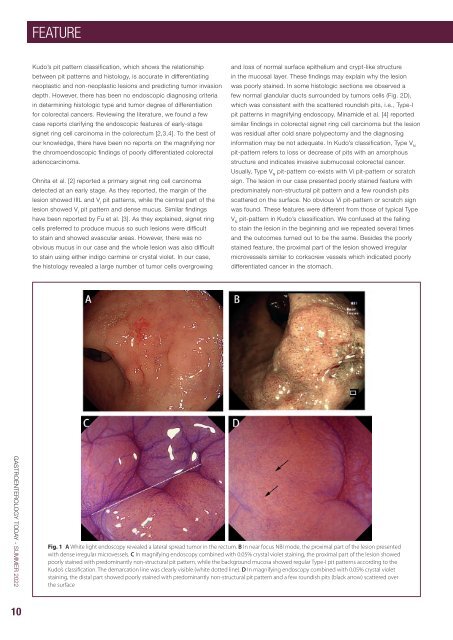
Ohnita et al. [2] reported a primary signet ring cell carcinoma<br />
detected at an early stage. As they reported, the margin of the<br />
lesion showed IIIL and V I<br />
pit patterns, while the central part of the<br />
lesion showed V I<br />
pit pattern and dense mucus. Similar findings<br />
have been reported by Fu et al. [3]. As they explained, signet ring<br />
cells preferred to produce mucus so such lesions were difficult<br />
to stain and showed avascular areas. However, there was no<br />
obvious mucus in our case and the whole lesion was also difficult<br />
to stain using either indigo carmine or crystal violet. In our case,<br />
the histology revealed a large number of tumor cells overgrowing<br />
and loss of normal surface epithelium and crypt-like structure<br />
in the mucosal layer. These findings may explain why the lesion<br />
was poorly stained. In some histologic sections we observed a<br />
few normal glandular ducts surrounded by tumors cells (Fig. 2D),<br />
which was consistent with the scattered roundish pits, i.e., Type-I<br />
pit patterns in magnifying endoscopy. Minamide et al. [4] reported<br />
similar findings in colorectal signet ring cell carcinoma but the lesion<br />
was residual after cold snare polypectomy and the diagnosing<br />
information may be not adequate. In Kudo’s classification, Type V N<br />
pit-pattern refers to loss or decrease of pits with an amorphous<br />
structure and indicates invasive submucosal colorectal cancer.<br />
Usually, Type V N<br />
pit-pattern co-exists with Vi pit-pattern or scratch<br />
sign. The lesion in our case presented poorly stained feature with<br />
predominately non-structural pit pattern and a few roundish pits<br />
scattered on the surface. No obvious Vi pit-pattern or scratch sign<br />
was found. These features were different from those of typical Type<br />
V N<br />
pit-pattern in Kudo’s classification. We confused at the failing<br />
to stain the lesion in the beginning and we repeated several times<br />
and the outcomes turned out to be the same. Besides the poorly<br />
stained feature, the proximal part of the lesion showed irregular<br />
microvessels similar to corkscrew vessels which indicated poorly<br />
differentiated cancer in the stomach.<br />
GASTROENTEROLOGY TODAY - SUMMER <strong>2022</strong><br />
Fig. 1 A White light endoscopy revealed a lateral spread tumor in the rectum. B In near focus NBI mode, the proximal part of the lesion presented<br />
with dense irregular microvessels. C In magnifying endoscopy combined with 0.05% crystal violet staining, the proximal part of the lesion showed<br />
poorly stained with predominantly non-structural pit pattern, while the background mucosa showed regular Type-I pit patterns according to the<br />
Kudo’s classification. The demarcation line was clearly visible (white dotted line). D In magnifying endoscopy combined with 0.05% crystal violet<br />
staining, the distal part showed poorly stained with predominantly non-structural pit pattern and a few roundish pits (black arrow) scattered over<br />
the surface<br />
10