Workshop on Polarized Electron Sources and Polarimeters
Workshop on Polarized Electron Sources and Polarimeters
Workshop on Polarized Electron Sources and Polarimeters

Create successful ePaper yourself
Turn your PDF publications into a flip-book with our unique Google optimized e-Paper software.
0<br />
55<br />
Y, mm X, mm 55<br />
1 mm<br />
1 mm<br />
0<br />
0 2 4 6 8 10 12<br />
Cooling time, s<br />
FWHMy, mm<br />
FWHMx, mm<br />
10<br />
8<br />
6<br />
4<br />
0<br />
25<br />
20<br />
15<br />
10<br />
5<br />
x=1.8 s<br />
0<br />
0 2 4 6 8 10 12<br />
Cooling time, s<br />
y=1.4 s<br />
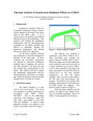
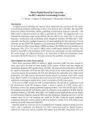
FIGURE 4. Transverse electr<strong>on</strong> cooling of 97 keV/u CF + [15]. Centre-of-mass positi<strong>on</strong>s of the C <strong>and</strong><br />
F neutral fragments vs time, recorded 12 m downstream of the electr<strong>on</strong> target by the imaging detector<br />
<strong>and</strong> indicating rms divergence angles of about 3∙10 -5 .<br />
Imaging detectors 12 m downstream of the electr<strong>on</strong> target were used to measure<br />
neutral recombinati<strong>on</strong> fragments <strong>and</strong> to analyze the dissociati<strong>on</strong> dynamics. The<br />
transverse momentum of the dissociati<strong>on</strong> fragments is recorded event by event using a<br />
spatially resolving multi-hit detector (two-dimensi<strong>on</strong>al fragment imaging) [22].<br />
For atomic i<strong>on</strong> beams the spatial profile of the neutral atoms produced by<br />
recombinati<strong>on</strong> in the electr<strong>on</strong> target directly reflects the angular divergence of the i<strong>on</strong><br />
beams [23]. For molecules, the imaging profile can not be used directly to m<strong>on</strong>itor<br />
transverse i<strong>on</strong> beam properties during the phase space cooling due to the significant<br />
kinetic energy release in the dissociati<strong>on</strong> process. However, this can be achieved by<br />
m<strong>on</strong>itoring the centre-of-mass of all fragment hit positi<strong>on</strong>s for a given recombinati<strong>on</strong><br />
event, which reflects the directi<strong>on</strong> of the molecular i<strong>on</strong> before it captured an electr<strong>on</strong><br />
in the target [24].<br />
Figure 4 (left) shows the distributi<strong>on</strong> spread of the centre-of-mass as a functi<strong>on</strong> of<br />
the cooling time derived from correlated two-hit events for both transverse directi<strong>on</strong>s.<br />
The FWHM of the centre-of-mass distributi<strong>on</strong> vs time is also shown in Fig. 4 (right).<br />
A transverse cooling time below 2 s was achieved, dem<strong>on</strong>strating a high cooling<br />
efficiency of the photocathode electr<strong>on</strong> beam. An estimati<strong>on</strong> of the cooling time from<br />
Eq. (2), assuming an isotropic electr<strong>on</strong> beam with 1 meV temperature, Lc=3.3 <strong>and</strong> the<br />
length of the target to be about 2.2% of the ring circumference, gives a value of about<br />
1.5 s for a cold i<strong>on</strong> beam <strong>and</strong> 6 s for a hot i<strong>on</strong> beam (with a diameter of the injected<br />
i<strong>on</strong> beam about 2 times larger than that of the electr<strong>on</strong> beam). After 6 s a FWHM of<br />
the centre-of-mass distributi<strong>on</strong> of 1 mm (horiz<strong>on</strong>tally) <strong>and</strong> 0.7 mm (vertically) was<br />
measured by the imaging detector. Assuming a l<strong>on</strong>gitudinal i<strong>on</strong> spread of about 5∙10 -5<br />
<strong>and</strong> using the horiz<strong>on</strong>tal TSR dispersi<strong>on</strong> of 2 m <strong>and</strong> the β functi<strong>on</strong>s of 3.9 m<br />
(horiz<strong>on</strong>tal) <strong>and</strong> 1.5 m (vertical) at the target secti<strong>on</strong>, the divergence <strong>and</strong> size of the<br />
i<strong>on</strong> beam can be derived. The divergence was found to be of about 3∙10 -5 in both<br />
transverse directi<strong>on</strong>s <strong>and</strong> the 1σ size was about 0.04 mm (vertically) <strong>and</strong> 0.2 mm<br />
(horiz<strong>on</strong>tally). The larger size of the i<strong>on</strong> beam in the horiz<strong>on</strong>tal directi<strong>on</strong> arises from