

3 - World Journal of Gastroenterology
3 - World Journal of Gastroenterology
3 - World Journal of Gastroenterology

Create successful ePaper yourself
Turn your PDF publications into a flip-book with our unique Google optimized e-Paper software.
Sun Y et al . Terahertz pulsed imaging and spectroscopy<br />
Figure 9 Photograph <strong>of</strong> the THz hand-held probe.<br />
Incident<br />
THz pulse<br />
Gum<br />
Enamel<br />
Dentin<br />
Pulp<br />
duced by Woodward et al [59] .<br />
Breast-conserving surgery is also an area <strong>of</strong> medicine<br />
which may benefit from THz imaging. Fitzgerald et al [65]<br />
conducted ex vivo studies <strong>of</strong> breast cancer to investigate the<br />
potential <strong>of</strong> THz imaging to aid the removal <strong>of</strong> breast cancer<br />
intra-operatively. In particular, they studied the feasibility<br />
<strong>of</strong> THz pulsed imaging to map the tumor margins on<br />
freshly excised human breast tissue. Good correlation was<br />
found for the area and shape <strong>of</strong> tumor in the THz images<br />
compared with that <strong>of</strong> histology. They also performed a<br />
spectroscopy study comparing the THz optical properties<br />
(absorption coefficient and refractive index) <strong>of</strong> the excised<br />
normal breast tissue and breast tumor. Both the absorption<br />
coefficient and refractive index were higher for tissue that<br />
contained tumor and this is a very positive indication that<br />
THz imaging could be used to detect margins <strong>of</strong> tumor<br />
and provide complementary information to techniques<br />
such as infrared and optical imaging, thermography, electrical<br />
impedance, and magnetic resonance imaging [66,67] .<br />
Since in vivo THz imaging is currently limited to sur-<br />
WJR|www.wjgnet.com<br />
1<br />
Detected pulse<br />
Figure 10 Schematic representation <strong>of</strong> the THz reflections from enamel.<br />
Reflection 1 is the reflection from the surface <strong>of</strong> the enamel and reflection 2 is<br />
the reflection from within the enamel due to tooth decay causing mineral loss.<br />
2<br />
1<br />
2<br />
Intensity (v)<br />
0.025<br />
0.015<br />
0.005<br />
-0.005<br />
-0.015<br />
-0.025<br />
-0.035<br />
14 16 18 20 22 24<br />
Optical delay (ps)<br />
From palm and plaster<br />
From palm only<br />
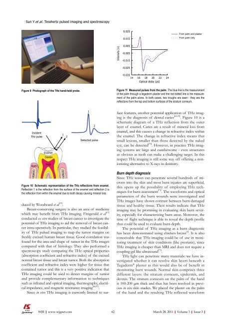
Figure 11 Measured pulses from the palm. The blue line is the measurement<br />
<strong>of</strong> the palm through a tegaderm plaster and the red dotted line is the measurement<br />
<strong>of</strong> the palm alone. In both cases, two troughs are seen - they are the<br />
reflections from the top and bottom surfaces <strong>of</strong> the stratum corneum.<br />
face features, another potential application <strong>of</strong> THz imaging<br />
is the diagnosis <strong>of</strong> dental caries [68,69] . Figure 10 is a<br />
schematic diagram <strong>of</strong> a THz reflection from the outer<br />
layer <strong>of</strong> enamel. Caries are a result <strong>of</strong> mineral loss from<br />
enamel, and this causes a change in refractive index within<br />
the enamel. The change in refractive index means that<br />
small lesions, smaller than those detected by the naked<br />
eye, can be detected [70] . However, in practice THz imaging<br />
systems are large and cumbersome - even structures<br />
as obvious as teeth can make a challenging target. In this<br />
respect THz imaging is still some way <strong>of</strong>f <strong>of</strong>fering a nonionizing<br />
alternative to X-rays in dentistry.<br />
Burn depth diagnosis<br />
Since THz waves can penetrate several hundreds <strong>of</strong> microns<br />
into the skin and most burn injuries are superficial,<br />
this opens up the possibility <strong>of</strong> employing THz techniques<br />
for burn assessment [71] . The waveforms and optical<br />
parameters <strong>of</strong> the burn wounds were investigated and<br />
THz images have shown contrast between burn-damaged<br />
tissue and healthy tissue. Their results indicate that THz<br />
imaging may be promising in evaluating skin burn severity,<br />
especially for characterizing burn areas. Moreover, the<br />
time <strong>of</strong> flight technique is able to reveal the depth pr<strong>of</strong>ile<br />
thus could be used to evaluate burn depth.<br />
The potential <strong>of</strong> THz imaging as a burn diagnostic<br />
has been demonstrated using chicken breast [6] . It is also<br />
conceivable that THz imaging could be <strong>of</strong> use in monitoring<br />
treatment <strong>of</strong> skin conditions (like psoriasis), since<br />
THz imaging is cheaper than MRI and does not require a<br />
coupling gel like ultrasound [72] .<br />
THz light can penetrate many materials: we have investigated<br />
whether it can resolve skin layers beneath a<br />
Tegaderm ® plaster as this would also be <strong>of</strong> benefit in<br />
monitoring burn wounds. Normal skin comprises three<br />
different layers: the stratum corneum, epidermis, and<br />
dermis. The stratum corneum on the palm <strong>of</strong> the hand<br />
is 100-200 μm thick and thus has been resolved in previous<br />
in vivo skin studies. We placed the plaster on the palm<br />
<strong>of</strong> the hand and the resulting THz reflected waveform<br />
62 March 28, 2011|Volume 3|Issue 3|


