

3 - World Journal of Gastroenterology
3 - World Journal of Gastroenterology
3 - World Journal of Gastroenterology

You also want an ePaper? Increase the reach of your titles
YUMPU automatically turns print PDFs into web optimized ePapers that Google loves.
6.9<br />
B<br />
C<br />
A<br />
Power (a.u.)<br />
6000<br />
4500<br />
3000<br />
1500<br />
0<br />
in silicone with an internal diameter <strong>of</strong> 2 mm and a 1 mm<br />
thick wall as in the case <strong>of</strong> the previous phantom. The<br />
third one, originating from a catheter (Surflo ® winged infusion<br />
set, Terumo ® , Belgium), had a 1 mm internal diameter<br />
and a 0.5 mm thick wall. The input and the output <strong>of</strong><br />
the phantom were composed <strong>of</strong> three-way taps (Disc<strong>of</strong>ix ® ,<br />
B. Braun, Melsungen, Germany) allowing linkage between<br />
the three pipes (Figure 3A). The phantom was connected<br />
to the same pump as previously described, providing a<br />
defined water flow rate set at 42.4 mL/min. Such an as-<br />
WJR|www.wjgnet.com<br />
1<br />
0 5 10 15 20<br />
t /s<br />
Linear raw data<br />
5 fps<br />
VRh5.8<br />
9.2<br />
MI 0.1<br />
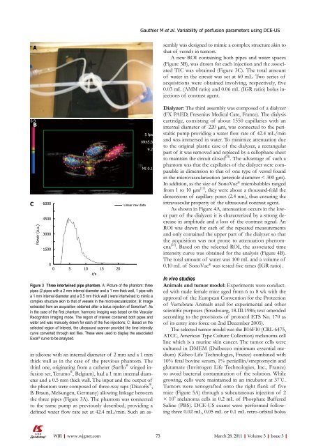
Figure 3 Three intertwined pipe phantom. A: Picture <strong>of</strong> the phantom: three<br />
pipes (2 pipes with a 2 mm internal diameter and a 1 mm thick wall, 1 pipe with<br />
a 1 mm internal diameter and a 0.5 mm thick wall ) were intertwined to mimic a<br />
complex structure akin to that <strong>of</strong> vessels in the microvascularization; B: Image<br />
extracted from an acquisition obtained after a bolus injection <strong>of</strong> SonoVue ® . As<br />
in the case <strong>of</strong> the first phantom, harmonic imaging was based on the Vascular<br />
Recognition Imaging mode. The region <strong>of</strong> interest contained both pipes and<br />
water and was manually drawn for each <strong>of</strong> the five injections; C: Based on the<br />
selected region <strong>of</strong> interest, the ultrasound scanner provided the time intensity<br />
curve converted through text files. These were used to display the associated<br />
Excel ® curve to be analyzed.<br />
Gauthier M et al . Variability <strong>of</strong> perfusion parameters using DCE-US<br />
sembly was designed to mimic a complex structure akin to<br />
that <strong>of</strong> vessels in tumors.<br />
A new ROI containing both pipes and water spaces<br />
(Figure 3B), was drawn for each injection and the associated<br />
TIC was obtained (Figure 3C). The total amount<br />
<strong>of</strong> water in the circuit was set at 60 mL. Two series <strong>of</strong><br />
acquisitions were obtained involving, respectively, five<br />
0.03 mL (AMM ratio) and 0.06 mL (IGR ratio) bolus injections<br />
<strong>of</strong> contrast agent.<br />
Dialyzer: The third assembly was composed <strong>of</strong> a dialyzer<br />
(FX PAED, Fresenius Medical Care, France). The dialysis<br />
cartridge, consisting <strong>of</strong> about 1550 capillaries with an<br />
internal diameter <strong>of</strong> 220 μm, was connected to the peristaltic<br />
pump providing a water flow rate <strong>of</strong> 42.4 mL/min<br />
and was immersed in water. To minimize attenuation due<br />
to the original plastic case <strong>of</strong> the dialyzer, a rectangular<br />
part <strong>of</strong> it was removed and replaced by a cellophane sheet<br />
to maintain the circuit closed [24] . The advantage <strong>of</strong> such a<br />
phantom was that the capillaries <strong>of</strong> the dialyzer were comparable<br />
in dimension to that <strong>of</strong> one type <strong>of</strong> vessel found<br />
in the microvascularization (arteriole diameter < 300 μm).<br />
In addition, as the size <strong>of</strong> SonoVue ® microbubbles ranged<br />
from 1 to 10 μm [12] , they were about a thousand-fold the<br />
dimensions <strong>of</strong> capillary pores (2.4 nm), thus ensuring the<br />
intravascular property <strong>of</strong> the ultrasound contrast agent.<br />
As shown in Figure 4A, attenuation occurs in the lower<br />
part <strong>of</strong> the dialyzer: it is characterized by a strong decrease<br />
in amplitude and a loss <strong>of</strong> the contrast signal. An<br />
ROI was drawn for each <strong>of</strong> the repeated measurements<br />
and only contained the upper part <strong>of</strong> the dialyzer so that<br />
the acquisition was not prone to attenuation phenomena<br />
[25] . Based on the selected ROI, the associated time<br />
intensity curve was obtained for the analysis (Figure 4B).<br />
The total amount <strong>of</strong> water was 100 mL and a volume <strong>of</strong><br />
0.10 mL <strong>of</strong> SonoVue ® was tested five times (IGR ratio).<br />
In vivo studies<br />
Animals and tumor model: Experiments were conducted<br />
with nude female mice aged from 6 to 8 wk with the<br />
approval <strong>of</strong> the European Convention for the Protection<br />
<strong>of</strong> Vertebrate Animals used for experimental and other<br />
scientific purposes (Strasbourg, 18.III.1986; text amended<br />
according to the provisions <strong>of</strong> protocol ETS No. 170 as<br />
<strong>of</strong> its entry into force on 2nd December 2005).<br />
The selected tumor model was the B16F10 (CRL-6475,<br />
ATCC, American Type Culture Collection) melanoma cell<br />
line which is a murine skin cancer. The tumor cells were<br />
cultured in DMEM (Dulbecco minimum essential medium)<br />
(Gibco Life Technologies, France) combined with<br />
10% fetal bovine serum, 1% penicillin/streptomycin and<br />
glutamate (Invitrogen Life Technologies, Inc., France)<br />
to avoid bacterial contamination <strong>of</strong> the solution. While<br />
growing, cells were maintained in an incubator at 37℃.<br />
Tumors were xenografted onto the right flank <strong>of</strong> five<br />
mice (Figure 5A) through a subcutaneous injection <strong>of</strong> 2<br />
× 10 6 melanoma cells in 0.2 mL <strong>of</strong> Phosphate Buffered<br />
Saline (PBS). DCE-US exams were performed following<br />
three 0.02 mL, 0.05 mL or 0.1 mL retro-orbital bolus<br />
73 March 28, 2011|Volume 3|Issue 3|


