

3 - World Journal of Gastroenterology
3 - World Journal of Gastroenterology
3 - World Journal of Gastroenterology

Create successful ePaper yourself
Turn your PDF publications into a flip-book with our unique Google optimized e-Paper software.
Figure 1 Pre-operative computed tomography scan <strong>of</strong> the chest without contrast<br />
media showing the large bilateral costal masses (white arrows) and an<br />
intracanalicular extradural mass (black arrow).<br />
Figure 2 Pre-operative “bone window” computed tomography scan <strong>of</strong> the<br />
chest without contrast media shows large bilateral, well circumscribed<br />
lobulated s<strong>of</strong>t tissue masses that cause widening <strong>of</strong> the ribs (short arrows)<br />
and periosteal reaction, without interruption <strong>of</strong> cortical bone (thin long<br />
arrow). Coexisting involvement <strong>of</strong> the sternum body (thick long arrow).<br />
Figure 3 Pre-operative “bone window” computed tomography scan without<br />
contrast media: the vertebral body is devoid <strong>of</strong> bony erosion and has a<br />
lacey appearance.<br />
Computed tomography (Figures 1-3) and magnetic resonance<br />
imaging (MRI, Figures 4-6) revealed bilateral paravertebral<br />
s<strong>of</strong>t-tissue masses at T4-L1 levels and a mass with<br />
the same features was seen inside the vertebral canal inducing<br />
spinal cord compression at T4-T9 levels. Furthermore,<br />
a marked medullary expansion <strong>of</strong> the bony structures was<br />
WJR|www.wjgnet.com<br />
Savini P et al . Extramedullary haematopoiesis with paraparesis<br />
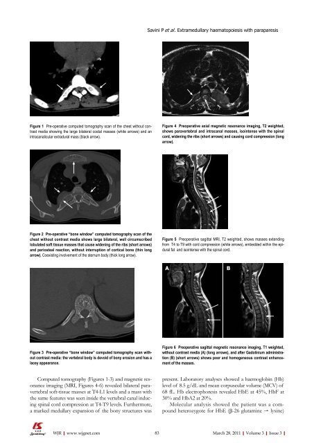
Figure 4 Preoperative axial magnetic resonance imaging, T2 weighted,<br />
shows paravertebral and intracanal masses, isointense with the spinal<br />
cord, widening the ribs (short arrows) and causing cord compression (long<br />
arrow).<br />
Figure 5 Preoperative sagittal MRI, T2 weighted, shows masses extending<br />
from T4 to T9 with cord compression (white arrows), embedded within the epidural<br />
fat and isointense with the spinal cord.<br />
A<br />
Figure 6 Preoperative sagittal magnetic resonance imaging, T1 weighted,<br />
without contrast media (A) (long arrows), and after Gadolinium administration<br />
(B) (short arrows) shows poor and homogeneous contrast enhancement<br />
<strong>of</strong> the masses.<br />
present. Laboratory analyses showed a haemoglobin (Hb)<br />
level <strong>of</strong> 8.5 g/dL and mean corpuscular volume (MCV) <strong>of</strong><br />
68 fL. Hb electrophoresis revealed HbE at 45%, HbF at<br />
30% and HbA2 at 20%.<br />
Molecular analysis showed the patient was a compound<br />
heterozygote for HbE (β-26 glutamine → lysine)<br />
83 March 28, 2011|Volume 3|Issue 3|<br />
B


