Ecole Nationale Supérieure Agronomique de Montpellier ... - CIAM
Ecole Nationale Supérieure Agronomique de Montpellier ... - CIAM
Ecole Nationale Supérieure Agronomique de Montpellier ... - CIAM

Create successful ePaper yourself
Turn your PDF publications into a flip-book with our unique Google optimized e-Paper software.
<strong>Ecole</strong> <strong>Nationale</strong> <strong>Supérieure</strong> <strong>Agronomique</strong> <strong>de</strong> <strong>Montpellier</strong><br />
Agro.<strong>Montpellier</strong><br />
T H E S E<br />
pour obtenir le gra<strong>de</strong> <strong>de</strong><br />
DOCTEUR<br />
DE L’ECOLE NATIONALE SUPERIEURE AGRONOMIQUE DE MONTPELLIER<br />
Discipline : Biologie <strong>de</strong>s Populations et Ecologie<br />
Formation Doctorale : Ressources Phytogénétiques et Interactions Biologiques<br />
<strong>Ecole</strong> Doctorale : 167: Biologie <strong>de</strong>s Systèmes Intégrés - Agronomie, Environnement<br />
présentée et soutenue publiquement<br />
par<br />
Gaël THEBAUD<br />
le 15 décembre 2005<br />
Titre :<br />
_______<br />
Etu<strong>de</strong> du développement spatio-temporel d’une maladie transmise par vecteur<br />
en intégrant modélisation statistique et expérimentation :<br />
cas <strong>de</strong> l’ESFY (European stone fruit yellows)<br />
_______<br />
JURY<br />
Jean-Noël BACRO Professeur à l’Université <strong>Montpellier</strong> II Prési<strong>de</strong>nt<br />
Avner BAR-HEN Professeur à l’Université Paris 13 Rapporteur<br />
Ivan SACHE Chargé <strong>de</strong> Recherche à l’INRA, Grignon Rapporteur<br />
Jean-Loup NOTTEGHEM Professeur à l’Agro.M, <strong>Montpellier</strong> Directeur <strong>de</strong> Thèse<br />
Manuel PLANTEGENEST Maître <strong>de</strong> Conférences à l’ENSAR, Rennes Examinateur<br />
Wolfgang JARAUSCH Chercheur à AlPlanta, Neustadt/W. Examinateur
Remerciements<br />
Je tiens à remercier Jean-Loup Nottéghem et Rachid Senoussi <strong>de</strong> m’avoir<br />
chaleureusement accueilli au sein <strong>de</strong>s Unités BGPI (<strong>Montpellier</strong>) et Biométrie<br />
(Avignon).<br />
Un très grand merci également à Joël C. 1 et Gérard Labonne pour leur jovialité<br />
quotidienne, pour leur disponibilité et pour avoir su me gui<strong>de</strong>r sans me bri<strong>de</strong>r.<br />
Je souhaite aussi remercier du fond du cœur tous les membres <strong>de</strong>s <strong>de</strong>ux équipes<br />
que j’ai eu plaisir à côtoyer pendant ces trois années.<br />
En particulier, je suis infiniment reconnaissant à celles et ceux qui se sont dévoués<br />
pour me transporter régulièrement entre la gare et le centre INRA <strong>de</strong> Montfavet. Un<br />
grand merci, donc, à Emilie, Sabrina, Nathalie et Pascal, qui ont souvent été <strong>de</strong> corvée.<br />
J’en profite pour remercier la SNCF pour les longues (voire très longues, parfois)<br />
heures <strong>de</strong> lecture qu’elle m’a aménagées.<br />
Enfin, mes plus tendres remerciements à Caroline pour sa relecture patiente <strong>de</strong> ce<br />
manuscrit et surtout pour son soutien pendant ces 3 années... et pour avoir su, ces<br />
<strong>de</strong>rniers temps, me partager avec un ordinateur sans trop s’en offusquer.<br />
Par contre, je ne remercie pas Chronos qui a été très glouton récemment.<br />
1 Joël Chadœuf ayant pour principe <strong>de</strong> ne pas accepter les remerciements, son nom a été soigneusement<br />
dissimulé ici.<br />
- 2 -
Glossaire<br />
A leur première occurrence, les mots définis dans ce glossaire figurent dans le texte en<br />
caractères gras et sont suivis d’un astérisque. La définition <strong>de</strong> certains termes variant selon les<br />
auteurs, aucune source n’est citée pour les définitions fournies : les définitions données ici<br />
précisent le sens qui a été retenu dans ce mémoire.<br />
Epidémiologie : Etu<strong>de</strong> <strong>de</strong>s facteurs <strong>de</strong> risque d’une maladie dans une population ; par<br />
extension, ces facteurs eux-mêmes et leurs conséquences sur la répartition <strong>de</strong>s cas <strong>de</strong><br />
maladie dans l’espace et/ou dans le temps.<br />
Etiologie : Etu<strong>de</strong> <strong>de</strong>s causes d’une maladie dans un individu ; par extension, ces causes ellesmêmes.<br />
Monocyclique/Polycyclique : Dans une épidémie monocyclique, les infections présentes<br />
dans une parcelle ne constituent pas une source d’inoculum interne ; l’évolution <strong>de</strong><br />
l’inci<strong>de</strong>nce n’est donc liée qu’à une succession <strong>de</strong> transmissions primaires. A l’inverse,<br />
dans une épidémie polycyclique, les infections présentes dans une parcelle sont à leur tour<br />
<strong>de</strong>s sources d’inoculum pour la parcelle ; l’évolution <strong>de</strong> l’inci<strong>de</strong>nce est alors liée à la fois<br />
aux transmissions primaires et aux transmissions secondaires.<br />
(Nested-) PCR (Polymerase Chain Reaction) : La réaction <strong>de</strong> PCR consiste à réaliser in vitro<br />
la duplication cyclique d’un fragment d’ADN à l’ai<strong>de</strong> d’une enzyme ; la présence du<br />
fragment amplifié peut ensuite être facilement détectée. Dans le cas d’une nested-PCR<br />
(ou PCR nichée), cette première amplification est suivie d’une secon<strong>de</strong> étape <strong>de</strong>stinée à<br />
amplifier un second fragment inclus dans le premier, ce qui augmente considérablement<br />
la sensibilité (et parfois la spécificité) <strong>de</strong> la métho<strong>de</strong>.<br />
Plante pérenne ou vivace : Végétal à durée <strong>de</strong> vie indéfinie (vivant 3 ans et plus, par<br />
opposition aux plantes annuelles ou bisannuelles).<br />
Polycyclique : cf. Monocyclique.<br />
Transmission primaire/secondaire : Les transmissions secondaires impliquent un pathogène<br />
issu <strong>de</strong> l’intérieur d’une population cible, souvent définie implicitement ; on observe alors<br />
<strong>de</strong>s infections secondaires dues à un inoculum secondaire (ou endo-inoculum). Par<br />
opposition, les transmissions primaires (et les infections primaires résultantes) impliquent<br />
un pathogène issu <strong>de</strong> l’extérieur <strong>de</strong> la population cible (inoculum primaire ou exoinoculum).<br />
Univoltin, ine : Qualifie le cycle biologique d’un insecte ayant une seule génération par an.<br />
- 3 -
~ Table <strong>de</strong>s Matières ~<br />
INTRODUCTION......................................................................................................................... 7<br />
I. Enjeux <strong>de</strong>s maladies émergentes ou ré-émergentes............................................................. 8<br />
A. Définition et causes <strong>de</strong>s (ré-)émergences ............................................................................. 8<br />
B. Enjeux économiques et sociaux <strong>de</strong>s (ré-)émergences......................................................... 10<br />
C. Enjeux scientifiques <strong>de</strong>s (ré-)émergences........................................................................... 10<br />
II. L’ESFY, une maladie grave mais dont l’épidémiologie est mal connue ........................ 12<br />
A. Une menace pour la filière agricole <strong>de</strong>s fruits à noyau ...................................................... 12<br />
1) Impact économique <strong>de</strong> l’ESFY sur la filière abricot, en France ...................................................12<br />
(a) Coût <strong>de</strong> la perte <strong>de</strong> récolte........................................................................................................................ 12<br />
(b) Coût <strong>de</strong> la lutte contre l’ESFY................................................................................................................. 13<br />
(c) Coût d’opportunité ................................................................................................................................... 13<br />
2) Impact environnemental <strong>de</strong> la maladie..........................................................................................13<br />
B. Etat <strong>de</strong> l’art concernant l’épidémiologie <strong>de</strong> l’ESFY........................................................... 13<br />
1) Historique <strong>de</strong> la maladie en France ..............................................................................................13<br />
2) Extension géographique <strong>de</strong> l’épidémie..........................................................................................14<br />
3) Symptomatologie............................................................................................................................15<br />
4) Gamme d’hôtes ..............................................................................................................................16<br />
5) Etiologie et diagnostic ...................................................................................................................17<br />
6) Vection ...........................................................................................................................................19<br />
(a) Biologie <strong>de</strong> C. pruni................................................................................................................................. 19<br />
(b) Caractéristiques <strong>de</strong> la vection .................................................................................................................. 21<br />
7) Incubation, latence et pério<strong>de</strong> infectieuse <strong>de</strong> la plante..................................................................21<br />
8) Propagation entre parcelles ..........................................................................................................22<br />
9) Bilan sur le cycle épidémique <strong>de</strong> l’ESFY.......................................................................................22<br />
C. Enjeux scientifiques............................................................................................................ 23<br />
III. Objectifs et stratégie d’étu<strong>de</strong> ............................................................................................ 23<br />
A. Objectifs.............................................................................................................................. 23<br />
B. Stratégie .............................................................................................................................. 24<br />
PARTIE I : IDENTIFIER DES FACTEURS DE RISQUE PAR UNE ENQUETE A<br />
L’ECHELLE D’UN BASSIN DE PRODUCTION .................................................................. 25<br />
I. Article I : “I<strong>de</strong>ntifying Risk Factors from a Survey with a Logistic Regression Mo<strong>de</strong>l:<br />
the Case of European Stone Fruit Yellows” ............................................................................ 26<br />
II. Bilan...................................................................................................................................... 45<br />
PARTIE II : IDENTIFIER LES CYCLES BIOLOGIQUES DE ‘CANDIDATUS<br />
PHYTOPLASMA PRUNORUM’ ET DE SON VECTEUR CACOPSYLLA PRUNI – DU<br />
TERRAIN AU LABORATOIRE ET VICE VERSA............................................................... 47<br />
I. Introduction........................................................................................................................... 48<br />
II. Détection spécifique et quantification <strong>de</strong> ‘Ca. P. prunorum’.......................................... 48<br />
A. Article II : “A Toolbox for the Specific Detection and Quantification of the<br />
Phytopathogenic Agent ‘Candidatus Phytoplasma prunorum’ in Plants and Insects” ........... 48<br />
B. Bilan.................................................................................................................................... 60<br />
III. Prévalence et transmissibilité <strong>de</strong> ‘Ca. P. prunorum’ dans les populations naturelles<br />
<strong>de</strong> son vecteur............................................................................................................................ 60<br />
A. Article III : “Survival of European Stone Fruit Yellows Phytoplasma Outsi<strong>de</strong> Fruit<br />
Crop Production Areas: a Case Study in Southeastern France”.............................................. 60<br />
B. Méta-analyse <strong>de</strong>s résultats expérimentaux européens ........................................................ 66<br />
1) Métho<strong>de</strong> du maximum <strong>de</strong> vraisemblance pour les tests par lots....................................................66<br />
2) Estimation : les vecteurs infectés ne sont pas tous infectieux........................................................67<br />
3) Y a-t-il <strong>de</strong>s tendances générales dans la prévalence <strong>de</strong>s phytoplasmes au sein <strong>de</strong>s populations<br />
vectrices ? ..........................................................................................................................................69<br />
- 4 -
C. Conclusions sur l’étu<strong>de</strong> <strong>de</strong>s vecteurs <strong>de</strong> l’ESFY en conditions naturelles ......................... 70<br />
IV. Article IV : “The Spread of European Stone Fruit Yellows is Regulated by the Life<br />
Cycle of its Vector and by the Growth Rate of the Hosted Phytoplasma, as Assessed by<br />
Real-Time PCR”........................................................................................................................ 70<br />
V. Bilan sur le fonctionnement <strong>de</strong> la vection.......................................................................... 83<br />
PARTIE III : TESTER DES HYPOTHESES SUR LE DEVELOPPEMENT DE<br />
L’ESFY EN VERGER................................................................................................................ 85<br />
I. Traduction <strong>de</strong>s hypothèses biologiques en hypothèses d’indépendance.......................... 86<br />
II. Analyse exploratoire <strong>de</strong>s motifs spatiaux et temporels.................................................... 88<br />
A. Un programme générique pour tester <strong>de</strong>s hypothèses d’indépendance.............................. 88<br />
B. Article V : “Spatio-Temporal Analysis of Disease Spread Provi<strong>de</strong>s Insights into the<br />
Epi<strong>de</strong>miology of European Stone Fruit Yellows”................................................................... 89<br />
C. Bilan.................................................................................................................................... 96<br />
III. Article VI : “Investigating Disease Spread Between Two Dates with Permutation<br />
Tests on a Lattice”..................................................................................................................... 96<br />
IV. Application à l’analyse <strong>de</strong> cartes pluriannuelles <strong>de</strong> l’ESFY en verger ....................... 115<br />
A. Présentation <strong>de</strong>s vergers étudiés ....................................................................................... 115<br />
B. Résultats et interprétations................................................................................................ 116<br />
1) Analyse <strong>de</strong> la répartition <strong>de</strong> l’ensemble <strong>de</strong>s arbres symptomatiques ..........................................116<br />
2) Analyse <strong>de</strong>s dépendances spatiales interannuelles......................................................................118<br />
V. Bilan sur les apports <strong>de</strong>s tests d’hypothèses.................................................................... 120<br />
PARTIE IV : SYNTHETISER L’INFORMATION DANS UN MODELE DE<br />
SIMULATION DES EPIDEMIES D’ESFY........................................................................... 123<br />
I. Hypothèses du modèle ........................................................................................................ 124<br />
II. Description du modèle ...................................................................................................... 125<br />
III. Perspectives ...................................................................................................................... 126<br />
CONCLUSION.......................................................................................................................... 127<br />
I. Conclusions sur l’épidémiologie <strong>de</strong> l’ESFY...................................................................... 128<br />
A. Conséquences <strong>de</strong>s résultats obtenus pour la gestion <strong>de</strong> la maladie .................................. 128<br />
1) Apports sur l’effet <strong>de</strong> la génétique <strong>de</strong>s arbres .............................................................................129<br />
2) Apports sur les transmissions primaires multiples ......................................................................129<br />
3) Apports sur les transmissions inter-annuelles .............................................................................129<br />
B. Perspectives....................................................................................................................... 131<br />
II. Conclusions sur la démarche retenue.............................................................................. 133<br />
REFERENCES BIBLIOGRAPHIQUES................................................................................ 137<br />
ANNEXES.................................................................................................................................. 147<br />
I. Annexe 1 : Programme R pour estimer une proportion à partir <strong>de</strong> tests groupés....... 148<br />
II. Annexe 2 : Programme R pour tester <strong>de</strong>s hypothèses d’indépendance par<br />
permutation............................................................................................................................. 150<br />
III. Annexe 3 : Programme R pour simuler le développement spatio-temporel <strong>de</strong><br />
l’ESFY dans un verger d’abricotier ..................................................................................... 153<br />
IV. Annexe 4 : “Testing Boolean Assumption in the Non Convex Case When a Boun<strong>de</strong>d<br />
Grain can be Assumed”........................................................................................................... 155<br />
- 5 -
~ Table <strong>de</strong>s Illustrations ~<br />
FIGURES<br />
Figure 1. Augmentation <strong>de</strong>s échanges planétaires. ....................................................................................................... 9<br />
Figure 2. Evolution <strong>de</strong> la température à la surface <strong>de</strong> la terre (1860-2000). ................................................................. 9<br />
Figure 3. Un cadre commun pour l’étu<strong>de</strong> <strong>de</strong>s maladies mal connues (émergentes ou non)........................................ 11<br />
Figure 4. Répartition géographique <strong>de</strong>s pays dans lesquels <strong>de</strong>s cas d’ESFY ont été mentionnés............................... 15<br />
Figure 5. Symptômes <strong>de</strong> l’ESFY. ............................................................................................................................... 16<br />
Figure 6. Hiérarchie <strong>de</strong> la sensibilité à l’ESFY parmi les Prunus............................................................................... 17<br />
Figure 7. Observation au microscope électronique <strong>de</strong> ‘Candidatus Phytoplasma prunorum’ .................................... 17<br />
Figure 8. Cacopsylla pruni, vecteur <strong>de</strong> l’ESFY. ......................................................................................................... 18<br />
Figure 9. Hiérarchie <strong>de</strong> la sensibilité à l’ESFY parmi les Prunus............................................................................... 19<br />
Figure 10. Evolution pluriannuelle <strong>de</strong>s dates <strong>de</strong> présence et <strong>de</strong>s effectifs <strong>de</strong>s sta<strong>de</strong>s adultes <strong>de</strong> C. pruni.. ................ 19<br />
Figure 11. Evolution synchrone <strong>de</strong>s effectifs <strong>de</strong>s sta<strong>de</strong>s adultes <strong>de</strong> C. pruni sur prunellier, prunier domestique,<br />
abricotier et myrobolan. ............................................................................................................................ 20<br />
Figure 12. Cycle <strong>de</strong> base <strong>de</strong> l’épidémie d’ESFY. ....................................................................................................... 22<br />
Figure 13. Connaissances initiales sur le cycle <strong>de</strong> C. pruni et <strong>de</strong> la vection <strong>de</strong> l’ESFY. ............................................ 23<br />
Figure 14. Stratégie d’étu<strong>de</strong> <strong>de</strong> l’épidémiologie <strong>de</strong> l’ESFY et organisation <strong>de</strong>s différentes approches envisagées. .. 24<br />
Figure 15. Connaissances acquises sur le cycle <strong>de</strong> C. pruni et sur la vection <strong>de</strong> l’ESFY. .......................................... 83<br />
Figure 16. Evolution temporelle <strong>de</strong> l’ESFY dans 4 vergers d’abricotier (cv. Polonais) greffés sur myrobolan ....... 115<br />
Figure 17. Caractéristiques spatiales <strong>de</strong>s arbres symptomatiques sur l’ensemble <strong>de</strong> la pério<strong>de</strong> <strong>de</strong> prospection....... 117<br />
Figure 18. Test d’indépendance totale entre les localisations <strong>de</strong>s arbres symptomatiques sur l’ensemble <strong>de</strong> la<br />
pério<strong>de</strong> <strong>de</strong> prospection.. .......................................................................................................................... 118<br />
Figure 19. Deux exemples <strong>de</strong> tests d’indépendance spatio-temporelle entre la fin et le milieu <strong>de</strong> la dynamique<br />
temporelle (1996-1999 vs. 1990-1995)................................................................................................... 119<br />
Figure 20. Cycle <strong>de</strong> la transmission <strong>de</strong> l’ESFY. ....................................................................................................... 124<br />
Figure 21. Schéma <strong>de</strong> l’algorithme <strong>de</strong> simulation d’une épidémie d’ESFY dans un verger d’abricotier. ................ 126<br />
Figure 22. Interrelations entre les différents “acteurs” conditionnant l’épidémiologie <strong>de</strong> l’ESFY........................... 128<br />
Figure 23. Différentes métho<strong>de</strong>s <strong>de</strong> lutte envisageables contre l’ESFY dans l’état actuel <strong>de</strong>s connaissances.......... 130<br />
Figure 24. Schéma général <strong>de</strong>s relations entre le système épidémique étudié, l’expérimentation, la modélisation<br />
et la stratégie <strong>de</strong> gestion <strong>de</strong> la maladie.................................................................................................... 135<br />
TABLEAUX<br />
Tableau 1. Liste <strong>de</strong>s pays dans lesquels les symptômes caractéristiques <strong>de</strong> l’ESFY et/ou l’agent pathogène<br />
associé ont été i<strong>de</strong>ntifiés. .......................................................................................................................... 14<br />
Tableau 2. Synthèse bibliographique sur les proportions <strong>de</strong> C. pruni porteurs <strong>de</strong> l’ESFY ou infectieux. .................. 68<br />
Tableau 3. Eléments <strong>de</strong> comparaison <strong>de</strong>s proportions <strong>de</strong> vecteurs porteurs <strong>de</strong> différents phytoplasmes.................... 69<br />
Tableau 4. Propriétés attendues <strong>de</strong>s motifs spatio-temporels selon les comportements du vecteur............................ 88<br />
Tableau 5. Caractéristiques <strong>de</strong>s vergers analysés...................................................................................................... 116<br />
Tableau 6. Propriétés biologiques retenues pour construire un modèle stochastique simulant le développement <strong>de</strong><br />
l’ESFY dans un verger d’abricotier. ....................................................................................................... 125<br />
Tableau 7. Avantages et inconvénients <strong>de</strong>s principales métho<strong>de</strong>s <strong>de</strong> lutte envisageables contre l’ESFY en verger<br />
d’abricotier.............................................................................................................................................. 131<br />
Tableau 8. Apports <strong>de</strong>s différentes approches dans l’analyse <strong>de</strong>s facteurs impliqués dans le développement<br />
spatio-temporel <strong>de</strong> l’ESFY...................................................................................................................... 133<br />
~ Table <strong>de</strong>s Articles ~<br />
Article I : “I<strong>de</strong>ntifying Risk Factors from a Survey with a Logistic Regression Mo<strong>de</strong>l: the Case of European<br />
Stone Fruit Yellows”................................................................................................................................. 26<br />
Article II : “A Toolbox for the Specific Detection and Quantification of the Phytopathogenic Agent ‘Candidatus<br />
Phytoplasma prunorum’ in Plants and Insects”......................................................................................... 48<br />
Article III : “Survival of European Stone Fruit Yellows Phytoplasma Outsi<strong>de</strong> Fruit Crop Production Areas: a<br />
Case Study in Southeastern France” ......................................................................................................... 60<br />
Article IV : “The Spread of European Stone Fruit Yellows is Regulated by the Life Cycle of its Vector and by<br />
the Growth Rate of the Hosted Phytoplasma, as Assessed by Real-Time PCR” ...................................... 70<br />
Article V : “Spatio-Temporal Analysis of Disease Spread Provi<strong>de</strong>s Insights into the Epi<strong>de</strong>miology of European<br />
Stone Fruit Yellows”................................................................................................................................. 89<br />
Article VI : “Investigating Disease Spread Between Two Dates with Permutation Tests on a Lattice”..................... 96<br />
- 6 -
Introduction<br />
- 7 -<br />
« Deux grands mobiles font agir les<br />
hommes : la peur et la nouveauté »<br />
(Machiavel)
L’ESFY (European stone fruit yellows) est une maladie ré-émergente touchant les arbres<br />
fruitiers à noyau (Prunus) en Europe. La démarche mise en œuvre pour l’étudier, présentée<br />
dans ce mémoire, a plus généralement vocation à être utilisée dans l’étu<strong>de</strong> <strong>de</strong> maladies<br />
émergentes et ré-émergentes, notamment dans une phase initiale d’exploration du<br />
fonctionnement épidémique. Cette démarche est donc davantage orientée vers l’étu<strong>de</strong> <strong>de</strong>s<br />
maladies dont l’épidémiologie est mal comprise que vers l’étu<strong>de</strong> <strong>de</strong>s maladies émergentes en<br />
un lieu mais déjà bien connues.<br />
I. Enjeux <strong>de</strong>s maladies émergentes ou ré-émergentes<br />
A. Définition et causes <strong>de</strong>s (ré-)émergences<br />
En prenant pour base une série <strong>de</strong> définitions plus ou moins spécifiques aux agents<br />
pathogènes <strong>de</strong> l’homme (Morse & Schlue<strong>de</strong>rberg, 1990 ; Morse, 1995 ; Institute of Medicine,<br />
1992 ; Taylor et al., 2001 ; Woolhouse et al., 2005), on peut définir <strong>de</strong> façon générale une<br />
maladie émergente ou ré-émergente comme une maladie dont l’inci<strong>de</strong>nce réelle 1 dans une<br />
population donnée augmente pour la première fois ou à la suite d’une modification durable <strong>de</strong><br />
son épidémiologie * . Notons que cette définition exclut les cas sporadiques et les maladies<br />
saisonnières, ainsi que les “pseudo émergences” dues uniquement à une meilleure mesure <strong>de</strong><br />
l’inci<strong>de</strong>nce réelle voire à un sursaut d’activité médiatique ou scientifique à l’égard d’une<br />
maladie donnée. Notons également que cette définition est relative car elle dépend <strong>de</strong> la<br />
population considérée : une maladie peut être émergente en un lieu et endémique ailleurs, par<br />
exemple. On trouvera <strong>de</strong> nombreux exemples <strong>de</strong> maladies émergentes végétales, animales et<br />
humaines dans Moffat (2001), Woolhouse (2002), An<strong>de</strong>rson et al. (2004), Morens et al.<br />
(2004) et Woolhouse et al. (2005), ainsi que dans la base <strong>de</strong> données ProMED 2 . Dans le cas<br />
<strong>de</strong>s maladies infectieuses, une émergence peut être provoquée par :<br />
(i) la modification <strong>de</strong> l’environnement <strong>de</strong> la relation hôte-(vecteur-)pathogène, y compris<br />
par un changement dans les mesures sanitaires ;<br />
(ii) l’augmentation <strong>de</strong>s contacts – directs ou indirects – existants entre un pathogène et<br />
son hôte, ou l’établissement d’un nouveau contact, par exemple par l’introduction<br />
d’un hôte sensible, par la migration d’un pathogène hors <strong>de</strong> son aire <strong>de</strong> répartition<br />
habituelle, ou par son introduction volontaire (bioterrorisme, lutte biologique) ou<br />
involontaire (échanges commerciaux) ;<br />
(iii) l’augmentation <strong>de</strong> la sensibilité <strong>de</strong> l’hôte, par exemple suite à une immunosuppression<br />
ou à l’absence <strong>de</strong> sélection pour la résistance ;<br />
(iv) un changement dans le génotype d’un agent pathogène préexistant, par mutation,<br />
recombinaison ou transfert horizontal <strong>de</strong> gènes.<br />
L’examen <strong>de</strong>s causes possibles d’émergence révèle que si la <strong>de</strong>rnière possibilité<br />
envisagée est probablement stable dans le temps, le contexte actuel est plus propice que<br />
jamais aux <strong>de</strong>ux premiers mécanismes d’émergence (Morse, 1995 ; Woolhouse, 2002 ;<br />
An<strong>de</strong>rson et al., 2004), principalement par l’action conjointe <strong>de</strong> l’augmentation <strong>de</strong>s échanges<br />
internationaux, du réchauffement climatique et <strong>de</strong> l’action <strong>de</strong> l’homme sur les milieux. Ainsi,<br />
le trafic aérien (Figure 1A) a été multiplié par 3 entre 1975 et 1995 (Penner et al., 1999) et les<br />
échanges internationaux, par exemple <strong>de</strong> fruits et légumes (Figure 1B), ont plus que quintuplé<br />
ces 40 <strong>de</strong>rnières années (FAOSTAT 3 , 2005). Outre l’hypothèse d’une attaque bioterroriste,<br />
1 Par opposition à l’inci<strong>de</strong>nce mesurée, qui peut être différente <strong>de</strong> l’inci<strong>de</strong>nce réelle pour <strong>de</strong> multiples raisons.<br />
Dans ce mémoire, conformément à Ahrens & Pigeot (2005), l’inci<strong>de</strong>nce est définie pour une maladie donnée<br />
comme la proportion <strong>de</strong> la population saine qui développe cette maladie par unité <strong>de</strong> temps, contrairement à la<br />
prévalence qui désigne la proportion <strong>de</strong> la population qui est mala<strong>de</strong> à une date donnée.<br />
2 Program for Monitoring Emerging Diseases : http://www.fas.org/promed/<br />
3 Food and Agriculture Organization of the United Nations : http://faostat.fao.org/<br />
- 8 -
l’intensification permanente <strong>de</strong> la circulation pacifique <strong>de</strong>s biens et <strong>de</strong>s personnes entre pays<br />
géographiquement éloignés entraîne presque mécaniquement une introduction plus fréquente<br />
<strong>de</strong> pathogènes, <strong>de</strong> vecteurs et d’hôtes nouveaux.<br />
A<br />
B<br />
Quantité <strong>de</strong> fruits et légumes<br />
échangés (Mt)<br />
1,4E+08<br />
1,2E+08<br />
1,0E+08<br />
8,0E+07<br />
6,0E+07<br />
4,0E+07<br />
2,0E+07<br />
(d’après Penner et al., 1999)<br />
0,0E+00<br />
1960 1970 1980<br />
Année<br />
1990 2000<br />
- 9 -<br />
Figure 1. Augmentation <strong>de</strong>s<br />
échanges planétaires.<br />
(A) Exemple du trafic<br />
aérien : les passagers sont<br />
plus nombreux et se<br />
déplacent plus loin, d’où<br />
l’augmentation observée<br />
(puis prédite) du nombre <strong>de</strong><br />
passagers-kilomètres. (B)<br />
Exemple du volume <strong>de</strong>s<br />
échanges <strong>de</strong> fruits et légumes<br />
entre pays (données<br />
FAOSTAT, 2005).<br />
En parallèle, on assiste actuellement au début d’un réchauffement climatique rapi<strong>de</strong><br />
(Figure 2) : selon l’International Panel on Climate Change (2001), la température moyenne a<br />
augmenté <strong>de</strong> 0,75°C lors <strong>de</strong>s 150 <strong>de</strong>rnières années et cette évolution <strong>de</strong>vrait s’accentuer dans<br />
les années à venir (selon les scénarios et les modèles, les intervalles <strong>de</strong> confiance s’éten<strong>de</strong>nt<br />
entre +0,6 et +5,5°C en 100 ans).<br />
(d’après IPCC, 2001)<br />
Figure 2. Evolution<br />
<strong>de</strong> la température à<br />
la surface <strong>de</strong> la<br />
terre (1860-2000).<br />
Or, on note déjà une relation entre l’augmentation <strong>de</strong> la température et le déplacement <strong>de</strong><br />
l’aire <strong>de</strong> répartition <strong>de</strong> certaines espèces (Parmesan & Yohe, 2003 ; Root et al., 2003) et on
peut s’attendre à ce que <strong>de</strong>s pathogènes, leurs hôtes et/ou d’éventuels vecteurs suivent cette<br />
tendance, contribuant ainsi à l’émergence <strong>de</strong> maladies dans <strong>de</strong> nouvelles zones ou dans <strong>de</strong><br />
nouvelles populations. Enfin, l’écologie <strong>de</strong>s pathosystèmes peut être modifiée par le climat,<br />
mais aussi par l’intervention directe <strong>de</strong> l’homme, par exemple par la modification <strong>de</strong>s<br />
systèmes <strong>de</strong> production agricoles : la réduction <strong>de</strong> la diversité génétique <strong>de</strong>s espèces<br />
domestiques, l’intensification <strong>de</strong> la production ou la modification <strong>de</strong> la lutte contre la maladie<br />
en sont <strong>de</strong>s exemples.<br />
B. Enjeux économiques et sociaux <strong>de</strong>s (ré-)émergences<br />
Les enjeux socio-économiques <strong>de</strong>s maladies infectieuses émergentes dépen<strong>de</strong>nt <strong>de</strong> l’hôte<br />
et, secondairement, <strong>de</strong>s caractéristiques épidémiques du pathogène. Si l’émergence concerne<br />
un pathogène <strong>de</strong> l’homme, ou un pathogène animal suspecté <strong>de</strong> pouvoir infecter l’homme, les<br />
enjeux <strong>de</strong> santé publique, économiques et sociaux peuvent être énormes (Morens et al., 2004 ;<br />
Weiss & McLean, 2004), comme l’ont déjà démontré les épidémies <strong>de</strong> SRAS (syndrome<br />
respiratoire aigu sévère) et <strong>de</strong> SIDA (syndrome d’immunodéficience acquise). Ainsi, le SIDA<br />
a été responsable <strong>de</strong> 3,1 millions <strong>de</strong> morts dans le mon<strong>de</strong> au cours <strong>de</strong> la seule année 2004<br />
(UNAIDS, 2004) ; en parallèle, l’émergence du SRAS a fait moins <strong>de</strong> 1000 morts mais<br />
l’impact <strong>de</strong> cette maladie sur l’économie du sud-est asiatique est estimé à 25 milliards<br />
d’Euros (WHO, 2003). L’émergence ou la ré-émergence <strong>de</strong> maladies qui ne sont pas<br />
susceptibles d’infecter l’homme peuvent engendrer <strong>de</strong>s famines et <strong>de</strong>s exo<strong>de</strong>s si elles touchent<br />
l’agriculture vivrière <strong>de</strong>s populations les plus pauvres, comme en Irlan<strong>de</strong> au XIX ème siècle à la<br />
suite <strong>de</strong> l’apparition du mildiou <strong>de</strong> la pomme <strong>de</strong> terre (dû à Phythophtora infestans). Plus<br />
fréquemment, on enregistre <strong>de</strong>s pertes économiques considérables pour la filière agricole<br />
concernée par la maladie, éventuellement associées à <strong>de</strong>s conflits politiques internationaux<br />
suite aux mesures <strong>de</strong> protection (ou <strong>de</strong> protectionnisme déguisé) prises à l’occasion <strong>de</strong> ces<br />
crises sanitaires. Parmi les nombreux exemples <strong>de</strong> maladies émergentes ou ré-émergentes<br />
économiquement dévastatrices, on peut citer la ré-émergence en 2001 <strong>de</strong> la fièvre aphteuse en<br />
Gran<strong>de</strong>-Bretagne dont le coût pour l’économie anglaise est estimé à 11 milliards d’Euros<br />
(Thompson et al., 2002), ou celle du chancre citrique en 1995 aux Etats-Unis qui a déjà coûté<br />
plus <strong>de</strong> 150 millions d’Euros juste pour le programme d’éradication (Brown, 2001).<br />
C. Enjeux scientifiques <strong>de</strong>s (ré-)émergences<br />
A l’inverse <strong>de</strong>s enjeux socio-économiques, les enjeux scientifiques sont très souvent<br />
i<strong>de</strong>ntiques quel que soit le type d’hôte considéré, qu’il s’agisse <strong>de</strong> questions théoriques ou <strong>de</strong><br />
questions plus immédiatement opérationnelles.<br />
Parmi les questions générales relatives à l’émergence, on peut citer les suivantes :<br />
- La fréquence <strong>de</strong>s émergences s’accélère-t-elle ?<br />
- Y a-t-il <strong>de</strong>s gran<strong>de</strong>s tendances qui prési<strong>de</strong>nt aux émergences (type <strong>de</strong> pathogène, type<br />
<strong>de</strong> cause, type <strong>de</strong> lieu) ?<br />
- Peut-on prédire les risques d’émergence ?<br />
La problématique <strong>de</strong> la prédiction <strong>de</strong>s émergences se heurte à un obstacle <strong>de</strong> taille :<br />
l’imprévisibilité du lieu et <strong>de</strong> la date <strong>de</strong> l’apparition <strong>de</strong> pathogènes complètement nouveaux<br />
(Weiss & McLean, 2004). Cependant, la prédiction est un enjeu important pour les<br />
émergences en lien avec <strong>de</strong>s pathogènes déjà connus ; en particulier, quand l’extension<br />
géographique d’un pathogène est limitée par le climat, le couplage d’un modèle<br />
épidémiologique avec <strong>de</strong>s scénarios climatiques afin <strong>de</strong> prévoir le déplacement possible <strong>de</strong>s<br />
aires <strong>de</strong> répartition <strong>de</strong>s pathogènes est un domaine <strong>de</strong> recherche actif (Brasier & Scott, 1994 ;<br />
Scherm & Yang, 1999 ; Sutherst, 2001 ; Mark & Hoddle, 2004 ; Pivonia & Yang, 2004 ;<br />
Yonow et al., 2004). Cette approche permet d’i<strong>de</strong>ntifier les émergences futures les plus<br />
probables, et <strong>de</strong> s’y préparer.<br />
- 10 -
Ceci nous amène aux enjeux liés à la prévention <strong>de</strong>s émergences. Bien que cet aspect soit<br />
éludé dans la plupart <strong>de</strong>s articles généraux consacrés aux émergences, une prévention réelle<br />
semble possible contre une partie <strong>de</strong>s émergences (par exemple, par le contrôle sanitaire <strong>de</strong>s<br />
marchandises échangées, par une gestion durable <strong>de</strong>s traitements et <strong>de</strong>s gènes <strong>de</strong> résistance,<br />
par la sélection d’espèces domestiques plus rustiques). Cependant, <strong>de</strong>vant l’infinité <strong>de</strong>s<br />
émergences possibles, les questions se posent plus souvent en termes <strong>de</strong> réactivité (Finley et<br />
al., 2004) qu’en termes <strong>de</strong> prévention totale. A cet égard, la conception <strong>de</strong> métho<strong>de</strong>s<br />
d’épidémio-surveillance permettant <strong>de</strong> détecter rapi<strong>de</strong>ment les émergences (puis <strong>de</strong> continuer<br />
à recueillir et analyser les données nécessaires au cours du temps) est un enjeu scientifique et<br />
organisationnel majeur (Institute of Medicine, 1992 ; Morse, 1995 ; Woolhouse, 2002 ;<br />
An<strong>de</strong>rson et al., 2004 ; Morens et al., 2004).<br />
Enfin, en cas d’émergence d’une maladie complètement nouvelle ou peu étudiée<br />
précé<strong>de</strong>mment, les enjeux scientifiques les plus immédiats sont <strong>de</strong> l’ordre <strong>de</strong> l’ai<strong>de</strong> à la<br />
décision : ils concernent l’i<strong>de</strong>ntification, l’acquisition et le transfert <strong>de</strong>s connaissances<br />
épidémiologiques permettant <strong>de</strong> contenir la maladie rapi<strong>de</strong>ment, efficacement et, si possible,<br />
durablement. Ces enjeux immédiats sont les mêmes que dans l’étu<strong>de</strong> épidémiologique <strong>de</strong> tout<br />
pathosystème mal connu, l’urgence en plus ; ils sont en outre souvent indépendants <strong>de</strong> la<br />
nature <strong>de</strong> l’hôte. On peut donc proposer un cadre général à l’étu<strong>de</strong> <strong>de</strong> ces problèmes (Figure<br />
3), basé sur le traitement en parallèle <strong>de</strong> différentes questions concourant à i<strong>de</strong>ntifier <strong>de</strong>s<br />
métho<strong>de</strong>s <strong>de</strong> lutte contre la maladie et à optimiser leur mise en œuvre en une stratégie<br />
cohérente.<br />
Etu<strong>de</strong>s<br />
<strong>de</strong> risque<br />
Risque<br />
d’émergence<br />
Emergence<br />
Développement <strong>de</strong> la maladie Régression<br />
Estimation du risque <strong>de</strong> persistance et<br />
du coût <strong>de</strong> la maladie<br />
- 11 -<br />
Risque <strong>de</strong><br />
ré-émergence<br />
Diagnostic<br />
I<strong>de</strong>ntification <strong>de</strong>s symptômes<br />
caractéristiques Détection spécifique<br />
Démonstration<br />
expérimentale<br />
<strong>de</strong>s processus<br />
biologiques<br />
<strong>de</strong> la présence <strong>de</strong> l’agent pathogène<br />
Détermination <strong>de</strong> l’étiologie <strong>de</strong> la maladie<br />
et <strong>de</strong> la biologie <strong>de</strong> sa<br />
Détermination<br />
transmission<br />
<strong>de</strong>s facteurs favorables<br />
à la maladie et à sa transmission<br />
Etu<strong>de</strong>s<br />
d’association<br />
Analyse <strong>de</strong>s facteurs <strong>de</strong> risque associés à la maladie<br />
Description quantifiée du développement spatio-<br />
Modélisation<br />
temporel <strong>de</strong> la maladie et tests<br />
Modélisation mécaniste du<br />
d’hypothèses<br />
développement spatio-temporel <strong>de</strong> la maladie<br />
Gestion <strong>de</strong> la<br />
I<strong>de</strong>ntification et mise en œuvre d’une<br />
maladie<br />
stratégie <strong>de</strong> gestion <strong>de</strong> la maladie<br />
Evaluation<br />
<strong>de</strong> la lutte<br />
Estimation a priori du rapport bénéfice/coût <strong>de</strong> la<br />
lutte contre la maladie<br />
Estimation a posteriori<br />
du rapport bénéfice/coût <strong>de</strong> la lutte contre la maladie<br />
Etu<strong>de</strong>s<br />
rétrospectives<br />
Analyse <strong>de</strong>s causes d’émergence, <strong>de</strong> maintien<br />
et <strong>de</strong> régression <strong>de</strong> la maladie<br />
Figure 3. Un cadre commun pour l’étu<strong>de</strong> <strong>de</strong>s maladies mal connues (émergentes ou non). L’objectif est <strong>de</strong><br />
fournir le plus rapi<strong>de</strong>ment possible les éléments scientifiques nécessaires à une gestion optimale <strong>de</strong> la<br />
maladie.<br />
L’intérêt <strong>de</strong> traiter ces questions en parallèle et <strong>de</strong> façon coordonnée rési<strong>de</strong> dans le gain<br />
<strong>de</strong> temps et dans la faible redondance entre les travaux <strong>de</strong>s différentes équipes impliquées<br />
(Finley et al., 2004), mais aussi dans l’enrichissement réciproque <strong>de</strong>s différentes approches.<br />
Cependant, mener sur une maladie donnée l’ensemble <strong>de</strong> ces tâches <strong>de</strong> façon coordonnée<br />
nécessiterait la création d’un vaste programme regroupant <strong>de</strong> nombreuses équipes <strong>de</strong>
echerche appartenant à <strong>de</strong>s disciplines différentes, ce qui ne saurait se justifier que par la<br />
gravité et l’urgence d’une situation donnée. Les émergences sont donc actuellement plutôt<br />
étudiées par <strong>de</strong>s spécialistes s’investissant dans une seule approche. Les collaborations<br />
pluridisciplinaires visant à utiliser simultanément différentes démarches mentionnées dans la<br />
Figure 3 sont rares, malgré les effets synergiques associés à la prise en compte immédiate <strong>de</strong>s<br />
connaissances nouvelles apportées par les autres approches. En particulier, la confrontation<br />
permanente entre les expérimentations et les modèles développés peut être particulièrement<br />
fructueuse pour lutter efficacement contre une maladie donnée. C’est dans cette optique que<br />
s’inscrit ma thèse, qui sera focalisée sur l’étu<strong>de</strong> par <strong>de</strong>s approches complémentaires <strong>de</strong><br />
l’épidémiologie <strong>de</strong> l’European stone fruit yellows (ESFY).<br />
II. L’ESFY, une maladie grave mais dont l’épidémiologie est mal connue<br />
Initialement nommée “dépérissement <strong>de</strong> l’abricotier par apoplexie” en France (Chabrolin,<br />
1924) et “leptonécrose” en Italie (Goidanich, 1934), puis rebaptisée “enroulement chlorotique<br />
<strong>de</strong> l’abricotier” ou ECA (Morvan & Castelain, 1965), la maladie qui nous intéresse est<br />
actuellement dénommée “European stone fruit yellows” <strong>de</strong>puis que Lorenz et al. (1994) ont<br />
établi la très forte proximité génétique entre les phytoplasmes associés à plusieurs<br />
dépérissements incurables <strong>de</strong>s Prunus en Europe.<br />
A. Une menace pour la filière agricole <strong>de</strong>s fruits à noyau<br />
1) Impact économique <strong>de</strong> l’ESFY sur la filière abricot, en France<br />
En 2004, la France était le 5 ème producteur mondial d’abricots (FAOSTAT 1 , 2005). La<br />
culture <strong>de</strong> l’abricotier est principalement localisée dans le Sud ; elle génère près <strong>de</strong> 5000<br />
UTA 2 (dont environ 50 % d’emplois permanents), ainsi que <strong>de</strong> nombreux emplois indirects<br />
dans la distribution et les industries <strong>de</strong> transformation (d’après AGRESTE, 2003).<br />
En France, l’ESFY est la maladie <strong>de</strong> l’abricotier qui a le plus <strong>de</strong> répercussions<br />
économiques, car les arbres mala<strong>de</strong>s <strong>de</strong>viennent improductifs, puis ils meurent ou sont<br />
arrachés (Lichou & Audubert, 1989). L’ESFY fait dépérir environ 5 % <strong>de</strong>s abricotiers tous les<br />
ans (Desvignes, 1999). Lors <strong>de</strong> la première manifestation connue <strong>de</strong> l’ESFY, en 1921 « la<br />
maladie a pu apparaître alors comme un véritable désastre dans les régions où domine<br />
l’Abricotier » avec un taux <strong>de</strong> mortalité <strong>de</strong>s arbres atteignant parfois 20, 30, voire 40 % en 1<br />
an (Chabrolin, 1924). Le coût <strong>de</strong>s maladies <strong>de</strong>s plantes en général est rarement chiffré ; celui<br />
<strong>de</strong> l’ESFY en particulier n’a, à notre connaissance, jamais été estimé. Au cours <strong>de</strong>s<br />
paragraphes suivants, nous évoquerons donc uniquement les différents coûts liés à l’ESFY<br />
sans les quantifier.<br />
(a) Coût <strong>de</strong> la perte <strong>de</strong> récolte<br />
En règle générale, les arbres mala<strong>de</strong>s dépérissent progressivement et finissent par mourir<br />
en quelques années après l’expression <strong>de</strong>s premiers symptômes (Chabrolin, 1924 ; Morvan<br />
1977). Pendant cette phase <strong>de</strong> dépérissement, leur vigueur diminue et ils portent moins <strong>de</strong><br />
fleurs, ce qui réduit leur ren<strong>de</strong>ment. Parfois, le mûrissement <strong>de</strong>s fruits et leur chute est<br />
prématurée ; la pulpe <strong>de</strong>s fruits peut brunir, sécher et se flétrir, ce qui les rend non<br />
commercialisables (Chabrolin, 1924 ; Morvan, 1977). La perte d’un arbre se traduit<br />
évi<strong>de</strong>mment par une perte <strong>de</strong> récolte sur plusieurs années, même si un autre arbre est replanté<br />
en remplacement.<br />
1 Food and Agriculture Organization of the United Nations : http://faostat.fao.org/<br />
2 Unité <strong>de</strong> Travail Annuel : équivalent à un emploi à temps plein pendant un an.<br />
- 12 -
(b) Coût <strong>de</strong> la lutte contre l’ESFY<br />
Il n’existe pas <strong>de</strong> traitement curatif autorisé contre les phytoplasmes : les produits du<br />
groupe <strong>de</strong> la tétracycline sont efficaces en conditions contrôlées (Llácer et al., 1976 ; Firrao et<br />
al., 2004), mais l’utilisation d’antibiotiques pour la protection <strong>de</strong>s plantes est interdite en<br />
Europe. L’amélioration génétique <strong>de</strong> l’espèce sensible est la métho<strong>de</strong> <strong>de</strong> lutte la plus durable,<br />
mais aussi la plus longue à aboutir. La lutte contre l’ESFY suit donc les principes<br />
prophylactiques qui sont les mêmes pour tous les phytoplasmes et la plupart <strong>de</strong>s virus :<br />
protection <strong>de</strong>s pépinières, plantation <strong>de</strong> matériel certifié, arrachage <strong>de</strong>s plantes mala<strong>de</strong>s, et<br />
traitements contre les insectes vecteurs. Actuellement, la principale métho<strong>de</strong> <strong>de</strong> lutte repose<br />
sur la détection précoce et l’arrachage <strong>de</strong>s arbres avec <strong>de</strong>s symptômes (afin <strong>de</strong> limiter le plus<br />
possible les transmissions secondaires * à partir <strong>de</strong> ces arbres infectieux), ce qui nécessite <strong>de</strong>s<br />
moyens humains supplémentaires, donc un coût.<br />
La suspicion d’une transmission par <strong>de</strong>s cica<strong>de</strong>lles (présentes surtout en fin d’été) avait<br />
incité certains arboriculteurs à réaliser en été <strong>de</strong>s traitements insectici<strong>de</strong>s <strong>de</strong> précaution – mais<br />
finalement en pure perte. La découverte du vecteur <strong>de</strong> la maladie (Carraro et al., 1998b) et la<br />
démonstration <strong>de</strong> sa présence au printemps dans les vergers du sud <strong>de</strong> la France (Labonne &<br />
Lichou, 2003 et 2004) suscite maintenant l’utilisation d’insectici<strong>de</strong>s au printemps, qui<br />
représentent également un surcoût (achat du produit, main d’œuvre, carburant) et peuvent<br />
nuire à l’image d’une culture traditionnellement peu traitée. Notons que dans certaines<br />
situations, l’utilisation d’insectici<strong>de</strong>s peut priver un arboriculteur d’un marché potentiel si son<br />
client (une gran<strong>de</strong> surface, le plus souvent) lui impose un cahier <strong>de</strong>s charges spécifiant<br />
l’absence <strong>de</strong> traitement insectici<strong>de</strong>.<br />
(c) Coût d’opportunité<br />
Le manque à gagner dû à l’ESFY est vraisemblablement très important. Il est lié au fait<br />
que l’ESFY est le premier facteur limitant l’extension <strong>de</strong> la culture du prunier japonais (P.<br />
salicina), très sensible à cette maladie, mais par ailleurs très intéressant économiquement<br />
(Duval, 1999).<br />
2) Impact environnemental <strong>de</strong> la maladie<br />
L’effet direct <strong>de</strong> l’ESFY sur les plantes sauvages paraît négligeable car les Prunus<br />
sauvages sont soit résistants, soit très tolérants (Carraro et al., 2002). L’ESFY peut néanmoins<br />
avoir un effet indirect sur l’environnement, via les métho<strong>de</strong>s <strong>de</strong> lutte utilisées. Ainsi, <strong>de</strong>puis<br />
2004, <strong>de</strong>ux spécialités insectici<strong>de</strong>s bénéficient d’une extension d’usage permettant <strong>de</strong> traiter<br />
contre le vecteur <strong>de</strong> la maladie. Ces insectici<strong>de</strong>s à large spectre peuvent avoir un impact<br />
négatif sur l’entomofaune <strong>de</strong>s vergers, en particulier sur les prédateurs naturels du vecteur <strong>de</strong><br />
l’ESFY. La découverte <strong>de</strong> prunelliers (P. spinosa) infectés (Jarausch et al., 2001b) et<br />
abondamment colonisés par le vecteur (Labonne & Lichou, 2004) risque également <strong>de</strong><br />
déclencher <strong>de</strong>s interventions (traitements, arrachage) sur les massifs <strong>de</strong> prunelliers sauvages et<br />
donc indirectement sur les espèces animales qui en dépen<strong>de</strong>nt. Ce type d’interventions est<br />
susceptible non seulement <strong>de</strong> réduire la biodiversité <strong>de</strong>s zones cultivées et <strong>de</strong> leurs abords,<br />
mais aussi <strong>de</strong> nuire à la régulation naturelle du vecteur par ses prédateurs inféodés aux<br />
prunelliers.<br />
B. Etat <strong>de</strong> l’art concernant l’épidémiologie <strong>de</strong> l’ESFY<br />
1) Historique <strong>de</strong> la maladie en France<br />
La première émergence documentée <strong>de</strong> l’ESFY a eu lieu en France en 1921, mais cette<br />
maladie était probablement présente <strong>de</strong> longue date à l’état endémique en France, voire en<br />
- 13 -
Europe. En effet, d’après Chabrolin (1924) : « Pour les années qui précè<strong>de</strong>nt 1921, les<br />
renseignements recueillis parmi les arboriculteurs sont assez concordants ; on peut en faire<br />
état. De tout temps on a observé <strong>de</strong>s cas <strong>de</strong> dépérissements <strong>de</strong> l’Abricotier par apoplexie dans<br />
la région. Ils avaient plus ou moins d’importance suivant les années, mais leur nombre s’est<br />
considérablement accru au cours <strong>de</strong>s <strong>de</strong>rnières années, jusqu’à atteindre son maximum en<br />
1921 ». Par la suite, étant donné l’absence chronique <strong>de</strong> données objectives sur l’inci<strong>de</strong>nce<br />
annuelle <strong>de</strong> la plupart <strong>de</strong>s maladies, on se base sur les indications <strong>de</strong>s acteurs du<br />
développement agricole. D’après eux, après une pério<strong>de</strong> <strong>de</strong> retour à un régime endémique à<br />
faible inci<strong>de</strong>nce, la maladie progresse <strong>de</strong>puis quelques années, en particulier sur les variétés<br />
les plus récentes, qui sont souvent les plus intéressantes économiquement (Lichou &<br />
Audubert, 1989 ; Mascherpa, 1996 ; Delgado, 1997 ; Breniaux, 2000). Cette maladie semble<br />
donc ré-émerger en France, mais aussi dans d’autres pays européens (Laimer Da Câmara<br />
Machado et al., 2001 ; Seljak & Petrovic, 2001 ; Ramel & Gugerli, 2004). Le lien évoqué<br />
avec le renouvellement <strong>de</strong>s variétés pourrait s’expliquer par <strong>de</strong>s évolutions concomitantes (par<br />
exemple une intensification du système <strong>de</strong> production), mais aussi par <strong>de</strong>s facteurs génétiques<br />
ou par la circulation <strong>de</strong> matériel infecté.<br />
2) Extension géographique <strong>de</strong> l’épidémie<br />
L’ESFY touche <strong>de</strong> nombreux pays européens, mais la maladie semble restreinte à cette<br />
zone (Tableau 1 et Figure 4).<br />
Tableau 1. Liste <strong>de</strong>s pays dans lesquels les symptômes caractéristiques <strong>de</strong> l’ESFY et/ou l’agent pathogène<br />
associé ont été i<strong>de</strong>ntifiés.<br />
Pays Date<br />
Métho<strong>de</strong><br />
d’i<strong>de</strong>ntification a Référence<br />
Albanie 2003 S+T Myrta et al., 2003<br />
Allemagne 1994 S+T Lorenz et al., 1994<br />
Angleterre 2000 S+M+T Davies & Adams, 2000<br />
Autriche 2001 S+T Laimer Da Câmara Machado et al., 2001<br />
Belgique 2004 S+T Olivier et al., 2004<br />
Bosnie-Herzégovine 2005 S+T Delic et al., 2005<br />
Bulgarie 2000 S+T Topchiiska et al., 2000<br />
Espagne 1994 S+T Lorenz et al., 1994<br />
France 1994 S+T Lorenz et al., 1994<br />
Grèce 1985 S+M Rumbos & Bosabalidis, 1985<br />
Hongrie 1994 S+T Lorenz et al., 1994<br />
Italie 1993 S+M+T Poggi Pollini et al., 1993<br />
République Tchèque 2001 S+T Navrátil et al., 2001<br />
Roumanie 1980 S+M Ploaie, 1980<br />
Serbie 1963 S Gavrilovic & Paunovic, 1963<br />
Slovénie 2001 S+T Brzin et al., 2001<br />
Suisse 2001 S+T Ramel et al., 2001<br />
Turquie 2000 S+T Jarausch et al., 2000b<br />
a<br />
S, symptômes ; M, microscopie ; T, techniques moléculaires<br />
- 14 -
Royaume-<br />
Uni<br />
Espagne<br />
Allemagne<br />
Belgique Rep.<br />
Tchèque<br />
France<br />
Suisse<br />
Autriche Hongrie<br />
Roumanie<br />
Italie Bosnie-<br />
Herz.<br />
Bulgarie<br />
Serbie-<br />
Slovénie<br />
Montén.<br />
Observation <strong>de</strong> symptômes typiques <strong>de</strong> l’ESFY<br />
Détection spécifique <strong>de</strong> ‘Ca. P. prunorum’<br />
- 15 -<br />
Albanie<br />
Grèce<br />
Turquie<br />
Figure 4. Répartition géographique <strong>de</strong>s pays dans lesquels <strong>de</strong>s cas d’ESFY ont été mentionnés. Cette<br />
maladie n’a jamais été décrite hors d’Europe.<br />
3) Symptomatologie<br />
L’intensité <strong>de</strong>s symptômes varie selon l’espèce cultivée, sa variété, son porte-greffe, les<br />
conditions pédoclimatiques locales (Chabrolin, 1924, Morvan 1977) et également selon les<br />
isolats du pathogène associé à la maladie (Kison & Seemüller, 2001). Plusieurs symptômes<br />
(Figure 5) évoquent un dérèglement physiologique : les entre-nœuds se raccourcissent (Figure<br />
5A), les fleurs sont peu nombreuses et apparaissent après les feuilles, la dormance hivernale<br />
est écourtée (feuillaison précoce), voire supprimée (Morvan, 1977). En été, les feuilles <strong>de</strong>s<br />
arbres les plus atteints sont plus petites et présentent une chlorose et un enroulement en cône<br />
caractéristiques (Morvan, 1977 ; Desvignes & Cornaggia, 1983), et la maturation <strong>de</strong>s fruits est<br />
perturbée (Chabrolin, 1924 ; Morvan 1977). Enfin, on observe une nécrose du phloème<br />
(Figure 5B) en cas d’hiver froid, puis un brusque dépérissement (apoplexie, Figure 5C) lors<br />
<strong>de</strong>s pério<strong>de</strong>s sèches <strong>de</strong> l’été (Chabrolin, 1924 ; Morvan & Castelain 1968). Les symptômes,<br />
initialement localisés, s’éten<strong>de</strong>nt à l’ensemble <strong>de</strong> l’arbre en 2 ans ou plus (Morvan, 1977).<br />
Outre ces symptômes spécifiques, les arbres atteints d’ESFY (Figure 5D) « se reconnaissent à<br />
leur aspect général, difficile à définir. C’est l’aspect languissant qu’ont tous les arbres<br />
souffreteux » (Chabrolin, 1924).<br />
En général, les arbres mala<strong>de</strong>s meurent en quelques années (Chabrolin, 1924 ; Morvan<br />
1977). Il arrive cependant que <strong>de</strong> rares arbres guérissent spontanément ; dans ce cas, un isolat<br />
faiblement pathogène (générant peu <strong>de</strong> symptômes) peut parfois être transmis par greffage sur<br />
un arbre sain (Castelain et al., 1997). L’inoculation préventive d’abricotiers sains par ces<br />
isolats dits « prémunitifs » dans une expérience <strong>de</strong> protection croisée semble montrer une<br />
certaine efficacité en verger contre les isolats agressifs d’ESFY, même si la vigueur <strong>de</strong>s arbres<br />
prémunis est un peu inférieure à celle <strong>de</strong>s arbres sains (Castelain et al., 1997). Cependant,<br />
l’absence <strong>de</strong> recul et <strong>de</strong> compréhension <strong>de</strong>s mécanismes impliqués dans la prémunition n’ont<br />
pas permis la généralisation <strong>de</strong> cette métho<strong>de</strong> qui requiert l’inoculation systématique <strong>de</strong>s<br />
arbres par un isolat faible.
A C<br />
B D<br />
Figure 5. Symptômes <strong>de</strong> l’ESFY. (A) Court-noué et débourrement précoce en hiver : les feuilles<br />
apparaissent avant les rares fleurs. (B) Nécrose du phloème : la ban<strong>de</strong> nécrosée suit les vaisseaux dans<br />
lesquels le phytoplasme a proliféré et s’arrête au niveau du point <strong>de</strong> greffe. (C) Arbre mort d’apoplexie<br />
pendant la sécheresse <strong>de</strong> l’été 2003. (D) Arbre dépérissant. (Photographies : G. Labonne, sauf (B) : C.<br />
Castelain)<br />
4) Gamme d’hôtes<br />
L’ESFY peut être transmis par greffage à la quasi-totalité <strong>de</strong>s Prunus (Morvan, 1977 ;<br />
Carraro et al., 2004a). Il semble que chaque espèce comporte <strong>de</strong>s variétés plus ou moins<br />
sensibles. Cependant, à partir d’une quantité assez importante d’observations et<br />
d’expérimentations, on peut indiquer les tendances générales qui se dégagent concernant la<br />
sensibilité <strong>de</strong>s différentes espèces (Figure 6). Le pêcher (P. persica) est décrit comme<br />
extrêmement sensible, <strong>de</strong> même que l’abricotier (P. armeniaca) et le prunier japonais (P.<br />
salicina) (Carraro et al., 1998a et 2004a ; Desvignes & Cornaggia, 1983 ; Goidanich, 1933 ;<br />
Kison & Semüller, 2001 ; Morvan, 1977). Le porte-greffe GF 8-1 (P. marianna) est moins<br />
sensible que l’abricotier (Morvan & Castelain, 1968 ; Desvignes et Cornaggia, 1983 ), ainsi<br />
que le mirabellier (P. insititia) (Kison & Semüller, 2001). Les espèces les moins sensibles<br />
manifestent peu ou pas <strong>de</strong> symptômes malgré la présence du phytoplasme et atténuent les<br />
symptômes <strong>de</strong>s greffons qui leur sont associés : il s’agit du myrobolan (P. cerasifera)<br />
(Chabrolin, 1924 ; Morvan & Castelain, 1968 ; Kison & Semüller, 2001 ; Carraro et al.,<br />
2004a), mais surtout du prunier (P. domestica) (Morvan, 1977 ; Desvignes & Cornaggia,<br />
1983 ; Carraro et al., 1998a ; Jarausch et al., 2000a ; Kison & Semüller, 2001), <strong>de</strong> l’amandier<br />
(P. amygdalus) (Morvan, 1977), et du prunellier (P. spinosa) (Morvan & Castelain, 1972 ;<br />
Carraro et al., 2004a). Le cerisier du Japon (P. serrulata) est sensible (Le<strong>de</strong>rer & Seemüller,<br />
- 16 -
1992) ; par contre, le cerisier doux (P. avium) apparaît très fortement résistant (Jarausch et al.,<br />
1999), <strong>de</strong> même que le cerisier à grappes (P. padus) et le bois <strong>de</strong> S te Lucie (P. mahaleb)<br />
(Carraro et al., 2002). A l’exception <strong>de</strong> ces cerisiers, tous les Prunus cités ont déjà été trouvés<br />
naturellement infectés par le pathogène associé à l’ESFY (Lorenz et al., 1994 ; Jarausch et al.,<br />
1998, 2001b ; Carraro et al., 2002). De nombreux autres hybri<strong>de</strong>s interspécifiques et Prunus<br />
plus rares ont également été contaminés expérimentalement (Morvan, 1977 ; Carraro et al.,<br />
2004a) ou naturellement (Jarausch et al., 1998, 2000b).<br />
Sensibilité<br />
Pêcher (P. persica)<br />
Abricotier (P. armeniaca)<br />
Prunier japonais (P. salicina)<br />
Cerisier du Japon (P. serrulata)<br />
GF 8-1 (P. marianna)<br />
Mirabellier (P. insititia)<br />
Myrobolan (P. cerasifera)<br />
Amandier (P. amygdalus)<br />
Prunier (P. domestica)<br />
Prunellier (P. spinosa)<br />
Cerisier à grappes (P. padus)<br />
Bois <strong>de</strong> S te Lucie (P. mahaleb)<br />
Cerisier doux (P. avium)<br />
5) Etiologie et diagnostic<br />
Figure 6. Hiérarchie <strong>de</strong> la sensibilité à l’ESFY parmi les Prunus.<br />
Cette synthèse <strong>de</strong> nombreuses observations et expérimentations<br />
n’a qu’une valeur qualitative, en particulier parce qu’il existe une<br />
certaine variabilité intra-spécifique <strong>de</strong> la sensibilité.<br />
Le pathogène associé à l’ESFY a également été<br />
détecté occasionnellement dans <strong>de</strong>s plantes n’appartenant<br />
pas au genre Prunus : noisetier (Corylus avellana)<br />
(Marcone et al., 1996) ; frêne (Fraxinus excelsior),<br />
micocoulier (Celtis australis) et églantier (Rosa canina)<br />
(Jarausch et al., 2001b) ; vigne (Vitis vinifera) (Duduk et<br />
al., 2004). La plupart <strong>de</strong> ces résultats restent toutefois à<br />
confirmer avec d’autres marqueurs moléculaires<br />
(Seemüller & Schnei<strong>de</strong>r, 2004). Enfin, le phytoplasme a<br />
également été transmis expérimentalement par cuscute à<br />
quelques hôtes herbacés (Morvan et al., 1973 ; Loi et al.,<br />
1995).<br />
La multiplicité <strong>de</strong>s noms qui ont été donnés à l’ESFY est symptomatique <strong>de</strong> la difficulté à<br />
définir l’étiologie * commune à différentes maladies <strong>de</strong>s arbres fruitiers européens, ce qui a<br />
longtemps limité la compréhension <strong>de</strong> son épidémiologie. Initialement, aucun organisme<br />
n’ayant été i<strong>de</strong>ntifié au microscope optique ni cultivé à partir <strong>de</strong>s arbres mala<strong>de</strong>s, l’ESFY a<br />
été considéré comme une maladie physiologique (Chabrolin, 1924), puis comme une maladie<br />
virale car elle était transmissible par greffage (Morvan, 1957), avant <strong>de</strong> découvrir grâce au<br />
microscope électronique à transmission la présence dans le phloème <strong>de</strong>s phytoplasmes<br />
(Morvan et al., 1973) (Figure 7), petites bactéries sans paroi d’un diamètre compris entre 0,2<br />
et 0,8 µm (Firrao et al., 2004).<br />
A B<br />
- 17 -<br />
Figure 7. Observation au<br />
microscope électronique <strong>de</strong><br />
‘Candidatus Phytoplasma<br />
prunorum’. Cellules du phloème<br />
(A) d’un abricotier mala<strong>de</strong> <strong>de</strong><br />
l’ESFY, et (B) d’une cuscute<br />
parasitant un arbre atteint <strong>de</strong><br />
l’ESFY. (Photographies :<br />
(A) Musetti et al., 2005 ;<br />
(B) Morvan et al., 1973)
Les phytoplasmoses les plus connues <strong>de</strong>s Prunus européens (rassemblées sous le nom<br />
d’ESFY) sont associées à un seul phytoplasme (Lorenz et al., 1994), nommé ‘Candidatus<br />
Phytoplasma prunorum’ (Seemüller & Schnei<strong>de</strong>r, 2004), possédant un génome <strong>de</strong> taille très<br />
réduite (630 kpb), dont une carte physique simple a été publiée (Marcone & Seemüller, 2001).<br />
Les postulats <strong>de</strong> Koch 1 n’ont été vérifiés pour aucun phytoplasme car ceux-ci n’ont jamais pu<br />
être cultivés sur un milieu acellulaire ; il existe cependant un faisceau d’arguments qui<br />
indiquent clairement que ‘Ca. P. prunorum’ est bien l’agent pathogène responsable <strong>de</strong><br />
l’ESFY : détection systématique <strong>de</strong> ‘Ca. P. prunorum’ dans les arbres présentant <strong>de</strong>s<br />
symptômes typiques <strong>de</strong> l’ESFY (Jarausch et al., 1998 ; Carraro et al., 1998a), observation <strong>de</strong>s<br />
symptômes <strong>de</strong> l’ESFY et <strong>de</strong> phytoplasmes dans <strong>de</strong>s plantes – initialement saines – greffées<br />
avec du matériel contaminé (Morvan et al., 1973), disparition <strong>de</strong>s symptômes après traitement<br />
avec <strong>de</strong> la tétracycline (Llácer et al., 1976) à laquelle les phytoplasmes sont sensibles, et<br />
détection <strong>de</strong> ‘Ca. P. prunorum’ dans le vecteur <strong>de</strong> l’ESFY (Carraro et al., 1998b).<br />
Le diagnostic <strong>de</strong> la maladie repose souvent sur l’observation <strong>de</strong>s symptômes<br />
caractéristiques. L’i<strong>de</strong>ntification <strong>de</strong> la maladie est parfois complétée par l’observation du<br />
pathogène en microscopie optique (avec du DAPI (4’,6-diamidino-2-phenylindole) comme<br />
colorant) ou en microscopie électronique, mais la métho<strong>de</strong> <strong>de</strong> référence repose sur les<br />
techniques <strong>de</strong> biologie moléculaire qui permettent <strong>de</strong> diagnostiquer l’ESFY par<br />
l’i<strong>de</strong>ntification spécifique <strong>de</strong> ‘Ca. P. prunorum’ (Seemüller & Schnei<strong>de</strong>r, 2004). En verger, le<br />
diagnostic est presque toujours basé sur la symptomatologie, éventuellement après greffage<br />
sur un indicateur sensible dans le cas <strong>de</strong> la production <strong>de</strong> matériel certifié (Desvignes &<br />
Cornaggia, 1983). L’observation directe <strong>de</strong>s symptômes est évi<strong>de</strong>mment inefficace pour les<br />
espèces tolérantes, mais parmi les espèces ou variétés sensibles il existe également <strong>de</strong>s arbres<br />
asymptomatiques infectés par ‘Ca. P. prunorum’ (Davies & Adams, 2000 ; Laimer Da<br />
Câmara Machado et al., 2001 ; Torres et al., 2004 ; Genini & Ramel, 2004), sans que la<br />
signification épidémiologique <strong>de</strong> ce fait soit établie (ces arbres sont-ils <strong>de</strong>s sources <strong>de</strong><br />
pathogènes ?).<br />
A B<br />
D<br />
C<br />
Figure 8. Cacopsylla pruni,<br />
vecteur <strong>de</strong> l’ESFY. Les<br />
adultes réimmigrants (A)<br />
quittent les conifères en fin<br />
d’hiver et se reproduisent sur<br />
les Prunus où <strong>de</strong>s groupes<br />
d’œufs (B) sont pondus le<br />
long <strong>de</strong>s nervures ; après<br />
éclosion, 5 sta<strong>de</strong>s larvaires<br />
(C) se succè<strong>de</strong>nt avant<br />
l’émergence <strong>de</strong>s jeunes<br />
adultes (D) qui quittent les<br />
Prunus pour les conifères en<br />
début d’été. (Photographies :<br />
G. Labonne)<br />
1 ème<br />
A la fin du XIX siècle, Koch a énoncé trois principes permettant <strong>de</strong> prouver rigoureusement qu’un organisme<br />
est l’agent étiologique d’une maladie donnée : (i) le pathogène putatif est présent chez tous les individus atteints<br />
par cette maladie, dans <strong>de</strong>s conditions permettant d’expliquer les observations pathologiques et cliniques ; (ii) le<br />
pathogène putatif n’est pas un organisme anodin dans d’autres situations ; (iii) après avoir été isolé à partir d’un<br />
individu mala<strong>de</strong> et multiplié en culture pure, il induit la même maladie quand on l’inocule à un individu<br />
initialement sain. Considérés par Koch lui-même comme <strong>de</strong>s directions générales, ces postulats ont par la suite été<br />
institués en véritable dogme (Fredricks & Relman, 1996).<br />
- 18 -
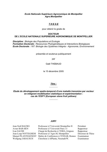
6) Vection<br />
Le vecteur <strong>de</strong> l’ESFY (Figure 8), Cacopsylla pruni Scopoli a été i<strong>de</strong>ntifié très<br />
tardivement (Carraro et al., 1998b). De ce fait, tous les paramètres épidémiologiques liés à la<br />
vection sont encore assez mal connus, qu’il s’agisse <strong>de</strong> la biologie du vecteur ou <strong>de</strong>s<br />
caractéristiques <strong>de</strong> la vection.<br />
(a) Biologie <strong>de</strong> C. pruni<br />
C. pruni est un Hémiptère appartenant<br />
à la famille <strong>de</strong>s psylles. Au printemps, lors<br />
<strong>de</strong> la reproduction <strong>de</strong>s adultes<br />
réimmigrants (<strong>de</strong> couleur foncée), les<br />
plantes hôtes <strong>de</strong> C. pruni sont <strong>de</strong>s Prunus.<br />
Les mesures d’abondance sur le terrain<br />
(Labonne & Lichou, 2004) et <strong>de</strong><br />
mortalité/fécondité au laboratoire<br />
(Carraro, 2004a) donnent <strong>de</strong>s résultats<br />
assez cohérents concernant les hôtes<br />
préférés <strong>de</strong> C. pruni (Figure 9) : le<br />
prunellier surtout, puis, en ordre<br />
décroissant, les pruniers (domestiques,<br />
japonais et myrobolan), l’abricotier, le<br />
pêcher et l’amandier ; les “cerisiers”<br />
(cerisier doux, cerisier à grappes, lauriercerise,<br />
bois <strong>de</strong> S te Lucie) sont moins<br />
propices au développement <strong>de</strong> C. pruni au<br />
laboratoire ; le mirabellier a également été<br />
signalé comme hôte naturel <strong>de</strong> ce psylle<br />
(Ossiannilsson, 1992).<br />
- 19 -<br />
Valeur hôte<br />
pour C. pruni<br />
Mirabellier (P. insititia)<br />
Prunellier (P. spinosa)<br />
Myrobolan (P. cerasifera)<br />
Prunier (P. domestica)<br />
Prunier japonais (P. salicina)<br />
Abricotier (P. armeniaca)<br />
Pêcher (P. persica)<br />
Amandier (P. amygdalus)<br />
Cerisier doux (P. avium)<br />
Bois <strong>de</strong> Ste Lucie (P. mahaleb)<br />
Cerisier à grappes (P. padus)<br />
Laurier-cerise (P. laurocerasus)<br />
Figure 9. Hiérarchie <strong>de</strong> la sensibilité à l’ESFY<br />
parmi les Prunus. Cette synthèse <strong>de</strong> quelques<br />
observations et expérimentations n’a qu’une<br />
valeur qualitative. Elle est basée sur l’abondance<br />
<strong>de</strong> C. pruni sur les Prunus dans la nature et/ou<br />
<strong>de</strong> sa longévité et <strong>de</strong> sa fécondité en conditions<br />
expérimentales.<br />
Au printemps, on trouve <strong>de</strong>s œufs et <strong>de</strong>s larves sur les plantes hôtes les plus favorables,<br />
puis <strong>de</strong> jeunes adultes émigrants (<strong>de</strong> couleur claire) qui disparaissent <strong>de</strong>s Prunus en début<br />
d’été. Une mesure pluriannuelle <strong>de</strong> la <strong>de</strong>nsité <strong>de</strong>s individus (Figure 10) indique que les dates<br />
<strong>de</strong> présence et les effectifs fluctuent légèrement d’une année à l’autre (Labonne & Lichou,<br />
2004). Cependant, pour une année donnée, l’évolution parallèle <strong>de</strong>s effectifs entre les<br />
différents Prunus (Figure 11) semble indiquer que C. pruni ne colonise pas ces espèces<br />
successivement mais bien <strong>de</strong> façon synchrone (Labonne & Lichou, 2004).<br />
Nombre <strong>de</strong> psylles<br />
120<br />
100<br />
80<br />
60<br />
40<br />
20<br />
0<br />
17/1<br />
31/1<br />
14/2<br />
28/2<br />
13/3<br />
27/3<br />
10/4<br />
24/4<br />
8/5<br />
22/5<br />
5/6<br />
2000<br />
Date<br />
2001 2002<br />
19/6<br />
3/7<br />
17/7<br />
Figure 10. Evolution<br />
pluriannuelle <strong>de</strong>s<br />
dates <strong>de</strong> présence et<br />
<strong>de</strong>s effectifs <strong>de</strong>s sta<strong>de</strong>s<br />
adultes <strong>de</strong> C. pruni.<br />
Les effectifs indiqués<br />
correspon<strong>de</strong>nt au<br />
battage <strong>de</strong> 20<br />
branches au-<strong>de</strong>ssus<br />
d’une toile <strong>de</strong> 1 m²<br />
dans une haie <strong>de</strong><br />
prunelliers située à<br />
Montbazin (Hérault).
Nombre <strong>de</strong> psylles<br />
Nombre <strong>de</strong> psylles<br />
120<br />
100<br />
80<br />
60<br />
40<br />
20<br />
0<br />
120<br />
100<br />
80<br />
60<br />
40<br />
20<br />
0<br />
17/1<br />
17/1<br />
31/1<br />
31/1<br />
14/2<br />
14/2<br />
28/2<br />
28/2<br />
13/3<br />
27/3<br />
Hérault 2002<br />
10/4<br />
24/4<br />
8/5<br />
22/5<br />
5/6<br />
19/6<br />
- 20 -<br />
3/7<br />
Prunellier<br />
Date<br />
Prunier Abricotier<br />
Pyrénées Orientales 2002<br />
13/3<br />
27/3<br />
10/4<br />
24/4<br />
8/5<br />
22/5<br />
5/6<br />
19/6<br />
Date<br />
Prunellier Myrobolan<br />
3/7<br />
17/7<br />
17/7<br />
Figure 11. Evolution synchrone<br />
<strong>de</strong>s effectifs <strong>de</strong>s sta<strong>de</strong>s adultes<br />
<strong>de</strong> C. pruni sur prunellier,<br />
prunier domestique, abricotier<br />
et myrobolan. Les effectifs<br />
indiqués correspon<strong>de</strong>nt au<br />
battage <strong>de</strong> 20 branches au<strong>de</strong>ssus<br />
d’une toile <strong>de</strong> 1 m².<br />
Le reste <strong>de</strong> l’année, les plantes “refuges” <strong>de</strong> C. pruni sont <strong>de</strong>s conifères (Conci et al.,<br />
1992 ; Lauterer, 1999). Les œufs et les larves n’ont jamais été observés sur conifères et il ne<br />
se déroule qu’un seul cycle sur les Prunus ; par conséquent, on peut supposer que C. pruni<br />
est une espèce univoltine * vivant environ un an (Conci et al., 1992 ; Ossiannilsson, 1992).<br />
Cependant, ce point reste à démontrer formellement.<br />
Les comportements <strong>de</strong> ce psylle sont certainement la partie la plus mal connue <strong>de</strong> sa<br />
biologie. Les larves peuvent se déplacer en marchant et les adultes en sautant ou en volant ;<br />
on ne connaît rien d’autre sur les distances et la fréquence <strong>de</strong>s déplacements <strong>de</strong> C. pruni si ce<br />
n’est que les adultes semblent capables <strong>de</strong> voler sur <strong>de</strong> longues distances, puisqu’on le trouve<br />
au printemps en nombre dans <strong>de</strong>s plaines situées à plusieurs dizaines <strong>de</strong> kilomètres <strong>de</strong>s zones<br />
refuges i<strong>de</strong>ntifiées. Par analogie avec son proche parent C. pyri, on sait qu’il ne faut pas<br />
négliger les vols <strong>de</strong> ce petit insecte sur <strong>de</strong> longues distances. En effet, Blomquist &<br />
Kirkpatrick (2002) ont estimé qu’en 18 ans, C. pyri s’est déplacé vers le sud <strong>de</strong>s Etats-Unis <strong>de</strong><br />
60 km/an en moyenne. De plus, parmi les Hémiptères, plusieurs espèces <strong>de</strong> pucerons, <strong>de</strong><br />
cica<strong>de</strong>lles et <strong>de</strong> psylles sont réputées migrer sur <strong>de</strong> très gran<strong>de</strong>s distances, probablement en<br />
profitant <strong>de</strong> courants aériens favorables (Byrne & Bellows, 1991 ; Loxdale & Lushai, 1999).<br />
On ne sait pas non plus si C. pruni possè<strong>de</strong> un comportement d’agrégation, mais <strong>de</strong> nombreux<br />
insectes émettent <strong>de</strong>s signaux acoustiques (Cocroft, 2005), en particulier <strong>de</strong>s psylles pendant<br />
la pério<strong>de</strong> <strong>de</strong> reproduction (D. M. Percy 1 , 2005). Enfin, l’existence <strong>de</strong> phéromones sexuelles a<br />
été démontrée chez un autre psylle du même genre, C. bi<strong>de</strong>ns (Soroker et al., 2004). Il n’est<br />
donc pas impossible qu’au moins l’un <strong>de</strong> ces procédés d’agrégation soit également utilisé par<br />
C. pruni.<br />
1 Diana M. Percy (20/01/2005) Song, sex and psyllid systematics : http://www.psyllids.org/psyllidsSOUND.htm
(b) Caractéristiques <strong>de</strong> la vection<br />
Les phytoplasmes sont en général transmis sur le mo<strong>de</strong> persistant (circulant multipliant)<br />
et sans transmission à la <strong>de</strong>scendance (Garnier et al., 2001). Il semble en être <strong>de</strong> même pour<br />
C. pruni. En effet, d’après Carraro et al. (2001), (i) les insectes infectieux sont capables <strong>de</strong><br />
transmettre l’ESFY pendant plusieurs semaines à plusieurs plantes-tests successivement ; (ii)<br />
certains <strong>de</strong>s tous premiers insectes capturés au début du printemps sont porteurs <strong>de</strong> ‘Ca. P.<br />
prunorum’ et immédiatement infectieux, ce qui suggère qu’ils ont conservé le phytoplasme<br />
pendant 9 mois environ (du début <strong>de</strong> l’été à la fin <strong>de</strong> l’hiver) ; et (iii) l’acquisition, la latence<br />
dans l’insecte et la transmission semblent durer en moyenne 3, 25 et 3 jours, respectivement.<br />
Ces longues durées sont caractéristiques du mo<strong>de</strong> <strong>de</strong> transmission circulant : la transmission<br />
n’a lieu que lors <strong>de</strong>s piqûres d’alimentation, ce qui nécessite que le vecteur déci<strong>de</strong> <strong>de</strong><br />
s’alimenter, puis dirige ses stylets jusqu’aux faisceaux conducteurs, pour finalement injecter<br />
<strong>de</strong> la salive (et <strong>de</strong>s phytoplasmes) dans le phloème puis ingérer la sève (et les phytoplasmes)<br />
qui y circulent. De plus, entre l’acquisition et la transmission, le phytoplasme doit se<br />
multiplier puis passer du tube digestif du vecteur à ses glan<strong>de</strong>s salivaires, via l’hémolymphe<br />
(Garnier et al., 2001). Tous ces processus <strong>de</strong>man<strong>de</strong>nt du temps.<br />
Les jeunes adultes émergents et les vieux adultes réimmigrants capturés sur les Prunus<br />
sont capables <strong>de</strong> transmettre l’ESFY (Carraro et al., 1998b, 2001, 2004c). Certains jeunes<br />
adultes sont donc infectieux avant <strong>de</strong> quitter le verger, mais il est probable que pour la<br />
majorité <strong>de</strong> ces individus porteurs du phytoplasme, la latence s’achève en <strong>de</strong>hors <strong>de</strong>s Prunus<br />
(Carraro et al., 2004c). Par contre, aucune expérience n’indique clairement si les larves sont<br />
capables <strong>de</strong> transmettre ‘Ca. P. prunorum’, si les jeunes adultes peuvent acquérir puis<br />
transmettre le phytoplasme avant <strong>de</strong> quitter les Prunus, ni si les adultes réimmigrants sont<br />
capables d’acquérir le pathogène puis <strong>de</strong> le transmettre avant la fin <strong>de</strong> leur vie. Ces<br />
paramètres sont pourtant fondamentaux car ils déterminent les échelles auxquelles se déroule<br />
le cycle <strong>de</strong> base <strong>de</strong> l’épidémie.<br />
7) Incubation, latence et pério<strong>de</strong> infectieuse <strong>de</strong> la plante<br />
La durée d’incubation est définie comme le temps écoulé entre la date <strong>de</strong> contamination<br />
et la date d’apparition <strong>de</strong>s symptômes, alors que la durée <strong>de</strong> latence correspond au temps<br />
écoulé entre la date <strong>de</strong> contamination et la date à laquelle la plante <strong>de</strong>vient infectieuse<br />
(Van<strong>de</strong>rplank, 1963). Certaines espèces ou variétés sont tolérantes (infectées, mais<br />
asymptomatiques) ; pour les autres, l’incubation dure <strong>de</strong> 7 à 12 mois lorsque la transmission<br />
est réalisée par greffage <strong>de</strong> matériel infecté (Morvan & Castelain, 1968 ; Giunchedi et al.,<br />
1983). Quand <strong>de</strong>s pruniers japonais <strong>de</strong> moins d’un an sont inoculés par <strong>de</strong>s groupes <strong>de</strong><br />
psylles, les premiers symptômes apparaissent 4 à 5 mois environ après la transmission<br />
(Carraro et al., 1998b, 2004a). En verger, selon l’espèce et la variété considérée, les premiers<br />
symptômes apparaissent 2 à 5 ans après la plantation <strong>de</strong> matériel sain (Morvan, 1977 ; Carraro<br />
et al., 1992 ; Labonne et al., 2000 ; Jarausch et al., 2001a). Les symptômes mettent ensuite au<br />
moins 2 ans à se propager à l’ensemble <strong>de</strong>s charpentières (Morvan, 1977) <strong>de</strong>s arbres mala<strong>de</strong>s.<br />
Il reste cependant plusieurs questions en suspens qui, à notre connaissance, n’ont pas été<br />
examinées, malgré leur importance, en particulier dans un contexte où la détection <strong>de</strong> la<br />
maladie est en général basée sur les symptômes (qui apparaissent après une pério<strong>de</strong><br />
d’incubation) : quelle est la durée <strong>de</strong> la latence ? Est-elle plus courte que la durée<br />
d’incubation ? Les vecteurs peuvent-ils acquérir le phytoplasme sur une plante qui n’a pas<br />
encore <strong>de</strong> symptômes ? Les vecteurs peuvent-ils acquérir le phytoplasme pendant toute la<br />
pério<strong>de</strong> où la plante présente <strong>de</strong>s symptômes (en particulier au début et à la fin <strong>de</strong> l’expression<br />
<strong>de</strong>s symptômes) ? Comment varient les pério<strong>de</strong>s d’incubation et <strong>de</strong> latence en fonction <strong>de</strong><br />
l’âge <strong>de</strong> la plante lors <strong>de</strong> son inoculation ?<br />
- 21 -
8) Propagation entre parcelles<br />
Compte tenu <strong>de</strong>s éléments fournis précé<strong>de</strong>mment, trois scénarios peuvent expliquer<br />
l’introduction <strong>de</strong> l’ESFY dans un verger : (i) l’arrivée <strong>de</strong> vecteurs réimmigrants infectieux<br />
ayant acquis le phytoplasme l’année précé<strong>de</strong>nte, impliquant l’environnement lointain du<br />
verger (zones <strong>de</strong> moyenne montagne), (ii) l’arrivée <strong>de</strong> vecteurs récemment infectés,<br />
impliquant les abords <strong>de</strong> la parcelle (vergers adjacents ou Prunus sauvages présents aux<br />
alentours), ou (iii) une introduction humaine. En particulier, les pépinières sont susceptibles<br />
<strong>de</strong> fournir du matériel contaminé, notamment si un arbre donneur <strong>de</strong> greffons mala<strong>de</strong> n’a pas<br />
été détecté (Morvan, 1977 ; Laimer Da Câmara Machado et al., 2001 ; Torres et al., 2004).<br />
9) Bilan sur le cycle épidémique <strong>de</strong> l’ESFY<br />
Les principales caractéristiques du cycle épidémique <strong>de</strong> l’ESFY sont résumées dans la<br />
Figure 12. La Figure 13 synthétise les connaissances disponibles sur le cycle <strong>de</strong> C. pruni et<br />
sur le cycle <strong>de</strong> la vection.<br />
Introduction <strong>de</strong><br />
l’ESFY dans le<br />
verger par un<br />
vecteur ou par<br />
l’homme<br />
3 jours<br />
Transmission par<br />
le vecteur<br />
Vecteur infectieux<br />
Arbre<br />
infecté<br />
2-5 ans ?<br />
Incubation dans l’arbre<br />
Latence dans l’arbre<br />
Arbre = Prunus<br />
Vecteur = C. pruni<br />
25 jours<br />
Latence dans le vecteur<br />
DISPERSION DU VECTEUR<br />
- 22 -<br />
Arbre symptomatique<br />
Arbre<br />
infectieux<br />
Acquisition par<br />
le vecteur<br />
Vecteur infecté<br />
Arbre<br />
mort<br />
3 jours<br />
Figure 12. Cycle <strong>de</strong> base <strong>de</strong> l’épidémie d’ESFY. En conditions naturelles, on ne sait pas à quelles échelles<br />
<strong>de</strong> temps et d’espace se déroule ce cycle. (Photographies : G. Labonne)
Conifères ?<br />
Rétention du<br />
phytoplasme ?<br />
Octobre<br />
Janvier<br />
Juillet<br />
Départ<br />
<strong>de</strong>s Prunus<br />
Retour sur<br />
les Prunus<br />
- 23 -<br />
Adultes<br />
réimmigrants<br />
(vieux)<br />
Avril<br />
Œufs<br />
Larves<br />
Adultes<br />
émergents<br />
(jeunes)<br />
Acquisition du<br />
phytoplasme ?<br />
Transmission<br />
du phytoplasme<br />
Acquisition du<br />
phytoplasme<br />
Transmission du<br />
phytoplasme ?<br />
Prunus<br />
en plaine<br />
Figure 13. Connaissances initiales sur le cycle <strong>de</strong> C. pruni et <strong>de</strong> la vection <strong>de</strong> l’ESFY. Les parties<br />
hachurées indiquent les points qui n’ont pas été établis avec certitu<strong>de</strong> lors <strong>de</strong> travaux précé<strong>de</strong>nts.<br />
(Photographies : N. Sauvion)<br />
C. Enjeux scientifiques<br />
Ce tour d’horizon <strong>de</strong>s connaissances accumulées sur l’ESFY <strong>de</strong>puis 1924 montre que<br />
cette maladie grave ré-émerge en Europe mais qu’elle reste encore assez mal comprise. En<br />
particulier, du fait <strong>de</strong> la découverte relativement récente <strong>de</strong> C. pruni, la vection <strong>de</strong> l’ESFY n’a<br />
été que partiellement étudiée, alors qu’il s’agit du moteur <strong>de</strong> l’épidémie. Il reste dans ce<br />
domaine <strong>de</strong>s lacunes dont l’étendue nuit à la compréhension du fonctionnement <strong>de</strong> l’épidémie<br />
et à l’i<strong>de</strong>ntification <strong>de</strong>s métho<strong>de</strong>s <strong>de</strong> lutte optimales contre cette maladie : dynamique <strong>de</strong><br />
population (en fonction du climat) et cycle biologique du vecteur, sta<strong>de</strong>s compétents pour<br />
acquérir et transmettre le phytoplasme, comportements et déplacements à courte distance<br />
(transmission <strong>de</strong> l’ESFY intra- et inter-vergers) et à longue distance (migrations), zones<br />
d’estivage et d’hivernage.<br />
D’autres questions concernant les différentes espèces <strong>de</strong> Prunus mériteraient également<br />
d’être examinées pour estimer leur contribution au développement <strong>de</strong> la maladie : valeur<br />
source, pério<strong>de</strong> infectieuse, d’incubation et <strong>de</strong> latence (en fonction du climat, et <strong>de</strong> l’âge <strong>de</strong> la<br />
plante lors <strong>de</strong> l’inoculation), durée relative <strong>de</strong> l’incubation et <strong>de</strong> la latence.<br />
Enfin, la gestion <strong>de</strong> la maladie pourrait bénéficier directement <strong>de</strong> l’amélioration <strong>de</strong>s<br />
métho<strong>de</strong>s et <strong>de</strong>s connaissances dans certains domaines : i<strong>de</strong>ntification <strong>de</strong>s pratiques culturales<br />
influençant la quantité <strong>de</strong> maladie, estimation (a priori et a posteriori) <strong>de</strong> l’efficacité <strong>de</strong><br />
différentes métho<strong>de</strong>s <strong>de</strong> lutte contre la maladie, compréhension du mécanisme <strong>de</strong> prémunition<br />
pour limiter les dégâts causés par l’ESFY.<br />
III. Objectifs et stratégie d’étu<strong>de</strong><br />
A. Objectifs<br />
Toutes les questions mentionnées ci-<strong>de</strong>ssus sont intéressantes sur un plan cognitif et/ou<br />
plus appliqué. Cependant, dans ce mémoire, seuls seront abordés les sujets susceptibles
d’améliorer à la fois la compréhension du processus <strong>de</strong> vection et la gestion <strong>de</strong> l’ESFY.<br />
L’objectif principal <strong>de</strong> ce travail est <strong>de</strong> montrer comment l’intégration résultant <strong>de</strong> l’étu<strong>de</strong><br />
pluridisciplinaire <strong>de</strong>s questions épidémiologiques (en particulier <strong>de</strong> la vection) améliore la<br />
compréhension <strong>de</strong>s épidémies d’une maladie à phytoplasme touchant <strong>de</strong>s plantes pérennes*.<br />
B. Stratégie<br />
La stratégie d’étu<strong>de</strong> retenue est résumée dans la Figure 14. L’analyse <strong>de</strong> la bibliographie a<br />
permis d’i<strong>de</strong>ntifier les facteurs <strong>de</strong> risque et les processus épidémiques déjà connus. Pour<br />
i<strong>de</strong>ntifier et quantifier les autres processus, on privilégie les démonstrations expérimentales<br />
directes, dans la mesure du possible. Cependant, l’étu<strong>de</strong> expérimentale <strong>de</strong> nombreux<br />
paramètres, processus et facteurs <strong>de</strong> risque est difficilement envisageable, pour <strong>de</strong>s questions<br />
<strong>de</strong> pertinence <strong>de</strong>s échelles (plantation <strong>de</strong> vergers), <strong>de</strong> moyens financiers ou techniques (suivi<br />
direct <strong>de</strong> petits insectes) et <strong>de</strong> durée (expérimentations pluriannuelles). Ainsi, dans la première<br />
partie <strong>de</strong> ce mémoire, les situations variées rencontrées à l’échelle d’une petite région <strong>de</strong><br />
production seront mises à profit pour étudier certains <strong>de</strong> ces facteurs, mais aussi pour émettre<br />
<strong>de</strong>s conjectures sur les processus <strong>de</strong> dissémination <strong>de</strong> l’ESFY dans l’espace. La secon<strong>de</strong> partie<br />
sera consacrée aux expérimentations visant à établir le potentiel infectieux <strong>de</strong>s différents<br />
sta<strong>de</strong>s du vecteur, ce qui conditionne très fortement les processus <strong>de</strong> dispersion <strong>de</strong> la maladie,<br />
et donc les motifs spatio-temporels formés par les arbres mala<strong>de</strong>s. La troisième partie<br />
montrera que les motifs attendus ont <strong>de</strong>s caractéristiques qui peuvent se traduire en termes<br />
d’hypothèses d’indépendance, et que la construction et l’application <strong>de</strong> tests d’hypothèses<br />
peuvent améliorer la compréhension <strong>de</strong>s processus <strong>de</strong> dispersion. Dans la <strong>de</strong>rnière partie, les<br />
connaissances acquises sur l’ESFY par les diverses approches utilisées seront formalisées et<br />
intégrées dans un modèle mécaniste à l’échelle <strong>de</strong> la parcelle <strong>de</strong>stiné à estimer les paramètres<br />
liés aux comportements – peu accessibles expérimentalement – du vecteur dans les vergers.<br />
Enfin, la conclusion situera ces résultats dans la perspective concrète <strong>de</strong> la gestion <strong>de</strong> l’ESFY<br />
et replacera la démarche suivie dans le cadre plus général <strong>de</strong> l’étu<strong>de</strong> <strong>de</strong>s maladies émergentes<br />
ou ré-émergentes.<br />
Analyse <strong>de</strong> la<br />
bibliographie<br />
Modèle<br />
exploratoire<br />
régional<br />
Tests<br />
d’hypothèses<br />
locales<br />
Démonstrations<br />
expérimentales<br />
Facteurs <strong>de</strong> risque<br />
Processus<br />
Paramètres<br />
- 24 -<br />
modulation<br />
Modèle<br />
mécaniste<br />
estimation<br />
Figure 14. Stratégie d’étu<strong>de</strong> <strong>de</strong> l’épidémiologie <strong>de</strong> l’ESFY et organisation <strong>de</strong>s différentes approches<br />
envisagées.
Partie I : I<strong>de</strong>ntifier <strong>de</strong>s<br />
facteurs <strong>de</strong> risque par<br />
une enquête à l’échelle<br />
d’un bassin <strong>de</strong><br />
production<br />
- 25 -
En cas d’émergence, l’étu<strong>de</strong> <strong>de</strong> données <strong>de</strong> terrain disponibles à gran<strong>de</strong> échelle permet <strong>de</strong><br />
profiter <strong>de</strong> la diversité <strong>de</strong>s situations préexistantes pour i<strong>de</strong>ntifier et hiérarchiser rapi<strong>de</strong>ment<br />
les facteurs <strong>de</strong> risque associés à la maladie considérée. C’est pourquoi la première phase <strong>de</strong> la<br />
stratégie décrite précé<strong>de</strong>mment consiste à analyser à l’échelle d’une région <strong>de</strong> production les<br />
corrélations entre la quantité d’ESFY et les caractéristiques <strong>de</strong>s vergers. Pour chaque verger,<br />
on dispose du nombre d’arbres plantés et du nombre d’arbres morts ou mala<strong>de</strong>s <strong>de</strong> l’ESFY<br />
<strong>de</strong>puis la plantation du verger jusqu’à 2004 (inci<strong>de</strong>nce cumulée <strong>de</strong> l’ESFY) et d’un certain<br />
nombre <strong>de</strong> variables explicatives potentielles (âge, exploitant, cultivar, porte-greffe, origine<br />
du matériel, surface, <strong>de</strong>nsité, coordonnées spatiales). La nature <strong>de</strong>s données et la volonté<br />
d’analyser finement les corrélations spatiales ont nécessité l’utilisation <strong>de</strong> métho<strong>de</strong>s peu<br />
courantes en épidémiologie végétale : modèle logistique surdispersé (pour gérer la variabilité<br />
extra-binomiale), bootstrap paramétrique (pour obtenir <strong>de</strong>s intervalles <strong>de</strong> confiance fiables sur<br />
les faibles proportions), test d’indépendance spatiale entre les résidus du modèle (pour<br />
signaler <strong>de</strong>s écarts aux hypothèses du modèle, potentiellement liés à la transmission <strong>de</strong> la<br />
maladie). Ces différents aspects sont présentés dans l’article ci-<strong>de</strong>ssous.<br />
I. Article I : “I<strong>de</strong>ntifying Risk Factors from a Survey with a Logistic<br />
Regression Mo<strong>de</strong>l: the Case of European Stone Fruit Yellows”<br />
Gaël Thébaud, Nicolas Sauvion, Joël Chadœuf, Arnaud Dufils and Gérard Labonne<br />
(Soumis à la revue Phytopathology)<br />
- 26 -
I<strong>de</strong>ntifying Risk Factors from a Survey with a Logistic Regression Mo<strong>de</strong>l:<br />
the Case of European Stone Fruit Yellows<br />
Gaël Thébaud, Nicolas Sauvion, Joël Chadœuf, Arnaud Dufils and Gérard Labonne<br />
First, second, and fifth authors: Institut national <strong>de</strong> la recherche agronomique (INRA), UMR<br />
BGPI, CIRAD TA 41/K, Campus international <strong>de</strong> Baillarguet, 34398 <strong>Montpellier</strong> Ce<strong>de</strong>x 5,<br />
France; first and third authors: INRA, Unité <strong>de</strong> Biométrie, Domaine Saint-Paul, Site<br />
Agroparc, 84914 Avignon Ce<strong>de</strong>x 9, France; and fourth author: Station Expérimentale La<br />
Pugère, Chemin <strong>de</strong> la Barque, 13370 Mallemort, France.<br />
Corresponding author: Gaël Thébaud, INRA - UMR BGPI, CIRAD TA 41/K, Campus<br />
international <strong>de</strong> Baillarguet, 34398 <strong>Montpellier</strong> Ce<strong>de</strong>x 5, France. E-mail address:<br />
thebaud@ensam.inra.fr.<br />
ABSTRACT<br />
Thébaud, G., Sauvion, N., Chadœuf, J., Dufils, A., and Labonne, G. I<strong>de</strong>ntifying risk factors<br />
from a survey with a logistic regression mo<strong>de</strong>l: the case of European stone fruit yellows.<br />
European stone fruit yellows (ESFY) is becoming a major economic problem for Prunus<br />
growers in Europe. The causal agent (‘Candidatus Phytoplasma prunorum’) and its vector<br />
(Cacopsylla pruni) have been i<strong>de</strong>ntified, but the present knowledge of the risk factors for this<br />
disease relies at best on specific experiments. To estimate the influence of several factors on<br />
disease inci<strong>de</strong>nce in the field, an exhaustive survey was performed on apricot and Japanese<br />
plum orchards in the Crau plain (France). After a preliminary multivariate exploration of the<br />
data, we used a logistic regression mo<strong>de</strong>l to analyze and predict the cumulative number of<br />
diseased trees on the basis of a set of quantitative (age, planting <strong>de</strong>nsity and area of the<br />
orchard) and categorical variables (species, cultivar and rootstock). Because of the nature of<br />
the data, we used an overdispersed binomial mo<strong>de</strong>l and we <strong>de</strong>veloped a parametric bootstrap<br />
procedure based on the beta-binomial distribution to obtain confi<strong>de</strong>nce intervals. Our results<br />
indicated that the age, species and cultivar of the scion were the major factors explaining the<br />
observed number of diseased trees. The planting <strong>de</strong>nsity and the rootstocks used in the zone<br />
un<strong>de</strong>r study were less significant, while the area of the orchard had no effect. The residuals of<br />
the mo<strong>de</strong>l showed that some explanatory variables had not been taken into account, since part<br />
of the remaining variability could be explained by a grower effect. The spatial distribution of<br />
the residuals suggested that one of the reasons for this grower effect was the correlation<br />
between orchards closer than 100 m, possibly caused by the flight behavior of infectious<br />
vectors. The impact of this survey on our perception of the epi<strong>de</strong>miology of ESFY is<br />
presented, as well as its role in the i<strong>de</strong>ntification of targets for future investigation.<br />
Keywords: epi<strong>de</strong>miology, generalized linear mo<strong>de</strong>l, Monte Carlo, Prunus armeniaca, Prunus salicina.<br />
INTRODUCTION<br />
A frequent goal of epi<strong>de</strong>miological studies is to highlight the factors that are highly<br />
correlated with disease inci<strong>de</strong>nce or severity, or even to point at causal factors explaining the<br />
emergence of a new disease. To these aims, the analysis of surveys can be seen as an<br />
introduction or an alternative to the experimental approach, in particular (i) for quarantine<br />
diseases, (ii) when many potential factors are consi<strong>de</strong>red, (iii) when the investigated factors<br />
are related to the agricultural landscape (e.g., size, shape, or <strong>de</strong>nsity of the plots), (iv) when an<br />
immense number of replicates would be necessary to achieve enough statistical power, or (v)<br />
- 27 -
when a lack of knowledge on the biological system could question the epi<strong>de</strong>miological<br />
significance of the experiments. For example, if an unknown insect transmits a disease, the<br />
study of risk factors by the experimental inoculation of plant material may not reflect field<br />
conditions because of the vector’s behavior, among other reasons. In contrast, analyzing<br />
survey data allows taking advantage of many “natural experiments” that occur un<strong>de</strong>r field<br />
conditions, where more factors can be investigated simultaneously, the si<strong>de</strong> effect being a<br />
lack of a priori control by an experimental <strong>de</strong>sign. Such observational studies are common in<br />
animal and human epi<strong>de</strong>miology, but are rarer in botanical epi<strong>de</strong>miology, probably because<br />
the networks for a high-quality data collection on a large scale are infrequent for plant<br />
diseases, and because many risk factors can be investigated directly through cost-effective<br />
experiments. Most of these surveys linked disease inci<strong>de</strong>nce with climatic parameters such as<br />
temperature and moisture. Soil structure or pH (40), and the quantity and proximity in space<br />
or time of putative sources of inoculum (12,38) have also been examined. Finally, only a few<br />
observational studies have characterized how the amount of disease was influenced by<br />
human-driven factors such as agricultural practices, control methods and the choice of the<br />
cultivated genotype (21,43,47). However, for some of these variables, prophylactic practices<br />
can be <strong>de</strong>fined so that the growers can make preventive choices to reduce disease inci<strong>de</strong>nce<br />
without the economic and environmental costs associated with the use of pestici<strong>de</strong>s. Our<br />
study focused on these human-driven factors because of high practical impact on the<br />
management of European stone fruit yellows (ESFY).<br />
ESFY is a systemic disease affecting the genus Prunus (32). It has been present in Europe<br />
since at least the beginning of the 20 th century (7,19), but its prevalence has increased in the<br />
last <strong>de</strong>ca<strong>de</strong>s (30) and ESFY is now a major economic problem on apricot (P. armeniaca) and<br />
Japanese plum (P. salicina) in Europe. We know that ‘Candidatus Phytoplasma prunorum’<br />
(42), the causal agent of the disease, is transmitted on the persistent mo<strong>de</strong> by Cacopsylla<br />
pruni (5), but the risk factors of ESFY are still poorly un<strong>de</strong>rstood. Standardized captures in<br />
the field (29) and experimental breeding (3,4) showed the vector’s strong host preference for<br />
blackthorn (P. spinosa), myrobalan (P. cerasifera), Japanese plum, and plum trees (P.<br />
domestica). Experimental vector transmissions and graft inoculations (3,4) <strong>de</strong>monstrated that<br />
there is a wi<strong>de</strong> range of susceptibility to ‘Ca. P. prunorum’ within the genus Prunus, with<br />
cherry trees being highly resistant (27) and Japanese plum being highly susceptible (19). Field<br />
evaluations (6) and experimental inoculations (16,26,28) also repeatedly indicated a<br />
differential sensitivity to infection between cultivars and between rootstocks. Although these<br />
factors have been tested individually, the relative contribution of several potential risk factors<br />
of ESFY (including agricultural practices) has never been investigated; however, such data<br />
would help prioritize the targets of control methods. Clarifying these contributions with an<br />
experimental approach would require an immense number of trees and a lot of time because<br />
the annual inci<strong>de</strong>nce in apricot is quite low. Thus, we preferred the alternative of analyzing<br />
survey data generated by a prophylactic program un<strong>de</strong>rtaken in France. This program intends<br />
to reduce the number of secondary transmissions and the regional pool of inoculum, on the<br />
basis of experience with Plum pox virus, another vector-borne disease of Prunus with a high<br />
economic impact.<br />
In the Crau plain (France), a partnership was initiated to collect epi<strong>de</strong>miologically<br />
relevant data, in addition to the data that were necessary for disease management. Several<br />
potential risk factors were recor<strong>de</strong>d to explain the <strong>de</strong>pen<strong>de</strong>nt variable, which was <strong>de</strong>fined as<br />
the number of diseased trees among a known number of exposed trees in each orchard. In<br />
or<strong>de</strong>r to analyze such binomial data, a generalized linear mo<strong>de</strong>l (GLM) with a logit link<br />
function (i.e., logistic regression) is generally preferred to the more classical linear mo<strong>de</strong>ls<br />
because it appropriately takes into account the binomial nature of the <strong>de</strong>pen<strong>de</strong>nt variable<br />
(8,18,25,35). However, since few studies have analyzed survey data with GLMs<br />
(1,13,36,38,43), we present in this paper a logistic regression mo<strong>de</strong>l for i<strong>de</strong>ntifying and<br />
quantifying risk factors of ESFY, through the analysis of a regional survey.<br />
- 28 -
MATERIALS AND METHODS<br />
Data Record and Selection. The survey exten<strong>de</strong>d insi<strong>de</strong> a square of approximately 25km<br />
si<strong>de</strong> in the Crau plain, an area of apricot production in southeastern France. Most of the<br />
data were collected in 2003 by well-trained technical staff and the database was updated in<br />
2004. In each orchard, 9 variables were recor<strong>de</strong>d. The <strong>de</strong>pen<strong>de</strong>nt variable was a count data:<br />
the cumulative number of diseased trees from the date of orchard planting to March 1994 (the<br />
term “inci<strong>de</strong>nce” will be used instead of “cumulative number of diseased trees” in the rest of<br />
the paper). Except for a few trees that were exclu<strong>de</strong>d from the analysis, ESFY was the only<br />
cause of tree <strong>de</strong>ath. The growers frequently removed the trees with typical symptoms and new<br />
trees were often replanted, but only the initial trees were consi<strong>de</strong>red in the analysis. Thus, the<br />
inci<strong>de</strong>nce was estimated on the basis of the number of trees that were <strong>de</strong>ad or removed, or<br />
with the characteristic winter symptom of early leafing. Some of the potential risk factors that<br />
we recor<strong>de</strong>d were directly related to the biological characteristics of the system (species and<br />
cultivar of the scion, rootstock, surface, planting <strong>de</strong>nsity, and age of the orchard). We also<br />
recor<strong>de</strong>d human factors such as the grower and the nurseries that provi<strong>de</strong>d the planting<br />
material (the abbreviated names of the variables are indicated in Table 1). The mean temporal<br />
evolution of ESFY in the study area was assessed in 517 apricot orchards. For more reliability<br />
in the estimated effects, we removed the plots with missing data and those with uncommon<br />
cultivars or rootstocks. The mean of the quantitative variables and the levels of the categorical<br />
variables in the final data subset (225 orchards and about 69,000 trees from 17 farms) are<br />
summarized in Table 1. The mean ESFY inci<strong>de</strong>nce in these orchards was 6.3%.<br />
Method Overview. Our general approach to the statistical analysis of this survey<br />
consisted of five successive steps: (i) a multivariate analysis to remove overly correlated<br />
variables from the mo<strong>de</strong>l; (ii) the most parsimonious overdispersed logistic regression mo<strong>de</strong>l<br />
was built by a stepwise selection of the variables; (iii) the a<strong>de</strong>quacy of this final mo<strong>de</strong>l was<br />
then checked by an analysis of the residuals and by assessing its predictive power for an<br />
external data set; (iv) we subsequently evaluated the relative influence of the different<br />
variables on disease inci<strong>de</strong>nce, and then (v) a test was performed on the residuals to track the<br />
remaining spatial covariates. Unless otherwise stated, all the analyses were performed with<br />
the R statistical software (41), version 2.0.1.<br />
Selection of the Variables. In or<strong>de</strong>r to avoid overloading the mo<strong>de</strong>l with redundant<br />
explanatory variables, a preliminary multivariate exploration of the data was un<strong>de</strong>rtaken. We<br />
first performed a normalized principal components analysis (23) on the quantitative variables<br />
(Y, AREA, AGE, DENS), and a multiple correspon<strong>de</strong>nce analysis (2) on the categorical<br />
variables (CLV, RST, OCLV, ORST). Then, a Hill and Smith analysis (22) was performed to<br />
mix these two analyses and thus to simultaneously analyze the relationships between all<br />
variables. The ADE-4 software (46) was used for the computation and generation of factorial<br />
maps.<br />
Logistic Regression Mo<strong>de</strong>l. To estimate simultaneously the influence of potential factors<br />
on ESFY inci<strong>de</strong>nce, we built a logistic regression mo<strong>de</strong>l (35). This GLM was <strong>de</strong>dicated to the<br />
analysis of proportions arising from binomial data: in each orchard, the numbers of exposed<br />
(ni) and affected trees (Yi) were known. The number of affected trees in the i th orchard was<br />
initially assumed to have a binomial distribution with probability pi for each tree to show<br />
symptoms, while a given combination of k explanatory factors <strong>de</strong>fined this probability pi.<br />
Thus, the mo<strong>de</strong>l could be written Yi|pi ~ B (ni,pi), where pi was <strong>de</strong>fined by ln(pi/(1-pi)) = a0 +<br />
a1,i (Fact1) + a2,i (Fact2) + … + ak,i (Factk). The best-fitting mo<strong>de</strong>l was obtained by a manual<br />
stepwise procedure for selecting significant variables and biologically meaningful second-<br />
- 29 -
or<strong>de</strong>r interactions. The R function glm was used for mo<strong>de</strong>l fitting by iteratively reweighted<br />
least squares.<br />
Mo<strong>de</strong>ling Overdispersion. In the mo<strong>de</strong>l <strong>de</strong>scribed above, the observed number of<br />
diseased trees (Yi) should have a binomial variance: Var(Yi) = ni pi (1-pi). However, in<br />
observational studies, the presence of overdispersion (extrabinomial variation, here) is very<br />
common (18,35), and it was also encountered in this study. Following Collett (8), we<br />
accounted for the extrabinomial variance with Williams’ iterative algorithm (48) because the<br />
number of trees was not i<strong>de</strong>ntical in all the orchards. Overdispersed mo<strong>de</strong>ls were fitted with<br />
the dispmod R package.<br />
Mo<strong>de</strong>l Assessment. The standard goodness-of-fit criterion for GLMs (the ratio between<br />
the residual <strong>de</strong>viance and the residual <strong>de</strong>grees of freedom) is meaningless for overdispersed<br />
mo<strong>de</strong>ls (8). Hence, several other procedures were carried out in or<strong>de</strong>r to check the mo<strong>de</strong>l.<br />
First, we examined the standardized <strong>de</strong>viance residuals in or<strong>de</strong>r to look for outlying values or<br />
for correlation with the linear predictors or with the variables (inclu<strong>de</strong>d or not in the final<br />
mo<strong>de</strong>l). Then, after weighting each point by the corresponding number of trees in the orchard,<br />
we compared the linear regression between observed and predicted values with their expected<br />
linear relationship. We also performed both an external validation and a robustness analysis<br />
by assessing the ability of the final mo<strong>de</strong>l to provi<strong>de</strong> an accurate estimate of inci<strong>de</strong>nce in an<br />
in<strong>de</strong>pen<strong>de</strong>nt data set composed of the 57 orchards exclu<strong>de</strong>d from the initial data because their<br />
rootstocks were unknown or un<strong>de</strong>r-represented. To obtain predictions in these new orchards,<br />
the parameters associated with the rootstock were replaced by the weighted mean of rootstock<br />
effects obtained after fitting the final mo<strong>de</strong>l.<br />
A characteristic of the data set was the low number of expected diseased trees in many<br />
orchards. Thus, for this discrete variable, the validity of confi<strong>de</strong>nce intervals based on<br />
asymptotic theorems could be questioned. Consequently, we used a parametric bootstrap<br />
approach (17) to <strong>de</strong>rive confi<strong>de</strong>nce intervals (i) for the predicted number of diseased trees in<br />
each orchard, and (ii) for the estimated value of the ak parameters. This bootstrap procedure<br />
was performed as follows: first, the fitted overdispersed binomial mo<strong>de</strong>l provi<strong>de</strong>d estimates of<br />
disease inci<strong>de</strong>nce in each orchard (pi) and of an overdispersion parameter φ. These parameters<br />
were used (as shown in the Appendix) to draw for each orchard 1,000 in<strong>de</strong>pen<strong>de</strong>nt<br />
realizations from a beta-binomial distribution, corresponding to a binomial law with extrabinomial<br />
variance (8,10,24). For the predicted number of diseased trees in each orchard, 95%<br />
confi<strong>de</strong>nce intervals could then be <strong>de</strong>rived from the quantiles 2.5% and 97.5% of the<br />
simulated distributions. After refitting the mo<strong>de</strong>l (with Williams’ procedure) on the 1,000<br />
simulated data sets, the subsequent 1,000 re-estimated values of each ak parameter were used<br />
similarly to <strong>de</strong>fine a 95% confi<strong>de</strong>nce interval around their mean value.<br />
Influence of the Risk Factors. The relative significance of the different factors was<br />
evaluated through <strong>de</strong>viance analysis. We initially assessed the significance of each variable<br />
alone. Afterwards, to evaluate the influence of each variable adjusted for the other variables,<br />
we used the final weights of the best overdispersed mo<strong>de</strong>l to fit reduced mo<strong>de</strong>ls in which each<br />
factor and its interactions with other factors were removed in turn. A chi-square test was then<br />
used to compare the full mo<strong>de</strong>l with each reduced mo<strong>de</strong>l. The influence of the variables was<br />
visualized by predicting the temporal evolution of the disease for different levels of the<br />
variables (<strong>de</strong>fault parameters to their mo<strong>de</strong> or mean: CLV = Orangered, RST = Peach, DENS<br />
= 384 trees/ha).<br />
Spatial Analysis of the Residuals. The coordinates of the orchards’ centroids were<br />
obtained from aerial orthophotographs (BD ORTHO, Institut Géographique National,<br />
France). The residuals could then be plotted at the location of the corresponding orchard for a<br />
- 30 -
visual inspection of their spatial pattern. The spatial <strong>de</strong>pen<strong>de</strong>nce of the residuals was further<br />
investigated with a nonparametric test relying on the empirical semivariogram (34). This<br />
function γ is based on the squared difference between the values of the residuals separated by<br />
a distance h±ε (i.e., residuals with coordinates s within the distance class h):<br />
1<br />
2<br />
γ(h) = ∑ ( r ( s+<br />
h)<br />
- r(<br />
s)<br />
) , where N(h) is the number of pairs of residuals r in the distance<br />
2N<br />
( h)<br />
N ( h)<br />
class h. We chose 32 meter-wi<strong>de</strong> distance classes (ε=16) in or<strong>de</strong>r to have at least 50 pairs of<br />
points in each class. Un<strong>de</strong>r the hypothesis of spatial in<strong>de</strong>pen<strong>de</strong>nce, the values of the residuals<br />
should be distributed at random among the locations of the orchards’ centroids. Therefore,<br />
this hypothesis of in<strong>de</strong>pen<strong>de</strong>nce was challenged by a random labeling test (14,39): the<br />
function γ(h) computed on the observed residuals was compared to 1,000 random reallocations<br />
of the values of these residuals. After or<strong>de</strong>ring the 1,000 simulated values, a bilateral P-value<br />
was computed, following Manly (33), as twice the proportion of simulated values more<br />
extreme or equal to the observed value of γ(h). A 95% confi<strong>de</strong>nce envelope was also <strong>de</strong>rived<br />
from the 25 th and 975 th values of γ(h). The hypothesis of spatial in<strong>de</strong>pen<strong>de</strong>nce between<br />
residuals should be rejected at the 5% significance level when the observed variogram is<br />
outsi<strong>de</strong> this envelope. However, when the mo<strong>de</strong>l is misspecified in some way (e.g., biased<br />
estimate or different variance for some levels, or significant variable not inclu<strong>de</strong>d), the<br />
grower’s trend to plant similar orchards si<strong>de</strong> by si<strong>de</strong> could also produce spatial correlation<br />
between residuals un<strong>de</strong>r the null hypothesis of in<strong>de</strong>pen<strong>de</strong>nce; as a precaution, the random<br />
labeling procedure was therefore adapted to perform the permutations insi<strong>de</strong> groups with<br />
homogeneous characteristics (see ref (33), pp. 182-199, for more <strong>de</strong>tails). Splitting the initial<br />
data set on the basis of the grower, cultivar and age of the orchards <strong>de</strong>fined 76 highly<br />
homogeneous groups that were used for conditioning the simulations.<br />
RESULTS<br />
Influence of the Species. The subset that was specially selected to analyze the species<br />
effect inclu<strong>de</strong>d 15 Japanese plum orchards and 10 apricot orchards with the same<br />
characteristics: all six to eight years old, on myrobalan rootstocks. The age of the orchards<br />
had no significant influence on disease inci<strong>de</strong>nce (not shown), thus the resulting mo<strong>de</strong>l for pi<br />
was simply: ln(pi/(1-pi)) = a1,Species. The estimated disease inci<strong>de</strong>nce (23.1% for the Japanese<br />
plum and 5.96% for the apricot plots) and the corresponding confi<strong>de</strong>nce intervals shown in<br />
Table 2 indicated that 6 to 8 years after planting, Japanese plum orchards were consi<strong>de</strong>rably<br />
more affected by ESFY than apricot orchards. An analysis of <strong>de</strong>viance showed that, even after<br />
allowing for overdispersion (φ = 0.019), this fourfold effect of the species was highly<br />
significant (P = 1.7×10 -14 , chi-square test on 1 df). Therefore, in the rest of the study, the few<br />
orchards planted with Japanese plum trees were exclu<strong>de</strong>d, so that our mo<strong>de</strong>l only analyzed the<br />
inci<strong>de</strong>nce of ESFY in apricot orchards.<br />
Multivariate Analysis. When initially inclu<strong>de</strong>d in the preliminary multivariate analysis,<br />
GRW completely <strong>de</strong>fined the first factorial plane and thus masked any correlation between<br />
the variables. Thus, it was treated as a supplementary variable in this analysis (Fig. 1), which<br />
showed that the data set was quite structured, because many categorical variables were<br />
interrelated. The projections of the orchards on the first factorial plane (Fig. 1A) could be<br />
subdivi<strong>de</strong>d into distinct groups (<strong>de</strong>noted I to V) sharing some specific combinations of the<br />
variables, as indicated by Fig. 1K and by a visual comparison between maps in Fig. 1A-J. The<br />
most significant multicorrelation involved sparse and ol<strong>de</strong>r orchards with cultivar 1 (Early<br />
Blush), origin 6, and rootstock 2 (Montclar) and 5 (GF 305) in group I, or <strong>de</strong>nse and young<br />
orchards with cultivar 2 (Goldrich), rootstock 4 (peach) and origins 3 and 9 in group II. The<br />
only <strong>de</strong>pen<strong>de</strong>nce between quantitative variables was the slight anticorrelation of AGE and<br />
- 31 -
DENS (r = -0.29). As expected, OCLV and ORST were almost completely correlated (Fig.<br />
1E-F and 1K). Additionally, these variables were strongly unbalanced and inclu<strong>de</strong>d 71<br />
missing values; hence they had to be removed. The supplementary variable GRW was clearly<br />
structured by the other variables (Fig. 1B), and thus strongly correlated to them. As we were<br />
more interested in these un<strong>de</strong>rlying correlates, the <strong>de</strong>scriptive variable GRW was not inclu<strong>de</strong>d<br />
in the mo<strong>de</strong>l until the ultimate step of the analysis. Finally, Fig. 1K showed that no<br />
explicative factor or variable was obviously correlated to the <strong>de</strong>pen<strong>de</strong>nt variable Y, further<br />
highlighting the need for an explicative mo<strong>de</strong>l.<br />
GLM. An initial GLM was built with the 5 remaining variables and their biologically<br />
meaningful interactions. AREA was then dropped because it was not significant (P = 0.24,<br />
chi-square test on 1 df). The <strong>de</strong>viance of the resulting mo<strong>de</strong>l was much higher than twice the<br />
number of <strong>de</strong>grees of freedom, a value that is used as a rule of thumb for the <strong>de</strong>tection of<br />
overdispersion (31). As the analysis of the residuals indicated no obvious problem with the<br />
mo<strong>de</strong>l, intra-orchard <strong>de</strong>pen<strong>de</strong>nce was the most probable cause of overdispersion. Thus the<br />
mo<strong>de</strong>l was corrected to allow extrabinomial variation (φ = 0.042) so as not to overestimate the<br />
significance of the effects. As the remaining 4 variables and 4 interactions were significant<br />
(all P-values < 4.3×10 -3 ), the final mo<strong>de</strong>l for pi was thus: ln(pi/(1-pi)) = a0 + a1,i (CLV) + a2,i<br />
(RST) + a3×AGEi + a4×DENSi + a5,i (CLV:AGE)×AGEi + a6,i (RST:AGE)×AGEi + a7,i<br />
(RST:DENS)×DENSi + a8×AGEi×DENSi.<br />
This mo<strong>de</strong>l was then checked. One point appeared to be overly influential on the basis of<br />
Cook’s distance (9). As the analyses provi<strong>de</strong>d consistent results with or without this point, it<br />
was not discar<strong>de</strong>d from the data set. The asymptotic results and parametric bootstrap<br />
distributions consistently i<strong>de</strong>ntified only one slightly outlying value that was conserved<br />
because the associated information was accurate. A quantile-quantile plot indicated that the<br />
normal distribution roughly approximated the distribution of the standardized <strong>de</strong>viance<br />
residuals (Fig. 2A). There was no indication of ina<strong>de</strong>quacy in the linear predictors because<br />
they were not correlated to the residuals. When plotted against the inclu<strong>de</strong>d variables, the<br />
residuals showed no particular trend other than a slight overestimation of ESFY inci<strong>de</strong>nce in<br />
young (2 to 4 year-old) orchards. The residuals were also randomly distributed with respect to<br />
the exclu<strong>de</strong>d variable AREA. On the contrary, the residuals significantly differed with respect<br />
to the grower (Fig. 2C) and origin of the planting material (Fig. 2D): the observed mean of the<br />
residuals was frequently outsi<strong>de</strong> the range expected un<strong>de</strong>r the null hypothesis of<br />
in<strong>de</strong>pen<strong>de</strong>nce. This fact indicates that the human factors not only summarized the other<br />
variables, but also had an additional significant effect that remained even after including some<br />
of the un<strong>de</strong>rlying factors in the mo<strong>de</strong>l.<br />
The weighted linear regression between observed and predicted inci<strong>de</strong>nce (Fig. 2B) had<br />
the equation: Observed = 0.946×Predicted + 0.003 (R 2 = 0.49, and standard error (SE) of 0.065<br />
and 0.006, respectively). On the logit scale, a R 2 of 0.42 confirmed that the fit of the mo<strong>de</strong>l<br />
was acceptable for an observational study: this mo<strong>de</strong>l captured half of the variability of the<br />
data with a relatively small number of parameters (21 of the initial 224 df were used). The<br />
same procedure carried out on the external data set resulted in a higher R 2 of 0.63, but the<br />
mo<strong>de</strong>l ten<strong>de</strong>d to overestimate the inci<strong>de</strong>nce, as shown by the equation of the regression line:<br />
Observed = 0.870×Predicted - 0.016 (SE = 0.088 and 0.009, respectively). Thus, <strong>de</strong>spite the<br />
lack of information on the rootstock in the validation data set, the mo<strong>de</strong>l still provi<strong>de</strong>d an<br />
acceptable prediction of ESFY inci<strong>de</strong>nce.<br />
The extent of the confi<strong>de</strong>nce intervals for some parameters of the mo<strong>de</strong>l (Table 3)<br />
confirmed that the 4 variables and 4 interactions inclu<strong>de</strong>d in the mo<strong>de</strong>l only explained part of<br />
the variability of the observed inci<strong>de</strong>nce. For these parameters, the discrepancy between<br />
bootstrap and the asymptotic confi<strong>de</strong>nce intervals was sometimes quite high (e.g., for the<br />
rootstocks), indicating that the assumptions of the asymptotic results were not fulfilled. Thus,<br />
in the rest of the study, we used the more reliable and more conservative bootstrap intervals.<br />
- 32 -
Influence of the Risk Factors. The mean temporal evolution of ESFY inci<strong>de</strong>nce in the<br />
study area (Fig. 3A) was summarized by the equation: Y=1/(1+e -0.198t+4.69 ). However, this<br />
binomial overdispersed mo<strong>de</strong>l was not satisfactory (R 2 = 0.1), thus indicating a major role for<br />
the other variables. The analysis of <strong>de</strong>viance for the one-variable mo<strong>de</strong>ls (Table 4) showed<br />
that the grower was the best single explanatory factor, much better than the cultivar or the<br />
other variables. However, because of the observed multicorrelation, it was statistically more<br />
accurate to assess the role of each variable after adjusting for the effect of the others. The<br />
corresponding <strong>de</strong>viance analysis (Table 5) showed that tree age was the most significant<br />
variable, followed by both <strong>de</strong>nsity and cultivar, and then the rootstock, with the area of the<br />
orchard having no influence on disease inci<strong>de</strong>nce. These results were robust to some<br />
alterations of the mo<strong>de</strong>l, because congruent conclusions (except that CLV became much more<br />
significant than DENS) were drawn when the same procedure was performed after fitting a<br />
simpler mo<strong>de</strong>l without the interaction terms (not shown). The interaction between the <strong>de</strong>nsity<br />
and the age of the orchard was more significant than any other interaction. No conclusion<br />
could be drawn from the mo<strong>de</strong>l concerning the effect of different levels of the factors,<br />
because the interactions were significant. The predictions for specific values were generally<br />
inconclusive as well, because of the wi<strong>de</strong> confi<strong>de</strong>nce intervals. However, the analysis of the<br />
mo<strong>de</strong>l without interaction terms indicated that the inci<strong>de</strong>nce was significantly higher on GF<br />
305 rootstock and on the cv. Orangered. This was further confirmed by the predicted ESFY<br />
progress in cv. Orangered and cv. Hargrand with the complete mo<strong>de</strong>l (Fig. 3B).<br />
Spatial Analysis of the Residuals. A simple map of the standardized <strong>de</strong>viance residuals<br />
clearly pointed out the spatial correlation between residuals. However, this correlation could<br />
have been at least partly explained by the spatial factor GRW (Fig. 2C), which had been<br />
discar<strong>de</strong>d from the mo<strong>de</strong>l. Thus, the mean of each level of this factor was <strong>de</strong>ducted from the<br />
corresponding residuals. Then, the grower-adjusted residuals were used to compute an<br />
empirical variogram and its confi<strong>de</strong>nce envelopes obtained by random labeling within<br />
homogeneous subgroups (Fig. 4). The first values of γ(h) (up to a distance of 100 m) were<br />
below or near the 95% confi<strong>de</strong>nce envelope (P-values ranging from
factor on disease inci<strong>de</strong>nce, with cv. Orangered more heavily infected than the other cultivars.<br />
This could be the result of a high attraction to the vector, or the consequence of a high level of<br />
susceptibility or sensitivity to the pathogen. Thus, to reduce the cost of ESFY, the growers<br />
should take into account the relative disease risk of the planted scion. The planting <strong>de</strong>nsity<br />
stood at a surprising third rank, which points out original speculative explanations: <strong>de</strong>nser<br />
orchards could be more attractive, could influence the mobility of the vector, or could speed<br />
up symptom expression in plants that are un<strong>de</strong>r more severe stress. The rootstock played an<br />
unexpected minor role in the system, which was confirmed by the relatively good prediction<br />
obtained even when it is unknown or different from the subset used to build the mo<strong>de</strong>l. This<br />
apparent discrepancy with previous observations indicating a major role of the rootstock in<br />
the evolution of the disease (37,28) may be caused by the excessive homogeneity of the<br />
rootstocks in the zone un<strong>de</strong>r study (80% of the rootstocks being peach cultivars). However, a<br />
competing explanatory hypothesis is that a faster visual <strong>de</strong>tection of diseased trees (enabled<br />
by acute symptoms) has a <strong>de</strong>creasing influence on the cumulative inci<strong>de</strong>nce as the orchards<br />
grow old. This could also explain the significant interaction between the age of the orchard<br />
and the rootstock.<br />
Human factors. Both human variables had a significant influence on ESFY inci<strong>de</strong>nce,<br />
but the growers had much more influence than the nurseries (Table 4 and Fig. 2C-2D). The<br />
grower was the best informative one-variable mo<strong>de</strong>l (Table 4). On the one hand, a large part<br />
of this high influence was the result of the correlation between the grower and many other<br />
variables (Fig. 1), with the grower being a good summary of several variables. On the other<br />
hand, the analysis of the residuals unequivocally <strong>de</strong>monstrated the existence of a grower<br />
effect not explained by the other variables (Fig. 2C). This result indicates that at least one<br />
grower-specific risk factor, though significant, was not inclu<strong>de</strong>d in the mo<strong>de</strong>l. In practice, a<br />
more in-<strong>de</strong>pth analysis of the differences in the agricultural practices of the growers with<br />
extremely low and extremely high inci<strong>de</strong>nce could point to the interesting factors. The level<br />
of prophylaxis, the insectici<strong>de</strong> protection, and the location of P. spinosa hedges are among the<br />
additional factors that could be investigated. Concerning the apparent nursery effect, we<br />
cannot completely rule out the possibility that some nurseries have differential levels of<br />
exposure to infectious vectors. However, it is more probably a case of confounding resulting<br />
from the strong correlation between growers and nurseries (Fig. 1B and 1E). The use of<br />
grower-adjusted residuals consi<strong>de</strong>rably reduces the variability between nurseries shown in<br />
Fig. 2D, whereas the symmetrical nursery-adjusted residuals only slightly attenuate the<br />
grower effect revealed in Fig. 2C (not shown).<br />
Spatial factors. The spatial <strong>de</strong>pen<strong>de</strong>nce was significant up to 100 m, and cannot be<br />
explained by an un<strong>de</strong>rlying grower effect because we used grower-adjusted residuals. Neither<br />
could it be the biologically uninteresting result of a spatial proximity between orchards with<br />
similar characteristics because we simulated the null hypothesis conditional on such<br />
similarity. Several hypotheses can be proposed to account for the remaining spatial<br />
<strong>de</strong>pen<strong>de</strong>nce. It might be indirectly caused by un<strong>de</strong>rlying physical spatial factors that have not<br />
been recor<strong>de</strong>d, such as the impact of soil characteristics on symptom expression. The spatial<br />
<strong>de</strong>pen<strong>de</strong>nce can also originate from some properties of the vectorial transmission of ESFY.<br />
The most obvious explanation comes from the presence in the data of cultivar mixtures within<br />
some plots (with very close centroids), where the vector could transmit the phytoplasma<br />
equivalently to one cultivar or the other. However, as this is not sufficient to give rise to the<br />
observed range of <strong>de</strong>pen<strong>de</strong>nce, other hypotheses are required, but they are more speculative.<br />
The population <strong>de</strong>nsity of C. pruni could be higher in some places, thereby forming small<br />
patches with a range of action limited to the adjacent orchards. The other processes that can<br />
play a role are either multiple primary or secondary transmissions occurring across orchards,<br />
mainly in a radius of approximately 100 m. For testing hypotheses related to these processes<br />
of disease spread, it would probably be interesting to analyze in <strong>de</strong>tail the individual trees and<br />
the spatiotemporal pattern of diseased trees (45).<br />
- 34 -
Mo<strong>de</strong>l Application<br />
This kind of mo<strong>de</strong>l-based analysis of a case study can raise questions related to its<br />
generality. The results showed that our mo<strong>de</strong>l was more efficient for i<strong>de</strong>ntifying and ranking<br />
the risk factors of ESFY than for predicting the evolution of this disease, because of the<br />
significant unexplained variability. However, the mo<strong>de</strong>l is suitable for an approximate<br />
evaluation of disease evolution in the Crau plain as far as it is not used to extrapolate to other<br />
levels of the factors (except for other rootstocks). This allows the participating growers to<br />
integrate the cost of disease control in the evaluation of the profitability of their orchards. Of<br />
course, outsi<strong>de</strong> the Crau plain, it would be inappropriate to make predictions with this mo<strong>de</strong>l.<br />
The results on the risk factors clearly show that even if the conclusions concerning particular<br />
levels of these factors may only be of local interest, the respective influence of the different<br />
factors are expected to be quite general. To this regard, the <strong>de</strong>monstration of a substantial<br />
effect of the grower should be highlighted because it indicates that, in addition to the choice<br />
of the cultivar, some agricultural practices can reduce or increase the inci<strong>de</strong>nce of ESFY.<br />
Validity of the GLM<br />
The data, by some of their features (<strong>de</strong>pen<strong>de</strong>nce and small binomial coefficients), did not<br />
strictly correspond to the assumptions of the logistic regression mo<strong>de</strong>l. As these problems<br />
frequently arise in the analysis of surveys with GLMs, we discuss how they have been<br />
<strong>de</strong>tected and handled.<br />
Depen<strong>de</strong>nce in the data. The spatial correlation between residuals can be seen as an<br />
unwelcome characteristic that does not meet the assumption of in<strong>de</strong>pen<strong>de</strong>nce between<br />
observations, which un<strong>de</strong>rlie most of the common statistical mo<strong>de</strong>ls. It can also be seen as a<br />
biologically meaningful feature that may motivate further investigation. Whatever the<br />
purpose, simple and versatile nonparametric tests based on random labeling can be inclu<strong>de</strong>d<br />
in the final steps of the analysis of the data to track spatial <strong>de</strong>pen<strong>de</strong>nce in the residuals<br />
(33,40). For this purpose, a multiscale exploration of spatial <strong>de</strong>pen<strong>de</strong>nce (e.g., using a<br />
variogram) or local indices could be a more powerful approach than global indices (such as<br />
Moran’s I or Geary’s C) to <strong>de</strong>tect residual spatial correlation at short-distance in a large-scale<br />
study. In this study, we <strong>de</strong>tected a significant spatial correlation up to 100 m. Taking this<br />
residual spatial correlation into account in the analysis is an interesting prospect for this study.<br />
However, building a statistically irreproachable mo<strong>de</strong>l to this aim would require <strong>de</strong>veloping<br />
cutting-edge spatial statistics methods that extend far beyond the scope of this article.<br />
Moreover, the hierarchy of the factors is expected to be quite robust to the incorporation of<br />
spatial effects in the mo<strong>de</strong>l, and the observed P-values (Table 5) are so low that all the<br />
corresponding effects would still be significant after an improbable 10-fold correction. It<br />
should also be noticed that the spatial correlation between residuals does not always challenge<br />
the assumption of in<strong>de</strong>pen<strong>de</strong>nce between the observations because it can be the consequence<br />
of land management (e.g., grouping i<strong>de</strong>ntical plots in clusters can also produce spatial<br />
correlation if the mo<strong>de</strong>l is slightly misspecified). In addition to the inter-orchards <strong>de</strong>pen<strong>de</strong>nce,<br />
the assumption of in<strong>de</strong>pen<strong>de</strong>nce can be challenged within the orchards, leading to<br />
extrabinomial variation. This phenomenon can be suspected when the fit of a binomial mo<strong>de</strong>l<br />
is unsatisfactory though nothing in the residuals indicates an incorrect specification of the<br />
mo<strong>de</strong>l (35). In this study, both sources of <strong>de</strong>pen<strong>de</strong>nce were i<strong>de</strong>ntified (i.e., intra- and interorchards).<br />
A previous analysis of the spatial pattern of diseased trees within an orchard also<br />
indicated <strong>de</strong>pen<strong>de</strong>nce between transmission events (44). These results can be partly explained<br />
by short-distance secondary transmissions. Moreover, as the phytoplasma is persistently<br />
transmitted by C. pruni (5), multiple transmissions can also account for the observed<br />
<strong>de</strong>pen<strong>de</strong>nce within (and to a lesser extent, between) orchards. Finally, it should be mentioned<br />
that the data experience a slight <strong>de</strong>pen<strong>de</strong>nt censoring, as some orchards have been removed in<br />
the past when the growers consi<strong>de</strong>red that they were too heavily infected. Thus the apparent<br />
temporal evolution un<strong>de</strong>restimates the real temporal evolution of the disease.<br />
- 35 -
Parametric bootstrap. When some predicted values (pi) are close to zero or when the<br />
number of individuals in some orchards (ni) is very low, the assumptions for the asymptotic<br />
results are not met. This situation can arise quite frequently in epi<strong>de</strong>miological surveys. In<br />
such circumstances, parametric bootstrap procedures could be used more frequently when<br />
satisfactory resampling mo<strong>de</strong>ls are available. Here, we expressed the parameters of the betabinomial<br />
distribution as a function of φ and pi (the overdispersion parameter and estimated<br />
proportions, respectively). To our knowledge, it is a new result that allows computing<br />
parametric bootstrap confi<strong>de</strong>nce intervals for overdispersed logistic mo<strong>de</strong>ls. In practice,<br />
bootstrap and asymptotic methods gave consistent results in the i<strong>de</strong>ntification of outliers, and<br />
similar confi<strong>de</strong>nce intervals for the species effect (Table 2) as well as for half of the<br />
parameters of the final mo<strong>de</strong>l (Table 3). For the other parameters, the bootstrap intervals were<br />
wi<strong>de</strong>r, hence more conservative.<br />
Concluding Remarks<br />
Performing a large-scale disease assessment, for a control program for example<br />
(11,12,20), can provi<strong>de</strong> reliable information for epi<strong>de</strong>miological studies. In such situations, a<br />
close collaboration with plant protection services from the initial steps is a prerequisite to<br />
ensure an optimal exploitation of the collected data. Otherwise, some data which are<br />
inexpensively and easily obtained and which are important for epi<strong>de</strong>miological exploitation<br />
could be omitted because of lack of interest in the control strategy. The analysis of such a<br />
survey with a logistic regression mo<strong>de</strong>l enabled the i<strong>de</strong>ntification and ranking of general risk<br />
factors for ESFY inci<strong>de</strong>nce. In summary, in addition to the obvious cumulative effect of the<br />
age, the main <strong>de</strong>terminant was the choice of the scion (the species being of major importance,<br />
followed by the cultivar). This result highlights the need for a rigorous experimental or field<br />
evaluation of the sensitivity of different cultivars to ESFY, as a basis for future genetic<br />
improvement. The planting <strong>de</strong>nsity and the rootstock appeared to play a secondary role, and at<br />
least one uni<strong>de</strong>ntified human factor had a significant impact. More <strong>de</strong>tailed investigations of<br />
the grower-specific agricultural practices would possibly i<strong>de</strong>ntify other risk factors, and the<br />
analysis of the spatiotemporal point pattern formed by the diseased trees would probably<br />
provi<strong>de</strong> insight into the transmission behavior of C. pruni.<br />
APPENDIX<br />
Here we show how the parameters of a beta-binomial mo<strong>de</strong>l can be <strong>de</strong>rived from an<br />
overdispersed binomial mo<strong>de</strong>l fitted by the Williams procedure (48) providing estimates of<br />
both pi and the overdispersion parameter φ.<br />
Let Yi be a binomial random variable: Yi ~ B(ni,pi). The variance of Yi is: Var(Yi) = ni pi<br />
(1-pi). As explained in pp.192-195 of Collett (8), the variance of an overdispersed binomial<br />
variable Yi can be written: Var(Yi) = ni pi (1-pi) (1+(ni-1)φ). This is in particular the variance<br />
of a beta-binomial random variable Yi in which Yi|Pi ~ B(ni,pi), Pi having a Beta distribution<br />
with E(Pi) = pi and Var(Pi) = φ pi (1-pi). As the mean and variance of a random variable Pi<br />
with a Beta distribution (with shape parameters αi and βi) are E(Pi) = αi/(αi+βi) = pi, and<br />
Var(Pi) = pi (1-pi)/(αi+βi+1), we obtain by i<strong>de</strong>ntification and resolution of the subsequent twoparameter<br />
equation:<br />
⎛ 1 ⎞<br />
⎛ 1 ⎞<br />
αi = pi⎜<br />
−1⎟<br />
and βi = (1-pi) ⎜ −1⎟<br />
.<br />
⎝φ<br />
⎠<br />
⎝φ<br />
⎠<br />
Thus, the parameters pi and φ estimated by Williams’ algorithm <strong>de</strong>fine the parameters αi<br />
and βi of the beta-binomial distribution. These parameters can then be used in a parametric<br />
bootstrap procedure to <strong>de</strong>rive confi<strong>de</strong>nce intervals for the predicted inci<strong>de</strong>nce in the orchards<br />
(pi) and for the parameters (ak) of the logistic regression mo<strong>de</strong>l.<br />
- 36 -
ACKNOWLEDGEMENTS<br />
We are much in<strong>de</strong>bted to the growers of Crau, to I. Ricavy and Y. Fradin (experimental<br />
station La Pugère), and to the Chambre d’Agriculture <strong>de</strong>s Bouches-du-Rhône for data<br />
collection, and for creating and actualizing the database. We wish to thank E. Klein and C.<br />
Bruchou for helpful discussions, J.-N. Bacro and S. Dallot for critically reviewing the<br />
manuscript and M. Hilf for improving the English. This study was partly fun<strong>de</strong>d by the<br />
Conseil Régional <strong>de</strong> PACA.<br />
LITERATURE CITED<br />
1. Aegerter, B. J., Nuñez, J. J., and Davis, R. M. 2003. Environmental factors affecting rose<br />
downy mil<strong>de</strong>w and <strong>de</strong>velopment of a forecasting mo<strong>de</strong>l for a nursery production system.<br />
Plant. Dis. 87:732-738.<br />
2. Burt, C. 1950. The factorial analysis of qualitative data. Br. J. Stat. Psychol. 3:166-185.<br />
3. Carraro, L., Ferrini, F., Ermacora, P., and Loi, N. 2002. Role of wild Prunus species in the<br />
epi<strong>de</strong>miology of European stone fruit yellows. Plant Pathol. 51:513-517.<br />
4. Carraro, L., Ferrini, F., Ermacora, P., and Loi, N. 2004. Transmission of European stone<br />
fruit yellows phytoplasma to Prunus species by using vector and graft transmission. Acta<br />
Hortic. 657:449-453.<br />
5. Carraro, L., Loi, N., and Ermacora, P. 2001. Transmission characteristics of the European<br />
stone fruit yellows phytoplasma and its vector Cacopsylla pruni. Eur. J. Plant Pathol.<br />
107:695-700.<br />
6. Carraro, L., Osler, R., Refatti, E., and Favali, M. A. 1992. Natural diffusion and<br />
experimental transmission of plum leptonecrosis. Acta Hortic. 309:285-290.<br />
7. Chabrolin, C. 1924. Quelques maladies <strong>de</strong>s arbres fruitiers <strong>de</strong> la vallée du Rhône. Annales<br />
<strong>de</strong>s Epiphyties 10:263-338.<br />
8. Collett, D. 1991. Mo<strong>de</strong>lling Binary Data. 1st ed. Chapman and Hall, London.<br />
9. Cook, R. D., and Weisberg, S. 1982. Residuals and Influence in Regression. Chapman and<br />
Hall, London.<br />
10. Crow<strong>de</strong>r, M. J. 1978. Beta-binomial anova for proportions. Appl. Stat. 27:34-37.<br />
11. Dallot, S., Gottwald, T., Labonne, G., and Quiot, J.-B. 2003. Spatial pattern analysis of<br />
Sharka disease (Plum pox virus strain M) in peach orchards of southern France.<br />
Phytopathology 93: 1543-1552.<br />
12. Dallot, S., Gottwald, T., Labonne, G., and Quiot, J.-B. 2004. Factors affecting the spread<br />
of Plum pox virus strain M in peach orchards subjected to roguing in France.<br />
Phytopathology 94:1390-1398.<br />
13. De Wolf, E. D., Mad<strong>de</strong>n, L. V., and Lipps, P. E. 2003. Risk assessment mo<strong>de</strong>ls for wheat<br />
Fusarium head blight epi<strong>de</strong>mics based on within-season weather data. Phytopathology<br />
93:428-435.<br />
14. Diggle, P. J. 1983. Statistical Analysis of Spatial Point Patterns. Mathematics in Biology.<br />
Aca<strong>de</strong>mic Press, London.<br />
15. Duval, H. 1999. Prunes japonaises : un défi à relever. L'Arboriculture Fruitière 524:35-41.<br />
16. Duval, H., Jullian, J.-P., and Lemaire, J.-M. 1999. Enroulement chlorotique <strong>de</strong><br />
l'abricotier : évaluation <strong>de</strong> la sensibilité <strong>de</strong>s principales variétés <strong>de</strong> prunier japonais... et du<br />
nouveau sur le vecteur. Phytoma 516:38-40.<br />
17. Efron, B., and Tibshirani, R. J. 1993. An Introduction to the Bootstrap. Chapman and<br />
Hall, New York.<br />
18. Garrett, K. A., Mad<strong>de</strong>n, L. V., Hughes, G., and Pfen<strong>de</strong>r, W. F. 2004. New applications of<br />
statistical tools in plant pathology. Phytopathology 94:999-1003.<br />
19. Goidanich, G. 1933. Un <strong>de</strong>perimento <strong>de</strong>i susini. Boll. Reg. Staz. Patol. Veg. Roma<br />
13:160-173.<br />
20. Gottwald, T. R., Sun, X., Riley, T., Graham, J. H., Ferrandino, F., and Taylor, E. L. 2002.<br />
Geo-referenced spatiotemporal analysis of the urban citrus canker epi<strong>de</strong>mic in Florida.<br />
- 37 -
Phytopathology 92:361-377.<br />
21. Hamel, C., Vujanovic, V., Nakano-Hylan<strong>de</strong>r, A., Jeannotte, R., and St-Arnaud, M. 2005.<br />
Factors associated with fusarium crown and root rot of asparagus outbreaks in Quebec.<br />
Phytopathology 95:867-873.<br />
22. Hill, M. O., and Smith, A. J. E. 1976. Principal component analysis of taxonomic data<br />
with multi-state discrete characters. Taxon 25:249-255.<br />
23. Hotelling, H. 1933. Analysis of a complex of statistical variables into principal<br />
components. J. Educ. Psychol. 24:417-441.<br />
24. Hughes, G., and Mad<strong>de</strong>n, L. V. 1993. Using the beta-binomial distribution to <strong>de</strong>scribe<br />
aggregated patterns of disease inci<strong>de</strong>nce. Phytopathology 83:759-763.<br />
25. Hughes, G., and Mad<strong>de</strong>n, L. V. 1995. Some methods allowing for aggregated patterns of<br />
disease inci<strong>de</strong>nce in the analysis of data from <strong>de</strong>signed experiments. Plant Pathol. 44:927-<br />
943.<br />
26. Jarausch, W., Eyquard, J. P., Lansac, M., Mohns, M., and Dosba, F. 2000. Susceptibility<br />
and tolerance of new French Prunus domestica cultivars to European stone fruit yellows<br />
phytoplasmas. J. Phytopathol. 148:489-493.<br />
27. Jarausch, W., Eyquard, J. P., Mazy, K., Lansac, M., and Dosba, F. 1999. High level of<br />
resistance of sweet cherry (Prunus avium L.) towards European stone fruit yellows<br />
phytoplasmas. Adv. Hortic. Sci. 13:108-112.<br />
28. Kison, H., and Seemuller, E. 2001. Differences in strain virulence of the European stone<br />
fruit yellows phytoplasma and susceptibility of stone fruit trees on various rootstocks to<br />
this pathogen. J. Phytopathol. 149:533-541.<br />
29. Labonne, G., and Lichou, J. 2004. Data on the life cycle of Cacopsylla pruni, Psyllidae<br />
vector of European stone fruit yellows (ESFY) phytoplasma, in France. Acta Hortic.<br />
657:465-470.<br />
30. Lemaire, J.-M., Jullian, J.-P., Au<strong>de</strong>rgon, J.-M., and Castelain, C. 1998. Enroulement<br />
chlorotique <strong>de</strong> l'abricotier : symptomatologie et gamme d'hôtes. L'Arboriculture Fruitière<br />
520:21-24.<br />
31. Lindsey, J. K. 1999. On the use of corrections for overdispersion. Appl. Stat. 48:553-561.<br />
32. Lorenz, K.-H., Dosba, F., Poggi-Pollini, C., Llácer, G., and Seemüller, E. 1994.<br />
Phytoplasma diseases of Prunus species in Europe are caused by genetically similar<br />
organisms. Z. Pflanzenk. Pflanzen. 101:567-575.<br />
33. Manly, B. F. J. 1991. Randomization and Monte Carlo Methods in Biology. Chapman and<br />
Hall, London, UK.<br />
34. Matheron, G. 1965. Les Variables Régionalisées et leur Estimation. Masson, Paris.<br />
35. McCullagh, P., and Nel<strong>de</strong>r, J. 1989. Generalized Linear Mo<strong>de</strong>l. Chapman and Hall,<br />
London.<br />
36. Mila, A. L., Carriquiry, A. L., and Yang, X. B. 2004. Logistic regression mo<strong>de</strong>ling of<br />
prevalence of soybean Sclerotinia stem rot in the north-central region of the United States.<br />
Phytopathology 94:102-110.<br />
37. Morvan, G. 1977. Apricot chlorotic leaf roll. EPPO Bull. 7:37-55.<br />
38. Nicot, P. C., and Rouse, D. I. 1987. Relationship between soil inoculum <strong>de</strong>nsity of<br />
Verticillium dahliae and systemic colonization of potato stems in commercial fields over<br />
time. Phytopathology 77.<br />
39. Ribeiro, P. J. J., Christensen, O. F., and Diggle, P. J. 2003. geoR and geoRglm: software<br />
for mo<strong>de</strong>l-based geostatistics. 3 rd International Workshop on Distributed Statistical<br />
Computing, March 20–22, Vienna, Austria.<br />
40. Rosso, P. H., and Hansen, E. M. 2003. Predicting Swiss needle cast disease distribution<br />
and severity in young Douglas-fir plantations in coastal Oregon. Phytopathology 93:790-<br />
798.<br />
41. R Development Core Team. 2004. R: A Language and Environment for Statistical<br />
Computing. R Foundation for Statistical Computing, Vienna, Austria.<br />
- 38 -
42. Seemüller, E., and Schnei<strong>de</strong>r, B. 2004. ‘Candidatus Phytoplasma mali’, ‘Candidatus<br />
Phytoplasma pyri’ and ‘Candidatus phytoplasma prunorum’, the causal agents of apple<br />
proliferation, pear <strong>de</strong>cline and European stone fruit yellows, respectively. Int. J. Syst.<br />
Bacteriol. 54:1217-1226.<br />
43. Shtienberg, D. 1996. Variables associated with intensity of Alternaria leaf spot in Pima<br />
cotton. Phytopathology 86:123-128.<br />
44. Thébaud, G., Labonne, G., Castelain, C., and Chadœuf, J. 2004. Spatio-temporal analysis<br />
of disease spread provi<strong>de</strong>s insights into the epi<strong>de</strong>miology of European stone fruit yellows.<br />
Acta Hortic. 657:471-476.<br />
45. Thébaud, G., Peyrard, N., Dallot, S., Calonnec, A., and Labonne, G. 2005. Investigating<br />
disease spread between two assessment dates with permutation tests on a lattice.<br />
Phytopathology. In Press.<br />
46. Thioulouse, J., Chessel, D., Dolé<strong>de</strong>c, S., and Olivier, J. M. 1997. ADE-4: a multivariate<br />
analysis and graphical display software. Stat. Comput. 7:75-83.<br />
47. Welham, S. J., Turner, J. A., Glad<strong>de</strong>rs, P., Fitt, B. D. L., Evans, N., and Baierl, A. 2004.<br />
Predicting light leaf spot (Pyrenopeziza brassicae) risk on winter oilseed rape (Brassica<br />
napus) in England and Wales, using survey, weather and crop information. Plant Pathol.<br />
53:713-724.<br />
48. Williams, D. A. 1982. Extra-binomial variation in logistic linear mo<strong>de</strong>ls. Appl. Stat.<br />
31:144-148.<br />
TABLE 1. Summary of the variables used in the preliminary multivariate analysis.<br />
Name of the variable<br />
Levels (frequency) or<br />
Abbreviated Full Mo<strong>de</strong><br />
Mean (range)<br />
Y Cumulative number of Depen<strong>de</strong>nt, 19.4 (0-144) trees<br />
diseased plants quantitative<br />
GRW Grower Categorical 17 levels (2-55 orchards/grower)<br />
CLV Cultivar Categorical Goldrich (27), Early Blush (33),<br />
Hargrand (66), Orangered (99)<br />
RST Rootstock Categorical Manicot - GF 1236 (11), Montclar (28),<br />
myrobalan (36), GF 305 (61), peach (89)<br />
OCLV Origin of the cultivar Categorical 8 levels (1-119 orchards/origin) + 71 NA a<br />
ORST Origin of the rootstock Categorical 9 levels (1-118 orchards/origin) + 71 NA<br />
AGE Age Quantitative 8.3 (2-15) years<br />
AREA Area Quantitative 0.73 (0.08-2.5) ha<br />
DENS<br />
a<br />
Not available<br />
Planting <strong>de</strong>nsity Quantitative 410 (238-571) trees/ha<br />
TABLE 2. Estimated mean and confi<strong>de</strong>nce intervals for the effect of the species on ESFY<br />
inci<strong>de</strong>nce (one-variable overdispersed GLM).<br />
Logit scale Response scale a<br />
Species Estimate ± SE Estimate<br />
- 39 -<br />
Bootstrap b<br />
confi<strong>de</strong>nce interval<br />
Asymptotic c<br />
confi<strong>de</strong>nce interval<br />
P. armeniaca -2.76 ± 0.209 0.060 0.038-0.085 0.039-0.089<br />
P. salicina -1.20 ± 0.097 0.231 0.197-0.265 0.197-0.268<br />
a The estimated disease inci<strong>de</strong>nce and the corresponding 95% confi<strong>de</strong>nce interval are back-<br />
transformed from the logit scale.<br />
b The parametric bootstrap procedure is <strong>de</strong>scribed in the text.<br />
c From a t-distribution on 23 df.
TABLE 3. Estimates and confi<strong>de</strong>nce intervals for the parameters a of the final overdispersed<br />
GLM.<br />
Parameter Estimate ± SE<br />
- 40 -<br />
Bootstrap b<br />
confi<strong>de</strong>nce interval<br />
Asymptotic c<br />
confi<strong>de</strong>nce interval<br />
(Intercept) -24.2 ± 6.36 -94.9 -16.8 -36.7 -11.7<br />
CLV Goldrich 1.31 ± 1.37 -1.06 4.34 -1.39 4.00<br />
CLV Hargrand -1.24 ± 1.20 -3.45 1.46 -3.61 1.13<br />
CLV Orangered 0.85 ± 1.17 -1.31 3.50 -1.45 3.15<br />
RST Montclar -0.12 ± 6.66 -10.5 67.7 -13.3 13.0<br />
RST myrobalan 9.87 ± 6.36 2.21 80.2 -2.66 22.4<br />
RST peach 11.5 ± 6.14 4.24 82.5 -0.61 23.6<br />
RST GF305 11.5 ± 6.10 4.48 82.5 -0.57 23.5<br />
AGE 1.54 ± 0.39 0.95 8.30 0.76 2.31<br />
DENS (×10 -2 ) 3.67 ± 1.53 0.46 8.42 0.65 6.69<br />
CLV Goldrich:AGE (×10 -1 ) -1.59 ± 1.57 -5.28 1.12 -4.67 1.50<br />
CLV Hargrand:AGE (×10 -1 ) 1.50 ± 1.21 -1.12 3.76 -0.89 3.89<br />
CLV Orangered:AGE (×10 -1 ) -0.08 ± 1.19 -2.71 2.20 -2.43 2.27<br />
RST Montclar:AGE -0.21 ± 0.34 -7.00 0.32 -0.88 0.46<br />
RST myrobalan:AGE -0.55 ± 0.34 -7.43 -0.07 -1.22 0.12<br />
RST peach:AGE -0.57 ± 0.33 -7.41 -0.09 -1.23 0.09<br />
RST GF305:AGE -0.59 ± 0.33 -7.40 -0.14 -1.24 0.06<br />
RST Montclar:DENS (×10 -2 ) 0.24 ± 1.54 -4.14 3.53 -2.79 3.27<br />
RST myrobalan:DENS (×10 -2 ) -1.36 ± 1.48 -5.79 1.67 -4.28 1.56<br />
RST peach:DENS (×10 -2 ) -1.67 ± 1.47 -6.16 1.45 -4.56 1.23<br />
RST GF305:DENS (×10 -2 ) -1.46 ±1.48 -6.04 1.51 -4.37 1.46<br />
AGE:DENS (×10 -3 ) -2.08 ± 0.53 -3.18 -1.08 -3.12 -1.04<br />
a All the values are on the logit scale.<br />
b The parametric bootstrap procedure is <strong>de</strong>scribed in the text.<br />
c From a t-distribution on 203 df.<br />
TABLE 4: Analysis of <strong>de</strong>viance for each one-variable GLM (without overdispersion)<br />
Variable Δdf a Deviance AIC b P-value c<br />
5761 6514<br />
GRW 16 4273 5057 < 10 -40<br />
CLV 3 4970 5728 < 10 -40<br />
OCLV 8 5298 6066 < 10 -40<br />
ORST 9 5338 6108 < 10 -40<br />
AREA 1 5449 6203 < 10 -40<br />
MAT 2 5543 6299 < 10 -40<br />
RST 4 5613 6374 5.8 × 10 -31<br />
AGE 1 5643 6397 1.7 × 10 -27<br />
DENS 1 5756 6510 2.1 × 10 -2<br />
a Difference between the number of <strong>de</strong>grees of freedom in the null mo<strong>de</strong>l and the number of<br />
<strong>de</strong>grees of freedom in each one-variable mo<strong>de</strong>l.<br />
b The mo<strong>de</strong>ls are sorted by increasing AIC (AIC = -2×log-likelihood + 2×df); a smaller value<br />
of AIC indicates a more parsimonious fit.<br />
c From a chi-square distribution on Δdf <strong>de</strong>grees of freedom.
TABLE 5: Analysis of <strong>de</strong>viance for each variable adjusted for the other effects (final mo<strong>de</strong>l)<br />
in the GLM.<br />
Variable Δdf a Deviance P-value b<br />
203.3<br />
Main effect and its interactions<br />
AGE 9 360.5 2.77 × 10 -29<br />
DENS 6 243.4 4.18 × 10 -7<br />
CLV 6 242.1 7.67 × 10 -7<br />
RST 12 250.5 4.19 × 10 -6<br />
Interaction only<br />
AGE:DENS 1 219.4 5.99 × 10 -5<br />
RST:AGE 4 223.2 5.13 × 10 -4<br />
CLV:AGE 3 218.5 1.64 × 10 -3<br />
RST:DENS 4 218.5 4.27 × 10 -3<br />
a Difference between the number of <strong>de</strong>grees of freedom in each reduced mo<strong>de</strong>l and the<br />
number of <strong>de</strong>grees of freedom in the full overdispersed mo<strong>de</strong>l.<br />
c From a chi-square distribution on Δdf <strong>de</strong>grees of freedom.<br />
- 41 -
Fig. 1. Factorial maps from a multivariate Hill & Smith analysis coupling the categorical and<br />
quantitative variables that <strong>de</strong>scribe the 225 apricot orchards. A, projections of the orchards on the<br />
first factorial plane (F1×F2, F1 and F2 representing respectively 13.8% and 8.5% of the total<br />
inertia). B-F, categorical variables. For a given variable, each circle represents the mean position<br />
(barycenter) of one modality on F1×F2, in connection with all the associated orchards. The<br />
supplementary variable GRW was not used to <strong>de</strong>fine F1×F2 (see in text). G-J, quantitative<br />
variables. For a given variable, each orchard is represented by a gray circle (positive value) or a<br />
white square (negative value) with an area proportional to the absolute value of the normalized<br />
variable. K, projection of the variables <strong>de</strong>fining F1×F2. Thin arrows: categorical variables; thick<br />
arrows: quantitative variables. Correlated variables have collinear vectors; the weight of the<br />
variables in the analysis increases with their distance from the center of the graph.<br />
- 42 -
Fig. 2. Examination of the mo<strong>de</strong>l fit. A, normal quantile-quantile plot of the residuals. B,<br />
linear regression of observed against predicted inci<strong>de</strong>nce (solid line), and expected line<br />
(dashed). C and D, boxplot of the residuals by grower (GRW) and origin of the planting<br />
material (OCLV), respectively. Points: mean of the residuals; brackets: 95% confi<strong>de</strong>nce<br />
interval for the expected mean un<strong>de</strong>r the null hypothesis of in<strong>de</strong>pen<strong>de</strong>nce between residuals<br />
and GRW or OCLV, respectively.<br />
- 43 -
Fig. 3. Mean temporal evolution of the disease. A, observed disease inci<strong>de</strong>nce in relation to<br />
the age of the orchards in the initial data set (517 orchards). B, predicted disease progress<br />
(bold lines) in the data subset including 225 orchards: cv. Orangered and cv. Hargrand, with<br />
the associated 95% bootstrap confi<strong>de</strong>nce envelopes (thin lines).<br />
Fig. 4. Bilateral random labeling test (α = 5%) of spatial in<strong>de</strong>pen<strong>de</strong>nce between the<br />
standardized <strong>de</strong>viance residuals. Solid line: variogram of the observed residuals; dotted line:<br />
mean of the simulated values; dashed lines: 95% confi<strong>de</strong>nce envelope. After a correction for<br />
the grower effect, the residuals were randomized conditional on the similarity between the<br />
orchards (see in text).<br />
- 44 -
II. Bilan<br />
Au-<strong>de</strong>là <strong>de</strong>s effets évi<strong>de</strong>nts (impact <strong>de</strong> l’âge du verger sur l’inci<strong>de</strong>nce cumulée <strong>de</strong>puis la<br />
plantation) et <strong>de</strong>s facteurs attendus compte tenu <strong>de</strong>s acquis bibliographiques (rôle <strong>de</strong> l’espèce<br />
cultivée, <strong>de</strong> la variété et – dans une moindre mesure – du porte-greffe), cette étu<strong>de</strong> a permis <strong>de</strong><br />
mettre en évi<strong>de</strong>nce l’influence importante <strong>de</strong>s pratiques culturales spécifiques à chaque<br />
arboriculteur, ainsi que <strong>de</strong>s effets spatiaux assez marqués (dépendance entre vergers distants<br />
<strong>de</strong> moins <strong>de</strong> 100 m). La plupart <strong>de</strong>s hypothèses émises pour expliquer cette dépendance sont<br />
liées aux modalités <strong>de</strong> la vection <strong>de</strong> l’ESFY : hétérogénéité locale <strong>de</strong> la <strong>de</strong>nsité <strong>de</strong>s vecteurs,<br />
transmissions primaires multiples ou transmissions secondaires entre vergers. La partie<br />
suivante présente les observations et les expériences complémentaires qui ont été effectuées<br />
afin d’éclaircir les caractéristiques <strong>de</strong> la vection <strong>de</strong> ‘Ca. P. prunorum’ par C. pruni.<br />
- 45 -
- 46 -
Partie II : I<strong>de</strong>ntifier les<br />
cycles biologiques <strong>de</strong><br />
‘Candidatus Phytoplasma<br />
prunorum’ et <strong>de</strong> son<br />
vecteur Cacopsylla<br />
pruni – du terrain au<br />
laboratoire et vice versa<br />
- 47 -<br />
« Rassemblons <strong>de</strong>s faits pour<br />
nous donner <strong>de</strong>s idées »<br />
(Buffon)
Ce chapitre est le fruit d’un travail réalisé en commun avec Michel Yvon et Gérard<br />
Labonne, dans lequel j’ai participé activement aux phases situées en amont et en aval <strong>de</strong>s<br />
expérimentations : définition <strong>de</strong>s objectifs et <strong>de</strong>s protocoles, analyse <strong>de</strong>s résultats et rédaction<br />
<strong>de</strong>s articles.<br />
I. Introduction<br />
La Figure 13 (p. 23) souligne combien la réalisation du cycle épidémique théorique<br />
présenté dans la Figure 12 (p. 22) dépend <strong>de</strong> l’adéquation entre le cycle biologique (voire<br />
physiologique) du vecteur, le cycle <strong>de</strong> végétation <strong>de</strong> son hôte, la disponibilité du pathogène<br />
dans l’hôte, et la durée <strong>de</strong> latence dans le vecteur. Ainsi, pour bien comprendre la biologie <strong>de</strong><br />
la transmission par le vecteur, il est indispensable <strong>de</strong> connaître avec certitu<strong>de</strong> la partie <strong>de</strong> son<br />
cycle biologique se déroulant hors <strong>de</strong>s Prunus, sur laquelle on a <strong>de</strong> fortes présomptions mais<br />
pas <strong>de</strong> preuve directe. Par ailleurs, du fait <strong>de</strong> la fenêtre relativement étroite pendant laquelle<br />
chaque génération du vecteur se nourrit sur les Prunus, la durée effective séparant sur le<br />
terrain l’acquisition du phytoplasme et sa transmission peut dépasser la durée théorique<br />
correspondante (qui est la somme du temps d’acquisition, <strong>de</strong> latence et <strong>de</strong> transmission). Il<br />
s’agit d’un point fondamental <strong>de</strong> l’épidémiologie <strong>de</strong> l’ESFY car une migration à longue<br />
distance entre l’acquisition et la transmission modifierait radicalement la compréhension et la<br />
modélisation <strong>de</strong>s épidémies d’ESFY, voire leur gestion. La stratégie choisie pour i<strong>de</strong>ntifier les<br />
caractéristiques <strong>de</strong> la vection consiste (i) dans un premier temps, à évaluer l’évolution <strong>de</strong>s<br />
taux d’insectes porteurs du phytoplasme sur le terrain (à la fois par <strong>de</strong>s suivis en conditions<br />
naturelles et par l’analyse <strong>de</strong> résultats comparables disponibles dans la bibliographie), et (ii)<br />
dans un <strong>de</strong>uxième temps, à approfondir et expliquer les observations <strong>de</strong> terrain en mesurant<br />
au laboratoire l’évolution <strong>de</strong> la quantité <strong>de</strong> phytoplasme dans C. pruni au cours du temps. En<br />
fin <strong>de</strong> chapitre, on reviendra au terrain en examinant les conséquences <strong>de</strong>s résultats acquis sur<br />
la propagation <strong>de</strong> l’ESFY. Mais, pour commencer, on doit disposer <strong>de</strong>s métho<strong>de</strong>s <strong>de</strong> détection<br />
les mieux adaptées aux objectifs poursuivis.<br />
II. Détection spécifique et quantification <strong>de</strong> ‘Ca. P. prunorum’<br />
Disposer <strong>de</strong> métho<strong>de</strong>s <strong>de</strong> détection sensibles et spécifiques est un point crucial pour<br />
améliorer le diagnostic et les étu<strong>de</strong>s épidémiologiques sur les pathogènes émergents, surtout<br />
si, comme le phytoplasme <strong>de</strong> l’ESFY, ils ne peuvent pas être cultivés. En l’occurrence, la<br />
mise au point d’outils sensibles et spécifiques permettant <strong>de</strong> détecter et <strong>de</strong> quantifier ‘Ca. P.<br />
prunorum’ dans son vecteur et dans les plantes constitue un préalable nécessaire aux étu<strong>de</strong>s<br />
portant sur la biologie <strong>de</strong> la vection. L’article suivant présente ces différentes techniques et<br />
leur intérêt respectif en fonction <strong>de</strong>s questions auxquelles on souhaite répondre.<br />
A. Article II : “A Toolbox for the Specific Detection and Quantification of the<br />
Phytopathogenic Agent ‘Candidatus Phytoplasma prunorum’ in Plants and<br />
Insects”<br />
Michel Yvon*, Gaël Thébaud*, Rémi Alary et Gérard Labonne<br />
(En préparation)<br />
- 48 -
A toolbox for the specific <strong>de</strong>tection and quantification of the phytopathogenic<br />
agent ‘Candidatus Phytoplasma prunorum’ in plants and insects<br />
Michel Yvon 1* , Gaël Thébaud 1* , Rémi Alary 2 and Gérard Labonne 1<br />
1 Institut National <strong>de</strong> la Recherche <strong>Agronomique</strong> (INRA), UMR BGPI, CIRAD TA 41/K,<br />
Campus international <strong>de</strong> Baillarguet, 34398 <strong>Montpellier</strong> Ce<strong>de</strong>x 5, France; 2 INRA, UMR PIA,<br />
2 Place Viala, 34060 <strong>Montpellier</strong> Ce<strong>de</strong>x 1, France.<br />
Abstract<br />
‘Candidatus Phytoplasma prunorum’ is the wall-less bacterium associated with European<br />
stone fruit yellows (ESFY), a severe disease of Prunus (mainly apricot and Japanese plum<br />
trees). It can be spread by one insect vector, Cacopsylla pruni, and by the tra<strong>de</strong> of infected<br />
material. The availability of PCR-based methods allowing a sensitive and specific <strong>de</strong>tection<br />
of ‘Ca. P. prunorum’ is crucial for this phytoplasma because, at present, it is uncultured and it<br />
cannot be <strong>de</strong>tected serologically. In contrast to the existing <strong>de</strong>tection tools, we <strong>de</strong>veloped a<br />
PCR test allowing both a sensitive and specific <strong>de</strong>tection of ‘Ca. P. prunorum’ in plants and<br />
insects. The sensitivity of this test was assessed by serial dilutions and its specificity was<br />
<strong>de</strong>monstrated both in silico and experimentally against several phytoplasmas, inclu<strong>de</strong>d the<br />
closely related ‘Ca. P. pyri’ (pear <strong>de</strong>cline) and ‘Ca. P. mali’ (apple proliferation). This test<br />
can also be used semi-quantitatively using real-time PCR. For studies requiring an absolute<br />
quantification of the phytoplasma load in C. pruni (thus including an internal standard for C.<br />
pruni), we <strong>de</strong>veloped quantitative real-time PCR assays for the two available chemistries.<br />
Keywords: diagnosis; epi<strong>de</strong>miology; primer; probe; Prunus armeniaca; Prunus salicina; quantitative PCR.<br />
1. Introduction<br />
Phytoplasmas are uncultivated plant pathogenic bacteria belonging to the class<br />
Mollicutes. These wall-less bacteria are linked with more than 100 plant diseases (Seemüller<br />
et al., 1998). The ‘Candidatus Phytoplasma’ genus is presently subdivi<strong>de</strong>d into 15 groups on<br />
the basis of their 16S ribosomal DNA (rDNA) sequence (Firrao et al., 2004), and ‘Candidatus<br />
Phytoplasma prunorum’ is a member of the 16SrX group. It is associated with European stone<br />
fruit yellows (ESFY) (Lorenz et al., 1994; Seemüller and Schnei<strong>de</strong>r, 2004), a disease<br />
affecting most of wild and cultivated Prunus species (Carraro et al., 2001; Jarausch et al.,<br />
1998). This disease – previously known as ‘apricot chlorotic leaf roll’ or ‘plum leptonecrosis’<br />
– is wi<strong>de</strong>spread in Europe and causes substantial economic loss because of the <strong>de</strong>cline and<br />
<strong>de</strong>ath of the infected trees (mainly apricot and Japanese plum trees). Only one insect species,<br />
Cacopsylla pruni, was i<strong>de</strong>ntified as a vector of this phytoplasma (Carraro et al., 1998), which<br />
can also be transmitted by the grafting of infected buds (Morvan, 1957) and the tra<strong>de</strong> of<br />
planting material. On cultivated stone fruit trees, ESFY generally induces yellows, <strong>de</strong>cline,<br />
and vegetative disor<strong>de</strong>rs with typical symptoms, such as early budbreak and leaf rolling<br />
(Lorenz et al., 1994). Nevertheless, the nature and intensity of these symptoms can differ<br />
<strong>de</strong>pending on the season, the host plant, and the isolate of ‘Ca. P. prunorum’ (Jarausch et al.,<br />
2000; Kison and Seemüller, 2001); some infected plants can even be asymptomatic (Carraro<br />
et al., 2002; Torres et al., 2004). Thus, for epi<strong>de</strong>miological studies on ESFY and disease<br />
management by plant protection services, more objective methods should be available to<br />
<strong>de</strong>tect and quantify ‘Ca. P. prunorum’ plants and in insects, either in experimental,<br />
commercial, or natural conditions.<br />
As no serological test is available and the phytoplasmas are yet uncultured, PCR<br />
amplification is the main <strong>de</strong>tection procedure, at present. The level of specificity of this<br />
* These authors contributed equally to this work.<br />
- 49 -
sensitive technique can be adapted to the objectives, through the <strong>de</strong>sign of different primer<br />
sets. For routine diagnosis and experiments, one frequently uses the PCR primers fU5/rU3<br />
(Lorenz et al., 1995) that were <strong>de</strong>signed to <strong>de</strong>tect all the phytoplasmas. However, these<br />
universal primers can match portions of the genome of some epi- or endophytic bacteria:<br />
some plants can shelter on their surface Acholeplasma sp. (Tully et al., 1994) or other bacteria<br />
that cross-react with the universal primers, generating false positives (Baric and Dalla-Via,<br />
2004; Skrzeczkowski et al., 2001). Moreover, several other phytoplasmas have been <strong>de</strong>tected<br />
in Prunus, some of which with a significant prevalence. Peach trees (P. persicae) clearly<br />
exemplify this situation: in the orchard, some symptoms of ESFY, western X-disease, and<br />
peach yellow leaf roll (PYLR) are similar (Kison et al., 1997). Furthermore, peach trees<br />
naturally infected by ‘Ca. P. phoenicium’ (Abou-Jawdah et al., 2002) or by ‘Ca. P. asteris’<br />
(Anfoka and Fattash, 2004) also showed yellows symptoms. Such yellows symptoms,<br />
typically displayed by phytoplasma-infected plants, can be confoun<strong>de</strong>d with ESFY symptoms<br />
and thus result in an erroneous diagnosis. The same i<strong>de</strong>ntification problems occur with the<br />
insects: of course they show no ‘Ca. P. prunorum’-specific symptom, and they contain a<br />
wealth of endogenous prokaryotes. In<strong>de</strong>ed, they carry gut and cuticle bacteria, symbionts (at<br />
least one species), and parasites (potentially including other phytoplasmas) that should not be<br />
<strong>de</strong>tected by primers <strong>de</strong>signed for ‘Ca. P. prunorum’ phytoplasma. Thus, a more specific<br />
method is often required for the <strong>de</strong>tection and specific i<strong>de</strong>ntification of ‘Ca. P. prunorum’ in<br />
plants and insects. Two group-specific primers (<strong>de</strong>tecting subcla<strong>de</strong>s within the genus ‘Ca.<br />
Phytoplasma’) are also used: fO1/rO1 (Lorenz et al., 1995) and R16(X)F1/R1 (Lee et al.,<br />
1995), but they amplify several phytoplasmas, including the phytoplasma associated with<br />
PYLR. Thus, a classical approach to the specific <strong>de</strong>tection of ‘Ca. P. prunorum’ relies on the<br />
amplification of a 16S rDNA fragment with generic primers followed by time- and resourceconsuming<br />
additional enzymatic digestions (Lorenz et al., 1995). A primer pair targeting a<br />
genomic DNA sequence has also been <strong>de</strong>signed to <strong>de</strong>tect ‘Ca. P. prunorum’ (Jarausch et al.,<br />
1998), but the specific <strong>de</strong>tection was obtained at the expense of sensitivity. Highly specific<br />
and sensitive primers would therefore be useful to <strong>de</strong>tect ‘Ca. P. prunorum’ in plants and<br />
insects.<br />
Quantitative real-time PCR (Q-PCR) can be used either as a very sensitive and specific<br />
<strong>de</strong>tection tool or, of course, as a way to quantify the phytoplasma in plants or insects. This<br />
method has already been <strong>de</strong>veloped for several phytoplasmas in plants (Baric and Dalla-Via,<br />
2004; Christensen et al., 2004; Wei et al., 2004) and insects (Jarausch et al., 2004). However,<br />
no protocol was available to quantify ‘Ca. P. prunorum’ in its vector, although this would<br />
allow consi<strong>de</strong>rable insights into the (spatio-) temporal dynamics of this phytoplasma within<br />
C. pruni.<br />
In this paper, we <strong>de</strong>scribe a new set of complementary tools for the specific <strong>de</strong>tection<br />
and/or quantification of ‘Ca. P. prunorum’ in its plant hosts and insect vector. These tools<br />
consist in (i) a specific primer pair that can be used either for the <strong>de</strong>tection of this<br />
phytoplasma for conventional PCR or for its quantification by real-time PCR with the SYBR<br />
Green <strong>de</strong>vice, and (ii) a set of TaqMan primers and probes for the absolute quantification of<br />
‘Ca. P. prunorum’. Primers and probe were available for the quantification of C. pruni DNA<br />
as an internal standard with these two chemistry.<br />
2. Materials and methods<br />
2.1. Sources of phytoplasma<br />
Healthy and ESFY-infected Prunus rootstocks (P. marianna GF 8-1, P. armeniaca cv.<br />
Manicot, P. persicae cv. Montclar) were grown in an insect-proof greenhouse. Several French<br />
ESFY isolates were used. The other phytoplasmas (fig.1A) were maintained on the<br />
experimental host Catharanthus roseus and were kindly provi<strong>de</strong>d by X. Foissac (INRA,<br />
- 50 -
Bor<strong>de</strong>aux). Plant samples were also collected in orchards or in blackthorn (P. spinosa) hedges<br />
in southeastern France. Petioles of C. roseus or phloem from woody shoots of Prunus (about<br />
0.5 g) were used.<br />
Overwintering adults C. pruni were collected from Prunus trees or from shelter conifers.<br />
Nymphs and adults of the new generation were reared in cages in a climatic chamber from the<br />
eggs laid by the overwintering adults. They were reared on either healthy or infected P.<br />
marianna, to provi<strong>de</strong> healthy or infected insects. Psyllids collected were conserved at –80°C<br />
in 1.5 ml Eppendorf tubes until DNA extraction.<br />
2.2. DNA extraction<br />
Collection of plant material and DNA extraction appeared to be crucial for obtaining<br />
accurate diagnosis without false positives. Preliminary trials had <strong>de</strong>monstrated that acci<strong>de</strong>ntal<br />
contamination from fingers, surfaces and tools could happen, particularly with experimental<br />
woody material with a high phytoplasma titer. Thus, the routine protocol inclu<strong>de</strong>d the<br />
disinfection of surfaces and experimental tools between samples and precautions to avoid<br />
touching the tested plant material.<br />
Total DNA from plant material was extracted using CTAB (cetyltrimethylammonium<br />
bromi<strong>de</strong>) as <strong>de</strong>scribed by Maixner et al. (1995). Total DNA of each insect was extracted by<br />
the same procedure with some modifications, following Marzachi et al. (1998). Each psyllid<br />
was ground in 40 µl of CTAB buffer with 3 µl of proteinase K (20 mg/ml) (Invitrogen) with a<br />
sterile micropestle, completed with 460 µl of CTAB buffer and incubated for 30 min at 65°C.<br />
The extraction was performed with 500 µl of chlorophorm-isoamyl alcohol (24:1). Then, 1 µl<br />
of Glycoblue (15 mg/ml) (Ambion) was ad<strong>de</strong>d to 350µl of cold isopropanol to improve<br />
precipitation and to dye DNA pellets in blue. Total DNA was washed with 70% ethanol and<br />
dried 30 min at 37°C in an incubator. Finally, plant and insect DNA was eluted in 100 µl and<br />
30 µl of sterile water, respectively and stored at –20°C.<br />
To control the reproducibility of DNA extraction, the amount of DNA extracted from<br />
each of 10 insects was measured with the Quant-iT PicoGreen quantification kit, using λDNA<br />
as a standard and according to the manufacturer’s instructions (Invitrogen). After excitation at<br />
480 nm, the fluorescence emission was measured at 526 nm with a F-2500 spectrofluorimeter<br />
(HITACHI SciencTec).<br />
2.3. Specific PCR<br />
The ESFY-specific primer pair ESFYf/r (Table 1) was based on the primers fAT/rPrus<br />
(Smart et al., 1996), with appropriate modifications (i.e., slight shifts). ESFYf was located in<br />
the 16S rDNA and ESFYr in the adjacent intergenic region of the phytoplasma genome,<br />
generating a 504 bp fragment. Sequence alignment using GenBank database showed that<br />
ESFYr had at least three mismatches with the sequences of the other phytoplasma species of<br />
the 16SrX group (fig.3), allowing the specific i<strong>de</strong>ntification of ‘Ca. P. prunorum’. Each<br />
reaction was performed in 20 µl including 1X PCR buffer (Invitrogen), 1.5 mM of MgCl2,<br />
125 µM of dNTP, 0.35 µM of each primer (Invitrogen), 1 unit of Taq polymerase<br />
(Invitrogen), 10–100 ng of template DNA. To find a good balance between sensitivity and<br />
specificity, a touch-down PCR was <strong>de</strong>signed with a high annealing temperature during the<br />
first cycles: 1 min of <strong>de</strong>naturation at 94°C, followed by 20 cycles of 30 s at 94°C, 20 s at<br />
65°C, 45 s at 72°C, and then 20 cycles of 30 s at 94°C, 20 s at 62°C, 45 s at 72°C.<br />
Amplification was carried out both in a T1 thermocycler (Biometra) and in a PT100<br />
thermocycler (MJ Research) applying different ramping rates to check the robustness of the<br />
method. Amplification products (8 µl) were analyzed by electrophoresis on 1.5% agarose gel<br />
in 0.5X TBE buffer and visualized using a UV transilluminator after ethidium bromi<strong>de</strong><br />
staining.<br />
- 51 -
2.4. Semi-quantitative real-time PCR<br />
The specific primer pair <strong>de</strong>signed for conventional PCR (ESFYf/r) could also be used<br />
in semi-quantitative PCR. The reactions were performed in 20 µl with Fullvelocity SYBR<br />
Green QPCR Master mix (Stratagene), 5 µl of a 10-fold dilution of the total DNA extract<br />
(from plant or insect), and 0.30 µM of each primer. Thermal cycle parameters were as<br />
follows: 5 min at 95°C (1 cycle) followed by 10 s at 95°C, 30 s at 62°C (40 cycles) and 1 min<br />
at 95°C, 30 s at 55°C, 30s at 95°C (1 cycle). Analyses were performed using the MX 3000P<br />
real-time PCR system and software (v.2 Stratagene).<br />
2.5. Quantitative real-time PCR<br />
The sequence <strong>de</strong>limited by ESFYf/r was not suited to <strong>de</strong>sign primers (ECAQf/r) and<br />
probe ECAQp. There (Table 1) were <strong>de</strong>signed only in the 16S rDNA region of the<br />
phytoplasma genome using the Primer Express (version 1.0) software (Applied Biosystems).<br />
The probe (Applied Biosystems) was labeled at its 5’-end with the fluorescent reporter dye<br />
VIC and at the 3’-end with the fluorescent quencher TAMRA (6-carboxy-tetramethylrhodamine).<br />
Sequence alignment using GenBank database showed the specificity of the <strong>de</strong>signed<br />
primers and probes in relation to other phytoplasmas belonging to the 16SrX group or<br />
occurring in stone fruit trees (fig.3).<br />
To take account of any variability in the yield of DNA extraction, an internal standard (an<br />
housekeeping gene from C. pruni) was used. As no sequence was available for C. pruni, we<br />
<strong>de</strong>signed a primer pair and a probe on a highly conserved portion of the 18S rDNA gene. The<br />
search for this gene was done with 7 C. pruni caught in different French areas to avoid<br />
possible intraspecific differences in the C. pruni populations. Total DNA from a single insect<br />
was extracted according to the method <strong>de</strong>scribed above. Primers were <strong>de</strong>signed on the highly<br />
conserved 18S rDNA gene. First, a 500 bp segment was amplified by standard PCR<br />
techniques using the primers previously <strong>de</strong>signed for this region in the Rotifera-<br />
Acanthocephala cla<strong>de</strong>s : 5’-CCACATCCAAGGAAGGCAGCAGGC-3’ (forward) and 5’-<br />
CCCGTGTTGAGTCAAATTAA-3’ (reverse) and according to the thermal cycle parameters<br />
<strong>de</strong>scribed by Miquelis et al. (2000).<br />
The PCR products were directly sequenced using an automated sequencer (Megabace,<br />
Amersham), and the 7 C. pruni sequences were <strong>de</strong>posited in GenBank (accession numbers:<br />
??). A BLAST search on the NBCI database was used to <strong>de</strong>tect any possible homology with<br />
known prokaryote sequences. We obtained the first DNA sequence of C. pruni (286 bp),<br />
which revealed some intraspecific genetic diversity within the 18S rDNA (outsi<strong>de</strong> the<br />
fragment chosen to <strong>de</strong>sign the primers). CPf/q/r were <strong>de</strong>signed in a slowly evolving zone of<br />
this gene, as confirmed by the complete homology with the corresponding sequence from<br />
Trioza eugeniae (Psyllidae). Thus, CPf/q/r are expected to amplify any C. pruni, and probably<br />
any Cacopsylla. However, this fragment is different from the homologous regions in the rest<br />
of living organisms. The probe was labeled at its 5’-end with the fluorescent reporter dye<br />
FAM (6-carboxy-fluorescein) and at the 3’-end with the fluorescent quencher dye, TAMRA<br />
(6-carboxy-tetramethyl-rhodamine). All the sequences of primers and TaqMan probes are<br />
listed in Table 1.<br />
Reactions were performed in 25 µl with 2X TaqMan Universal PCR Master mix (Applied<br />
Biosystems), on 5µl of total DNA. Psyllid DNA and ‘Ca. P. prunorum’ DNA quantification<br />
required an optimization of the primers and probes concentrations. Optimized concentrations<br />
were respectively 200 nM of CPf, 200 nM of CPr, 200 nM of probe and 600 nM of ECAQf,<br />
600 nM of ECAQr, 250 nM of probe. Thermal cycle parameters were as follows: 2 min at<br />
50°C (1 cycle) and 10 min at 95°C (1 cycle) followed by 15 s at 95°C and 1 min at 60°C (40<br />
- 52 -
cycles). The analyses were performed using the ABI Prism 7700 and the Sequence Detector<br />
software (v.1.6.3, Applied Biosystems). Samples were tested in triplicate.<br />
CPf/r primers could also be effective with the SYBR Green chemistry. The reactions<br />
were performed in the same conditions <strong>de</strong>scribe in semi-quantitative real time paragraph but<br />
with 0,20µM of each primer.<br />
2.6. Standard curves<br />
Plasmids containing the interest genes and the housekeeping genes were used as standard<br />
template. The ‘Ca. P. prunorum’ gene and the C. pruni genes were amplified by classic PCR<br />
with the <strong>de</strong>signed primers. PCR products were purified with Wizard SV gel and PCR clean up<br />
System (Promega), then cloned in a plasmid vector with the pGEM-T Easy Vector System II<br />
(Promega) and purified with Wizard Plus SV Minipreps DNA Purification System (Promega).<br />
Plasmid DNA references samples (standard template) were quantified with an Ultraspec 3000<br />
spectrophotometer (Pharmacia Biotech Ltd, Cambrig<strong>de</strong>, UK) to evaluate the target copy<br />
number per pg. Standard curves were generated by performing three in<strong>de</strong>pen<strong>de</strong>nt serial<br />
dilution of these DNA references samples with known concentrations (fig. 4) and a negative<br />
control. Comparing the threshold cycle (CT) for unknown samples to this standard curve<br />
enabled to quantify the target copy number. CT is inversely proportional to the log of the<br />
initial copy number (Higuchi et al., 1993). The slope (S) of this graph was used to <strong>de</strong>termine<br />
the reaction efficiency. The PCR efficiency was calculated as 10 -1/S -1. After the real-time<br />
PCR, the copy number of the unknown samples could be interpolated using the linear<br />
regression mo<strong>de</strong>ling the standard curve.<br />
3. Results<br />
3.1. Reproducibility of insect DNA extraction<br />
A sample of 10 total DNA extracts of individual insects was analyzed with the<br />
PicoGreen dsDNA quantification kit to estimate the quality of the extraction of insect DNA.<br />
The extraction yield varied little, as the mean DNA concentration was 246.3 ng/ml with a<br />
standard error of only 6.17 ng/ml.<br />
3.2. Validation of specific PCR<br />
The specificity and the sensitivity were tested in comparison with universal ribosomal<br />
primers fU5/rU3 (Lorenz et al., 1995). The primer specificity was checked against 7 different<br />
phytoplasmas (Fig.1A). ESFYf/r <strong>de</strong>tected only the 2 ESFY isolates while the other<br />
phytoplasmas were also amplified by fU5/ rU3. Three in<strong>de</strong>pen<strong>de</strong>nt repetitions were ma<strong>de</strong><br />
with the same result. The sensitivity of ESFYf/r was monitored by serial dilutions consisting<br />
in 1 µl of total DNA extract from a plant infected by a local ESFY isolate into increasing<br />
volumes of healthy plant DNA extracts. The primer pair ESFYf/r was slightly less sensitive<br />
that fU5/rU3 (limit dilution of 5. 10 -4 and 10 -4 , respectively) (Fig.1B).<br />
3.3. Validation of the real-time PCR<br />
The primers ESFYf/r <strong>de</strong>signed to be used in conventional PCR were also efficient with a<br />
semi-quantitative PCR with SYBR Green chemistry. Specific <strong>de</strong>tection was performed in<br />
plants and in insects. The slope of standard (fig. not shown) curve allows to calculate a PCR<br />
efficiency of 95%. The specificity of ECAQf/r and CPf/r primers, <strong>de</strong>signed to avoid the<br />
unspecific amplification of prokaryotic organisms inclu<strong>de</strong>d in total DNA extraction, was<br />
confirmed by conventional PCR (Fig. 2).<br />
- 53 -
Quantification of ‘Ca. P. prunorum’ DNA<br />
The primers were <strong>de</strong>signed on the 16S ribosomal gene and <strong>de</strong>limited a fragment of 108<br />
bp. The linear regression of that standard curve attested the accuracy of the dilution. The slope<br />
of standard curve (fig.4) allows to calculate a PCR efficiency of 95%.<br />
Quantification of C. pruni DNA<br />
On the highly conserved 18S rDNA, a region of 232 bp was specific to Cacopsylla and<br />
<strong>de</strong>posited in GenBank. The primers <strong>de</strong>signed on this sequence <strong>de</strong>limited a fragment of 92 bp.<br />
No signal was <strong>de</strong>tected with healthy psyllids. This control showed that no prokaryote inclu<strong>de</strong>d<br />
in the vector was amplified. The slope of the standard curve (not shown) corresponds to a<br />
PCR efficiency > of 98% with two chemistries.<br />
4. Discussion<br />
In this study, our aim was to propose a set of PCR-based tools for the <strong>de</strong>tection and<br />
quantification of ‘Ca. P. prunorum’, the phytoplasma associated with the European stone fruit<br />
yellows disease. Thus, we obtained and validated a pair of primers suitable for classical or<br />
quantitative PCR in plants or insects, and a set of primers and probes to quantify ‘Ca. P.<br />
prunorum’ in its vector.<br />
All the primers and probes were based on rDNA of C. pruni and ‘Ca. P. prunorum’. The<br />
rDNA is the most frequently sequenced portion of the genomes throughout the tree of life,<br />
providing good knowledge of the nucleoti<strong>de</strong> diversity in this zone within and between<br />
species. In addition, rDNA genes are duplicated in ‘Ca. P. prunorum’ (Marcone and<br />
Seemüller, 2001), which doubles the sensitivity of the <strong>de</strong>tection relatively to other genes.<br />
However, the 16S rDNA is so highly conserved within the 16SrX group that it has been<br />
consi<strong>de</strong>red as inappropriate to <strong>de</strong>sign primers discriminating ‘Ca. P. pyri’ from ‘Ca. P.<br />
prunorum’ (Malisano et al., 1996). Hence the interest of <strong>de</strong>signing a primer on the subsequent<br />
intergenic spacer (IS), which is less conserved.<br />
The choice between the available molecular tools <strong>de</strong>pends on the required level of<br />
specificity and sensitivity. If the issue is to <strong>de</strong>tect any phytoplasma in a given sample, specific<br />
primers for the genus ‘Ca. Phytoplasma’ are welcome, even if only one phytoplasma has been<br />
previously recognized in the tested species. If one’s aim is to <strong>de</strong>tect the presence of ‘Ca. P.<br />
prunorum’ in plants or insects, specific primers such as ESFYf/r are more appropriate. Using<br />
these primers with the <strong>de</strong>scribed touch-down PCR program allows a reliable, specific, and<br />
sensitive <strong>de</strong>tection of ‘Ca. P. prunorum’ in plants or insects, suitable for routine diagnostic.<br />
These primers are slightly less sensitive than fU5/rU3 universal primers; they can<br />
nevertheless <strong>de</strong>tect the pathogen in 5×10 -5 dilutions. If the highest sensitivity is required (e.g.,<br />
for <strong>de</strong>tecting the phytoplasma in plant species with a high level of resistance), ESFYf/r can be<br />
used in a nested PCR after a first round of amplification with an outer phytoplasma-specific<br />
primer pair such as PA2F/PA2R (Heinrich et al., 2001). The use of ESFYf/r in routine<br />
diagnostic has generated no unspecific amplification of other prokaryotic organisms inclu<strong>de</strong>d<br />
in the total DNA extract.<br />
Despite the length of the amplicon (504 bp), these primers can also provi<strong>de</strong> semiquantitative<br />
information for plant or insect samples through comparing the real-time<br />
amplification of the tested DNA to serial dilutions of standard DNA of known concentration<br />
(e.g., a plasmid containing the target gene). The result is only semi-quantitative because no<br />
precise information is inclu<strong>de</strong>d as regards the amount of tested material. This quantity can be<br />
roughly estimated by expressing the result as a number of targets per mg of fresh plant<br />
material or per insect. Therefore, using ESFYf/r for real-time PCR is a valuable alternative to<br />
the classical PCR-electrophoresis procedure (in particular for high-throughput applications<br />
such as routine diagnosis) because it provi<strong>de</strong>s more information without any post-PCR<br />
manipulation.<br />
- 54 -
The above semi-quantification relies on the assumption of a constant yield of DNA<br />
extraction. Even if our results show that this assumption can be fulfilled for C. pruni, an<br />
internal standard is required for the precise quantification of ‘Ca. P. prunorum’. Thus,<br />
ECAQf/p/r were <strong>de</strong>signed to quantify ‘Ca. P. prunorum’ 16S rDNA and CPf/p/r were<br />
<strong>de</strong>signed to quantify C. pruni 18S rDNA. None of these sets of primers and probes are strictly<br />
specific at the species level, but they have been <strong>de</strong>signed in parts of the rDNA that are highly<br />
divergent from the rest of the available sequences (except for some of the closer relatives<br />
species). Thus, ECAQf/p/r should be used when no mixed-phytoplasma infections occurs in<br />
the insect; this is not a major limitation because quantification studies often involve<br />
experimentally inoculated material with only one phytoplasma. For quantifying ‘Ca. P.<br />
prunorum’ in host plants, the plant-specific primers and probe <strong>de</strong>fined elsewhere (Christensen<br />
et al., 2004) can be used to standardize the data.<br />
In conclusion, new molecular tools are now available for ‘Ca. P. prunorum’ diagnosis and<br />
quantification, as well as for further studies on the epi<strong>de</strong>miology of ESFY and on the<br />
phytoplasma/vector interactions.<br />
ACKNOWLEDGMENTS<br />
This work was partly foun<strong>de</strong>d by a grant INRA-DADP / Région Languedoc-Roussillon<br />
and a grant INRA EpiEmerge.<br />
LITERATURE CITED<br />
Abou Jawdah, Y., A. Karakashian, H. Sobh, M. Martini, and I. M. Lee. 2002. An epi<strong>de</strong>mic of almond witches'broom<br />
in Lebanon: classification and phylogenetic relationships of the associated phytoplasma. Plant<br />
Disease 86 (5):477-484.<br />
Anfoka, G. H., and I. Fattash. 2004. Detection and i<strong>de</strong>ntification of aster yellows (16SrI) phytoplasma in peach<br />
trees in Jordan by RFLP analysis of PCR-amplified products (16S rDNAs). Journal of Phytopathology 152<br />
(4):210-214.<br />
Baric, S., and J. Dalla-Via. 2004. A new approach to apple proliferation <strong>de</strong>tection: a highly sensitive real-time<br />
PCR assay. Journal of Microbiological Methods 57 (1):135-145.<br />
Carraro, L., R. Osler, N. Loi, P. Ermacora, and E. Refatti. 1998. Transmission of European stone fruit yellows<br />
phytoplasma by Cacopsylla pruni. Journal of Plant Pathology 80 (3):233-239.<br />
Carraro, L., N. Loi, and P. Ermacora. 2001. Transmission characteristics of the European stone fruit yellows<br />
phytoplasma and its vector Cacopsylla pruni. European Journal of Plant Pathology 107 (7):695-700.<br />
Carraro, L., F. Ferrini, P. Ermacora, and N. Loi. 2002. Role of wild Prunus species in the epi<strong>de</strong>miology of<br />
European stone fruit yellows. Plant Pathology 51 (4):513-517.<br />
Christensen, N. M., M. Nicolaisen, M. Hansen, and A. Schulz. 2004. Distribution of phytoplasmas in infected<br />
plants as revealed by real-time PCR and bioimaging. Molecular Plant-Microbe Interactions 17 (11):1175-<br />
1184.<br />
Firrao, G., M. An<strong>de</strong>rsen, A. Bertaccini, E. Boudon, J. M. Bove, X. Daire, R. E. Davis, J. Fletcher, M. Garnier, K.<br />
S. Gibb, D. E. Gun<strong>de</strong>rsen-Rindal, N. Harrison, C. Hiruki, B. C. Kirkpatrick, P. Jones, C. R. Kuske, I. M. Lee,<br />
L. Liefting, C. Marcone, S. Namba, B. Schnei<strong>de</strong>r, B. B. Sears, E. Seemuller, C. D. Smart, C. Streten, and K.<br />
Wang. 2004. 'Candidatus Phytoplasma', a taxon for the wall-less, non-helical prokaryotes that colonize plant<br />
phloem and insects. International Journal of Systematic and Evolutionary Microbiology 54:1243-1255.<br />
Heinrich, M., S. Botti, L. Caprara, W. Arthofer, S. Strommer, V. Hanzer, H. Katinger, A. Bertaccini, and M.<br />
Laimer Da Câmara Machado. 2001. Improved <strong>de</strong>tection methods for fruit tree phytoplasmas. Plant<br />
Molecular Biology Reporter 19 (2):169-179.<br />
Higuchi, R., C. Fockler, G. Dollinger, and R. Watson. 1993. Kinetic PCR analysis - Real-time monitoring of<br />
DNA amplification reactions. Bio-Technology 11 (9):1026-1030.<br />
Jarausch, W., M. Lansac, C. Saillard, J. M. Broquaire, and F. Dosba. 1998. PCR assay for specific <strong>de</strong>tection of<br />
European stone fruit yellows phytoplasmas and its use for epi<strong>de</strong>miological studies in France. European<br />
Journal of Plant Pathology 104 (1):17-27.<br />
Jarausch, W., J. P. Eyquard, M. Lansac, M. Mohns, and F. Dosba. 2000. Susceptibility and tolerance of new<br />
French Prunus domestica cultivars to European stone fruit yellows phytoplasmas. Journal of Phytopathology<br />
148 (7-8):489-493.<br />
Jarausch, W., T. Peccerella, N. Schwind, B. Jarausch, and G. Krczal. 2004. Establishment of a quantitative real-<br />
- 55 -
time PCR assay for the quantification of apple proliferation phytoplasmas in plants and insects. Acta<br />
Horticulturae 657:415-420.<br />
Kison, H., B. C. Kirkpatrick, and E. Seemuller. 1997. Genetic comparison of the peach yellow leaf roll agent<br />
with European fruit tree phytoplasmas of the apple proliferation group. Plant Pathology 46 (4):538-544.<br />
Kison, H., and E. Seemüller. 2001. Differences in strain virulence of the European stone fruit yellows<br />
phytoplasma and susceptibility of stone fruit trees on various rootstocks to this pathogen. Journal of<br />
Phytopathology 149 (9):533-541.<br />
Lee, I. M., A. Bertaccini, M. Vibio, and D. E. Gun<strong>de</strong>rsen. 1995. Detection of multiple phytoplasmas in perennial<br />
fruit trees with <strong>de</strong>cline symptoms in Italy. Phytopathology 85 (6):728-735.<br />
Lorenz, K. H., F. Dosba, C. Poggi Pollini, G. Llacer, and E. Seemuller. 1994. Phytoplasma diseases of Prunus<br />
species in Europe are caused by genetically similar organisms. Zeitschrift fur Pflanzenkrankheiten und<br />
Pflanzenschutz 101 (6):567-575.<br />
Lorenz, K. H., B. Schnei<strong>de</strong>r, U. Ahrens, and E. Seemuller. 1995. Detection of the apple proliferation and pear<br />
<strong>de</strong>cline phytoplasmas by PCR amplification of ribosomal and nonribosomal DNA. Phytopathology 85<br />
(7):771-776.<br />
Maixner, M., U. Ahrens, and E. Seemuller. 1995. Detection of the German grapevine yellows<br />
(Vergilbungskrankheit) MLO in grapevine, alternative hosts and a vector by a Specific PCR procedure.<br />
European Journal of Plant Pathology 101 (3):241-250.<br />
Malisano, G., G. Firrao, and R. Locci. 1996. 16S rDNA-<strong>de</strong>rived oligonucleoti<strong>de</strong> probes for the differential<br />
diagnosis of plum leptonecrosis and apple proliferation phytoplasmas. Bulletin OEPP 26 (2):421-428.<br />
Marcone, C., and E. Seemuller. 2001. A chromosome map of the European stone fruit yellows phytoplasma.<br />
Microbiology Reading 147 (5):1213-1221.<br />
Marzachi, C., F. Veratti, and D. Bosco. 1998. Direct PCR <strong>de</strong>tection of phytoplasmas in experimentally infected<br />
insects. Annals of Applied Biology 133 (1):45-54.<br />
Miquelis, A., J. F. Martin, E. W. Carson, G. Brun, and A. Gilles. 2000. Performance of 18S rDNA helix E23 for<br />
phylogenetic relationships within and between the Rotifera-Acanthocephala cla<strong>de</strong>s. Comptes Rendus <strong>de</strong><br />
l'Aca<strong>de</strong>mie <strong>de</strong>s Sciences, Série III 323 (10):925-941.<br />
Morvan, G. 1957. Mise en évi<strong>de</strong>nce <strong>de</strong> l'action d'un virus dans le dépérissement <strong>de</strong> l'abricotier. Comptes Rendus<br />
<strong>de</strong> l'Aca<strong>de</strong>mie d'Agriculture <strong>de</strong> France 43:13-14.<br />
Seemüller, E., C. Marcone, U. Lauer, A. Ragozzino, and M. Goschl. 1998. Current status of molecular<br />
classification of the phytoplasmas. Journal of Plant Pathology 80 (1):3-26.<br />
Seemüller, E., and B. Schnei<strong>de</strong>r. 2004. 'Candidatus Phytoplasma mali', 'Candidatus Phytoplasma pyri' and<br />
'Candidatus phytoplasma prunorum', the causal agents of apple proliferation, pear <strong>de</strong>cline and European<br />
stone fruit yellows, respectively. International Journal of Systematic and Evolutionary Microbiology<br />
54:1217-1226.<br />
Skrzeczkowski, L. J., W. E. Howell, K. C. Eastwell, and T. D. Cavileer. 2001. Bacterial sequences interfering in<br />
<strong>de</strong>tection of phytoplasma by PCR using primers <strong>de</strong>rived from the ribosomal RNA operon. Acta Horticulturae<br />
(550):417-424.<br />
Smart, C. D., B. Schnei<strong>de</strong>r, C. L. Blomquist, L. J. Guerra, N. A. Harrison, U. Ahrens, K. H. Lorenz, E.<br />
Seemuller, and B. C. Kirkpatrick. 1996. Phytoplasma-specific PCR primers based on sequences of the 16S-<br />
23S rRNA spacer region. Applied and Environmental Microbiology 62 (8):2988-2993.<br />
Torres, E., M. P. Martin, S. Paltrinieri, A. Vila, R. Masalles, and A. Bertaccini. 2004. Spreading of ESFY<br />
phytoplasmas in stone fruit in Catalonia (Spain). Journal of Phytopathology 152 (7):432-437.<br />
Tully, J. G., R. F. Whitcomb, D. L. Rose, J. M. Bove, P. Carle, N. L. Somerson, D. L. Williamson, and S.<br />
E<strong>de</strong>ngreen. 1994. Acholeplasma brassicae sp. nov. and Acholeplasma palmae sp. nov., 2 non-sterol-requiring<br />
mollicutes from plant surfaces. International Journal of Systematic Bacteriology 44 (4):680-684.<br />
Wei, W., S. Kakizawa, S. Suzuki, H. Y. Jung, H. Nishigawa, S. Miyata, K. Oshima, M. Ugaki, T. Hibi, and S.<br />
Namba. 2004. In planta dynamic analysis of onion yellows phytoplasma using localized inoculation by insect<br />
transmission. Phytopathology 94 (3):244-250.<br />
- 56 -
Table 1. Sequence of the primers and probes <strong>de</strong>signed to <strong>de</strong>tect or quantify ‘Ca. P. prunorum’<br />
and C. pruni DNA.<br />
Name Sequence (5’→ 3’)<br />
‘Ca. P. prunorum’ 16S rDNA<br />
ESFYf (forward) CCATCATTTAGTTGGGCACT<br />
ESFYr (reverse) ATAGGCCCAAGCCATTATTG<br />
ECAQf<br />
AAACGACTGCTAAGACTGGATATGAA<br />
(forward)<br />
ECAQp (probe) VIC-CCCGCAAGGGTATGCTGAGAGATGG<br />
ECAQr (reverse) TTACCAACTAACTAATGTGCCGCA<br />
C. pruni 18S rDNA<br />
CPf (forward) CAAGTACGTCCCCGTTGATCA<br />
CPp (probe) FAM-TTAGAGGTTCGAAGGCGATCAGATACCGC<br />
CPr (reverse) GCTGGCTGACATCGTTTATGG<br />
Table 2. Gui<strong>de</strong>lines to the choice of the primer set <strong>de</strong>pending on the purpose (i.e.,<br />
i<strong>de</strong>ntification or quantification).<br />
Source Conventional Semi-quantitative<br />
Quantitative PCR<br />
organism specific PCR specific PCR ‘Ca. P. prunorum’ Internal control<br />
Insect ESFYf/r ESFYf/r ECAQf/p/r CP f/p/r<br />
Plant ESFYf/r ESFYf/r (see Christensen et al., 2004)<br />
- 57 -
A B<br />
Fig. 1. Specificity (A) and sensitivity (B) validation to ESFYf/r primers by conventional PCR. Agarose gel<br />
(1.5%) showing amplification products obtained by universal primer fU5/rU3. (A) phytoplasmas: (1) ‘Ca. P.<br />
prunorum’ laboratory; (2) ‘Ca. P. prunorum’ field isolate; (3) ‘Ca. P. asteris’ (European aster yellows); (4) Peach<br />
Western X; (5-6) ‘Ca. P. mali’ (apple proliferation, strains AP and AT, respectively); (7) ‘Ca. P. phoenicium’;<br />
(8) ‘Ca. P. pyri’ (pear <strong>de</strong>cline); (9) Stolbur; (10) Healthy sample (Catharantus roseus); M, 100 pb DNA marker.<br />
(B) Dilution series of ‘Ca. P. prunorum’ laboratory isolate: (1) 10 -1 ; (2) 5 10 -2 ; (3) 10 -2 ; (4) 5 10 -3 ; (5) 10 -3 ; (6)<br />
510 -4 ; (7) 10 -4 ; (8) 5 10 -5 ; (9) 10 -5 ; (10) 510 -6 ; (11) 10 -6 ; (12) Healthy sample (C. roseus); (13) PCR mix. All the<br />
isolates were kindly provi<strong>de</strong>d by X. Foissac (INRA, Bor<strong>de</strong>aux) except ‘Ca. P. prunorum’ field isolate.<br />
A<br />
1500 bp-<br />
600 bp-<br />
100 bp-<br />
M 1 2 3 4 5 6 7 8 9 10 M<br />
M 1 2 3 4 5 6 7 8 9 10 11 12 13 M<br />
Fig. 2. phytoplasma specificity of ECAQf/r (A) and Psyllid specificity of CPf/r (B). (A) Agarose gel (2%): M:<br />
size marker; (1) ‘Ca. P. prunorum’ laboratory; (2) ‘Ca. P. prunorum’ orchard isolate; (3) ‘Ca. P. asteris’ (Aster<br />
Yellow European); (4) Peach Western X; (5-6) ‘Ca. P. mali’ (apple proliferation, strains AP and AT,<br />
respectively); (7) ‘Ca. P. phoenicium’; (8) ‘Ca. P. pyri’ (Pear Decline); (9) Stolbur; (10) Healthy plant sample<br />
(Catharantus roseus); (11) Infected psyllid; (12) Healthy psyllid; (13) PCR mix. (B) Agarose gel (2%): M, size<br />
marker; (1) Cacopsylla pruni (near Perpignan); (2) C. pruni (near <strong>Montpellier</strong>); (3) Psyllid sp. (4) Aphid (Myzus<br />
persicae); (5) Escherichia coli; (6) Operator contamination; (7) PCR mix.<br />
- 58 -<br />
M 1 2 3 4 5 6 7 8 9 10 11 12 13 M<br />
1500 bp-<br />
600 bp-<br />
100 bp-<br />
B<br />
M 1 2 3 4 5 6 7 M
ESFY G1R (X)<br />
ESFY G2 (X)<br />
AP15R (X)<br />
PD1R A<br />
(X)<br />
SpaWB (X)<br />
BWB (X)<br />
OY-M (I)<br />
AlmWB (IX)<br />
WX (III)<br />
CCATCATTTAGT--TGGGCACT 464 bp CAA------TAATGG-CTTGGGCCTAT<br />
............--........ 464 bp ...------......-...........<br />
............--........ 463 bp A..------......T.CG........<br />
............--........ 465 bp T..------......-.CG........<br />
............--........ 463 bp AT.------.T.C..TT.G........<br />
...G....C...--....G... 459 bp .T.------.G.------A........<br />
...G..CG..A.GG....G... 485 bp TT.ATCTTT.T.A.ATTAA........<br />
.G.C..CA..A.GG..A..... 468 bp TTT-------TGATAT.C.........<br />
...G...G..A.GA....G... 467 bp TT.-------.GGCATTAA........<br />
B ECAQf ECAQp ECAQr<br />
ESFY G1 R (X)<br />
ESFY G2 (X)<br />
AP15 R (X)<br />
PD1 R (X)<br />
SpaWB (X)<br />
BWB (X)<br />
OY-M (I)<br />
AlmWB (IX)<br />
WX (III)<br />
ESFYf ESFYr<br />
AAACGACTGCTAAGACTGGATATGAA 30 bp CCCGC----AAGGGTATGCTGAGAGATGGGCTTGCGGCACATTAGTTAGTTGGTAA<br />
.......................... .....----...............................................<br />
......................G... .....----...........A...................................<br />
......................G... .....----...............................................<br />
......................G... ..T.A----...................A...........................<br />
......T...............G... ..T..----..A..........A........................T........<br />
......................G..G ..TAG--CA.TA........T..G..G.A.......T.................GG<br />
....AGT...............G... ..TTTTTCGG.A........T.A...G.........C...................<br />
....AGT...............G... T.TT.TTT-G.A........T.AG..G.........A..................G<br />
Fig. 3. Sequence alignment of the 16S-IS-23S rDNA region showing the specificity of the <strong>de</strong>signed primers and<br />
probes in relation to other phytoplasmas belonging to the 16SrX group or occurring in stone fruit trees. The 16S<br />
rDNA groups are indicated in parenthesis. (A) ESFYf/r primers for the specific <strong>de</strong>tection and semi-quantification<br />
of ‘Ca. P. prunorum’ in plants or insects. (B) ECAQf/p/r primers and probe for the quantification of ‘Ca. P.<br />
prunorum’ in insects.<br />
Fig. 4. Real-time simplex PCR standard curves on ‘Ca. P. prunorum’ DNA reference sample dilution.<br />
- 59 -
B. Bilan<br />
Ce travail a permis <strong>de</strong> disposer d’une technique pour détecter spécifiquement l’agent<br />
responsable <strong>de</strong> l’ESFY dans <strong>de</strong>s plantes ou <strong>de</strong>s insectes ; <strong>de</strong>s protocoles <strong>de</strong> quantification du<br />
phytoplasme dans son vecteur ont également été définis. Ces métho<strong>de</strong>s peuvent ensuite être<br />
utilisées dans <strong>de</strong>ux types d’étu<strong>de</strong>s présentées dans les paragraphes suivants : un suivi<br />
exploratoire du taux d’infection du vecteur en conditions naturelles, et une série d’expériences<br />
en conditions contrôlées sur les capacités <strong>de</strong> C. pruni à acquérir et à transmettre le<br />
phytoplasme.<br />
III. Prévalence et transmissibilité <strong>de</strong> ‘Ca. P. prunorum’ dans les<br />
populations naturelles <strong>de</strong> son vecteur<br />
On souhaite connaître l’évolution <strong>de</strong>s capacités d’acquisition et <strong>de</strong> transmission <strong>de</strong> C.<br />
pruni au cours <strong>de</strong> sa vie pour déterminer en particulier si chaque sta<strong>de</strong> peut acquérir et<br />
transmettre le phytoplasme <strong>de</strong> l’ESFY pendant la pério<strong>de</strong> qu’il passe sur les Prunus. Une<br />
première approche a consisté à explorer le fonctionnement du système en conditions<br />
naturelles. La proportion d’insectes porteurs du phytoplasme a été suivie d’une part sur les<br />
lieux d’hivernage i<strong>de</strong>ntifiés, et d’autre part au cours <strong>de</strong> la pério<strong>de</strong> passée par le vecteur dans<br />
un massif <strong>de</strong> prunelliers isolé contenant <strong>de</strong>s plantes infectées par ‘Ca. P. prunorum’.<br />
A. Article III : “Survival of European Stone Fruit Yellows Phytoplasma Outsi<strong>de</strong><br />
Fruit Crop Production Areas: a Case Study in Southeastern France”<br />
Michel Yvon, Gérard Labonne et Gaël Thébaud<br />
Acta Horticulturae (2004) 657 : 477-481<br />
- 60 -
Survival of European Stone Fruit Yellows Phytoplasma Outsi<strong>de</strong> Fruit<br />
Crop Production Areas : a Case Study in Southeastern France<br />
M. Yvon, G. Labonne and G. Thébaud<br />
INRA, UMR BGPI, 2 place Viala, 34060 <strong>Montpellier</strong>, France<br />
Keywords: ESFY, epi<strong>de</strong>miology, Cacopsylla pruni, Prunus spinosa, overwintering<br />
ABSTRACT<br />
The aim of the study was to assess the role of blackthorn (Prunus spinosa) in the<br />
epi<strong>de</strong>miological cycle of ESFY and to search if an epi<strong>de</strong>miological cycle may exist<br />
in<strong>de</strong>pendantly of stone fruit orchards. A typical blackthorn hedge was chosen in an area free<br />
of fruit orchards. A sample of 58 plants was tested for ESFY and 2 plants were <strong>de</strong>tected<br />
infected. Samples of the population of Cacopsylla pruni (vector of ESFY) found on the hedge<br />
were taken at regular intervals from February to June and were tested for the presence of<br />
ESFY. The proportion of reimmigrants (C. pruni arriving after overwintering to reproduce on<br />
Prunus) infected remained stable during their 3 months of presence. Infected individuals were<br />
also <strong>de</strong>tected in the new generation. On overwintering sites (conifers in a montainous area<br />
several km apart on the North-West), 2 C. pruni were <strong>de</strong>tected infected by ESFY. From these<br />
results it is suggested that an epi<strong>de</strong>miological cycle of ESFY can be achieved in wild<br />
reservoirs of the phytoplasma even in the absence of Prunus orchards. On the other hand, the<br />
blackthorn hedge do not seem to be a very efficient reservoir of ESFY.<br />
INTRODUCTION<br />
European stone fruit yellows phytoplasma (ESFY-P) damages mainly apricot (Prunus<br />
armeniaca) and Japanese plum (P. salicina) crops, but can also be hosted by other Prunus<br />
species, either cultivated (P. amygdalus, P. domestica), used as rootstock (P. cerasifera, P.<br />
marianna) or wild (P. spinosa).<br />
In the south of France, the <strong>de</strong>sease on apricot trees (apricot chlorotic leaf roll) has been<br />
noticed many years ago (Chabrolin, 1924) and is consi<strong>de</strong>red to be the first cause of <strong>de</strong>cline of<br />
this crop in some areas (Lemaire et al., 1998). During the last years, large apricot and<br />
Japanese plum orchards were planted over previously uncultivated areas. In some cases a high<br />
prevalence of ESFY has been observed in the new orchards. Thus the question of the arrival<br />
of the first inoculum in these new areas arised.<br />
The first inoculum can originate from planting material, from infectious vectors comming<br />
from abroad or from wild reservoirs of phytoplasma which act as sources for the vectors.<br />
Each of these 3 origins is possible : althought a strict certification scheme is used for Prunus<br />
trees, infected plants are sometimes noticed in nurseries ; wild spontaneous or subspontaneous<br />
Prunus reservoirs of ESFY phytoplasma were <strong>de</strong>monstrated (Carraro et al., 2002, Jarausch et<br />
al , 2001) ; the vector has been i<strong>de</strong>ntified as the psyllid Cacopsylla pruni (Carraro et al.,<br />
1998). It is suspected to migrate between Prunus and conifers (Ossiannilsson, 1992 ; Lauterer,<br />
1999) and to be infectious at the time it arrives on its Prunus host-plants (reimmigrants) to<br />
reproduce (Carraro et al., 2001).<br />
In this work, we search if an epi<strong>de</strong>miological cycle may exist in<strong>de</strong>pendantly of stone fruit<br />
orchards and can constitute a threat for new plantations. We investigated the infection status<br />
of the psyllid population from a typical P. spinosa hedge far from any area of infected Prunus<br />
orchards and then the status of infection of 2 populations at their overwintering sites.<br />
MATERIAL AND METHODS<br />
Location of the Blackthorn Plants<br />
The study was carried out in the Languedoc plain, in an area of « garrigue» vegetation<br />
and vineyards. The first stone fruit orchards were 25 to 30 km apart and they are grown un<strong>de</strong>r<br />
- 61 -
the same climatic and altitu<strong>de</strong> conditions. Only a few plum and cherry trees are planted in<br />
private gar<strong>de</strong>ns in the vicinity of the site.<br />
The chosen hedge of blackthorns (P. spinosa) was about 50 m long and 2 to 5 m wi<strong>de</strong>, on<br />
the bor<strong>de</strong>r of a cultivated field. Several other clumps or hedges of P. spinosa are sprea<strong>de</strong>d all<br />
around.<br />
A sample of C. pruni had been taken one year before in this area to verify that some<br />
ESFY infected individuals could be found.<br />
Location of Overwintering Sites<br />
Several individuals of C. pruni were found on conifers (Abies, Picea and Pinus), in what<br />
seem overwintering sites of the psyllid. The first site was 25 km on the West at a 700 m<br />
altitu<strong>de</strong> (Séranne). The second site was 35 km on the North-West at a 1300 m altitu<strong>de</strong> (Lingas<br />
mountain).<br />
Collected Samples of C. pruni<br />
Samples of C. pruni (200 individuals when possible) were collected with a beating tray.<br />
The reimmigrants (adults coming from their overwintering site at the end of the winter and<br />
morphologically distinguishable by their dark color) were collected each week from the end<br />
of February to the end of May. In May and June, 4 samples of adults of the new generation<br />
(adults coming from eggs laid on the P. spinosa plants by the reimmigrants, distinguishable<br />
by their light color) were collected. When possible, 40 C. pruni of each sample were tested<br />
individually.<br />
Collected Samples of P. spinosa<br />
58 regularly spaced P. spinosa plants were labelled along the bor<strong>de</strong>r of the hedge. One<br />
shoot per plant was then collected to be tested by PCR. The plants <strong>de</strong>tected infected were cut<br />
into distinct parts according to their morphology and each part was tested by PCR to obtain an<br />
assessment of the distribution of the phytoplasma in the plant.<br />
Detection method of ESFY Phytoplasma<br />
ESFY-P was <strong>de</strong>tected by simple and nested-PCR both for plants and insects. DNA was<br />
extracted from plants (0.3-0.5 g of phloem) or individual psyllid using a CTAB method as<br />
<strong>de</strong>scribed by Maixner et al. (1995). A nested-PCR using universal phytoplasma primers P1/P7<br />
(Schnei<strong>de</strong>r et al., 1995) then U3/U5 (Lorenz et al.,1995) was used to <strong>de</strong>tect phytoplasma in<br />
psyllid or plant extracts. DNA amplification was carried out in a Biometra T1 thermocycler.<br />
For P1/P7 a <strong>de</strong>naturation step of 4 min at 94°C was followed by 40 cycles of 1 min at 94°C, 1<br />
min at 55°C, 1 min at 72°C and en<strong>de</strong>d by a final step of 4 min at 72°C. Then, the product was<br />
diluted 1:30 th and 1 µl was used in a second reaction with U3/U5. A first <strong>de</strong>naturation step of<br />
1 min at 92°C was followed by 35 cycles of 30 sec at 92°C, 30 sec at 55°C, 45 sec at 72°C<br />
and a final step of 4 min at 72°C. Amplification products (8µl) were analyzed by 1% agarose<br />
gel electrophoresis. DNA was stained with ethydium bromi<strong>de</strong> and visualized on a UV<br />
transilluminator.<br />
A simple PCR was run on positive samples with a pair of ESFY specific primers<br />
(protocole to be published) to confirm the i<strong>de</strong>ntity of ESFY. An additional confirmation was<br />
done on a sample of positive plants and psyllids by sequencing the internal spacer of 16S-23S<br />
rRNA genes.<br />
RESULTS - DISCUSSION<br />
P. spinosa Plants<br />
Two plants out of 58 were <strong>de</strong>tected infected. One plant was entirely infected (14/14<br />
infected parts). Only one part (1/24) was <strong>de</strong>tected infected for the second plant. This indicates<br />
that only a small proportion of the hedge could provi<strong>de</strong> an ESFY source for C. pruni.<br />
- 62 -
C. pruni Reimmigrants<br />
The percentage of infected reimmigrants of C. pruni seems to remain stable during their<br />
entire period of presence on blackthorns (table 1). Only one statistically significant difference<br />
appeared on the 7 th May, at the end of the life period for the reimmigrants : more infected C.<br />
pruni were <strong>de</strong>tected by nested-PCR. To explain this difference, several hypotheses can be<br />
ma<strong>de</strong> : 1) Some adults came from an other infected clump of P. spinosa ; 2) the reimmigrants<br />
were mainly collected on an infected part of the hedge ; 3) the reimmigrants have had enough<br />
time to accumulate or multiply the phytoplasma to a <strong>de</strong>tectable amount. The third hypothesis<br />
seems to be the most likely as the simple PCR <strong>de</strong>tected only 1 C. pruni infected, which is<br />
equivalent to the other periods .<br />
C. pruni Adults of New Generation<br />
The adults of the new generation collected during the end of May to mid-June had very<br />
variable infection rates, from 2 to 49 %. According to the fact that the hedge is not uniformly<br />
infected, an hypothesis could be that the collected adults were agregated either on infected or<br />
healthy branches of P. spinosa.<br />
From the 43 individuals <strong>de</strong>tected infected by nested-PCR, only 7 were <strong>de</strong>tected infected<br />
by the simple PCR. Thus it seems that only a few individuals fly away to their overwintering<br />
sites with a large amount of phytoplasma.<br />
C. pruni at Overwintering Sites<br />
No C. pruni could be found on conifers in the area of the studied blackthorn hedge<br />
althought numerous Pinus sp are present. On the contrary, C. pruni were regularly caught on<br />
conifers at both mountainous sites (table 2) which thus seem to be overwintering sites. At the<br />
end of the winter, C. pruni diseappeared from conifers and could be found on P. spinosa.<br />
From 88 C. pruni caught on conifers and tested by nested-PCR, 2 were <strong>de</strong>tected infected (1<br />
from each site). Both were also <strong>de</strong>tected infected when using the simple PCR.<br />
CONCLUSIONS<br />
In this case study on a <strong>de</strong>fined blackthorn hedge, we <strong>de</strong>monstrate : 1) that the hedge can<br />
host infected reimmigrants during all the reproductive period of C. pruni ; 2) that it can<br />
provi<strong>de</strong> ESFY infected C. pruni in the emerging new generation ; 3) that some infected C.<br />
pruni can be found on conifers in overwintering sites at the end of the winter (which prove<br />
that the same insects migrate from Prunus to conifers and that the phytoplasma is retained<br />
through all the period from July to the end of winter).<br />
The overall observations suggest that an epi<strong>de</strong>miological cycle of ESFY can be achieved<br />
in wild reservoirs of the phytoplasma even in the absence of Prunus orchards. On the other<br />
hand, the blackthorn hedge do not seem to be an efficient reservoir of ESFY able to change<br />
healthy reimmigrants landing on it into infected reimmigrants. Thus, for neighbouring<br />
orchards, a blackthorn hedge would be more a reservoir for the vector and its natural<br />
ennemies than a direct source of infection for the fruit trees.<br />
LITERATURE CITED<br />
Carraro L., Osler R., Loi N., Ermacora P., Refatti E., 1998.Transmission of european stone<br />
fruit yellows phytoplasma by Cacopsylla pruni. Journal of Plant Pathology 80 : 233-239.<br />
Carraro L., Loi N., Ermacora P., 2001.Transmission characteristics of the european stone fruit<br />
yellows phytoplasma and its vector Cacopsylla pruni. European Journal of Plant<br />
Pathology, 107 : 695-700.<br />
Carraro L., Ferrini F., Ermacora P., Loi N., 2002. Role of wild Prunus species in the<br />
epi<strong>de</strong>miology of European stone fruit yellows. Plant Pathology, 51 : 513-517.<br />
Chabrolin C., 1924. Quelques maladies <strong>de</strong>s arbres fruitiers <strong>de</strong> la Vallée du Rhône. Annales<br />
<strong>de</strong>s Epiphyties 10 : 265-333.<br />
- 63 -
Jarausch W., Danet J. L., Labonne G., Dosba F., Broquaire J. M., Saillard C., Garnier<br />
M.,2001. Mapping the spread of apricot chlorotic leaf roll (ACLR) in southern France and<br />
implication of Cacopsylla pruni as a vector of European stone fruit yellows (ESFY)<br />
phytoplasmas. Plant Pathology 50 : 782-790.<br />
Lauterer P., 1999. Results of the investigations on Hemipterain Moravia, ma<strong>de</strong> by the<br />
Moravian Museum (Psylloi<strong>de</strong>a 2). Acta Musei Moraviae, Scientiae biologicae 84 : 71-<br />
151.<br />
Lemaire J.M., Julian J.P., Au<strong>de</strong>rgon J.M., Castelain C., 1998. Enroulement chlorotique <strong>de</strong><br />
l’abricotier : symptomatologie et gamme d’hôtes. L’arboriculture fruitière 520 21-24.<br />
Lorenz K.H.., Schnei<strong>de</strong>r B., Ahrens U., Seemüller E., 1995. Detection of the apple<br />
proliferation and pear <strong>de</strong>cline phytoplasmas by PCR amplification of ribosomal and non<br />
ribosomal DNA. Phytopathology 85 : 771-776.<br />
Maixner, M., Ahrens, U., Seemuller, E., 1995. Detection of the German grapevine yellows<br />
(Vergilbungskrankheit) MLO in grapevine, alternative hosts and a vector by a specific<br />
PCR procedure. European Journal of Plant Pathology 101 : 241-250.<br />
Ossiannilsson F., 1992. The Psylloi<strong>de</strong>a (Homoptera) of Fennoscandia and Denmark. E.J.<br />
Brill, Lei<strong>de</strong>n, 347pp.<br />
Schnei<strong>de</strong>r B., Seemüller E., Smart CD., Kirkpatrick BC., 1995. Phylogenetic classification of<br />
plant pathogenic mycoplasma-like organisms or phytoplasma. In: Razin R and Tully JG<br />
(eds), Molecular and Diagnostic Procedures in Mycoplasmology, Vol 1, pp. 369-380,<br />
Aca<strong>de</strong>mic Press, San Diego.<br />
- 64 -
Table 1. Detection of ESFY from C. pruni on a blackthorn hedge.<br />
Date :<br />
Collected<br />
C. pruni<br />
Tested<br />
C. pruni<br />
ESFY<br />
<strong>de</strong>tected by<br />
nested PCR<br />
- 65 -<br />
% infected<br />
ESFY<br />
<strong>de</strong>tected<br />
by simple<br />
PCR<br />
% infected<br />
Reimmigrants :<br />
28/02/2002 175 40 2 5.0% 1 2.5%<br />
08/03/2002 220 40 4 10.0% 1 2.5%<br />
12/03/2002 240 60 11 18.3% 1 2.5 a 1.7%<br />
21/03/2002 218 40 1 2.5% 0 2.5 a 0%<br />
27/03/2002 200 60 3 5.0% 1 1.7%<br />
05/04/2002 200 40 4 10.0% 2 5.0%<br />
12/04/2002 47 40 3 7.5% 1 2.5%<br />
18/04/2002 131 40 4 10.0% 0 2.5 a 0%<br />
25/04/2002 60 40 1 2.5% 1 2.5%<br />
30/04/2002 80 40 1 2.5% 1 2.5%<br />
07/05/2002 46 46 15 32.6% 1 2.5 a 2.2%<br />
15/05/2002 8 8 0 0.0% 0 0.0%<br />
24/05/2002 6 6 0 0.0% 0 0.0%<br />
New generation :<br />
24/05/2002 200 40 19 47.5% 1 2.5%<br />
29/05/2002 140 64 1 1.6%<br />
13/06/202 109 49 24 49.0% 5 10.2%<br />
19/06/2002 20 20 2 10.0%<br />
a Erratum to the published version.<br />
Table 2. Detection of ESFY from C. pruni on conifers at 2 overwintering sites.<br />
Site 1 : Séranne (700 m) Site 2 : Lingas (1300 m)<br />
Date : Collected ESFY <strong>de</strong>tected Date : Collected ESFY <strong>de</strong>tected<br />
C. pruni: positive/tested<br />
C. pruni: positive/tested:<br />
05/01/2003 9 1 / 8 12/01/2003 * 5 0 / 5<br />
25/01/2003 11 0 / 11 26/01/2003 * 6 0 / 6<br />
08/02/2003 15 0 / 15 29/03/2003 * 22 1 / 22<br />
22/02/2003 20 0 / 18 06/04/2003 * 3 0 / 3<br />
08/03/2003 Change to P. spinosa<br />
total<br />
* snow on the trees<br />
1 / 52 total 1 / 36
B. Bilan<br />
Au cours <strong>de</strong> cette étu<strong>de</strong>, <strong>de</strong>s sites d’hivernage ont été i<strong>de</strong>ntifiés dans le sud <strong>de</strong> la France :<br />
il s’agit <strong>de</strong> conifères appartenant aux genres Abies, Picea et Pinus situés en moyenne<br />
montagne. Environ 2 % <strong>de</strong> C. pruni infectés se trouvent sur ces sites, ce qui renforce<br />
fortement les présomptions sur la capacité du vecteur à conserver le phytoplasme pendant les<br />
9 mois passés sur les conifères. Dans la haie <strong>de</strong> prunellier contenant <strong>de</strong>s plantes infectées, la<br />
proportion <strong>de</strong> psylles réimmigrants infectés évolue peu au cours du temps ; en particulier, la<br />
proportion <strong>de</strong> réimmigrants très infectés (détection possible en PCR * simple) reste la même<br />
que dans les populations hivernantes. Malgré cela, on trouve <strong>de</strong> nombreux psylles émergents<br />
infectés. Les quelques prunelliers atteints par l’ESFY constituent donc une source <strong>de</strong><br />
phytoplasme. Ces observations indiquent-elles que les adultes réimmigrants ne multiplient pas<br />
le phytoplasme ou que les plantes infectieuses étaient trop peu nombreuses dans la haie<br />
étudiée ? Ces résultats (en particulier la proportion d’individus porteurs) sont-ils universels ou<br />
spécifiques à la zone étudiée ? Pour le savoir, nous avons regroupé et comparé les résultats<br />
obtenus dans différents travaux réalisés en Europe.<br />
C. Méta-analyse <strong>de</strong>s résultats expérimentaux européens<br />
Depuis l’i<strong>de</strong>ntification du vecteur, neuf étu<strong>de</strong>s permettent d’estimer les proportions<br />
d’insectes porteurs <strong>de</strong> ‘Ca. P. prunorum’ ou infectieux en conditions naturelles. En règle<br />
générale, ces quantités ne figurent pas dans les articles mais elles peuvent être calculées<br />
d’après les résultats reportés (issus le plus souvent <strong>de</strong> tests groupés 1 ).<br />
1) Métho<strong>de</strong> du maximum <strong>de</strong> vraisemblance pour les tests par lots<br />
Les tests par lots considérés ici donnent une réponse binaire pour l’ensemble du lot : le lot<br />
est positif (quand au moins l’un <strong>de</strong>s individus du lot est infecté) ou négatif (quand tous les<br />
individus du lot sont sains). On cherche à déterminer τ, la probabilité pour qu’un individu<br />
choisi au hasard dans une population soit positif. Pour simplifier les expérimentations quand τ<br />
est a priori faible, les individus sont testés par lots ; la plupart du temps, plusieurs lots<br />
d’effectif i<strong>de</strong>ntique sont testés au cours d’une expérience. Malheureusement, les résultats sont<br />
alors souvent exprimés en pourcentage <strong>de</strong> lots positifs, ce qui ne donne pas directement<br />
l’information qui nous intéresse, c’est-à-dire la valeur <strong>de</strong> τ. De plus, dès lors que les lots ont<br />
<strong>de</strong>s effectifs différents, il est impossible <strong>de</strong> cumuler les résultats obtenus dans une même<br />
expérience et a fortiori <strong>de</strong> comparer différentes expériences. Il est donc nécessaire <strong>de</strong><br />
déterminer τ ) , un estimateur <strong>de</strong> τ palliant ces défauts. Nous avons choisi un estimateur simple,<br />
intuitif et asymptotiquement sans biais, celui du maximum <strong>de</strong> vraisemblance, c’est-à-dire la<br />
valeur <strong>de</strong> τ pour laquelle la probabilité d’observer les valeurs obtenues expérimentalement est<br />
maximale.<br />
Chaque individu a la probabilité τ d’être positif. Sous l’hypothèse d’indépendance entre<br />
individus, le résultat du test du i ème lot composé <strong>de</strong> Ii individus suit une loi <strong>de</strong> Bernoulli avec<br />
Ii<br />
la probabilité p = 1− ( 1−<br />
τ)<br />
d’être positif, c’est-à-dire <strong>de</strong> comporter au moins 1 individu<br />
positif. Si on répète <strong>de</strong> façon indépendante cette expérience <strong>de</strong> Bernoulli sur Li lots <strong>de</strong> même<br />
effectif Ii, le nombre <strong>de</strong> lots positifs Ni suit une loi binomiale <strong>de</strong> paramètre p.<br />
ni<br />
ni<br />
Li<br />
−ni<br />
Par conséquent, P ( N = n ) = C p ( 1−<br />
p)<br />
.<br />
i<br />
i<br />
L<br />
i<br />
1 Afin <strong>de</strong> conserver une cohérence d’ensemble, les étu<strong>de</strong>s reposant sur <strong>de</strong>s tests individuels (Fialová et al., 2004 ;<br />
Yvon et al., 2004) sont analysées <strong>de</strong> la même façon que les autres, c’est-à-dire en considérant que les tests sont<br />
effectués sur <strong>de</strong>s lots <strong>de</strong> 1 individu.<br />
- 66 -
Si l’expérience est répétée <strong>de</strong> façon indépendante pour k valeurs différentes <strong>de</strong> Ii et Li, la<br />
vraisemblance <strong>de</strong> l’ensemble <strong>de</strong>s k expériences s’écrit : v = ∏ P ( Ni<br />
= ni<br />
) .<br />
i=<br />
1<br />
Après avoir remplacé p par son expression en fonction <strong>de</strong> τ, la log-vraisemblance <strong>de</strong><br />
l’ensemble <strong>de</strong>s k expériences s’écrit :<br />
k<br />
⎡ ⎛ ni<br />
Ii<br />
V=ln(v)= ⎜ [ 1−<br />
( 1−<br />
τ)<br />
]<br />
∑<br />
i=<br />
1<br />
⎢<br />
ln C ⎣ ⎝<br />
L<br />
i<br />
- 67 -<br />
n<br />
i<br />
k<br />
× ( 1−<br />
τ)<br />
I ( L −n<br />
)<br />
On cherche sur l’intervalle [0,1] la valeur <strong>de</strong> τ qui maximise V, ce qui implique que la<br />
)<br />
τ = τ tel que :<br />
dérivée <strong>de</strong> V par rapport à τ s’annule en ce point τmax. On doit donc trouver max<br />
k<br />
Ii<br />
−1<br />
n × − τ<br />
k<br />
i Ii<br />
( 1 max ) 1<br />
∑<br />
= ∑[<br />
Ii<br />
( Li<br />
− ni<br />
) ]<br />
Ii<br />
i=<br />
1 1−<br />
( 1−<br />
τmax<br />
) 1−<br />
τmax<br />
i=<br />
1<br />
)<br />
Quand tous les groupes ont le même effectif (k=1), on obtient facilement τ = 1−<br />
I 1−<br />
p . Dans<br />
le cas général, on obtient l’estimateur à l’ai<strong>de</strong> d’un algorithme <strong>de</strong> minimisation, faute d’avoir<br />
pu résoudre analytiquement cette équation.<br />
Un intervalle <strong>de</strong> confiance à α % peut être obtenu à partir <strong>de</strong> v(τmax). En effet, on peut<br />
vraisemblance<br />
<strong>de</strong> la vraie valeur <strong>de</strong> τ v(<br />
τ)<br />
définir le rapport : R =<br />
= , tel que -2ln(R) suive<br />
vraisemblance<br />
<strong>de</strong> la valeur <strong>de</strong> τmax<br />
v(<br />
τmax<br />
)<br />
asymptotiquement une loi du χ² à 1 <strong>de</strong>gré <strong>de</strong> liberté (la démonstration figure par exemple<br />
( 1)<br />
dans Saporta (1990), p. 327). Soit K le quantile d’ordre 1-α <strong>de</strong> la loi du χ² à 1 <strong>de</strong>gré <strong>de</strong><br />
1−α ( 1)<br />
K1<br />
liberté, on a donc : P( v( τ) ≥ vlim<br />
) =1-α , où<br />
2 −α<br />
v lim = v(<br />
τmax<br />
) × e .<br />
Si τ ) est différent <strong>de</strong> 0 et <strong>de</strong> 1, vlim possè<strong>de</strong> <strong>de</strong>ux antécé<strong>de</strong>nts par la fonction <strong>de</strong><br />
vraisemblance qui définissent les <strong>de</strong>ux bornes d’un intervalle <strong>de</strong> confiance <strong>de</strong> niveau 1-α<br />
autour <strong>de</strong> τ ) . Le programme permettant <strong>de</strong> calculer τ ) (et l’intervalle <strong>de</strong> confiance associé) à<br />
l’ai<strong>de</strong> du logiciel R (R Development Core Team, 2004) figure en Annexe 1. Il a été utilisé<br />
pour obtenir les estimations présentées dans les paragraphes suivants.<br />
2) Estimation : les vecteurs infectés ne sont pas tous infectieux<br />
Le Tableau 2 rassemble les résultats permettant d’évaluer la prévalence et/ou la<br />
transmission <strong>de</strong> l’ESFY par les populations naturelles <strong>de</strong> C. pruni. Il ressort <strong>de</strong> l’ensemble <strong>de</strong><br />
ces essais que la proportion maximale d’insectes porteurs du phytoplasme est <strong>de</strong> l’ordre <strong>de</strong><br />
25 % (<strong>de</strong>ux échantillons) ; en moyenne, cette proportion est plutôt <strong>de</strong> l’ordre <strong>de</strong> 15 % dans les<br />
zones où la prévalence <strong>de</strong> l’ESFY est forte et <strong>de</strong> 5 % environ hors <strong>de</strong>s zones <strong>de</strong> verger (à<br />
plusieurs dizaines <strong>de</strong> km). La prévalence <strong>de</strong> l’ESFY dans la population vectrice est donc<br />
modérée compte tenu <strong>de</strong> la sensibilité <strong>de</strong> la nested-PCR*, capable <strong>de</strong> détecter aussi bien les<br />
insectes infectieux que ceux qui se sont nourris récemment sur <strong>de</strong>s plantes mala<strong>de</strong>s et qui<br />
peuvent ne jamais <strong>de</strong>venir infectieux. Les essais <strong>de</strong> transmission permettent d’estimer la<br />
proportion <strong>de</strong> vecteurs infectieux, qui globalement semble relativement faible, en tout cas plus<br />
faible que la proportion <strong>de</strong> vecteurs porteurs du phytoplasme. Dans les zones où l’ESFY est<br />
endémique, on compte 4 % <strong>de</strong> vecteurs réimmigrants infectieux, ce qui – dans les cas où la<br />
comparaison est possible – ne diffère pas significativement <strong>de</strong> la proportion porteuse du<br />
phytoplasme, les intervalles <strong>de</strong> confiance étant largement chevauchants. A l’inverse, la<br />
proportion <strong>de</strong>s psylles émergents infectieux (environ 2 %) est significativement inférieure à la<br />
proportion <strong>de</strong> psylles porteurs et à la proportion <strong>de</strong> réimmigrants infectieux (méta-analyse <strong>de</strong>s<br />
5 articles <strong>de</strong> Carraro et al.). Ce <strong>de</strong>rnier résultat indique (i) une durée <strong>de</strong> latence s’étendant au-<br />
-1<br />
i<br />
i<br />
i<br />
⎞⎤<br />
⎟<br />
⎠<br />
⎥ .<br />
⎦
<strong>de</strong>là du moment où C. pruni quitte son hôte <strong>de</strong> reproduction, et/ou (ii) une mortalité<br />
importante <strong>de</strong>s psylles émergents au début <strong>de</strong>s essais <strong>de</strong> transmission, (iii) une mortalité<br />
importante <strong>de</strong>s insectes porteurs pendant l’hivernage, (iv) une redistribution <strong>de</strong>s psylles à<br />
gran<strong>de</strong> échelle. Par une comparaison à plus large échelle, une tendance à l’hétérogénéité se<br />
<strong>de</strong>ssine entre pays. En particulier, la proportion d’individus infectieux dans les zones d’étu<strong>de</strong><br />
en France semble un peu moins élevée qu’en Italie.<br />
Tableau 2. Synthèse bibliographique sur les proportions <strong>de</strong> C. pruni porteurs <strong>de</strong> l’ESFY ou infectieux.<br />
Age <strong>de</strong>s vecteurs<br />
Réimmigrants Emergents<br />
Infectés a,b Infectieux a,c Infectés a,b Infectieux a,c<br />
Origine <strong>de</strong>s vecteurs<br />
(PAYS d ) Référence e<br />
2,3 %<br />
0,8 % Verger <strong>de</strong> prunier et myrobolan, Carraro et<br />
[1,2 – 4]<br />
[0,2 – 2,2] ESFY endémique (IT) al., 1998b<br />
6,9 %<br />
2 % Verger <strong>de</strong> prunier japonais très Carraro et<br />
[2,3 – 16,7]<br />
[0,9 – 3,8] contaminé par l’ESFY (IT) al., 2001<br />
9,7 % 3,8 %<br />
Vergers <strong>de</strong> Prunus, Carraro et<br />
[4,5 – 17,5] [1,9 – 7,1]<br />
ESFY endémique (IT) al., 2002<br />
Initiaux Initiaux<br />
12,2 % 8,6 %<br />
[8,1 – 17,5] [5,4 – 12,9] 10,1 % 1,7 % Vergers <strong>de</strong> prunier et <strong>de</strong> prunier *Carraro et<br />
Tardifs<br />
26,8 %<br />
[19,7 – 34,8]<br />
[6,9 – 14,1] [0,6 – 4] japonais, ESFY endémique (IT) al., 2004c<br />
16,7 % 3,5 %<br />
Prunus variés, Carraro et<br />
[9,2 – 27] [2,6 – 4,6]<br />
ESFY endémique (IT) al., 2004a<br />
4 %<br />
Prunier, myrobolan et prunellier, Jarausch et<br />
[2,6 – 6]<br />
ESFY endémique (FR) al., 2001a<br />
2,6 %<br />
Prunelliers, très loin <strong>de</strong>s cultures *Duriez,<br />
[0,1 – 10,9]<br />
Sur conifères<br />
<strong>de</strong> Prunus (FR) 2003<br />
2,3 %<br />
[0,4 – 6,9]<br />
Sur Prunus<br />
9,8 %<br />
[7,4 – 12,6]<br />
26,6 %<br />
[20,4 – 33,5]<br />
Conifères en moyenne montagne<br />
et prunelliers très loin <strong>de</strong>s<br />
cultures <strong>de</strong> Prunus (FR)<br />
Yvon et al.,<br />
2004<br />
18,5 %<br />
Vergers et Prunus sauvages dans Fialová et<br />
[12,9 – 25]<br />
<strong>de</strong>s zones variées (CZ) al., 2004<br />
a<br />
Les valeurs indiquées correspon<strong>de</strong>nt aux proportions estimées et à un intervalle <strong>de</strong> confiance à 95 %.<br />
b<br />
Détection par nested-PCR.<br />
c<br />
Durées d’inoculation variable mais toujours supérieure à 4 jours (et souvent jusqu’à la mort <strong>de</strong>s insectes).<br />
d<br />
Pays dans lequel l’étu<strong>de</strong> a été réalisée : IT, Italie ; FR, France ; CZ, République Tchèque.<br />
e<br />
Pour les références précédées d’un astérisque, les valeurs indiquées ont été calculées par les auteurs.<br />
Enfin, dans les quelques étu<strong>de</strong>s où C. pruni a été confiné sur <strong>de</strong>s plantes sources d’ESFY<br />
(Carraro et al., 2001 ; Labonne, communication personnelle), le taux <strong>de</strong> transmission est aussi<br />
faible que celui obtenu à partir <strong>de</strong>s individus prélevés dans la nature. Si cette tendance se<br />
confirmait, il faudrait expliquer (i) pourquoi on n’obtient pas 100 % d’insectes infectieux et<br />
(ii) pourquoi les psylles élevés sur <strong>de</strong>s plantes sources ne sont pas plus efficaces que ceux<br />
prélevés sur le terrain. Un défaut dans le protocole d’acquisition-transmission en conditions<br />
contrôlées peut fournir une réponse unique à cette double question. Si, au contraire, ce<br />
protocole est optimal, alors la première question peut indiquer une latence longue, une<br />
répartition hétérogène du pathogène dans la plante source, ou l’incompétence vectorielle<br />
d’une partie <strong>de</strong> la population vectrice ; la <strong>de</strong>uxième question peut signaler que, sur le terrain,<br />
tous les vecteurs ont accès à au moins une plante source <strong>de</strong> phytoplasme.<br />
- 68 -
Il y a cependant <strong>de</strong>ux réserves à considérer pour interpréter les proportions estimées <strong>de</strong><br />
vecteurs infectieux : (i) une hypothèse <strong>de</strong> la métho<strong>de</strong> d’estimation est que tous les vecteurs<br />
infectieux du groupe ont transmis, donc le refus <strong>de</strong> s’alimenter ou la mort <strong>de</strong> certains vecteurs<br />
aboutit à une sous-estimation <strong>de</strong> la proportion d’insectes infectieux ; en revanche, le maintien<br />
<strong>de</strong>s vecteurs sur <strong>de</strong>s plantes-tests au-<strong>de</strong>là <strong>de</strong> la date à laquelle ils quittent les Prunus surestime<br />
le pouvoir infectieux <strong>de</strong> la population naturelle ; (ii) l’application <strong>de</strong> la métho<strong>de</strong> d’estimation<br />
suppose que tous les événements soient indépendants, ce qui peut être faux en particulier si<br />
l’une <strong>de</strong>s plantes-tests utilisées est très résistante à l’infection ou si la probabilité individuelle<br />
<strong>de</strong> transmission dépend <strong>de</strong> la <strong>de</strong>nsité <strong>de</strong>s insectes sur la plante. Dans le cas ou l’on cherche à<br />
évaluer la proportion d’insectes porteurs du phytoplasme par un test moléculaire, il est<br />
probable que la détection <strong>de</strong>s vecteurs peu infectés soit d’autant meilleure que l’effectif <strong>de</strong><br />
chaque lot testé est faible (dilution du phytoplasme dans les psylles sains).<br />
3) Y a-t-il <strong>de</strong>s tendances générales dans la prévalence <strong>de</strong>s phytoplasmes au sein <strong>de</strong>s<br />
populations vectrices ?<br />
On peut rapprocher la prévalence <strong>de</strong> ‘Ca. P. prunorum’ dans les populations <strong>de</strong> C. pruni<br />
échantillonnées et le taux d’infection <strong>de</strong> différents vecteurs <strong>de</strong> phytoplasmes touchant <strong>de</strong>s<br />
plantes pérennes (Tableau 3). Pour que cette comparaison ne soit pas biaisée par la sensibilité<br />
<strong>de</strong> la métho<strong>de</strong> <strong>de</strong> détection choisie (PCR ou nested-PCR), les étu<strong>de</strong>s sur l’ESFY comparant<br />
ces <strong>de</strong>ux métho<strong>de</strong>s figurent dans le Tableau 3.<br />
Tableau 3. Eléments <strong>de</strong> comparaison <strong>de</strong>s proportions <strong>de</strong> vecteurs porteurs <strong>de</strong> différents phytoplasmes.<br />
Maladie Vecteur<br />
ESFY<br />
ESFY<br />
Cacopsylla<br />
pruni<br />
Cacopsylla<br />
pruni<br />
Proportion <strong>de</strong> vecteurs infectés a<br />
Age <strong>de</strong>s<br />
vecteurs Nested-PCR PCR Référence b<br />
Réimmigrants<br />
initiaux<br />
tardifs<br />
Emergents<br />
Réimmigrants<br />
sur conifères<br />
sur Prunus<br />
Emergents<br />
Réimmigrants<br />
12,2 % [8,1 – 17,5]<br />
26,8 % [19,7 – 34,8]<br />
10,1 % [6,9 – 14,1]<br />
2,3 % [0,4 – 6,9]<br />
9,8 % [7,4 – 12,6]<br />
26,6 % [20,4 – 33,5]<br />
3,1 % [2,3 – 3,9]<br />
- 69 -<br />
9,1 % [6 – 13]<br />
12,5 % [9 – 16,7]<br />
7,8 % [5,3 – 10,9]<br />
2,3 % [0,4 – 6,9]<br />
2 % [1 – 3,5]<br />
4,3 % [1,4 – 9,2]<br />
*Carraro et al.<br />
(2004c et<br />
communication<br />
personnelle)<br />
Yvon et al., 2004<br />
Apple Cacopsylla<br />
Te<strong>de</strong>schi et al.,<br />
proliferation melanoneura Emergents 0,5 % [0,03 – 2,1]<br />
2003<br />
Apple Cacopsylla Réimmigrants 12 %<br />
*Jarausch et al.,<br />
proliferation picta Emergents 14 – 23 %<br />
2004b<br />
Pear <strong>de</strong>cline Cacopsylla<br />
pyricola<br />
3 – 17 %<br />
*Davies et al.,<br />
1995<br />
Pear <strong>de</strong>cline Cacopsylla<br />
pyricola<br />
13,8 – 19 %<br />
*Blomquist &<br />
Kirkpatrick, 2002<br />
Elm<br />
yellows<br />
Macropsis<br />
mendax<br />
5,6 % [0,3 – 23]<br />
Carraro et al.,<br />
2004b<br />
Grapevine<br />
yellows<br />
Hyalesthes<br />
obsoletus<br />
7 – 34 %<br />
*Weber &<br />
Maixner, 1998<br />
a<br />
Les valeurs indiquées correspon<strong>de</strong>nt aux proportions estimées et à un intervalle <strong>de</strong> confiance à 95 %.<br />
b<br />
Pour les références précédées d’un astérisque, les valeurs indiquées ont été calculées par les auteurs.<br />
Il apparaît que les valeurs typiques présentées dans le Tableau 3 (entre 5 et 15 % pour une<br />
détection par nested-PCR) sont assez proches <strong>de</strong>s valeurs obtenues pour d’autres couples<br />
phytoplasme-Cacopsylla. Il s’agit ainsi d’un point commun supplémentaire entre les<br />
pathosystèmes <strong>de</strong> l’ESFY, du Pear <strong>de</strong>cline et <strong>de</strong> l’Apple proliferation, trois maladies causées<br />
par <strong>de</strong>s phytoplasmes appartenant au même groupe transmis par <strong>de</strong>s psylles du genre
Cacopsylla et touchant <strong>de</strong>s arbres fruitiers appartenant à la famille <strong>de</strong>s rosacées. Plus<br />
généralement, la cica<strong>de</strong>lle Macropsis mendax et le cixii<strong>de</strong> Hyalesthes obsoletus ont <strong>de</strong>s taux<br />
d’infection du même ordre que les psylles, ce qui peut indiquer – sous l’hypothèse d’une<br />
mobilité comparable <strong>de</strong>s vecteurs – que la prévalence <strong>de</strong> ces phytoplasmoses parmi les<br />
plantes hôtes reste dans une gamme similaire. En effet, du fait <strong>de</strong> la sensibilité <strong>de</strong> la détection,<br />
le taux d’insectes porteurs du pathogène représente probablement moins le potentiel<br />
infectieux du vecteur que l’état sanitaire <strong>de</strong> ses plantes hôtes dans la zone échantillonnée.<br />
Sous certaines conditions, on peut d’ailleurs y voir un moyen original et économique <strong>de</strong><br />
comparer, entre différentes zones, la prévalence d’une maladie transmise par vecteur.<br />
Enfin, la comparaison <strong>de</strong>s <strong>de</strong>ux étu<strong>de</strong>s sur l’ESFY ayant utilisé en parallèle la PCR<br />
classique et la nested-PCR souligne <strong>de</strong>ux phénomènes intéressants qui montrent l’intérêt <strong>de</strong><br />
ne pas s’appuyer exclusivement sur la métho<strong>de</strong> <strong>de</strong> détection la plus sensible : (i)<br />
contrairement à ce qu’on observe en nested-PCR, la proportion <strong>de</strong> vecteurs réimmigrants<br />
détectés par PCR classique (vecteurs très infectés) n’augmente pas <strong>de</strong> façon significative au<br />
cours du temps par rapport à celle mesurée sur les vecteurs en fin d’hivernage, que la zone<br />
étudiée soit en France et éloignée <strong>de</strong>s vergers ou au milieu <strong>de</strong> vergers touchés par l’ESFY en<br />
Italie ; (ii) par contre, la proportion moyenne <strong>de</strong> C. pruni très infectés diffère selon la zone<br />
géographique considérée. Ce <strong>de</strong>rnier point conforte les résultats exposés précé<strong>de</strong>mment<br />
concernant les taux <strong>de</strong> transmission. Bien entendu, il faudrait réaliser une étu<strong>de</strong> multilocale<br />
pour confirmer ces résultats qui pourraient n’être représentatifs que du site expérimental<br />
choisi dans chaque pays.<br />
D. Conclusions sur l’étu<strong>de</strong> <strong>de</strong>s vecteurs <strong>de</strong> l’ESFY en conditions naturelles<br />
Au vu <strong>de</strong>s différents travaux permettant d’estimer les proportions <strong>de</strong> C. pruni porteurs <strong>de</strong><br />
l’ESFY et infectieux, nous pouvons bâtir le scénario explicatif suivant, cohérent avec<br />
l’ensemble <strong>de</strong>s résultats : certains vecteurs hivernants sont porteurs du pathogène (0,4 à<br />
13 % dans les zones étudiées), dont une forte proportion transmet le phytoplasme lors du<br />
retour sur les Prunus. Suite à leur passage sur <strong>de</strong>s Prunus source <strong>de</strong> phytoplasme, certains <strong>de</strong>s<br />
psylles réimmigrants acquièrent le phytoplasme, qui s’accumule rarement à un niveau<br />
permettant sa détection en PCR simple. Ces adultes pon<strong>de</strong>nt sur <strong>de</strong>s plantes saines ou<br />
infectées, et – selon la prévalence <strong>de</strong> la maladie dans la zone – les larves et les adultes<br />
émergents acquièrent le phytoplasme en proportion variable. Une partie <strong>de</strong>s vecteurs<br />
émergents accumulent assez <strong>de</strong> phytoplasme pour qu’il soit détecté en PCR simple, mais très<br />
peu (même parmi ceux qui sont nés sur <strong>de</strong>s plantes infectieuses) <strong>de</strong>viennent infectieux avant<br />
<strong>de</strong> quitter les Prunus en conditions naturelles ou <strong>de</strong> mourir sur ces plantes en conditions<br />
expérimentales. La majorité <strong>de</strong>s vecteurs ne sont infectieux qu’à leur retour sur les Prunus<br />
l’année suivante.<br />
Les expériences présentées dans l’article suivant ont pour objectifs (i) <strong>de</strong> tester en<br />
conditions contrôlées le scénario présenté dans le paragraphe précé<strong>de</strong>nt, en suivant la<br />
cinétique d’accumulation <strong>de</strong> ‘Ca. P. prunorum’ au cours du cycle <strong>de</strong> C. pruni à l’ai<strong>de</strong> <strong>de</strong> la<br />
métho<strong>de</strong> <strong>de</strong> quantification présentée au paragraphe II.A, et (ii) <strong>de</strong> vali<strong>de</strong>r expérimentalement<br />
la partie hypothétique du cycle du vecteur en déplaçant manuellement <strong>de</strong>s groupes <strong>de</strong> vecteurs<br />
entre Prunus et conifères au moment <strong>de</strong>s migrations présumées.<br />
IV. Article IV : “The Spread of European Stone Fruit Yellows is Regulated<br />
by the Life Cycle of its Vector and by the Growth Rate of the Hosted<br />
Phytoplasma, as Assessed by Real-Time PCR”<br />
Gaël Thébaud, Michel Yvon et Gérard Labonne<br />
(En préparation)<br />
- 70 -
The spread of European stone fruit yellows is regulated by the life cycle of<br />
its vector and by the growth rate of the hosted phytoplasma, as assessed by<br />
real-time PCR<br />
G. Thébaud, M. Yvon, and G. Labonne<br />
Institut National <strong>de</strong> la Recherche <strong>Agronomique</strong>, UMR BGPI, CIRAD TA 41/K, Campus<br />
international <strong>de</strong> Baillarguet, 34398 <strong>Montpellier</strong> ce<strong>de</strong>x 5, France.<br />
ABSTRACT<br />
The spread of ‘Candidatus Phytoplasma prunorum’ by its vector Cacopsylla pruni has<br />
been intensively studied these last years, but some issues about the biology of the vector and<br />
about the transmission processes remain unclear and prevent a clear un<strong>de</strong>rstanding of the<br />
epi<strong>de</strong>miology of the disease. In this work, we obtained new information on the overwintering<br />
of C. pruni and we measured the evolution of the quantities of phytoplasma in the insects<br />
after acquisition. The cycle of C. pruni was completed for the first time, <strong>de</strong>monstrating<br />
directly that it is a univoltine species. From the data obtained at the overwintering sites, we<br />
hypothesized that the overwintering is conditioned by long distance migrations from Prunus<br />
to conifers (in the range of several tens of kilometers). When C. pruni are grown on an<br />
infected plant, they accumulate the phytoplasma and then multiply it so that at the end of the<br />
overwintering period the phytoplasma concentration is at its uppermost value. Adults of the<br />
new generation, though highly infected, had low transmission efficiency. Reimmigrants that<br />
arrive healthy to reproduce on their host plant did not appear to have enough time to acquire<br />
the phytoplasma from an infected plant and then to transmit it to another plant. It seems thus<br />
that the infected overwintering reimmigrants are the main efficient vectors of the phytoplasma<br />
and that the dissemination of the disease require consi<strong>de</strong>ring both local and regional scales.<br />
Keywords: Apricot, psyllid, Q-PCR, secondary spread.<br />
INTRODUCTION<br />
European stone fruit yellows (ESFY) is a disease damaging mainly apricot (Prunus<br />
armeniaca) and Japanese plum (P. salicina) orchards in Europe. It is due to a phytoplasma for<br />
which the name ‘Candidatus Phytoplasma prunorum’ has been proposed (Seemüller &<br />
Schnei<strong>de</strong>r, 2004). It is spread by the psyllid Cacopsylla pruni (Carraro et al., 1998).<br />
C. pruni is a European and Middle-Asiatic species (Lauterer, 1999). In the entomological<br />
records, the species is <strong>de</strong>scribed as a univoltine species reproducing on Prunus sp., mainly P.<br />
spinosa, and overwintering mainly on conifers (Ossiannilsson, 1992; Hodkinson & White,<br />
1979; Lauterer, 1999). In France, its distribution, host preference and period of presence on<br />
Prunus have been studied (Labonne & Lichou, 2004), and the insect has been found in all the<br />
areas where it was searched, either on wild or cultivated Prunus. The period of presence on<br />
Prunus corresponds to the reproduction of the insect. It extends, with some variations<br />
according to the geographical area, from the beginning of February to the end of June or July.<br />
Two successive morphs were observed during this period: a dark-winged form corresponding<br />
to the reimmigrants coming back on Prunus after overwintering; a light-colored form<br />
corresponding to the adults of the new generation. However, the knowledge about the<br />
overwintering period is very poor and the biological cycle of the species has not been<br />
completed until now, so that the assertions about it are not <strong>de</strong>monstrated.<br />
C. pruni has been found infected by the phytoplasma in several countries (Italy: Carraro<br />
et al., 1998; Poggi Pollini et al., 2004; France: Jarausch et al., 2001; Spain: Laviña et al.,<br />
2004; Switzerland: Ramel et al., 2001; Czech Republic: Fialová et al., 2004; Bosnia-<br />
Herzegovina: Carraro et al., 2005). Both reimmigrants and adults of the new generation are<br />
- 71 -
infected and infectious in natural populations (Carraro et al., 2004). During the last six years,<br />
the transmission properties of ‘Ca. P. prunorum’ by C. pruni have been intensively studied,<br />
mainly by Carraro et al. (1998, 2001, 2002, 2004). They measured the proportions of infected<br />
and infectious insects in orchards un<strong>de</strong>r their conditions; they <strong>de</strong>monstrated that the<br />
phytoplasma was persistently transmitted; they characterized the minimum acquisition (4<br />
days) and transmission (2 days) periods; they showed that the latency period lasted at least 2<br />
weeks. However, two important points for the epi<strong>de</strong>miology of the disease remain unclear:<br />
the persistence of the phytoplasma in its vector during the overwintering period and the<br />
possibility of successful transmission by reimmigrants if they acquire the phytoplasma after<br />
their return on an infected plant. These two points <strong>de</strong>termine if transmission from infected<br />
plants to healthy plants occurs within a year or between years, which is of major concern to<br />
<strong>de</strong>fine the basic scale of ESFY epi<strong>de</strong>mics and to improve its control since C. pruni migrates<br />
between years.<br />
The aim of this work was to obtain a clear <strong>de</strong>monstration of the cycle of the insect, to get<br />
information about what happens to the phytoplasma in the vector during the overwintering<br />
period, to assess the possibility of the reimmigrants to acquire and transmit the phytoplasma<br />
during their reproductive period on Prunus, and to connect the biology of the vector to the<br />
transmission processes to improve the knowledge on the epi<strong>de</strong>miology of ESFY. The<br />
overwintering of C. pruni was investigated during the last years by searching for the insect,<br />
mainly on conifers, in different places at a regional scale and by setting samples of C. pruni<br />
on conifers. A real-time PCR method was <strong>de</strong>signed and used to measure precisely the<br />
evolution of the phytoplasma titer in different stages of the insect.<br />
MATERIAL AND METHODS<br />
Collecting of C. pruni<br />
Reimmigrant C. pruni were collected on blackthorn (Prunus spinosa) hedges during the<br />
months of February to May. The hedges are located in a plain at the north of <strong>Montpellier</strong><br />
(Languedoc-Roussillon, France) with “garrigue” and vineyard landscapes. The first stone fruit<br />
orchards are 25 to 30 km away, and only a few Prunus trees are planted in private gar<strong>de</strong>ns in<br />
the vicinity of the sites.<br />
Overwintering C. pruni were searched on conifers (Abies, Picea, and Pinus) in the whole<br />
Languedoc area. Samples were mainly collected either on the first line of a high plateau<br />
25 km on the west at an altitu<strong>de</strong> of 700 m (Séranne: site 1), or in the mountains 50 km on the<br />
north-west at an altitu<strong>de</strong> of 1300 m (Aigoual: site 2).<br />
Samples of C. pruni were collected with a beating tray. The reimmigrants were collected<br />
each week on a <strong>de</strong>fined blackthorn hedge with a standardized protocol (20 P. spinosa<br />
sampled). During the same period, overwintering C. pruni were searched on Abies sp. trees in<br />
the site 1 (sampling of 20 min each time).<br />
Life cycle of C. pruni<br />
To complete the life cycle of C. pruni, adults of the new generation were obtained on<br />
Prunus marianna ‘GF 8-1’ in climatic chambers and <strong>de</strong>posited during July and August on<br />
conifers at site 1 on Abies sp., at site 2 on Picea abies and near the laboratory (site 3) on Pinus<br />
halepensis. They were maintained on the shoots insi<strong>de</strong> sleeve cages ma<strong>de</strong> of fine-meshed<br />
tissue closed at both extremities until they were collected in February of the following year.<br />
To test the hypothesis that some C. pruni could overwinter on P. spinosa plants,<br />
experiments were ma<strong>de</strong> on a natural bush of P. spinosa where the psyllid was regularly<br />
collected. A part of the bush was enclosed un<strong>de</strong>r two cages in December after the <strong>de</strong>parture of<br />
the new generation but before the arrival of the reimmigrants. Each cage enclosed a surface of<br />
1.8 × 1.8 m. Two sticky blue traps (which had proven to be attractive for C. pruni) were set<br />
- 72 -
insi<strong>de</strong> the cage. The cages were removed and the traps controlled the following year, after the<br />
arrival of C. pruni on the P. spinosa plants. The experiment was repeated twice.<br />
Acquisition of ‘Ca. P. prunorum’ by C. pruni from infected plants<br />
Reimmigrants of C. pruni were collected from natural populations on P. spinosa, either<br />
near <strong>Montpellier</strong> or on the Larzac plateau. To acquire the phytoplasma, the insects were set on<br />
infected plants covered by sleeve cages. The source plants were previously tested to confirm<br />
the presence of ‘Ca. P. prunorum’. They were mainly P. marianna inoculated by grafting with<br />
the same isolate of ‘Ca. P. prunorum’ one or two years before the experiments. To avoid any<br />
problem which could be linked to the Prunus species or to the phytoplasma isolate, a small<br />
number of acquisition experiments were also ma<strong>de</strong> with other isolates and 2 other species (P.<br />
armeniaca, P. salicina). After a <strong>de</strong>fined time of acquisition, the insects were set on healthy<br />
test plants (young cuttings of P. marianna) by groups of 5 to 15 adults. To achieve enough<br />
statistical power for <strong>de</strong>tecting an effect of a preliminary acquisition on the transmission<br />
efficiency 1155 and 630 C. pruni were inclu<strong>de</strong>d as control in transmission experiments in<br />
2003 and 2004, respectively.<br />
Infected nymphs and adults of the new generation were obtained in climatic chambers<br />
from eggs laid by the reimmigrants on the previously <strong>de</strong>scribed source plants. Psyllids were<br />
collected 20 days after inoculation when still alive. Test plants were then sprayed with an<br />
insectici<strong>de</strong> and incubated in an insect-proof greenhouse.<br />
Detection of ‘Ca. P. prunorum’<br />
Detection of ‘Ca. P. prunorum’ in plants and insects was performed by PCR using the<br />
primer pair ESFYf/r <strong>de</strong>scribed previously (Yvon et al., in preparation). The test plants were<br />
checked for phytoplasma infection the year following the inoculation.<br />
Quantification of ‘Ca. P. prunorum’ in C. pruni<br />
The quantities of phytoplasma in samples of C. pruni were measured by real-time PCR<br />
using the TaqMan method <strong>de</strong>scribed in Yvon et al. (in preparation). As the extraction process<br />
proved to be highly reproducible, the number of phytoplasma <strong>de</strong>tected in each insect is<br />
generally used directly. However, to give an estimate of the absolute quantity of phytoplasma<br />
in a given class of insect, the number of phytoplasma <strong>de</strong>tected was expressed relatively to the<br />
quantity of cells of each insect (<strong>de</strong>termined by the internal standard of psyllid DNA) and this<br />
quantity was then weighted by the mean quantity of cells per class of individual (nymph,<br />
emergent or reimmigrant). By using this calculation, the number of cells was supposed to be<br />
constant insi<strong>de</strong> each class, the number of cells calculated for each individual being the result<br />
of its specific extraction quality.<br />
The psyllids came either from natural populations, or from rearing in controlled<br />
conditions and overwintering on conifers in sites 1 and 2 (Table 1). The insects coming from<br />
natural populations were supposed to be generally healthy as the proportion of infected<br />
individuals was only 2.6% in another experiment with the same populations and 3.1% at the<br />
overwintering sites. Only one insect which successfully transmitted the phytoplasma to a<br />
healthy P. marianna could be recovered and its phytoplasma titer was measured. A small<br />
number of male and female reimmigrants were compared for the quantity of phytoplasma<br />
they have accumulated after a 21-day acquisition period on the same infected plant.<br />
RESULTS<br />
Overwintering of C. pruni<br />
Entomological data on the overwintering of C. pruni indicate mainly conifers as shelter<br />
plants during winter. But the possibility for the vector to overwinter on some parts of P.<br />
- 73 -
spinosa was not totally exclu<strong>de</strong>d, as indicated by Lauterer (1999) and based on the occurrence<br />
of another Cacopsylla species, C. pyri, in bark crevices of its host (pear tree) during winter.<br />
The experiments carried out during two successive years by enclosing parts of a bush of P.<br />
spinosa gave negative results: no C. pruni was collected on the traps un<strong>de</strong>r the cages although<br />
C. pruni reimmigrants were found on the other parts of the bush.<br />
During the overwintering period of the 4 last years, an episodic survey was carried out on<br />
the conifers in the whole area around <strong>Montpellier</strong> (Table 2). We were unable to <strong>de</strong>tect C.<br />
pruni on the Pinus halepensis surrounding the blackthorn hedges and bushes in the plain<br />
(except one individual found in December 2003 on a P. halepensis on the top of a hill). C.<br />
pruni was found on P. halepensis in small numbers but regularly on the first line of hills north<br />
of <strong>Montpellier</strong>. It was found regularly on Abies sp. at site 1 and at some places on the plateau<br />
north of site 1 on Pinus nigra. It was found regularly in the whole mountainous area including<br />
site 2 on Abies alba, Picea abies, and Pinus sylvestris. Additional samplings in site 1 and site<br />
2 indicated a heterogeneous distribution of C. pruni with apparent accumulation in specific<br />
parts of the forest.<br />
Enclosing C. pruni on conifer shoots un<strong>de</strong>r sleeve cages allowed recovering surviving C.<br />
pruni at the end of their natural overwintering period at all 3 sites. The proportion of<br />
surviving adults insi<strong>de</strong> these cages was irregular (0% to 54%) but generally small (Table 3).<br />
For each site, a sample of surviving C. pruni was recovered and set on a P. marianna<br />
plant. The eggs laid on each plant <strong>de</strong>veloped normally in nymphs and new adults. Thus, for<br />
the first time, the biological cycle of C. pruni has been completed, <strong>de</strong>monstrating directly that<br />
the individuals found on conifers are the same than those reproducing on Prunus and that<br />
there is only 1 generation per year.<br />
C. pruni were sampled weekly on P. spinosa and then on Abies at site 1 (Fig. 1). The<br />
highest numbers of adults of the new generation occurred in weeks 23 and 24 on P. spinosa.<br />
Later samplings gave only a few individuals and not any one after week 27. On conifers, the<br />
first C. pruni was collected on week 26 and the highest numbers were obtained on week 30<br />
and after. Thus, the period of emigration of the new generation from P. spinosa in the plain<br />
correspon<strong>de</strong>d roughly to the period of arrival of the insects on conifers. However, the<br />
synchronism is not strict as there is at least a gap of 2 weeks between the <strong>de</strong>crease of C. pruni<br />
populations on the monitored P. spinosa hedge and their increase on conifers on site 1.<br />
Detection of ‘Ca. P. prunorum’ in overwintering C. pruni<br />
Samples of C. pruni collected on conifers in winter at sites 1 and 2 were checked for the<br />
presence of the phytoplasma. One out of 52 was <strong>de</strong>tected infected at site 1, and 7 out of 204 at<br />
site 2. This result <strong>de</strong>monstrates that the phytoplasma persists in its vector during the<br />
overwintering period.<br />
Transmission of ‘Ca. P. prunorum’ by C. pruni<br />
Transmission experiments carried out in 2003 and 2004 with reimmigrants indicated that<br />
about 0.5% of the sampled populations of C. pruni were infectious (confi<strong>de</strong>nce intervals:<br />
[0.19% - 1.13%] in 2003; [0.04% - 1.14%] in 2004). Whatever the duration of the acquisition<br />
period on infected plants and the duration of the transmission period, we were unable to find<br />
out any significant increase of the transmission efficiency, <strong>de</strong>spite the rather large number of<br />
insects tested (Table 4).<br />
The adults of the new generation reared on infected plants were able to infect a few test<br />
plants but they showed a transmission efficiency of only 0.6% (Table 5). As the emerging<br />
adults of C. pruni exhibit a strong emigration behavior from their Prunus hosts, it can be<br />
thought that the feeding behavior of the new adults on Prunus plants can prevent the<br />
transmission of the phytoplasma. Thus, 16-day old nymphs reared on infected plants were<br />
<strong>de</strong>posited on healthy test plants in comparison to young adults. There was no significant<br />
difference in the transmission efficiency between nymphs and young adults.<br />
- 74 -
Quantification of ‘Ca. P. prunorum’ in C. pruni<br />
When C. pruni was reared on infected plants, the quantity of phytoplasma increased<br />
during the time from a mean of 5.4×10 4 phytoplasma in the nymphs collected 19 to 21 days<br />
after hatching to 2.0×10 7 phytoplasma in the adults at the end of the overwintering period<br />
(Fig. 2). As the highest phytoplasma titer was found in C. pruni overwintering on conifers, it<br />
can be assumed that the phytoplasma is conserved or multiplied in the insects outsi<strong>de</strong> their<br />
reproduction host. Within each experimental modality, the variability in the phytoplasma<br />
quantities measured is rather low, but in the sample of 85-day old and in the sample of<br />
overwintering C. pruni, a few individuals seemed to have lost almost all the phytoplasma. We<br />
hypothesize that these individuals were not able to multiply the phytoplasma.<br />
Even after 1 day on an infected plant, the reimmigrants contained a measurable quantity<br />
of phytoplasma, clearly differentiating them from the control insects. This <strong>de</strong>monstrates that<br />
they have fed on the plants and have acquired the phytoplasma. The mean quantity of<br />
phytoplasma measured after 1, 2, 10 or 21 days of acquisition remained around 10 4<br />
phytoplasmas <strong>de</strong>tected and the quantities measured in each individual are quite similar one to<br />
another. After they were transferred from infected plants to healthy plants, the quantity of<br />
phytoplasma differed greatly <strong>de</strong>pending on the individuals (Fig. 3). Many insects lost almost<br />
completely the phytoplasma (particularly those remaining only 1 day on the infected plant). In<br />
other insects, the quantity of phytoplasma reached values between 10 6 and 10 7 . This 100-fold<br />
increase relatively to the previous values can be explained only by a multiplication of the<br />
phytoplasma insi<strong>de</strong> the insects after a latency period. Through these results, the latency period<br />
can be evaluated around 30 days after acquisition.<br />
No significant difference appeared between the samples of 4 males and 9 females (not<br />
shown). The comparison of gave. In the unique individual that was recovered after a<br />
successful transmission, the number of <strong>de</strong>tected phytoplasma was the higher (1.9×10 8 ) than in<br />
any other tested C. pruni.<br />
DISCUSSION<br />
For the first time, the entire life cycle of C. pruni was experimentally completed. It<br />
allowed obtaining several insects which were reared on source plants. It was thus possible to<br />
test these insects after the overwintering period (8 months later) and it is the first time that the<br />
evolution of the quantities of a phytoplasma has been monitored by quantitative PCR in its<br />
vector over the lifetime of the insect.<br />
Real-time PCR offers the possibility to quantify phytoplasmas in plants or insects. The<br />
SYBR green technology has recently be used for quantifying the titer of ‘Ca. P. mali’ (apple<br />
proliferation) in plants (Torres et al., 2005; Jarausch et al., 2004) or in insects (Jarausch et al.,<br />
2004) and TaqMan technology in plants (Christensen et al., 2004). Here we used the TaqMan<br />
technology to quantify ‘Ca. P. prunorum’ in its vector. The first pair of primers and probe<br />
obtained for a psyllid was associated to new primers and probe for the phytoplasma.<br />
For the insects reared on infected plants, the phytoplasma titer increased after the<br />
emergence of adults, kept up during the overwintering period and was at the uppermost titer at<br />
the end of the overwintering period, when the insects return to their reproduction hosts. These<br />
measures are in agreement with the <strong>de</strong>tection of some infected C. pruni in natural populations<br />
found on conifers during winter (Yvon et al., 2004). They are also in agreement with the<br />
<strong>de</strong>tection of infected C. pruni in the first reimmigrants caught in orchards (Carraro et al.,<br />
2004).<br />
The quantities of phytoplasma measured in nymphs and young emigrants is much lower<br />
(between 10 4 and 10 5 ) than in the old emigrants (around 10 7 ). With these values, it seems<br />
unlikely that nymphs and young emigrants could efficiently transmit the phytoplasma to a<br />
plant. This is in agreement with the absence of difference in the experiment where either<br />
infected nymphs or adults were set on healthy test plants. Although the nymphs necessarily<br />
- 75 -
fed on the plants, they did not transmit more than the adults set later: they appeared to be<br />
unable to infect the plants at this stage. This is also in agreement with the high quantity (10 8 )<br />
phytoplasma observed in the unique infectious C. pruni tested.<br />
Old emigrants were able to inoculate a few test plants in the transmission experiments,<br />
but the proportion of infectious insects remained low (0.6%). This is in agreement with the<br />
results of Carraro et al. (2003; 2004). The quantity of phytoplasma measured in these insects<br />
did not explain the low proportion of infection, as it reached its highest value in the old<br />
emigrants. The feeding behavior of the emigrants may be the cause of the low transmission<br />
rate. In natural conditions, emigrants did not remain a long time on Prunus and in<br />
transmission experiments most insects died when they were set on the healthy test plants. We<br />
hypothesize that the newly emerged adults are repelled by the Prunus and generally do not<br />
feed on it.<br />
The quantity of phytoplasma in reimmigrants that have had an access to an infected plant<br />
is quite low (around 10 4 ) and almost the same for acquisition periods between 1 and 21 days.<br />
But at the end of the transmission period, 20 days later, the quantity of phytoplasma measured<br />
in some insects was much higher. It can be assumed that during the first step the phytoplasma<br />
just accumulated in the insects but that a multiplication took place after at least 3 weeks post<br />
acquisition. Nine C. pruni out of the 28 tested showed such an increase of the quantity of<br />
phytoplasma. Only one insect exhibited a value higher than 10 7 , similar to what was measured<br />
in the old emigrants reared on infected plants. Even in this case, it cannot be certain that the<br />
quantity of phytoplasma resulted from the acquisition on the infected plant as the 28 insects<br />
came from a natural population in which a proportion of about 3% of insects are infected. The<br />
experiments ma<strong>de</strong> by comparing the proportions of transmission from natural populations of<br />
reimmigrants with populations that have had an access to an infected plant <strong>de</strong>monstrated no<br />
difference, even with the insects which had 40 days post acquisition. Thus, these results<br />
question the ability of the reimmigrants to acquire and transmit ‘Ca. P. prunorum’ if they are<br />
not infected before the overwintering period: even if they acquire the phytoplasma by landing<br />
and feeding on an infected plant, only very few of them will multiply the phytoplasma fast<br />
enough to be infectious before their <strong>de</strong>ath.<br />
Thus, from the transmission experiments and the measures of the quantity of<br />
phytoplasma, the nymphs, the emigrants, and the healthy reimmigrants appeared to be much<br />
less efficient vectors of ‘Ca. P. prunorum’ than the reimmigrants which arrive infected from<br />
the overwintering sites.<br />
The dynamics of C. pruni on the observed P. spinosa hedge indicated that the emerging<br />
adults leave their host plants quickly: the peak of emergence lasted only 2 weeks. As there<br />
seemed to be a good correlation between the dynamics of C. pruni at several sites in the same<br />
area (Labonne & Lichou, 2004), a large number of C. pruni will leave their host plant at the<br />
same time to go to another plant or area. Most of the reports of C. pruni out of Prunus plants<br />
indicated conifers as shelter plants, and we were also unable to <strong>de</strong>tect the insect on other<br />
plants. The search for C. pruni on conifers in the plain, near the sites where P. spinosa were<br />
abundant and where the insect reproduce gave an almost negative result. The closest site<br />
where a significant <strong>de</strong>nsity of C. pruni was <strong>de</strong>tected is the first bor<strong>de</strong>r of the Larzac plateau<br />
(site 1), about 15 km west from the reproduction area that we observed. The highest <strong>de</strong>nsity of<br />
C. pruni was found in a mountainous site (site 2) were P. spinosa and other wild Prunus are<br />
absent or very rare at a distance of several kilometers. All of these facts suggest the<br />
hypothesis that C. pruni migrates on distances of several kilometers or more after its<br />
emergence on Prunus, probably by using the dominant winds which blow from the sea to the<br />
land at this season. The results of forced overwintering on conifers suggested also that the<br />
survival, while possible in plain (Table 3), might be more efficient when the insects are in<br />
altitu<strong>de</strong>.<br />
The synthesis of the results on the life cycle of C. pruni and of its transmission efficiency<br />
at different stages of its life suggests the following scenario for the dissemination of ‘Ca. P.<br />
- 76 -
prunorum’: the infectious reimmigrants would be the main vectors of the phytoplasma; they<br />
would inoculate susceptible plants when they return to reproduce on Prunus at the end of the<br />
winter; emerging psyllids would get infected when living on an infected plant, either<br />
cultivated or wild and, after migrating to overwinter on the conifers, the insects would either<br />
conserve or multiply the phytoplasma. In this scenario, the wild Prunus may play a central<br />
role because they produce numerous vectors and they are reservoirs of the phytoplasma<br />
(Carraro et al., 2002); on the contrary, most of the cultivated Prunus are treated with<br />
insectici<strong>de</strong>s and some of them are second-hand hosts to the vector, and thus their contribution<br />
to the pool of C. pruni is limited. Some questions remain about the distances and trajectory of<br />
the migratory flights, but this scenario implies a regional scale for the spread of the<br />
phytoplasma, as the migrations of C. pruni seem to occur at distances of several tens of<br />
kilometers.<br />
ACKNOWLEDGMENTS<br />
This work was partly supported by the INRA / Région Languedoc-Roussillon program<br />
PSDR and by the INRA AIP EpiEmerge. The experimental overwintering of C. pruni was<br />
un<strong>de</strong>rtaken with the collaboration of ONF and Parc National <strong>de</strong>s Cévennes.<br />
REFERENCES<br />
Carraro L., Osler R., Loi N., Ermacora P., Refatti E. (1998). Transmission of European<br />
stone fruit yellows phytoplasma by Cacopsylla pruni. Journal of Plant Pathology 80: 233-239<br />
Carraro L., Loi N., Ermacora P. (2001). Transmission characteristics of the European<br />
stone fruit yellows phytoplasma and its vector Cacopsylla pruni. European Journal of Plant<br />
Pathology 107: 695-700<br />
Carraro L., Ferrini F., Ermacora P., Loi N. (2002). Role of wild Prunus species in the<br />
epi<strong>de</strong>miology of European stone fruit yellows. Plant Pathology 51: 513-517<br />
Carraro L., Ferrini F., Labonne G., Ermacora P., Loi N. (2004). Seasonal infectivity of<br />
Cacopsylla pruni, vector of European stone fruit yellows phytoplasma. Annals of Applied<br />
Biology 144: 191-195<br />
Christensen N. M., Nicolaisen M., Hansen M., Schulz A., 2004. Distribution of<br />
phytoplasmas in infected plants as revealed by real-time PCR and bioimaging. Molecular<br />
Plant-Microbe Interactions 17 (11): 1175-1184<br />
Delic D., Martini M., Ermacora P., Carraro L., Myrta A. (2005). First report of fruit tree<br />
phytoplasmas and their psyllid vectors in Bosnia and Herzegovina. Journal of Plant Pathology<br />
87: 149-150<br />
Fialova R., Navratil M., Valova P., Kocourek F., Poncarova-Vorackova Z., Lauterer P.<br />
(2004). Epi<strong>de</strong>miology of European stone fruit yellows phytoplasma in the Czech Republic.<br />
Acta Horticulturae 657: 483-487<br />
Hodkinson I.D., White I.M., 1979. Homoptera Psylloi<strong>de</strong>a. Handbooks for the<br />
i<strong>de</strong>ntification of British insects II (5). Royal Entomological Society of London<br />
Jarausch W., Danet J. L., Labonne G., Dosba F., Broquaire J.M., Saillard C., Garnier M.<br />
(2001). Mapping the spread of apricot chlorotic leaf roll (ACLR) in southern France and<br />
implication of Cacopsylla pruni as a vector of European stone fruit yellows (ESFY)<br />
phytoplasmas. Plant Pathology 50: 782-790<br />
Jarausch W.; Peccerella T.; Schwind N.; Jarausch B., Krczal G., 2004.Establishment of a<br />
quantitative real-time PCR assay for the quantification of apple proliferation phytoplasmas in<br />
plants and insects. Acta Horticulturae 657: 415-420<br />
Labonne G., Lichou J. (2004). Data on the life cycle of Cacopsylla pruni, psyllidae vector<br />
of European stone fruit yellows (ESFY) phytoplasma, in France. Acta Horticulturae 657: 465-<br />
470<br />
- 77 -
Lauterer P., 1999. Results of the investigations on Hemipterain Moravia, ma<strong>de</strong> by the<br />
Moravian Museum (Psylloi<strong>de</strong>a 2). Acta Musei Moraviae, Scientiae biologicae 84: 71-151.<br />
Laviña A., Sabate J., Garcia-Chapa M., et al. (2004). Occurrence and epi<strong>de</strong>miology of<br />
European stone fruit yellows phytoplasma in Spain. Acta Horticulturae 657: 489-494<br />
Ossiannilsson F., 1992. The Psylloi<strong>de</strong>a (Homoptera) of Fennoscandia and Denmark. E.J.<br />
Brill, Lei<strong>de</strong>n, 347 pp.<br />
Poggi Pollini C.P., Bissani R., Giunchedi L., et al. (2004). Detection of European stone<br />
fruit yellows phytoplasma (ESFYP) in Homoptera insects and in wild stone fruit trees<br />
collected in peach orchards in Northern Italy. Acta Horticulturae 657: 513-518<br />
M. E. Ramel, P. Gugerli, L. Bourquin, J. d. Meyer and L. Schaub, 2001. Characterization<br />
of apricot chlorotic leaf roll and <strong>de</strong>tection of ESFY phytoplasma in western Switzerland.<br />
Revue Suisse <strong>de</strong> Viticulture, Arboriculture et Horticulture 33: 279-286<br />
Seemüller E., Schnei<strong>de</strong>r B. (2004). ‘Candidatus Phytoplasma mali’, ‘Candidatus<br />
Phytoplasma pyri’ and ‘Candidatus Phytoplasma prunorum’, the causal agents of apple<br />
proliferation, pear <strong>de</strong>cline and European stone fruit yellows, respectively. International<br />
Journal of Systematic and Evolutionary Microbiology 54: 1217-1226<br />
Torres E., Bertolini E., Cambra M., Montón C., Martín M.P., 2005. Real-time PCR for<br />
simultaneous and quantitative <strong>de</strong>tection of quarantine phytoplasmas from apple proliferation<br />
(16SrX) group. Molecular and Cellular Probes 19: 334-340<br />
Table 1. Origins of the individual C. pruni tested in real-time PCR<br />
Stage Origin<br />
Nymphs controlled conditions on P. marianna<br />
New generation controlled conditions on P. marianna<br />
Overwintering adults controlled conditions on P. marianna then<br />
(181 day old) site 2 on Picea abies<br />
Overwintering adults controlled conditions on P. marianna then<br />
(319 day old) site 1 on Abies sp.<br />
Reimmigrants natural population from P. spinosa<br />
(except Ri21)<br />
Reimmigrants Ri21 controlled conditions<br />
(healthy adults overwintering on site 1)<br />
- 78 -
Table 2. Occurrence of Cacopsylla pruni on conifers in the Languedoc area (2001-2005).<br />
Each sample was obtained in 20 minutes with a beating tray (except those indicated *)<br />
Site Altitu<strong>de</strong> Host plant Occurrence a Mean b<br />
Gigean 170 m Pinus nigra & P. halepensis 0/1 0<br />
Fabrègues 200 m Pinus nigra & P. halepensis 0/3 0<br />
Mas <strong>de</strong> Londres 180 m Pinus nigra & P. halepensis 0/3 0<br />
Villeveyrac 100 m Pinus halepensis 0/2 0<br />
Montferrier<br />
(site 3)<br />
100 m Pinus halepensis 0/4 0<br />
Vailhauques 300 m Pinus halepensis 1/1 1<br />
St Guilhem le<br />
Désert<br />
650 m Pinus nigra salzmanni 2/2 1<br />
Les Lavagnes<br />
(site 1)<br />
700 m Abies sp 24/26 8.4<br />
Les Lavagnes 700 m Pinus nigra 0/2 0<br />
Les Lavagnes 700 m Cedrus sp 0/3 0<br />
Les Lavagnes -<br />
Coupette<br />
710 m Abies sp 1/1 4<br />
Les Lavagnes -<br />
Sauvie<br />
560 m Abies sp & Pinus sp. 0/1 0<br />
Les Lavagnes - 660 m Pinus nigra 0/1 0<br />
Laret<br />
La Vacquerie 700 m Pinus nigra 0/1 0<br />
La Couvertoira<strong>de</strong> 800 m Pinus nigra & P. sylvestris 5/5 2 (+ 125*)<br />
Millau 800 m Pinus nigra & P. sylvestris 0/1 0<br />
LeCaylar 800 m Pinus nigra 1/1 1<br />
LeCaylar 800 m Cedrus sp 0/1 0<br />
L'Escandorgue 780 m Pinus nigra 0/2 0<br />
L'Escandorgue 780 m Pseudotsuga menziesii 0/1 0<br />
Col du Minier 1260 m Abies alba & Picea abies 1/1 2<br />
L'espérou - MF 1300 m Abies alba & Picea abies 2/2 3.5<br />
L'espérou 1230 m Abies alba & Picea abies 0/1 0<br />
Les Pises (site 2) 1260 m Abies alba, Picea abies, Pinus<br />
nigra<br />
5/5 13.2<br />
Col <strong>de</strong> l'Homme 1340 m Abies alba & Picea abies 1/1 4<br />
Mort<br />
Col <strong>de</strong> Faubel 1320 m Picea abies 1/3 1<br />
Camprieu 1130 m Abies alba & Picea abies 1/3 1 (+ 51*)<br />
Douch 1020 m Pinus nigra & P. sylvestris 1/1 2<br />
a Number of samples with C. pruni / total number of samples<br />
b Mean number of insects collected per sample<br />
* Intensive sampling during several hours<br />
- 79 -
Table 3. Proportion of Cacopsylla pruni alive after overwintering on conifers.<br />
Number of C. pruni<br />
set from June to C. pruni alive on Percentage<br />
Site August 2004 February 2005 of survival<br />
1 1380 113 8.19%<br />
2* 1020 11 1.08%<br />
3 975 12 1.23%<br />
* Fungal epi<strong>de</strong>mics killed a large proportion of the psyllid populations at this site (both<br />
natural populations and laboratory psyllids insi<strong>de</strong> sleeve cages).<br />
Table 4. Transmission of ‘Ca. P. prunorum’ by C. pruni reimmigrants collected from natural<br />
populations on P. spinosa after acquisition on infected Prunus sources.<br />
Modality 2003 * 2004 *<br />
control 6 / 1155 2 / 630<br />
1 day acquisition 4 / 400<br />
7-10 days acquisition 6 / 1235<br />
20 days acquisition 1 / 200 4 / 480<br />
* Infectious C. pruni / total number tested<br />
Table 5. Transmission of ‘Ca. P. prunorum’ by C. pruni new generation after rearing on<br />
healthy or infected Prunus sources; the psyllid were <strong>de</strong>posited either at the nymph or adult<br />
stage.<br />
Rearing plant Stage at the <strong>de</strong>position Experiment 2004 *<br />
healthy adult 0 / 620<br />
infected nymph 3 / 300<br />
infected adult 2 / 600<br />
* Infectious C. pruni / total number tested<br />
- 80 -
Number of C. pruni on P. spinos<br />
40<br />
35<br />
30<br />
25<br />
20<br />
15<br />
10<br />
5<br />
C. pruni 2004<br />
0<br />
0<br />
6 11 16 21 26 31 36 41 46 51<br />
Week<br />
- 81 -<br />
reimmigrants<br />
new generation<br />
on conifers<br />
Fig. 1. Assessment by sampling on plants of the period of presence and <strong>de</strong>nsity of C. pruni<br />
reimmigrants and emigrants on Prunus spinosa and period of presence of the emigrants on<br />
conifers. The sampling inclu<strong>de</strong>s either 20 P. spinosa or 20 min on conifers.<br />
phytoplasma quantity<br />
1,E+08<br />
1,E+07<br />
1,E+06<br />
1,E+05<br />
1,E+04<br />
1,E+03<br />
1,E+02<br />
1,E+01<br />
1,E+00<br />
Ec38 1-1<br />
Ec65 1-1<br />
Li19a 1a-2<br />
Li19a 2a-2<br />
Li19a 2a-4<br />
Li19a 1b-3<br />
Li19a 1b-5<br />
Li21a 2-6<br />
C. pruni reared on infected plants<br />
Li21a 2-8<br />
Ei38a 1a-1<br />
Ei38a 1b-3<br />
Ei38a 1b-5<br />
Ei44a 1-2<br />
Ei44a 1-4<br />
Ei65a 1a-1<br />
Ei65a 1b-3<br />
Ei65a 1b-5<br />
individual C. pruni<br />
control nymphs new gen 38 d new gen 44 d new gen 65 d new gen 85 d wint 181 wint 319<br />
Fig 2. Quantity of phytoplasma <strong>de</strong>tected in individual C. pruni reared on an infected plant.<br />
Control: adults of the new generation reared on a healthy plant; nymphs: nymphs 19 or 21<br />
days after hatching; new gen 33, 44, 65, 85: adults of the new generation 33, 44, 65 and 85<br />
day-old (calculated after egg hatching); wint 181, 319: adults 181 and 319 day-old<br />
overwintering on conifers.<br />
Ei65a 2a-2<br />
Ei65a 2a-4<br />
Ei65b 1a-1<br />
Ei65b 1b-3<br />
Ei65b 1b-5<br />
Ei65b 2b-2<br />
Ei65b 2b-4<br />
Ri181W 1a-1<br />
Number of C. pruni on conifers<br />
Ri181W 1b-3<br />
Ri319W 2-2<br />
18<br />
16<br />
14<br />
12<br />
10<br />
8<br />
6<br />
4<br />
2<br />
Ri319W 2-4<br />
Ri319W 2-6
phytoplasma quantity<br />
1,E+08<br />
1,E+07<br />
1,E+06<br />
1,E+05<br />
1,E+04<br />
1,E+03<br />
1,E+02<br />
1,E+01<br />
1,E+00<br />
Rc50 1a-1<br />
Rc50 1c-4<br />
Rc21 1-2<br />
Rc21 1-5<br />
Ri1a 1-3<br />
Ri1a 2-1<br />
C. pruni after acquisition on an infected plant<br />
Ri1a 2-4<br />
Ri2a 1-2<br />
Ri2a 1-5<br />
Ri10a 1-3<br />
Ri10a 2-1<br />
Ri10a 2-4<br />
Ri21a 2-2<br />
Ri21af 2-1<br />
control acq 1 d acq 2 d acq 10 d<br />
individual C. pruni<br />
acq 21 d acq 1 d +tr acq 10 d +tr acq 20 d +tr<br />
Fig 3. Quantity of phytoplasma <strong>de</strong>tected in individual reimmigrant C. pruni after acquisition<br />
on an infected plant. Control: adults reimmigrants from a natural population; acq 1, 2, 10,<br />
21d: reimmigrants tested just after a 1, 2, 10 or 21 day acquisition period on an infected plant;<br />
acq 1, 10, 21 d + tr: reimmigrants tested after 1(or 2), 10 or 21 day acquisition period on an<br />
infected plant followed by a 20 day period of transmission on a healthy plant.<br />
- 82 -<br />
Ri21af 2-4<br />
Ri21af 2-7<br />
Ri21af 2-10<br />
Ri1b 1-3<br />
Ri1b 2-1<br />
Ri1b 2-4<br />
Ri2b 1-2<br />
Ri10b 1-1<br />
Ri10b 1-4<br />
Ri10b 2-2<br />
Ri10b 2-5<br />
Ri20b 1b-3<br />
Ri20b 2a-1<br />
Ri20b 2a-4
V. Bilan sur le fonctionnement <strong>de</strong> la vection<br />
Ce travail apporte un double éclairage sur l’épidémiologie <strong>de</strong> l’ESFY. Concernant la<br />
biologie <strong>de</strong> C. pruni, son cycle hypothétique a été vérifié expérimentalement : C. pruni est<br />
donc bien un insecte univoltin passant le printemps sur <strong>de</strong>s Prunus et le reste <strong>de</strong> l’année sur<br />
<strong>de</strong>s conifères ; <strong>de</strong> plus, la quasi-synchronicité observée entre la brusque disparition du vecteur<br />
en plaine et sa brusque apparition dans les zones d’hivernage accrédite l’hypothèse <strong>de</strong><br />
migrations entre ces <strong>de</strong>ux types <strong>de</strong> zones.<br />
Concernant les modalités <strong>de</strong> la vection, les expérimentations en conditions contrôlées<br />
permettent <strong>de</strong> mieux comprendre les observations réalisées en conditions naturelles, en<br />
accédant à une échelle inférieure dans la <strong>de</strong>scription <strong>de</strong>s mécanismes biologiques sousjacents.<br />
On peut ainsi affiner le scénario décrit précé<strong>de</strong>mment. En effet, 8 <strong>de</strong>s 10 individus<br />
infectés expérimentalement puis maintenus en captivité sur conifères pendant plus <strong>de</strong> 8 mois<br />
sont très infectés, ce qui démontre définitivement la rétention longue <strong>de</strong> ce phytoplasme. Tous<br />
les sta<strong>de</strong>s multiplient efficacement le phytoplasme. Cependant, rares sont les vecteurs qui, en<br />
une durée compatible avec leur présence sur les Prunus en conditions naturelles, accumulent<br />
le phytoplasme à un niveau équivalent à celui atteint chez les adultes réimmigrants (jusqu’à<br />
20 millions par insecte). Si l’on suppose que ce haut niveau d’infection est nécessaire à la<br />
transmission, alors on comprend mieux la forte corrélation entre le taux <strong>de</strong> détection et le taux<br />
<strong>de</strong> transmission pour les premiers psylles réimmigrants, ainsi que le faible taux <strong>de</strong><br />
transmission <strong>de</strong>s vecteurs émergents par rapport à leur taux d’infection. Tout indique que<br />
l’essentiel <strong>de</strong>s transmissions se fait environ 9 mois après l’acquisition, par les adultes<br />
réimmigrants ayant acquis le phytoplasme dans leur jeunesse, même s’il manque encore une<br />
démonstration directe <strong>de</strong> ce résultat majeur aux conséquences multiples. Ainsi, la<br />
complémentarité et la cohérence <strong>de</strong>s résultats obtenus en conditions naturelles et<br />
expérimentales permettent <strong>de</strong> lever certaines inconnues du cycle épidémique <strong>de</strong> l’ESFY, et<br />
d’en présenter une version actualisée (Figure 15), à comparer avec la Figure 13, page 23.<br />
Epicéa,<br />
pin, sapin<br />
en moyenne<br />
montagne<br />
Rétention du<br />
phytoplasme<br />
Octobre<br />
Janvier<br />
Juillet<br />
Départ<br />
<strong>de</strong>s Prunus<br />
Retour sur<br />
les Prunus<br />
Adultes<br />
réimmigrants<br />
(vieux)<br />
- 83 -<br />
Œufs<br />
Avril<br />
Larves<br />
Adultes<br />
émergents<br />
(jeunes)<br />
Acquisition du<br />
phytoplasme<br />
Transmission<br />
du phytoplasme<br />
Acquisition du<br />
phytoplasme<br />
Transmission du<br />
phytoplasme<br />
Prunus<br />
en plaine<br />
Figure 15. Connaissances acquises sur le cycle <strong>de</strong> C. pruni et sur la vection <strong>de</strong> l’ESFY. Les transmissions<br />
se font surtout à une échelle interannuelle. L’acquisition puis la transmission par les adultes réimmigrants<br />
(flèche en pointillés) reste incertaine ; si elle est possible, il s’agit d’un événement rare. (Photo : N. Sauvion)
A partir <strong>de</strong>s connaissances acquises sur la vection et sur l’interaction vecteur-abricotier,<br />
on peut émettre <strong>de</strong>s hypothèses simples sur le développement spatio-temporel attendu <strong>de</strong> la<br />
maladie dans un verger d’abricotier. La partie suivante décrit la construction <strong>de</strong> tests<br />
d’indépendance relativement génériques, utilisés ici pour i<strong>de</strong>ntifier les hypothèses biologiques<br />
les plus cohérentes avec les propriétés statistiques <strong>de</strong>s motifs spatio-temporels observés dans<br />
les vergers.<br />
- 84 -
Partie III : Tester <strong>de</strong>s<br />
hypothèses sur le<br />
développement <strong>de</strong> l’ESFY<br />
en verger<br />
« Quand vous avez éliminé l’impossible, ce qui reste,<br />
même improbable, doit être la vérité »<br />
(Conan Doyle)<br />
- 85 -
Lors <strong>de</strong> l’émergence d’une maladie, la localisation spatio-temporelle <strong>de</strong>s individus<br />
mala<strong>de</strong>s est souvent le premier indice – et parfois le seul – susceptible <strong>de</strong> gui<strong>de</strong>r le chercheur<br />
vers les processus biologiques qui en sont responsables. Dans le cas <strong>de</strong> l’ESFY, la découverte<br />
du vecteur (Carraro et al., 1998b) a permis <strong>de</strong> réaliser <strong>de</strong>s avancées sur les propriétés <strong>de</strong><br />
l’acquisition et <strong>de</strong> la transmission du phytoplasme par une approche expérimentale.<br />
Cependant, il est nettement plus difficile <strong>de</strong> mettre en œuvre <strong>de</strong>s expérimentations pour<br />
i<strong>de</strong>ntifier les déplacements du vecteur dans <strong>de</strong>s vergers d’abricotier où il est très peu<br />
abondant. La solution choisie repose donc sur l’étu<strong>de</strong> indirecte <strong>de</strong>s déplacements <strong>de</strong>s vecteurs<br />
infectieux via la marque qu’ils peuvent laisser sur les arbres où ils se sont nourris : l’ESFY.<br />
En effet, quand un vecteur est le seul responsable <strong>de</strong> la transmission d’une maladie, les<br />
motifs spatio-temporels <strong>de</strong>ssinés par les plantes inoculées (<strong>de</strong> coordonnées x, y et t) sont le<br />
fruit <strong>de</strong> trois phénomènes : les variations d’abondance du vecteur, ses comportements <strong>de</strong><br />
dispersion à l’échelle considérée, et les propriétés <strong>de</strong> la vection sensu stricto (c’est-à-dire<br />
l’acquisition, la latence et la transmission). Il faut également tenir compte <strong>de</strong> la durée<br />
d’incubation dans la plante car, en conditions naturelles, la donnée observée n’est pas la date<br />
d’inoculation mais une date plus tardive à laquelle la métho<strong>de</strong> <strong>de</strong> détection choisie (la<br />
symptomatologie, dans notre cas) est efficace. Aux phénomènes précé<strong>de</strong>mment mentionnés<br />
s’ajoute donc la réaction <strong>de</strong> la plante au pathogène, qui peut être binaire (réussite ou échec <strong>de</strong><br />
la transmission) ou quantitative (durée avant la détection ; en cas <strong>de</strong> détection basée sur les<br />
symptômes, il s’agit <strong>de</strong> la durée d’incubation). Dans un premier temps (et tant que rien ne<br />
démontre le contraire), on se basera sur les résultats d’inoculation expérimentale pour faire<br />
l’hypothèse d’une incubation relativement courte pour tous les arbres infectés, <strong>de</strong> l’ordre d’un<br />
an ou <strong>de</strong>ux. Les propriétés <strong>de</strong> la vection sont dans ce cas le principal déterminant <strong>de</strong>s motifs<br />
spatio-temporels observés mais, lors <strong>de</strong>s interprétations, on conservera à l’esprit la possibilité<br />
d’une incubation plus longue dans la plante.<br />
La démarche retenue consiste à éliminer progressivement les hypothèses explicatives les<br />
moins vraisemblables compte tenu <strong>de</strong>s éléments acquis expérimentalement et <strong>de</strong>s motifs<br />
spatio-temporels observés, afin <strong>de</strong> cerner peu à peu les hypothèses les plus probables pour<br />
expliquer le développement spatio-temporel <strong>de</strong> la maladie dans les vergers d’abricotier.<br />
Concrètement, il s’agit <strong>de</strong> tester la compatibilité entre les scénarios <strong>de</strong> vection élaborés dans<br />
la partie précé<strong>de</strong>nte et les coordonnées <strong>de</strong>s plantes mala<strong>de</strong>s. La première étape <strong>de</strong> cette<br />
approche indirecte est donc d’élaborer une série d’hypothèses testables sur les motifs spatiotemporels<br />
à partir <strong>de</strong>s scénarios biologiques retenus.<br />
I. Traduction <strong>de</strong>s hypothèses biologiques en hypothèses d’indépendance<br />
Pour qu’un psylle émergent réalise <strong>de</strong>s transmissions dans un verger d’abricotier, il<br />
semble nécessaire qu’il soit pondu sur une plante source, et il doit être présent sur un<br />
abricotier sain entre le moment où il peut transmettre la maladie (après la fin <strong>de</strong> la latence) et<br />
celui où il quitte les Prunus. Or, l’abricotier n’est pas un hôte très apprécié par C. pruni : on<br />
trouve très peu le vecteur dans les vergers d’abricotier, et dans ce cas, principalement sur <strong>de</strong>s<br />
“pruniers” (P. domestica et P. cerasifera) utilisés comme porte-greffes (Labonne & Lichou,<br />
2004). On peut observer <strong>de</strong>s pontes sur les abricotiers en conditions naturelles (Schaub &<br />
Monneron, 2003), mais la longévité et la fécondité du psylle sur l’abricotier étant réduites sur<br />
cet hôte (Carraro et al., 2004a), on constate qu’en situation <strong>de</strong> choix entre <strong>de</strong>s abricotiers et<br />
<strong>de</strong>s Prunus plus favorables, les pontes et les larves sont peu nombreuses sur les abricotiers<br />
(Labonne & Lichou, 2004). Comme les transmissions expériementales indiquent que seule<br />
une faible proportion <strong>de</strong> ces rares vecteurs émergents présents sur les abricotiers est capable<br />
<strong>de</strong> transmettre le phytoplasme avant <strong>de</strong> quitter les Prunus, il est très peu probable que les<br />
psylles émergents contribuent à transmettre la maladie entre abricotiers au sein d’un même<br />
verger. Ils ne seront donc pas considérés par la suite. On supposera que les vecteurs<br />
- 86 -
émergents migrent dans <strong>de</strong>s massifs forestiers éloignés et qu’à leur retour, ils se redistribuent<br />
sur une large zone, indépendamment <strong>de</strong> l’endroit d’où ils proviennent (et où ils ont acquis le<br />
phytoplasme).<br />
Il reste les transmissions et les comportements <strong>de</strong>s vecteurs réimmigrants. On supposera<br />
(au vu <strong>de</strong>s résultats <strong>de</strong> PCR quantitative) qu’ils ne sont pas capables d’acquérir puis <strong>de</strong><br />
transmettre le phytoplasme au cours du même printemps. Il n’est pas exclu que C. pruni se<br />
déplace en groupes <strong>de</strong> quelques individus dans les vergers pendant sa pério<strong>de</strong> <strong>de</strong><br />
reproduction. Cependant, étant donné son manque d’affinité pour l’abricotier et la faible<br />
proportion <strong>de</strong> vecteurs réimmigrants infectieux, il est improbable que plusieurs vecteurs<br />
infectieux se déplacent dans un même groupe, car cela impliquerait la présence dans le verger<br />
<strong>de</strong> groupes d’une taille incompatible avec les observations <strong>de</strong> terrain. On considérera donc<br />
que les vecteurs infectieux sont indépendants. On peut d’ailleurs vérifier cette hypothèse sur<br />
les motifs spatiaux <strong>de</strong>s arbres mala<strong>de</strong>s observés dans les vergers en testant l’hypothèse<br />
d’indépendance spatiale entre les différentes “taches” d’arbres mala<strong>de</strong>s, ce qui est l’objet d’un<br />
article présenté en Annexe 4.<br />
Bien que l’on ait déjà éliminé les scénarios les moins probables, il en reste <strong>de</strong> nombreux<br />
autres à tester sur la base <strong>de</strong>s motifs spatiaux attendus. Ces différents cas <strong>de</strong> figure<br />
correspon<strong>de</strong>nt à tous les croisements possibles <strong>de</strong> trois comportements élémentaires : le choix<br />
du premier arbre inoculé (SCENARIO A : aléatoire ; SCENARIO B : près d’un bord ; SCENARIO<br />
C : dans certaines zones plus favorables), le nombre d’arbres inoculés par chaque vecteur<br />
infectieux (SCENARIO 1 : un au plus ; SCENARIO 2 : plus <strong>de</strong> un), et l’attraction exercée par les<br />
plantes mala<strong>de</strong>s, par exemple du fait d’un débourrement précoce (SCENARIO a : attraction ;<br />
SCENARIO i : indifférence). Ainsi, le scénario le plus simple concernant le comportement d’un<br />
vecteur infectieux dans un verger d’abricotier (SCENARIO A1i) consiste à considérer qu’il<br />
arrive seul, par erreur et aléatoirement, puis se nourrit sur un arbre au plus avant <strong>de</strong> repartir en<br />
terres plus hospitalières à la recherche <strong>de</strong> ses congénères. Dans ce cas, les contaminations<br />
observées chaque année <strong>de</strong>vraient être indépendantes (entre elles et par rapport aux<br />
contaminations précé<strong>de</strong>ntes) et i<strong>de</strong>ntiquement distribuées (réparties complètement au hasard<br />
sur la surface du verger). Dans les autres scénarios, les psylles peuvent effectuer plusieurs<br />
piqûres <strong>de</strong> nutrition sur <strong>de</strong>s arbres proches ou arriver préférentiellement dans une partie<br />
localisée du verger. Les motifs spatio-temporels attendus sous les différents scénarios sont<br />
regroupés dans le Tableau 4. Tous ces scénarios sont simplifiés car ils n’explicitent pas<br />
l’impact <strong>de</strong> la réaction <strong>de</strong> la plante, ni la possibilité que du matériel infecté ait été planté à la<br />
création du verger.<br />
Il apparaît que certains <strong>de</strong>s scénarios envisagés (A2a, C1a, C1i, C2a et C2i) ne peuvent<br />
pas être départagés sur la base <strong>de</strong> ces seuls critères. Il faudrait alors s’intéresser à la forme <strong>de</strong>s<br />
agrégats ou à l’intensité <strong>de</strong> l’agrégation. En théorie, on peut différencier les autres<br />
comportements, mais en pratique, la puissance <strong>de</strong>s tests statistiques nécessaires peut être un<br />
facteur limitant. Leur disponibilité aussi ; d’où la présentation dans la partie suivante d’un<br />
programme permettant <strong>de</strong> tester <strong>de</strong>s hypothèses d’indépendance sur une grille (les arbres sont<br />
plantés sur les nœuds d’une grille), puis son utilisation dans le cadre d’une étu<strong>de</strong> exploratoire<br />
(par <strong>de</strong>s tests d’hypothèses) <strong>de</strong>s motifs spatiaux et temporels observés dans <strong>de</strong>s vergers.<br />
- 87 -
Tableau 4. Propriétés attendues <strong>de</strong>s motifs spatio-temporels selon les comportements du vecteur.<br />
Motif initial <strong>de</strong>s<br />
Relation entre les plantes<br />
mala<strong>de</strong>s à <strong>de</strong>s dates<br />
Motif final <strong>de</strong>s<br />
Scénario plantes mala<strong>de</strong>s<br />
successives<br />
plantes mala<strong>de</strong>s<br />
A1a Aléatoire Agrégation Agrégé<br />
A1i Aléatoire Indépendance Aléatoire<br />
A2a Agrégé Agrégation Agrégé<br />
A2i Agrégé Indépendance Agrégé<br />
B1a<br />
Aléatoire, sachant<br />
l’effet <strong>de</strong> bord<br />
Agrégation, sachant<br />
l’effet <strong>de</strong> bord<br />
Agrégé, sachant<br />
l’effet <strong>de</strong> bord<br />
B1i<br />
Aléatoire, sachant<br />
l’effet <strong>de</strong> bord<br />
Indépendance, sachant<br />
l’effet <strong>de</strong> bord<br />
Aléatoire, sachant<br />
l’effet <strong>de</strong> bord<br />
B2a<br />
Agrégé, sachant<br />
l’effet <strong>de</strong> bord<br />
Agrégation, sachant<br />
l’effet <strong>de</strong> bord<br />
Agrégé, sachant<br />
l’effet <strong>de</strong> bord<br />
B2i<br />
Agrégé, sachant<br />
l’effet <strong>de</strong> bord<br />
Indépendance, sachant<br />
l’effet <strong>de</strong> bord<br />
Agrégé, sachant<br />
l’effet <strong>de</strong> bord<br />
C1a Agrégé Agrégation Agrégé<br />
C1i Agrégé Agrégation Agrégé<br />
C2a Agrégé Agrégation Agrégé<br />
C2i Agrégé Agrégation Agrégé<br />
II. Analyse exploratoire <strong>de</strong>s motifs spatiaux et temporels<br />
On peut parfois souhaiter tester autre chose que les hypothèses d’indépendance présentées<br />
dans le paragraphe précé<strong>de</strong>nt mais, en général, ces hypothèses peuvent être testées dans tout<br />
verger ayant fait l’objet d’un suivi systématique : on cherchera donc le plus souvent à savoir<br />
si un groupe <strong>de</strong> points est réparti <strong>de</strong> façon aléatoire dans le verger, ou si plusieurs groupes <strong>de</strong><br />
points sont indépendants entre eux. Par conséquent, à l’instar <strong>de</strong> l’étu<strong>de</strong> expérimentale <strong>de</strong> la<br />
vection exposée dans la partie précé<strong>de</strong>nte, la première étape pour analyser le développement<br />
spatio-temporel <strong>de</strong> la maladie consiste à créer un outil permettant <strong>de</strong> réaliser avec fiabilité <strong>de</strong>s<br />
analyses classiques “en routine”, c’est-à-dire en minimisant la nécessité <strong>de</strong> réadapter tout test<br />
statistique aux particularités <strong>de</strong> chaque verger étudié.<br />
A. Un programme générique pour tester <strong>de</strong>s hypothèses d’indépendance<br />
Le programme réalisé regroupe dans un ensemble cohérent plusieurs modules <strong>de</strong>stinés à<br />
effectuer <strong>de</strong> manière flexible différents tests d’indépendance spatiale basés sur <strong>de</strong>s<br />
permutations. La flexibilité introduite concerne le type <strong>de</strong> test, la forme <strong>de</strong>s données (arbres<br />
sains ou mala<strong>de</strong>s, date <strong>de</strong>s premiers symptômes), la statistique <strong>de</strong> test et le choix <strong>de</strong>s<br />
permutations. Ainsi, ce programme est un outil d’analyse pour la plupart <strong>de</strong>s cartes <strong>de</strong><br />
maladie car il permet en général <strong>de</strong> gérer les plantes manquantes, les vergers <strong>de</strong> forme variée,<br />
les plantations non régulières et les structures sous-jacentes connues. Pour explorer les<br />
propriétés <strong>de</strong>s jeux <strong>de</strong> données disponibles, la statistique <strong>de</strong> test la plus utilisée par la suite est<br />
basée sur la distribution (cumulée) <strong>de</strong>s distances entre tous les points :<br />
Qc(d) = ∑ N( δ ) ∑ Nsim<br />
( δ ) = Nc(d) / N csim(<br />
d ) ,<br />
δ ≤d δ ≤d<br />
où Nc(d) est le nombre <strong>de</strong> couples <strong>de</strong> plantes symptomatiques plus proches qu’une distance d,<br />
et N csim(<br />
d ) est le nombre moyen simulé <strong>de</strong> couples <strong>de</strong> plantes symptomatiques plus proches<br />
qu’une distance d. Une variante <strong>de</strong> cette statistique <strong>de</strong> test, Vc(d), est utilisée en complément<br />
pour ses propriétés légèrement différentes (plus sensible que Qc(d) pour détecter <strong>de</strong><br />
l’agrégation sauf pour les fortes proportions <strong>de</strong> plantes mala<strong>de</strong>s). Cette statistique <strong>de</strong> test est<br />
- 88 -
i<strong>de</strong>ntique à Qc(d) sauf qu’elle est basée sur la distribution <strong>de</strong>s distances entre chaque point et<br />
son plus proche voisin, uniquement. Enfin, une autre variante <strong>de</strong> ce test est utilisée pour<br />
i<strong>de</strong>ntifier un éventuel effet <strong>de</strong> bord, qui est la forme d’hétérogénéité spatiale la plus<br />
fréquemment rencontrée lors <strong>de</strong> l’étu<strong>de</strong> d’une maladie dans <strong>de</strong>s parcelles agricoles en<br />
conditions <strong>de</strong> production. La statistique Bc,i(d) repose sur les distances entre chaque plante<br />
symptomatique et les différents bords i du verger, et Bc(d) repose sur la distance entre chaque<br />
plante symptomatique et le bord le plus proche. En première approximation, les distances aux<br />
bords simulées sont obtenues par la redistribution aléatoire <strong>de</strong>s positions <strong>de</strong>s arbres mala<strong>de</strong>s<br />
parmi l’ensemble <strong>de</strong>s arbres sains ou mala<strong>de</strong>s. Par défaut, les statistiques <strong>de</strong> test choisies<br />
n’apportent pas d’information directionnelle, mais une option permet <strong>de</strong> les calculer<br />
uniquement sur le rang ou perpendiculairement au rang.<br />
Un aperçu 1 du programme R obtenu et enrichi au fur et à mesure <strong>de</strong>s analyses, figure en<br />
Annexe 2. Dans l’article présenté ci-<strong>de</strong>ssous, il a été utilisé pour analyser les distributions<br />
spatiales <strong>de</strong>s arbres mala<strong>de</strong>s dans 4 vergers adjacents plantés la même année et suivis pendant<br />
17 ans. Les analyses exploratoires présentées dans cet article sont <strong>de</strong>stinées à tirer profit <strong>de</strong>s<br />
différentes combinaisons variété/porte-greffe pour suggérer <strong>de</strong>s hypothèses sur les processus<br />
intervenant dans l’introduction et le développement <strong>de</strong> l’ESFY au cours du temps.<br />
B. Article V : “Spatio-Temporal Analysis of Disease Spread Provi<strong>de</strong>s Insights into<br />
the Epi<strong>de</strong>miology of European Stone Fruit Yellows”<br />
Gaël Thébaud, Gérard Labonne, Clau<strong>de</strong> Castelain et Joël Chadœuf<br />
Acta Horticulturae (2004) 657 : 471-476<br />
1 L’intégration au mémoire <strong>de</strong>s 1400 lignes <strong>de</strong> la version actuelle <strong>de</strong> ce programme aurait présenté peu d’intérêt,<br />
donc seules les principales fonctions sont brièvement décrites, dont l’une un peu plus en détail, comme exemple.<br />
- 89 -
Spatio-temporal Analysis of Disease Spread Provi<strong>de</strong>s Insights into the<br />
Epi<strong>de</strong>miology of European Stone Fruit Yellows<br />
Gaël Thébaud, Clau<strong>de</strong> Castelain, Joël Chadœuf<br />
and Gérard Labonne<br />
UMR BGPI<br />
Institut National <strong>de</strong> la<br />
Recherche <strong>Agronomique</strong><br />
<strong>Montpellier</strong>, France<br />
Station <strong>de</strong> Pathologie Végétale<br />
INRA<br />
Domaine Saint-Maurice<br />
Montfavet, France<br />
- 90 -<br />
Unité <strong>de</strong> Biométrie<br />
INRA, Domaine Saint-Paul<br />
Site Agroparc<br />
Avignon, France<br />
Keywords: inci<strong>de</strong>nce curve, disease map, simulation, permutation, ESFY, apricot chlorotic<br />
leaf roll, statistics.<br />
ABSTRACT<br />
European stone fruit yellows (ESFY) is caused by a phytoplasma and transmitted by<br />
Cacopsylla pruni. As it is becoming a major threat in Europe for Prunus orchards, we need<br />
more knowledge on many fundamental epi<strong>de</strong>miological processes of this disease. Up to now,<br />
the spread of ESFY in an orchard has not been analysed on a statistical basis, and the<br />
un<strong>de</strong>rlying mechanisms are poorly documented: How is the pathogen introduced into an<br />
orchard? How long are the incubation, latent and infectious periods for the infected trees?<br />
What is the current range of the dissemination? Can we estimate transmission parameters<br />
from the observed patterns? To begin addressing these questions, we adopted a hypothesis<br />
testing approach to the spatio-temporal correlations between the symptomatic trees. The case<br />
study presented here is based on a long-term (17 years) survey and mapping of symptomatic<br />
trees in a group of 4 adjacent apricot orchards. After a <strong>de</strong>scription of the temporal dynamics<br />
of the disease and of its spatio-temporal pattern, we tested hypotheses on the proximity<br />
between symptomatic trees. Our results show the unevenness of disease introduction and/or<br />
symptom expression within this group of orchards. We also <strong>de</strong>monstrate that the infected<br />
trees are too close from one to another to be the result of in<strong>de</strong>pen<strong>de</strong>nt infections. The<br />
epi<strong>de</strong>miological significance of the results is discussed.<br />
INTRODUCTION<br />
The European stone fruit yellows (ESFY) phytoplasma is the causal agent of apricot<br />
chlorotic leaf roll and other <strong>de</strong>cline diseases affecting trees of the Genus Prunus (Lorenz et<br />
al., 1994). Symptoms of ESFY have been reported for a long time in French orchards<br />
(Chabrolin, 1924), but the intensity of recent outbreaks highlights that this disease is a major<br />
threat for the new cultivars of apricot (Prunus armeniaca L.) and for Japanese plum (P.<br />
salicina Lindl.). Recently, significant epi<strong>de</strong>miological results have been produced:<br />
i<strong>de</strong>ntification of the vector Cacopsylla pruni Scopoli (Carraro et al., 1998), <strong>de</strong>tection of wild<br />
plant hosts for the phytoplasma (Jarausch et al., 2001b), and characterisation of vector<br />
transmission in controlled conditions (Carraro et al., 2001). At present, one of the major<br />
challenges is to un<strong>de</strong>rstand the biological processes that are responsible for the spread of the<br />
disease at the scale of an orchard, and at a regional scale.<br />
Although an increasing number of spatial or spatio-temporal data sets are being collected,<br />
few of them (Labonne et al., 2000; Jarausch et al., 2001a) have been used to analyse the<br />
spread of ESFY. This work aims at (i) illustrating how a clear visual summary of the data can<br />
suggest hypotheses and (ii) showing that a framework based on statistical tests of these<br />
hypotheses is useful to gain insights into the epi<strong>de</strong>miology of a disease.
MATERIALS AND METHODS<br />
Characteristics of the Group of Orchards<br />
The 4 contiguous plots, planted in 1982, are located in the Rhone Valley near the city of<br />
Valréas (Vaucluse, France). They are quite isolated from other orchards (the closest one is<br />
about 3 km away). Their size ranges from 68 to 596 trees, and the planting distance is 5 m<br />
within and across rows. Each orchard was planted with a different combination<br />
rootstock/cultivar. Three apricot cultivars (‘Polonais’, ‘Rouge <strong>de</strong> Fournès’ and ‘Mo<strong>de</strong>sto’),<br />
and four rootstocks were used: myrobalan (P. cerasifera), GF 8-1 (P. marianna), GF 31<br />
(P. cerasifera × P. salicina) and P. armeniaca ‘Manicot’ (for more <strong>de</strong>tails, see Fig. 1A).<br />
Symptomatic trees were not removed. Most of them died within about 2 years. Before 1993,<br />
<strong>de</strong>ad trees were replaced; after this year, they were just cut.<br />
Disease Assessment<br />
From March 1984 to March 2000, this group of orchards was inspected twice a year (in<br />
March and October). The trees with typical ESFY symptoms (early bud break, paper-like<br />
rolled leaves, off-season growth) were located on a map. The trees with new symptoms in<br />
March were pooled with those recor<strong>de</strong>d in October the year before, because the vectors live<br />
on Prunus from March to July (Labonne and Lichou, 2003). Replanted trees were not taken<br />
into account in the analyses in or<strong>de</strong>r to avoid additional uncontrolled variability because of<br />
age or cultivar differences.<br />
Statistical Methods<br />
1. Temporal Analysis. We <strong>de</strong>picted both annual disease inci<strong>de</strong>nce and cumulative<br />
disease inci<strong>de</strong>nce. Inci<strong>de</strong>nce is <strong>de</strong>fined as the ratio of the number of new symptomatic trees to<br />
the number of susceptible trees (living trees that never showed symptoms).<br />
2. Spatio-temporal Analysis. The data set was cut into 4 distinct periods (1983-85; 1986-<br />
89; 1990-95; 1996-99) based on the temporal evolution of disease inci<strong>de</strong>nce. We first tested<br />
the evenness of disease introduction between the 4 orchards during the years 1983-85 and<br />
1986-89, so we calculated the likelihood to see no symptomatic tree insi<strong>de</strong> 2 plots out of 4,<br />
un<strong>de</strong>r the assumption of a random introduction of the disease.<br />
In a second test, we inspected the spatial randomness of diseased trees for each period and<br />
also for the whole duration of the epi<strong>de</strong>mic. We used a method based on distances between all<br />
possible pairs of points and related to 2DCLASS (Gray et al., 1986; Nelson et al., 1992). For<br />
each possible pair of symptomatic trees, the distance between the two points was computed<br />
and assigned to a distance class (noted d). Then we performed 1000 simulations un<strong>de</strong>r the<br />
spatial randomness hypothesis by reallocating (randomly) an equal number of diseased trees<br />
within each orchard (thus conserving the structure of the group of orchards). For each<br />
simulation (noted s), the number of pairs of symptomatic trees in the distance class d, noted<br />
Nsim(s,d), was counted. Then for each of the 21 distance classes, we calculated R(d) which is<br />
the ratio between the number of observed pairs Nobs(d), and the mean number of simulated<br />
pairs Nsim (d) . Each R(d), mathematically <strong>de</strong>fined below, was then plotted with its 95%<br />
confi<strong>de</strong>nce interval.<br />
1000<br />
N<br />
∑<br />
s=<br />
sim(s,<br />
d)<br />
Nobs(d)<br />
1<br />
R(d) = , with Nsim (d) =<br />
Nsim(d)<br />
1000<br />
For the first test, the likelihood was straightforward to obtain, whereas for distance class<br />
tests the statistic R(d) was compared to an empirical 95% confi<strong>de</strong>nce interval (Diggle,1983)<br />
estimated after 1000 simulations of the null hypothesis (the 25 th and 975 th smallest values of<br />
the simulated statistic were used). The points lying outsi<strong>de</strong> the confi<strong>de</strong>nce interval indicate<br />
distance classes that <strong>de</strong>part from the null hypothesis.<br />
- 91 -
RESULTS<br />
Data Summary<br />
Fig. 1B shows that a greyscale map (of common use to summarise topography, for<br />
example) allows an intuitive overview of how the disease spreads in the orchards in space and<br />
time. For the temporal patterns, Fig. 1A enhances the contrasting evolution of annual disease<br />
inci<strong>de</strong>nce in the 4 adjacent orchards during the years 1983-1989.<br />
Introduction of the Disease<br />
During the years 1983-1985 (respectively 1986-1989), the probability to observe no<br />
symptomatic tree in orchards 1 and 3 (resp. 3 and 4) un<strong>de</strong>r the hypothesis of a random<br />
introduction of the disease over the 4 orchards (25 (resp. 41) random inoculations) is:<br />
⎛ N1<br />
+ N<br />
25<br />
3 ⎞<br />
⎛ N 3 + N − 9<br />
41<br />
4 ⎞<br />
P1 = ⎜1−<br />
⎟ P2 = ⎜1<br />
−<br />
⎟<br />
⎝ N1<br />
+ N 2 + N 3 + N 4 ⎠<br />
⎝ N1<br />
+ N 2 + N 3 + N 4 − 25 ⎠<br />
where Ni is the number of trees in the i th orchard (see Fig. 1A for the values of Ni). Both<br />
probabilities are very low (P1 = 7.5×10 -11 and P2 = 4.4×10 -4 ), which indicates a very significant<br />
uneven timing of expression of ESFY in the 4 adjacent orchards.<br />
Spatial Characteristics within Each Period of the Epi<strong>de</strong>mic<br />
Whatever the period consi<strong>de</strong>red, there was always an excess (significant or not) of pairs<br />
of points for the first two distance classes, and often up to 35 m (data not shown). Fig. 2 is<br />
given as an example, because spatial aggregation (corresponding to an high R in<strong>de</strong>x) becomes<br />
obvious when we consi<strong>de</strong>r the whole epi<strong>de</strong>mic: the points for the first distance classes are<br />
clearly outsi<strong>de</strong> their respective 95% confi<strong>de</strong>nce intervals.<br />
DISCUSSION<br />
Methodological Consi<strong>de</strong>rations<br />
1. Data Summary. Although significant methodological work have been <strong>de</strong>voted to the<br />
analysis of spatio-temporal binary data in plant epi<strong>de</strong>miology, more basic aspects have been<br />
neglected, which could hamper the optimal analysis of data sets. For example, a plot of the<br />
disease progress curve may not be the most meaningful representation for this kind of<br />
epi<strong>de</strong>mics, because its cumulative nature can hi<strong>de</strong> temporal patterns (e.g. cyclic patterns).<br />
Thus we chose to plot both annual inci<strong>de</strong>nce and cumulative inci<strong>de</strong>nce curves (which<br />
corresponds to the classical disease progress curves). For the mapping, our visual display<br />
contrasts with typical spatio-temporal maps (for example: Jarausch et al., 2001a; Pethybridge<br />
and Mad<strong>de</strong>n, 2003) in that a single map allows a direct perception of disease spread in time<br />
and space. As every summary, our representation has some limits: it is only ma<strong>de</strong> for binary<br />
data and it is not appropriate when one wants to take account of changes in the sanitary status<br />
of infected plants (recovery or replanting of healthy material).<br />
2. Permutation Methods. These non-parametric tests are frequently used in the literature<br />
and specific computer applications based on these methods have been <strong>de</strong>dicated to the<br />
analysis of regularly spaced binary data, as 2DCLASS (Nelson et al., 1992) and STCLASS<br />
(Nelson, 1995) which allow routine analysis of spatial patterns of plant diseases. In our study,<br />
each plot was planted with a different combination of rootstock and cultivar, which had an<br />
obvious impact on the temporal evolution of the disease in each orchard (Fig. 1A), so the<br />
permutations were ma<strong>de</strong> insi<strong>de</strong> each orchard. If spatial randomness is to be tested in a slightly<br />
different context (e.g. irregularly spaced plants), this method can be adapted easily. Our first<br />
approach also contrasts with two-dimensional distance class analyses in that the Euclidian<br />
distance between points is the only criteria to <strong>de</strong>fine a distance class (i.e. no directional<br />
information was used). This choice increases the number of pairs in each distance class and<br />
- 92 -
allows summarising in one graph (i) R: the observed excess of points as a function of distance<br />
and (ii) a 95% confi<strong>de</strong>nce interval for each distance class.<br />
Epi<strong>de</strong>miological Interpretations<br />
There are at least three assumptions that can account for the observed in<strong>de</strong>pen<strong>de</strong>nt<br />
evolution of symptom expression in the 4 orchards during the first years of the epi<strong>de</strong>mic: (i)<br />
ESFY has been introduced with the planting material, (ii) the rootstock and/or the cultivar<br />
influence the incubation period, (iii) the vectors have been attracted by 2 cultivars and/or<br />
rootstocks. The pattern for the years 1986-1989 suggests the secondary hypothesis of a<br />
gradient of infectious vectors coming from outsi<strong>de</strong> into the upper orchards.<br />
For the second test, we can give several interpretations of the spatial <strong>de</strong>pen<strong>de</strong>nce<br />
between the nearest neighbours within each group of years: (i) the vectors (progeny inclu<strong>de</strong>d)<br />
infect several neighbouring trees, (ii) short-distance secondary spread and symptom<br />
expression occurs within each group of years, (iii) attractiveness and/or symptom expression<br />
are conditional to an un<strong>de</strong>rlying factor which acts at a short range.<br />
As far as spatial randomness is concerned, each test provi<strong>de</strong>s a clear answer. However,<br />
the matter in epi<strong>de</strong>miology is more often to <strong>de</strong>termine the cause of a given non-randomness<br />
than to simply reject randomness. Therefore, we gave for each test a set of plausible<br />
explanatory assumptions, which are non-exclusive: a pattern can be the result of two<br />
phenomena or more. In or<strong>de</strong>r to link the observations to their cause, it is necessary to keep in<br />
mind that the whole survey relies on the visual <strong>de</strong>tection of the first typical symptoms, so we<br />
have to be aware of the potential role of the incubation period.<br />
These results suggest three major explanatory hypotheses: (i) several neighbouring trees<br />
are probably infected by each vector (or group of vectors), (ii) the combination<br />
rootstock/cultivar could influence the length of the incubation period and (iii) the planting of<br />
diseased material is suspected. These last two points remind us that the impact of the disease<br />
on production can be <strong>de</strong>creased if the planting of highly susceptible cultivars is avoi<strong>de</strong>d and if<br />
all the necessary steps are taken to guarantee the initial sanitary status of the material<br />
(Labonne and Lichou, 2003).<br />
Concluding remarks<br />
Our preliminary results show that a case study, although limited in time and space, is a<br />
way to gain insights into the epi<strong>de</strong>miology of a disease if one carries out a <strong>de</strong>tailed analysis of<br />
both spatial and temporal patterns (a <strong>de</strong>tailed analysis of annual inci<strong>de</strong>nce could provi<strong>de</strong> more<br />
information, but this is out of the scope of this article). The flexible approach of hypothesis<br />
testing is a first step towards mo<strong>de</strong>lling because it summarises and emphasizes the features of<br />
disease spread. These statistical tests also foster the statement of explanatory assumptions,<br />
which can then be inclu<strong>de</strong>d in the conceptual framework of a mechanistic mo<strong>de</strong>l. Our future<br />
work will be <strong>de</strong>dicated to <strong>de</strong>sign a mo<strong>de</strong>l that explicitly takes the vectors into account.<br />
ACKNOWLEDGEMENTS<br />
We thank Dr. Sylvie Dallot for her critical analysis of the manuscript.<br />
LITERATURE CITED<br />
Carraro, L., Loi, N. and Ermacora, P. 2001. Transmission characteristics of the European<br />
stone fruit yellows phytoplasma and its vector Cacopsylla pruni. Eur. J. Plant Pathol.<br />
107:695-700.<br />
Carraro, L., Osler, R., Loi, N., Ermacora, P. and Refatti, E. 1998. Transmission of European<br />
stone fruit yellows phytoplasma by Cacopsylla pruni. J. Plant Pathol. 80:233-239.<br />
Chabrolin, C. 1924. Quelques maladies <strong>de</strong>s arbres fruitiers <strong>de</strong> la vallée du Rhône. Annales <strong>de</strong>s<br />
Epiphyties. 10:263-338.<br />
- 93 -
Diggle, P.J. 1983. Statistical Analysis of Spatial Point Patterns. Mathematics in Biology<br />
Series, R. Sibson and J.E. Cohen (eds.). Aca<strong>de</strong>mic Press, London, UK.<br />
Gray, S.M., Moyer, J.W. and Bloomfield, P. 1986. Two-dimensional distance class mo<strong>de</strong>l for<br />
quantitative <strong>de</strong>scription of virus-infected plant distribution lattices. Phytopathology.<br />
76:243-248.<br />
Jarausch, W., Danet, J.-L., Labonne, G., Dosba, F., Broquaire, J.-M., Saillard, C. and Garnier,<br />
M. 2001a. Mapping the spread of apricot chlorotic leaf roll (ACLR) in southern France<br />
and implication of Cacopsylla pruni as a vector of European stone fruit yellows (ESFY)<br />
phytoplasmas. Plant Pathol. 50:782-790.<br />
Jarausch, W., Jarausch Wehrheim, B., Danet, J.-L., Broquaire, J.-M., Dosba, F., Saillard, C.<br />
and Garnier, M. 2001b. Detection and i<strong>de</strong>ntification of European stone fruit yellows and<br />
other phytoplasmas in wild plants in the surroundings of apricot chlorotic leaf rollaffected<br />
orchards in southern France. Eur. J. Plant Pathol. 107:209-217.<br />
Labonne, G., Broquaire, J.-M., Jarausch, W., Freydier, M. and Quiot, J.-B. 2000. Enroulement<br />
chlorotique <strong>de</strong> l’abricotier : la base d'une stratégie <strong>de</strong> lutte en vergers d'abricotiers.<br />
Phytoma. 530:32-35.<br />
Labonne, G. and Lichou, J. 2003. Enroulement chlorotique <strong>de</strong> l’abricotier : le point sur le<br />
vecteur Cacopsylla pruni et les implications relatives à la lutte contre la maladie.<br />
L’Arboriculture Fruitière. 571:XXIX-XXXII.<br />
Lorenz, K.H., Dosba, F., Poggi Pollini, C., Llacer, G. and Seemuller, E. 1994. Phytoplasma<br />
diseases of Prunus species in Europe are caused by genetically similar organisms.<br />
Zeitschrift fur Pflanzenkrankheiten und Pflanzenschutz. 101:567-575.<br />
Nelson, S.C. 1995. STCLASS - spatiotemporal distance class analysis software for the<br />
personal computer. Plant Dis. 79:643-648.<br />
Nelson, S.C., Marsh, P.L. and Campbell, C.L. 1992. 2DCLASS, a two-dimensional distance<br />
class analysis software for the personal computer. Plant Dis. 76:427-432.<br />
Pethybridge, S.J. and Mad<strong>de</strong>n, L.V. 2003. Analysis of spatiotemporal dynamics of virus<br />
spread in an Australian hop gar<strong>de</strong>n by stochastic mo<strong>de</strong>ling. Plant Dis. 87:56-62.<br />
- 94 -
(A)<br />
(B)<br />
Fig. 1. Summary of the spatio-temporal spread of ESFY in 4 contiguous apricot orchards. The<br />
or<strong>de</strong>r of the four graphics corresponds to their spatial disposition on the map. A: Annual and<br />
cumulative inci<strong>de</strong>nce from March 1984 to March 2000. B: Spatio-temporal map of ESFY.<br />
Each square symbolize one tree, darker squares <strong>de</strong>noting ol<strong>de</strong>r symptom expression. The lines<br />
symbolise the limits of each orchard. In the text, orchards are referred to by the number on<br />
their left.<br />
0 50 100 150 200<br />
Distance (meters)<br />
Fig. 2. Spatial <strong>de</strong>pen<strong>de</strong>nce between trees that showed symptoms between 1983 and 1999. The<br />
ratio R is plotted as a function of distance and is figured by points. The empirical 95%<br />
confi<strong>de</strong>nce envelope for the null hypothesis (spatial randomness) is represented by two<br />
dashed lines.<br />
1<br />
2<br />
- 95 -<br />
3<br />
4
C. Bilan<br />
Cet article indique que l’hypothèse la plus simple mentionnée en introduction (A1i) n’est<br />
pas recevable : les motifs observés ne sont pas compatibles avec une seule transmission pour<br />
chaque vecteur arrivant <strong>de</strong> façon aléatoire et indépendante <strong>de</strong>s plantes mala<strong>de</strong>s, même si on<br />
tient compte <strong>de</strong> l’hétérogénéité <strong>de</strong>s variétés dans ce “méta-verger” ou si on ne regar<strong>de</strong> que les<br />
contaminations finales (pour élu<strong>de</strong>r la question du matériel initial potentiellement infecté). Ce<br />
qui apparaît encore plus clairement, c’est que la combinaison porte-greffe/cultivar a un impact<br />
crucial sur le développement temporel <strong>de</strong> la maladie, et plus particulièrement sur la durée<br />
avant l’observation <strong>de</strong>s premiers arbres mala<strong>de</strong>s (et non sur l’inci<strong>de</strong>nce cumulée finale,<br />
puisque dans 3 <strong>de</strong>s 4 vergers, l’inci<strong>de</strong>nce cumulée 17 ans après plantation est sensiblement la<br />
même). Les explications les plus cohérentes avec ces caractéristiques sont l’effet <strong>de</strong> la<br />
combinaison porte-greffe/cultivar sur la durée d’incubation, et/ou la plantation <strong>de</strong> matériel<br />
contaminé dans 2 <strong>de</strong>s 4 vergers.<br />
Pour aller plus loin dans l’analyse, on a besoin <strong>de</strong> tester si la localisation <strong>de</strong>s plantes<br />
mala<strong>de</strong>s dépend <strong>de</strong>s plantes symptomatiques aux dates précé<strong>de</strong>ntes. Il s’agit donc cette fois <strong>de</strong><br />
tester l’indépendance spatiale entre <strong>de</strong>ux ensembles <strong>de</strong> points qui sont eux-mêmes<br />
potentiellement structurés spatialement. Ce test existe déjà en milieu continu, mais pas sur <strong>de</strong>s<br />
grilles régulières comme les vergers, où il faut corriger la statistique <strong>de</strong> test pour qu’elle<br />
prenne en compte la censure générée par les arbres manquants et par les arbres mala<strong>de</strong>s à la<br />
première date (qui ne guérissent pas). L’article suivant présente la métho<strong>de</strong> qui a été<br />
développée pour surmonter les difficultés propres à ce type <strong>de</strong> données.<br />
III. Article VI : “Investigating Disease Spread Between Two Dates with<br />
Permutation Tests on a Lattice”<br />
Gaël Thébaud, Nathalie Peyrard, Sylvie Dallot, Agnès Calonnec et Gérard Labonne<br />
(Phytopathology, sous presse)<br />
- 96 -
Investigating Disease Spread between Two Assessment Dates with<br />
Permutation Tests on a Lattice<br />
Gaël Thébaud, Nathalie Peyrard, Sylvie Dallot, Agnès Calonnec, and Gérard Labonne<br />
First, third, and fifth authors: Institut national <strong>de</strong> la recherche agronomique (INRA), UMR<br />
BGPI, CIRAD TA 41/K, Campus international <strong>de</strong> Baillarguet, 34398 <strong>Montpellier</strong> Ce<strong>de</strong>x 5,<br />
France; first and second authors: INRA, Unité <strong>de</strong> Biométrie, Domaine Saint-Paul, Site<br />
Agroparc, 84914 Avignon Ce<strong>de</strong>x 9, France; and fourth author: INRA, UMR Santé Végétale,<br />
71 avenue Edouard Bourlaux, BP 81, 33883 Villenave-d’Ornon Ce<strong>de</strong>x, France.<br />
Corresponding author: Gaël Thébaud; E-mail address: thebaud@ensam.inra.fr<br />
ABSTRACT<br />
Thébaud, G., Peyrard, N., Dallot, S., Calonnec, A., and Labonne, G. xxxx. Investigating<br />
disease spread between two assessment dates with permutation tests on a lattice.<br />
Phytopathology #:xxx-xxx.<br />
Mapping and analyzing the disease status of individual plants within a study area at<br />
successive dates can give insight into the processes involved in the spread of a disease. We<br />
propose a permutation method to analyze such spatiotemporal maps of binary data (healthy or<br />
diseased plants) in regularly spaced plantings. It requires little prior information on the causes<br />
of disease spread and handles missing plants and censored data. A Monte Carlo test is used to<br />
assess whether the location of newly diseased plants is in<strong>de</strong>pen<strong>de</strong>nt of the location of<br />
previously diseased plants. The test takes account of the significant spatial structures at each<br />
date in or<strong>de</strong>r to separate nonrandomness caused by the structure at one date from<br />
nonrandomness caused by the <strong>de</strong>pen<strong>de</strong>nce between newly diseased plants and previously<br />
diseased plants. If there is a nonrandom structure at both dates, in<strong>de</strong>pen<strong>de</strong>nt patterns are<br />
simulated by randomly shifting the entire pattern observed at the second date. Otherwise,<br />
in<strong>de</strong>pen<strong>de</strong>nt patterns are simulated by randomly reallocating the positions of one group of<br />
diseased plants. Simulated and observed patterns of disease are then compared through<br />
distance-based statistics. The performance of the method and its robustness are evaluated by<br />
its ability to accurately i<strong>de</strong>ntify simulated in<strong>de</strong>pen<strong>de</strong>nt and <strong>de</strong>pen<strong>de</strong>nt bivariate point patterns.<br />
Additionally, two real-world spatiotemporal maps with contrasting disease progress illustrate<br />
how the tests can provi<strong>de</strong> valuable clues about the processes of disease spread. This method<br />
can supplement biological investigations and be used as an exploratory step before <strong>de</strong>veloping<br />
a specific mechanistic mo<strong>de</strong>l.<br />
Additional keywords: censoring, distance class, European stone fruit yellows,<br />
nonparametric, Plum pox virus, toroidal shift.<br />
One of the main goals of plant disease epi<strong>de</strong>miologists is to un<strong>de</strong>rstand the un<strong>de</strong>rlying<br />
processes of epi<strong>de</strong>mics. When the epi<strong>de</strong>miology of a disease is poorly known, for example in<br />
the case of an emerging disease (2,37), the initial questions often inclu<strong>de</strong> the following: How<br />
to summarize the progress of the disease in time and space? Is there secondary spread at the<br />
scale consi<strong>de</strong>red, i.e., do previously diseased plants provi<strong>de</strong> the inoculum that infects the<br />
newly diseased plants? What are the biological processes responsible for disease spread?<br />
Answering these questions can help i<strong>de</strong>ntify control strategies, <strong>de</strong>sign sampling procedures<br />
(21,23), or build mechanistic mo<strong>de</strong>ls (15).<br />
Un<strong>de</strong>rstanding plant disease epi<strong>de</strong>miology was initially based on the analysis of the<br />
temporal progress of diseases (41), with the introduction of correction factors (basically, a<br />
reduction of the rate of disease progress) to take into account the effect of disease clustering<br />
- 97 -
on temporal dynamics (5,25,42). However, temporal analysis has limitations when it is used<br />
to draw conclusions about the un<strong>de</strong>rlying processes of disease spread because many different<br />
phenomena can generate the same temporal progress (4,5). Recently, the spatial patterns of<br />
plant diseases have been investigated more systematically. Plant species whose health status<br />
can be mapped individually are frequently grown in regular lattices (e.g., orchards, vineyards,<br />
some vegetable crops, and commercial forest plots). This article focuses on the exploratory<br />
analysis of such maps as an initial step in epi<strong>de</strong>miological studies. Because it requires few<br />
assumptions and little prior knowledge of the processes of spread, the nonparametric<br />
approach based on distances between mapped individuals (10,36,40) is well suited to such<br />
preliminary epi<strong>de</strong>miological studies. It was popularized in plant disease epi<strong>de</strong>miology by<br />
Gray et al. (19), who <strong>de</strong>veloped a specific two-dimensional method to i<strong>de</strong>ntify the spatial<br />
patterns of diseased individuals using multiple Monte Carlo tests. Subsequently, several<br />
specific software packages have been <strong>de</strong>veloped, including 2DCLASS (29), STCLASS<br />
(27,28), and 2DCORR (13), which have been used to analyze spatial patterns of plant diseases<br />
(17,18,32,38). Spatial analyses not only i<strong>de</strong>ntify disease clustering, but can also suggest<br />
processes of disease spread (a directional effect along rows, for example, often indicates a<br />
spread by way of cultural interventions or plant contacts). However, when disease is<br />
significantly clustered, several explanations can be given: within-field spread from previously<br />
diseased plants, of course, but also proximity to an external source of inoculum (e.g., along<br />
field bor<strong>de</strong>rs), local heterogeneity in plant susceptibility, successive inoculations (for vectorborne<br />
diseases with persistent transmission), or planting of a batch of infected plants (19).<br />
Multiple interpretations of clustering allow few <strong>de</strong>ductions about the biological processes that<br />
are responsible for the observed spatial pattern, mainly due to a possible confounding between<br />
spatial and temporal effects (39). Therefore, additional information is necessary to gain<br />
insight into the processes of disease spread.<br />
The spatiotemporal pattern of diseased plants can be used to go one step further because it<br />
provi<strong>de</strong>s a way to distinguish the basic spatial pattern of the disease from its temporal<br />
progress. Then it becomes possible to test hypotheses that are more directly related to the<br />
biological processes of spread: Are the newly diseased plants located at random? Is there a<br />
spatial relationship between newly diseased plants and previously diseased plants? Nelson<br />
(28) provi<strong>de</strong>d a technique to answer the first question using a permutation test in which the<br />
positions of missing plants and previously diseased plants are fixed during the simulations of<br />
the null hypothesis. However, in his approach to the second question, randomness of disease<br />
increase is tested globally: nonrandomness caused by the spatial relationship between newly<br />
diseased plants and previously diseased plants is not separated from nonrandomness caused<br />
by the spatial structure within newly diseased plants. Mugglestone et al. (26) <strong>de</strong>veloped a test<br />
of in<strong>de</strong>pen<strong>de</strong>nce between diseased plants at two dates on a lattice, restricted to the situation<br />
where diseased plants at the first assessment date are scarce and located at random.<br />
The aim of the work presented here is to <strong>de</strong>velop a nonparametric method to assess<br />
whether the location of newly diseased plants <strong>de</strong>pends on the location of previously diseased<br />
plants within regular plantings. At each date, diseased plants can be clustered or not, and this<br />
information is taken into account: the expected patterns un<strong>de</strong>r the null hypothesis are<br />
simulated consistently with the observed spatial pattern within each group. Thus, the method<br />
is based on a set of three Monte Carlo tests, corresponding to different combinations of<br />
patterns at time 1 and time 2. After presenting these tests, their accuracy and robustness are<br />
evaluated with computer-simulated data sets, and they are applied to real-world<br />
spatiotemporal disease maps.<br />
MATERIALS AND METHODS<br />
Overview. The method <strong>de</strong>scribed here is <strong>de</strong>dicated to the analysis of a common kind of<br />
binary data sets that are collected when diseased and healthy plants are mapped over time, and<br />
- 98 -
when some plants may be missing for a reason unrelated to the disease. The systems have the<br />
following characteristics: (i) plants are regularly spaced along and across rows, but the<br />
distance between two plants along rows can differ from the distance across rows; (ii) a plant<br />
must be either missing, healthy, diseased at time 1, or diseased at time 2; and (iii) diseased<br />
plants at time 1 are still diseased at time 2 (successive inoculations of the same plant cannot<br />
be observed; roguing followed by replanting is not taken into account). These features<br />
correspond primarily to systemic diseases (5) of vegetables, vines, fruit trees, and commercial<br />
forest trees. However, our method can also be used in the analysis of other pathosystems that<br />
can reasonably be reduced to a binary spatiotemporal data set (quantitative data transformed<br />
into binary data, batch analyses, a field divi<strong>de</strong>d into a grid of contiguous diseased or healthy<br />
quadrats).<br />
The tested null hypothesis (H0) is: “the location of diseased plants at the second<br />
assessment date is in<strong>de</strong>pen<strong>de</strong>nt of the location of previously diseased plants”. The value of a<br />
statistic measuring the spatial <strong>de</strong>pen<strong>de</strong>nce between these two groups of plants is compared<br />
with its distribution un<strong>de</strong>r H0. To this aim, a set of expected patterns is simulated by<br />
randomizing the pattern observed at one date without altering its significant spatial structures.<br />
A crucial rule in the simulations is that the probability for a given plant to be in a given status<br />
must be the same in simulated data as it had been in the observed data (this implies, for<br />
example, that a diseased plant in an expected pattern must not appear at the location of a<br />
missing plant in the actual data).<br />
Observed and simulated patterns are then compared through distance class analysis: in<br />
both observed and simulated data, every pair of plants composed of one t1 case and one t2 case<br />
is assigned to a distance class (‘t1 cases’ <strong>de</strong>noting diseased plants at time 1, and ‘t2 cases’<br />
<strong>de</strong>noting healthy plants at time 1 that are diseased at time 2). This class is exclusively<br />
<strong>de</strong>termined by the distance between plants, regardless of orientation. The subsequent<br />
statistical test is based on the radial cumulative frequency of distances between t1 - t2 pairs of<br />
cases (i.e., the number of t1 - t2 pairs of cases within a circle of increasing radius): using this<br />
cumulative function (13,22,36) instead of a non-cumulative function increases the power of<br />
the test for the initial distance classes and reduces the variability of the curves. Then, a test<br />
statistic is <strong>de</strong>fined to provi<strong>de</strong> a direct evaluation of the cumulative <strong>de</strong>parture of the observed<br />
pattern from H0. To assess the significance of this <strong>de</strong>parture, we compute the same statistic on<br />
the observed data and on 1000 Monte Carlo simulations of H0 (which preserves the i<strong>de</strong>ntified<br />
spatial structures – or their absence – within each group). For each distance class, once sorted,<br />
the simulated values with a rank corresponding to the upper and lower significance thresholds<br />
<strong>de</strong>fine a confi<strong>de</strong>nce interval for the statistic un<strong>de</strong>r H0. In addition, for this bilateral test, an<br />
approximate P-value is computed, following Manly (24), as twice the proportion of simulated<br />
values more extreme than or equal to the observed value.<br />
For a given distance class (and a given significance level, e.g., 5%) the test can<br />
provi<strong>de</strong> three different results: (i) the number of pairs is too high to be compatible with the<br />
in<strong>de</strong>pen<strong>de</strong>nce between t1 cases and t2 cases; (ii) the number of pairs is too low to be<br />
compatible with the in<strong>de</strong>pen<strong>de</strong>nce between t1 cases and t2 cases; and (iii) we cannot exclu<strong>de</strong><br />
the possibility that t1 cases and t2 cases result from in<strong>de</strong>pen<strong>de</strong>nt causes. Here, we always<br />
assume that nonrandomness can be caused either by an excessive aggregation or by an<br />
excessive regularity, thus we use a two-si<strong>de</strong>d test with a global 5% significance divi<strong>de</strong>d in<br />
two symmetrical parts. Of course, if an excess of regularity is impossible, then a one-si<strong>de</strong>d<br />
test should be performed with a 5% significance level. We also applied these general<br />
principles to implement directional tests along and across rows, the only difference being that<br />
the computed distances only involve plants that are located within the same row (or interrow).<br />
To test the in<strong>de</strong>pen<strong>de</strong>nce conditional on the intra-group spatial structures, the most<br />
general test preserves the pattern within both t1 cases and t2 cases. However, the systematic<br />
inclusion of a spatial structure that may not exist can reduce the power of the test. To avoid<br />
- 99 -
such a situation, we suggest taking into account the pattern of t1 cases or t2 cases only when<br />
they are significantly structured (Table 1): when both patterns are structured, t2 cases are<br />
randomly shifted relative to t1 cases (Test 3); when only one of the two patterns is<br />
significantly structured, the non-structured pattern is randomized (Test 1 and Test 2); when<br />
none of the two patterns is significantly structured, t2 cases are randomized (Test 1). Hence, in<br />
the absence of prior knowledge of the expected spatial patterns, a preliminary analysis of the<br />
spatial pattern at each date is required to <strong>de</strong>ci<strong>de</strong> which kind of randomization should be<br />
applied (and to which set of points). This analysis should be carried out with a method that<br />
correctly handles missing plants; if permutations are used, the location of t1 cases should be<br />
fixed during the random permutations of t2 cases. Subsequent to this preliminary analysis, one<br />
of the following three tests can be applied.<br />
Random pattern at date 2 (Test 1). Test 1 is performed when the preliminary analysis<br />
indicates that the spatial repartition of newly diseased plants (t2 cases) shows no significant<br />
<strong>de</strong>parture from randomness, whatever the spatial pattern of t1 cases. As in STCLASS (28), the<br />
locations of both missing plants and t1 cases are fixed, and t2 cases are randomly reallocated<br />
among the other plants. But instead of consi<strong>de</strong>ring the distances between all diseased plants,<br />
we compute a test statistic that is only based on the distances between t1 cases and t2 cases.<br />
Since the representation of cumulative frequencies leads to graphical scaling effects that can<br />
mask a significant <strong>de</strong>parture from randomness in the first distance classes (3), counts are<br />
standardized by the mean of simulated values, leading to:<br />
Qc(d) = ∑ N( δ ) ∑ Nsim<br />
( δ ) = Nc(d) / N csim(<br />
d ) , (1)<br />
δ ≤d δ ≤d<br />
Nc(d) being the number of t1 - t2 pairs of cases closer than or equal to distance d, and<br />
( d ) being the mean number of simulated t1 - t2 pairs of cases closer than or equal to<br />
N csim<br />
distance d.<br />
If the observed value of Qc(d) in a given distance class is significantly different from the<br />
simulated set of values (strictly higher than the 975 th value or strictly smaller than the 25 th , for<br />
a bilateral test with a 5% significance level and 1000 simulations), we can conclu<strong>de</strong> that the<br />
location of t2 cases <strong>de</strong>pends on the location of t1 cases, i.e., disease did not spread in a random<br />
way.<br />
Structured pattern at date 2 only (Test 2). Test 2 is performed when t2 cases are<br />
significantly structured whereas no spatial structure could be i<strong>de</strong>ntified within t1 cases. This<br />
test involves random permutations (reallocations) of t1 cases among all plants except missing<br />
plants. Thus, some simulated t1 cases can overlay t2 cases. This is impossible in real<br />
spatiotemporal surveys where multiple infections of the same plant cannot be <strong>de</strong>tected (t1<br />
cases hi<strong>de</strong> future potential t2 cases, thus no plant will belong both to t1 cases and t2 cases in<br />
the observed data). The principles of permutation tests require <strong>de</strong>aling with the resultant<br />
censoring. Hence, t2 cases on which t1 cases are reallocated after a simulation round must be<br />
exclu<strong>de</strong>d, as <strong>de</strong>monstrated in the Appendix: such t2 cases are not taken into account in the<br />
computation of the distances on both observed and simulated data. As each randomization<br />
leads to the exclusion of different t2 cases, the test statistic has been adapted from usual<br />
permutation tests (6,33). Let φ be a given permutation of t1 cases among all plants excluding<br />
missing plants, let Y1 be the set of t1 cases in the actual data, Y1,i φ the set of t1 cases after the i th<br />
permutation, and R2,i the subset of t2 cases in the actual data that are not censored by t1 cases<br />
after the i th permutation. After each simulation, we compute the distances between noncensored<br />
observed t2 cases and either the simulated t1 cases or the observed t1 cases. The<br />
associated functions are Ci φ (d) = f (R2,i , Y1,i φ , d) and Ci(d) = f (R2,i , Y1 , d), respectively, where<br />
f <strong>de</strong>notes the cumulative number of cases closer than or equal to distance d. The test is based<br />
on the difference between these functions computed on simulated and observed data. As the<br />
variability of Ci φ (d) – Ci(d) increases along with the distance d, it is divi<strong>de</strong>d by d to improve<br />
- 100 -
the graphical display by an appropriate scaling (which does not affect the result of the test),<br />
leading to: Sc,i(d) = [Ci φ (d) – Ci(d)] / d. (2)<br />
If t1 cases and t2 cases are in<strong>de</strong>pen<strong>de</strong>nt, the expected value of the test statistic Sc,i(d) is<br />
equal to 0; thus for each distance class d, the whole set of values of Sc,i(d) is compared with 0.<br />
For a bilateral test with a significance level α = 5%, if all of the lowest 97.5% values are<br />
negative at a given distance, there is an excess of pairs of diseased plants in the observed data<br />
for this distance class. Symmetrically, if the highest 97.5% values are all positive at a given<br />
distance, there is a lack of pairs of diseased plants. Both situations indicate a spatial<br />
relationship between t1 cases and t2 cases (either a positive or a negative association).<br />
Structured patterns at both date 1 and date 2 (Test 3). Test 3 is performed when both<br />
t1 cases and t2 cases are significantly structured, to test the in<strong>de</strong>pen<strong>de</strong>nce between the whole<br />
pattern of t1 cases and the whole pattern of t2 cases. This spatiotemporal test is based on<br />
Lotwick and Silverman’s test (22), adapted following a method to cope with the censoring<br />
(7,33) caused by missing plants and t1 cases. In or<strong>de</strong>r to preserve the existing spatial<br />
structures, the permutations that are applied to t2 cases are restricted to shifts of the whole set<br />
of t2 cases. As in the original paper by Lotwick and Silverman (22), the torus convention is<br />
used to connect the opposite edges of the plot, with the result that all distances are measured<br />
on this torus. Thus, for practical reasons, only rectangular plots can be used (excluding data<br />
located beyond the largest rectangular subset from the spatiotemporal map). We show on a<br />
hypothetical disease map (Fig. 1A) the initial steps for performing Test 3. A toroidal shift by a<br />
number of plants along and across rows (<strong>de</strong>noted φ) can be applied to the whole map (Fig.<br />
1B). In the test, the observed positions of t1 cases are preserved whereas the whole set of t2<br />
cases is shifted on the torus. As in the previous test, some of the shifted t2 cases are censored<br />
and some of the observed t2 cases (overlaid by the shifted censoring pattern) have to be<br />
exclu<strong>de</strong>d in or<strong>de</strong>r to conserve the same distribution of distances in observed and simulated<br />
patterns (see Appendix and Fig. 1C). Thus only the points that belong to R'2,i φ and to R'2,i are<br />
used in the calculation of the number of t1 - t2 pairs of cases closer than or equal to distance d.<br />
R'2,i φ <strong>de</strong>notes the subset of shifted t2 cases that are not censored (by observed t1 cases or<br />
observed missing plants) after the i th shift (open triangles in Fig. 1D). R'2,i is symmetrically<br />
<strong>de</strong>fined as the subset of observed t2 cases that are not censored (by shifted t1 cases or shifted<br />
missing plants) after the i th shift (filled triangles in Fig. 1D). The associated functions are<br />
C'i φ (d) = f (Y1, R'2,i φ , d) and C'i(d) = f (Y1, R'2,i , d).<br />
The remaining steps of this test are similar to what has been <strong>de</strong>scribed above (repeated<br />
computation of Sc,i(d) and comparison with 0) except for one <strong>de</strong>tail: the maximum number of<br />
different shifts is the product of the number of rows by the number of plants by row. Thus, the<br />
total number of possible toroidal shifts is much less than the total number of possible<br />
permutations, which can result in a lack of power for small lattices. When the number of<br />
plants is lower or slightly above the pre<strong>de</strong>fined number (e.g., 1000) of expected patterns to<br />
simulate, all possible shifts are performed one after another rather than performing random<br />
toroidal shifts. Doing so leads to an exact test instead of an approximation and can also reduce<br />
the computing time required. All the tests were programmed and performed using the<br />
statistical R language (34). The source co<strong>de</strong> is available upon request to the corresponding<br />
author.<br />
Numerical validation. A simulation study was performed to evaluate the type I error and<br />
the power of the method, as well as its robustness to some <strong>de</strong>viation from key assumptions. In<br />
this evaluation, the first four distance classes were consi<strong>de</strong>red. The proportion of 1000 virtual<br />
patterns that is rejected by the test is an estimate of the true level of type I error when<br />
in<strong>de</strong>pen<strong>de</strong>nt patterns are simulated; it estimates the statistical power when <strong>de</strong>pen<strong>de</strong>nt patterns<br />
are simulated. These patterns were generated on a lattice as follows: (i) two in<strong>de</strong>pen<strong>de</strong>nt<br />
Poisson processes for the in<strong>de</strong>pen<strong>de</strong>nce between two random patterns, which could<br />
correspond to two waves of in<strong>de</strong>pen<strong>de</strong>ntly incoming vectors; (ii) two in<strong>de</strong>pen<strong>de</strong>nt Neyman-<br />
Scott processes for the in<strong>de</strong>pen<strong>de</strong>nce between two clustered patterns, which could correspond<br />
- 101 -
to two in<strong>de</strong>pen<strong>de</strong>nt waves of incoming vectors causing primary infections of the persistent<br />
mo<strong>de</strong>; (iii) a Poisson process giving rise to an eight-neighbor contact process for the<br />
<strong>de</strong>pen<strong>de</strong>nce between two random patterns, which could correspond to a short-distance<br />
secondary transmission of the non-persistent mo<strong>de</strong> after random primary infections; and (iv) a<br />
Neyman-Scott process giving rise to a secondary Neyman-Scott process for the <strong>de</strong>pen<strong>de</strong>nce<br />
between two clustered patterns, which could correspond to multifocal epi<strong>de</strong>mics.<br />
To evaluate the robustness of the method to some <strong>de</strong>viation from its assumptions, we also<br />
simulated: (i) t1 patterns <strong>de</strong>pen<strong>de</strong>nt on missing plants by a Neyman-Scott process using<br />
missing plants as centers, and (ii) a bor<strong>de</strong>r effect by an inhomogeneous Neyman-Scott process<br />
with the distance between each center and the left si<strong>de</strong> of the lattice following an exponential<br />
distribution with mean 1/3 of the lattice width.<br />
For all patterns, the positions of missing plants were assigned at random. For random<br />
patterns, the average number of t2 cases was half that of t1 cases; for clustered patterns, t2<br />
cases were twice the number of t1 cases, on average. To avoid edge effects, a 10-plant-wi<strong>de</strong><br />
outer margin was ad<strong>de</strong>d to the observation window before simulating Neyman-Scott<br />
processes. For each Neyman-Scott process, cluster centers were located at random (the<br />
probability for each position to be a center being 0.013), and marked points were created<br />
around these centers at a distance following an exponential law with mean 2.5. Unless<br />
otherwise stated, the simulations were performed on a 50 × 20 observation window (1 × 1 unit<br />
lattice) with a total of 20% disease inci<strong>de</strong>nce and with 2% missing plants.<br />
Experimental data sets. To illustrate the method, we analyzed data sets from two<br />
different vector-borne diseases showing contrasting spatiotemporal patterns. The first data set<br />
(Fig. 2A) is a map of trees affected by European stone fruit yellows (ESFY) in an apricot<br />
orchard of 720 trees (15 rows of 48 trees each), planted in 1977 on a 4 × 4 m lattice. Typical<br />
symptoms of this phytoplasma disease (e.g., off-season growth, yellowing, and leaf roll) were<br />
recor<strong>de</strong>d twice a year, starting in 1983; by 1994, 91 trees had expressed symptoms and 21<br />
were missing for reasons unrelated to ESFY (mainly after showing symptoms of bacterial<br />
canker). To test the in<strong>de</strong>pen<strong>de</strong>nce between early and late infections, we ma<strong>de</strong> two groups of<br />
plants on the basis of the temporal evolution of annual inci<strong>de</strong>nce: t1 cases expressed<br />
symptoms before 1988 and t2 cases expressed symptoms between 1989 and 1994.<br />
The second data set (Fig. 2B) is from a peach orchard affected by Plum pox virus (PPV)<br />
strain M. The 663 trees (13 rows of 51 trees each) were planted in 1989 on a 2 × 5 m lattice<br />
(within rows and between rows, respectively). Each tree was inspected visually for specific<br />
PPV symptoms (e.g., vein clearing and mosaic) from 1992 to 1994. By the end of 1994, 169<br />
trees had shown symptoms. Symptomatic trees from the first two assessment dates were<br />
pooled in the analyses because not all trees had been correctly examined for PPV symptoms<br />
on the first assessment.<br />
The preliminary i<strong>de</strong>ntification of spatial patterns within each group of cases in the ESFY-<br />
and PPV-infected orchards was performed using radial correlation analysis (13), which is<br />
<strong>de</strong>signed specifically for discrete data; for the analysis at the second assessment date, t1 cases<br />
were enco<strong>de</strong>d as missing plants. When using the tests presented in this article, we always<br />
<strong>de</strong>fined the size of the distance classes as the minimal distance between the trees.<br />
RESULTS<br />
Simulated data sets. More than 25,000 simulated patterns were tested to <strong>de</strong>termine the<br />
performance of the test in various situations. As shown in Table 2, for all the simulated<br />
stationary patterns, the true level of type I error is approximately equal to the pre<strong>de</strong>fined 5%<br />
level. Despite the censoring of some observations, the power of the test is very high (> 90%)<br />
and increases at the second distance class because of the cumulative nature of the test. For<br />
low disease inci<strong>de</strong>nce, the test has slightly less power and is somewhat conservative because<br />
of ties (the risk to wrongly reject the hypothesis of in<strong>de</strong>pen<strong>de</strong>nce is then < 5%). Table 3<br />
- 102 -
shows that the proportion of missing plants has a minor impact on the power of the test (at<br />
least up to 10%), whereas a <strong>de</strong>creasing lattice size reduces the power of the test, which<br />
nevertheless remains high in a 12 × 30 lattice (approximately 90%). Concerning the<br />
robustness of the test to violations of its two basic assumptions, the method is not robust to<br />
the simulated bor<strong>de</strong>r effect because the type I error grows from 5% to > 15%. Conversely, the<br />
<strong>de</strong>pen<strong>de</strong>nce between t1 cases and missing plants (2% located at random) has no major effect<br />
(Table 4).<br />
ESFY data set. The preliminary analysis using 2DCORR indicated that neither t1 cases<br />
nor t2 cases significantly differed from a random pattern, even without applying the<br />
conservative Bonferroni correction. The cumulative probability function almost perfectly<br />
matched what would be expected if diseased plants were located at random (not shown).<br />
Consequently, in or<strong>de</strong>r to analyze the <strong>de</strong>pen<strong>de</strong>nce between early and late infections, we<br />
performed Test 1. This bilateral test, with a global 5% significance level and 1,000<br />
simulations of H0, indicated a trend toward intra-row aggregation between t1 cases and t2<br />
cases: P = 0.084 within a distance of two trees (Fig. 3A).<br />
PPV data set. The preliminary analysis indicated that both t1 cases and t2 cases were<br />
significantly clustered (with a P-value below the Bonferroni threshold) for the first distance<br />
class along rows for t1 cases, and for the first distance class along and across rows for t2 cases.<br />
This was confirmed by a formal Kolmogorov-Smirnov test for t2 cases (significant at<br />
P < 0.05). Hence, to analyze the <strong>de</strong>pen<strong>de</strong>nce between early and late infections, we used Test<br />
3. This bilateral test with a global 5% significance level and 663 simulations of H0 indicated<br />
that t1 cases and t2 cases were tightly aggregated, with P-values ranging from P = 0.012 to<br />
P = 0.045 up to a distance of 10 m (Fig. 3B).<br />
DISCUSSION<br />
The analysis of epi<strong>de</strong>mics simultaneously in space and time can provi<strong>de</strong> crucial<br />
indications about the process of disease spread. Up to now, however, we were lacking a<br />
nonparametric method to handle disease clustering at each date and disease censoring by t1<br />
diseased plants and by missing plants. This has hampered the initial exploration of<br />
spatiotemporal data when plants are regularly spaced and mapped individually. In this article,<br />
we have presented a framework to test the hypothesis that the location of newly diseased<br />
plants is in<strong>de</strong>pen<strong>de</strong>nt of the location of previously diseased plants. In this permutation method<br />
<strong>de</strong>dicated to the exploration of spatiotemporal data, the significant spatial structures at each<br />
date and the censoring are taken into account. This method can provi<strong>de</strong> indications on the role<br />
of short-distance plant-to-plant transmission, which is fundamental for both epi<strong>de</strong>miological<br />
studies and disease management.<br />
The validation of our method on numerous simulated bivariate point patterns shows that it<br />
is powerful in the <strong>de</strong>tection of <strong>de</strong>pen<strong>de</strong>nt patterns (even for a relatively small lattice size such<br />
as 12 × 30). It also shows that, when in<strong>de</strong>pen<strong>de</strong>nt patterns are simulated, the rate of rejection<br />
of the hypothesis of in<strong>de</strong>pen<strong>de</strong>nce is around the pre<strong>de</strong>fined 5% significance level, or slightly<br />
below for small lattices. Of course, the accuracy and power of the method could be assessed<br />
for other combinations of the parameters that <strong>de</strong>fine the processes; the properties of the test in<br />
any specific situation can be studied numerically as exemplified in this article, by simulating<br />
the appropriate patterns.<br />
The analysis of data sets from ESFY and PPV epi<strong>de</strong>mics <strong>de</strong>monstrates the practical<br />
application of this method that allowed testing general spatiotemporal hypotheses related to<br />
the processes of disease spread for both diseases. For example, as the epi<strong>de</strong>miology of ESFY<br />
remains poorly characterized, secondary transmission at the orchard scale is still an open<br />
question. The test of in<strong>de</strong>pen<strong>de</strong>nce indicated a trend toward within-row aggregation between<br />
the trees showing symptoms early in the epi<strong>de</strong>mic and those <strong>de</strong>veloping disease later; this<br />
trend is consistent with short-range intra-orchard transmission. However, the same analyses<br />
- 103 -
should be performed on several representative orchards to confirm that this trend is, in fact, a<br />
general feature of the spread of ESFY.<br />
The second example addressed the spatiotemporal spread of PPV-M in a peach orchard.<br />
Natural epi<strong>de</strong>mics caused by the PPV-M strain frequently result in highly clustered patterns<br />
of disease (9). However, when disease management is strict within the orchard (systematic<br />
visual i<strong>de</strong>ntification and roguing of symptomatic trees) and immediately effective, there<br />
should be no spatial <strong>de</strong>pen<strong>de</strong>nce between new (exogenous) infections and previously diseased<br />
trees. Thus, analyzing the distances between diseased trees that have been rogued and trees<br />
that showed symptoms the year following the removal can provi<strong>de</strong> clues about the<br />
effectiveness of this control method. As the aggregation between newly and previously<br />
symptomatic plants was very strong, it appears clearly that the continuing progress of the<br />
disease within this orchard was principally driven by internal processes, <strong>de</strong>spite the<br />
implemented control method.<br />
The analysis of epi<strong>de</strong>miological data goes from an acute observation of disease patterns to<br />
a comprehensive un<strong>de</strong>rstanding of epi<strong>de</strong>miological processes. Among the corresponding<br />
approaches that are currently used to study spatiotemporal data, our tests occupy an<br />
intermediate position between methods that provi<strong>de</strong> a <strong>de</strong>tailed summary of the spatiotemporal<br />
patterns (28) and methods for estimating parameters of disease spread (15). Testing<br />
in<strong>de</strong>pen<strong>de</strong>nce of diseased plants between two dates is attractive because this hypothesis is<br />
linked to the process of disease spread, and it naturally associates spatial and temporal<br />
patterns. Thus, such tests complement the approaches that provi<strong>de</strong> precise <strong>de</strong>scriptions of the<br />
patterns through the empirical comparison of spatial analyses at successive dates (17) or<br />
through the separate analysis of the temporal and spatial features of a disease (2,37,38). When<br />
the plants are mapped individually as healthy or diseased, analyses based on point patterns<br />
use more information than quadrat-based techniques and usually provi<strong>de</strong> a more <strong>de</strong>tailed<br />
insight into the data. Conversely, if disease notation is quantitative or if the health status is<br />
assessed for a group of plants, an analysis with other methods like spatio-temporal<br />
autocorrelation analysis (8,35) or other quadrat-based methods (5) could be more appropriate.<br />
Our method is suitable to analyze poorly known diseases, because it makes no assumption<br />
about unknown proximity patterns or distribution functions. Hence, distribution-free<br />
hypothesis tests can play a role in the construction of mechanistic mo<strong>de</strong>ls, as they can be used<br />
in the initial steps of mo<strong>de</strong>ling, when one has to <strong>de</strong>ci<strong>de</strong> which basic mechanisms and<br />
assumptions have to be inclu<strong>de</strong>d. The test statistic Qc(d) can also be used to assess the fit of a<br />
stochastic mo<strong>de</strong>l to a data set, through a Monte-Carlo test where the confi<strong>de</strong>nce envelopes are<br />
<strong>de</strong>fined by repeated realizations of the mo<strong>de</strong>l and computation of the corresponding values of<br />
Qc(d). When sufficient information is available on the basic processes of spread and on the<br />
distribution of the associated parameters, parametric analysis and more specifically<br />
mechanistic mo<strong>de</strong>ling is probably the most powerful approach to gain insights into disease<br />
spread and to estimate epi<strong>de</strong>miological parameters.<br />
In addition to its contribution to plant disease epi<strong>de</strong>miology, our method can also be<br />
adapted to analyze spatiotemporal data or censored spatial data in ecology. A test using<br />
random shifts to investigate the spatial <strong>de</strong>pen<strong>de</strong>nce between two point patterns given the<br />
spatial structure within each group of points has already been <strong>de</strong>scribed in a continuous space<br />
(10,22) and used in spatial ecology (1,11,14,16,31), mainly to study the interactions between<br />
two populations (competition or facilitation). In continuous space, random shifts of points do<br />
not generate censoring, because the probability of a point being shifted to the exact position of<br />
another point is null. On the contrary, when plants are grown in a regular lattice, simulated<br />
points can censor actual points so these events had to be taken into account. Despite this<br />
fundamental difference, our tests might be applied to cope with censored data in the analysis<br />
of ecological data in which no plant can grow in a portion of the region un<strong>de</strong>r study (e.g.,<br />
rocky or floo<strong>de</strong>d zones) while the remain<strong>de</strong>r of the area is homogenous. In combination with<br />
- 104 -
the approach <strong>de</strong>veloped by Pélissier and Goreaud (30) to <strong>de</strong>termine the limits of such<br />
inhospitable areas, the method could provi<strong>de</strong> a way to test the interactions between two<br />
populations. Moreover, Test 1 and Test 2 can be directly applied to irregular plantings, so<br />
they can be used to analyze disease spread between two dates in natural landscapes and in<br />
rows of heterogeneous <strong>de</strong>nsity. Of course, the method can also be adapted if one is interested<br />
in the proximity pattern between events that do not cause censoring, i.e., in which the two<br />
events can be observed simultaneously (e.g., two diseases causing discernable symptoms on<br />
the same plant, or two insect species). Besi<strong>de</strong>s these modifications that could broa<strong>de</strong>n the<br />
application of this method, we i<strong>de</strong>ntified some limitations that can be the starting point for<br />
future work, which could lead to more sophisticated analyses of spatiotemporal data.<br />
In common with many methods of spatial analysis, the tests <strong>de</strong>scribed in this paper<br />
require a few assumptions; it is necessary to specify them and to point out their implications<br />
for the interpretation of the results. First, we assume that missing plants are in<strong>de</strong>pen<strong>de</strong>nt of<br />
disease, so the <strong>de</strong>monstration of their spatial <strong>de</strong>pen<strong>de</strong>nce modifies the interpretation. For<br />
example, if missing plants are associated with t1 cases, <strong>de</strong>pending on the objectives it might<br />
be relevant to consi<strong>de</strong>r them as early diseased plants and to incorporate them into t1 cases.<br />
However, at least when few missing plants are located at random, the <strong>de</strong>pen<strong>de</strong>nce between t1<br />
cases and missing plants has no major impact on the test (Table 4).<br />
The second assumption is that the process is stationary (i.e., when shifted, its statistical<br />
properties are invariant). When the preliminary analysis shows a significant bor<strong>de</strong>r effect in<br />
disease inci<strong>de</strong>nce, our tests would often <strong>de</strong>tect a nonrandom association between the two<br />
dates (Table 4). The observed bor<strong>de</strong>r effect is an obvious cause to this association, which can<br />
hi<strong>de</strong> other potential sources of <strong>de</strong>pen<strong>de</strong>nce that are more interesting in un<strong>de</strong>rstanding disease<br />
spread. Thus, for non-stationary processes, our test should not be used as is: the null<br />
hypothesis should inclu<strong>de</strong> any existing bor<strong>de</strong>r effect, and more specific tests will be required.<br />
Similarly, any un<strong>de</strong>rlying structure in the field that is related to the disease (e.g.,<br />
heterogeneous soil, sowing date, cultivar, etc.) violates the assumption of stationarity. In these<br />
situations, random permutations and shifts should be performed within each stationary subset;<br />
afterwards, the distances can be combined in a global test.<br />
Toroidal distances in Test 3 artificially group together distant points located near the two<br />
vertical or horizontal edges of the plot. Thus, Test 3 is more suitable when the size of the<br />
lattice wi<strong>de</strong>ns, because the weight of these edge effects in the analysis <strong>de</strong>creases. It is<br />
noteworthy that Test 3 computes all distances on a torus, with the result that observed and<br />
simulated patterns are consistent: on a torus, the distribution of distances within a shifted<br />
group of points does not differ from the distances within the same group before shift. Hence,<br />
when we simulate in<strong>de</strong>pen<strong>de</strong>nt patterns on a small lattice (12 × 30), the type I error is still<br />
around 5% (Table 3). The way the censoring is handled in Test 2 and Test 3 induces a<br />
drawback: the exclusion of part of the data slightly reduces the power of these tests. A<br />
solution to both issues (the use of toroidal shifts and the handling of censored data) is to<br />
i<strong>de</strong>ntify the point process that generates the observed pattern of t2 cases and to simulate the<br />
null hypothesis according to this point process (16). However, i<strong>de</strong>ntifying a point process<br />
from a unique censored realization is a difficult task because different processes can produce<br />
the same pattern.<br />
Further <strong>de</strong>velopment could improve some aspects of the tests. For example, currently, we<br />
only seek directional aggregation (or repulsion) along rows or across rows. Yet a major<br />
benefit of two-dimensional distance class analysis comes from its ability to <strong>de</strong>tect a<br />
directional spread of diseases potentially driven by wind or by plant contacts (12,13,19). So,<br />
additional work could intend to allow the <strong>de</strong>tection of wind-driven spread or to incorporate<br />
the tests in a two-dimensional framework. It is possible to generate a map-like output because<br />
- 105 -
non-cumulative versions of Qc(d) and Sc(d) can be computed for each distance-orientation<br />
class. However, when the phenomenon un<strong>de</strong>r study is isotropic, splitting distance classes into<br />
distance-orientation classes lowers the statistical power because of the <strong>de</strong>crease in the number<br />
of pairs of plants in each class. The resulting lack of statistical power, in combination with the<br />
problem of multiple testing (12) restricts the use of such a two-dimensional analysis to<br />
diagnostic purposes, in the exploratory phase of studies. As shown by Ferrandino (13), the<br />
two approaches could be combined for providing both a two-dimensional insight into the<br />
phenomenon (with limited statistical power) and a non-directional test based on plants less<br />
than a distance apart from each plant (with better statistical properties), like Qc(d) and Sc(d).<br />
Another feature of our method is that in contrast to other spatiotemporal methods (15,35),<br />
generalizing the method to more than two dates implies a clear reassessment of the hypothesis<br />
that one wants to test. In its present form, even when three or more dates are available, the test<br />
of in<strong>de</strong>pen<strong>de</strong>nce only uses two dates or two groups of dates. However, several assessment<br />
dates can be combined, for example on the basis of the incubation period if it is longer than<br />
the time interval between two successive visual assessments. Another possibility is that<br />
several paired dates or groups of dates can be analyzed in an exploratory fashion. However,<br />
such a choice reduces the statistical value of the tests if they are not corrected to take account<br />
of multiple testing (12), because it increases the risk to <strong>de</strong>tect a spurious significant<br />
<strong>de</strong>pen<strong>de</strong>nce. Thus, in or<strong>de</strong>r to limit the number of tests, the hypothesis should be clearly<br />
stated (e.g., the pattern at each date is in<strong>de</strong>pen<strong>de</strong>nt of the pattern at the date before) and<br />
groups should be <strong>de</strong>ci<strong>de</strong>d a priori. However, to <strong>de</strong>tect a general trend in a spatiotemporal data<br />
set, it should be possible to build a global test that synthesizes the distances computed on<br />
several in<strong>de</strong>pen<strong>de</strong>nt pairs of dates.<br />
In this article, we have <strong>de</strong>veloped a versatile framework for testing hypotheses on the<br />
in<strong>de</strong>pen<strong>de</strong>nce of the positions of diseased plants at two dates, even when there is censoring<br />
and spatial <strong>de</strong>pen<strong>de</strong>nce within each group of plants. The method can be used as <strong>de</strong>scribed, or<br />
adapted to test the spatial <strong>de</strong>pen<strong>de</strong>nce between two groups of points in other systems. By<br />
testing hypotheses of in<strong>de</strong>pen<strong>de</strong>nce, our aim is to make inferences about the biological<br />
processes of disease spread, in the line of the position promoted by Hughes and Mad<strong>de</strong>n (20):<br />
“Ultimately it will be <strong>de</strong>sirable to forge links between statistical <strong>de</strong>scriptions of spatial<br />
patterns of plant disease and observations and theories about the dispersal of plant pathogens<br />
and disease vectors”. However, such links are indirect: as we do not test the biological<br />
mechanism itself but its consequence, we obtain clues rather than a <strong>de</strong>finitive <strong>de</strong>monstration<br />
of the biological processes (10). Thus, the <strong>de</strong>monstration of <strong>de</strong>pen<strong>de</strong>nce between two dates<br />
should not be interpreted in terms of direct transmission without consi<strong>de</strong>ring the competing<br />
explanations (e.g., in relation to the latent period, the infectious period, and the susceptibility<br />
of the plants). Therefore, in or<strong>de</strong>r to unveil major features of spatiotemporal data sets and to<br />
select the most likely explanatory processes, we suggest to use the method <strong>de</strong>scribed here as<br />
one element in a broa<strong>de</strong>r exploratory approach rather than as a unique ready-ma<strong>de</strong> tool.<br />
Successive hypothesis tests can be used because their flexibility allows progressive<br />
refinements of the initial hypothesis. Thus, testing in<strong>de</strong>pen<strong>de</strong>nce between two disease<br />
assessment dates becomes one step in a more general question-oriented strategy. It is also<br />
possible to combine the results of several complementary methods <strong>de</strong>dicated to the analysis of<br />
disease inci<strong>de</strong>nce data in time or space (18,37). These multiple analyses provi<strong>de</strong> a nuanced<br />
perception of disease progress: they allow testing of hypothetical processes and they are a<br />
basis to envisage new hypotheses. Furthermore, the best way to get a comprehensive view of<br />
an epi<strong>de</strong>mic is to use an integrated approach including spatiotemporal data analyses alongsi<strong>de</strong><br />
experimental studies.<br />
- 106 -
APPENDIX<br />
Test 2. This section corresponds to the theory for the permutation test when the diseased<br />
plants at date 2 show a spatial structure whereas the diseased plants at date 1 are not<br />
structured. Z1 and Z2 <strong>de</strong>note the corresponding un<strong>de</strong>rlying point processes, which are partly<br />
censored by an in<strong>de</strong>pen<strong>de</strong>nt censoring point pattern X corresponding to missing plants. In this<br />
situation, the positions of observed t1 cases ( Z ∩ X ) are in<strong>de</strong>pen<strong>de</strong>nt and i<strong>de</strong>ntically<br />
distributed (i.e., at random). The tested hypothesis (H0) is: “The two point processes Z1 and Z2<br />
are in<strong>de</strong>pen<strong>de</strong>nt”.<br />
Y1 = Z1<br />
∩ X and Y2 = Z 2 ∩ X are the subsets of diseased plants that are not censored by<br />
X at dates 1 and 2, respectively. Since Z1 and Z2 are in<strong>de</strong>pen<strong>de</strong>nt (un<strong>de</strong>r H0) and are<br />
in<strong>de</strong>pen<strong>de</strong>nt of X, Z1|X is in<strong>de</strong>pen<strong>de</strong>nt of Z2|X. This implies that Y1|X is in<strong>de</strong>pen<strong>de</strong>nt of Y2|X.<br />
Let Y be the result of a permutation φ of Y1 (on X ). If L <strong>de</strong>notes the probability law,<br />
L<br />
φ<br />
1<br />
φ<br />
( Y1<br />
1<br />
X ) = L(<br />
Y X ) . This fact, together with the in<strong>de</strong>pen<strong>de</strong>nce between Y1|X and Y2|X, leads<br />
φ<br />
φ<br />
( 1 1 2<br />
1 1 2<br />
to L Y , Y , Y X ) = L(<br />
Y , Y , Y X ) .<br />
Then, for any transformation f, L f ( Y , Y , Y ) X ] = L[<br />
f ( Y , Y , Y ) X ].<br />
The case f ( u,<br />
v,<br />
w)<br />
= ( w ∩ u ∩ v,<br />
u)<br />
yields:<br />
- 107 -<br />
1<br />
φ<br />
φ<br />
[ 1 1 2<br />
1 1 2<br />
φ<br />
L(<br />
Y2<br />
∩ Y1<br />
∩Y1<br />
, Y1<br />
X ) = L(<br />
Y2<br />
∩Y1<br />
∩Y1<br />
, Y1<br />
X ) .<br />
With the simplified notations used in the text, this can be written L(R2, Y1) = L(R2, Y1 φ ):<br />
the distribution of the distances between observed t1 cases and observed t2 cases which are not<br />
overlaid by a t1 case after permutation is the same as the distribution of distances between t1<br />
cases after permutation and observed t2 cases which are not overlaid by a t1 case after<br />
permutation.<br />
Test 3. The <strong>de</strong>monstration corresponding to Test 3 (to be applied when there is a<br />
significant spatial structure at both dates) is more straightforward. We assume that Z1 and Z2<br />
are in<strong>de</strong>pen<strong>de</strong>nt of the external censoring X (missing plants), that the point processes are<br />
stationary and invariant by toroidal shifts. The tested hypothesis (H0) is: “The two point<br />
processes Z1 and Z2 are in<strong>de</strong>pen<strong>de</strong>nt”. Un<strong>de</strong>r these assumptions and for a given toroidal shift<br />
φ, Z2 is in<strong>de</strong>pen<strong>de</strong>nt of Z1, X, Z1∪Z1 φ and X∪X φ . Thus, their joint probability law is invariant<br />
for any in<strong>de</strong>pen<strong>de</strong>nt transformation of Z2.<br />
Applying φ to Z2 leads to: L(Z2, Z1, X, Z1∪Z1 φ , X∪X φ ) = L(Z2 φ , Z1, X, Z1∪Z1 φ , X∪X φ ).<br />
Hence, for any transformation f:<br />
L[f(Z2, Z1, X, Z1∪Z1 φ , X∪X φ )]=L[f(Z2 φ , Z1, X, Z1∪Z1 φ , X∪X φ )].<br />
The case f ( u,<br />
v,<br />
w,<br />
x,<br />
y)<br />
= ( v ∩ w,<br />
u ∩ x ∩ y)<br />
leads to:<br />
φ<br />
φ<br />
φ<br />
L[ Z1<br />
∩ X , Z 2 ∩ ( Z1<br />
∪ Z1<br />
) ∩ ( X ∪ X )] = L[<br />
Z1<br />
∩ X , Z 2 ∩ ( Z1<br />
∪ Z1<br />
) ∩ ( X ∪ X )] .<br />
With the simplified notations used in the text, this can be written L(Y1, R'2) = L(Y1, R'2 φ ):<br />
the distribution of the distances between observed t1 cases and observed t2 cases which are not<br />
overlaid by a shifted t1 case or a shifted missing plant is the same as the distribution of<br />
distances between observed t1 cases and shifted t2 cases which are not overlaid by a t1 case or<br />
a missing plant.<br />
ACKNOWLEDGMENTS<br />
We wish to thank Etienne Klein for fruitful discussions during the preparation of the<br />
manuscript and Clive Bock for correcting a previous version of the text. We are also grateful<br />
to Clau<strong>de</strong> Castelain and to the Fédération Régionale <strong>de</strong> Défense contre les Organismes<br />
Nuisibles (FREDON) of Languedoc-Roussillon who kindly provi<strong>de</strong>d the two disease maps<br />
used in this paper.<br />
φ<br />
φ<br />
φ<br />
φ
LITERATURE CITED<br />
1. Barot, S., Gignoux, J., and Menaut, J. C. 1999. Demography of a savanna palm tree:<br />
predictions from comprehensive spatial pattern analyses. Ecology 80:1987-2005.<br />
2. Bassanezi, R. B., Bergamin, A., Amorim, L., Gimenes-Fernan<strong>de</strong>s, N., Gottwald, T. R., and<br />
Bové, J.-M. 2003. Spatial and temporal analyses of citrus sud<strong>de</strong>n <strong>de</strong>ath as a tool to<br />
generate hypotheses concerning its etiology. Phytopathology 93:502-512.<br />
3. Besag, J. E. 1977. Discussion on Dr Ripley's paper. J. R. Stat. Soc. B 39:193-195.<br />
4. Campbell, C. L. 1986. Interpretation and uses of disease progress curves for root diseases.<br />
Pages 38-54 in: Plant Disease Epi<strong>de</strong>miology, vol 1: Population Dynamics and<br />
Management, K. J. Leonard and W. E. Fry eds. Macmillan, New York.<br />
5. Campbell, C. L., and Mad<strong>de</strong>n, L. V. 1990. Introduction to Plant Disease Epi<strong>de</strong>miology.<br />
Wiley, New York.<br />
6. Chadœuf, J., Brix, A., Pierret, A., and Allard, D. 2000. Testing local <strong>de</strong>pen<strong>de</strong>nce of spatial<br />
structures on images. J. Microsc. 200:32-41.<br />
7. Chadœuf, J., Capowiez, Y., Kretzschmar, A., and Dessart, H. 1997. Testing interaction<br />
between a random area process and another spatial process: the analysis of spatial patterns<br />
of soil sections. Acta Stereol. 16:251-258.<br />
8. Cliff, A. D., and Ord, J. K. 1981. Spatial Processes: Mo<strong>de</strong>ls and Applications. Pion Ltd.,<br />
London.<br />
9. Dallot, S., Gottwald, T., Labonne, G., and Quiot, J.-B. 2003. Spatial pattern analysis of<br />
sharka disease (Plum pox virus strain M) in peach orchards of southern France.<br />
Phytopathology 93: 1543-1552.<br />
10. Diggle, P. J. 1983. Statistical Analysis of Spatial Point Patterns. Aca<strong>de</strong>mic Press, London.<br />
11. Duncan, R. P. 1991. Competition and the coexistence of species in a mixed podocarp<br />
stand. J. Ecol. 79:1073-1084.<br />
12. Ferrandino, F. J. 1996. Two-dimensional distance class analysis of disease-inci<strong>de</strong>nce<br />
data: problems and possible solutions. Phytopathology 86:685-691.<br />
13. Ferrandino, F. J. 1998. Past nonrandomness and aggregation to spatial correlation:<br />
2DCORR, a new approach for discrete data. Phytopathology 88:84-91.<br />
14. Forget, P. M., Mercier, F., and Collinet, F. 1999. Spatial patterns of two ro<strong>de</strong>nt-dispersed<br />
rain forest trees Carapa procera (Meliaceae) and Vouacapoua americana<br />
(Caesalpiniaceae) at Paracou, French Guiana. J. Trop. Ecol. 15:301-313.<br />
15. Gibson, G. J., Kleczkowski, A., and Gilligan, C. A. 2004. Bayesian analysis of botanical<br />
epi<strong>de</strong>mics using stochastic compartmental mo<strong>de</strong>ls. Proc. Natl. Acad. Sci. USA 101:12120-<br />
12124.<br />
16. Goreaud, F., and Pélissier, R. 2003. Avoiding misinterpretation of biotic interactions with<br />
the intertype K12-function: population in<strong>de</strong>pen<strong>de</strong>nce vs. random labelling hypotheses. J.<br />
Veg. Sci. 14:681-692.<br />
17. Gottwald, T. R., Avinent, L., Llacer, G., Hermoso <strong>de</strong> Mendoza, A., and Cambra, M.<br />
1995. Analysis of the spatial spread of sharka (plum pox virus) in apricot and peach<br />
orchards in eastern Spain. Plant Dis. 79:266-278.<br />
18. Gottwald, T. R., Cambra, M., Moreno, P., Camarasa, E., and Piquer, J. 1996. Spatial and<br />
temporal analyses of citrus tristeza virus in eastern Spain. Phytopathology 86:45-55.<br />
19. Gray, S. M., Moyer, J. W., and Bloomfield, P. 1986. Two-dimensional distance class<br />
mo<strong>de</strong>l for quantitative <strong>de</strong>scription of virus-infected plant distribution lattices.<br />
Phytopathology 76:243-248.<br />
20. Hughes, G., and Mad<strong>de</strong>n, L. V. 1992. Aggregation and inci<strong>de</strong>nce of disease. Plant Pathol.<br />
41:657-660.<br />
21. Hughes, G., McRoberts, N., Mad<strong>de</strong>n, L. V., and Gottwald, T. R. 1997. Relationships<br />
between disease inci<strong>de</strong>nce at two levels in a spatial hierarchy. Phytopathology 87:542-550.<br />
22. Lotwick, H. W., and Silverman, B. W. 1982. Methods for analysing spatial processes of<br />
- 108 -
several types of points. J. R. Stat. Soc. B 44:406-413.<br />
23. Mad<strong>de</strong>n, L. V., and Hughes, G. 1999. Sampling for plant disease inci<strong>de</strong>nce.<br />
Phytopathology 89:1088-1103.<br />
24. Manly, B. F. J. 1991. Randomization and Monte Carlo Methods in Biology. Chapman &<br />
Hall, London.<br />
25. McRoberts, N., Hughes, G., and Mad<strong>de</strong>n, L. V. 1996. Incorporating spatial variability<br />
into simple disease progress mo<strong>de</strong>ls for crop pathogens. Aspects of Applied Biology<br />
46:75-82.<br />
26. Mugglestone, M. A., Lee, B. Y. Y., Roff, M. N. M., Jones, P., Plumb, R. T., and<br />
Deadman, M. L. 1996. Point process mo<strong>de</strong>lling of BYMV inci<strong>de</strong>nce in lupins. Aspects of<br />
Applied Biology 46:87-94.<br />
27. Nelson, S. C. 1995. STCLASS - spatiotemporal distance class analysis software for the<br />
personal computer. Plant Dis. 79:643-648.<br />
28. Nelson, S. C. 1995. Spatiotemporal distance class analysis of plant disease epi<strong>de</strong>mics.<br />
Phytopathology 85:37-43.<br />
29. Nelson, S. C., Marsh, P. L., and Campbell, C. L. 1992. 2DCLASS, a two-dimensional<br />
distance class analysis software for the personal computer. Plant Dis. 76:427-432.<br />
30. Pélissier, R., and Goreaud, F. 2001. A practical approach to the study of spatial structure<br />
in simple cases of heterogeneous vegetation. J. Veg. Sci. 12:99-108.<br />
31. Penttinen, A., Stoyan, D., and Henttonen, H. M. 1992. Marked point-processes in forest<br />
statistics. Forest Sci. 38:806-824.<br />
32. Pethybridge, S. J., Wilson, C. R., Ferrandino, F. J., and Leggett, G. W. 2000. Spatial<br />
analyses of viral epi<strong>de</strong>mics in Australian hop gar<strong>de</strong>ns: implications for mechanisms of<br />
spread. Plant Dis. 84:513-515.<br />
33. Peyrard, N., Calonnec, A., Bonnot, F., and Chadœuf, J. 2005. Explorer un jeu <strong>de</strong> données<br />
sur grille par tests <strong>de</strong> permutation. Revue <strong>de</strong> Statistiques Appliquées LIII:59-78.<br />
34. R Development Core Team. 2004. R: A Language and Environment for Statistical<br />
Computing. R Foundation for Statistical Computing, Vienna, Austria.<br />
35. Reynolds, K. M., and Mad<strong>de</strong>n, L. V. 1988. Analysis of epi<strong>de</strong>mics using spatio-temporal<br />
autocorrelation. Phytopathology 78:240-246.<br />
36. Ripley, B. D. 1977. Mo<strong>de</strong>lling spatial patterns. J. R. Stat. Soc. B 39:172-212.<br />
37. Roumagnac, P., Pruvost, O., Chiroleu, F., and Hughes, G. 2004. Spatial and temporal<br />
analyses of bacterial blight of onion caused by Xanthomonas axonopodis pv. allii.<br />
Phytopathology 94:138-146.<br />
38. Smith, M. C., Page, W. W., Holt, J., and Kyetere, D. 2000. Spatial dynamics of maize<br />
streak virus disease epi<strong>de</strong>mic <strong>de</strong>velopment in maize fields. Int. J. Pest Manage. 46:55-66.<br />
39. Thébaud, G., Labonne, G., Castelain, C., and Chadœuf, J. 2004. Spatio-temporal analysis<br />
of disease spread provi<strong>de</strong>s insights into the epi<strong>de</strong>miology of European stone fruit yellows.<br />
Acta Hortic. 657:471-476.<br />
40. Upton, G., and Fingleton, B. 1985. Spatial Data Analysis by Example. vol. 1: Point<br />
Pattern and Quantitative Data. Wiley, Chichester.<br />
41. Van<strong>de</strong>rplank, J. E. 1963. Plant Diseases: Epi<strong>de</strong>mics and Control. Aca<strong>de</strong>mic Press, New<br />
York.<br />
42. Waggoner, P. E., and Rich, S. 1981. Lesion distribution, multiple infection, and the<br />
logistic increase of plant disease. Proc. Natl. Acad. Sci. USA 78:3292-3295.<br />
- 109 -
TABLE 1. Three tests of in<strong>de</strong>pen<strong>de</strong>nce to investigate disease spread on a lattice between two<br />
assessment dates, with a <strong>de</strong>cision rule <strong>de</strong>pending on the pattern of diseased plants at each<br />
assessment date.<br />
Pattern of t1 cases a<br />
Pattern of<br />
t2 cases b Random Structured<br />
Random Test 1<br />
Structured Test 2<br />
(permutation<br />
(permutation of t2 cases)<br />
of t1 cases)<br />
a Diseased plants at the first assessment date.<br />
b Diseased plants at the second assessment date.<br />
Test 3<br />
(toroidal shift<br />
of t2 cases)<br />
TABLE 2. Type I error and power (distance classes 1 to 4) of the tests of spatial in<strong>de</strong>pen<strong>de</strong>nce<br />
between diseased plants at two assessment dates on a lattice when the patterns at both dates<br />
are either random or clustered, for three levels of disease inci<strong>de</strong>nce.<br />
Disease Distance class 1 Distance class 2 Distance class 3 Distance class 4<br />
inci<strong>de</strong>nce In<strong>de</strong>pen<strong>de</strong>nt a Depen<strong>de</strong>nt b In<strong>de</strong>pen<strong>de</strong>nt Depen<strong>de</strong>nt<br />
Random<br />
patterns<br />
In<strong>de</strong>pen<strong>de</strong>nt Depen<strong>de</strong>nt In<strong>de</strong>pen<strong>de</strong>nt Depen<strong>de</strong>nt<br />
3% 0.025 0.922 0.025 0.999 0.034 0.984 0.039 0.894<br />
14% 0.037 0.999 0.052 1.000 0.038 0.991 0.049 0.880<br />
25%<br />
Clustered<br />
patterns<br />
0.039 0.997 0.046 1.000 0.047 0.977 0.046 0.815<br />
3% 0.022 0.829 0.032 0.945 0.036 0.946 0.037 0.926<br />
14% 0.030 0.999 0.039 1.000 0.051 0.999 0.046 0.997<br />
25% 0.040 1.000 0.049 1.000 0.052 1.000 0.051 1.000<br />
a<br />
The level of type I error is the probability to wrongly reject the (null) hypothesis of in<strong>de</strong>pen<strong>de</strong>nce when<br />
in<strong>de</strong>pen<strong>de</strong>nt patterns are simulated.<br />
b<br />
The power of the test is the probability to correctly reject the (null) hypothesis of in<strong>de</strong>pen<strong>de</strong>nce when<br />
<strong>de</strong>pen<strong>de</strong>nt patterns are simulated.<br />
TABLE 3. Type I error and power of the test of spatial in<strong>de</strong>pen<strong>de</strong>nce between clusters of<br />
diseased plants at two assessment dates on a lattice, in relation to the proportion of missing<br />
plants and lattice size.<br />
Distance class 1 Distance class 2 Distance class 3 Distance class 4<br />
In<strong>de</strong>pen<strong>de</strong>nt a Depen<strong>de</strong>nt b In<strong>de</strong>pen<strong>de</strong>nt Depen<strong>de</strong>nt In<strong>de</strong>pen<strong>de</strong>nt Depen<strong>de</strong>nt In<strong>de</strong>pen<strong>de</strong>nt Depen<strong>de</strong>nt<br />
Proportion of<br />
missing plants<br />
2% 0.049 1.000 0.056 1.000 0.063 1.000 0.059 0.998<br />
6% 0.033 1.000 0.041 1.000 0.051 1.000 0.050 0.999<br />
10% 0.034 0.999 0.037 1.000 0.046 1.000 0.040 0.999<br />
Lattice size<br />
12 × 30 0.026 0.818 0.044 0.893 0.041 0.796 0.027 0.631<br />
20 × 50 0.049 1.000 0.056 1.000 0.063 1.000 0.059 0.998<br />
30 × 75 0.051 1.000 0.049 1.000 0.058 1.000 0.041 1.000<br />
a The level of type I error is the probability to wrongly reject the (null) hypothesis of in<strong>de</strong>pen<strong>de</strong>nce when<br />
in<strong>de</strong>pen<strong>de</strong>nt patterns are simulated.<br />
b The power of the test is the probability to correctly reject the (null) hypothesis of in<strong>de</strong>pen<strong>de</strong>nce when<br />
<strong>de</strong>pen<strong>de</strong>nt patterns are simulated.<br />
- 110 -
TABLE 4. Type I error and power of the test of spatial in<strong>de</strong>pen<strong>de</strong>nce between clusters of<br />
diseased plants at two assessment dates on a lattice, in relation to a <strong>de</strong>parture from the<br />
assumptions of stationarity and in<strong>de</strong>pen<strong>de</strong>nce of missing plants.<br />
Bor<strong>de</strong>r<br />
effect c<br />
Aggregation<br />
with missing<br />
plants d<br />
Distance class 1 Distance class 2 Distance class 3 Distance class 4<br />
In<strong>de</strong>pen<strong>de</strong>nt a Depen<strong>de</strong>nt b In<strong>de</strong>pen<strong>de</strong>nt Depen<strong>de</strong>nt In<strong>de</strong>pen<strong>de</strong>nt Depen<strong>de</strong>nt In<strong>de</strong>pen<strong>de</strong>nt Depen<strong>de</strong>nt<br />
0.135 1.000 0.146 1.000 0.161 1.000 0.151 1.000<br />
0.035 0.994 0.052 0.998 0.048 1.000 0.044 0.996<br />
a The level of type I error is the probability to wrongly reject the (null) hypothesis of in<strong>de</strong>pen<strong>de</strong>nce when<br />
in<strong>de</strong>pen<strong>de</strong>nt patterns are simulated.<br />
b The power of the test is the probability to correctly reject the (null) hypothesis of in<strong>de</strong>pen<strong>de</strong>nce when<br />
<strong>de</strong>pen<strong>de</strong>nt patterns are simulated.<br />
c The simulated patterns is inhomogeneous, with an excessive number of diseased plants near one bor<strong>de</strong>r of the<br />
plot.<br />
d Missing plants are simulated at random locations, and diseased plants at time 1 are simulated around them.<br />
- 111 -
Fig. 1. Handling censoring patterns caused on newly diseased plants (t2 cases) by missing<br />
plants and previously diseased plants (t1 cases) in the test based on toroidal shifts (Test 3, see<br />
text). The observed pattern (A) is randomly shifted (B) on a torus (1 unit to the bottom and 1<br />
unit to the left, in this example). The superposition of observed and shifted patterns (C)<br />
exclu<strong>de</strong>s some t2 cases and results in the final sets of selected points (D). The exclu<strong>de</strong>d points<br />
are observed t2 cases censored by the shift of either a missing plant or a t1 case;<br />
symmetrically, any shifted t2 case censored by an observed missing plant or t1 case is<br />
exclu<strong>de</strong>d. The distances between all t1 cases and the subsets of observed and shifted t2 cases<br />
that have not been censored during the shift are used to compute the test statistic Sc,i(d).<br />
- 112 -
Fig. 2. Spatiotemporal pattern of diseased trees in two orchards affected by: A, European<br />
stone fruit yellows; B, Plum pox virus. In both orchards, white, black, and gray squares<br />
represent healthy trees, initial infections (t1 cases) and later infections (t2 cases), respectively.<br />
The crosses symbolize missing trees.<br />
- 113 -
Fig. 3. Bilateral permutation test (α = 5%) of in<strong>de</strong>pen<strong>de</strong>nce between initial and later<br />
infections on a lattice. The points represent the observed values of the distance-based test<br />
statistic, and gray levels indicate the corresponding P-values. The dotted lines are the mean<br />
values of the criteria and the dashed lines correspond to the upper and lower 2.5% thresholds<br />
for each distance class un<strong>de</strong>r the hypothesis of in<strong>de</strong>pen<strong>de</strong>nce. A, Trend toward aggregation<br />
within row between the two groups of plants infected by the European stone fruit yellows<br />
(confi<strong>de</strong>nce intervals are based on 1,000 random permutations; Test 1, see text). B,<br />
Significant aggregation between newly diseased plants and previously diseased plants in the<br />
orchard infected by Plum pox virus (confi<strong>de</strong>nce intervals are based on 663 toroidal shifts; Test<br />
3, see text).<br />
- 114 -
IV. Application à l’analyse <strong>de</strong> cartes pluriannuelles <strong>de</strong> l’ESFY en verger<br />
L’objectif <strong>de</strong> l’analyse du développement spatial <strong>de</strong> la maladie dans un verger au cours <strong>de</strong><br />
plusieurs années successives est <strong>de</strong> tester l’existence d’une dépendance spatio-temporelle,<br />
mais aussi, le cas échéant, <strong>de</strong> pouvoir distinguer les trois hypothèses explicatives résiduelles<br />
(la transmission secondaire ayant été éliminée sur <strong>de</strong>s bases biologiques) : l’existence dans le<br />
verger <strong>de</strong> zones préférentielles pour le vecteur (bords compris), l’attraction par les plantes<br />
mala<strong>de</strong>s et les contaminations multiples par un même vecteur suivies <strong>de</strong> durées d’incubation<br />
variables selon les plantes. La première étape consiste donc à vérifier si on observe une<br />
accumulation <strong>de</strong>s cas à proximité <strong>de</strong>s bords, ce qui est assez attendu si le vecteur provient du<br />
proche environnement (haies ou vergers voisins). Ensuite, l’étu<strong>de</strong> du niveau d’agrégation <strong>de</strong>s<br />
arbres mala<strong>de</strong>s dans chacun <strong>de</strong>s vergers disponibles permettra (i) <strong>de</strong> confirmer ou d’infirmer<br />
les conclusions obtenues à partir du seul méta-verger D1-4 (Article V), et (ii) <strong>de</strong> choisir le test<br />
d’indépendance le plus approprié pour pouvoir ensuite étudier les relations spatiales entre les<br />
arbres mala<strong>de</strong>s à <strong>de</strong>s dates différentes.<br />
A. Présentation <strong>de</strong>s vergers étudiés<br />
Les <strong>de</strong>ux groupes <strong>de</strong> vergers étudiés, distants <strong>de</strong> 3 km, se situent dans la même zone du<br />
sud-est <strong>de</strong> la France, à proximité <strong>de</strong> Valréas (Vaucluse). Les arbres y ont été suivis<br />
individuellement pendant une quinzaine d’années par Clau<strong>de</strong> Castelain (INRA, Avignon), et<br />
les arbres avec <strong>de</strong>s symptômes d’ESFY ont été repérés tous les ans. Le premier groupe <strong>de</strong><br />
vergers a déjà été présenté dans l’Article V ; le <strong>de</strong>uxième groupe est également constitué <strong>de</strong> 4<br />
parcelles assez proches les unes <strong>de</strong>s autres mais non jointives. Les caractéristiques <strong>de</strong>s vergers<br />
étudiés sont résumées dans le Tableau 5 et l’évolution temporelle <strong>de</strong> l’ESFY dans les vergers<br />
BH1-4 est représentée sur la Figure 16. Les motifs temporels <strong>de</strong>s vergers BH1-4 sont<br />
visiblement bien plus concordants que ceux <strong>de</strong>s vergers D1-4, indiquant un comportement<br />
plus homogène vis-à-vis <strong>de</strong> la maladie, probablement dû à l’homogénéité génétique du<br />
matériel végétal planté.<br />
Inci<strong>de</strong>nce annuelle<br />
Inci<strong>de</strong>nce annuelle<br />
7%<br />
6%<br />
5%<br />
4%<br />
3%<br />
2%<br />
1%<br />
0%<br />
5%<br />
4%<br />
3%<br />
2%<br />
1%<br />
0%<br />
83<br />
83<br />
BH1 (125 arbres)<br />
85<br />
85<br />
87<br />
87<br />
89<br />
89<br />
91<br />
91<br />
93<br />
93<br />
95<br />
95<br />
97<br />
Années<br />
Inci<strong>de</strong>nce annuelle Inci<strong>de</strong>nce cumulée<br />
BH2 (565 arbres)<br />
97<br />
40%<br />
30%<br />
20%<br />
10%<br />
0%<br />
Années<br />
Inci<strong>de</strong>nce annuelle Inci<strong>de</strong>nce cumulée<br />
40%<br />
30%<br />
20%<br />
10%<br />
0%<br />
Inci<strong>de</strong>nce cumulée<br />
Inci<strong>de</strong>nce cumulée<br />
Inci<strong>de</strong>nce annuelle<br />
Inci<strong>de</strong>nce annuelle<br />
- 115 -<br />
12%<br />
10%<br />
8%<br />
6%<br />
4%<br />
2%<br />
0%<br />
3,0%<br />
2,5%<br />
2,0%<br />
1,5%<br />
1,0%<br />
0,5%<br />
0,0%<br />
83<br />
BH3 (50 arbres)<br />
85<br />
87<br />
89<br />
91<br />
93<br />
95<br />
97<br />
Années<br />
Inci<strong>de</strong>nce annuelle Inci<strong>de</strong>nce cumulée<br />
BH4 (720 arbres)<br />
83<br />
84<br />
85<br />
86<br />
87<br />
88<br />
89<br />
90<br />
91<br />
92<br />
93<br />
94<br />
Années<br />
Inci<strong>de</strong>nce annuelle Inci<strong>de</strong>nce cumulée<br />
Figure 16. Evolution temporelle <strong>de</strong> l’ESFY dans 4 vergers d’abricotier (cv. Polonais) greffés sur<br />
myrobolan.<br />
40%<br />
30%<br />
20%<br />
10%<br />
0%<br />
40%<br />
30%<br />
20%<br />
10%<br />
0%<br />
Inci<strong>de</strong>nce cumulée<br />
Inci<strong>de</strong>nce cumulée
Tableau 5. Caractéristiques <strong>de</strong>s vergers analysés. Ces différents vergers sont situés dans la vallée du<br />
Rhône, à proximité <strong>de</strong> Valréas.<br />
Nom Date <strong>de</strong> Nombre Variété<br />
Distance <strong>de</strong><br />
plantation d’arbres d’abricotier Porte-greffe (espèce) plantation<br />
D1 1981-1982 596 Polonais Myrobolan (P. cerasifera) 5 × 5<br />
D2 1981-1982 414 Polonais GF 8-1 (P. marianna) 5 × 5<br />
D3 1981-1982 147<br />
Rouge <strong>de</strong><br />
Fournès<br />
GF 31<br />
(P. cerasifera × P. salicina)<br />
5 × 5<br />
D4 1981-1982 68 Mo<strong>de</strong>sto Manicot (P. sources) 5 × 5<br />
BH1 1977-1978 125 Polonais Myrobolan (P. cerasifera) 4 × 4<br />
BH2 1977-1978 565 Polonais Myrobolan (P. cerasifera) 4 × 4<br />
BH3 1977-1978 50 Polonais Myrobolan (P. cerasifera) 4 × 4<br />
BH4 1976-1977 720 Polonais Myrobolan (P. cerasifera) 4 × 4<br />
B. Résultats et interprétations<br />
1) Analyse <strong>de</strong> la répartition <strong>de</strong> l’ensemble <strong>de</strong>s arbres symptomatiques<br />
La première colonne <strong>de</strong> la Figure 17 démontre que dans les vergers étudiés, les plantes<br />
mala<strong>de</strong>s ne s’accumulent pas à proximité <strong>de</strong>s bords, et apparaissent même parfois au centre<br />
du verger, en particulier dans le méta-verger D1-4 qui est très isolé. Une étu<strong>de</strong> plus détaillée<br />
pour chacun <strong>de</strong>s bords (non montré ici) ne fait pas non plus apparaître d’agrégation<br />
significative le long d’un bord donné ; toutefois, les fonctions <strong>de</strong> distribution <strong>de</strong>s distances<br />
ten<strong>de</strong>nt souvent à avoir <strong>de</strong>s comportements différents selon le bord considéré, ce qui n’est pas<br />
surprenant car on effectue <strong>de</strong>s tests multiples sur <strong>de</strong>s motifs agrégés. Ces résultats nous<br />
amènent à conclure que C. pruni n’entre pas dans les vergers étudiés par simple diffusion à<br />
partir <strong>de</strong> l’environnement immédiat (haies, autres vergers). Dans le cas <strong>de</strong> D1-4, il semble<br />
plutôt que les vecteurs sont attirés au centre du verger, à moins que l’effet observé ne soit que<br />
la conséquence fortuite <strong>de</strong> la forte agrégation <strong>de</strong>s arbres mala<strong>de</strong>s régnant dans ce verger<br />
(i<strong>de</strong>ntifiée dans l’Article V). Au vu <strong>de</strong> l’ensemble <strong>de</strong>s résultats, on privilégiera l’hypothèse<br />
d’une arrivée <strong>de</strong>s vecteurs indépendante <strong>de</strong>s bords du verger (il reste à déterminer si les arbres<br />
mala<strong>de</strong>s – qui ne sont pas arrachés dans ces parcelles – sont attractifs pour les vecteurs).<br />
La secon<strong>de</strong> colonne <strong>de</strong> la Figure 17 et la Figure 2 <strong>de</strong> l’Article V démontrent que sur<br />
l’ensemble <strong>de</strong> la pério<strong>de</strong> considérée, les arbres mala<strong>de</strong>s sont soit significativement agrégés<br />
(D1-4, BH1, BH2), soit ten<strong>de</strong>nt à l’être (BH3, BH4). Une analyse plus précise dans les trois<br />
vergers où l’on dispose <strong>de</strong> la puissance statistique nécessaire (Figure 18) montre que<br />
l’agrégation observée est relativement équilibrée entre le rang et l’inter-rang, même si<br />
l’utilisation <strong>de</strong> la statistique <strong>de</strong> test Qc(d) indique que la portée <strong>de</strong> la dépendance tend<br />
généralement à être un peu plus gran<strong>de</strong> sur le rang (non montré ici). Pour l’instant, le scénario<br />
retenu pour expliquer cette dépendance relativement isotrope à courte distance est que les<br />
vecteurs se déplacent par <strong>de</strong>s vols courts approximativement isotropes. Cependant, le Tableau<br />
4 indique que <strong>de</strong> nombreux processus biologiques peuvent être à l’origine d’une agrégation<br />
quand on considère l’ensemble <strong>de</strong>s arbres mala<strong>de</strong>s <strong>de</strong>puis la plantation du verger. L’analyse<br />
<strong>de</strong>s dépendances spatiales entre <strong>de</strong>s années successives est un moyen <strong>de</strong> trier ces différents<br />
processus possibles en fonction <strong>de</strong> leur adéquation aux motifs spatio-temporels observés.<br />
- 116 -
Verger<br />
D1-4<br />
Verger<br />
BH1<br />
Verger<br />
BH2<br />
Verger<br />
BH3<br />
Verger<br />
BH4<br />
B c = (Proportion cumulée d’arbres symptomatiques) / (Simulation moyenne)<br />
Arbres symptomatiques<br />
plus près d’un bord <strong>de</strong> la parcelle<br />
qu'une distance donnée<br />
0.8 0.9 1.0 1.1 1.2<br />
0.6 0.8 1.0 1.2 1.4 1.6<br />
0.6 0.8 1.0 1.2 1.4<br />
0.6 0.7 0.8 0.9 1.0 1.1 1.2 1.3<br />
0.6 0.8 1.0 1.2 1.4<br />
0 10 20 30 40 50 60 70<br />
0 2 4 6 8 10 12 14<br />
0 5 10 15 20 25<br />
0 1 2 3 4 5 6<br />
0 5 10 15 20 25<br />
Classes <strong>de</strong> distance en mètres<br />
- 117 -<br />
V c = (Proportion cumulée d’arbres symptomatiques) / (Simulation moyenne)<br />
P-value<br />
0.4 0.6 0.8 1.0 1.2 1.4<br />
0.8 0.9 1.0 1.1 1.2 1.3<br />
0.7 0.8 0.9 1.0 1.1 1.2<br />
[0,0.001)<br />
[0.001,0.01)<br />
[0.01,0.05)<br />
[0.05,0.1)<br />
[0.1,0.2)<br />
> 0.2<br />
Arbres symptomatiques<br />
plus près <strong>de</strong> leur plus proche<br />
voisin qu'une distance donnée<br />
0.7 0.8 0.9 1.0 1.1 1.2<br />
0 1 2 3 4 5 6<br />
0 5 10 15<br />
0 1 2 3 4 5 6<br />
0 5 10 15<br />
Classes <strong>de</strong> distance en mètres<br />
Figure 17. Caractéristiques spatiales <strong>de</strong>s arbres symptomatiques sur l’ensemble <strong>de</strong> la pério<strong>de</strong> <strong>de</strong><br />
prospection. La colonne <strong>de</strong> gauche représente la distance entre les plantes mala<strong>de</strong>s et le bord du verger ;<br />
la colonne <strong>de</strong> droite indique les résultats du test d’indépendance totale entre les localisations <strong>de</strong>s arbres<br />
symptomatiques (cf. Article V pour le méta-verger D1-4). Les cercles représentent la statistique <strong>de</strong> test<br />
observée et les lignes pointillées forment une enveloppe <strong>de</strong> confiance (à 5 % pour la distance aux bords et<br />
10 % pour les distances entre points) basée sur 1000 permutations.
Verger<br />
D1-4<br />
Verger<br />
BH2<br />
Verger<br />
BH4<br />
V c = (Proportion cumulée d’arbres symptomatiques) / (Simulation moyenne)<br />
0.90 0.95 1.00 1.05 1.10 1.15<br />
0.8 1.0 1.2 1.4<br />
0.6 0.8 1.0 1.2 1.4<br />
Arbres symptomatiques plus près <strong>de</strong> leur plus<br />
proche voisin qu'une distance donnée<br />
P -value :<br />
(mesuré uniquement sur le rang)<br />
0 20 40 60<br />
(mesuré uniquement sur le rang)<br />
0 20 40 60 80<br />
(mesuré uniquement sur le rang)<br />
0 10 20 30 40 50<br />
Classes <strong>de</strong> distance<br />
le long du rang (m)<br />
[0,0.001)<br />
[0.001,0.01)<br />
- 118 -<br />
0.9 1.0 1.1 1.2<br />
0.8 1.0 1.2 1.4<br />
0.6 0.8 1.0 1.2 1.4<br />
0 20 40 60 80<br />
[0.01,0.05)<br />
[0.05,0.1)<br />
(mesuré uniquement sur l'inter-rang)<br />
(mesuré uniquement sur l'inter-rang)<br />
0 10 20 30 40<br />
(mesuré uniquement sur l'inter-rang)<br />
0 10 20 30 40<br />
Classes <strong>de</strong> distance<br />
dans l’inter-rang (m)<br />
[0.1,0.2)<br />
> 0.2<br />
Figure 18. Test d’indépendance totale entre les localisations <strong>de</strong>s arbres symptomatiques sur l’ensemble <strong>de</strong><br />
la pério<strong>de</strong> <strong>de</strong> prospection. Agrégation directionnelle (colonne <strong>de</strong> gauche, le long du rang ; colonne <strong>de</strong><br />
droite, sur l’inter-rang). Les cercles représentent la statistique <strong>de</strong> test observée (Vc) et les lignes pointillées<br />
forment une enveloppe <strong>de</strong> confiance à 10 % basée sur 1000 permutations.<br />
2) Analyse <strong>de</strong>s dépendances spatiales interannuelles<br />
Pour <strong>de</strong>s questions <strong>de</strong> puissance <strong>de</strong>s tests statistiques, il est nécessaire <strong>de</strong> disposer <strong>de</strong><br />
suffisamment d’arbres mala<strong>de</strong>s dans les groupes dont on analyse la dépendance spatiale. La<br />
plupart du temps, on regroupe donc les cas qui sont apparus au cours <strong>de</strong> 3 à 6 années<br />
successives ; les vergers BH1 et BH3 ne sont pas analysés ici car ils comportent trop peu<br />
d’arbres. Dans les autres vergers, les regroupements sont basés sur l’évolution <strong>de</strong> l’inci<strong>de</strong>nce
annuelle (Figure 1 <strong>de</strong> l’Article V et Figure 16). Il arrive que pour une année donnée, le<br />
nombre d’arbres mala<strong>de</strong>s soit suffisamment élevé pour pouvoir tester la dépendance entre<br />
cette année-là et les années précé<strong>de</strong>ntes ou suivantes. Sinon, la délimitation <strong>de</strong>s groupes <strong>de</strong><br />
dates – nécessairement un peu arbitraire – a permis <strong>de</strong> former les ensembles suivants :<br />
- cas initiaux : 1983 (1983-1985 pour BH2) ;<br />
- début <strong>de</strong> la dynamique : 1983-1989 ;<br />
- milieu <strong>de</strong> la dynamique : 1990-1995 ;<br />
- fin <strong>de</strong> la dynamique : 1996-1999.<br />
Dépendance intra-groupe<br />
1996-1999 1990-1995<br />
Dépendance entre groupes<br />
(1996-1999 vs. 1990-1995)<br />
Qc = (Proportion cumulée d’arbres<br />
symptomatiques) / (Simulation moyenne)<br />
Sc = Ecart cumulé entre les nombres<br />
<strong>de</strong> couples simulés et observés<br />
Verger D2<br />
Paires d’arbres symptomatiques plus proches qu'une distance donnée<br />
0.6 0.8 1.0 1.2 1.4 1.6 1.8<br />
0.5 1.0 1.5 2.0<br />
-3 -2 -1 0 1 2 3<br />
0 10 20 30 40 50 60<br />
0 10 20 30 40 50 60<br />
0 10 20 30 40 50<br />
Classes <strong>de</strong> distance<br />
en mètres<br />
P -value :<br />
[0,0.001)<br />
[0.001,0.01)<br />
- 119 -<br />
Q c = (Proportion cumulée d’arbres symptomatiques) / (Simulation moyenne)<br />
0.6 0.8 1.0 1.2 1.4<br />
0.0 0.5 1.0 1.5 2.0 2.5 3.0<br />
[0.01,0.05)<br />
[0.05,0.1)<br />
Verger D3<br />
0 10 20 30 40 50<br />
0 10 20 30 40 50<br />
g p q<br />
Test 3 Test 1<br />
(405 translations)<br />
0.6 0.8 1.0 1.2 1.4 1.6<br />
0 10 20 30 40 50<br />
Classes <strong>de</strong> distance<br />
en mètres<br />
[0.1,0.2)<br />
> 0.2<br />
Figure 19. Deux exemples <strong>de</strong> tests d’indépendance spatio-temporelle entre la fin et le milieu <strong>de</strong> la<br />
dynamique temporelle (1996-1999 vs. 1990-1995). Les <strong>de</strong>ux premières lignes présentent le résultat <strong>de</strong>s<br />
tests effectués à l’intérieur <strong>de</strong> chacun <strong>de</strong>s 2 groupes <strong>de</strong> dates, basés sur la statistique Qc(d). La <strong>de</strong>rnière<br />
ligne présente le résultat du test d’indépendance entre les 2 groupes (colonne <strong>de</strong> gauche : dépendance non<br />
significative entre groupes <strong>de</strong> dates dans la sous-parcelle D2 ; colonne <strong>de</strong> droite : dépendance significative<br />
entre groupes <strong>de</strong> dates dans la sous-parcelle adjacente, D3). Les cercles représentent la statistique <strong>de</strong> test<br />
observée et les lignes pointillées forment une enveloppe <strong>de</strong> confiance à 10 % basée sur 1000 permutations,<br />
sauf indication contraire.<br />
Conformément à la démarche proposée dans l’Article VI, les motifs à l’intérieur <strong>de</strong><br />
chaque groupe <strong>de</strong> dates sont analysés préalablement au test d’indépendance entre groupes afin<br />
<strong>de</strong> choisir le test le plus approprié (cf. Figure 19). On détecte une agrégation intra-groupe
significative pour 13 <strong>de</strong>s 21 groupes <strong>de</strong> dates analysés, y compris pour 5 <strong>de</strong>s 9 groupes ne<br />
contenant qu’une ou <strong>de</strong>ux années (D1 et D2 en 90-91 ; BH2 en 1986 ; BH4 en 1988 et 1993).<br />
A la lumière <strong>de</strong>s durées d’incubation attendues (environ 1 ou 2 ans), ce résultat pourrait<br />
s’interpréter comme l’effet <strong>de</strong> transmissions successives réalisées par un seul vecteur sur <strong>de</strong>s<br />
arbres voisins. Dans ce cas, une durée d’incubation variable <strong>de</strong>vrait suffire à induire un peu<br />
d’agrégation interannuelle puisque <strong>de</strong>s arbres voisins inoculés par un même vecteur<br />
pourraient être répartis dans <strong>de</strong>ux groupes successifs. Malgré cela, seul 1 test sur les 23<br />
effectués avec la statistique Qc(d) – et même aucun avec Vc(d) – révèle une dépendance<br />
interannuelle significative (Figure 19, secon<strong>de</strong> colonne : verger D3, dépendance entre la fin et<br />
le milieu <strong>de</strong> la dynamique). Par ailleurs, seuls 4 autres tests réalisés avec l’une <strong>de</strong> ces <strong>de</strong>ux<br />
statistiques indiquent une tendance forte à l’agrégation (P-value ∈ [0,05 – 0,10]). La Figure<br />
19 montre le test significatif et un exemple <strong>de</strong> résultat non significatif (obtenu en testant la<br />
dépendance entre les mêmes dates au sein <strong>de</strong> la parcelle voisine appartenant au méta-verger<br />
D1-4).<br />
Le manque <strong>de</strong> puissance <strong>de</strong>s tests ne paraît pas être le seul facteur à incriminer, dans la<br />
mesure où on parvient à détecter <strong>de</strong> l’agrégation à l’intérieur <strong>de</strong> la plupart <strong>de</strong>s groupes <strong>de</strong><br />
dates (les cas observés à une date donnée sont donc souvent plus proches entre eux que <strong>de</strong>s<br />
cas précé<strong>de</strong>nts ou suivants). Par conséquent, s’il existe un processus biologique qui génère <strong>de</strong><br />
la dépendance entre les groupes <strong>de</strong> points testés, il est peu marqué. Pour s’en assurer, il serait<br />
donc nécessaire <strong>de</strong> développer <strong>de</strong>s tests plus puissants pour poursuivre ces analyses plus en<br />
profon<strong>de</strong>ur. En particulier, on pourrait construire un test global en regroupant les distances<br />
entre tous les groupes <strong>de</strong> points séparés par un nombre donné d’années dans un verger donné,<br />
ou en regroupant pour l’ensemble <strong>de</strong>s vergers les distances entre 2 groupes <strong>de</strong> dates<br />
prédéfinis.<br />
Si l’on se base sur le Tableau 4, le scénario A2i serait donc le plus probable au vu <strong>de</strong><br />
l’ensemble <strong>de</strong>s motifs spatiaux et <strong>de</strong> leur évolution temporelle (agrégation sur l’ensemble <strong>de</strong>s<br />
dates, pas d’agrégation le long <strong>de</strong>s bords, très peu <strong>de</strong> dépendance spatiale entre dates). Ainsi,<br />
ces motifs pourraient être dus à <strong>de</strong>s vecteurs infectieux qui arrivent indépendamment les uns<br />
<strong>de</strong>s autres et au hasard dans le verger (en particulier, ni à proximité <strong>de</strong>s bords, ni à proximité<br />
<strong>de</strong>s arbres mala<strong>de</strong>s), puis qui se déplacent par quelques vols courts et isotropes entre lesquels<br />
ils transmettent le phytoplasme, mais sans réaliser <strong>de</strong> transmissions secondaires. Le scénario<br />
proposé n’est pas bâti sur <strong>de</strong>s preuves, mais plutôt sur un faisceau d’interprétations<br />
compatibles avec les phénomènes observés dans un échantillon relativement restreint <strong>de</strong><br />
vergers. Ce scénario est le “modèle nul” que l’on conservera tant qu’aucun élément nouveau<br />
ne viendra le remettre sérieusement en cause.<br />
V. Bilan sur les apports <strong>de</strong>s tests d’hypothèses<br />
Les tests d’hypothèses décrits et utilisés mettent en évi<strong>de</strong>nce certaines dépendances dans<br />
la répartition spatio-temporelle <strong>de</strong>s arbres mala<strong>de</strong>s <strong>de</strong> l’ESFY. Ils ont permis <strong>de</strong> proposer un<br />
scénario explicatif simple et cohérent avec les motifs spatio-temporels observés. Ainsi, à<br />
partir <strong>de</strong> suivis spatio-temporels détaillés, on peut avoir une idée plus précise sur les<br />
processus biologiques impliqués dans la progression <strong>de</strong> l’ESFY dans les vergers ; en<br />
particulier, les éléments du scénario qui concernent les comportements du vecteur en<br />
conditions <strong>de</strong> production auraient été difficiles à déterminer expérimentalement.<br />
La faiblesse <strong>de</strong> cette approche est liée au manque <strong>de</strong> connaissances sur la durée<br />
d’incubation dans la plante en fonction <strong>de</strong> la combinaison porte-greffe/cultivar utilisée, et sur<br />
sa variabilité et sa dépendance <strong>de</strong> conditions externes qui peuvent être hétérogènes dans<br />
l’espace (stress hydrique) ou dans le temps (conditions climatiques). L’autre difficulté<br />
- 120 -
consiste à disposer <strong>de</strong> vergers à la fois suffisamment uniformes (variétés et porte-greffes) et<br />
initialement sains, afin <strong>de</strong> restreindre le domaine <strong>de</strong>s explications possibles aux motifs<br />
observés. Les données disponibles sont fréquemment issues <strong>de</strong> vergers commerciaux dont on<br />
ne connaît pas l’état sanitaire initial ou <strong>de</strong> vergers <strong>de</strong> plants hybri<strong>de</strong>s expérimentaux, issus <strong>de</strong><br />
noyaux – donc sains car la transmission naturelle <strong>de</strong>s phytoplasmes requiert obligatoirement<br />
un vecteur (Garnier et al., 2001) – mais issus <strong>de</strong> différents croisements donc génétiquement<br />
hétérogènes et fréquemment regroupés spatialement par famille. Dans le premier cas, il est<br />
préférable <strong>de</strong> n’apporter <strong>de</strong>s conclusions sur les motifs spatiaux qu’à partir d’une durée<br />
supérieure à la durée d’incubation <strong>de</strong>s jeunes plants ; dans le <strong>de</strong>uxième cas, pourvu que<br />
l’ascendance <strong>de</strong> chaque plant soit connue (et, dans l’idéal, que le dispositif soit randomisé par<br />
rapport à cet effet génétique), on peut tenter <strong>de</strong> modéliser l’effet du génotype sur la proportion<br />
<strong>de</strong> plantes mala<strong>de</strong>s et conditionner les tests spatiaux par cet effet.<br />
On constate aussi que si les connaissances biologiques acquises sont trop restreintes pour<br />
pouvoir éliminer a priori une partie <strong>de</strong>s scénarios, il est très difficile d’analyser les motifs<br />
spatio-temporels d’une maladie dans une démarche purement hypothético-déductive.<br />
L’analyse <strong>de</strong>s données repose alors plutôt sur l’exploration <strong>de</strong>s gran<strong>de</strong>s propriétés statistiques<br />
<strong>de</strong>s motifs spatio-temporels et la proposition <strong>de</strong> quelques interprétations vraisemblables. On<br />
rencontre à cette occasion les défauts habituels <strong>de</strong>s approches indirectes et exploratoires : le<br />
domaine <strong>de</strong>s possibles peut parfois être restreint mais les preuves n’abon<strong>de</strong>nt pas. Cette<br />
approche contribue toutefois à l’exploration d’un système et au tri entre différents scénarios<br />
explicatifs, en particulier dans le cas <strong>de</strong>s maladies dont le mo<strong>de</strong> <strong>de</strong> transmission n’a pas été<br />
i<strong>de</strong>ntifié.<br />
Plus généralement, cette partie a montré comment on peut tester <strong>de</strong>s hypothèses reliées<br />
aux processus biologiques responsables <strong>de</strong> la dispersion d’une maladie dans une plantation<br />
régulière. Les tests d’hypothèses développés ici peuvent d’ailleurs être utilisés pour répondre<br />
à <strong>de</strong>s questions complètement différentes, dont le point commun est <strong>de</strong> pouvoir être formulées<br />
en termes d’hypothèses d’indépendance entre points sur <strong>de</strong>s grilles régulières.<br />
- 121 -
- 122 -
Partie IV : Synthétiser<br />
l’information dans un<br />
modèle <strong>de</strong> simulation <strong>de</strong>s<br />
épidémies d’ESFY<br />
« On fait <strong>de</strong> la science avec <strong>de</strong>s faits comme une<br />
maison avec <strong>de</strong>s pierres ; mais une accumulation<br />
<strong>de</strong> faits n’est pas plus une science qu’un tas <strong>de</strong><br />
pierres n’est une maison »<br />
(H. Poincaré)<br />
- 123 -
Les approches complémentaires mises en œuvre dans les parties précé<strong>de</strong>ntes ont permis<br />
d’i<strong>de</strong>ntifier <strong>de</strong> façon progressive différentes propriétés <strong>de</strong> l’épidémiologie <strong>de</strong> l’ESFY. Ainsi,<br />
on peut “se faire une idée” sur le développement attendu <strong>de</strong> la maladie dans un verger<br />
d’abricotier. L’enjeu est alors <strong>de</strong> transformer ce modèle mental implicite en un modèle dont<br />
les objectifs sont <strong>de</strong> synthétiser les informations acquises, d’évaluer l’adéquation entre<br />
modèle et données et d’estimer certains paramètres et leur variabilité. Dans ce but, on a choisi<br />
<strong>de</strong> concevoir un modèle <strong>de</strong> simulation explicite, mécaniste, individu-centré, spatialisé et<br />
stochastique. Le choix d’une modélisation mécaniste est un moyen <strong>de</strong> veiller au réalisme du<br />
modèle et au sens biologique <strong>de</strong> ses paramètres ; le choix d’un modèle individu-centré permet<br />
<strong>de</strong> se placer à une échelle correspondant aux données observées et aux mécanismes modélisés<br />
(comportements individuels <strong>de</strong>s vecteurs). L’intégration <strong>de</strong>s informations spatiales concernant<br />
ces individus est nécessaire pour pouvoir simuler <strong>de</strong>s métho<strong>de</strong>s <strong>de</strong> lutte potentielles ayant une<br />
composante spatiale (arrachage préventif, pièges attractifs, traitements partiels) et pour juger<br />
<strong>de</strong> l’adéquation <strong>de</strong>s processus retenus avec les processus réels (via les motifs spatio-temporels<br />
observés dans les vergers). La relative complexité <strong>de</strong>s phénomènes en jeu à l’échelle<br />
individuelle et la prise en compte explicite <strong>de</strong> l’espace impose alors d’avoir recours à la<br />
simulation. Enfin, l’introduction d’une part <strong>de</strong> stochasticité dans le modèle traduit la nature<br />
aléatoire <strong>de</strong>s trajectoires <strong>de</strong>s vecteurs, mais elle permet aussi d’estimer la variabilité <strong>de</strong>s<br />
paramètres, ce qui essentiel dans une perspective <strong>de</strong> prédiction.<br />
I. Hypothèses du modèle<br />
A partir d’un ensemble <strong>de</strong> possibilités initialement très vaste, les parties précé<strong>de</strong>ntes ont<br />
permis <strong>de</strong> bâtir un scénario probable et relativement simple du cycle <strong>de</strong> la transmission <strong>de</strong> la<br />
maladie dans les vergers d’abricotier (Figure 20), où les motifs spatio-temporels générés par<br />
C. pruni ne dépen<strong>de</strong>nt que du nombre <strong>de</strong> vecteurs réimmigrants infectieux et <strong>de</strong> leurs<br />
déplacements (fréquence, distance).<br />
A B<br />
R S<br />
N S<br />
E S<br />
Vecteur Contacts Plante<br />
hôte<br />
R L<br />
N L<br />
E L<br />
R I<br />
N I<br />
E I<br />
- 124 -<br />
S<br />
L<br />
I<br />
M<br />
Vecteur Contacts Plante<br />
hôte<br />
R S<br />
N S<br />
Acquisition<br />
Transmission<br />
Figure 20. Cycle <strong>de</strong> la transmission <strong>de</strong> l’ESFY. R et E, vecteurs adultes réimmigrants et émergents,<br />
respectivement ; N, larves ; S, sain ; L, latent ; I, infectieux ; M : mort. (A) Cycle hypothétique initial. (B)<br />
Cycle probable dans les vergers d’abricotier, pouvant être réduit à la partie grisée quand les vecteurs<br />
émergents migrent loin du verger, le reste traduisant l’évolution interannuelle à gran<strong>de</strong> échelle.<br />
Le Tableau 6 présente les différentes hypothèses du modèle, ainsi que les moyens utilisés<br />
pour s’assurer <strong>de</strong> leur vraisemblance.<br />
R I<br />
N I<br />
E I<br />
S<br />
L<br />
I<br />
M
Tableau 6. Propriétés biologiques retenues pour construire un modèle stochastique simulant le<br />
développement <strong>de</strong> l’ESFY dans un verger d’abricotier.<br />
Phénomène<br />
concerné Hypothèse retenue Motivation du choix<br />
Acquisition /<br />
Transmission<br />
Résultats issus <strong>de</strong> la bibliographie ;<br />
Les seules transmissions sont<br />
méta-analyse, tests <strong>de</strong> transmission et<br />
dues aux vecteurs réimmigrants<br />
suivi expérimental <strong>de</strong> la multiplication du<br />
ayant acquis le pathogène ailleurs<br />
pathogène dans C. pruni, i<strong>de</strong>ntification <strong>de</strong><br />
l’année précé<strong>de</strong>nte<br />
sites d’hivernage<br />
Hypothèse initiale vraisemblable si les<br />
Les vecteurs infectieux<br />
déplacements sont peu fréquents et si<br />
transmettent le pathogène à<br />
tous les arbres sont sensibles ; modifiable<br />
chaque déplacement<br />
si <strong>de</strong> futurs résultats la remettent en cause<br />
Les vecteurs infectieux sont<br />
indépendants entre eux<br />
Résultat issu <strong>de</strong> la bibliographie<br />
Les vecteurs infectieux arrivent<br />
Tests d’hypothèses<br />
complètement au hasard dans le (indépendance par rapport aux arbres<br />
verger<br />
mala<strong>de</strong>s et pas d’effets <strong>de</strong> bord)<br />
Comportement Le vecteur se déplace par <strong>de</strong>s vols<br />
<strong>de</strong>s vecteurs courts en nombre poissonnien<br />
Tests d’hypothèses ;<br />
hypothèse poissonnienne à vali<strong>de</strong>r<br />
Les vols du vecteur sont isotropes Tests d’hypothèses<br />
Nombre <strong>de</strong><br />
vecteurs<br />
infectieux<br />
Etat <strong>de</strong>s<br />
plantes<br />
II. Description du modèle<br />
La distance <strong>de</strong>s vols suit une loi<br />
exponentielle<br />
Le vecteur ne s’arrête pas sur les<br />
arbres morts (il fait un autre vol)<br />
Le nombre <strong>de</strong> vecteurs infectieux<br />
arrivant dans la parcelle suit une<br />
loi <strong>de</strong> Poisson<br />
- 125 -<br />
Rien n’indique que ce choix classique<br />
soit inadapté ici ; modifiable si nécessaire<br />
Traduit l’ouverture du couvert végétal<br />
Hypothèse initiale simplificatrice, à<br />
remplacer par une loi <strong>de</strong> Poisson<br />
surdispersée si le climat accentue la<br />
variabilité ; la proportion <strong>de</strong> vecteurs<br />
infectieux est probablement constante à<br />
l’échelle <strong>de</strong> la durée d’un verger (inertie)<br />
Le verger est initialement sain Situation idéale<br />
L’incubation dans la plante dure<br />
au moins 1 an et suit une loi<br />
exponentielle<br />
Hypothèse issue <strong>de</strong> la bibliographie, mais<br />
à préciser expérimentalement<br />
Le Tableau 6 suffit presque à décrire le modèle construit. Il s’agit d’un modèle individucentré<br />
contenant <strong>de</strong>ux types d’individus car chaque arbre du verger et chaque vecteur<br />
infectieux est représenté. Les arbres sont fixes (évi<strong>de</strong>mment) et peuvent être dans 4 états<br />
(sains, en incubation, symptomatiques, manquants). Seuls les vecteurs infectieux sont<br />
représentés ; ils sont mobiles et leur position peut évoluer plusieurs fois au cours d’une année.<br />
L’état <strong>de</strong>s individus peut être modifié lorsqu’ils interagissent (contact entre <strong>de</strong>s vecteurs<br />
infectieux et <strong>de</strong>s arbres sains) ou lorsque le temps passe (réinitialisation <strong>de</strong> la population <strong>de</strong><br />
vecteurs chaque année, sortie d’incubation pour les plantes). Ainsi, ce modèle simule un<br />
processus comportant <strong>de</strong>ux pas <strong>de</strong> temps différents, car certaines modifications ont lieu au<br />
cours du printemps et les autres se déroulent à une échelle pluriannuelle (Figure 21). Le<br />
programme <strong>de</strong> simulation correspondant figure en Annexe 3.
Pas <strong>de</strong> temps inter-annuel<br />
Arrivée aléatoire <strong>de</strong>s vecteurs<br />
Actualisation<br />
<strong>de</strong> l’état<br />
<strong>de</strong>s plantes<br />
en incubation<br />
III. Perspectives<br />
Transmission<br />
Déplacements<br />
Disparition (migration)<br />
Pas <strong>de</strong><br />
temps<br />
saisonnier<br />
- 126 -<br />
Figure 21. Schéma <strong>de</strong> l’algorithme <strong>de</strong> simulation<br />
d’une épidémie d’ESFY dans un verger<br />
d’abricotier. Le processus simulé implique <strong>de</strong>ux<br />
pas <strong>de</strong> temps différents : une évolution rapi<strong>de</strong> <strong>de</strong><br />
la localisation <strong>de</strong>s vecteurs infectieux (et <strong>de</strong> l’état<br />
<strong>de</strong>s arbres inoculés) au cours d’une année, et une<br />
évolution interannuelle <strong>de</strong> l’état <strong>de</strong> tout le<br />
système.<br />
La construction <strong>de</strong> ce modèle simple met en lumière le nombre <strong>de</strong> processus à connaître<br />
pour obtenir un modèle spatial réaliste. Cependant, même quand certaines propriétés du<br />
système restent inconnues, on peut utiliser un modèle <strong>de</strong> ce type pour simuler un grand nombre<br />
<strong>de</strong> réalisations d’un scénario potentiel (basé sur <strong>de</strong>s “dires d’experts”, par exemple). De façon<br />
similaire aux tests d’hypothèses présentés dans ce mémoire, on peut alors comparer les motifs<br />
spatio-temporels simulés à ceux qu’on observe sur le terrain, par exemple sur la base <strong>de</strong> la<br />
distribution temporelle <strong>de</strong>s arbres mala<strong>de</strong>s, ou sur la base <strong>de</strong> la distribution <strong>de</strong>s distances entre<br />
arbres mala<strong>de</strong>s (ce qui revient alors à effectuer un test <strong>de</strong> Monte Carlo paramétrique plutôt que<br />
non-paramétrique). Cependant, au lieu <strong>de</strong> définir a priori la valeur <strong>de</strong> tous les paramètres, on<br />
peut utiliser ce modèle <strong>de</strong> simulation pour estimer les paramètres inconnus en minimisant<br />
l’écart entre modèle et données, par exemple en se basant sur les statistiques Qc(d) et Vc(d). Ces<br />
utilisations du modèle <strong>de</strong> simulation obtenu feront l’objet <strong>de</strong> travaux futurs.
Conclusion<br />
- 127 -<br />
« Il importe <strong>de</strong> penser globalement<br />
et d’agir localement »<br />
(R. Dubos)
L’objectif <strong>de</strong> ce travail était <strong>de</strong> parvenir à une meilleure compréhension du fonctionnement<br />
épidémique <strong>de</strong> l’ESFY, par le biais d’un ensemble coordonné d’approches directes et<br />
indirectes. Les connaissances relativement limitées initialement disponibles sur les processus<br />
<strong>de</strong> dispersion du pathogène donnent à la démarche suivie et à la plupart <strong>de</strong>s métho<strong>de</strong>s d’analyse<br />
un caractère exploratoire mais également suffisamment générique pour qu’elles puissent être<br />
transposées à l’étu<strong>de</strong> d’autres maladies émergentes ou ré-émergentes, ou simplement mal<br />
connues. Pour pouvoir i<strong>de</strong>ntifier le mo<strong>de</strong> <strong>de</strong> propagation <strong>de</strong> l’ESFY dans les vergers, il était<br />
essentiel d’entreprendre un travail pluridisciplinaire combinant <strong>de</strong>s démonstrations<br />
expérimentales, <strong>de</strong>s tests d’hypothèses sur les données <strong>de</strong> terrain et un modèle intégrant les<br />
facteurs et les processus ainsi mis en évi<strong>de</strong>nce. S’il a fallu développer <strong>de</strong>s métho<strong>de</strong>s<br />
moléculaires <strong>de</strong> détection et <strong>de</strong> quantification spécifiquement <strong>de</strong>stinées aux recherches sur<br />
l’ESFY, il a également été nécessaire <strong>de</strong> mettre en œuvre <strong>de</strong>s métho<strong>de</strong>s génériques d’analyse –<br />
par <strong>de</strong>s tests d’hypothèses – <strong>de</strong>s motifs spatio-temporels résultant <strong>de</strong> l’expansion d’une maladie<br />
dans une plantation régulière. D’autres métho<strong>de</strong>s d’analyse, pas ou peu employées<br />
précé<strong>de</strong>mment dans le domaine <strong>de</strong> l’épidémiologie végétale, ont été utilisées : analyse<br />
multivariée et régression logistique surdispersée puis analyse spatiale <strong>de</strong>s résidus du modèle,<br />
bootstrap paramétrique, estimation et intervalles <strong>de</strong> confiance pour <strong>de</strong>s tests par lots.<br />
L’application <strong>de</strong> l’ensemble <strong>de</strong> ces métho<strong>de</strong>s au cas <strong>de</strong> l’ESFY a permis <strong>de</strong> mieux comprendre<br />
le fonctionnement <strong>de</strong> ce système épidémique. Les résultats obtenus, replacés dans le contexte<br />
<strong>de</strong> la gestion <strong>de</strong> l’ESFY, fournissent <strong>de</strong>s éléments permettant d’optimiser la stratégie actuelle<br />
<strong>de</strong> lutte contre cette maladie.<br />
I. Conclusions sur l’épidémiologie <strong>de</strong> l’ESFY<br />
A. Conséquences <strong>de</strong>s résultats obtenus pour la gestion <strong>de</strong> la maladie<br />
Pour ne pas déroger à la coutume (voir, par exemple, l’apologie du triangle rédigée par L.<br />
J. Francl 1 en 2001), on peut représenter sous forme d’un tétraèdre les interactions ayant lieu<br />
entre les protagonistes du système épidémique étudié (Figure 22).<br />
Figure 22. Interrelations entre les différents<br />
“acteurs” conditionnant l’épidémiologie d’une<br />
maladie transmise par vecteur.<br />
Gérer la maladie consiste à intervenir sur l’un <strong>de</strong>s composants du pathosystème (sommet)<br />
ou sur l’interaction entre <strong>de</strong>ux composants (arrête). Les résultats obtenus au cours <strong>de</strong> ce travail<br />
portent sur l’interaction plante-pathogène (susceptibilité ou sensibilité variétale, durée<br />
d’incubation), sur l’interaction plante-vecteur (cycle biologique et déplacements du vecteur,<br />
attraction variétale) et sur l’interaction vecteur-pathogène (cinétique <strong>de</strong> multiplication,<br />
prévalence du pathogène dans la population vectrice et capacité <strong>de</strong> transmission). L’effet <strong>de</strong><br />
l’environnement n’a pas été étudié pour l’instant, bien qu’il agisse probablement sur le<br />
1 Leonard J. Francl (2001) The disease triangle: a plant pathological paradigm revisited. The Plant Health<br />
Instructor. http://www.apsnet.org/education/InstructorCommunication/TeachingArticles/Francl/<br />
- 128 -
fonctionnement du système épidémique. En pratique, ce composant du système est celui sur<br />
lequel les possibilités d’action sont les plus limitées (action sur le micro-environnement par<br />
l’irrigation ou la fertilisation, par exemple). Cependant, la connaissance <strong>de</strong> l’impact du climat<br />
sur le développement, voire sur les <strong>de</strong>nsités <strong>de</strong> C. pruni pourrait permettre d’optimiser les<br />
traitements insectici<strong>de</strong>s.<br />
1) Apports sur l’effet <strong>de</strong> la génétique <strong>de</strong>s arbres<br />
L’analyse <strong>de</strong> l’enquête réalisée à l’échelle régionale et les tests d’hypothèses à l’échelle<br />
d’un verger composite montrent <strong>de</strong> façon répétée – et cohérente avec les résultats<br />
expérimentaux issus <strong>de</strong> la bibliographie – l’existence d’un effet majeur <strong>de</strong> la combinaison<br />
variété/porte-greffe sur la dynamique <strong>de</strong> la maladie. Ces résultats indiquent qu’il existe une<br />
variabilité génétique sous-jacente susceptible d’être exploitée dans <strong>de</strong>s programmes<br />
d’amélioration <strong>de</strong>s Prunus pour créer <strong>de</strong>s cultivars plus résistants (ou au moins plus tolérants).<br />
Cette question est d’autant plus cruciale que le développement extrêmement rapi<strong>de</strong> <strong>de</strong> l’ESFY<br />
dans les vergers <strong>de</strong> prunier japonais remet sérieusement en cause leur rentabilité dans un<br />
contexte <strong>de</strong> lutte par arrachage <strong>de</strong>s arbres infectés. La résistance à l’ESFY peut donc être un<br />
critère <strong>de</strong> rentabilité majeur dans certaines situations, susceptible <strong>de</strong> modifier les choix <strong>de</strong><br />
variétés.<br />
2) Apports sur les transmissions primaires multiples<br />
Les différentes parties du mémoire indiquent que les vecteurs inoculent fréquemment<br />
plusieurs arbres relativement proches les uns <strong>de</strong>s autres. La complémentarité <strong>de</strong>s approches est<br />
flagrante sur ce point car l’enquête régionale nous a conduit à soupçonner ce phénomène qui<br />
peut expliquer la surdispersion et la dépendance spatiale entre vergers ; les tests d’hypothèses<br />
ont ensuite i<strong>de</strong>ntifié une agrégation spatiale souvent modérée (mais bien réelle) entre les arbres<br />
mala<strong>de</strong>s ; enfin, tests d’hypothèses et résultats expérimentaux ont permis <strong>de</strong> privilégier les<br />
transmissions primaires multiples par rapport aux interprétations alternatives <strong>de</strong> l’agrégation<br />
observée entre les arbres mala<strong>de</strong>s (effets <strong>de</strong> bords, attractivité <strong>de</strong>s arbres symptomatiques,<br />
transmissions secondaires). L’hypothèse <strong>de</strong> transmissions successives réalisées par le même<br />
individu est donc la plus probable pour expliquer la dépendance spatiale observée. Ce nouvel<br />
élément signifie concrètement qu’une métho<strong>de</strong> <strong>de</strong> lutte efficace contre le vecteur <strong>de</strong>vra<br />
s’attacher à réduire le produit du nombre <strong>de</strong> vecteurs et du nombre <strong>de</strong> transmissions par<br />
vecteur. Par exemple, un insectici<strong>de</strong> à effet lent qui augmenterait la fréquence <strong>de</strong>s<br />
déplacements et <strong>de</strong>s piqûres d’alimentation <strong>de</strong> C. pruni pourrait être inefficace. Les <strong>de</strong>ux<br />
insectici<strong>de</strong>s actuellement homologués ne posent probablement pas ce genre <strong>de</strong> problème car ils<br />
sont à base <strong>de</strong> lambda-cyhalothrine, une matière active réputée pour son effet “choc” du fait <strong>de</strong><br />
son action par contact (ACTA, 2003).<br />
3) Apports sur les transmissions interannuelles<br />
La quantification <strong>de</strong> la cinétique <strong>de</strong> multiplication du phytoplasme dans le vecteur confirme<br />
et explique les observations <strong>de</strong> terrain et les expérimentations indiquant que, dans les vergers<br />
d’abricotier, l’essentiel <strong>de</strong>s transmissions a lieu l’année suivant l’acquisition du phytoplasme.<br />
Cette donnée nouvelle étaye les préconisations visant à focaliser la lutte sur les adultes<br />
réimmigrants (Labonne et Lichou, 2003 et 2004). D’autre part, la majorité <strong>de</strong>s transmissions<br />
intervenant après une phase <strong>de</strong> migration (probablement à longue distance), on attend très peu<br />
<strong>de</strong> transmissions secondaires dans les vergers d’abricotier. D’ailleurs, ni les transmissions<br />
secondaires, ni l’attraction <strong>de</strong>s vecteurs par les plantes mala<strong>de</strong>s ne semblent nécessaires pour<br />
obtenir les motifs spatio-temporels observés : les nouveaux cas d’ESFY seraient donc<br />
indépendants <strong>de</strong> la présence <strong>de</strong> plantes symptomatiques dans le verger. Or, l’un <strong>de</strong>s objectifs <strong>de</strong><br />
l’arrachage <strong>de</strong>s abricotiers mala<strong>de</strong>s dans la stratégie actuelle <strong>de</strong> lutte contre l’ESFY est <strong>de</strong><br />
- 129 -
éduire l’inoculum secondaire présent dans le verger. La pertinence à une échelle locale <strong>de</strong> cet<br />
objectif <strong>de</strong> l’arrachage serait alors remise en question. En revanche, dans les vergers contenant<br />
<strong>de</strong> nombreuses pousses <strong>de</strong> porte-greffe favorables à C. pruni (myrobolan, prunier), les vecteurs<br />
restent et pon<strong>de</strong>nt plus fréquemment, donc les transmissions secondaires – même en faible<br />
proportion – pourraient constituer un risque à cause du grand nombre <strong>de</strong> vecteurs présents ; ce<br />
risque est encore plus manifeste dans les vergers <strong>de</strong> prunier japonais. Dans ces situations, la<br />
suppression <strong>de</strong>s arbres mala<strong>de</strong>s reste probablement un moyen <strong>de</strong> limiter la progression <strong>de</strong> la<br />
maladie vers les arbres situés à proximité, à condition que les arbres asymptomatiques ne<br />
constituent pas une source d’inoculum majeure et cachée.<br />
Le <strong>de</strong>uxième objectif <strong>de</strong> la lutte contre la maladie par l’arrachage <strong>de</strong>s plantes mala<strong>de</strong>s se<br />
situe à l’échelle <strong>de</strong> la région <strong>de</strong> production. Il vise à limiter le nombre <strong>de</strong> plantes sources à<br />
gran<strong>de</strong> échelle, et ainsi à réduire la proportion <strong>de</strong> vecteurs infectieux l’année suivante. Cet<br />
objectif ne peut être atteint que si les sources cachées d’inoculum (les plantes infectieuses mais<br />
asymptomatiques, les Prunus sauvages, les vergers non prospectés) représentent une part<br />
mineure <strong>de</strong>s sources <strong>de</strong> phytoplasme. Or, les Prunus cultivés sensibles ne constituent peut-être<br />
que la partie visible <strong>de</strong>s populations sources d’ESFY, car on trouve <strong>de</strong>s porteurs sains <strong>de</strong> ‘Ca.<br />
P. prunorum’ à la fois parmi les Prunus cultivés et les Prunus sauvages. Il reste à quantifier la<br />
fréquence et la valeur source <strong>de</strong> ces plantes ; si elles s’avéraient être importantes, la lutte contre<br />
l’ESFY sur la base <strong>de</strong> l’arrachage <strong>de</strong>s arbres symptomatiques serait remise en question dans les<br />
vergers d’abricotier sur <strong>de</strong>s porte-greffes peu favorables à C. pruni. Cette situation montre<br />
l’intérêt d’utiliser plusieurs approches complémentaires permettant d’avoir une vision<br />
suffisamment complète <strong>de</strong> l’épidémiologie <strong>de</strong> la maladie considérée, car on peut être confronté,<br />
comme ici, à une conjonction <strong>de</strong> propriétés biologiques rendant inefficace une réponse<br />
classique. Cette situation montre également l’intérêt <strong>de</strong> prévoir, dès la mise en place d’une<br />
métho<strong>de</strong> <strong>de</strong> lutte, les moyens d’en évaluer l’efficacité.<br />
Dans l’état actuel <strong>de</strong>s connaissances, il semble nécessaire <strong>de</strong> continuer à combiner <strong>de</strong>s<br />
mesures <strong>de</strong>stinées à réduire l’impact local <strong>de</strong> la maladie et <strong>de</strong>s mesures visant à réduire la<br />
quantité d’inoculum à une gran<strong>de</strong> échelle correspondant aux distances <strong>de</strong> migration du vecteur.<br />
Les mesures actuellement envisageables sont indiquées sur la Figure 23 ; le Tableau 7 présente<br />
leurs limites respectives, ainsi que les composantes <strong>de</strong> l’épidémie visées par les métho<strong>de</strong>s <strong>de</strong><br />
lutte.<br />
Réduction régionale <strong>de</strong> l’inoculum<br />
Arrachage <strong>de</strong>s plantes mala<strong>de</strong>s<br />
Limitation <strong>de</strong> la plantation<br />
d’espèces sources<br />
Protection et contrôle<br />
<strong>de</strong>s pépinières<br />
Réduction <strong>de</strong> l’attractivité<br />
Suppression <strong>de</strong>s drageons<br />
et utilisation <strong>de</strong> plantes<br />
peu attractives<br />
Répulsifs<br />
Régulation locale du vecteur<br />
Traitements insectici<strong>de</strong>s contre<br />
les vecteurs réimmigrants<br />
Pièges attractifs<br />
- 130 -<br />
Réduction <strong>de</strong>s dégâts<br />
Utilisation <strong>de</strong> plantes plus<br />
résistantes ou tolérantes<br />
Suppression <strong>de</strong>s<br />
charpentières mala<strong>de</strong>s<br />
dès les 1ers symptômes<br />
Prémunition par <strong>de</strong>s<br />
isolats atténués<br />
Régulation régionale_<br />
du vecteur<br />
Traitements insectici<strong>de</strong>s<br />
Lutte biologique avec<br />
un agent spécifique<br />
Pièges attractifs ou<br />
confusion sexuelle<br />
Figure 23.<br />
Différentes<br />
métho<strong>de</strong>s <strong>de</strong> lutte<br />
envisageables<br />
contre l’ESFY dans<br />
l’état actuel <strong>de</strong>s<br />
connaissances. Les<br />
pointillés indiquent<br />
les métho<strong>de</strong>s qui<br />
risquent <strong>de</strong> n’être<br />
que marginalement<br />
efficaces ; les<br />
caractères gris<br />
indiquent les<br />
métho<strong>de</strong>s reposant<br />
sur <strong>de</strong>s moyens qui<br />
ne sont pas encore<br />
disponibles.
Tableau 7. Avantages et inconvénients <strong>de</strong>s principales métho<strong>de</strong>s <strong>de</strong> lutte envisageables contre l’ESFY en<br />
verger d’abricotier.<br />
Métho<strong>de</strong> <strong>de</strong> lutte Action Inconvénients<br />
Arrachage <strong>de</strong>s plantes<br />
mala<strong>de</strong>s à gran<strong>de</strong> échelle<br />
Limitation <strong>de</strong> la plantation<br />
d’espèces sources<br />
Protection et contrôle<br />
<strong>de</strong>s pépinières<br />
Suppression immédiate<br />
<strong>de</strong>s charpentières mala<strong>de</strong>s<br />
Prémunition avec <strong>de</strong>s<br />
isolats atténués<br />
Choix ou amélioration <strong>de</strong>s<br />
variétés (plantes plus<br />
tolérantes au pathogène)<br />
Choix ou amélioration <strong>de</strong>s<br />
variétés (plantes plus<br />
résistantes au pathogène<br />
ou répulsives pour le<br />
vecteur)<br />
Suppression <strong>de</strong>s drageons<br />
pendant tout le printemps<br />
Traitements insectici<strong>de</strong>s<br />
dans les vergers<br />
Pièges attractifs<br />
Régulation régionale du<br />
vecteur (insectici<strong>de</strong>s, lutte<br />
biologique, pièges<br />
attractifs, confusion<br />
sexuelle)<br />
B. Perspectives<br />
Réduction <strong>de</strong><br />
l’inoculum régional<br />
Réduction <strong>de</strong><br />
l’inoculum régional<br />
Réduction à gran<strong>de</strong><br />
échelle <strong>de</strong>s<br />
infections initiales<br />
Réduction <strong>de</strong>s<br />
dégâts sur les arbres<br />
mala<strong>de</strong>s<br />
Réduction <strong>de</strong>s<br />
dégâts sur les arbres<br />
mala<strong>de</strong>s<br />
Réduction <strong>de</strong>s<br />
dégâts sur les arbres<br />
mala<strong>de</strong>s<br />
Réduction durable<br />
<strong>de</strong>s transmissions<br />
Réduction du<br />
nombre <strong>de</strong> vecteurs<br />
Réduction du<br />
nombre <strong>de</strong> vecteurs<br />
Réduction du<br />
nombre <strong>de</strong> vecteurs<br />
Réduction <strong>de</strong>s<br />
contaminations à<br />
gran<strong>de</strong> échelle<br />
- 131 -<br />
Coût <strong>de</strong> la prospection et <strong>de</strong> la<br />
coordination ; inutile si les vergers sont <strong>de</strong>s<br />
sources négligeables d’inoculum<br />
Peu réaliste si les espèces sources<br />
sont rentables<br />
Coût <strong>de</strong> la surveillance<br />
Coût d’une surveillance permanente ;<br />
inutile si le pathogène atteint le tronc <strong>de</strong><br />
l’arbre avant les premiers symptômes<br />
Réduction <strong>de</strong> la productivité <strong>de</strong>s arbres<br />
sains prémunis ; coût <strong>de</strong> production et <strong>de</strong><br />
distribution du “vaccin” ; ignorance du<br />
mécanisme en jeu<br />
Augmentation du nombre <strong>de</strong> sources<br />
cachées d’inoculum pour les arbres<br />
sensibles <strong>de</strong> la région<br />
Peu réaliste quand les critères sanitaires<br />
sont secondaires ; durée nécessaire pour<br />
i<strong>de</strong>ntifier et introgresser <strong>de</strong>s gènes <strong>de</strong><br />
résistance<br />
Surcoût<br />
Surcoût ; impacts sur l’environnement ;<br />
perte <strong>de</strong> marchés<br />
Surcoût ; coûts <strong>de</strong> R&D ; effet partiel<br />
et incertain (élimination <strong>de</strong>s vecteurs<br />
présents ou attraction d’autres vecteurs)<br />
Coût <strong>de</strong>s produits et <strong>de</strong> la coordination <strong>de</strong><br />
leur utilisation ; difficulté pour couvrir<br />
toute la zone ; risque d’apparition <strong>de</strong><br />
résistances à moyen terme ; risque <strong>de</strong><br />
déstabiliser l’écosystème associé à C.<br />
pruni et <strong>de</strong> générer d’autres problèmes ;<br />
coûts <strong>de</strong> R&D pour les solutions nouvelles<br />
Nous avons obtenu un faisceau d’arguments plus ou moins directs qui indiquent <strong>de</strong> façon<br />
concordante que les vecteurs arrivent dans les vergers et transmettent le phytoplasme<br />
responsable <strong>de</strong> l’ESFY <strong>de</strong> façon indépendante <strong>de</strong> la position <strong>de</strong>s arbres précé<strong>de</strong>mment<br />
mala<strong>de</strong>s. L’agrégation observée ne serait donc que le résultat du déplacement <strong>de</strong>s psylles<br />
initialement infectieux. La perspective la plus immédiate <strong>de</strong> ce travail consiste à prolonger<br />
l’analyse <strong>de</strong>s “cartes <strong>de</strong> maladie” disponibles à l’ai<strong>de</strong> <strong>de</strong>s outils développés, afin d’affiner les<br />
conclusions obtenues et <strong>de</strong> vérifier leur généralité. Si l’absence <strong>de</strong> transmission secondaire est
confirmée par <strong>de</strong>s étu<strong>de</strong>s expérimentales directes, la modélisation précise <strong>de</strong>s déplacements<br />
du vecteur perdra une partie <strong>de</strong> ses justifications. En effet, cette étape resterait utile dans la<br />
perspective lointaine <strong>de</strong> l’utilisation <strong>de</strong> pièges attractifs, mais l’objectif immédiat d’optimiser<br />
la lutte locale basée sur l’arrachage <strong>de</strong>viendrait caduc.<br />
La suite logique <strong>de</strong> ce travail est donc <strong>de</strong> quantifier directement les capacités <strong>de</strong><br />
transmission <strong>de</strong>s différents sta<strong>de</strong>s du vecteur selon la date <strong>de</strong> l’acquisition, le préalable étant<br />
<strong>de</strong> disposer d’une métho<strong>de</strong> d’élevage efficace et comparable avec les conditions naturelles.<br />
Les comportements du vecteur (attraction par les plantes mala<strong>de</strong>s, par les autres vecteurs,<br />
fréquence <strong>de</strong>s déplacements) méritent également d’être étudiés avec attention, que ce soit<br />
expérimentalement ou par <strong>de</strong>s tests d’hypothèses sur d’autres vergers. Enfin, dans une optique<br />
plus finalisée, la modélisation prédictive <strong>de</strong>s dates <strong>de</strong> présence du vecteur et <strong>de</strong> son abondance<br />
dans les vergers en fonction <strong>de</strong> paramètres climatiques peut permettre d’optimiser les<br />
traitements insectici<strong>de</strong>s ; cependant, dans un premier temps, une approche plus empirique<br />
(impliquant par exemple <strong>de</strong>s captures <strong>de</strong> C. pruni sur les prunelliers) peut jouer ce rôle.<br />
Le <strong>de</strong>uxième axe à développer pour mieux comprendre la dynamique épidémique<br />
concerne la pério<strong>de</strong> infectieuse <strong>de</strong>s arbres (en particulier les liens entre durée <strong>de</strong> latence et<br />
durée d’incubation) en fonction du climat et <strong>de</strong> leur âge lors <strong>de</strong> l’inoculation par les vecteurs.<br />
En effet, la méconnaissance <strong>de</strong> ce paramètre démultiplie les interprétations possibles <strong>de</strong>s<br />
motifs spatio-temporels observés. De plus, ce phénomène introduit dans le système une inertie<br />
à prendre en compte en théorie comme en pratique. Son évaluation directe pose <strong>de</strong> sérieux<br />
problèmes expérimentaux faute <strong>de</strong> métho<strong>de</strong> fiable pour obtenir <strong>de</strong> gran<strong>de</strong>s quantités<br />
d’insectes infectieux mais elle mérite d’être entreprise au vu <strong>de</strong>s enjeux. L’analyse <strong>de</strong> suivis<br />
temporels dans <strong>de</strong>s vergers issus <strong>de</strong> semis pourrait être un moyen indirect et complémentaire<br />
d’estimer la durée d’incubation <strong>de</strong>s jeunes arbres.<br />
Enfin, un troisième aspect fondamental pour comprendre le fonctionnement du système a<br />
été très peu abordé au cours <strong>de</strong> ce mémoire : il s’agit d’estimer précisément la contribution<br />
relative <strong>de</strong>s Prunus sauvages et domestiques à la dynamique épidémique. Si l’impact potentiel<br />
<strong>de</strong>s plantes sauvages infectieuses sur la gestion <strong>de</strong> la maladie a déjà été précisé au début <strong>de</strong><br />
cette conclusion, le thème <strong>de</strong>s échanges entre les plantes sauvages et domestiques ouvre<br />
également <strong>de</strong>s perspectives intéressantes sur un plan plus théorique. En effet, l’étu<strong>de</strong> d’une<br />
éventuelle spécialisation <strong>de</strong>s populations <strong>de</strong> C. pruni en fonction <strong>de</strong> la plante hôte et l’examen<br />
<strong>de</strong>s migrations du vecteur entre Prunus et conifères pourraient révéler <strong>de</strong>s facettes inattendues<br />
<strong>de</strong> l’épidémiologie <strong>de</strong> l’ESFY et témoigner <strong>de</strong>s liens entre la génétique <strong>de</strong>s populations et<br />
l’épidémiologie. Les informations nouvelles ainsi obtenues pourraient être intégrées dans un<br />
modèle dépassant l’échelle spatiale et temporelle du verger, <strong>de</strong>stiné par exemple à analyser les<br />
rétroactions potentielles sur le système épidémique en cas d’introduction massive d’une<br />
espèce très sensible ou très résistante.<br />
Sur le plan méthodologique, l’expérience acquise a permis <strong>de</strong> s’apercevoir que l’absence<br />
<strong>de</strong> tests d’hypothèses génériques basés sur les motifs spatio-temporels <strong>de</strong>ssinés par les plantes<br />
mala<strong>de</strong>s peut ralentir la phase exploratoire <strong>de</strong>s recherches sur l’épidémiologie d’une maladie.<br />
Certains <strong>de</strong>s articles présentés dans ce mémoire ont donc visé à combler partiellement cette<br />
lacune en proposant <strong>de</strong>s tests pour <strong>de</strong>s hypothèses relativement classiques. Il apparaît<br />
clairement que la préexistence d’un ensemble <strong>de</strong> tests regroupés dans un cadre flexible<br />
permettrait d’accélérer les analyses, améliorant ainsi la réactivité face à une émergence. En ce<br />
qui concerne l’organisation <strong>de</strong> cette phase exploratoire initiale, il semble d’ailleurs que l’une<br />
<strong>de</strong>s premières démarches à entreprendre suite à une émergence soit <strong>de</strong> mettre en place un<br />
essai multilocal dans une zone contaminée, avec <strong>de</strong>s parcelles <strong>de</strong> taille suffisante constituées<br />
<strong>de</strong> plantes présentant toutes les garanties sanitaires et dont un lot témoin serait conservé dans<br />
un local protégé pendant la durée <strong>de</strong> l’essai. La répartition spatio-temporelle <strong>de</strong>s plantes<br />
- 132 -
mala<strong>de</strong>s correspondrait alors uniquement à la propagation naturelle <strong>de</strong> la maladie : une partie<br />
<strong>de</strong>s motifs observés (et <strong>de</strong>s hypothèses explicatives) serait ainsi avantageusement éliminée.<br />
Une autre voie qui pourrait nécessiter le développement ou la transposition <strong>de</strong> métho<strong>de</strong>s<br />
spécifiques concerne l’estimation <strong>de</strong>s paramètres d’un modèle à partir <strong>de</strong> l’observation <strong>de</strong>s<br />
positions <strong>de</strong>s plantes mala<strong>de</strong>s.<br />
Ces différentes pistes <strong>de</strong> recherche s’inscrivent dans la démarche proposée et illustrée au<br />
cours <strong>de</strong> ce mémoire, car elles s’appuient sur la complémentarité entre expérimentation et<br />
modélisation afin <strong>de</strong> répondre à <strong>de</strong>s questions épidémiologiques, tout en impliquant d’autres<br />
disciplines.<br />
II. Conclusions sur la démarche retenue<br />
Pour conclure, on peut insister sur la stratégie <strong>de</strong> recherche qui a été choisie, car elle<br />
constitue l’apport le plus générique <strong>de</strong> ce travail centré sur un pathosystème spécifique.<br />
L’idée directrice est simple : l’efficacité <strong>de</strong> l’exploration initiale d’un système épidémique<br />
mal connu repose sur la combinaison d’approches et <strong>de</strong> disciplines différentes. Ainsi, le<br />
Tableau 8 résume les facteurs analysés par les différentes approches retenues.<br />
Tableau 8. Apports <strong>de</strong>s différentes approches dans l’analyse <strong>de</strong>s facteurs impliqués dans le développement<br />
spatio-temporel <strong>de</strong> l’ESFY.<br />
Expéri- Enquête<br />
Bibliographie mentation régionale<br />
• cycle du • âge du verger<br />
• i<strong>de</strong>ntité du vecteur<br />
vecteur • multiplication<br />
puis rétention<br />
• plantes hôtes du pathogène<br />
principales<br />
• potentiel<br />
du pathogène<br />
infectieux du<br />
et du vecteur<br />
vecteur selon<br />
son âge<br />
• une partie <strong>de</strong>s<br />
modalités <strong>de</strong> • proportion<br />
la vection d’insectes<br />
infectieux<br />
• génétique du<br />
greffon<br />
• génétique du<br />
porte-greffe<br />
• structure du<br />
parcellaire<br />
• pratiques <strong>de</strong><br />
l’arboriculteur<br />
• origine du<br />
matériel<br />
• agrégation <strong>de</strong>s<br />
transmissions<br />
a<br />
Apports attendus dans <strong>de</strong> futurs travaux.<br />
- 133 -<br />
Tests<br />
d’hypothèses Modèle mécaniste a<br />
• rôle <strong>de</strong>s bords<br />
• isotropie <strong>de</strong>s<br />
déplacements<br />
• influence <strong>de</strong>s<br />
arbres avec <strong>de</strong>s<br />
symptômes<br />
.<br />
• effet <strong>de</strong>s variétés<br />
• nature <strong>de</strong>s<br />
transmissions<br />
• nombre <strong>de</strong> vecteurs<br />
infectieux par an<br />
• distance et fréquence<br />
<strong>de</strong>s déplacements<br />
<strong>de</strong>s vecteurs<br />
• durée d’incubation<br />
Si l’on se réfère à la Figure 3 (p. 11) présentant un cadre d’étu<strong>de</strong> pour les maladies mal<br />
connues, cinq <strong>de</strong>s huit voies d’étu<strong>de</strong> mentionnées ont été abordées. En effet, la stratégie<br />
choisie combine l’étu<strong>de</strong> <strong>de</strong> l’association entre pratiques culturales et prévalence <strong>de</strong> la maladie,<br />
l’i<strong>de</strong>ntification expérimentale <strong>de</strong> propriétés <strong>de</strong> la vection, <strong>de</strong>s tests d’hypothèses sur la<br />
localisation spatio-temporelle <strong>de</strong>s arbres mala<strong>de</strong>s et la modélisation <strong>de</strong>s processus sousjacents<br />
; notre approche implique également, mais <strong>de</strong> façon plus marginale, un investissement<br />
dans le diagnostic et <strong>de</strong>s suggestions sur la gestion <strong>de</strong> la maladie.<br />
La gestion optimale d’une maladie prend en compte ses propriétés épidémiologiques<br />
plutôt que d’apporter une réponse stéréotypée et maximaliste. Dans cette optique, il est<br />
nécessaire <strong>de</strong> se baser sur une conjonction d’approches différentes, car chaque approche prise<br />
individuellement peut être trompeuse. L’ESFY en est un bon exemple, puisque la<br />
confrontation <strong>de</strong>s différentes approches employées indique que la transmission se déroule
essentiellement à une échelle pluriannuelle, ce qui signifie que cette maladie est<br />
fondamentalement <strong>de</strong> type monocyclique * alors qu’une étu<strong>de</strong> préliminaire pouvait laisser<br />
penser le contraire (Labonne et al., 2000). Cet élément nouveau remet partiellement en cause<br />
la gestion <strong>de</strong> la maladie par l’arrachage <strong>de</strong>s plantes mala<strong>de</strong>s car cette pratique est plutôt<br />
<strong>de</strong>stinée à lutter contre les maladies polycycliques * . Ainsi, la synergie qui découle du<br />
dialogue entre observation, expérimentation et modélisation permet d’i<strong>de</strong>ntifier rapi<strong>de</strong>ment,<br />
parmi l’ensemble <strong>de</strong>s fonctionnements possibles, les points cruciaux <strong>de</strong> l’épidémiologie d’une<br />
maladie, les hypothèses les plus probables, les développements méthodologiques nécessaires<br />
et les métho<strong>de</strong>s <strong>de</strong> lutte pertinentes. Cette démarche est résumée dans la Figure 24 qui<br />
représente les interactions entre le système réel, les approches expérimentales, les différentes<br />
formes <strong>de</strong> modélisation (allant du modèle implicite au modèle mathématique, en passant par<br />
les tests d’hypothèses), et les métho<strong>de</strong>s <strong>de</strong> lutte contre la maladie.<br />
Comme l’illustre le système biologique étudié, l’épidémiologie est une discipline à très<br />
fort potentiel intégratif (largement sous-exploité), car la compréhension d’un système<br />
épidémique peut reposer sur la conjonction d’approches et <strong>de</strong> disciplines différentes et<br />
requiert souvent d’explorer plusieurs échelles (temporelles, spatiales, niveaux d’organisation<br />
du vivant). L’intégration peut donc être transversale (par l’éclairage croisé <strong>de</strong> différentes<br />
disciplines), mais aussi horizontale (par la nécessaire prise en compte <strong>de</strong>s interactions ayant<br />
lieu au sein du tétraèdre épidémique présenté en Figure 22), et verticale (en s’intéressant à la<br />
dispersion du pathogène, mais aussi aux mécanismes biologiques sous-jacents, et à la fonction<br />
<strong>de</strong>s différents protagonistes du système). On peut donc espérer que le concept<br />
d’épidémiologie intégrative passera un jour du statut <strong>de</strong> concept nouveau à celui <strong>de</strong><br />
pléonasme.<br />
- 134 -
Expérimentations<br />
expérimentales<br />
Suggérer<br />
Evaluer a<br />
posteriori<br />
Vali<strong>de</strong>r<br />
Epidémies<br />
naturelles<br />
Estimer<br />
Interpréter<br />
Explorer Démontrer<br />
Modèles du<br />
système<br />
épidémique<br />
Stratégie<br />
<strong>de</strong> lutte<br />
contre la<br />
maladie<br />
- 135 -<br />
Système<br />
épidémique réel<br />
Enrichir<br />
Processus<br />
sous-jacents<br />
Explorer :<br />
- tester <strong>de</strong>s hypothèses<br />
- établir <strong>de</strong>s corrélations<br />
- simuler <strong>de</strong>s scénarios<br />
Evaluer<br />
a priori<br />
Résumer<br />
Organiser<br />
Agir sur<br />
Figure 24. Schéma général <strong>de</strong>s relations entre le système épidémique étudié, l’expérimentation, la<br />
modélisation et la stratégie <strong>de</strong> gestion <strong>de</strong> la maladie.
- 136 -
Références<br />
bibliographiques<br />
- 137 -
1. Abou Jawdah Y., Karakashian A., Sobh H., Martini M., and Lee I. M. (2002) An epi<strong>de</strong>mic of almond<br />
witches' broom in Lebanon: classification and phylogenetic relationships of the associated phytoplasma.<br />
Plant Disease 86:477-484.<br />
2. ACTA (2003) In<strong>de</strong>x Phytosanitaire. Association <strong>de</strong> Coordination Technique Agricole, Paris.<br />
3. Aegerter B. J., Nuñez J. J., and Davis R. M. (2003) Environmental factors affecting rose downy mil<strong>de</strong>w<br />
and <strong>de</strong>velopment of a forecasting mo<strong>de</strong>l for a nursery production system. Plant Disease 87:732-738.<br />
4. AGRESTE (2003) Enquête sur la Structure <strong>de</strong>s Vergers en 2002, Chiffres et Données Agriculture. SCEES,<br />
Paris.<br />
5. Ahrens W., and Pigeot I., eds. (2005) Handbook of Epi<strong>de</strong>miology. Edited by. Springer-Verlag, Berlin.<br />
6. An<strong>de</strong>rson P. K., Cunningham A. A., Patel N. G., Morales F. J., Epstein P. R., and Daszak P. (2004)<br />
Emerging infectious diseases of plants: pathogen pollution, climate change and agrotechnology drivers.<br />
Trends in Ecology & Evolution 19:535-544.<br />
7. Anfoka G. H., and Fattash I. (2004) Detection and i<strong>de</strong>ntification of aster yellows (16SrI) phytoplasma in<br />
peach trees in Jordan by RFLP analysis of PCR-amplified products (16S rDNAs). Journal of Phytopathology<br />
152:210-214.<br />
8. Baric S., and Dalla-Via J. (2004) A new approach to apple proliferation <strong>de</strong>tection: a highly sensitive realtime<br />
PCR assay. Journal of Microbiological Methods 57:135-145.<br />
9. Barot S., Gignoux J., and Menaut J. C. (1999) Demography of a savanna palm tree: predictions from<br />
comprehensive spatial pattern analyses. Ecology 80:1987-2005.<br />
10. Bassanezi R. B., Bergamin A., Amorim L., Gimenes-Fernan<strong>de</strong>s N., Gottwald T. R., and Bové J.-M.<br />
(2003) Spatial and temporal analyses of citrus sud<strong>de</strong>n <strong>de</strong>ath as a tool to generate hypotheses concerning its<br />
etiology. Phytopathology 93:502-512.<br />
11. Besag J. E. (1977) Discussion on Dr Ripley's paper. Journal of the Royal Statistical Society, series B<br />
39:193-195.<br />
12. Blomquist C. L., and Kirkpatrick B. C. (2002) Frequency and seasonal distribution of pear psylla infected<br />
with the pear <strong>de</strong>cline phytoplasma in California pear orchards. Phytopathology 92:1218-1226.<br />
13. Brasier C. M., and Scott J. K. (1994) European oak <strong>de</strong>clines and global warming: a theoretical assessment<br />
with special reference to the activity of Phytophthora cinnamomi. EPPO Bulletin 24:221-232.<br />
14. Breniaux D. (2000) Bilan phytosanitaire arboriculture fruitière 1999. Phytoma 525:14-18.<br />
15. Brown K. (2001) Florida fights to stop citrus canker. Science 292:2275-2276.<br />
16. Brzin J.et al. (2001) First results on laboratory analyses of phytoplasmas on fruit trees. 5 th Slovenian<br />
Conference on Plant Protection, Catez ob Savi, Slovenia, 6-8 March 2001.<br />
17. Burt C. (1950) The factorial analysis of qualitative data. British Journal of Statistical Psychology 3:166-185.<br />
18. Byrne D. N., and Bellows T. S. J. (1991) Whitefly biology. Annu. Rev. Entomol. 36:431-457.<br />
19. Campbell C. L. (1986) Interpretation and uses of disease progress curves for root diseases. Pages 38-54<br />
in: Plant Disease Epi<strong>de</strong>miology, vol 1: Population Dynamics and Management, K. J. Leonard and W. E. Fry<br />
eds. Macmillan, New York.<br />
20. Campbell C. L., and Mad<strong>de</strong>n L. V. (1990) Introduction to Plant Disease Epi<strong>de</strong>miology. Wiley, New York.<br />
21. Carraro L., Osler R., Refatti E., and Favali M. A. (1992) Natural diffusion and experimental transmission of<br />
plum leptonecrosis. Acta Horticulturae 309:285-290.<br />
22. Carraro L., Loi N., Ermacora P., and Osler R. (1998a) High tolerance of European plum varieties to plum<br />
leptonecrosis. European Journal of Plant Pathology 104:141-145.<br />
23. Carraro L., Osler R., Loi N., Ermacora P., and Refatti E. (1998b) Transmission of European stone fruit<br />
yellows phytoplasma by Cacopsylla pruni. Journal of Plant Pathology 80:233-239.<br />
24. Carraro L., Loi N., and Ermacora P. (2001) Transmission characteristics of the European stone fruit<br />
yellows phytoplasma and its vector Cacopsylla pruni. European Journal of Plant Pathology 107:695-700.<br />
25. Carraro L., Ferrini F., Ermacora P., and Loi N. (2002) Role of wild Prunus species in the epi<strong>de</strong>miology of<br />
European stone fruit yellows. Plant Pathology 51:513-517.<br />
26. Carraro L., Ferrini F., Ermacora P., and Loi N. (2004a) Transmission of European stone fruit yellows<br />
phytoplasma to Prunus species by using vector and graft transmission. Acta Horticulturae 657:449-453.<br />
27. Carraro L., Ferrini F., Ermacora P., Loi N., Martini M., and Osler R. (2004b) Macropsis mendax as a<br />
vector of elm yellows phytoplasma of Ulmus species. Plant Pathology 53:90-95.<br />
28. Carraro L., Ferrini F., Labonne G., Ermacora P., and Loi N. (2004c) Seasonal infectivity of Cacopsylla<br />
pruni, vector of European stone fruit yellows phytoplasma. Annals of Applied Biology 144:191-195.<br />
29. Castelain C., Chastelliere M. G., Jullian J. P., Morvan G., and Lemaire J. M. (1997) La prémunition<br />
contre l'enroulement chlorotique <strong>de</strong> l'abricotier. Phytoma 493:39-44.<br />
- 138 -
30. Chabrolin C. (1924) Quelques maladies <strong>de</strong>s arbres fruitiers <strong>de</strong> la vallée du Rhône. Annales <strong>de</strong>s Epiphyties<br />
10:263-338.<br />
31. Chadœuf J., Capowiez Y., Kretzschmar A., and Dessart H. (1997) Testing interaction between a random<br />
area process and another spatial process: the analysis of spatial patterns of soil sections. Acta Stereologica<br />
16:251-258.<br />
32. Chadœuf J., Brix A., Pierret A., and Allard D. (2000) Testing local <strong>de</strong>pen<strong>de</strong>nce of spatial structures on<br />
images. Journal of Microscopy 200:32-41.<br />
33. Christensen N. M., Nicolaisen M., Hansen M., and Schulz A. (2004) Distribution of phytoplasmas in<br />
infected plants as revealed by real-time PCR and bioimaging. Molecular Plant-Microbe Interactions 17:1175-<br />
1184.<br />
34. Cliff A. D., and Ord J. K. (1981) Spatial Processes: Mo<strong>de</strong>ls and Applications. Pion Ltd., London.<br />
35. Cocroft R. B. (2005) Vibrational communication facilitates cooperative foraging in a phloem-feeding insect.<br />
Proceedings of the Royal Society B-Biological Sciences 272:1023-1029.<br />
36. Collett D. (1991) Mo<strong>de</strong>lling Binary Data. Chapman and Hall, London, UK.<br />
37. Conci C., Rapisarda C., and Tamanini L. (1992) Annotated catalogue of the Italian Psylloi<strong>de</strong>a - First part<br />
(Insecta Homoptera). Atti <strong>de</strong>ll' Acca<strong>de</strong>mia Roveretana <strong>de</strong>gli Agiati ser. VII, 2B:104-107.<br />
38. Cook R. D., and Weisberg S. (1982) Residuals and Influence in Regression. Chapman and Hall, London.<br />
39. Crow<strong>de</strong>r M. J. (1978) Beta-binomial anova for proportions. Applied Statistics 27:34-37.<br />
40. Dallot S., Gottwald T., Labonne G., and Quiot J.-B. (2003) Spatial pattern analysis of sharka disease<br />
(Plum pox virus strain M) in peach orchards of southern France. Phytopathology 93: 1543-1552.<br />
41. Dallot S., Gottwald T., Labonne G., and Quiot J.-B. (2004) Factors affecting the spread of Plum pox virus<br />
strain M in peach orchards subjected to roguing in France. Phytopathology 94:1390-1398.<br />
42. Davies D. L., Barbara D. J., and Clark M. F. (1995) The <strong>de</strong>tection of MLOs associated with pear <strong>de</strong>cline in<br />
pear trees and pear psyllids by polymerase chain reaction. Acta Horticulturae 386:484-488.<br />
43. Davies D. L., and Adams A. N. (2000) European stone fruit yellows phytoplasmas associated with a<br />
<strong>de</strong>cline disease of apricot in southern England. Plant Pathology 49:635-639.<br />
44. De Wolf E. D., Mad<strong>de</strong>n L. V., and Lipps P. E. (2003) Risk assessment mo<strong>de</strong>ls for wheat Fusarium head<br />
blight epi<strong>de</strong>mics based on within-season weather data. Phytopathology 93:428-435.<br />
45. Delgado M. (1997) Enroulement chlorotique <strong>de</strong> l'abricotier : <strong>de</strong>s pistes <strong>de</strong> recherche. L'Arboriculture<br />
Fruitière 507:25-27.<br />
46. Delic D., Martini M., Ermacora P., Carraro L., and Myrta A. (2005) First report of fruit tree phytoplasmas<br />
and their psyllid vectors in Bosnia and Herzegovina. Journal of Plant Pathology 87:150-150.<br />
47. Desvignes J. C., and Cornaggia D. (1983) Observation on apricot chlorotic leaf roll (ACLR): sensitiveness<br />
of different Prunus species, <strong>de</strong>tection, spread in plum orchards. Acta Horticulturae 130:249-256.<br />
48. Desvignes J. C. (1999) Maladies à virus <strong>de</strong>s arbres fruitiers. CTIFL, Paris.<br />
49. Diggle P. J. (1983) Statistical Analysis of Spatial Point Patterns, Mathematics in Biology. Aca<strong>de</strong>mic Press,<br />
London.<br />
50. Duduk B., Botti S., Ivanovic M., Krstic B., Dukic N., and Bertaccini A. (2004) I<strong>de</strong>ntification of<br />
phytoplasmas associated with grapevine yellows in Serbia. Journal of Phytopathology 152:575-579.<br />
51. Duncan R. P. (1991) Competition and the coexistence of species in a mixed podocarp stand. Journal of<br />
Ecology 79:1073-1084.<br />
52. Duriez S. (2003) Caractérisation <strong>de</strong> facteurs influençant l'épidémiologie <strong>de</strong> l'ESFY (European stone fruit<br />
yellows). Mémoire <strong>de</strong> stage <strong>de</strong> fin d’étu<strong>de</strong>s, <strong>Ecole</strong> <strong>Nationale</strong> <strong>Supérieure</strong> <strong>Agronomique</strong> <strong>de</strong> Rennes.<br />
53. Duval H. (1999) Prunes japonaises : un défi à relever. L'Arboriculture Fruitière 524:35-41.<br />
54. Duval H., Jullian J. P., and Lemaire J. M. (1999) Enroulement chlorotique <strong>de</strong> l'abricotier : évaluation <strong>de</strong> la<br />
sensibilité <strong>de</strong>s principales variétés <strong>de</strong> prunier japonais... et du nouveau sur le vecteur. Phytoma 516:38-40.<br />
55. Efron B., and Tibshirani R. J. (1993) An Introduction to the Bootstrap. Chapman and Hall, New York.<br />
56. Ferrandino F. J. (1996) Two-dimensional distance class analysis of disease-inci<strong>de</strong>nce data: problems and<br />
possible solutions. Phytopathology 86:685-691.<br />
57. Ferrandino F. J. (1998) Past nonrandomness and aggregation to spatial correlation: 2DCORR, a new<br />
approach for discrete data. Phytopathology 88:84-91.<br />
58. Fialová R., Navrátil M., Válová P., Lauterer P., Kocourek F., and Poncarová-Vorácková Z. (2004)<br />
Epi<strong>de</strong>miology of European stone fruit yellows phytoplasma in the Czech Republic. Acta Horticulturae<br />
657:483-487.<br />
59. Finlay B. B., See R. H., and Brunham R. C. (2004) Rapid response research to emerging infectious<br />
diseases: lessons from SARS. Nature Reviews Microbiology 2:602-607.<br />
- 139 -
60. Firrao G.et al. (2004) 'Candidatus Phytoplasma', a taxon for the wall-less, non-helical prokaryotes that<br />
colonize plant phloem and insects. International Journal of Systematic and Evolutionary Microbiology<br />
54:1243-1255.<br />
61. Forget P. M., Mercier F., and Collinet F. (1999) Spatial patterns of two ro<strong>de</strong>nt-dispersed rain forest trees<br />
Carapa procera (Meliaceae) and Vouacapoua americana (Caesalpiniaceae) at Paracou, French Guiana.<br />
Journal of Tropical Ecology 15:301-313.<br />
62. Fredricks D. N., and Relman D. A. (1996) Sequence-based i<strong>de</strong>ntification of microbial pathogens: a<br />
reconsi<strong>de</strong>ration of Koch's postulates. Clinical Microbiology Reviews 9:18-33.<br />
63. Garnier M., Foissac X., Gaurivaud P., Laigret F., Renaudin J., Saillard C., and Bové J.-M. (2001)<br />
Mycoplasmas, plants, insect vectors: a matrimonial triangle. Comptes Rendus <strong>de</strong> l'Aca<strong>de</strong>mie <strong>de</strong>s Sciences<br />
324:923-928.<br />
64. Garrett K. A., Mad<strong>de</strong>n L. V., Hughes G., and Pfen<strong>de</strong>r W. F. (2004) New applications of statistical tools in<br />
plant pathology. Phytopathology 94:999-1003.<br />
65. Gavrilovic M., and Paunovic S. (1963) Apoplexy of apricot grown in Yugoslavia and mesures to control it.<br />
Annales <strong>de</strong>s Epiphyties 14:144-150.<br />
66. Genini M., and Ramel M. E. (2004) Distribution of European stone fruit yellows phytoplasma in apricot trees<br />
in Western Switzerland. Acta Horticulturae 657:455-458.<br />
67. Gibson G. J., Kleczkowski A., and Gilligan C. A. (2004) Bayesian analysis of botanical epi<strong>de</strong>mics using<br />
stochastic compartmental mo<strong>de</strong>ls. Proceedings of the National Aca<strong>de</strong>my of Sciences of the United States of<br />
America 101:12120-12124.<br />
68. Giunchedi L., Poggi-Pollini C., and Credi R. (1983) Susceptibility of stone fruit trees to the Japanese<br />
plum-tree <strong>de</strong>cline causal agent. Acta Horticulturae 130:285-290.<br />
69. Goidanich G. (1933) Un <strong>de</strong>perimento <strong>de</strong>i susini. Bollettino <strong>de</strong>lla Stazione di Patologia Vegetale, Roma<br />
13:160-173.<br />
70. Goidanich G. (1934) La leptonecrosi <strong>de</strong>i ciliegi e <strong>de</strong>gli albicocchi. Bollettino <strong>de</strong>lla Stazione di Patologia<br />
Vegetale, Roma 14:531-540.<br />
71. Goreaud F., and Pélissier R. (2003) Avoiding misinterpretation of biotic interactions with the intertype K12function:<br />
population in<strong>de</strong>pen<strong>de</strong>nce vs. random labelling hypotheses. Journal of Vegetation Science 14:681-<br />
692.<br />
72. Gottwald T. R., Avinent L., Llácer G., Hermoso <strong>de</strong> Mendoza A., and Cambra M. (1995) Analysis of the<br />
spatial spread of sharka (plum pox virus) in apricot and peach orchards in eastern Spain. Plant Disease<br />
79:266-278.<br />
73. Gottwald T. R., Cambra M., Moreno P., Camarasa E., and Piquer J. (1996) Spatial and temporal analyses<br />
of citrus tristeza virus in eastern Spain. Phytopathology 86:45-55.<br />
74. Gottwald T. R., Sun X., Riley T., Graham J. H., Ferrandino F., and Taylor E. L. (2002) Geo-referenced<br />
spatiotemporal analysis of the urban citrus canker epi<strong>de</strong>mic in Florida. Phytopathology 92:361-377.<br />
75. Gray S. M., Moyer J. W., and Bloomfield P. (1986) Two-dimensional distance class mo<strong>de</strong>l for quantitative<br />
<strong>de</strong>scription of virus-infected plant distribution lattices. Phytopathology 76:243-248.<br />
76. Hamel C., Vujanovic V., Nakano-Hylan<strong>de</strong>r A., Jeannotte R., and St-Arnaud M. (2005) Factors<br />
associated with fusarium crown and root rot of asparagus outbreaks in Quebec. Phytopathology 95:867-873.<br />
77. Heinrich M., Botti S., Caprara L., Arthofer W., Strommer S., Hanzer V., Katinger H., Bertaccini A., and<br />
Laimer Da Câmara Machado M. (2001) Improved <strong>de</strong>tection methods for fruit tree phytoplasmas. Plant<br />
Molecular Biology Reporter 19:169-179.<br />
78. Higuchi R., Fockler C., Dollinger G., and Watson R. (1993) Kinetic PCR analysis - Real-time monitoring of<br />
DNA amplification reactions. Bio-Technology 11:1026-1030.<br />
79. Hill M. O., and Smith A. J. E. (1976) Principal component analysis of taxonomic data with multi-state<br />
discrete characters. Taxon 25:249-255.<br />
80. Hodkinson I. D., and White I. M. (1979) Homoptera Psylloi<strong>de</strong>a. Handbooks for the i<strong>de</strong>ntification of British<br />
insects. Royal Entomological Society of London II.<br />
81. Hotelling H. (1933) Analysis of a complex of statistical variables into principal components. Journal of<br />
Educational Psychology 24:417-441.<br />
82. Hughes G., and Mad<strong>de</strong>n L. V. (1992) Aggregation and inci<strong>de</strong>nce of disease. Plant Pathology 41:657-660.<br />
83. Hughes G., and Mad<strong>de</strong>n L. V. (1993) Using the beta-binomial distribution to <strong>de</strong>scribe aggregated patterns<br />
of disease inci<strong>de</strong>nce. Phytopathology 83:759-763.<br />
84. Hughes G., and Mad<strong>de</strong>n L. V. (1995) Some methods allowing for aggregated patterns of disease inci<strong>de</strong>nce<br />
in the analysis of data from <strong>de</strong>signed experiments. Plant Pathology 44:927-943.<br />
85. Hughes G., McRoberts N., Mad<strong>de</strong>n L. V., and Gottwald T. R. (1997) Relationships between disease<br />
inci<strong>de</strong>nce at two levels in a spatial hierarchy. Phytopathology 87:542-550.<br />
- 140 -
86. Institute of Medicine (1992) Emerging Infections: Microbial Threats to Health in the United States. National<br />
Aca<strong>de</strong>my Press, Washington.<br />
87. International Panel on Climate Change (2001) Bilan 2001 <strong>de</strong>s Changements Climatiques : Rapport <strong>de</strong><br />
Synthèse. Cambridge University Press, New York.<br />
88. Jarausch W., Lansac M., Saillard C., Broquaire J.-M., and Dosba F. (1998) PCR assay for specific<br />
<strong>de</strong>tection of European stone fruit yellows phytoplasmas and its use for epi<strong>de</strong>miological studies in France.<br />
European Journal of Plant Pathology 104:17-27.<br />
89. Jarausch W., Eyquard J. P., Mazy K., Lansac M., and Dosba F. (1999) High level of resistance of sweet<br />
cherry (Prunus avium L.) towards European stone fruit yellows phytoplasmas. Advances in Horticultural<br />
Science 13:108-112.<br />
90. Jarausch W., Eyquard J. P., Lansac M., Mohns M., and Dosba F. (2000a) Susceptibility and tolerance of<br />
new French Prunus domestica cultivars to European stone fruit yellows phytoplasmas. Journal of<br />
Phytopathology 148:489-493.<br />
91. Jarausch W., Saillard C., Broquaire J.-M., Garnier M., and Dosba F. (2000b) PCR-RFLP and sequence<br />
analysis of a non-ribosomal fragment for genetic characterization of European stone fruit yellows<br />
phytoplasmas infecting various Prunus species. Molecular and Cellular Probes 14:171-179.<br />
92. Jarausch W., Danet J.-L., Labonne G., Dosba F., Broquaire J.-M., Saillard C., and Garnier M. (2001a)<br />
Mapping the spread of apricot chlorotic leaf roll (ACLR) in southern France and implication of Cacopsylla<br />
pruni as a vector of European stone fruit yellows (ESFY) phytoplasmas. Plant Pathology 50:782-790.<br />
93. Jarausch W., Jarausch Wehrheim B., Danet J.-L., Broquaire J.-M., Dosba F., Saillard C., and Garnier<br />
M. (2001b) Detection and i<strong>de</strong>ntification of European stone fruit yellows and other phytoplasmas in wild<br />
plants in the surroundings of apricot chlorotic leaf roll-affected orchards in southern France. European<br />
Journal of Plant Pathology 107:209-217.<br />
94. Jarausch W., Peccerella T., Schwind N., Jarausch B., and Krczal G. (2004a) Establishment of a<br />
quantitative real-time PCR assay for the quantification of apple proliferation phytoplasmas in plants and<br />
insects. Acta Horticulturae 657:415-420.<br />
95. Jarausch B., Schwind N., Jarausch W., and Krczal G. (2004b) Overwintering adults and springtime<br />
generation of Cacopsylla picta (synonym C. costalis) can transmit apple proliferation phytoplasmas. Acta<br />
Horticulturae 657:409-413.<br />
96. Kison H., Kirkpatrick B. C., and Seemüller E. (1997) Genetic comparison of the peach yellow leaf roll<br />
agent with European fruit tree phytoplasmas of the apple proliferation group. Plant Pathology 46:538-544.<br />
97. Kison H., and Seemüller E. (2001) Differences in strain virulence of the European stone fruit yellows<br />
phytoplasma and susceptibility of stone fruit trees on various rootstocks to this pathogen. Journal of<br />
Phytopathology 149:533-541.<br />
98. Labonne G., Broquaire J.-M., Jarausch W., Freydier M., and Quiot J.-B. (2000) Enroulement chlorotique<br />
<strong>de</strong> l'abricotier : la base d'une stratégie <strong>de</strong> lutte en vergers d'abricotiers. Phytoma 530:32-35.<br />
99. Labonne G., and Lichou J. (2003) Enroulement chlorotique <strong>de</strong> l'abricotier : le point sur le vecteur<br />
Cacopsylla pruni et les implications relatives à la lutte contre la maladie. L'Arboriculture Fruitière 571:XXIX-<br />
XXXII.<br />
100. Labonne G., and Lichou J. (2004) Data on the life cycle of Cacopsylla pruni, Psyllidae vector of European<br />
stone fruit yellows (ESFY) phytoplasma, in France. Acta Horticulturae 657:465-470.<br />
101. Laimer Da Câmara Machado M., Paltrinieri S., Hanzer V., Arthofer W., Strommer S., Martini M.,<br />
Pondrelli M., and Bertaccini A. (2001) Presence of European stone fruit yellows (ESFY or 16SrX-B)<br />
phytoplasmas in apricots in Austria. Plant Pathology 50:130-135.<br />
102. Lauterer P. (1999) Results of the investigations on Hemiptera in Moravia, ma<strong>de</strong> by the Moravian Museum<br />
(Psylloi<strong>de</strong>a 2). Acta Musei Moraviae, Scientiae biologicae 84:71-151.<br />
103. Laviña A., Sabaté J., García-Chapa M., Batlle A., and Torres E. (2004) Occurrence and epi<strong>de</strong>miology of<br />
European stone fruit yellows phytoplasma in Spain. Acta Horticulturae 657:489-494.<br />
104. Le<strong>de</strong>rer W., and Seemüller E. (1992) Demonstration of mycoplasmas in Prunus species in Germany.<br />
Journal of Phytopathology 134:89-96.<br />
105. Lee I. M., Bertaccini A., Vibio M., and Gun<strong>de</strong>rsen D. E. (1995) Detection of multiple phytoplasmas in<br />
perennial fruit trees with <strong>de</strong>cline symptoms in Italy. Phytopathology 85:728-735.<br />
106. Lemaire J.-M., Jullian J.-P., Au<strong>de</strong>rgon J.-M., and Castelain C. (1998) Enroulement chlorotique <strong>de</strong><br />
l'abricotier : symptomatologie et gamme d'hôtes. L'Arboriculture Fruitière 520:21-24.<br />
107. Lichou J., and Audubert A. (1989) L'abricotier. CTIFL, Paris.<br />
108. Lindsey J. K. (1999) On the use of corrections for overdispersion. Applied Statistics 48:553-561.<br />
109. Llácer G., Sanchez-Capuchino J. A., Forner J. B., Bono R., and Casanova R. (1976) Tetracycline<br />
treatments of stone fruit trees affected by apricot chlorotic leaf roll. Acta Horticulturae 67:141-148.<br />
- 141 -
110. Loi N., Carraro L., Musetti R., Pertot I., and Osler R. (1995) Dod<strong>de</strong>r transmission of two different MLOs<br />
from plum trees affected by "leptonecrosis". Acta Horticulturae 386:465-470.<br />
111. Lorenz K.-H., Dosba F., Poggi Pollini C., Llácer G., and Seemüller E. (1994) Phytoplasma diseases of<br />
Prunus species in Europe are caused by genetically similar organisms. Zeitschrift fur Pflanzenkrankheiten<br />
und Pflanzenschutz 101:567-575.<br />
112. Lorenz K.-H., Schnei<strong>de</strong>r B., Ahrens U., and Seemüller E. (1995) Detection of the apple proliferation and<br />
pear <strong>de</strong>cline phytoplasmas by PCR amplification of ribosomal and nonribosomal DNA. Phytopathology<br />
85:771-776.<br />
113. Lotwick H. W., and Silverman B. W. (1982) Methods for analysing spatial processes of several types of<br />
points. Journal of the Royal Statistical Society, series B 44:406-413.<br />
114. Loxdale H. D., and Lushai G. (1999) Slaves of the environment: the movement of herbivorous insects in<br />
relation to their ecology and genotype. Philosophical Transactions of the Royal Society of London Series B-<br />
Biological Sciences 354:1479-1495.<br />
115. Mad<strong>de</strong>n L. V., and Hughes G. (1999) Sampling for plant disease inci<strong>de</strong>nce. Phytopathology 89:1088-1103.<br />
116. Maixner M., Ahrens U., and Seemüller E. (1995) Detection of the German grapevine yellows<br />
(Vergilbungskrankheit) MLO in grapevine, alternative hosts and a vector by a specific PCR procedure.<br />
European Journal of Plant Pathology 101:241-250.<br />
117. Malisano G., Firrao G., and Locci R. (1996) 16S rDNA-<strong>de</strong>rived oligonucleoti<strong>de</strong> probes for the differential<br />
diagnosis of plum leptonecrosis and apple proliferation phytoplasmas. Bulletin OEPP 26:421-428.<br />
118. Manly B. F. J. (1991) Randomization and Monte Carlo Methods in Biology. Chapman & Hall, London.<br />
119. Marcone C., Ragozzino A., and Seemüller E. (1996) Association of phytoplasmas with the <strong>de</strong>cline of<br />
European hazel in southern Italy. Plant Pathology 45:857-863.<br />
120. Marcone C., and Seemüller E. (2001) A chromosome map of the European stone fruit yellows<br />
phytoplasma. Microbiology Reading 147:1213-1221.<br />
121. Mark M. S., and Hoddle S. (2004) The potential adventive geographic range of glassy-winged<br />
sharpshooter, Homalodisca coagulata and the grape pathogen Xylella fastidiosa: implications for California<br />
and other grape growing regions of the world. Crop Protection 23:691-699.<br />
122. Marzachi C., Veratti F., and Bosco D. (1998) Direct PCR <strong>de</strong>tection of phytoplasmas in experimentally<br />
infected insects. Annals of Applied Biology 133:45-54.<br />
123. Mascherpa A.-L. (1996) Enroulement chlorotique <strong>de</strong> l'abricotier : état <strong>de</strong>s lieux <strong>de</strong>s vergers gardois.<br />
L'Exploitant Agricole 807:3-4.<br />
124. Matheron G. (1965) Les Variables Régionalisées et leur Estimation. Masson, Paris.<br />
125. McCullagh P., and Nel<strong>de</strong>r J. (1989) Generalized Linear Mo<strong>de</strong>l. Chapman and Hall, London.<br />
126. McRoberts N., Hughes G., and Mad<strong>de</strong>n L. V. (1996) Incorporating spatial variability into simple disease<br />
progress mo<strong>de</strong>ls for crop pathogens. Pages 75-82 in: Mo<strong>de</strong>lling in Applied Biology: Spatial Aspects. The<br />
Association of Applied Biologists, Wellesbourne.<br />
127. Mila A. L., Carriquiry A. L., and Yang X. B. (2004) Logistic regression mo<strong>de</strong>ling of prevalence of soybean<br />
Sclerotinia stem rot in the north-central region of the United States. Phytopathology 94:102-110.<br />
128. Miquelis A., Martin J.-F., Carson E. W., Brun G., and Gilles A. (2000) Performance of 18S rDNA helix<br />
E23 for phylogenetic relationships within and between the Rotifera-Acanthocephala cla<strong>de</strong>s. Comptes<br />
Rendus <strong>de</strong> l'Aca<strong>de</strong>mie <strong>de</strong>s Sciences, Série III 323:925-941.<br />
129. Moffat A. S. (2001) Finding new ways to fight plant diseases. Science 292:2270-2273.<br />
130. Morens D. M., Folkers G. K., and Fauci A. S. (2004) The challenge of emerging and re-emerging<br />
infectious diseases. Nature 430:242-249.<br />
131. Morse S. S., and Schlue<strong>de</strong>rberg A. (1990) Emerging viruses: the evolution of viruses and viral diseases.<br />
Journal of Infectious Diseases 162:1-7.<br />
132. Morse S. S. (1995) Factors in the Emergence of Infectious-Diseases. Emerging Infectious Diseases 1:7-15.<br />
133. Morvan G. (1957) Mise en évi<strong>de</strong>nce <strong>de</strong> l'action d'un virus dans le dépérissement <strong>de</strong> l'abricotier. Comptes<br />
Rendus <strong>de</strong> l'Aca<strong>de</strong>mie d'Agriculture <strong>de</strong> France 43:13-14.<br />
134. Morvan G., and Castelain C. (1965) Recherche d'indicateurs pour les virus liés à l'Enroulement chlorotique<br />
<strong>de</strong> l'Abricotier. Zastita Bilja 85:419-425.<br />
135. Morvan G., and Castelain C. (1968) L'enroulement chlorotique <strong>de</strong> l'abricotier et la nécrose du liber.<br />
Annales <strong>de</strong>s Epiphyties 19:247-268.<br />
136. Morvan G., and Castelain C. (1972) Induction d'une résistance <strong>de</strong> l'abricotier à l'enroulement chlorotique<br />
par le greffage sur Prunus spinosa L. Annales <strong>de</strong> Phytopathologie 4:418 (abstr.).<br />
137. Morvan G., Giannotti J., and Marchoux G. (1973) Untersuchungen über die Ätiologie <strong>de</strong>s chlorotischen<br />
Blattrolls <strong>de</strong>s Aprikosenbaums: Nachweis von Mykoplasmen. Phytopathologische Zeitschrift 76:33-38.<br />
- 142 -
138. Morvan G. (1977) Apricot chlorotic leaf roll. EPPO Bulletin 7:37-55.<br />
139. Mugglestone M. A., Lee B. Y. Y., Roff M. N. M., Jones P., Plumb R. T., and Deadman M. L. (1996) Point<br />
process mo<strong>de</strong>lling of BYMV inci<strong>de</strong>nce in lupins. Pages 87-94 in: Mo<strong>de</strong>lling in Applied Biology: Spatial<br />
Aspects. The Association of Applied Biologists, Wellesbourne.<br />
140. Musetti R., di Toppi L. S., Martini M., Ferrini F., Loschi A., Favali M. A., and Osler R. (2005) Hydrogen<br />
peroxi<strong>de</strong> localization and antioxidant status in the recovery of apricot plants from European Stone Fruit<br />
Yellows. European Journal of Plant Pathology 112:53-61.<br />
141. Myrta A., Ermacora P., Stamo B., and Osler R. (2003) First report of phytoplasma infections in fruit trees<br />
and grapevine in Albania. Journal of Plant Pathology 85:64-64.<br />
142. Navrátil M., Válová P., Fialová R., Petrová K., Fránová J., Nebesárová J., Poncarová Vorácková Z.,<br />
and Karesová R. (2001) Survey for stone fruit phytoplasmas in the Czech Republic. Acta Horticulturae<br />
550:377-382.<br />
143. Nelson S. C., Marsh P. L., and Campbell C. L. (1992) 2DCLASS, a two-dimensional distance class<br />
analysis software for the personal computer. Plant Disease 76:427-432.<br />
144. Nelson S. C. (1995a) STCLASS - spatiotemporal distance class analysis software for the personal<br />
computer. Plant Disease 79:643-648.<br />
145. Nelson S. C. (1995b) Spatiotemporal distance class analysis of plant disease epi<strong>de</strong>mics. Phytopathology<br />
85:37-43.<br />
146. Nicot P. C., and Rouse D. I. (1987) Relationship between soil inoculum <strong>de</strong>nsity of Verticillium dahliae and<br />
systemic colonization of potato stems in commercial fields over time. Phytopathology 77.<br />
147. Olivier T., Kummert J., and Steyer S. (2004) First <strong>de</strong>tection of European stone fruit yellows phytoplasma<br />
(ESFY) in Belgium. Acta Horticulturae 657:519-521.<br />
148. Ossiannilsson F. (1992) The Psylloi<strong>de</strong>a (Homoptera) of Fennoscandia and Denmark. vol. 26, Fauna<br />
Entomologica Scandinavica. Brill E. J., Lei<strong>de</strong>n.<br />
149. Parmesan C., and Yohe G. (2003) A globally coherent fingerprint of climate change impacts across natural<br />
systems. Nature 421:37-42.<br />
150. Penner J., Griggs D., Lister D., Dokken D. J., and McFarland M. (1999) Aviation and the Global<br />
Atmosphere. Cambridge University Press, New York.<br />
151. Penttinen A., Stoyan D., and Henttonen H. M. (1992) Marked point-processes in forest statistics. Forest<br />
Science 38:806-824.<br />
152. Pethybridge S. J., Wilson C. R., Ferrandino F. J., and Leggett G. W. (2000) Spatial analyses of viral<br />
epi<strong>de</strong>mics in Australian hop gar<strong>de</strong>ns: implications for mechanisms of spread. Plant Disease 84:513-515.<br />
153. Pethybridge S. J., and Mad<strong>de</strong>n L. V. (2003) Analysis of spatiotemporal dynamics of virus spread in an<br />
Australian hop gar<strong>de</strong>n by stochastic mo<strong>de</strong>ling. Plant Disease 87:56-62.<br />
154. Peyrard N., Calonnec A., Bonnot F., and Chadœuf J. (2005) Explorer un jeu <strong>de</strong> données sur grille par<br />
tests <strong>de</strong> permutation. Revue <strong>de</strong> Statistiques Appliquées LIII:59-78.<br />
155. Pélissier R., and Goreaud F. (2001) A practical approach to the study of spatial structure in simple cases of<br />
heterogeneous vegetation. Journal of Vegetation Science 12:99-108.<br />
156. Pivonia S., and Yang X. B. (2004) Assessment of the potential year-round establishment of soybean rust<br />
throughout the world. Plant Disease 88:523-529.<br />
157. Ploaie P. G. (1980) Dovezi experimentale privind prezenta micoplasmelor (Mollicutes-mycoplasmatales) in<br />
caisii cu simptome <strong>de</strong> <strong>de</strong>clin si rolul acestora in procesul <strong>de</strong> pieire a caisului. Analele Institutului <strong>de</strong> Cercetari<br />
pentru Protectia Plantelor 16:29-34.<br />
158. Poggi Pollini C., Giunchedi L., and Gambin E. (1993) Presence of mycoplasma-like organisms in peach<br />
trees in northern-central Italy. Phytopathologia Mediterranea 32:188-192.<br />
159. Poggi Pollini C., Bissani R., Giunchedi L., Mori N., Dradi D., and Visigalli T. (2004) Detection of<br />
European stone fruit yellows phytoplasma (ESFYP) in Homoptera insects and in wild stone fruit trees<br />
collected in peach orchards in Northern Italy. Acta Horticulturae 657:513-518.<br />
160. Ramel M.-E., Gugerli P., Bourquin L., <strong>de</strong> Meyer J., and Schaub L. (2001) Caractérisation <strong>de</strong><br />
l'enroulement chlorotique <strong>de</strong> l'abricotier et détection du phytoplasme ESFY en Suisse roman<strong>de</strong>. Revue<br />
Suisse <strong>de</strong> Viticulture, Arboriculture et Horticulture 33:279-286.<br />
161. Ramel M.-E., and Gugerli P. (2004) Epi<strong>de</strong>miological survey of European stone fruit yellows phytoplasma in<br />
two orchards in Western Switzerland. Acta Horticulturae 657:459-463.<br />
162. Reynolds K. M., and Mad<strong>de</strong>n L. V. (1988) Analysis of epi<strong>de</strong>mics using spatio-temporal autocorrelation.<br />
Phytopathology 78:240-246.<br />
163. Ribeiro P. J. J., Christensen O. F., and Diggle P. J. (2003) geoR and geoRglm: software for mo<strong>de</strong>l-based<br />
geostatistics. 3 rd International Workshop on Distributed Statistical Computing, March 20–22, Vienna, Austria.<br />
164. Ripley B. D. (1977) Mo<strong>de</strong>lling spatial patterns. Journal of the Royal Statistical Society, series B 39:172-212.<br />
- 143 -
165. Root T. L., Price J. T., Hall K. R., Schnei<strong>de</strong>r S. H., Rosenzweig C., and Pounds J. A. (2003) Fingerprints<br />
of global warming on wild animals and plants. Nature 421:57-60.<br />
166. Rosso P. H., and Hansen E. M. (2003) Predicting Swiss needle cast disease distribution and severity in<br />
young Douglas-fir plantations in coastal Oregon. Phytopathology 93:790-798.<br />
167. Roumagnac P., Pruvost O., Chiroleu F., and Hughes G. (2004) Spatial and temporal analyses of bacterial<br />
blight of onion caused by Xanthomonas axonopodis pv. allii. Phytopathology 94:138-146.<br />
168. Rumbos I. C., and Bosabalidis A. M. (1985) Mycoplasmalike organisms associated with <strong>de</strong>clined plum<br />
trees in Greece. Zeitschrift fur Pflanzenkrankheiten und Pflanzenschutz 92:47-54.<br />
169. R Development Core Team (2004) R: A Language and Environment for Statistical Computing. R<br />
Foundation for Statistical Computing, Vienna, Austria.<br />
170. Saporta G. (1990) Probabilités, Analyse <strong>de</strong> Données et Statistiques. Technip, Paris.<br />
171. Schaub L., and Monneron A. (2003) Phénologie <strong>de</strong> Cacopsylla pruni, vecteur <strong>de</strong> l'enroulement chlorotique<br />
<strong>de</strong> l'abricotier. Revue Suisse <strong>de</strong> Viticulture, Arboriculture et Horticulture 35:123-125.<br />
172. Scherm H., and Yang X. B. (1999) Risk assessment for sud<strong>de</strong>n <strong>de</strong>ath syndrome of soybean in the northcentral<br />
United States. Agricultural Systems 59:301-310.<br />
173. Schnei<strong>de</strong>r B., Seemüller E., Smart C. D., and Kirkpatrick B. C. (1995) Phylogenetic classification of plant<br />
pathogenic mycoplasma-like organisms or phytoplasma. Pages 369-380 in: Molecular and Diagnostic<br />
Procedures in Mycoplasmology, R. Razin and J. G. Tully eds. Aca<strong>de</strong>mic Press, San Diego.<br />
174. Seemüller E., Marcone C., Lauer U., Ragozzino A., and Goschl M. (1998) Current status of molecular<br />
classification of the phytoplasmas. Journal of Plant Pathology 80:3-26.<br />
175. Seemüller E., and Schnei<strong>de</strong>r B. (2004) 'Candidatus Phytoplasma mali', 'Candidatus Phytoplasma pyri' and<br />
'Candidatus phytoplasma prunorum', the causal agents of apple proliferation, pear <strong>de</strong>cline and European<br />
stone fruit yellows, respectively. International Journal of Systematic and Evolutionary Microbiology 54:1217-<br />
1226.<br />
176. Seljak G., and Petrovic N. (2001) An overview of the presence and research of the phytoplasma diseases<br />
on grapevine and fruit trees in Slovenia. Sodobno Kmetijstvo 34:466-471.<br />
177. Shtienberg D. (1996) Variables associated with intensity of Alternaria leaf spot in Pima cotton.<br />
Phytopathology 86:123-128.<br />
178. Skrzeczkowski L. J., Howell W. E., Eastwell K. C., and Cavileer T. D. (2001) Bacterial sequences<br />
interfering in <strong>de</strong>tection of phytoplasma by PCR using primers <strong>de</strong>rived from the ribosomal RNA operon. Acta<br />
Horticulturae:417-424.<br />
179. Smart C. D., Schnei<strong>de</strong>r B., Blomquist C. L., Guerra L. J., Harrison N. A., Ahrens U., Lorenz K.-H.,<br />
Seemüller E., and Kirkpatrick B. C. (1996) Phytoplasma-specific PCR primers based on sequences of the<br />
16S-23S rRNA spacer region. Applied and Environmental Microbiology 62:2988-2993.<br />
180. Smith M. C., Page W. W., Holt J., and Kyetere D. (2000) Spatial dynamics of maize streak virus disease<br />
epi<strong>de</strong>mic <strong>de</strong>velopment in maize fields. International Journal of Pest Management 46:55-66.<br />
181. Soroker V., Talebaev S., Harari A. R., and Wesley S. D. (2004) The role of chemical cues in host and<br />
mate location in the pear psylla Cacopsylla bi<strong>de</strong>ns (Homoptera : Psyllidae). Journal of Insect Behavior<br />
17:613-626.<br />
182. Sutherst R. W. (2001) The vulnerability of animal and human health to parasites un<strong>de</strong>r global change.<br />
International Journal for Parasitology 31:933-948.<br />
183. Taylor L. H., Latham S. M., and Woolhouse M. E. J. (2001) Risk factors for human disease emergence.<br />
Philosophical Transactions of the Royal Society of London Series B-Biological Sciences 356:983-989.<br />
184. Te<strong>de</strong>schi R., Visentin C., Alma A., and Bosco D. (2003) Epi<strong>de</strong>miology of apple proliferation (AP) in<br />
northwestern Italy: evaluation of the frequency of AP-positive psyllids in naturally infected populations of<br />
Cacopsylla melanoneura (Homoptera : Psyllidae). Annals of Applied Biology 142:285-290.<br />
185. Thébaud G., Labonne G., Castelain C., and Chadœuf J. (2004) Spatio-temporal analysis of disease<br />
spread provi<strong>de</strong>s insights into the epi<strong>de</strong>miology of European stone fruit yellows. Acta Horticulturae 657:471-<br />
476.<br />
186. Thébaud G., Peyrard N., Dallot S., Calonnec A., and Labonne G. (2005) Investigating disease spread<br />
between two assessment dates with permutation tests on a lattice. Phytopathology In Press.<br />
187. Thioulouse J., Chessel D., Dolé<strong>de</strong>c S., and Olivier J. M. (1997) ADE-4: a multivariate analysis and<br />
graphical display software. Statistics and Computing 7:75-83.<br />
188. Thompson D., Muriel P., Russell D., Osborne P., Bromley A., Rowland M., Creigh-Tyte S., and Brown<br />
C. (2002) Economic costs of the foot and mouth disease outbreak in the United Kingdom in 2001. Revue<br />
Scientifique et Technique <strong>de</strong> l'Office International <strong>de</strong>s Epizooties 21:675-687.<br />
189. Topchiiska M., Marcone C., and Seemüller E. (2000) Detection of pear <strong>de</strong>cline and European stone fruit<br />
yellows in Bulgaria. Zeitschrift fur Pflanzenkrankheiten und Pflanzenschutz 107:658-663.<br />
- 144 -
190. Torres E., Martin M. P., Paltrinieri S., Vila A., Masalles R., and Bertaccini A. (2004) Spreading of ESFY<br />
phytoplasmas in stone fruit in Catalonia (Spain). Journal of Phytopathology 152:432-437.<br />
191. Torres E., Bertolini E., Cambra M., Monton C., and Martin M. P. (2005) Real-time PCR for simultaneous<br />
and quantitative <strong>de</strong>tection of quarantine phytoplasmas from apple proliferation (16SrX) group. Molecular and<br />
Cellular Probes 19:334-340.<br />
192. Tully J. G., Whitcomb R. F., Rose D. L., Bové J.-M., Carle P., Somerson N. L., Williamson D. L., and<br />
E<strong>de</strong>ngreen S. (1994) Acholeplasma brassicae sp. nov. and Acholeplasma palmae sp. nov., 2 non-sterolrequiring<br />
mollicutes from plant surfaces. International Journal of Systematic Bacteriology 44:680-684.<br />
193. UNAIDS (2004) AIDS epi<strong>de</strong>mic update. UNAIDS, Geneva.<br />
194. Upton G., and Fingleton B. (1985) Spatial Data Analysis by Example. Volume 1: Point Pattern and<br />
Quantitative Data. John Wiley & Sons, Chichester.<br />
195. Van<strong>de</strong>rplank J. E. (1963) Plant Diseases: Epi<strong>de</strong>mics and Control. Aca<strong>de</strong>mic Press, New York.<br />
196. Waggoner P. E., and Rich S. (1981) Lesion distribution, multiple infection, and the logistic increase of plant<br />
disease. Proceedings of the National Aca<strong>de</strong>my of Sciences of the United States of America 78:3292-3295.<br />
197. Weber A., and Maixner M. (1998) Survey of populations of the planthopper Hyalesthes obsoletus Sign.<br />
(Auchenorrhyncha, Cixiidae) for infection with the phytoplasma causing grapevine yellows in Germany.<br />
Journal of Applied Entomology 122:375-381.<br />
198. Wei W.et al. (2004) In planta dynamic analysis of onion yellows phytoplasma using localized inoculation by<br />
insect transmission. Phytopathology 94:244-250.<br />
199. Weiss R. A., and McLean A. R. (2004) What have we learnt from SARS? Philosophical Transactions of the<br />
Royal Society of London Series B-Biological Sciences 359:1137-1140.<br />
200. Welham S. J., Turner J. A., Glad<strong>de</strong>rs P., Fitt B. D. L., Evans N., and Baierl A. (2004) Predicting light leaf<br />
spot (Pyrenopeziza brassicae) risk on winter oilseed rape (Brassica napus) in England and Wales, using<br />
survey, weather and crop information. Plant Pathology 53:713-724.<br />
201. WHO (2003) Severe Acute Respiratory Syndrome (SARS): Status of the Outbreak and Lessons for the<br />
Immediate Future. World Health Organization, Geneva, 20 May 2003.<br />
202. Williams D. A. (1982) Extra-binomial variation in logistic linear mo<strong>de</strong>ls. Applied Statistics 31:144-148.<br />
203. Woolhouse M. E. J. (2002) Population biology of emerging and re-emerging pathogens. Trends in<br />
Microbiology 10:S3-S7.<br />
204. Woolhouse M. E. J., Haydon D. T., and Antia R. (2005) Emerging pathogens: the epi<strong>de</strong>miology and<br />
evolution of species jumps. Trends in Ecology & Evolution 20:238-244.<br />
205. Yonow T., Kriticos D. J., and Medd R. W. (2004) The potential geographic range of Pyrenophora<br />
semeniperda. Phytopathology 94:805-812.<br />
206. Yvon M., Labonne G., and Thébaud G. (2004) Survival of European stone fruit yellows phytoplasma<br />
outsi<strong>de</strong> fruit crop production areas: a case study in southeastern France. Acta Horticulturae 657:477-481.<br />
Sources Internet citées :<br />
Program for Monitoring Emerging Diseases : http://www.fas.org/promed/<br />
Food and Agriculture Organization of the United Nations : http://faostat.fao.org/<br />
Diana M. Percy (20/01/2005) Song, sex and psyllid systematics : http://www.psyllids.org/psyllidsSOUND.htm<br />
Leonard J. Francl (2001) The disease triangle: a plant pathological paradigm revisited. The Plant Health<br />
Instructor: http://www.apsnet.org/education/InstructorCommunication/TeachingArticles/Francl/<br />
- 145 -
- 146 -
Annexes<br />
- 147 -
I. Annexe 1 : Programme R pour estimer une proportion à partir <strong>de</strong> tests<br />
groupés<br />
#------------------------------------------------------------------------#<br />
# Du fait <strong>de</strong>s bornes choisies pour l’intervalle (1e-8 ; 1-1e-8), ce #<br />
# ce programme n’est pas adapté pour estimer <strong>de</strong>s proportions d’individus #<br />
# très faibles ou très élevées. #<br />
#------------------------------------------------------------------------#<br />
EntreDon
if (estim!=1) W2.99) text(B2,0,paste("1 -",signif(1-B2,3)),adj=c(1,1))<br />
else text(B2,0,signif(B2,3),adj=c(1,1))<br />
}<br />
cat("\n") ; cat("Matrice <strong>de</strong>s effectifs observés : \n")<br />
names(jDon)
II. Annexe 2 : Programme R pour tester <strong>de</strong>s hypothèses d’indépendance<br />
par permutation<br />
#######################################################################################<br />
# Fonction <strong>de</strong> calcul <strong>de</strong>s classes <strong>de</strong> distances entre cas dans toutes les directions. #<br />
# #<br />
# Pour chaque classe <strong>de</strong> distance, on représente un intervalle <strong>de</strong> confiance sous H0 #<br />
# et éventuellement un co<strong>de</strong> pour la P-value. #<br />
# A est le tableau fourni par l’utilisateur contenant les informations nécessaires #<br />
# sur chaque arbre. #<br />
# Les paramètres d'entrée <strong>de</strong> la fonction sont les colonnes <strong>de</strong> A où se trouvent les #<br />
# coordonnées X et Y, ainsi que la condition qui désigne les cas. Il y a aussi les #<br />
# distances inter- et intra-rang ainsi qu'un booléen indiquant si on souhaite ramener #<br />
# les distances réelles à <strong>de</strong>s distances en nombre d'arbres. #<br />
# Le paramètre "trq" représente la proportion <strong>de</strong> la diagonale au-<strong>de</strong>là <strong>de</strong> laquelle #<br />
# on souhaite arrêter la représentation <strong>de</strong>s classes <strong>de</strong> distance. #<br />
# Dans tout le programme, les arbres manquants doivent avoir pour valeur -1. #<br />
# Quand il existe <strong>de</strong>s sous-groupes, les permutations sont réalisées indépendamment #<br />
# dans chaque sous-groupe, mais les distances sont calculées globalement. #<br />
# Lors d'une analyse, on doit choisir entre <strong>de</strong>s statistiques <strong>de</strong> test calculées dans #<br />
# toutes les directions [do1D=c(F,F)], dans le sens du rang [do1D=c(T,F)], dans le #<br />
# sens <strong>de</strong> l'inter-rang [do1D=c(F,T)]. Plusieurs statistiques <strong>de</strong> distance peuvent être #<br />
# choisies simultanément : EP, distribution <strong>de</strong>s d entre points ; EPcum, i<strong>de</strong>m cumulé #<br />
# PPV, distribution <strong>de</strong>s d au plus proche voisin ; Bd, d aux bords ; Var, variogramme. #<br />
# Pour tracer un variogramme à partir <strong>de</strong> variables continues, attribuer n'importe #<br />
# quelle valeur à valCas et utiliser conti=T. #<br />
#######################################################################################<br />
GenDClass
if ((sum(Do[1:4])!=0)&(sum(do1D)==0)) dCas
#######################################################################################<br />
# Test 1 : Indépendance entre 2 groupes <strong>de</strong> points quand le groupe censuré est CSR. #<br />
# #<br />
# Fonction <strong>de</strong> calcul <strong>de</strong>s classes <strong>de</strong> distance dans toutes les directions, entre les #<br />
# cas appartenant à <strong>de</strong>ux groupes distincts. Cette fonction est initialement prévue pr #<br />
# <strong>de</strong>s situations où la censure (Cas1, manquants) est fixée lors <strong>de</strong>s permutations. #<br />
# Calculer ces distances est donc valable quand les cas censurés (Cas2) ne présentent #<br />
# pas <strong>de</strong> différence significative par rapport à une répartition spatiale aléatoire. #<br />
# Les classes <strong>de</strong> distance calculées sont comparées à leur intervalle <strong>de</strong> confiance #<br />
# sous H0 impliquant une redistribution partielle mais pas <strong>de</strong> censure à gérer. #<br />
# Les paramètres d'entrée <strong>de</strong> la fonction sont les colonnes <strong>de</strong> A où se trouvent les #<br />
# coordonnées X et Y, ainsi que la condition qui désigne les cas. Il y a aussi les #<br />
# distances inter- et intra-rang ainsi qu'un booléen indiquant si on souhaite ramener #<br />
# les distances réelles à <strong>de</strong>s distances en nombre d'arbres. #<br />
# Le paramètre "trq" représente la proportion <strong>de</strong> la diagonale au-<strong>de</strong>là <strong>de</strong> laquelle on #<br />
# souhaite arrêter la représentation <strong>de</strong>s classes <strong>de</strong> distance. #<br />
# Quand on souhaite redistribuer les Cas2 avec une probabilité différente selon les #<br />
# plantes il faut entrer ces probabilités dans une colonne supplémentaire et indiquer #<br />
# lors <strong>de</strong> l'appel <strong>de</strong> la fonction le numéro <strong>de</strong> colP. Il faut aussi renseigner valCas. #<br />
# Quand il existe <strong>de</strong>s sous-groupes, les permutations sont réalisées indépendamment #<br />
# dans chaque sous-groupe, mais les distances sont calculées globalement. #<br />
#######################################################################################<br />
Gen12permNC
III. Annexe 3 : Programme R pour simuler le développement spatiotemporel<br />
<strong>de</strong> l’ESFY dans un verger d’abricotier<br />
# Fonction <strong>de</strong> création d’un verger<br />
# (N ligne, N colonne, d inter-rang, d intra-rg, manquant<br />
InitArbres
dst
IV. Annexe 4 : “Testing Boolean Assumption in the Non Convex Case When<br />
a Boun<strong>de</strong>d Grain can be Assumed”<br />
Joël Chadœuf, Jean-Noël Bacro et Gaël Thébaud<br />
(En préparation)<br />
- 155 -
- 156 -
Testing boolean assumption in the non convex<br />
case when a boun<strong>de</strong>d grain can be assumed<br />
Chadœuf J, Bacro JN & Thébaud G<br />
October 29, 2005<br />
Abstract<br />
Spatial in<strong>de</strong>pen<strong>de</strong>nce of objects is a strong assumption when using<br />
Boolean mo<strong>de</strong>ls. Methods to test it have then been <strong>de</strong>veloped,<br />
but only when the objects are convex. We propose here to replace<br />
this assumption by a bound assumption of the objects, which can be<br />
more easily assumed when mo<strong>de</strong>ling spatial patterns in ecology and<br />
agricultural science. A test is then proposed, based on the length of<br />
the voids of the intersection between transect lines and a dilation of<br />
the original process by a disk whose diameter is the bound of the object.<br />
Its application is shown to several examples, together with its<br />
extension to an epi<strong>de</strong>miological case on orchards, where this problem<br />
comes from.
1 introduction<br />
Boolean mo<strong>de</strong>ls are used in spatial statistics to mo<strong>de</strong>l the spatial<br />
repartition of objects. They assume that these objects, generally<br />
called grains, are in<strong>de</strong>pen<strong>de</strong>ntly located and have in<strong>de</strong>pen<strong>de</strong>nt shapes.<br />
The boolean mo<strong>de</strong>l is then obtained as the union of all these objects.<br />
Boolean mo<strong>de</strong>ls are used in very different fields as for example in<br />
ecology where they can be used to <strong>de</strong>scribe the spatial repartition of<br />
plants when these plants cannot be assumed punctual (Diggle 1981),<br />
or in soil science when mo<strong>de</strong>ling the soil surface roughness (Bertuzzi<br />
et al 1995, Kamphorst et al 2005), this surface being consi<strong>de</strong>red as the<br />
union of in<strong>de</strong>pen<strong>de</strong>nt 3D shapes, the easiest one being the half-sphere.<br />
A less classical case can be encountered in spatial epi<strong>de</strong>miology<br />
when looking at illnesses transmitted by insects on regularly planted<br />
plots. In illnesses like ESFY, which <strong>de</strong>velop on orchards, insects spend<br />
only a short time of their life cycle on the orchard and spend the other<br />
part of their life cycle on pine trees several kilometers away. The<br />
phytoplasm which is transmitted by the insect can be transmitted<br />
only after spending part of its life cycle insi<strong>de</strong> the insect. Therefore<br />
it is admitted that the ESFY cases are generated by insects already<br />
carrying a phytoplasma when they arrived on the orchard. Moreover,<br />
once this large fly from pine trees to the orchard is done, the insects<br />
perform only a few flies at short distance (to the nearest trees or the<br />
next ones), unless it is disturbed and will perform a very large one.<br />
Assuming in<strong>de</strong>pen<strong>de</strong>nce of insects, i.e. that they arrive in<strong>de</strong>pen<strong>de</strong>ntly<br />
to each other and in<strong>de</strong>pen<strong>de</strong>ntly to trees already carrying ESFY, the<br />
pattern of ESFY trees can be consi<strong>de</strong>red as the sampling of a boolean<br />
process, where each object is formed by the union of the surfaces<br />
formed by meshes centered on the successive insect positions on the<br />
canopy, supposed to cover all the orchard area. For young trees whose<br />
canopies do not connect, the same applies if one can assume that the<br />
arrival point process is Poisson on the area formed by the union of the<br />
tree canopies.<br />
In all these cases, in<strong>de</strong>pen<strong>de</strong>nce assumption is a crucial assumption<br />
as it influences all statistical properties of the mo<strong>de</strong>l. Testing it is then<br />
a preliminary step before going to spatial mo<strong>de</strong>ling when only global<br />
statistical properties are of interest, or an important step for itself<br />
in cases like plant epi<strong>de</strong>miology as it can give some insight in insect
ehavior, which is difficult to observe directly.<br />
Such a test has been <strong>de</strong>veloped by Laslett( Laslett 1985, Cressie<br />
1991, Molchanov 1997). It reposes on the analysis of the spatial pattern<br />
of observed extrema. It remains then very general, the only<br />
nee<strong>de</strong>d assumption being that of convexity. On the other si<strong>de</strong>, this<br />
assumption is seldom fulfilled in agricultural science and ecology where<br />
convexity assumption can be more easily replaced by other assumptions<br />
as for example boun<strong>de</strong>dness of the grain, on the basis of biological<br />
knowledge. In the ESFY example given above, a grain is the<br />
union of plants contaminated by one insect and will not generally be<br />
convex. Field observation on insect behavior can lead to a maximum<br />
number of short distance fly and a maximum distance of fly, and the<br />
bound will be given as the product of the two parameters. In ecology,<br />
boolean mo<strong>de</strong>ls can be used to <strong>de</strong>scribe the spatial repartition of<br />
plants whose canopy can be observed. Un<strong>de</strong>rlying assumption is then<br />
that seedlings are in<strong>de</strong>pen<strong>de</strong>ntly spread and grow in<strong>de</strong>pen<strong>de</strong>ntly, a<br />
bush being an isolated plant or the union of several plants with intercepted<br />
canopies. The canopy of one plant will not be convex, except<br />
on some species with very regular growth.<br />
As mentioned by Molchanov (1997), testing the boolean assumption<br />
without any hypothesis on the grain is impossible, and we propose<br />
here to replace the convexity assumption by a boun<strong>de</strong>dness condition.<br />
The principle of the test, presented in the first section, is first to dilate<br />
the process in one dimension, then sample transect lines parallel to<br />
that direction and compare the length distribution of voids conditionally<br />
to the total void length with the expected distribution of voids<br />
un<strong>de</strong>r the assumption of total randomness. In a second section we<br />
show on three simulated examples, one where the typical point process<br />
is Poisson one where the typical point process is a Strauss process<br />
and one where the typical point process is an aggregated point process,<br />
how the test works. In a third section we apply the test on two<br />
examples, one issued from soil surfaces <strong>de</strong>scription, the second issued<br />
from ecology and interested in buxus distribution. In a fourth section,<br />
we show how the test can be adapted in the ESFY epi<strong>de</strong>miological<br />
example, and how finiteness of the orchards can be <strong>de</strong>alt with. Limits<br />
of the method will be discussed in a last section.
2 The boolean mo<strong>de</strong>l in IR 2<br />
2.1 the boolean mo<strong>de</strong>l<br />
Definition of the boolean mo<strong>de</strong>l can be found in (Cressie 1991, Molchanov<br />
1997, Stoyan 1995) together with an extensive <strong>de</strong>scription of its properties.<br />
For the following, we just recall its <strong>de</strong>finition.<br />
Let X = (Xi) i∈IN , an homogeneous Poisson point process and M =<br />
(Mi) i∈IN a set of measurable in<strong>de</strong>pen<strong>de</strong>nt random sets. The boolean<br />
mo<strong>de</strong>l B = (X, M) is <strong>de</strong>fined as B = <br />
i Xi ⊕ Mi. In the following, we<br />
assume that the grains are boun<strong>de</strong>d, that is, there exists 0 < r < ∞<br />
so that Mi ∈ B(0, r).<br />
If b is a boun<strong>de</strong>d set, if Sb = S ⊕ b <strong>de</strong>notes the dilation of S by b,<br />
then Bb = <br />
i Xi ⊕ (Mi ⊕ b) is a boolean process. Therefore, if B is a<br />
boolean process with boun<strong>de</strong>d grain, the process obtained by dilating<br />
each grain by a disk of radius r (resp. by a an oriented segment of<br />
length r) is a boolean process. In the first case, the intersection of<br />
any dilated grain by a given line D is a segment. In the second case,<br />
the intersection of any dilated grain by a given line D parallel to the<br />
dilating segment is also a segment.<br />
2.2 the intersection of a boolean mo<strong>de</strong>l by a<br />
straight line<br />
If D is a given straight line, the intersection B ∩ D of the boolean<br />
process B by D is a boolean process. This can be easily un<strong>de</strong>rstood<br />
in the case of boun<strong>de</strong>d grains: grains Xi⊕Mi intersecting D have their<br />
typical point Xi in a cylin<strong>de</strong>r C of width 2r and axe parallel to D.<br />
B ∩ D is then equal to P (Xi) ⊕ Mi ∩ D where P is the orthogonal<br />
projection of C onto D. Therefore, the intersection by a line D of<br />
the dilation of the boun<strong>de</strong>d boolean process by a segment of length<br />
r parallel to D is a boolean process of segments. If Di is a series<br />
of parallel lines separated by more than r, the boolean processes Bi<br />
<strong>de</strong>fined as the intersections with the lines Di of the dilation of B by<br />
the segment of length r parallel to D0 are in<strong>de</strong>pen<strong>de</strong>nt.<br />
If B0 is a boolean segment process on the lines, let us <strong>de</strong>note Vi<br />
the void segments, that is the largest segments not intercepting B0.<br />
The length li of the void segments are in<strong>de</strong>pen<strong>de</strong>nt and exponentially
distributed. So, the length distribution of the n segments Vi, knowing<br />
that n i=1 li = L, is the distribution of segments obtained by throwing<br />
n − 1 points uniformly in<strong>de</strong>pen<strong>de</strong>ntly on a segment of length L.<br />
2.3 The proposed test<br />
Let B ∩ W the observation of the boolean process B through a rectangular<br />
sampling window W = [0, a] × [0, b]. Suppose that the grains<br />
of the boolean process are boun<strong>de</strong>d by a positive bound r.<br />
We propose in a first step to dilate the observed process by the segment<br />
[0, r] parallel to one of the si<strong>de</strong>s of W , say the first one, then restrict<br />
the observation of the dilated process to the window Wr = [r, a]×[0, b]<br />
so as to avoid bor<strong>de</strong>r effect.<br />
In a second step, we consi<strong>de</strong>r a series of N parallel transects Di,<br />
parallel to the first si<strong>de</strong> of W , and separated by r. The K intersections<br />
Di∩Br ∩Wr are then K in<strong>de</strong>pen<strong>de</strong>nt realizations of a boolean segment<br />
process on the line observed through a segment of length a − r.<br />
In a third step, we consi<strong>de</strong>r the length of the void segments li,j,<br />
j ≤ Ji of Di ∩ Br ∩ Wr which do not intercept the bor<strong>de</strong>r of Wr.<br />
Un<strong>de</strong>r the boolean assumption, the distribution of the (li,j) lengths<br />
knowing the total length of Ji uncensored void segments per transect<br />
Li = Ji<br />
j=1 li,j > 0 is the distribution of length of the consecutive segments<br />
obtained by throwing Ji − 1 points randomly uniformly on the<br />
segments Li.<br />
The statistics we propose to use is then the length distribution of<br />
1 Ni=1 Ji void segments g(x, (li,j)) = N<br />
j=1 1I {li,j≤x}<br />
i=1 Ji<br />
The test can be performed by either:<br />
• comparing the observed statistics to its individual confi<strong>de</strong>nce<br />
band, obtained by simulation,<br />
• computing the p-value of the observed segment length variance.<br />
A <strong>de</strong>tailed <strong>de</strong>scription of such procedure can be found for example in<br />
Diggle (1981) in the case of mapped point pattern exploration.
2.4 grid approximation<br />
In practice, one very often does not observe the area process in W ,<br />
but observe its realization on a regular grid, the realization at the<br />
center of each cell being 1 or 0 whether the cell intersects the boolean<br />
process or not. Let U = (us)s∈S be the intersection of the grid with<br />
W , vs = {x ∈ W ; || x − us ||= mins<br />
′(|| x − us ′ ||)}<br />
This is for example the case when mapping bush coverage by using<br />
aerial photography, as presented in the following. The observed<br />
process is then X = (xs)s∈S, where xs = 1I {vs∩B=∅}<br />
This can be also the case when looking at a plant disease transmitted<br />
by insects in orchards. Suppose that the trees are old enough<br />
so that their canopies form a continuous surface. Orchard trees being<br />
of the same age, species and variety, the canopies of all trees are more<br />
or less similar, and equal to v0, the area of the cell centered at 0, as<br />
soon as they do not overlap. If insects arrive and move in<strong>de</strong>pen<strong>de</strong>ntly<br />
in the canopy, let us <strong>de</strong>note Y = (Yi) the process of first insect arrival,<br />
Zi = ∪j∈Ji uij the set of the successive relative positions of insect i<br />
from Yi. The sets Yi + Zi are in<strong>de</strong>pen<strong>de</strong>nt from each other. If insects<br />
arrive in in<strong>de</strong>pen<strong>de</strong>nt i<strong>de</strong>ntically distributed small groups, let Yi be<br />
the center of a group, Zi the relative positions of insects of group i<br />
around Yi. Then, one gets also that the sets Yi + Zi are in<strong>de</strong>pen<strong>de</strong>nt<br />
from each other. Suppose that these insects feed on trees and transmit<br />
an illness observed at the tree level when feeding. The observed<br />
process of tree illness X is then the intersection of us with the boolean<br />
process Y + Z + v0.<br />
In all these cases, if the value of Xs1 , .., Xsn on n consecutive points<br />
of the grid is null, then the intersection of the original boolean process<br />
with the segment [Xs1 , Xsn] is empty, and two consecutive empty segments<br />
on the grid are of in<strong>de</strong>pen<strong>de</strong>nt length. The same tests as above<br />
can be applied by replacing randomness of segment limits on [0, Li]<br />
by randomness of segment limits on [0, Li]∩IN and conditioning of the<br />
number of segments.<br />
2.5 testing with an assumption as general as<br />
possible<br />
There are only few methods for testing the boolean mo<strong>de</strong>l (see for<br />
example Molchanov 1997) and the convexity of the grain is essential<br />
for all the methods. The most popular approach comes from Laslett’s
ule which transforms the tangent points of the boolean mo<strong>de</strong>l to<br />
an homogeneous Poisson process with the same intensity as the un<strong>de</strong>rlying<br />
boolean mo<strong>de</strong>l (Stoyan et al. 1995, Molchanov 1997). The<br />
test is then based on a Poisson test of the transformed process. But<br />
Laslett’s transformation explicitely uses the positions of the extrema<br />
of the grains and in practice these are often unknown. Moreover, when<br />
these positions are available it is only with an error. How such errors<br />
will affect the transformation is unknown but clearly errors add up and<br />
may seriously affect the conclusion of the test. In<strong>de</strong>ed, in practice, the<br />
convexity assumption of the grain appears as a serious limitation. To<br />
get around this problem, one i<strong>de</strong>a is to dilate the grain, but dilation<br />
allows to tend towards the convexity without any garantee to reach<br />
it. As an example, a regular star will need a large dilation before nonconvexity<br />
becomes negligeable. As shown before, our test approach<br />
only needs a boun<strong>de</strong>d grain assumption and as a consequence allows<br />
to work in more general contexts.<br />
3 simulation examples<br />
Two simulated examples were performed in or<strong>de</strong>r to check the procedure.<br />
In the first one, we used a boolean mo<strong>de</strong>l with non-convex<br />
grain to see if the boolean assumption was well <strong>de</strong>tected when taking<br />
into account the non-convexity as proposed above, and rejected if not<br />
taken into account. In the second one, we consi<strong>de</strong>red a Strauss point<br />
process, on which we attached the same non-convex grains, in or<strong>de</strong>r to<br />
test if the boolean assumption was rejected when taking into account<br />
the non-convexity.<br />
3.1 boolean mo<strong>de</strong>l<br />
Figure 1(a) presents the boolean mo<strong>de</strong>l realization. The Poisson process<br />
intensity is λ = 24.3 whereas each grain is formed of 3 discs of<br />
radius r = 0.044 disposed on an horizontal line and separated by a<br />
distance d = 0.066. Observation is ma<strong>de</strong> on a 5×5 window.<br />
Bound was chosen equal to b = 0.244, distance between consecutive<br />
horizontal transects was equal to b. Figure 1(b) illustrates the result<br />
of the dilation on the original process observed on the intersection<br />
with the transects and Wr.<br />
Figure 1(c) presents the result of the test when no dilation of the
original process is performed. The observed segment length distribution<br />
remains generally outsi<strong>de</strong> the interval confi<strong>de</strong>nce band, leading<br />
to a rejection of the boolean assumption. Similarly, the thick line<br />
representing the p-value p(x) remains below 5%.<br />
Figure 1(d) presents the result of the test after dilation. The observed<br />
segment length distribution is presented in thin plain line with<br />
its 95% individual confi<strong>de</strong>nce band in broken line. The observed curve<br />
lies insi<strong>de</strong> its individual confi<strong>de</strong>nce band un<strong>de</strong>r the boolean assumption.<br />
The thick broken line presents the changes of the p-value p(x).<br />
This last function is never below 0.42, corresponding to no rejection<br />
of the boolean assumption.<br />
In this case, not taking into account the non-convexity may lead<br />
to falsely reject the boolean assumption.<br />
3.2 Strauss mo<strong>de</strong>l of typical points<br />
The Strauss mo<strong>de</strong>l used was <strong>de</strong>fined with the following parameters.<br />
Point process intensity was λ = 24.3, inhibition parameter c = 0.5<br />
and inhibition distance r = 0.44. Same grains as above are attached<br />
to each point. Observation window was a 5×5 square. For testing, the<br />
bound was chosen equal to b = 0.244, distance between consecutive<br />
horizontal transects was equal to b.<br />
Figure 2(a) presents a realization of the process, figure 2(b) the<br />
result of the dilation of this realization observed on the intersection<br />
with the transects.<br />
Figure 2(c) presents the result of the test when no dilation is performed.<br />
The observed segment length distribution lies outsi<strong>de</strong> the<br />
individual confi<strong>de</strong>nce band build un<strong>de</strong>r H0, its p-value function being<br />
always near 0, so that the boolean assumption is rejected. However,<br />
rejection is not so much due to a lack of short length segments as<br />
expected from the Strauss process, but to an excess due to the grain<br />
non-convexity. Not taking into account the grain shape but just looking<br />
at such curves may then lead to misinterpretations, as for example<br />
concluding the typical point process is not Poisson but an aggregative<br />
process instead of a regular one.<br />
Figure 2(d) presents the result of the test after dilation of the original<br />
process. The observed segment length lies outsi<strong>de</strong> the confi<strong>de</strong>nce<br />
bound for short lengths, leading to rejection of the boolean assumption,<br />
but the curve lies near the limits of the confi<strong>de</strong>nce bound.<br />
From the test procedure itself, in the case of a regular process,
large void segments will clearly appear less often as expected un<strong>de</strong>r<br />
the boolean assumption. The same result seems to be also true for<br />
small segments too but, because of the non-convexity, a grain could<br />
give several small void segments. In other words, ignorance of the nonconvexity<br />
of the grain could then introduce a balancing of the small<br />
segments distribution and thus lead to a false conclusion regarding<br />
to the un<strong>de</strong>rlying process. In such a case, taking into account the<br />
non-convexity of the grain is essential.<br />
3.3 test power<br />
Two series of tests were conducted in or<strong>de</strong>r to look at the test power<br />
changes. A first series is based on a Poisson distribution of typical<br />
points, a second one on a Strauss distribution. For each of them,<br />
point process characteristics are the same as above: λ = 24.3 in both<br />
cases and c = 0.5, r = 0.44 for the Strauss process.<br />
We ma<strong>de</strong> vary the window area (5 × 5, 10 × 10, 15 × 15) in or<strong>de</strong>r<br />
to look at how the mixing property well applied on the tests results,<br />
and the distance between circles, i.e. the non-convexity of the grain<br />
(d = k ∗ 0.066 with k = 1 . . . 4). Bound used in the test varied consequently.<br />
100 simulations were performed in each case. Statistic used<br />
is the p-value of the segment length variance, which follows a uniform<br />
distribution un<strong>de</strong>r boolean assumption.<br />
Figure 3 presents the results in Q-Q plots. Curves corresponding<br />
to the boolean process are given in red, those for the area process<br />
based on Strauss distribution in black. The thicker the line, the larger<br />
the distance between consecutive circles in one grain, the more the<br />
non convexity.<br />
Un<strong>de</strong>r boolean assumption, Q-Q plot curves (in red) stay well<br />
around the diagonal curve. The larger the window, the closer the<br />
set of curves corresponding to a lower variability in the test. Un<strong>de</strong>r<br />
Strauss assumption, the larger the window, the better the test power<br />
for all distances d between consecutive circles. For the smaller window,<br />
the test is not powerful for the largest d (d = 0.198 and d = 0.264).<br />
In fact, for such d, the bound used becomes large and the coverage<br />
by the dilated grain is high, so that the number of segments at our<br />
disposal to perform the test drops.
4 data set examples<br />
4.1 soil surface mo<strong>de</strong>ling<br />
An experiment was conducted in INRA to study small scale roughness<br />
measurement techniques and mo<strong>de</strong>ling. Roughness is an important<br />
factor in soil surface as it greatly influences water storage and runoff.<br />
Several mo<strong>de</strong>ls have been proposed to mo<strong>de</strong>l this roughness, among<br />
them boolean processes which offer the advantage of taking explicitly<br />
the notion of clods, these one being represented as union of grains.<br />
In this case, soil clods were arranged on two 1 square meter areas,<br />
one in a seemingly random manner, the other one in rows of<br />
about 25cm. Surface height was measured on a squared grid of 2mm<br />
lag by a automated laser equipment. Figure 4a and 4b present the<br />
area processes obtained by looking at part of the process above 10cm<br />
height, after removal of the trend due to rows in the second case. Un<strong>de</strong>r<br />
boolean 3-D surface assumption, this area process is a boolean<br />
process.<br />
Bound was taken as equal to 0.08m. Figures 4a and 4b present the<br />
two process areas, Figures 5a and 5b the cumulative distribution function<br />
of the segment length of the intersection of the dilated process by<br />
the horizontal transects. One may notice an excess of small segments<br />
with respect to the boolean assumption in the isotropic case, a lack of<br />
such segments for the anisotropic case. Interestingly, the test based on<br />
the variance of the segment length rejects boolean assumption in the<br />
anisotropic case (p-value=0.018), whereas it does not in the isotropic<br />
one (p-value=0.16), due to the distribution of large segments.<br />
4.2 bushes spatial repartition<br />
Buxus bushes <strong>de</strong>velop on former pasture areas unused for several years<br />
in regions like Causse Méjean in France, from where the picture presented<br />
in figure 4c is taken. Buxus is a slow growing plant which is<br />
invading these pastures since 50 years due to changes in agricultural<br />
practices. Invasion is done by both bush expansion and seed spread.<br />
In this picture, bushes larger than 30cm thickness are present and<br />
constitute the initial source of seeds on which dispersion mo<strong>de</strong>ls are<br />
applied. For a given buxus coverage, invasion speed will then <strong>de</strong>pend<br />
on seed source spatial repartition. Bush mapping on large scales is<br />
not possible, and one as to rely on bush spatial repartition mo<strong>de</strong>ls,
together with local measurements at several places to take into account<br />
possible large scale intensity variations. Knowing whether a<br />
boolean mo<strong>de</strong>l is an acceptable mo<strong>de</strong>l or whether an more specific<br />
one is necessary is a preliminary step to invasion prediction before<br />
<strong>de</strong>ciding which kinds of measurements have to be done.<br />
The observed pasture field figure 4c is a 130m×160m area. The<br />
bound was taken as equal to 9m, largely above the increase in thickness<br />
of buxus during 50 years. Estimated annual growth of buxus in this<br />
region is 1cm/year in each direction. Such a large bound allowed for<br />
presence of some large bushes 50 years ago for agricultural practices<br />
as for example field <strong>de</strong>limitation or litter for sheep.<br />
Figure 5b presents the segment length distribution curve. The<br />
curve lies insi<strong>de</strong> the confi<strong>de</strong>nce band, near the upper limit. The test<br />
based on variance of these lengths rejects the boolean assumption with<br />
a p-value of 0.001.<br />
4.3 vegetal epi<strong>de</strong>miology<br />
Figure 6a presents the health status of apricot trees in five young<br />
orchards, planted in 1999, where ESFY is present in southeastern<br />
France. Total amount of trees is 5794, total number of diseased trees<br />
is 231. Distance between consecutive planting lines is 6m, distance<br />
between consecutive trees is 3m. ESFY observation was ma<strong>de</strong> in XXX<br />
Figure 6b illustrates the inter-event distance computed on diseased<br />
trees, together with its confi<strong>de</strong>nce band at level 95% un<strong>de</strong>r in<strong>de</strong>pen<strong>de</strong>nce<br />
assumption, conditionally to the number of diseased trees in<br />
each orchard. In<strong>de</strong>pen<strong>de</strong>nce assumption is rejected in favour of an<br />
aggregative pattern of diseased trees.<br />
The vector of ESFY is an insect 2mm long, cacopsylla pruni, who<br />
does not stay in orchards nor their neighborhood during winter, but<br />
has been found in pine forests during this period. After a long fly it<br />
arrives on orchard trees at very low <strong>de</strong>nsities. Then they tend to stay<br />
on the same tree, except maybe for mating, several males being then<br />
on the same tree as a female or nearby, or some small movements,<br />
whereas females do not interact. ESFY is then transmitted during<br />
feeding. Un<strong>de</strong>r these assumptions, one may then assume that ESFY<br />
pattern is the result of the addition of individual in<strong>de</strong>pen<strong>de</strong>nt patches.<br />
To test it, we performed the proposed test, assuming that the<br />
diameter is less than 4 consecutive trees (12m). Result of the test is<br />
shown in Figure 6c. The observed curve lies well insi<strong>de</strong> the confi<strong>de</strong>nce
and and the assumption is not rejected.<br />
5 conclusion<br />
Testing for boolean assumption can be done in the case of non-convex<br />
grains. However if one does not want an asymptotic approach as<br />
the one proposed by Molchanov, one has to rely on boun<strong>de</strong>dness of<br />
the grain. From that point of view it can be noted that the same<br />
assumption is necessary for its intensity estimation (Schmitt 1991).<br />
The proposed method relies on the completion of the empty spaces<br />
insi<strong>de</strong> a grain along horizontal lines, so as to obtain a classical convex<br />
boolean mo<strong>de</strong>l on the line. Molchanov’s proposal, dilation of the<br />
boolean mo<strong>de</strong>l, then applying Laslett transform and checking for Poisson<br />
assumption of the translated tangence points relies through the<br />
dilation on the same i<strong>de</strong>a : completing the empty spaces insi<strong>de</strong> the<br />
grain. However, the dilation being then done in all directions, the<br />
proportion of points covered by the dilated process is heavier, as one<br />
has to ensure that all the dilated grains are more or less convex. Moreover,<br />
even if a bound of the grain is known, no insurance is given how<br />
much the final grain is near convexity. Dilating a star with five long<br />
branches is a typical example in this case.<br />
More and more data sets are acquired by automatic equipments,<br />
where data are given as images. In such cases and for convex grains, it<br />
can be very difficult to locate a tangence point if the convexity radius<br />
is large. Therefore applying Laslett transform on dilated images of<br />
the original process needs to find an acceptable compromize between<br />
a large dilation, necessary to approach convexity, and a small dilation,<br />
to keep a convexity radius small enough with respect to the pixel size.<br />
The method we propose avoids the necessity of such a compromize,<br />
which is difficult to accept in practice.<br />
References<br />
Bertuzzi P., Garcia-Sanchez L., Chadœuf J., Guerif J., Goulard<br />
M. & Monestiez P. 1995. Mo<strong>de</strong>lling surface roughness by a boolean<br />
approach. EJSS 46, 215-220.<br />
Cressie N. 1991. Statistics for Spatial Data. Wiley, New-York.<br />
Diggle P. 1981 Binary mosaics and the spatial pattern of heather.<br />
Biometrics, 37, 531-539.
Kamphorst E.C. , Chadøeuf J. , Jetten V. & and Guérif J.<br />
2005. Generating 3D soil surfaces from 2D height measurements to<br />
<strong>de</strong>termine <strong>de</strong>pression storage. Catena 62, 2-3, 189-205.<br />
Laslett GM, Cressie N & Liow S. 1985. Intensity estimation<br />
in a spatial mo<strong>de</strong>l of overlapping particles. Unpublished manuscript,<br />
Division of Mathematics and Statistics, CSIRO, Melbourne.<br />
Molchanov I. 1997. Statistics of the Boolean Mo<strong>de</strong>l for Practionners<br />
and Mathematicians. Wiley, New-York.<br />
Schmitt 1991. Estimation of the <strong>de</strong>nsity in a stationary Boolean<br />
mo<strong>de</strong>l. J. Appl. Proba., 28,, 702-708.<br />
Stoyan D, Kendall WS & Mecke J 1995. Stochastic geometry<br />
and its applications Wiley, New-York.
List of Figures<br />
• Figure 1: (a) realization of a boolean process with Poisson intensity<br />
24.3, each grain being the union of three discs of radius<br />
0.044 lying on an horizontal axis separated by d = 0.066 (b)<br />
observation on the transect lines of the dilation of Figure 1(a).<br />
Bound is equal to 0.244. (c) length distribution of the void segments<br />
of Figure 1(a), computed on the same transect lines as on<br />
Figure 1(b), together with its confi<strong>de</strong>nce band un<strong>de</strong>r in<strong>de</strong>pen<strong>de</strong>nce<br />
assumption. In red : p-value of the length distribution at<br />
each distance. (d) length distibution of the void segments of Figure<br />
1(b), together with its confi<strong>de</strong>nce band un<strong>de</strong>r in<strong>de</strong>pen<strong>de</strong>nce<br />
assumption. In red : p-value of the length distribution at each<br />
distance.<br />
• Figure 2: (a) realization of a Strauss process with intensity<br />
24.3, inhibition parameter c=0.5 and inhibition distance 0.44.<br />
Same grains are attached as in Figure 1(a) (b), (c), (d) similar<br />
to Figure 1(b,c,d) with bound 0.244.<br />
• Figure 3: p-values of the test based on void segment variance<br />
against p-values un<strong>de</strong>r in<strong>de</strong>pen<strong>de</strong>nce assumption. Same typical<br />
point processes are used as in Figures 1(a) and 2(a). Same grains<br />
as in Figures 1(a) and 2(a) are used, but with disks separating<br />
distances varying regularly from 0.066 (thin lines) to 0.264(thick<br />
lines). Three observed window sizes were consi<strong>de</strong>red: (a) 450 x<br />
450, (b) 900 x 900, (c) 1350 x 1350.<br />
• Figure 4: Data examples. (a) isotropic soil surface (0.5m x<br />
0.5m) intersection at 10cm height from the lowest point. (b)<br />
isotropic soil surface (0.5m x 0.5m) intersection at 10cm height<br />
from the lowest point. (c) buxus repartition on a 130m x 160m<br />
area.<br />
• Figure 5: segment length distribution of the void segments computed<br />
on examples 4(a,b,c) with bounds equal to 80mm, 80mm<br />
and 9m respectively.<br />
• Figure 6: (a) health status of apricot trees in 4 orchards. Diseased<br />
trees are in red. (b) inter-event distance distribution of<br />
diseased trees and confi<strong>de</strong>nce band un<strong>de</strong>r in<strong>de</strong>pen<strong>de</strong>nce assumption.<br />
(c) length distribution of void segments, chosen bound is<br />
equal to 12m.
c.d.f<br />
0.0 0.2 0.4 0.6 0.8 1.0<br />
0 1 2 3 4 5<br />
0 1 2 3 4 5<br />
initial<br />
0 0.22 0.44 0.66 0.88 1.1<br />
length<br />
c.d.f.<br />
0.0 0.2 0.4 0.6 0.8 1.0<br />
0 1 2 3 4 5<br />
0 1 2 3 4 5<br />
dilate<br />
0 0.22 0.44 0.66 0.88 1.1<br />
length
c.d.f.<br />
0.0 0.2 0.4 0.6 0.8 1.0<br />
0 1 2 3 4 5<br />
0 1 2 3 4 5<br />
initial<br />
0 0.22 0.44 0.66 0.88 1.1<br />
length<br />
c.d.f.<br />
0.0 0.2 0.4 0.6 0.8<br />
0 1 2 3 4 5<br />
0 1 2 3 4 5<br />
dilate<br />
0 0.22 0.44 0.66 0.88 1.1<br />
length
0.0 0.2 0.4 0.6 0.8 1.0<br />
0.0 0.2 0.4 0.6 0.8 1.0<br />
0.0 0.2 0.4 0.6 0.8 1.0<br />
taille <strong>de</strong> la fenetre 450<br />
Poisson<br />
Strauss<br />
0.0 0.2 0.4 0.6 0.8 1.0<br />
taille <strong>de</strong> la fenetre 900<br />
Poisson<br />
Strauss<br />
0.0 0.2 0.4 0.6 0.8 1.0<br />
taille <strong>de</strong> la fenetre 1350<br />
Poisson<br />
Strauss<br />
0.0 0.2 0.4 0.6 0.8 1.0
200 400 600 800 1000<br />
200 400 600 800 1000<br />
30 60 90 120 150<br />
200 400 600 800 1000<br />
200 400 600 800 1000<br />
30 60 90 120<br />
18
c.d.f.<br />
c.d.f.<br />
c.d.f.<br />
0.0 0.2 0.4 0.6 0.8 1.0<br />
0.0 0.2 0.4 0.6 0.8 1.0<br />
0.0 0.2 0.4 0.6 0.8<br />
0 0.04 0.08 0.12 0.16 0.2<br />
length<br />
0 0.04 0.08 0.12 0.16 0.2<br />
length<br />
0 20 40 60 80 100<br />
length
0 100 200 300 400<br />
inter−event distance distribution<br />
0.000 0.004 0.008 0.012<br />
psupdilat[1, ]<br />
0.0 0.2 0.4 0.6 0.8<br />
0 100 200 300 400<br />
5 10<br />
distance<br />
15 20<br />
dilate<br />
2 4 6 8 10<br />
In<strong>de</strong>x
RESUME<br />
Les maladies (ré-)émergentes peuvent être à l’origine <strong>de</strong> graves crises économiques, voire<br />
sociales. L’enjeu immédiat dans ce champ <strong>de</strong> recherche est d’acquérir les connaissances<br />
épidémiologiques permettant <strong>de</strong> gérer ces maladies. Une démarche visant à répondre à cet enjeu est<br />
présentée et appliquée à une maladie <strong>de</strong>s Prunus ré-émergente en Europe : l’ESFY (European stone<br />
fruit yellows). Cette maladie provoque un dépérissement incurable touchant surtout les abricotiers et<br />
les pruniers japonais. Elle est due à un phytoplasme (‘Candidatus Phytoplasma prunorum’)<br />
spécifiquement transmis par Cacopsylla pruni sur le mo<strong>de</strong> persistant. Nous avons analysé les facteurs<br />
<strong>de</strong> risque et les processus épidémiques <strong>de</strong> l’ESFY en intégrant plusieurs approches : un modèle<br />
statistique à l’échelle régionale pour analyser les facteurs corrélés à la prévalence <strong>de</strong> l’ESFY, <strong>de</strong>s<br />
expérimentations sur le cycle du vecteur et sur le potentiel infectieux <strong>de</strong> ses différents sta<strong>de</strong>s, et <strong>de</strong>s<br />
tests d’hypothèses basés sur la localisation <strong>de</strong>s arbres mala<strong>de</strong>s. Les approches statistiques soulignent<br />
l’impact majeur <strong>de</strong> la combinaison variété/porte-greffe sur la dynamique <strong>de</strong> l’ESFY. Les expériences<br />
prouvent que C. pruni est un vecteur univoltin dont les jeunes sta<strong>de</strong>s acquièrent le phytoplasme, le<br />
multiplient, puis le conservent pendant la pério<strong>de</strong> d’estivage et d’hivernage qu’ils passent sur <strong>de</strong>s<br />
conifères (hôtes alternatifs). Selon le scénario le plus probable issu <strong>de</strong> la confrontation <strong>de</strong>s différentes<br />
approches, seuls les vecteurs réimmigrants infectés <strong>de</strong>puis l’année précé<strong>de</strong>nte transmettraient l’ESFY<br />
dans les vergers d’abricotier ; ils y arriveraient au hasard et indépendamment les uns <strong>de</strong>s autres, puis<br />
ils réaliseraient souvent <strong>de</strong>s inoculations primaires successives à courte distance : la maladie serait<br />
donc monocyclique dans les vergers d’abricotier. Ce scénario a été inclus dans un modèle <strong>de</strong><br />
simulation à l’échelle du verger, exploitable par la suite pour estimer les paramètres liés aux<br />
comportements locaux du vecteur.<br />
TITLE<br />
Studying the spatio-temporal spread of a vector-borne disease by the integration of statistical<br />
mo<strong>de</strong>lling and experimentation: the case of ESFY (European stone fruit yellows)<br />
ABSTRACT<br />
Emerging and re-emerging diseases can give rise to serious economical – and even social – crises.<br />
Improving the knowledge that allows coping with such diseases is an immediate stake in this field of<br />
research. An approach to this issue is proposed and applied to European stone fruit yellows (ESFY), a<br />
disease of Prunus trees that re-emerges in Europe. This disease is responsible for an incurable <strong>de</strong>cline,<br />
mainly on apricot and Japanese plum. It is caused by a phytoplasma (‘Candidatus Phytoplasma<br />
prunorum’) specifically transmitted by Cacopsylla pruni on the persistent mo<strong>de</strong>. We analysed the risk<br />
factors and the processes of ESFY epi<strong>de</strong>mics through integrating several approaches: a statistical<br />
mo<strong>de</strong>l at a regional scale for analysing the factors correlated to ESFY prevalence, experiments on the<br />
cycle of the vector and on the potential infectivity of its different stages, and hypothesis tests based on<br />
the location of diseased trees. The statistical approaches highlight the major impact on disease<br />
dynamics of the cultivar/rootstock combination. The experiments <strong>de</strong>monstrate that C. pruni is a<br />
univoltine vector whose young stages acquire the phytoplasma, multiply it, and then conserve it during<br />
their summering and overwintering on conifers (alternative hosts). In the most probable scenario<br />
arising from the comparison of the different approaches, the reimmigrants infected since the year<br />
before would be the only efficient vectors of ESFY in apricot orchards, where they would land at<br />
random and in<strong>de</strong>pen<strong>de</strong>ntly; then, they would often perform several short-distance primary<br />
inoculations: therefore, this disease would be monocyclic in apricot orchards. This scenario was<br />
incorporated into a simulation mo<strong>de</strong>l at the orchard scale, which, in the future, will unable estimating<br />
the parameters linked to the local behaviour of the vector.<br />
DISCIPLINE : Biologie <strong>de</strong>s Populations et Ecologie<br />
MOTS-CLES : Enquête, épidémiologie, insecte, PCR quantitative, permutation, plante<br />
pérenne, Prunus armeniaca, Prunus salicina, spatial, stochastique, vection.<br />
LABORATOIRES D’ACCUEIL<br />
UMR BGPI INRA, Unité <strong>de</strong> Biométrie<br />
CIRAD TA 41/K Domaine S t Paul<br />
Campus international <strong>de</strong> Baillarguet Site Agroparc<br />
34 398 MONTPELLIER ce<strong>de</strong>x 5 84 914 AVIGNON ce<strong>de</strong>x 9<br />
- 157 -