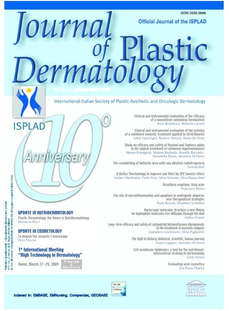
Vol. 5, n. 1, January-March 2009 1th International ... - Salute per tutti
Vol. 5, n. 1, January-March 2009 1th International ... - Salute per tutti
Vol. 5, n. 1, January-March 2009 1th International ... - Salute per tutti

You also want an ePaper? Increase the reach of your titles
YUMPU automatically turns print PDFs into web optimized ePapers that Google loves.
<strong>Vol</strong>. 5, n. 1, <strong>January</strong>-<strong>March</strong> <strong>2009</strong><br />
UPDATE IN NUTRIDERMATOLOGY<br />
Plastic Dermatology: the future is NutriDermatology<br />
Mariuccia Bucci<br />
UPDATE IN COSMETOLOGY<br />
To deepen the cosmetic’s knowledge<br />
Piera Fileccia<br />
1 th <strong>International</strong> Meeting<br />
“High Technology in Dermatology”<br />
Rome, <strong>March</strong> 27-29, <strong>2009</strong><br />
P r o g r a m<br />
&<br />
A b s t r a c t s<br />
Indexed in: EMBASE, EMNursing, Compendex, GEOBASE<br />
Clinical and instrumental evaluation of the efficacy<br />
of a nanosilicon containing formulation<br />
Enzo Berardesca, Manuela Carrera<br />
Clinical and instrumental evaluation of the activity<br />
of a combined cosmetic treatment applied to stretchmarks<br />
Adele Sparavigna, Beatrice Tenconi, Ileana De Ponti<br />
Study on efficacy and safety of Rucinol and Sophora alpha<br />
in the topical treatment of cutaneous hy<strong>per</strong>melanoses<br />
Marina Romagnoli, Martina Burlando, Rossella Boccardo,<br />
Alessandra Pavesi, Antonino Di Pietro<br />
The remodelling of buttocks area with non ablative radiofrequency<br />
Fiorella Bini<br />
A Reflex Thechnology to improve sun filter by SPF booster effect<br />
Stefano Manfredini, Paola Ziosi, Silvia Vertuani, Silvia Bustacchini<br />
Acneiform eruptions. Kelp acne<br />
Francesco Bruno<br />
The role of microinflammation and apoptosis in androgenic alopecia:<br />
new therapeutical strategies<br />
Paola Bezzola, Elisabetta Sorbellini<br />
Horny layer molecular structure: a new theory<br />
for hydrophilic molecules free diffusion through the skin<br />
Andrea Fratter<br />
Long-term efficacy and safety of calcipotriol/betamethasone dipropionate<br />
in the treatment of psoriasis vulgaris<br />
Giampiero Girolomoni, Silvia Pugliarello<br />
The light in history: historical, scientific, human journey<br />
Ivano Luppino, Antonino Di Pietro<br />
Cell membrane lipidomics: a tool for the nutritional/<br />
nutriceutical strategy in dermatology<br />
Carla Ferreri<br />
Evaluating acne cosmetica<br />
Zoe Diana Draelos
Cari soci e amici,<br />
inizia <strong>per</strong> l'ISPLAD un nuovo anno denso di impegni, iniziative e ambiziosi traguardi a cominciare dal 1 th<br />
<strong>International</strong> Meeting “Hight Technology in Dermatology” che si svolgerà a Roma dal 27 al 29 Marzo.<br />
È singolare che in un momento dove <strong>tutti</strong> si lamentano e vengono spaventati da questa crisi, l’ISPLAD<br />
proceda più forte che mai. Questo si spiega con la professionalità, l’impegno e la correttezza con cui ogni<br />
nostro socio svolge il proprio lavoro e con l'alto valore scientifico dell’ISPLAD, che in questi anni si è sempre<br />
impegnata a favorire la crescita professionale dei Dermatologi.<br />
La dimostrazione sta anche nel fatto che le aziende del settore cavalcano sempre con grande entusiasmo<br />
l e n o s t re iniziative: a<br />
l o ro è indirizzato il<br />
mio più sincero ringraziamento.<br />
C’è grande fermento intorno<br />
a noi <strong>per</strong> il prossimo importante evento<br />
ISPLAD-ADOI dal titolo “Cute e annessi<br />
cutanei” che si terrà a Castellaneta<br />
Marina (TA ) dal 28 al 30 maggio <strong>2009</strong><br />
p resso la struttura Nova Ya rd i n i a .<br />
Sono state già registrate molte iscrizioni e<br />
ogni giorno se ne aggiungono di nuove.<br />
In quell’occasione s<strong>per</strong>o di festeggiare ,<br />
n u m e rosi, i primi 10 anni dell’ISPLAD.<br />
Insomma, altro che crisi! Infatti ci rivolgiamo<br />
a testa alta verso nuovi obiettivi,<br />
supportati dal nostro organo ufficiale, il<br />
J o u rnal of Plastic Derm a t o l o g y, di cui<br />
segnalo la speciale versione grafica e la<br />
recente indicizzazione in Scopus.<br />
La serietà paga sempre!<br />
Da ultimo, <strong>per</strong>mettetemi un ricord o<br />
dello stimato collega Innocenzi: una<br />
grave <strong>per</strong>dita come uomo e come Dermatologo.<br />
Tutta l'ISPLAD è vicina alla<br />
sua famiglia e ai suoi collaboratori che<br />
con lui condividevano l’attività.<br />
Di seguito il ricordo dell'amico Francesco<br />
Bruno.<br />
Un ricordo di Daniele Innocenzi<br />
Dear Members and Friends,<br />
A new year full of undertakings, initiatives, and ambitious goals is starting for<br />
ISPLAD. We commence with the 1 th <strong>International</strong> Meeting [High Technology in<br />
Dermatology”, which will take place in Rome from the 27 th -29 th of <strong>March</strong>.<br />
It is striking that at a time in which everyone is complaining about and is frightened by<br />
the economic crisis, ISPLAD is moving forward with more strength than ever before .<br />
This is a direct result of the maximum professionalism, effort, and correctness that<br />
every one of our members has put into his work. It is also a consequence of the high<br />
scientific value of ISPLAD, which has tenaciously encouraged the professional gro w t h<br />
of dermatologists throughout the last years. Further testament to this is the great enthusiasm<br />
that companies in the dermatology sector continuously show for our initiatives.<br />
I would like to take this opportunity to <strong>per</strong>sonally thank them for their support.<br />
There is mounting anticipation and enthusiasm for the next important ISPLAD-<br />
ADOI event, a meeting entitled “Skin and Skin Adnexa” that will take place at the<br />
Nova Yardinia in Castellaneta Marina (TA) from the 28 th -30 th of May, <strong>2009</strong>. Many<br />
people have already signed up for this event and the number of registered participants<br />
increases with each passing day. I hope to see as many of you there as possible<br />
so we can celebrate the first ten years of ISPLAD together.<br />
Despite the crisis, we march towards new objectives. Our official publication, the<br />
Journal of Plastic Dermatology, even has a new special graphic version and Scopus<br />
Index. It always pays to work hard!<br />
Let us take a moment to remember our highly respected colleague, Professor<br />
Innocenzi. As an esteemed man and dermatologist, his passing is a great loss to us<br />
all. ISPLAD shares this pain with his family and the colleagues who have stood by<br />
him in all his endeavours. The following lines are written by Francesco Bruno in<br />
memory of our dear friend.<br />
Antonino Di Pietro<br />
Il giorno 18 febbraio <strong>2009</strong> è mancato un amico di noi <strong>tutti</strong>, non soltanto di noi dermatologi, ma di <strong>tutti</strong> coloro che l’hanno conosciuto. Non voglio<br />
pensare neppure <strong>per</strong> un attimo al crudele destino che quel giorno, in quell’ora, in quel punto preciso, l’attendeva. Nessuno di noi, credenti e non,<br />
lo può accettare. Voglio ricordare soltanto quel ragazzone, semplice, simpatico, coltissimo che mi ha abbracciato qualche mese fa all’“Acne day”,<br />
facendomi i complimenti <strong>per</strong> la mia misera relazione, consegnandomi il libro sulla “Dermatite seborroica”, al quale avevamo collaborato insieme,<br />
ringraziandomi <strong>per</strong> essere stato presente alla riunione. Facendomi sentire, <strong>per</strong> un attimo, importante. Sì! Perché Daniele, che aveva una produzione<br />
scientifica di livello straordinario, faceva sentire importante chiunque lavorasse con lui: i colleghi, i giovani, le hostess, i tecnici. Poi... ti ringraziava<br />
sempre, ma spesso eri tu che dovevi essergli grato. Quando mi ha dato l’onore di far parte dell’“Italian Acne Board”, me lo chiese come se<br />
il privilegio fosse il suo. Il vuoto che lascerà sarà incolmabile, sia <strong>per</strong> le sue doti scientifiche, sia <strong>per</strong> le sue grandi doti umane. Quel tragico giorno,<br />
apprendo la notizia da Elisabetta Perosino, dopo un minuto mi chiama Giuseppe Micali, poi Antonino Di Pietro. Ci siamo sentiti in un attimo<br />
membri di una stessa famiglia. E come una vera famiglia, siamo stretti e vicini alla moglie e alle tre figlie che, anche in questo momento così doloroso,<br />
possono solo essere fiere di avere avuto un papà come lui!<br />
Ciao Daniele! Francesco Bruno<br />
Journal of Plastic Dermatology <strong>2009</strong>; 5, 1 1
Daniele Innocenzi: un mio grande amico<br />
Rispondo con profonda commozione all’invito di scrivere alcune righe <strong>per</strong> condividere con Voi il ricord o<br />
del mio grande amico Daniele Innocenzi che ci ha lasciato tragicamente la mattina del 18 febbraio <strong>2009</strong>.<br />
Mi risulta molto difficile <strong>per</strong> la drammaticità dell’evento, che stordisce me, i suoi amici ed i colleghi che lo hanno<br />
stimato, che inevitabilmente rischia di deviare il contenuto di questi pensieri verso una esternazione del mio dolore<br />
piuttosto che ricordare il valore umano e professionale della <strong>per</strong>sona.<br />
Conosco Daniele da circa 30 anni, abbiamo condiviso lunghi anni della nostra crescita umana, <strong>per</strong>sonale e pro f e s s i o n a l e .<br />
Subito si è instaurata una forte amicizia.<br />
La sua amicizia è stata un bene prezioso <strong>per</strong> me e <strong>per</strong> <strong>tutti</strong> coloro che hanno avuto il privilegio di condividerla,<br />
e sarà un ricordo indelebile, non <strong>per</strong> la sua simpatia brillante, <strong>per</strong> la sua convivialità, <strong>per</strong> la sua risata contagiosa,<br />
bensì <strong>per</strong> il valore profondo che dava a questo termine, <strong>per</strong> l’attenzione sincera e prodiga che ha sempre riservato<br />
e concesso ai suoi amici.<br />
Ha sinceramente creduto nell’alto valore morale, civile e sociale del medico. Professionalmente, si è formato presso<br />
la Clinica Dermatologica dell’Università di Roma, ed attualmente ricopriva la carica di Direttore della Clinica<br />
Dermatologica, Polo Pontino, dell’Università di Roma La Sapienza.<br />
Daniele Innocenzi aveva in animo, profondamente e chiaramente, le finalità professionali del dermatologo e del<br />
dermatologo che ha scelto di dedicarsi all’insegnamento, con il costante intento di elevare il valore scientifico della<br />
Dermatologia. Lo hanno testimoniato i riconoscimenti <strong>per</strong>sonali ottenuti, la sua capacità organizzativa, il seguito di<br />
studenti che sapeva appassionare <strong>per</strong> il suo amore e dedizione <strong>per</strong> la materia, la sincera stima dei suoi colleghi.<br />
C redeva nei suoi Maestri e conosceva il significato e l’impegno che tale ruolo comporta. Aveva iniziato, con d e d i z i o n e<br />
e grande promessa, a costru i re una sua scuola e divenire anche lui un Maestro, ma il destino, la tragica fatalità, hanno<br />
voluto che questo non si potesse arr i c c h i re.<br />
Ne sentirò, ne sentiremo <strong>tutti</strong> la mancanza.<br />
Sono certo che il tempo non sopirà il suo ricordo, ma eleverà il riconoscimento del suo valore.<br />
Luca Bianchi<br />
Professore Associato di Dermatologia<br />
Università di Roma Tor Vergata<br />
Journal of Plastic Dermatology <strong>2009</strong>; 5, 1 3
Journal of Plastic Dermatology<br />
Editor<br />
Antonino Di Pietro (Italy)<br />
Editor in Chief<br />
Francesco Bruno (Italy)<br />
Co-Editors<br />
Bernd Rüdiger Balda (Austria)<br />
Salvador Gonzalez (USA)<br />
Pedro Jaen (Spain)<br />
Associate Editors<br />
Francesco Antonaccio (Italy)<br />
Mariuccia Bucci (Italy)<br />
Franco Buttafarro (Italy)<br />
Ornella De Pità (Italy)<br />
Giulio Ferranti (Italy)<br />
Andrea Giacomelli (Italy)<br />
Alda Malasoma (Italy)<br />
Steven Nisticò (Italy)<br />
Elisabetta Perosino (Italy)<br />
Andrea Romani (Italy)<br />
Nerys Roberts (UK)<br />
Editorial Board<br />
Lucio Andreassi (Italy)<br />
Kenneth Arndt (USA)<br />
H.S. Black (USA)<br />
Lucia Brambilla (Italy)<br />
Günter Burg (Switzerland)<br />
Michele Carruba (Italy)<br />
Vincenzo De Sanctis (Italy)<br />
Aldo Di Carlo (Italy)<br />
Robin Eady AJ (UK)<br />
Paolo Fabbri (Italy)<br />
Ferdinando Ippolito (Italy)<br />
Giuseppe Micali (Italy)<br />
Martin Charles Jr Mihm (USA)<br />
Joe Pace (Malta)<br />
Lucio Pastore (Italy)<br />
Gerd Plewig (Germany)<br />
Riccarda Serri (Italy)<br />
Adele Sparavigna (Italy)<br />
Abel Torres (USA)<br />
Stefano Veraldi (Italy)<br />
Umberto Veronesi (Italy)<br />
Managing Editor<br />
Antonio Di Maio<br />
English editing<br />
Rewadee Anujapad<br />
Direttore Responsabile Pietro Cazzola<br />
Direttore Generale Armando Mazzù<br />
Direttore Marketing Antonio Di Maio<br />
Consulenza grafica Piero Merlini<br />
Impaginazione Stefania Cacciaglia<br />
R e g i s t r. Tribunale di Milano n. 102 del 14/02/2005<br />
Scripta Manent s.n.c. Via Bassini, 41 - 20133 Milano<br />
Tel. 0270608091/0270608060 - Fax 0270606917<br />
E-mail: scriman@tin.it<br />
Abbonamento annuale (3 numeri) Euro 39,00<br />
Pagamento: conto corrente postale n. 20350682<br />
intestato a: Edizioni Scripta Manent s.n.c.,<br />
via Bassini 41- 20133 Milano<br />
Stampa: Arti Grafiche Bazzi, Milano<br />
Sommario<br />
pag. 7 Clinical and instrumental evaluation of the efficacy<br />
of a nanosilicon containing formulation<br />
Enzo Berardesca, Manuela Carrera<br />
pag. 13 Clinical and instrumental evaluation of the activity of a combined<br />
cosmetic treatment applied to stretchmarks<br />
Adele Sparavigna, Beatrice Tenconi, Ileana De Ponti<br />
pag. 21 Efficacia e tollerabilità di Rucinol e Sophora alpha nel trattamento topico delle<br />
i<strong>per</strong>melanosi in monoterapia e combinato a peeling chimico, laserterapia e IPL<br />
Marina Romagnoli, Martina Burlando,<br />
Rossella Boccardo, Alessandra Pavesi, Antonino Di Pietro<br />
pag. 31 Il rimodellamento dei glutei con radiofrequenza non ablativa<br />
Fiorella Bini<br />
pag. 37 A Reflex Thechnology to improve sun filter by SPF booster effect<br />
Stefano Manfredini, Paola Ziosi, Silvia Vertuani, Silvia Bustacchini<br />
pag. 43 Le eruzioni acneiformi. Kelp acne (acne da alghe)<br />
Francesco Bruno<br />
pag. 47 Il ruolo della microinfiammazione e dell’apoptosi nella alopecia androgenetica:<br />
nuove strategie terapeutiche<br />
Paola Bezzola, Elisabetta Sorbellini<br />
pag. 59 Struttura fisico-molecolare dello strato corneo: nuova teoria<br />
<strong>per</strong> descrivere il flusso transcutaneo di molecole idrofile<br />
Andrea Fratter<br />
pag. 69 Efficacia e tollerabilità a lungo termine dell’associazione calcipotriolo/betametasone<br />
dipropionato nel trattamento della psoriasi volgare<br />
Giampiero Girolomoni, Silvia Pugliarello<br />
pag. 77 La luce nella storia<br />
Ivano Luppino, Antonino Di Pietro<br />
UPDATE IN NUTRIDERMATOLOGY<br />
pag. 83 Dermatologia Plastica: il futuro è la NutriDermatologia<br />
Mariuccia Bucci<br />
pag. 85 Cell membrane lipidomics: a tool for the nutritional/nutriceutical<br />
strategy in dermatology<br />
Carla Ferreri<br />
UPDATE IN COSMETOLOGY<br />
pag. 93 To deepen the cosmetic’s knowledge<br />
Piera Fileccia<br />
pag. 95 Evaluating acne cosmetica<br />
Zoe Diana Draelos<br />
pag. 99 1 th <strong>International</strong> Meeting “High Technology in Dermatology”<br />
Rome, <strong>March</strong> 27-29, <strong>2009</strong><br />
P r o g r a m<br />
&<br />
A b s t r a c t s<br />
È vietata la riproduzione totale o parziale,<br />
con qualsiasi mezzo, di articoli, illustrazioni<br />
e fotografie senza l’autorizzazione scritta dell’Editore.<br />
L’Editore non risponde dell’opinione espressa dagli<br />
Autori degli articoli.<br />
Ai sensi della legge 675/96 è possibile in qualsiasi<br />
momento opporsi all’invio della rivista<br />
comunicando <strong>per</strong> iscritto la propria decisione a:<br />
Edizioni Scripta Manent s.n.c.<br />
Via Bassini, 41 - 20133 Milano<br />
Journal of Plastic Dermatology <strong>2009</strong>; 5, 1 5
Enzo Berardesca<br />
Manuela Carrera<br />
Dermatological Institute<br />
San Gallicano, Rome.<br />
Clinical and instrumental evaluation<br />
of the efficacy of a nanosilicon<br />
containing formulation<br />
SU M M A R Y<br />
I ntroduction<br />
Silicon is a component of pro t e o g l y c a n<br />
complexes that interlace with collagen and contribute<br />
to structural integrity. Specifically, some<br />
suggest that silicon-oxygen bridges (-O-Si-O-)<br />
make up structural elements in the muco-polysaccharides<br />
found in connective tissues.<br />
F u r t h e r m o re, silicon promotes the synthesis of<br />
p roline and hydro x y p roline; principal amino<br />
Clinical and instrumental evaluation<br />
of the efficacy of a nanosilicon<br />
containing formulation<br />
Silicon is a component of elastin and collagen and increasing evidence suggests that<br />
silicon is a nutrient of concern for both bone and skin. It has been proven that silicon<br />
deprivation affects collagen formation and collagen deposition in many tissues.<br />
The main source of silicon is the diet, but the bioavailability of this nutrient fro m<br />
solid foods is not assessed. The transformation of silicon contained in zeolite by<br />
means of nanotechnology has led to the production of nanosilicon. The particles of<br />
nanosilicon raise dimensions of 10-20 nm with an increased active surface and a<br />
higher bioavailability which facilitate the cellular absorption. Nanosilicon is very<br />
important in skin hydration because of its capacity to bind a large amount of water<br />
and to normalize the contents of glycosaminoglycan.<br />
This study has been undertaken in order to evaluate the efficacy of a nanosilicon containing<br />
formulation to improve skin hydration and elasticity. Thirty healthy female<br />
subjects entered the study. Fifteen patients were randomized to receive the placebo<br />
and fifteen patients received the active compound; all the patients were instructed to<br />
take the food supplement twice a day for the first twenty days and then once a day<br />
for the next twenty days. All the patients enrolled were instructed to apply the<br />
nanosilicon containing solution on the right forearm four times a day. The patients<br />
were evaluated with non invasive methods at baseline, after 20 days and at the end<br />
of the study, after 40 days. Statistical analysis was <strong>per</strong>formed using t-test and Anova<br />
and values of p < 0.05 were considered statistically significant. After twenty days in<br />
both groups there was a statistically significant improvement of skin hydration on<br />
the right forearm as well as a significant improvement of the barrier function as<br />
shown by the decrease of the TEWL. The elasticity was increased only in the group<br />
who take the active compound and the results were statistically significant. The evaluation<br />
<strong>per</strong>formed at the end of the study confirmed this trend.<br />
The consumption of the food supplement and the topical use of the solution both<br />
containing nanosilicon has proven to be effective for the improvement of skin<br />
hydration, barrier function and elasticity.<br />
KE Y W O R D S: Skin hydration and elasticity, Nanosilicon<br />
acids in collagen and central elements in defining<br />
its primary and secondary structure. Incre a s i n g<br />
evidence suggests that silicon is a nutrient of<br />
c o n c e rn for both bone and skin. It has been<br />
p roven that silicon deprivation affects collagen<br />
formation and collagen deposition in many tissues<br />
and that levels of this mineral in the skin<br />
d e c rease with aging 1 .<br />
Journal of Plastic Dermatology <strong>2009</strong>; 5, 1 7
8<br />
E. Berardesca, M. Carrera<br />
Diet is the main source of silicon but the<br />
bioavailability of this nutrient from solid foods<br />
is poor 2 . The transformation of silicon contained<br />
in zeolite by means of nanotechnology<br />
has led to the production of nanosilicon. “Nano”<br />
concerns a world of molecules with specific<br />
pro<strong>per</strong>ties and behaviour very different from<br />
the “Macro” ones. The particles of nanosilicon<br />
raise dimensions of 10-20 nm with an increased<br />
active surface and a higher bioavailibility which<br />
facilitates the cellular absorption.<br />
im of the study<br />
A<br />
This study has been undertaken in<br />
order to evaluate the efficacy of a topical and/or<br />
oral formulation containing nanosilicon for the<br />
improvement of skin hydration and elasticity.<br />
aterials and methods<br />
M<br />
Thirty healthy female subjects entered<br />
the study. Inclusion and exclusion criteria were<br />
as follows:<br />
Inclusion criteria:<br />
• Female;<br />
• Subjects willing to refrain from using other<br />
topical products on test area during the study;<br />
• Subjects willing to not ingest drugs and/or<br />
alimentary integrator that could interfere<br />
with the results of the study;<br />
• Subjects who have signed the written<br />
informed consent;<br />
• Subjects willing to avoid sun exposure on<br />
test area during the study.<br />
Exclusion criteria:<br />
• All the conditions that are not included in<br />
the inclusion criteria;<br />
• Pregnant or nursing subjects;<br />
• Immunocompromising diseases;<br />
• Allergies;<br />
• Subjects with significant history or current<br />
evidence of any psychological disorder that<br />
in the investigator's opinion would preclude<br />
enrolment into the study.<br />
Fifteen patients were randomized to receive the<br />
placebo and fifteen patients received the active<br />
compound; all the patients were instructed to<br />
take the food supplement twice a day for the<br />
Journal of Plastic Dermatology <strong>2009</strong>; 5, 1<br />
first twenty days and then once a day for the<br />
next twenty days. All the patients enrolled were<br />
instructed to apply the nanosilicon containing<br />
solution on the right forearm four times a day.<br />
The patients were evaluated with non invasive<br />
methods at baseline, after 20 days and at the<br />
end of the study, after 40 days. Several parameters<br />
have been monitored as follows:<br />
H y d r a t i o n . Skin surface hydration was<br />
determined with the Corneometer ! CM 825<br />
(Courage & Khazaka, Germany). The measuring<br />
principle of this instrument is based on<br />
capacitance measurement of a dieletric<br />
medium. Any change in the dielectric constant<br />
due to skin surface hydration variation<br />
alters the capacitance of a precision measuring<br />
capacitor.<br />
B a rrier function. The measurement of the<br />
t r a n s e p i d e rmal water loss (TEWL) is the most<br />
important parameter for evaluating the eff iciency<br />
of the skin water barrier. The state of<br />
the barrier function is assessed by the<br />
Tewameter TM 200 (Courage & Khazaka,<br />
G e rm a n y). When the barrier function is re d uced,<br />
the values of TEWL are high. The meas<br />
u rement of the water evaporation is based<br />
on the diffusion principle in an open chamb<br />
e r. The density gradient is measured indirectly<br />
by the two pairs of sensors (tem<strong>per</strong>ature<br />
and relative humidity) inside the hollow<br />
cylinder and is analysed by a micro p ro c e s s o r.<br />
Elasticity. The elasticity of the skin is evaluated<br />
by the Cutometer ! MPA 580 (Courage<br />
& Khazaka, Germany). The measuring principle<br />
is based on the suction method.<br />
Negative pressure is created in the device<br />
and the skin is drawn into the a<strong>per</strong>ture of<br />
the probe. The resistance of the skin to be<br />
sucked up by the negative pressure (firmness)<br />
and its ability to return in its original<br />
position (elasticity) are displayed as curves<br />
at the end of each measurement.<br />
Skin smoothness, skin roughness, scaliness,<br />
w r i n k l e s . These four clinical parameters<br />
describe quantitatively and qualitatively the<br />
skin surface; they are measured by a special<br />
U V-light video camera (Vi s i o s c a n ! V C ,<br />
Courage & Khazaka, Germ a n y). The images of<br />
the skin surface are elaborated by a particular<br />
s o f t w a re that <strong>per</strong>mits the calculation of the<br />
parameters. Thus, before and after tre a t m e n t<br />
comparisons of the same skin site enable a<br />
comparison of trends in skin condition.<br />
Until now, profilometry has been the only
method to determine the condition of skin<br />
surface, other than subjective evaluation. By<br />
means of Visioscan ! VC skin can be monitored<br />
optically, using a new method called<br />
Surface Evaluation of the Living Skin (SELS).<br />
The parameters evaluated have been developed<br />
in various clinical studies in order to<br />
be <strong>per</strong>fectly suitable to characterize the skin<br />
surface. Skin smoothness (Sesm) is the calculation<br />
from the average width and depth of<br />
the wrinkles. Skin roughness (SEr) is the<br />
opposite parameter. The s c a l i n e s s ( S E s c )<br />
measures the level of dryness of the stratum<br />
corneum. The wrinkles (SEw) derives from<br />
the proportion of horizontal and vertical<br />
wrinkles.<br />
Measurements have been <strong>per</strong>formed at 20 ± °C<br />
of environmental tem<strong>per</strong>ature with a relative<br />
humidity of 45 ± 5% according to guidelines<br />
published for these techniques.<br />
Statistical analysis was <strong>per</strong>formed using t-test<br />
and Anova and values of p < 0.05 were considered<br />
statistically significant.<br />
A clinical evaluation has been <strong>per</strong>formed using<br />
a numerical scale (0-10) to assess the patients’<br />
<strong>per</strong>ception of skin condition concerning two<br />
parameters: hydration and elasticity.<br />
R esults<br />
Concerning hydration, after twenty<br />
days in both groups there was a statistically significant<br />
improvement of skin hydration on the<br />
treated forearm (p < 0.05) and the evaluation<br />
<strong>per</strong>formed at the end of the study confirmed<br />
this trend.<br />
Clinical and instrumental evaluation of the efficacy of a nanosilicon containing formulation<br />
Concerning the barrier function, after 20 days<br />
and at the end of the study, there was a statistically<br />
significant reduction of TEWL (p < 0.05),<br />
which indicates an improvement of barrier<br />
function, on both treated and untreated forearms<br />
of patients taking the active compound as<br />
well as on the treated forearm of patients taking<br />
placebo.<br />
After 20 days there was no improvement of<br />
skin elasticity, whereas it was increased after 40<br />
days, at the end of the study but only on the<br />
t reated forearm of the patients taking the active<br />
compound as shown by the results of the<br />
C u t o m e t e r ! M PA 580 (enhancement of R2 and<br />
R5). The enhancement of skin elasticity was<br />
statistically significant (p < 0.05). There was<br />
n o i m p rovement of skin elasticity neither in<br />
t h e g roup taking the placebo nor on the<br />
u n t reated forearm of patients taking the active<br />
c o m p o u n d .<br />
In the group taking the active product, the evaluation<br />
<strong>per</strong>formed by means of Visioscan ! VC<br />
has shown a statistically significant (p < 0.05)<br />
improvement of skin roughness (variance) and<br />
texture contrast on both forearms (treated and<br />
untreated) and a statistically significant reduction<br />
of scaliness (SEsc) but only on the treated<br />
forearm. Texture parameter “contrast” indicates<br />
a general overview over the state of skin, a good<br />
skin condition shows low contrast values.<br />
Te x t u re parameter “v a r i a n c e” indicates skin<br />
roughness and the parameter Sesc evaluates the<br />
hydration level of stratum corneum, the smaller<br />
Sesc, the more is the hydration level.<br />
All the patients reported improvement of skin<br />
hydration on the treated forearm, where a s<br />
patients taking the active product also reported<br />
improvement in skin elasticity.<br />
Figures 1 and 2.<br />
Improvement of skin hydration on the treated forearm in subjects taking food supplement containing nanosilicon.<br />
Journal of Plastic Dermatology <strong>2009</strong>; 5, 1<br />
9
10<br />
E. Berardesca, M. Carrera<br />
Figure 3.<br />
Improvement of skin hydration on the treated forearm<br />
in subjects taking placebo.<br />
Figure 5.<br />
Improvement of barrier function on the untreated forearm<br />
in subjects taking food supplement containing nanosilicon.<br />
Figure 9.<br />
Improvement of skin condition on the treated forearm<br />
in subjects taking food supplement containing nanosilicon.<br />
Journal of Plastic Dermatology <strong>2009</strong>; 5, 1<br />
Figure 4.<br />
Improvement of barrier function on the treated forearm<br />
in subjects taking food supplement containing nanosilicon.<br />
Figure 6.<br />
Improvement of barrier function on the treated forearm<br />
in subjects taking placebo.<br />
Figures 7 and 8.<br />
Improvement of skin elasticity on the treated forearm in subjects taking food supplement containing nanosilicon.<br />
Figure 10.<br />
Improvement of skin condition on the untreated forearm<br />
in subjects taking food supplement containing nanosilicon.
Figure 11..<br />
Improvement of skin roughness on the treated forearm<br />
in subjects taking food supplement containing nanosilicon.<br />
C onclusion<br />
Topical nanosilicon used alone significantly<br />
enhances skin hydration and improves<br />
barrier function.<br />
Clinical and instrumental evaluation of the efficacy of a nanosilicon containing formulation<br />
Figure 12.<br />
Improvement of skin roughness on the untreated forearm<br />
in subjects taking food supplement containing nanosilicon.<br />
Figures 13 and 14.<br />
Self-assessment of skin hydration in patients taking food supplement containing nanosilicon (Fig. 13) and in patients taking placebo (Fig. 14).<br />
Figure 15.<br />
Self-assessment of skin elasticity in patients taking food<br />
supplement containing nanosilicon.<br />
Barrier function is also improved by the food<br />
supplement containing nanosilicon used alone.<br />
Topical nanosilicon and food supplement containing<br />
nanosilicon act synergically for the<br />
improvement of skin elasticity.<br />
The intake of food supplement containing<br />
nanosilicon is effective for the improvement of<br />
general skin condition and the improvement of<br />
skin roughness.<br />
R eferences<br />
1. Carlisle EM. Silicon. In: O'Dell, B.L. &<br />
Sunde, R.A., Ed. Handbook of nutrionally essential elements.<br />
New York: Marcel Dekker, 1997; 603-618.<br />
2. Seaborn CD and Nielsen FH. Silicon deprivation<br />
decreases collagen formation in wounds and bone, and<br />
ornithine transaminase enzyme activity in liver. Biol Trace<br />
Element Res 2002; 89:251-261.<br />
Journal of Plastic Dermatology <strong>2009</strong>; 5, 1<br />
11
Adele Sparavigna<br />
Beatrice Tenconi<br />
Ileana De Ponti<br />
DermIng, Clinical Research<br />
and Bioengineering Institute, Monza.<br />
Clinical and instrumental evaluation<br />
of the activity of a combined cosmetic<br />
treatment applied to stretchmarks<br />
SU M M A R Y<br />
I ntroduction<br />
Topical treatment of stre t c h m a r k s<br />
(striae cutis atrophicae et distensae) is hardly successful<br />
especially for old lesions, which can be<br />
considered as scars following distension and<br />
rupture of dermal fibres. The ideal treatment for<br />
s t retchmarks should provide a stimulus to<br />
fibroblasts in the dermis as well as an improved<br />
epidermal turnover. A new treatment based on<br />
the alternate use of two creams was applied to<br />
s t retchmarks: the first formulation (D i s p a r i<br />
cream) was conceived to exert an exfoliating<br />
effect while the second one (Pari cream) to regenerate<br />
the skin with its growth factors and<br />
protecting agents.<br />
omposition of “Dispari” cream<br />
C<br />
The formulation of Dispari cre a m c o ntains<br />
actives which carry out an exfoliant and<br />
lightening activity, accelerating skin cell turn o v e r<br />
and improving micro c i rc u l a t i o n .<br />
Fruit AHA (alpha hydroxy acids) (30%): a mixt<br />
u re of acids obtained from fruit, which have a<br />
purifying action on the skin and make it bright. It<br />
is a mixture based on a standardized extraction<br />
Clinical and instrumental evaluation<br />
of the activity of a combined cosmetic<br />
treatment applied to s t r e t c h m a r k s<br />
Aim of this study is to evaluate by clinical and non invasive instrumental evaluations<br />
the activity of a cosmetic treatment combining 2 diff e rent products (Pari and Dispari<br />
c ream) on healthy female volunteers presenting striae cutis (stretchmarks) of re c e n t<br />
and/or old appearance. The two products were applied on alternative days, once a day,<br />
for a <strong>per</strong>iod of 16 weeks. At the end of the treatment <strong>per</strong>iod, very significant results were<br />
observed in terms of clinical evaluation and instrumental data, re g a rding the deepness<br />
of the lesions, skin elasticity and smoothness of skin surface. The activity of the tre a tment<br />
was also well <strong>per</strong>ceived by the subjects.<br />
KE Y W O R D S: Stretchmarks, Topical treatment, Skin elasticity, Skin smoothness<br />
f rom five diff e rent plants: blueberry, sugarc a n e ,<br />
maple sugar, orange tree and lemon tre e .<br />
Five alpha hydroxy acids are extracted from<br />
these plants: glycolic acid, lactic acid, citric<br />
acid, malic acid and tartaric acid.<br />
Glycolic acid, indeed the most known AHA,<br />
is a scrub that makes the skin smooth and<br />
bright and stimulates the synthesis of collagen<br />
and glycosaminoglycans (GAG).<br />
Lactic acid is a very good hydrating agent.<br />
Citric acid is a very good “booster” of collagen.<br />
Malic acid and tartaric acid increase the skin<br />
elasticity.<br />
H y a l u ronic acid: essential structural constituent<br />
of deep skin layers. Hyaluronic acid enters the<br />
composition of the most esteemed cosmetic<br />
p reparations, because of its hydrating effect due<br />
to the molecule’s ability to keep water and theref<br />
o re increase skin turg o r. At the same time,<br />
t h rough mechanisms of interaction with dermal<br />
f i b roblasts, it stimulates the production of new<br />
collagen and eliminates free radicals.<br />
Kojic acid: substance with a depigmentating<br />
action. It is widely used in cosmetic field to<br />
fight aging.<br />
Journal of Plastic Dermatology <strong>2009</strong>; 5, 1 13
14<br />
A. Sparavigna, B. Tenconi, I. De Ponti<br />
Retinol: synonym of vitamin A. Liposoluble<br />
vitamin which protects and stimulates the<br />
growth of skin.<br />
H y d rolyzed soya pro t e i n s: rich in amino acids,<br />
they have an anti-inflammatory action.<br />
Centella extract (Centella asiatica): it helps the<br />
microcirculation and improves the <strong>per</strong>meability<br />
of vessels. It has an anti-cellulite action.<br />
Biovin: a natural antioxidant element extracted<br />
from vine (Vitis vinifera) composed of oligomeric<br />
proanthocyanidins and resveratrol.<br />
Through an activity that can be defined as “phytokinetic<br />
synergy”, it protects cell membrane<br />
phospholipids from oxidation and blocks the<br />
chain reaction of free radicals. It is essential, in<br />
synergy with vitamin C, for the synthesis of collagen<br />
and for the protective effect on microcirculation<br />
vessels. Its capability of metallic ions<br />
chelating makes it an effective “radical scavenger”<br />
having an antipollution action.<br />
Phytic acid complex: it is a mixture made of<br />
phytic acid, aloe vera and ascorbic acid bound to<br />
silicone in a biologically active way. It has an<br />
antioxidant and anti-inflammatory action. It<br />
stimulates the cell turnover and has an exfoliating<br />
action.<br />
Sweet almond oil (P runus amygdalus dulcis oil) :<br />
emollient, soothing, nutritious and lenitive. It is<br />
rich in proteins, glucides, mineral salts, vitamin A<br />
and vitamins of group B. It is suitable for all types<br />
of skin, prevents skin aging and it is used for dry<br />
and reddened skin such as childre n ’s sensitive<br />
skin. It is very effective against stre t c h m a r k s ,<br />
especially during the pregnancy <strong>per</strong>iod or while<br />
being on a slimming diet. It is advised to apply it<br />
on “critical are a s ” such as bosom, hips and thighs,<br />
anyway on clear skin, with a soft massage in ord e r<br />
to facilitate the oil absorption, which penetrates<br />
in deep layers and releases active principles.<br />
Sericin: silk is a natural product and researches<br />
in this field have demonstrated that this protein<br />
is able to inhibit the effects of some<br />
enzymes responsible for skin aging and can<br />
reduce wrinkles and dark spots. The sericin put<br />
on the skin forms a film that makes it extremely<br />
soft and silken. It has a remarkable long-lasting<br />
hydrating effect that benefits the integrity of<br />
the skin hydrolipidic barrier.<br />
Journal of Plastic Dermatology <strong>2009</strong>; 5, 1<br />
Sericin is used in its integral form (400 kDa); its<br />
action is not due to the non-structured pool of<br />
amino acids, but to the molecule’s spatial structure<br />
and its bonds of coordination and bioadhesion<br />
to the skin surface.<br />
Safety, sensory agreeableness and efficacy have<br />
been scientifically demonstrated.<br />
omposition of “Pari” cream<br />
C<br />
The formulation of Pari cream contains<br />
actives which carry out a re-epithelising<br />
anti-oxidant and anti-age activity.<br />
Argan oil (Argania spinosa): it has been used for<br />
centuries in cosmetic field because it contains<br />
strong doses of unsaturated fatty acid and of vitamin<br />
E. It has a hydrating and nourishing<br />
action. It regenerates tissues and make the skin<br />
elastic and bright.<br />
Rose hip oil (extracted from Rosa mosqueta<br />
seeds): it has a hydrating and cicatrizing effect.<br />
It has the capability to reduce scars, stretch<br />
marks and acne marks. Rosa mosqueta is a wild<br />
rose coming from Asia, than spread all over<br />
Europe and finally introduced in South America<br />
by Spaniards during their conquering <strong>per</strong>iod.<br />
Today it is thriving in Chile.<br />
The oil got from the cold pressing of its seeds<br />
has a very high concentration of polyunsaturated<br />
fatty acids (linoleic 41%, linolenic 39%)<br />
which are essential for the synthesis of<br />
prostaglandins, substances responsible for cell<br />
regeneration processes and the renewal of skin<br />
tissues. Besides essential fatty acids, there is also<br />
trans-retinoic acid, an isomer of vitamin A,<br />
whose rejuvenation action on the skin has been<br />
demonstrated: its application makes the skin<br />
fresher, smoother and more elastic.<br />
Rose hip oil is there f o re effective on scars caused<br />
by stretchmarks, sunburns and skin spots due to<br />
aging. It minimizes facial expression wrinkles and<br />
p revents pre m a t u re aging of skin tissues.<br />
Phytodermine C: of vegetable origin, it has a<br />
high hydrating power and increases the skin<br />
elasticity.<br />
Spirulina extract: it is an extract of a small blue<br />
alga very rich in proteins and essential amino<br />
acids. It contains also vitamin A and E. It has a<br />
s t rong antioxidant action.
Clinical and instrumental evaluation of the activity of a combined cosmetic treatment applied to stretchmarks<br />
Ascorbic acid: vitamin C essential for the<br />
organism: it stimulates the synthesis of interferon,<br />
the carnitine biosynthesis and makes iron<br />
absorption easier.<br />
It can play an important role in stretchmark and<br />
wound healing.<br />
Colostrum H1: bovine colostrum coming<br />
from breedings rigorously selected and certified,<br />
which is collected within the first hour<br />
from delivery (H1) in order to get the maximum<br />
concentration of growth factors and cell<br />
regeneration, as Fibroblast Growth Factor (FGF)<br />
and Epithelial Growth Factor (EGF), lysozyme,<br />
biotin (vitamin H) and cytokines.<br />
Naturally rich in immunoglobulin (antibody Ig<br />
G 1 ) and telomerase (cellular repair enzyme),<br />
colostrum is not subject to chemical-physical<br />
processes that can damage or inactivate its<br />
essential and useful principles.<br />
Colostrum H1 has a much higher concentration<br />
of active factors than commercial colostrum,<br />
which is generally collected within 24 hours after<br />
d e l i v e ry. This produces an immediate cosmetic<br />
e ffect, which is visible right from the first hours<br />
and long-lasting. It also has a regenerating, re v italizing<br />
and anti-age action on the skin.<br />
Pari cream also contains Biovin and Sericin, as<br />
well as Dispari cream.<br />
escription of the study<br />
D<br />
Aim of this study was to evaluate clinically<br />
and by non invasive instrumental evaluations<br />
the activity of the combined cosmetic tre a tment,<br />
Pari cre a m and Dispari cre a m, on stre t c hmarks.<br />
Clinical and instrumental evaluations<br />
w e re <strong>per</strong>formed in basal condition and after 8<br />
and 16 weeks of treatment. It was also aim of the<br />
study to evaluate cosmetic acceptability by the<br />
volunteers and products efficacy and tolerance<br />
both by investigator and volunteers. The study<br />
was planned as an open trial conducted by 1<br />
c e n t re and one investigator. The two diff e re n t<br />
p roducts were to be applied on alternative days.<br />
Each included subject had to apply altern a t i v e l y<br />
the study products on skin areas affected by<br />
s t retchmarks once a day for a <strong>per</strong>iod of 16 weeks<br />
with a light massage. The study foresaw 3 visits:<br />
a basal visit (T0), an intermediate visit after eight<br />
weeks (T8) and a final visit at the end of the<br />
t reatment, after sixteen weeks (T16). The study<br />
was started out on 24 informed, adult, healthy,<br />
female volunteers (age range: 18-57 years,<br />
m e a n = 40) affected by stretchmarks of recent or<br />
old appearance. During the course of the study<br />
one subject dropped for reasons not related to<br />
the study. The volunteers signed a written<br />
informed consent and accepted not to deviate<br />
f rom their normal alimentary and life habits for<br />
the month preceding the test and for the entire<br />
duration of the study. Moreover unprotected sun<br />
and UV light exposure were avoided. At basal<br />
conditions (T0) during the clinical examination<br />
the investigator assigned to each subject a skin<br />
a rea, affected by stretchmarks, to be treated and<br />
to be submitted to clinical and instrumental<br />
evaluations. Selected skin area was kept the<br />
same for the entire study duration. Clinical and<br />
instrumental evaluations and standardised photographic<br />
re c o v e ry were <strong>per</strong>formed at all visits<br />
(T0, T8 and 16 weeks). In order to avoid possible<br />
bias to the evaluations, during 3 hours before<br />
the instrumental measurements the volunteers<br />
could not smoke, drink coffee or alcohol. No<br />
cosmetic product could be used on the skin test<br />
a rea for 2 hours before the visit. All evaluations<br />
w e re <strong>per</strong>formed under standard enviro n m e n t a l<br />
conditions (Te m p e r a t u re = 22+\-2°C; Relative<br />
Humidity ≤ 60%). Before each visit the volunteers<br />
were acclimatised under relax conditions<br />
for at least 10-15 min.<br />
Clinical evaluations were <strong>per</strong>formed according<br />
to the following visual scores:<br />
Striae clinical score<br />
– Grade 1: < 10 striae, length < 3 cm and thickness < 5 mm<br />
– Grade 2: > 10 striae, length < 3 cm and thickness > 5 mm<br />
– Grade 3: > 10 striae, length > 3 cm and thickness < 5 mm<br />
– Grade 4: > 10 striae, length > 3 cm and thickness > 5 mm<br />
Erythema, oedema, atrophy<br />
– Absent 1<br />
– Light 2<br />
– Moderate 3<br />
– Severe 4<br />
Non invasive instrumental evaluations were the<br />
following:<br />
Electrical capacitance of skin (Hydration)<br />
was measured by the instrument Corneometer<br />
CM820 (Courage - Khazaka, Köln, Germany). To<br />
reduce the variability of measurements, three<br />
repeated measures on the same skin area were<br />
<strong>per</strong>formed and their adjusted mean was considered<br />
as the real measure value.<br />
Journal of Plastic Dermatology <strong>2009</strong>; 5, 1<br />
15
16<br />
A. Sparavigna, B. Tenconi, I. De Ponti<br />
Skin elasticity was measured through the instrument<br />
D e rmal To rque Meter (D i a - S t ron Ltd.,<br />
U K). This instrument is based on the principle of<br />
the torsion given to the skin surface by a pro b e<br />
made of two circles that adhere to the skin thro ugh<br />
shaped adhesive tapes and provides the parameters<br />
listed below:<br />
– U e: immediate extensibility;<br />
– U f: final extensibility;<br />
– U v: viscoelasticity;<br />
– U r: immediate elastic recovery.<br />
Entity of stretchmarks was evaluated by computerised<br />
image analysis <strong>per</strong>formed on skin<br />
replicas, obtained using silicone rubber, according<br />
to a standardized method. Images of the<br />
replicas were then acquired by a stereomicroscope<br />
connected to an analogical video-camera.<br />
The evaluation was conducted through a computerised<br />
image elaboration.<br />
Waviness of stretchmark surface was expressed<br />
by the parameter Wt, representing the<br />
deepness of striae aspect (Norm DIN 4774).<br />
M o rfometrical analysis of skin surf a c e, detected<br />
on skin replicas as well, allowed the evaluation<br />
of the irregularity of the micro relief (su<strong>per</strong>ficial<br />
cutaneous network). This evaluation was<br />
done by means of Fast Fourier Tr a n s f o rm.<br />
Finally, at the end of the treatment (T16), the<br />
investigator submitted each volunteer to an<br />
interview in order to fill in the form dedicated<br />
to cosmetic acceptability and self <strong>per</strong>ception of<br />
the activity of the treatment on stretchmarks.<br />
E thics<br />
The test started after the evaluation of<br />
the documentation provided by the Sponsor,<br />
including the composition of the products, a<br />
declaration that the products are submitted to<br />
the E.E.C. legislation, the normal conditions of<br />
use of the products and eventual caution. Each<br />
volunteer was precisely informed about the<br />
study, its risks and benefits, a consent form<br />
being completed and signed.<br />
R esults<br />
Clinical evaluation<br />
The treatment was well tolerated, no<br />
adverse event occurred during the study. As far<br />
as efficacy is concerned, already after 8 weeks of<br />
Journal of Plastic Dermatology <strong>2009</strong>; 5, 1<br />
treatment it was observed an improvement of<br />
average clinical scores in treated areas, as<br />
reported in the following table:<br />
T0 T8 T16<br />
Global evaluation 3 2,5 2,2<br />
Erythema 1,8 1,4 1,1<br />
Oedema 1,6 1,3 1,0<br />
Atrophy 2,8 2,2 1,9<br />
In particular, at the end of the treatment (16<br />
weeks) it was found an important and clinically<br />
relevant reduction of the striae clinical<br />
score versus basal conditions (Dunnett test<br />
p < 0.05) corresponding to a reduction of the<br />
visual score of at least 1 grade in 59% of treated<br />
subjects; this result underlines in particular<br />
the reduction of striae length and thickness.<br />
Ve ry relevant and significant results were<br />
obtained also on erythema (59% of improved<br />
subjects) and on atrophy (68%). Evident clinical<br />
results are shown in Figures 1 and 2.<br />
nstrumental evaluation<br />
I<br />
Striae mean deepness (profilometry)<br />
Image analysis of skin replicas (prof<br />
i l o m e t ry), showed a statistically significant<br />
average improvement of Wt parameter (Wt<br />
mean value: -31% at T8 and -44% at T16,<br />
Dunnet test p < 0.05 vs. T0, Figure 3). As confirmed<br />
by the clinical evaluations, this result<br />
highlighted the reducing activity of the products<br />
under study on the appearance of striae.<br />
Moreover, the analysis of this instrumental data<br />
was also based on the onset of striae, as follows:<br />
Striae onset 10 years<br />
Wt reduction (mm) % -2 6 % -4 6 % -4 5 %<br />
N. of subjects 3 9 1 0<br />
This analysis, based on the <strong>per</strong>iod of onset of<br />
s t retchmarks, highlighted the significant activity<br />
exerted by the combined treatment on old striae.<br />
Torsiometry (skin plastoelasticity)<br />
The improvements of the considered<br />
torsiometric parameters, Ue (immediate extensibility)<br />
Uf (final extensibility), Uv (viscoelasticity)
Figure 3.<br />
Profilometry:<br />
average<br />
deepness of<br />
s t r e t c h m a r k s<br />
(Wt) (mm).<br />
Clinical and instrumental evaluation of the activity of a combined cosmetic treatment applied to stretchmarks<br />
Figure 1.<br />
Comparison of a treated site at T0, T8 and T16 (<strong>Vol</strong>. n. 19).<br />
and Ur (immediate elastic recovery) were all statistically<br />
significant at the end of the study. The<br />
Figure 2.<br />
Comparison of a treated site at T0, T8 and T16 (<strong>Vol</strong>. n. 24).<br />
following <strong>per</strong>centages of increase underline the<br />
activity of the treatment in improving skin elasticity<br />
and softness.<br />
Percentage of improvement v s baseline<br />
T8 T16<br />
Ue 14.2% 24,4%<br />
Uf 18.8% 24,9%<br />
Uv 27,4% 25,9%<br />
Ur 3,8% 13,7%<br />
Skin electrical capacitance<br />
(hydration)<br />
Short term evaluation of electrical<br />
capacitance of treated skin, after 2 hours fro m<br />
the first application of each of the two pro d-<br />
Journal of Plastic Dermatology <strong>2009</strong>; 5, 1<br />
17
18<br />
A. Sparavigna, B. Tenconi, I. De Ponti<br />
ucts gave an increment of hydration corresponding<br />
to 18% for D i s p a r i c ream and to 22%<br />
for P a r i c ream.<br />
Microrelief surface evaluation<br />
Although this evaluation, <strong>per</strong>formed<br />
at T8, did not show a statistical significant variation<br />
of cutaneous surface microrelief regularity,<br />
a tendencial regularizing activity of the treatment<br />
on skin microrelief (+5%) was found.<br />
Self-evaluation questionnaire<br />
The overall activity of the treatment<br />
on striae was self <strong>per</strong>ceived by the participating<br />
subjects in 72% of cases.<br />
C onclusions<br />
It is common clinical ex<strong>per</strong>ience that<br />
topical treatment of stretchmarks is hardly successful<br />
especially for old lesions, which can be<br />
considered as scars caused by distension and<br />
rupture of dermal fibres. On the contrary, striae<br />
of recent onset may undergo spontaneous resolution.<br />
The combined treatment under study<br />
was able to provide a stimulus to fibroblasts in<br />
the dermis as well as an improved epidermal<br />
turnover. Instrumental measurements of striae<br />
deepness and skin elasticity, clinical evaluations<br />
and self evaluation questionnaire provided statistically<br />
significant results, especially for striae<br />
dating more than two years.<br />
L ectures<br />
Khanna S, Venojarvi M, Roy S, Sharma N,<br />
Trikha P, Bagchi D, Bagchi M, Sen CK. Dermal wound healing<br />
pro<strong>per</strong>ties of redox-active grape seed proanthocyanidins.<br />
Free Radic Biol Med 2002; 33:1089-96.<br />
Baliga MS, Katiyar SK. Chemoprevention of photocarcinogenesis<br />
by selected dietary botanicals. Photochem Photobiol<br />
Sci 2006; 5:243-53.<br />
Lee AR. Enhancing dermal matrix regeneration and biomechanical<br />
pro<strong>per</strong>ties of 2nd degree-burn wounds by EGFimpregnated<br />
collagen sponge dressing. Arch Pharm Res<br />
2005; 28:1311-6.<br />
Carter K. Growth factors: the wound healing therapy of the<br />
f u t u re. Br J Community Nurs 2003; 8:S15-6, S18-9, S22-3.<br />
Bhora FY, Dunkin BJ, Batzri S, Aly HM, Bass BL, Sidawy AN,<br />
H a rmon JW. Effect of growth factors on cell proliferation and<br />
epithelialization in human skin. J Surg Res 2005; 59:236-44.<br />
Journal of Plastic Dermatology <strong>2009</strong>; 5, 1<br />
Thapa BR. Health factors in colostrum. Indian J Pediatr<br />
2005; 72:849-52.<br />
Zhang YQ. Applications of natural silk protein sericin in<br />
biomaterials. Biotechnol Adv 2002; 20:91-100.<br />
Minoura N, Aiba S, Gotoh Y, Tsukada M, Imai Y.<br />
Attachment and growth of cultured fibroblast cells on silk<br />
protein matrices. J Biomed Mater Res 1995; 29:1215-21.<br />
Corcuff P, Chatenay F, Leveque JL. A fully automated system<br />
to study skin surface patterns. Int J Cosm Sci 1984,<br />
6:167-186.<br />
De Rigal J, Leveque JL. In vivo measurement of the stratum<br />
c o rneum elasticity. Bioengineering and the skin 1985; 1:13-23.<br />
Elsner P., Berardesca E., Maibach H. Bioengineering of the<br />
skin: Water and the stratum corneum. CRC Press, Boca<br />
Raton, 1994.<br />
F e rn a y, Vo l t a i re. The World Medical Association “Wo r l d<br />
Medical Association Declaration of Helsinki”, Hong-Kong,<br />
1 9 8 9 .<br />
Hoppe U, Sauermann G, Lunderstadt R. Quantitative<br />
analysis of the skin surface by means of digital signal processing.<br />
J Soc Cosmet Chem 1985; 36:105-123.<br />
ICH Harmonised Tripartite Guideline - Guideline for Good<br />
Clinical Practice. <strong>International</strong> Conference on<br />
Harmonisation of Technical Requirements for Registration<br />
of Pharmaceuticals for Human Use, 1996.<br />
Mitts T, Jimenez F, Hinek A. Skin biopsy analysis reveals<br />
predisposition to stretchmark formation. Aesthetic Surgery<br />
Journal, 2005; 25:593-600.<br />
Prall JK. The effectiveness of cosmetic products in alleviating<br />
a range skin dryness conditions as determined by clinical<br />
and instrumental techniques. <strong>International</strong> Journal of<br />
Cosmetic Science 1986; 8:159-174.<br />
Sachs L. Applied statistics: a handbook of techniques.<br />
Heidelberg: Springer, 1981; 536-539.<br />
Salter DC et al. Skin mechanics measured in vivo using torsion:<br />
a new and accurate model more sensitive to age, sex<br />
and moisturizing treatment. Preprints 17t h I F S C C<br />
<strong>International</strong> Congress Yokohama 1992; 758-780.<br />
Setaro M., Sparavigna A. Morphological Evaluation of Skin<br />
Surface by Fourier’s Transform: A Study on the Phase and<br />
the Modulus” Preprints of 12th <strong>International</strong> Symposium<br />
on Bioengineering and the Skin, Boston 1998.<br />
Setaro M., Sparavigna A. Irregularity skin index (ISI): a<br />
tool to evaluate skin surface texture. Skin Research and<br />
Technology 2001; 7:159-163.<br />
Sparavigna A., Setaro M. Misurazione delle proprietà meccaniche<br />
della cute mediante metodi di torsione. In:<br />
“Diagnostica non invasiva in dermatologia”. A cura di<br />
Stefania Seidenari, EDRA. Medical Publishing & New<br />
Media, Milano, 1998, pp 323-328.<br />
Wilhelm KP, Elsner P, Berardesca E, Maibach HI.<br />
Bioengineering of the skin: Skin surface imaging and analysis.<br />
Boca Raton: CRC Press 1997.
Marina Romagnoli 1<br />
Martina Burlando 2<br />
Rossella Boccardo 3<br />
Alessandra Pavesi 4<br />
Antonino Di Pietro 5<br />
1 Direttore Unità O<strong>per</strong>ativa di Dermatologia<br />
e Laser - Istituto Biomedical, Genova.<br />
2 Dermatologo - Istituto Biomedical, Genova.<br />
3 Tecnico Cosmetologo - Università di Genova.<br />
4 Medico Specializzando in Dermatologia<br />
Clinica Dermatologica, Genova.<br />
5 Direttore Servizio di Dermatologia<br />
Ospedale di Inzago, Milano.<br />
Efficacia e tollerabilità di Rucinol<br />
e Sophora alfa nel trattamento topico<br />
delle i<strong>per</strong>melanosi in monoterapia<br />
e combinato a peeling chimico,<br />
laserterapia e IPL<br />
SU M M A R Y<br />
M elanogenesi<br />
Il normale colore cutaneo è il risultato<br />
della combinazione di quattro pigmenti che<br />
sono localizzati sia nell’epidermide, che nel<br />
derma. Nell’epidermide si trovano la melanina<br />
prodotta <strong>per</strong> via endogena (bruno-marrone) ed<br />
i carotenoidi derivanti dall’apporto esogeno<br />
(giallo); mentre nel derma sono presenti l’emoglobina<br />
ossigenata (rosso) nei capillari e l’emoglobina<br />
ridotta (blu) nelle venule. Tra questi<br />
pigmenti la melanina, sintetizzata dai melanociti,<br />
è il principale determinante delle differenze<br />
di colore cutaneo.<br />
I melanociti, localizzati nello strato basale dell’epidermide,<br />
sono cellule provviste di particolari<br />
arborizzazioni (dendriti) che vengono<br />
proiettate sia su<strong>per</strong>iormente, verso i cheratinociti<br />
dello strato spinoso del Malpighi, sia lateralmente,<br />
verso il derma. Nel citoplasma dei<br />
Study on efficacy and safety<br />
o f Rucinol and Sophora alpha in<br />
t h e topical treatment of cutaneous<br />
h y p e r m e l a n o s e s<br />
Cutaneous hy<strong>per</strong>melanoses are usually the result of an increased epidermal or dermal<br />
melanin pigmentation. Inflammatory processes and sun light exposure are the most frequent<br />
causes of the acquired skin hy<strong>per</strong>pigmentations.<br />
In this article we review the results of four years clinical ex<strong>per</strong>ience of a new bleaching<br />
agent containing rucinol serum 0.3% and sophora alpha 1%.<br />
This compound was applied as monotherapy or combined with chemical peel or laser<br />
therapy in more than 300 patients with acquired skin hy<strong>per</strong>pigmentations.<br />
The results show that rucinol serum 0.3% plus sophora alpha 1% is effective and safe in<br />
i m p roving acquired skin hy<strong>per</strong>pigmentations both in monotherapy and combined with<br />
chemical peel or laser therapy.<br />
KE Y W O R D S: Cutaneous hy<strong>per</strong>melanoses, Rucinol, Sophora alpha<br />
melanociti sono dis<strong>per</strong>si particolari organuli,<br />
chiamati melanosomi, che si presentano come<br />
particelle sferiche od ellissoidali circondate da<br />
una membrana, con una struttura interna altamente<br />
organizzata in lamelle concentriche,<br />
simili a sfoglie; i melanosomi sono la sede di<br />
formazione della melanina. Quando la melanina<br />
si è formata, viene trasferita dai melanociti ai<br />
cheratinociti con un meccanismo particolare: i<br />
cheratinociti inglobano le estremità dendritiche<br />
dei melanociti ripiene di melanina <strong>per</strong> fusione<br />
delle membrane cellulari, avviene così lo scarico<br />
della melanina nei cheratinociti. Ciascun<br />
melanocita epidermico trasferisce in questo<br />
modo i melanosomi in circa 36 cheratinociti<br />
vicini, costituendo un’entità biologica chiamata<br />
unità epidermo-melanica 1 .<br />
Durante la maturazione e l’ascesa dei cheratino-<br />
Journal of Plastic Dermatology <strong>2009</strong>; 5, 1 21
22<br />
M. Romagnoli, M. Burlando, R. Boccardo, A. Pavesi, A. Di Pietro<br />
citi verso la su<strong>per</strong>ficie dell’epidermide la melanina<br />
viene degradata dall’azione di enzimi idrolitici.<br />
Con la <strong>per</strong>dita dello strato corneo (desquamazione),<br />
vengono rimossi anche i pigmenti<br />
residui di melanina; ciò rende la melanizzazione<br />
dell’epidermide un processo dinamico,<br />
in continuo rinnovamento.<br />
La principale funzione della melanina è quella di<br />
p ro t e g g e re gli strati più bassi della cute dalle<br />
radiazioni ultraviolette, disponendosi a cappello<br />
sul nucleo dei cheratinociti basali. L’ e s p o s i z i o n e<br />
ai raggi ultravioletti accelera sia la sintesi di melanina<br />
che il passaggio dei melanosomi ai cheratinociti,<br />
dando luogo all’abbronzatura.<br />
I due principali induttori di melanogenesi sono<br />
lo stimolo attinico (sia UVA che UVB) e lo stimolo<br />
infiammatorio, fenomeni responsabili dell’abb<br />
ronzatura, ma anche del fotoinvecchiamento e<br />
delle i<strong>per</strong>pigmentazioni postinfiammatorie 2 .<br />
I <strong>per</strong>melanosi<br />
La mancanza di un linguaggio standardizzato<br />
<strong>per</strong> l’inquadramento dei disordini<br />
della pigmentazione rende spesso difficile la<br />
comunicazione; proporremo di seguito quella<br />
pubblicata da G. Nordlund et al. nel 2006, che<br />
definisce le i<strong>per</strong>melanosi come un gruppo di<br />
patologie cutanee caratterizzate da un aumento,<br />
localizzato o diffuso, di melanina nella cute con<br />
normale densità di melanociti, le i<strong>per</strong>melanocitosi<br />
come alterazioni del pigmento dovute ad<br />
aumento sia dei melanociti che della melanina e<br />
le i<strong>per</strong>pigmentazioni più genericamente come<br />
i<strong>per</strong>cromie di tipo melanotico 3 .<br />
La melanina in eccesso può essere depositata a<br />
livello epidermico e/o dermico; nel primo caso si<br />
avrà in su<strong>per</strong>ficie una pigmentazione bruno nerastra,<br />
nel secondo una tonalità grigio-bluastra.<br />
Importante ausilio <strong>per</strong> la diagnosi delle pigmentazioni<br />
a livello epidermico nei fototipi da 1<br />
a 3 è la luce di Wood (raggi UV lunghi, 352 nm)<br />
che accentua il contrasto tra area i<strong>per</strong>pigmentata<br />
e cute sana.<br />
Le alterazioni della pigmentazione possono essere<br />
dovute a difetti di diversi processi biologici:<br />
La formazione del melanosoma nel melanocita;<br />
La melanizzazione dei melanosomi;<br />
Il trasferimento dei melanosomi dai melanociti<br />
ai cheratinociti;<br />
Il trasporto dei melanosomi nei cheratinociti<br />
verso la su<strong>per</strong>ficie cutanea.<br />
Journal of Plastic Dermatology <strong>2009</strong>; 5, 1<br />
Esistono numerose classificazioni delle i<strong>per</strong>pigmentazioni:<br />
si distinguono forme congenite e<br />
forme acquisite, localizzate e diffuse, epidermiche<br />
e dermiche.<br />
L’ i<strong>per</strong>pigmentazione post-infiammatoria è<br />
prevalentemente dermica e si manifesta dopo<br />
un fenomeno flogistico che interessa i cheratinociti<br />
dello strato basale.<br />
Può rappre s e n t a re l’esito di patologie cutanee,<br />
più frequentemente quelle con intere s s a m e n t o<br />
della membrana basale (acne, lichen, lupus,<br />
morfea, herpes), di esiti chirurgici, di traumi fisici<br />
(ustioni, abrasioni) e di trattamenti medici<br />
(peeling, crioterapia, laser, ipl, farmaci).<br />
All’istologia si riscontra un danno dei cheratinociti<br />
basali, un aumento dei melanociti ed incontinenza<br />
del pigmento con melanofagi nel derma.<br />
Le lentigo solari sono lesioni maculari i<strong>per</strong>pigmentate<br />
di diametro variabile da pochi millimetri<br />
a qualche centimetro; tendono ad essere multiple,<br />
ad aumentare di dimensioni nel tempo,<br />
non si scuriscono dopo esposizione al sole, né<br />
scompaiono al cessare delo stimolo attinico.<br />
Sono localizzate tipicamente in sedi fotoesposte,<br />
soprattutto al volto, al collo, al décolleté, al dorso<br />
delle mani ed alla su<strong>per</strong>ficie estensoria degli<br />
a v a m b r a c c i .<br />
Non deve essere sottovalutato il potenziale impatto<br />
sociale negativo di questa patologia che appare<br />
in aree altamente visibili del corpo e che è considerata<br />
uno dei principali segni di fotoaging tanto<br />
da essere presente in più del 90% dei pazienti<br />
caucasici con più di 50 anni.<br />
In queste lesioni l’accumulo melanico è <strong>per</strong> lo<br />
più nello strato basale dell’epidermide e sono<br />
sempre associati segni microscopici di danno<br />
attinico 4 .<br />
Il melasma è un’i<strong>per</strong>pigmentazione cutanea<br />
che colpisce, nel 90% dei casi, il sesso femminile<br />
ed i fototipi scuri nei quali rappresenta la<br />
principale manifestazione di fotoaging cutaneo.<br />
Spesso consiste in macule pigmentate larghe e<br />
diffuse con bordi policiclici o arciformi; la pigmentazione<br />
può essere lineare (come una banda<br />
che scorre giù <strong>per</strong> le guance) o guttata (macchie<br />
pigmentate a confetti).<br />
Si identificano tre tipi clinici di melasma a<br />
seconda della sede di comparsa:<br />
centrofacciale;<br />
malare;<br />
mandibolare.
Il colore delle macchie è generalmente uniforme<br />
da marrone chiaro a marrone scuro, ma può<br />
anche avere un colore marrone variegato.<br />
Le sedi su cui si manifesta il melasma sono<br />
quelle fotoesposte: la fronte (in forma di archi al<br />
di sopra delle sopracciglia), la glabella, il dorso<br />
del naso, le guance con un aspetto a farfalla, il<br />
labbro su<strong>per</strong>iore con aspetto a baffo, il mento e<br />
talvolta il décolleté, o la su<strong>per</strong>ficie estensoria<br />
degli avambracci.<br />
Può manifestarsi con una singola lesione, ma di<br />
solito le lesioni sono multiple e disposte simmetricamente<br />
sul volto.<br />
I fattori sicuramente più importanti nella patogenesi<br />
del melasma sono la predisposizione<br />
genetica e l’esposizione solare 3 .<br />
Gli UVB possono stimolare i cheratinociti a prod<br />
u r re citochine ad azione sia mitogena che melanogenetica<br />
nei confronti dei melanociti.<br />
Fra le citochine, quelle che rivestono un ruolo<br />
importante sono: l’endotelina 1 (ET1), il stem cell<br />
factor (SCF), il fattore di crescita dei fibroblasti e<br />
l’ormone stimolante gli !-melanociti (MSH).<br />
È stato inoltre dimostrato che i vecchi cheratinociti<br />
(danneggiati da sole) rispetto ai nuovi<br />
cheratinociti hanno una maggiore capacità di<br />
stimolare la melanogenesi.<br />
I fibroblasti dermici, stimolati dal fotoinvecchiamento,<br />
producono anche loro citochine<br />
come il SCF ed il fattore di crescita epatocitario<br />
in grado di stimolare i melanociti degli strati<br />
epidermici sovrastanti.<br />
Un ruolo chiave nella patogenesi del melasma è<br />
svolto dalle mast cellule.<br />
Queste presentano sulla loro su<strong>per</strong>ficie un<br />
recettore <strong>per</strong> la cKit tirosinasi. Le mast cellule<br />
stimolate a loro volta dal SCF inducono una<br />
cascata infiammatoria che è alla base della genesi<br />
del melasma.<br />
Non solo fattori estrinseci, come il danno solare,<br />
regolano la i<strong>per</strong>espressione delle mast cellule,<br />
ma anche fattori intrinseci, legati all’espressione<br />
del cKit. Questo potrebbe spiegare come<br />
mai, a parità di danno solare subito, un soggetto<br />
può svillupare melasma con più facilità di un<br />
altro ed il <strong>per</strong>ché della disomogeneità della pigmentazione<br />
in zone egualmente fotoesposte 5 .<br />
È stato inoltre dimostrato che il grado di danno<br />
solare e quindi di infiammazione, non correla<br />
col MASI (Melasma Area and Severity Index) 6 ,<br />
un’ulteriore riprova che esistono fattori intrinseci,<br />
a tutt’oggi non ancora del tutto chiari, che<br />
svolgono un ruolo chiave nella patogenesi del<br />
melasma.<br />
Efficacia e tollerabilità di Rucinol e Sophora alfa nel trattamento topico delle i<strong>per</strong>melanosi<br />
Colpendo in prevalenza il volto, il melasma ha<br />
un importante impatto psicologico sulla qualità<br />
di vita; inoltre è caratteristicamente resistente<br />
alle comuni terapie 7 .<br />
T erapia<br />
delle i<strong>per</strong>pigmentazioni<br />
Si basa su tre tipologie di intervento:<br />
C o r rezione dei fattori di rischio (<strong>per</strong> esempio,<br />
sospensione dei farmaci favorenti) fotopro t ezione<br />
(filtri UVA, UVB e norme comportamentali)<br />
e prevenzione dell’infiammazione.<br />
Modulazione dell’attività melanocitaria attraverso<br />
l’uso di molecole che interferiscano su<br />
vari livelli del processo melanogenetico:<br />
1 . molecole che agiscono prima della sintesi<br />
di melanina inibendo la trascrizione o la<br />
glicosilazione della tirosinasi (tre t i n o i n a ,<br />
s o p h o r a -!) ;<br />
2. molecole che agiscono durante la sintesi<br />
di melanina riducendo l’attività tirosinasica<br />
e <strong>per</strong>ossidasica ed agendo come scavengers<br />
dei radicali liberi (idrochinone, resveratrolo,<br />
acido cogico, acido azelaico, arbutina,<br />
acido ascorbico, rucinolo);<br />
3. molecole che agiscono dopo la sintesi di<br />
melanina aumentando la degradazione<br />
della tirosinasi ed inibendo il trasferimento<br />
dei melanosomi (acido tioctico, estratti<br />
della soia, alfa tocoferolo, rucinolo, acido<br />
linoleico, niacinamide).<br />
Rimozione del pigmento in eccesso attraverso<br />
l’azione di esfolianti corneo-cheratolitici<br />
(!, ", cheto-idrossiacidi, retinoidi) 8 .<br />
Nel caso in cui questi tre approcci terapeutici<br />
non siano sufficienti o richiedano tempi tro p p o<br />
lunghi, è possibile integrare l’azione delle terapie<br />
topiche domiciliari con trattamenti strumentali<br />
( L a s e r-IPL- peeling- dermoabrasione, etc.).<br />
A tutt’oggi lo schiarente dimostratosi più efficace<br />
<strong>per</strong> il trattamento delle i<strong>per</strong>melanosi è l’idrochinone<br />
in concentrazione su<strong>per</strong>iore o uguale al<br />
4%, soprattutto se coadiuvato dall’azione della<br />
tretinoina e del flucinolone acetonide.<br />
Il limite di questa formulazione, all’interno<br />
della Comunità Economica Europea, è dato<br />
dalla sua re<strong>per</strong>ibilità in sola forma galenica e dai<br />
rischi e dalle difficoltà che ne conseguono, so-<br />
Journal of Plastic Dermatology <strong>2009</strong>; 5, 1<br />
23
24<br />
M. Romagnoli, M. Burlando, R. Boccardo, A. Pavesi, A. Di Pietro<br />
prattutto <strong>per</strong> le patologie che richiedono lunghi<br />
<strong>per</strong>iodi di applicazione. È <strong>per</strong> questo motivo<br />
che continua la ricerca di molecole schiarenti<br />
efficaci ma gravate da minor rischio di effetti<br />
collaterali 9 .<br />
R ucinolo<br />
Il re s o rcinolo è una molecola con attività<br />
cheratolitica, antisettica e melanore g o l a t r i c e<br />
in grado di inibire la tirosinasi, enzima chiave<br />
nella sintesi della melanina, ma il suo impiego è<br />
limitato dall’instabilità<br />
e dal potenziale<br />
i r r i t a n t e .<br />
Il rucinolo (4n<br />
- b u t i l re s o rcinolo)<br />
è un<br />
suo derivato<br />
dotato di<br />
m a g g i o re stabilità;<br />
allo 0,3%<br />
è molto eff i c a c e<br />
nella regolazione della<br />
melanosintesi ed è ben tollerato.<br />
Il rucinolo ha un duplice meccanismo d’azione<br />
che si svolge all’interno dei melanociti (Figura 1):<br />
1. inibisce in maniera competitiva l’attività<br />
dell’enzima tirosinasi, che catalizza la<br />
2. Inibizione della TRP-1<br />
OH<br />
Journal of Plastic Dermatology <strong>2009</strong>; 5, 1<br />
OH<br />
CH 2 CH 2 CH 2 CH 3<br />
tappa iniziale della sintesi di melanina, <strong>per</strong><br />
la sua maggiore affinità rispetto al substrato<br />
naturale;<br />
2. inibisce l’attività della TRP1 (tyrosinaserelated<br />
protein 1), che controlla la tappa<br />
finale del processo, quella che porta alla<br />
formazione di eumelanina.<br />
In questo modo il rucinolo controlla la melanogenesi<br />
inibendo sia la produzione di melanina<br />
in toto che, selettivamente, quella di eumelanina,<br />
maggior responsabile della pigmentazione<br />
bruna 1 0 .<br />
La sophora ! modula l’attività melanocitaria<br />
dall’esterno, entrando in competizione a livello<br />
recettoriale con l’!-MSH, ormone ipofisario<br />
attivante i melanociti (Figura 2).<br />
La sinergia di queste due sostanze è stata studiata<br />
al fine di ottenere una maggior efficacia terapeutica<br />
ed una maggiore velocità d’azione del<br />
trattamento, il tutto senza penalizzare la già elevata<br />
tollerabilità del prodotto a base di rucinolo.<br />
copo dello studio<br />
S<br />
Valutazione retrospettiva di 4 anni di<br />
utilizzo di un cosmetico schiarente a base di<br />
Rucinol ® allo 0,3% e Sophora ! ® all’1% utilizzato<br />
in pazienti con melasma, i<strong>per</strong>pigmentazioni<br />
postinfiammatorie e lentigo solari:<br />
1. Inibizione della Tirosinasi<br />
Figura 1.<br />
Meccanismo d’azione<br />
del rucinolo.
Figura 2.<br />
Meccanismo d’azione<br />
di sophora !.<br />
1. Efficacia schiarente, tollerabilità e compliance<br />
in melasma ed i<strong>per</strong>pigmentazioni<br />
postinfiammatorie.<br />
2. Riduzione dell’insorgenza di i<strong>per</strong>pigmentazioni<br />
postinfiammatorie dopo trattamento<br />
delle lentigo solari con mezzi fisici.<br />
3. Tollerabilità e compliance dei principi attivi<br />
nella formulazione in siero <strong>per</strong> terapie<br />
prolungate nel tempo fino a 2 anni.<br />
4. Tollerabilità e compliance del siero utilizzato<br />
come terapia domiciliare in pazienti trattati<br />
con laser, IPL, e peeling chimico.<br />
ateriali e metodi<br />
M<br />
Sono stati studiati retrospettivamente<br />
488 pazienti con melasma, i<strong>per</strong>pigmentazioni<br />
postinfiammatorie e lentigo solari, visitati dal<br />
2004 al 2008.<br />
Al momento della prima visita (T0) erano stati<br />
raccolti in una cartella clinica i dati anagrafici,<br />
anamnestici e l’esame obiettivo; a <strong>tutti</strong> era stata<br />
effettuata fotografia ed ai pazienti con melasma<br />
era stato calcolato il MASI (6).<br />
Di questi:<br />
222 hanno iniziato terapia con sola fotoprotezione<br />
spf 30-50 e cosmetico idratante abituale<br />
(casi controllo).<br />
266 hanno abbinato ai topici suddetti<br />
R u c i n o l ® 0,3% + Sophora ! ® 1% in siero<br />
applicato mattina e sera su tutta la su<strong>per</strong>ficie<br />
del volto.<br />
Di questi pazienti 150 (56%) pre s e n t a v ano<br />
lentigo, 80 (30%) melasma e 36 (13%)<br />
i<strong>per</strong>pigmentazioni postinfiammatorie.<br />
Efficacia e tollerabilità di Rucinol e Sophora alfa nel trattamento topico delle i<strong>per</strong>melanosi<br />
Dopo 30 giorni (T1) venivano ripetuti l’esame<br />
obiettivo, la fotografia ed il calcolo del MASI<br />
<strong>per</strong> un primo giudizio di efficacia e venivano<br />
valutate la compliance e la tollerabilità del prodotto<br />
schiarente attraverso la valutazione anamnestica<br />
ed obiettiva della presenza di:<br />
1. prurito e/o bruciore;<br />
2. eritema, edema, desquamazione, eruzioni<br />
acneiche;<br />
3. untuosità e/o xerosi cutanea;<br />
4. d i fficoltà dell’utilizzo dell’abituale make-up;<br />
5. richiesta da parte del paziente di un prodotto<br />
alternativo.<br />
In 172 dei 266 pazienti trattati con Rucinol ®<br />
0,3% + Sophora ! ® 1% sono state successivamente<br />
abbinate altre scelte terapeutiche così rapp<br />
resentate: 49 (28%) peeling chimico (soluzione<br />
di J e s s n e r), 10 (6%) laser CO 2 frazionato, 25<br />
(14%) IPL, 10 (6%) laser frazionale non ablativo<br />
a 1540 nm, 70 (41%) laser QS a 532 nm, 8 (5%)<br />
peeling chimico associato a laser frazionale<br />
(Figura 3).<br />
La terapia topica con Rucinol ® 0,3% + Sophora<br />
! ® 1% è stata interrotta <strong>per</strong> i 2 giorni successivi<br />
nei pazienti che hanno eseguito IPL, laser frazionale<br />
1540 e laser QS e fino al termine della<br />
desquamazione (circa 5 gg) nei pazienti che<br />
hanno eseguito peeling e laser CO 2 frazionato.<br />
La seconda valutazione (T2) veniva eseguita a<br />
distanza di 30 giorni dal termine del ciclo di<br />
trattamenti ambulatoriali, intervallo variabile in<br />
base alla patologia ed ai singoli trattamenti,<br />
quindi complessivamente dopo:<br />
2 mesi di utilizzo del siero <strong>per</strong> lentigo trattate<br />
con laser frazionale (Active Fx Encore<br />
Lumenis 90-100 mJ 80-100Hz D2) o con<br />
laser QS 532 nm (Versa Pulse Aesthetic<br />
Lumenis 0.9/1.2 mJ);<br />
3 mesi di utilizzo del siero <strong>per</strong> lentigo<br />
trattate con laser frazionale 1540 nm (S t a r<br />
Lux 300 Palomar manipolo 15 mm 15 msec<br />
6/12 mJ);<br />
S C E LT E T E R A P E U T I C H E<br />
Figura 3. Terapie ambulatoriali abbinate.<br />
Journal of Plastic Dermatology <strong>2009</strong>; 5, 1<br />
25
26<br />
M. Romagnoli, M. Burlando, R. Boccardo, A. Pavesi, A. Di Pietro<br />
5 mesi di utilizzo del siero <strong>per</strong> lentigo trattate<br />
con IPL (Star Lux 300 Palomar Green<br />
10-20 msec 36-40 mJ);<br />
7 mesi di utilizzo del siero <strong>per</strong> melasma ed<br />
i<strong>per</strong>pigmentazioni postinfiammatorie trattate<br />
con peeling chimico a base di soluzione<br />
di Jessner (2-3 passaggi);<br />
8 mesi di utilizzo del siero <strong>per</strong> melasma<br />
trattato con peeling chimico e laser frazionale<br />
1540 nm (Star Lux 300 Palomar manipolo<br />
15 mm 15 msec 6/12 mJ).<br />
A T2 sono state valutate l’efficacia schiarente del<br />
cosmetico nel melasma e nelle i<strong>per</strong>pigmentazioni<br />
postinfiammatorie, l’incidenza di i<strong>per</strong>pigmentazioni<br />
postinfiammatorie secondarie a terapia<br />
ambulatoriale in 150 pazienti trattati <strong>per</strong> lentigo<br />
rispetto a 132 pazienti che non avevano utilizzato<br />
il siero. Sono stati inoltre rivalutate la tollerabilità<br />
e la compliance riferite agli stessi parametri<br />
utilizzati al tempo T1 dopo un minimo di 2 mesi<br />
ed un massimo di 8 mesi di utilizzo.<br />
Degli iniziali 266 pazienti che hanno applicato<br />
terapia topica con Rucinol ® 0,3% + Sophora ! ®<br />
1%, 32 affetti da melasma hanno continuato<br />
tale terapia <strong>per</strong> 2 anni (T3); al tempo T3 sono<br />
stati rivalutati <strong>tutti</strong> i parametri considerati nello<br />
studio.<br />
R isultati<br />
Al tempo T1 i pazienti che effettuavano<br />
terapia topica con Rucinol ® 0,3% + Sophora<br />
! ® 1% risultavano (Figura 4):<br />
Migliorati: 22/80 (27%) pazienti con melasma,<br />
5/150 (3%) pazienti con lentigo e 10/36<br />
(27%) pazienti con i<strong>per</strong>pigmentazioni<br />
postinfiammatorie.<br />
Stazionari: 54/80 (67%) pazienti con melasma,<br />
145/150 (96%) pazienti con lentigo e<br />
26/36 (72%) pazienti con i<strong>per</strong>pigmentazioni<br />
postinfiammatorie.<br />
Peggiorati: 4/80 (5%) pazienti con melasma e<br />
nessuno con lentigo o i<strong>per</strong>pigmentazioni<br />
postinfiammatorie.<br />
A T1, i casi controllo risultavano (Figura 5):<br />
Migliorati: 3/69 (4%) pazienti con melasma,<br />
3/30 (10%) pazienti con i<strong>per</strong>pigmentazioni<br />
Journal of Plastic Dermatology <strong>2009</strong>; 5, 1<br />
Figura 4. Pazienti in terapia<br />
con Rucinol ® 0,3% + Sophora ! ® 1%, risultati a T1.<br />
Figura 5. Casi controllo, risultati a T1.<br />
postinfiammatorie e 0/130 (0%) pazienti<br />
con lentigo.<br />
Stazionari: 56/69 (82%) pazienti con melasma,<br />
<strong>tutti</strong> i pazienti con lentigo e 23/30 (76%)<br />
pazienti con i<strong>per</strong>pigmentazioni postinfiammatorie.<br />
Peggiorati: 10/69 (14%) pazienti con melasma,<br />
nessun paziente con lentigo e 4/30 (13%)<br />
pazienti con i<strong>per</strong>pigmentazioni postinfiammatorie.<br />
Al tempo T1 263 (98%) dei 266 pazienti trattati<br />
con Rucinol ® 0,3% + Sophora ! ® 1% non ha riferito<br />
l’insorgenza di alcun effetto avverso, indipendentemente<br />
dal fototipo e dal biotipo; 3/266 (1%)<br />
pazienti hanno dovuto interro m p e re la terapia<br />
<strong>per</strong> insorgenza di dermatite da contatto.<br />
Nessun paziente ha riportato effetti indesiderati<br />
quali untuosità o secchezza cutanea e difficoltà<br />
nell’applicazione dell’abituale make-up.
Figura 6.<br />
Efficacia T2.<br />
A T2 l’efficacia è risultata soddisfacente nell’80%<br />
dei casi di melasma ed i<strong>per</strong>pigmentazioni postinfiammatorie<br />
in terapia combinata con trattamen-<br />
Efficacia e tollerabilità di Rucinol e Sophora alfa nel trattamento topico delle i<strong>per</strong>melanosi<br />
MASI=1.8 MASI=0.9<br />
Prima e dopo 1 mese di sola terapia domiciliare con Rucinol ® 0,3% + Sophora ! ®<br />
ti ambulatoriali, così come verificato dalla documentazione<br />
fotografica, dal MASI index e da un<br />
giudizio clinico di medico e paziente.<br />
MASI=3.6 MASI=2.7 MASI=2<br />
MASI=6.1 MASI=1.9<br />
Prima e dopo terapia domiciliare con Rucinol ® 0,3% + Sophora ! ® e peeling chimico.<br />
Controlli Rucinol e<br />
Sophora !<br />
Per quanto riguarda le lentigo trattate con laser<br />
o IPL, si è verificato un 2% di i<strong>per</strong>pigmentazioni<br />
postinfiammatorie secondarie nei pazienti<br />
in terapia con Rucinol ® 0,3% + Sophora ! ®<br />
1%, rispetto ad un 3,8% nei casi controllo (Fig<br />
u r a 6 ) .<br />
Al tempo T2, su 172 pazienti che hanno eff e ttuato<br />
terapia combinata, 169 (98,5%) hanno tollerato<br />
ottimamente l’uso del cosmetico schiare nte,<br />
2 (1%) hanno riferito la comparsa di eritema<br />
e di bruciore, 1 (0,5%) di desquamazione ed in<br />
Journal of Plastic Dermatology <strong>2009</strong>; 5, 1<br />
27
28<br />
M. Romagnoli, M. Burlando, R. Boccardo, A. Pavesi, A. Di Pietro<br />
nessun paziente è stato riscontrato prurito<br />
(Figura 7).<br />
Figura 7. Tollerabilità T2.<br />
S e m p re in questo sottogruppo di pazienti, 159<br />
(92,5%) hanno avuto un’ottima compliance,<br />
1 (0,5%) paziente ha manifestato secchezza<br />
cutanea e 12 (7%) hanno richiesto la sostituzione<br />
del cosmetico <strong>per</strong> motivi economici<br />
(Figura 8).<br />
Figura 8. Compliance T2.<br />
Journal of Plastic Dermatology <strong>2009</strong>; 5, 1<br />
Al tempo T3 <strong>tutti</strong> e 32 (100%) i pazienti con melasma<br />
che hanno prolungato a 2 anni la terapia<br />
topica con Rucinol ® 0,3% + Sophora ! ® 1 %<br />
hanno tollerato ottimamente il cosmetico: nessuno<br />
ha dovuto interro m p e rne l’utilizzo <strong>per</strong> intolleranza.<br />
Alla sospensione della terapia è stata notata<br />
una tendenza al peggioramento del melasma.<br />
C onclusioni<br />
Lo studio ha dimostrato che la formulazione<br />
in siero a base di Rucinol ® 0,3% e Sophora<br />
! ® 1% è efficace come schiarente su melasma<br />
ed i<strong>per</strong>pigmentazioni postinfiammatorie<br />
<strong>per</strong> lunghi <strong>per</strong>iodi di utilizzo in monoterapia ed<br />
ha ridotto l’incidenza di i<strong>per</strong>pigmentazioni postinfiammatorie<br />
nei casi di lentigo in cui è stata<br />
associata terapia strumentale. Il cosmetico è<br />
risultato ottimamente tollerato sia in abbinamento<br />
alle terapie ambulatoriali che <strong>per</strong> <strong>per</strong>iodi di<br />
utilizzo pro l u n g a t i .<br />
La formulazione in siero si è dimostrata adatta, <strong>per</strong><br />
tollerabilità e compliance, a varie tipologie di<br />
pazienti, indipendentemente dal sesso, fototipo e<br />
biotipo; è inoltre di facile applicazione e <strong>per</strong>mette<br />
l’utilizzo contemporaneo di altri cosmetici quali<br />
idratanti, fotoprotettori e make-up.<br />
Nonostante l’utilizzo a lungo termine (2 anni)<br />
della molecola nei nostri 32 pazienti non abbia<br />
dato adito ad eventi avversi di alcun tipo, riteniamo<br />
indispensabili ulteriori studi su una casistica<br />
più ampia a conferma di questo dato.<br />
Alla luce dei risultati ottenuti, riteniamo che l’utilizzo<br />
di questa molecola nelle lentigo solari<br />
possa avere un’importanza soprattutto nella prevenzione<br />
delle i<strong>per</strong>pigmentazioni postinfiammatorie<br />
da terapia strumentale che costituisce a<br />
tutt’oggi la prima scelta terapeutica.<br />
2006 <strong>2009</strong><br />
Prima e dopo 1 anno dalla fine delle terapie.
B ibliografia<br />
1. Rebora A, Cainelli T, Gianetti A. Manuale<br />
d i D e rmatologia Medica e Chiru rgica. McGraw-Hill<br />
Companies. Cap. 1-2:1-23.<br />
2. Grimes P, Nordlund JJ, Pandya AG, Taylor S, Rendon<br />
M, Ortonne JP. Increasing our understanding of pigmentary<br />
disorders. J Am Acad Dermatol 2006; 54 (5 suppl 2):<br />
S 2 5 5 - 6 1 .<br />
3. Nordlund JJ, Ortonne JP, Cestari T, Grimes P, Chan H.<br />
Confusions about color: formulating a more precise lexicon for<br />
pigmentation, pigmentary disorders, and abnormalities of<br />
“ c h romatics”. J Am Acad Dermatol 2006; 54 (5 suppl<br />
2):S291-7.<br />
4. Ortonne JP, Pandya AG, Lui H, Hexsel D. Treatment of<br />
solar lentigines. J Am Acad Dermatol. 2006; 54 (5 suppl<br />
2 ) : S 2 6 2 - 7 1 .<br />
5. Grimes PE. Melasma: etiologic and therapeutic considerations.<br />
Arch Dermatol 1995; 131:1453-7.<br />
6. Balkrishnan R, Mc Michael AJ, Camacho FT, Saltzberg<br />
F, Housman TS, Grummer S, Feldman SR, Chren M.<br />
Development and validation of a health-related quality of<br />
Efficacia e tollerabilità di Rucinol e Sophora alfa nel trattamento topico delle i<strong>per</strong>melanosi<br />
life instrument for women with melasma. Br J Dermatol<br />
2003; 149:572-577.<br />
7. Hernandez-Barrera R, Torres-Alvarez B, Costanedo-<br />
Cazares JP, Oras-Ovallet C, Moncada B. Solar elastosis and<br />
presence of mast cells key features in the pathogenesis of<br />
melasma. Clin Exp Dermatol 2008; 33:305-8.<br />
8. Balkrishnon R, McMichael AJ, Camacho FT, Saltzberg F,<br />
Houman TS, Grummer S, Feldman SR, Mchren M.<br />
Development and validation of a health-related quality of<br />
life instrument for women with melasma. Br J Dermatol<br />
2003; 149:572-7.<br />
9. Rendon M, Bern e b u rg M, Arellano I, Picardo M.<br />
Treatment of melasma. J Am Acad Dermatol 2006; 54 (5<br />
suppl 2):S272-81.<br />
10. Gupta AK, Gover MD, Nouri K, Taylor S. The treatment<br />
of melasma: a review of clinicals trials. J Am Acad<br />
Dermatol 2006; 55:1048-65.<br />
11. Khemis A, Kaiafa A, Queille-Roussel C, Duteil L,<br />
Ortonne JP. Evaluation of efficacy and safety of rucinol<br />
serum in patients with melasma: a randomized controlled<br />
trial. Br J Dermatol 2007; 156:997-1004.<br />
Journal of Plastic Dermatology <strong>2009</strong>; 5, 1<br />
29
Specialista in Dermatologia e Venereologia,<br />
Firenze.<br />
GIRTeF- Gruppo Italiano Radiofrequenze e<br />
Terapia Fotodinamica.<br />
Il rimodellamento dei glutei<br />
con radiofrequenza non ablativa<br />
Fiorella Bini The remodelling of buttocks area<br />
SU M M A R Y with non ablative radiofrequency<br />
I ntroduzione<br />
Negli ultimi anni la ricerca della bellezza<br />
e della giovinezza della pelle ha portato<br />
allo sviluppo di nuove metodiche mediche e<br />
chirurgiche capaci di conferire levigatezza e turgore<br />
alla cute, ma soprattutto di contrastare la<br />
lassità cutanea, che rappresenta l’inestetismo<br />
più difficilmente trattabile.<br />
Ancor più pressante è stata la richiesta di tecniche<br />
non invasive, che consentissero di curare la<br />
pelle senza alcun rischio né effetti collaterali,<br />
senza dolore e particolarmente senza il fastidio<br />
di lunghi tempi di recu<strong>per</strong>o post-trattamento.<br />
Recentemente è stata introdotta una metodica<br />
non chirurgica, la Radiofrequenza Non Ablativa<br />
(RFNA), che si è dimostrata capace di rispondere<br />
a queste aspettative: rimodellare la cute<br />
efficacemente ed in modo non invasivo.<br />
R i c o rdiamo che le radiofrequenze sono onde<br />
e l e t t romagnetiche capaci di pro v o c a re nei tessuti<br />
trattati una modificazione delle cariche molecolari<br />
e un movimento di ioni: la naturale re s i s t e nza<br />
che la cute vi oppone (impedenza) determina<br />
c a l o re, come è espresso nella Legge di Ohm.<br />
E = I 2 x R x t<br />
E = calore (Joule); I = corrente (Amps)<br />
R = impedenza del tessuto (Ohm); t = tempo<br />
Sagging of buttocks area is a common result of chronoaging: improvement of skin laxity<br />
can be difficult to achieve. The object of this study was to evaluate the safety and eff icacy<br />
of a nonablative radiofrequency device in tightening tissue. The results were positive<br />
in 80% of cases, without posto<strong>per</strong>ative morbidity, including down time and complic<br />
a t i o n s .<br />
KE Y W O R D S: Skin laxity, Non ablative radiofrequency, Buttocks<br />
La profondità di penetrazione nei tessuti e il<br />
grado dell’effetto termico variano in base a:<br />
F re q u e n z a della corrente emessa: più<br />
bassa è la frequenza, maggiore è la profondità<br />
di penetrazione. Ad esempio con una<br />
f requenza di 1 MHz si raggiunge una<br />
profondità di circa 8 mm, invece con una<br />
frequenza su<strong>per</strong>iore, pari a 6 MHz si raggiunge<br />
una profondità inferiore, pari a 4<br />
mm. In conclusione la profondità di penetrazione<br />
è inversamente proporzionale alla frequenza<br />
emessa.<br />
Caratteristiche di conduttività del tessuto<br />
trattato. Tanto maggiore è l’impedenza<br />
del tessuto, cioè la resistenza al movimento<br />
degli elettroni, tanto maggiore è il calore<br />
prodotto. Quindi il calore dipende oltre<br />
che dalla quantità di corrente e dal tipo di<br />
emissione, anche dal tipo di tessuto trattato.<br />
Il tessuto adiposo è quello con maggiore<br />
impedenza e quindi nei distretti corporei<br />
dove questo è più abbondante si sviluppa<br />
il maggiore effetto termico. In questi<br />
distretti il trattamento risulta quindi più<br />
efficace.<br />
Con la RFNA si sviluppa un calore endogeno,<br />
controllato ed omogeneo, a carico del derma<br />
su<strong>per</strong>ficiale e profondo, del tessuto adiposo fino<br />
alla fascia muscolare esclusa, ad una profondità<br />
in genere compresa fra 3 e 8 mm 1-3 .<br />
Il calore prodotto determina un danno termico<br />
Journal of Plastic Dermatology <strong>2009</strong>; 5, 1 31
32<br />
F. Bini<br />
a carico delle fibre collagene con rottura dei<br />
legami intramolecolari e riarrangiamento delle<br />
fibre su<strong>per</strong>ficiali e profonde in modo stereoscopico,<br />
cioè a 360°, con riorganizzazione delle<br />
linee di tensione cutanea in modo fisiologico 4, 5 .<br />
Oltre ad una contrazione delle fibre collagene,<br />
immediata e con effetto progressivo nelle settimane<br />
successive al trattamento, si assiste anche<br />
ad una stimolazione dei fibroblasti ad o<strong>per</strong>a di<br />
numerose sostanze (Heat Shock Proteins HSP<br />
60 e 70, interleuchine e altre citochine) prodotte<br />
ad o<strong>per</strong>a del calore 6, 7 .<br />
L’aumentata attività dei fibroblasti si traduce<br />
nella sintesi di nuovo collagene con aumento<br />
della densità dermica nelle settimane successive<br />
al trattamento, ampiamente descritte in letteratura<br />
8-13 .<br />
Il calore determina inoltre l’attivazione del<br />
microcircolo: l’aumentato scambio di sostanze<br />
fra i tessuti ed i vasi sanguigni provoca un<br />
migliorato drenaggio ed un maggior apporto di<br />
nutrienti ed ossigeno.<br />
Il trattamento provoca anche una alterazione<br />
della carica elettrica adipocitaria con conseguente<br />
attività lipolitica.<br />
La lassità della regione glutea è un inestetismo<br />
particolarmente fastidioso <strong>per</strong> le pazienti in<br />
quanto evidente target di invecchiamento cutaneo,<br />
difficile da trattare con metodiche sia chirurgiche<br />
che, a maggior ragione, non invasive.<br />
Dato il meccanismo di azione della RFNA e i<br />
risultati nettamente positivi ottenuti al volto e<br />
collo, abbiamo utilizzato questa metodica <strong>per</strong> il<br />
trattamento della lassità dei glutei correlata al<br />
cronoinvecchiamento.<br />
Inoltre, ricordando che il tessuto adiposo possiede<br />
la maggior impedenza provocando una<br />
maggior quantità di calore, la regione glutea,<br />
così ricca di adipe, rappresenta una sede particolarmente<br />
indicata <strong>per</strong> questo trattamento.<br />
ateriali e metodi<br />
M<br />
Abbiamo trattato 20 donne di età<br />
compresa fra 45 e 70 anni, in buona salute, che<br />
presentavano una lassità cutanea nella zona glutea<br />
correlata alla fisiologica <strong>per</strong>dita di tono <strong>per</strong><br />
cronoinvecchiamento.<br />
Sono stati rispettati i seguenti criteri di esclusione:<br />
Gravidanza;<br />
Collagenopatie;<br />
Presenza di pacemaker;<br />
Journal of Plastic Dermatology <strong>2009</strong>; 5, 1<br />
Protesi metalliche nella zona da trattare;<br />
Patologie a carico di organi interni;<br />
Altri trattamenti dermoplastici concomitanti;<br />
Assunzione di farmaci;<br />
Attività fisica particolarmente intensa;<br />
Diete.<br />
È stato richiesto il consenso informato preliminare.<br />
La valutazione del grado di lassità basale e degli<br />
eventuali cambiamenti, in assenza di specifiche<br />
metodiche di misurazione, è stata fatta <strong>per</strong><br />
mezzo di:<br />
1. Valutazione fotografica con fotocamera<br />
digitale e parametri standardizzati. Le<br />
pazienti venivano fotografate con camera<br />
digitale ad alta risoluzione in stazione eretta<br />
(la posizione supina elimina gli effetti della<br />
forza gravitazionale sulla lassità cutanea), a<br />
distanza e posizione standardizzate. La valutazione<br />
delle immagini è stata affidata ad<br />
o<strong>per</strong>atori diversi da quelli che avevano eseguito<br />
il trattamento; i risultati sono stati<br />
valutati in base al livello di miglioramento<br />
raggiunto e quantizzati in ottimi, buoni,<br />
scarsi, nulli.<br />
2. Questionario di autovalutazione compilato<br />
dalle pazienti, in cui venivano richieste<br />
informazioni circa il trattamento, come la<br />
eventuale presenza e l’intensità del dolore<br />
durante l’intervento, i disturbi insorti successivamente,<br />
le modificazioni oggettive e<br />
soggettive a carico del distretto trattato ed<br />
infine la propria valutazione del risultato<br />
conseguito e il grado di soddisfazione.<br />
Il questionario veniva proposto alle pazienti al<br />
termine del ciclo terapeutico; la valutazione ed<br />
il grado di soddisfazione venivano richiesti<br />
anche dopo 1 e 3 mesi dalla conclusione.<br />
È stato utilizzato un apparecchio a radiofrequenza<br />
monopolare a trasferimento elettrico<br />
capacitivo con potenza 40 Watt, con frequenza<br />
1,3 MHz (Plasma RF, EPEM s.r.l.).<br />
Il trasferimento elettrico di tipo capacitivo <strong>per</strong>mette<br />
alla corrente in radiofrequenza di trasferirsi<br />
sul corpo del paziente attraverso l’applicazione<br />
su<strong>per</strong>ficiale di un elettrodo movibile isolato<br />
da un sottile spessore di Nylon.<br />
Le pazienti sono state sottoposte a 7 sedute a<br />
cadenza settimanale <strong>per</strong> un totale mediamente<br />
di 8 settimane.
Tabella 2.<br />
Questionario di autovalutazione.<br />
Dopo aver adattato la frequenza applicata al tessuto<br />
della paziente in modo da consentire il<br />
miglior trasferimento di energia, il manipolo<br />
veniva fatto scorrere ripetutamente sulla zona<br />
da trattare con movimenti lineari e circolari in<br />
base alle linee di trazione cutanea, previa applicazione<br />
di un gel.<br />
Ogni seduta si protraeva <strong>per</strong> un tempo totale di<br />
30 minuti (15 <strong>per</strong> ciascun lato).<br />
Non sono state necessarie né procedure di raffreddamento<br />
della cute né l’impiego di anestetici<br />
o analgesici prima o durante il trattamento.<br />
Terminata la seduta, la paziente riprendeva<br />
immediatamente le proprie attività, senza bisogno<br />
di medicazioni o di alcun tempo di recu<strong>per</strong>o<br />
post trattamento.<br />
R isultati<br />
In base alla valutazione dei risultati<br />
fotografici, si sono avute modificazioni giudicate<br />
ottime in 6 soggetti, buone in 12, scarse in 2<br />
e nulle in nessun caso (Tabella 1).<br />
I risultati sono stati minori nel caso di due<br />
pazienti di 58 e di 65 anni, magre, con lassità<br />
importante e sottocute scarsamente rappresentato,<br />
con glutei quasi “svuotati”.<br />
L’osservazione dei re<strong>per</strong>ti fotografici ha evidenziato<br />
in <strong>tutti</strong> i casi un miglioramento del tono<br />
della cute: si è verificato un “ricompattamento”<br />
Tabella 1.<br />
Risultati in base alla valutazione fotografica.<br />
Ottimi 6 soggetti<br />
Buoni 12 soggetti<br />
Scarsi 2 soggetti<br />
Nulli 0 soggetti<br />
Dolore (0-3) 0<br />
Disturbi post-trattamento (0-3) 0<br />
Texture cutanea (0-3) 2 (60%), 3 (40%)<br />
Sensazione di aumentata tensione (0-3) 1 (20%), 2 (40%), 3 (40%)<br />
Risultato (nullo, buono, ottimo) buono (70%), ottimo (30%)<br />
Grado di soddisfazione (0-10) 7 -10<br />
Il rimodellamento dei glutei con radiofrequenza non ablativa<br />
dell’epidermide, che ha recu<strong>per</strong>ato elasticità e<br />
turgore.<br />
Questo dato è stato evidente sin dal primo trattamento.<br />
Il miglioramento dell’aspetto della cute è stato<br />
ottenuto, seppure in maniera meno eclatante,<br />
anche nei casi in cui la lassità cutanea di partenza<br />
era meno grave.<br />
Il ricompattamento dell’epidermide si è manifestato<br />
anche attraverso l’attenuazione delle strie<br />
distensae; pur essendo ormai stabilizzate, le<br />
strie albae si sono ridotte di ampiezza ed hanno<br />
attenuato il colorito <strong>per</strong>laceo.<br />
Il profilo corporeo è cambiato: si è osservato un<br />
risollevamento dei glutei con riduzione dei<br />
macronoduli e delle depressioni cutanee e conseguentemente<br />
un rimodellamento di questo<br />
distretto. Il recu<strong>per</strong>o della silhouette in alcune<br />
pazienti è stato particolarmente evidente e si è<br />
tradotto in una migliorata vestibilità dei capi di<br />
abbigliamento.<br />
In <strong>tutti</strong> i casi c’è stato un netto miglioramento<br />
della texture cutanea: grazie all’attivazione del<br />
microcircolo il trofismo è migliorato e la pelle si<br />
presenta più idratata e vascolarizzata, di colorito<br />
meno pallido.<br />
I cambiamenti sono stati evidenti sin dalla prima<br />
seduta, incrementandosi pro g ressivamente col<br />
ripetersi delle applicazioni (Figura 1).<br />
Il questionario di autovalutazione compilato<br />
dalle pazienti ha dato i risultati riportati in<br />
Tabella 2. Anzitutto va sottolineato la non invasività<br />
del trattamento: nessuna paziente ha<br />
lamentato fastidio o dolore durante la seduta né<br />
successivamente, ritornando immediatamente<br />
alle proprie attività senza bisogno di medicazioni<br />
o di un <strong>per</strong>iodo di convalescenza.<br />
Da rilevare la sensazione soggettiva di una magg<br />
i o re compattezza della cute riferita dalle<br />
pazienti. Il livello di soddisfazione si è mantenuto<br />
costante dopo 1 e 3 mesi dal termine del<br />
trattamento.<br />
D iscussione<br />
Fino ad oggi il trattamento della lassità<br />
della regione glutea era piuttosto problematico:<br />
la chirurgia non costituiva il trattamento di<br />
elezione e le metodiche non invasive a disposizione<br />
non davano risultati soddisfacenti e duraturi<br />
nel tempo.<br />
La RFNA rappresenta una valida alternativa in<br />
quanto questa metodica è:<br />
Journal of Plastic Dermatology <strong>2009</strong>; 5, 1<br />
33
34<br />
F. Bini<br />
P R I M A D O P O P R I M A D O P O<br />
Journal of Plastic Dermatology <strong>2009</strong>; 5, 1<br />
Figura 1.<br />
Radiofrequenza non ablativa: alcuni casi prima e dopo il trattamento.
Efficace: ne abbiamo visto i risultati favorevoli<br />
nella totalità dei casi. Anche quando<br />
l’incremento del tono cutaneo è stato meno<br />
eclatante si è registrato comunque un miglioramento<br />
del trofismo e della texture<br />
della cute.<br />
Non invasiva:si esegue in ambulatorio, non<br />
necessita di anestesia né di medicazioni<br />
preliminari o post-intervento.<br />
Innocua, come dimostrato dal suo impiego<br />
in medicina sin dagli anni 40.<br />
Priva di effetti collaterali: non si sono verificate<br />
ustioni, ecchimosi o altri eventi negativi<br />
e in nessun caso è stato necessario<br />
sospendere il trattamento.<br />
Abbinabile ad altre metodiche classicamente<br />
utilizzate nel distretto cosce-glutei (meso<br />
o microterapia, elettrolipolisi, laserterapia,<br />
cavitazione, liposuzione ecc).<br />
Non presenta limitazioni legate al fototipo:<br />
possono essere trattati <strong>tutti</strong> i tipi di cute e<br />
in ogni <strong>per</strong>iodo dell’anno.<br />
È stato scelto un intervallo fra sedute di circa 7<br />
giorni <strong>per</strong>ché sia in letteratura che secondo la<br />
nostra es<strong>per</strong>ienza con trattamenti di RF in altre<br />
zone, questo intervallo determina una ottimizzazione<br />
dei risultati rispetto a cadenze più lunghe:<br />
la modificazione del collagene sembra più<br />
marcata rispetto a quanto si osserva con intervalli<br />
maggiori.<br />
Per ottenere risultati migliori è fondamentale la<br />
presenza di un sottocute ben rappresentato,<br />
come testimoniato dai due casi che presentavano<br />
glutei particolarmente magri, quasi svuotati<br />
e che hanno raggiunto risultati inferiori.<br />
La presenza di un sottocute valido sembrerebbe<br />
più importante anche rispetto al fattore età; la<br />
paziente più anziana (settantenne) ma con tessuto<br />
sottocutaneo abbondante ha risposto bene<br />
al trattamento.<br />
I risultati sono rimasti stabili nel tempo a conferma<br />
di quanto riportato in letteratura: la contrazione<br />
del collagene esistente e la formazione<br />
di nuovo avvengono progressivamente nell’arco<br />
di 2-3 mesi e fino a 6 mesi dopo il trattamento.<br />
Il giudizio delle pazienti e del dermatologo è<br />
senz’altro positivo: è comunque indispensabile<br />
mantenere le aspettative entro i limiti di un<br />
Il rimodellamento dei glutei con radiofrequenza non ablativa<br />
risultato scientificamente attuabile, illustrando<br />
chiaramente le possibilità ed i limiti di questa<br />
metodica e escludendo dal trattamento chi vi si<br />
affacci con aspettative eccessive e quindi irrealizzabili.<br />
B ibliografia<br />
1. Kim KH, Geronemus RG. Nonablative laser<br />
and light therapies for skin rejuvenation. Arch Facial Plast<br />
Surg 2004; 6:398.<br />
2. Rinaldi F et al. Radiofrequenza non ablativa nel ringiovanimento<br />
cutaneo. Milano: Poletto Ed., 2005.<br />
3. Zelickson BD et al. Histological and ultrastructural evaluation<br />
of effects of a RF based nonablative dermal remodelling<br />
device. Lasers in Surgery and Medicine supplement<br />
2003; 15:9-13.<br />
4. Ruiz- Esparza J, Gomez JB. The medical face lift: a non<br />
invasive, non surgical approach to tissue tightening in facial<br />
skin using non ablative radiofrequency. Dermatol Surg<br />
2003; 29:325-32.<br />
5. Guttman C. RF heating promising for skin tightening.<br />
Browlifting. Cosmetic Surgery Times, 2002; 5:7.<br />
6. Le Pillower-Prost A. Fibroblasts: what’s new in cellular<br />
biology? J Cosmetic & Laser Ther 2003; 5:232-238.<br />
7. Cirillo P, Pepe NR, Benci M. Principi teorici dell’utilizzo<br />
della radiofrequenza nelle ptosi cutanee della faccia. Hi<br />
Tech Dermo, II, 1/2007; 53-60.<br />
8. Fitzpatrick R et al. Multicenter study of non invasive<br />
radiofrequency for <strong>per</strong>iorbital tissue tightening. Laser Surg<br />
Med 2003; 33:232-242.<br />
9. Ruiz-Esparza J. Noninvasive lower eyelid blepharoplasty:<br />
a new technique using nonablative radiofrequency on<br />
<strong>per</strong>iorbital skin. Dermatol Surg 2004; 30:125.<br />
10. Aster TS, Tanzi E. Improvement of neck and cheek laxity<br />
with a nonablative radiofrequency device: a lifting ex<strong>per</strong>ience.<br />
Dermatol Surg 2004; 30:503.<br />
11. Bini F, Comacchi C. Il rimodellamento dei glutei con<br />
r a d i o f requenza non ablativa. ISPLAD - 2° Congre s s o<br />
Internazionale di Dermatologia Plastica - Milano, 6-8<br />
Marzo 2008.<br />
12. Rinaldi F, Sorbellini E, Bezzola P. Trattamento della lassità<br />
cutanea dell’addome mediante radiofrequenza non<br />
ablativa con manipolo bipolare con tecnica FACES. J Plast<br />
Dermatol 2006; 2,3:17-22.<br />
13. Dessy LA, Onesti MG. Smagliature cutanee rilassate,<br />
trattamento con radiofrequenza. Tema Medicina n. 2, anno<br />
XII, 5-9.<br />
Journal of Plastic Dermatology <strong>2009</strong>; 5, 1<br />
35
Stefano Manfredini 1<br />
Paola Ziosi 2<br />
Silvia Vertuani 2<br />
Silvia Bustacchini 3<br />
1 Dipartimento Scienze Farmaceutiche,<br />
Università di Ferrara.<br />
2 Ambrosialab Srl, Ferrara.<br />
3 Direzione Medica, R & S Affari Regolatori<br />
IDI Farmaceutici Srl, Pomezia, Roma.<br />
A Reflex Technology to improve<br />
sun filter by SPF booster effect<br />
SU M M A R Y<br />
I ntroduzione<br />
Un approccio moderno alla fotoprotezione<br />
deve garantire una protezione ad ampio<br />
spettro dalla radiazione ultravioletta, ma deve<br />
anche porre attenzione alle problematiche di<br />
sicurezza d’uso derivanti dall’applicazione topica<br />
di filtri solari. Un buon filtro solare dovre b b e<br />
p o s s e d e re numerosi requisiti, tra cui l’efficacia, la<br />
stabilità e la tollerabilità. In realtà alcuni filtri possono<br />
pre s e n t a re problemi di fotoinstabilità e<br />
intolleranze. La forte energia di attivazione prodotta<br />
dalla radiazione ultravioletta sul filtro solare<br />
può indurre modificazioni nella struttura del filt<br />
ro di tipo reversibile o irreversibile. In quet’ultimo<br />
si osserva riduzione o <strong>per</strong>dita del potere filtrante<br />
e protettivo e formazione di specie potenzialmente<br />
dannose.<br />
Una riduzione della capacità filtrante UVB porta<br />
ad una riduzione del Sun Protection Factor (SPF) e<br />
quindi ad un maggior rischio di eritema, mentre<br />
una riduzione della capacità filtrante UVA porta a<br />
danni di tipo cronico e può passare inosserv a t a<br />
essendo riscontrabili a lungo termine.<br />
A seguito dell’aumento delle osservazioni di<br />
effetti collaterali legati all’uso di filtri organici e<br />
non ad elevate concentrazioni di uso, l’attenzione<br />
al problema della tossicologia dei filtri si è<br />
notevolmente acutizzata in questi ultimi anni.<br />
In questo ambito, l’utilizzo di materie prime<br />
inerti ad effetto boosting (potenziamento) rappresenta<br />
un approccio interessante nello sviluppo<br />
di prodotti solari innovativi negli aspetti di<br />
sicurezza ed efficacia.<br />
A Reflex Technology to improve<br />
sun filter by SPF booster effect<br />
The Reflex Technology (SPF – booster effect) increases efficiency solar filters<br />
t h rough reflection of sun rays. This approach consents to achieve the expected protection<br />
with a consistent reduction of organic and inorganic sun filters.<br />
KE Y W O R D S: Reflex Te c h n o l o g y, Sun filter, Sun protection factor<br />
ateriali e metodi<br />
M<br />
Styrene/Acrylates Copolymer, il polimero<br />
acrilico utilizzato, agisce incrementando il<br />
fattore di protezione di un prodotto solare, grazie<br />
ad un’azione di scattering delle radiazioni<br />
ultraviolette. Le particelle di forma sferica,<br />
vuote internamente, hanno un diametro esterno<br />
di appena 400 nm.<br />
L’aria contenuta all’interno della struttura sferica,<br />
che ha un indice di rifrazione diverso rispetto a<br />
quello del guscio polimerico, agisce come un<br />
c e n t ro di scattering <strong>per</strong> le radiazioni ultraviolette.<br />
Il polimero non assorbe le radiazioni ultraviolette,<br />
ma le sfere agiscono come efficienti centri<br />
di dis<strong>per</strong>sione, che ottimizzano l’assorbimento<br />
da parte dei filtri solari organici e inorganici,<br />
aumentando la probabilità che la radiazione UV<br />
incidente incontri gli ingredienti UV filtranti<br />
presenti.<br />
Formulazioni<br />
– CV1: emulsione O/A caratterizzata dalla presenza<br />
di pochi ingredienti e una coppia di<br />
filtri chimici, che assorbono prevalentemente<br />
le radiazioni UVB (ethylhexylmethoxycinnamate,<br />
oxybenzone, 9%);<br />
– C V 3: differisce dalla CV1 <strong>per</strong> la maggior concentrazione<br />
di filtri (e t h y l h e x y l m e t h o x y c i n n am<br />
a t e, o x y b e n z o n e, 15%);<br />
– C V 5: è un’emulsione complessa dal punto di<br />
vista qualitativo, rispetto alle precedenti, e<br />
contiene una miscela costituita da due filtri<br />
(disodium phenyl dibenzimidazole t e t r a s u l f o n a t e,<br />
Journal of Plastic Dermatology <strong>2009</strong>; 5, 1 37
38<br />
S.Manfredini, P. Ziosi, S. Vertuani, S. Bustacchini<br />
diethylamino hydroxybenzoyl hexyl benzoate) e<br />
2 UVB (e t h y l h e x y l m e t h o x y c i n n a m a t e, o x y b e nz<br />
o n e) ;<br />
– CV7: ha una composizione qualitativa analoga<br />
a CV1, ma con doppia concentrazione<br />
di polimero (10%).<br />
Come confronto, sono state utilizzate emulsioni<br />
che differiscono dalle precedenti <strong>per</strong> la sola<br />
assenza di SPF booster, denominate rispettivamente<br />
CV2, CV4, CV6, CV8.<br />
Al fine di evidenziare l’efficacia dell’SPF Booster<br />
nel ridurre la quantità di filtri necessari all’ottenimento<br />
di un determinato SPF sono state formulate<br />
CV9, CV10 e CV11, con un contenuto<br />
di filtri crescente (rispettivamente 5, 6 e 8%).<br />
Valutazione del fattore di protezione<br />
solare in vitro<br />
La valutazione s<strong>per</strong>imentale del valore<br />
di SPF in vitro di ciascuna emulsione e dei re l a t ivi<br />
controlli, è stata calcolata secondo il metodo di<br />
D i ffey e Robson utilizzando uno spettro f o t o m e t ro<br />
a doppio raggio UV-VIS (Jasco, mod V- 5 3 0 p c ,<br />
Sennin-cho 2-chome, Hachoiji, Tokyo, Japan) .<br />
Il preparato solare è stato applicato in modo<br />
uniforme su di un supporto costituito da un<br />
cerotto particolarmente poroso e <strong>per</strong>meabile ai<br />
raggi ultravioletti (Transpore TM 3M, ST Paul,<br />
Minn, USA) alla dose di 2 mg/cm 2 .<br />
È stata misurata la curva di assorbimento spettrale<br />
relativa alla regione UVA e UVB (da 400 a<br />
290 nm).<br />
Il risultato rappresenta la media di 6 misurazioni.<br />
Infine, sono stati registrati i dati di assorbenza<br />
del valore medio, da 400 a 290 nm, utilizzando<br />
i valori presi ad intervalli di 5 nm ed<br />
elaborati utilizzando l’equazione di Diff e y e<br />
Robson, ottenendo conseguentemente i valori di<br />
protezione solare. È stato calcolato anche il<br />
valore di lambda critica (-c).<br />
Valutazione del fattore di protezione in vivo<br />
Delle formulazioni CV1 e CV2 è stato<br />
valutato il valore di SPF in vivo. Il test è stato<br />
condotto su 6 volontari adulti di ambo i sessi.<br />
Per l’irraggiamento è stato utilizzato un simulatore<br />
di luce solare (Multiport UV Solar Simulator<br />
Model 601, 150 W, Glenside, P, USA) che emana<br />
radiazioni ultraviolette nella regione compresa tra<br />
290 e 400 nm, eliminando le componenti visibili<br />
e dell’infrarosso. È costituito da sei uscite indipendenti<br />
che possono essere usate contemporaneamente<br />
durante il test. I prodotti da testare<br />
sono stati distribuiti su un’area di 35 cm 2 s u l l a<br />
Journal of Plastic Dermatology <strong>2009</strong>; 5, 1<br />
schiena dei volontari partecipanti al test. La dose<br />
di un prodotto applicata era di 2,00+/-0,04<br />
m g / c m 2 . La misurazione della MED è stata eff e ttuata<br />
dopo 10-15 minuti dall’applicazione del<br />
p rodotto solare. Nelle medesime quantità dei<br />
p rodotti solari è stato applicato anche uno stand<br />
a rd di riferimento con un SPF noto.<br />
Trascorse 24 ore dall’irraggiamento con la lampada<br />
è stata osservata la formazione o meno dell’eritema<br />
cutaneo fotoindotto. L’SPF del pro d o t t o<br />
è stato calcolato come media aritmetica di <strong>tutti</strong> i<br />
valori di SPF individuale ottenuti dal panel di<br />
soggetti selezionati ed è espresso con un decimale<br />
dopo la virgola. Sono stati calcolati la media<br />
dei valori SPF ottenuti dai volontari, la deviazione<br />
standard e il limite di confidenza al 95%.<br />
R isultati<br />
I dati relativi alla valutazione del valore<br />
di SPF in vitro delle emulsioni cosmetiche<br />
testate sono riportati in Figura 1.<br />
Figura 1.<br />
1 - CV3: si osserva un aumento del 54% di protezione rispetto a CV4.<br />
2 - CV5: l’aumento del fattore di protezione è del 42% rispetto al controllo (CV6).<br />
3 - CV7: l’aumento del fattore di protezione è dell’86% rispetto al controllo (CV2).<br />
Il grafico evidenzia che tutte le formulazioni<br />
contenenti il polimero ad effetto booster presentano<br />
un fattore di protezione solare su<strong>per</strong>iore<br />
rispetto al controllo.<br />
La formulazione CV1 contenente un 5% di<br />
polimero SPF booster, possiede un valore di<br />
SPF in vitro pari a 24, ovvero un valore del 37%<br />
su<strong>per</strong>iore a quello della stessa formulazione<br />
priva del booster (CV2). Il dato è confermato<br />
dal test in vivo, nel quale si è riscontrato un<br />
aumento del 45%.
Figura 2.<br />
La formulazione CV1 ha una composizione<br />
molto semplice ed è caratterizzata dalla presenza<br />
di una miscela binaria di filtri chimici, che<br />
assorbono prevalentemente le radiazioni UVB.<br />
Il calcolo della lambda critica (Figura 2) effettuato<br />
dallo strumento ha evidenziato che, nonostante<br />
l’esigua concentrazione di filtri che<br />
assorbono nella parte UVA dello spettro, l’aggiunta<br />
delle sfere polimeriche ne innalza il valore<br />
(da 355 a 365,5).<br />
In tutte le formulazioni testate l’inserimento del booster di SPF<br />
porta ad un aumento della lambda critica.<br />
Figura 3.<br />
Analogamente, anche nel caso dell’emulsione<br />
CV3 si osservava un aumento del 54% di protezione<br />
rispetto a quello della formulazione CV4,<br />
priva del booster SPF.<br />
Questi dati hanno evidenziato che all’aumentare<br />
della concentrazione di filtri presenti all’in-<br />
Il valore di SPF di CV10 è 24, analogo a quello di CV1.<br />
È necessario aggiungere il 6% di filtri chimici <strong>per</strong> avere lo stesso SPF dell’analoga<br />
emulsione contenente il 5% di SPF booster.<br />
A Reflex Technology to improve sun filter by SPF booster effect<br />
terno del prodotto solare, aumentava l’effetto<br />
boosting di SPF. Questa evidenza trova giustificazione<br />
nel fatto che, da un punto di vista statistico,<br />
quanto più la matrice cosmetica è affollata<br />
di filtri solari, maggiore è la probabilità che le<br />
radiazioni ultraviolette “deviate” dalla sferetta<br />
polimerica li incontrino e da essi vengano riflesse<br />
(se fisici), o assorbite (se chimici).<br />
Il valore di SPF in vitro della formulazione CV5,<br />
costituita da una miscela complessa di eccipienti<br />
e filtri, era circa 31.<br />
L’aumento del fattore di protezione è del 42%<br />
rispetto al controllo (CV6); nel caso delle emulsioni<br />
precedenti è emerso che l’efficacia del<br />
polimero è proporzionale alla concentrazione<br />
dei filtri presenti; questo ultimo dato sembra<br />
indicare che la complessità della formulazione<br />
non ne inficia l’effetto boosting.<br />
I n o l t re, è stato valutato il valore di SPF di una<br />
formulazione, CV7, che differisce da CV1 unicamente<br />
<strong>per</strong> la concentrazione di polimero (10<br />
anziché 5%). Il risultato ottenuto ha dimostrato<br />
che all’aumentare del contenuto del booster, e a<br />
parità di concentrazione di filtri, il fattore di protezione<br />
incrementa in modo pro p o r z i o n a l e .<br />
Infatti, l’aumento di SPF di CV1 è del 37%,<br />
quello di CV7 è dell’86% rispetto al comune<br />
c o n t rollo (CV2). È importante inoltre sottolineare<br />
che l’inserimento del 10% di polimero nella<br />
formulazione consente anche di raggiungere un<br />
v a l o re di lambda critica > 370 nm (MORE),<br />
indice di una buona protezione su tutta la banda<br />
s o l a re ultravioletta.<br />
Infine, è stato calcolato il valore di SPF in vitro<br />
di altre formulazioni (CV9, CV10, CV11), che<br />
d i fferiscono dall’emulsione CV2 <strong>per</strong> la maggiore<br />
concentrazione di filtri (ne contengono rispettivamente<br />
5, 6 , 8% in più), <strong>per</strong> verificare la<br />
quantità di ingredienti attivi necessaria <strong>per</strong> ragg<br />
i u n g e re il fattore di protezione solare della<br />
formulazione CV1 (analoga alla CV2 ma con<br />
5% di copolimero ) .<br />
Il valore di protezione solare dell’emulsione<br />
CV10 calcolato spettrofotometricamente era 24,<br />
analogo a quello della formulazione CV1.<br />
È quindi lecito aff e r m a re che è necessario<br />
aggiungere il 6% di filtri chimici <strong>per</strong> avere lo<br />
stesso SPF dell’analoga emulsione contenente il<br />
5% di polimero.<br />
In altre parole, relativamente alle formulazioni<br />
testate, l’inserimento di un 5% di booster di SPF,<br />
oggetto dello studio, consente di ridurre drasticamente<br />
la concentrazione di filtri mantenendo<br />
le stesse <strong>per</strong>formance protettive (Figura 3).<br />
Journal of Plastic Dermatology <strong>2009</strong>; 5, 1<br />
39
40<br />
S.Manfredini, P. Ziosi, S. Vertuani, S. Bustacchini<br />
C onclusioni<br />
In questo lavoro è stata valutata l’efficacia<br />
di un copolimero acrilico ad effetto SPF<br />
booster, in grado di potenziare la capacità di<br />
assorbimento e riflessione dei filtri, <strong>per</strong>mettendo<br />
di raggiungere la protezione desiderata (SPF)<br />
con una minore quantità di attivi. Le sfere polimeriche<br />
che non possiedono proprietà UV filtranti,<br />
agiscono come efficienti centri di dis<strong>per</strong>sione,<br />
ottimizzando l’assorbimento da parte dei<br />
filtri solari organici e inorganici, grazie ad un’azione<br />
di scattering che aumenta la pro b a b i l it<br />
à di incidenza della radiazione UV incidente<br />
etture consigliate<br />
L Ziosi P, Manfredini S, Brazzo F, et al. SPF Booster.<br />
Nuovo approccio nello sviluppo di Prodotti Solari. Va l u t a z i o n e<br />
di efficacia in vitro ed in vivo. Cosmetic Technology 2006;<br />
9 : 1 1 - 1 5 .<br />
Maier T, Korting HC. Sunscreens – which and what for?<br />
Skin Pharmacol Physiol 2005; 18: 253-262.<br />
Tuchinda C, Lim HM, Osterwalder U, Rougier A. Novel<br />
emerging sunscreen. Technologies Dermatol Clin 2006;<br />
24:105-117.<br />
Pattison Dl, Davies MJ. Actions of ultraviolet light on cellular<br />
structures. EXS 2006; 96:131-157.<br />
Moan J, Dahlback A, Setlow RB. Epidemiological support for<br />
an hypothesis for melanoma induction indicating a role for<br />
U VA radiation. Phototochem Photobiol 1999; 70: 243-247.<br />
Foley P, Nixon R, Marks R, Frowen K, Thompson S. The<br />
f requency of reactions to sunscreens: the result of a lon-<br />
Journal of Plastic Dermatology <strong>2009</strong>; 5, 1<br />
verso i filtri solari presenti nella formula. Inoltre,<br />
il tipo di formulazione è in grado di influenzare<br />
il risultato in maniera significativa (dati<br />
non riportati) e <strong>per</strong>tanto non è possibile estrapolare<br />
direttamente i risultati ottenuti in formulazioni<br />
diverse da quelle presentate. L’approccio<br />
è stato confermato mediante valutazioni<br />
in vitro e in vivo del valore di SPF delle formulazioni.<br />
Tali caratteristiche rendono queste formulazioni<br />
di particolare interesse nello sviluppo di prodotti<br />
destinati a pelli sensibili, e all’infanzia,<br />
laddove la sicurezza e l’efficacia devono essere<br />
requisiti primari del prodotto.<br />
gitudinal population based study on the regular use of<br />
s u n s c reens in Australia. Br J Dermatol 1993; 128:512-<br />
5 1 8 .<br />
Loprieno N, Loprieno G. La valutazione della fotossicità<br />
degli ingredienti nelle analisi della sicurezza dei cosmetici<br />
nell’Unione Europea. Cosm Dermatol 1999; 64:67-103.<br />
Hayden CGJ, Roberts MS, Benson HA. Systemic absortion<br />
of sunscreens after topical application. Lancet 1997;<br />
350:863-864.<br />
Schlumpt M, Cotton B, Conscience M, Haller V, et al. In<br />
vitro and in vivo estrogenicity of UV screens Env Health<br />
Persp 2001; 109:239-244.<br />
Koda T, Umezu T, Kamata R, Morohoshi K et al.<br />
U t e ro t rophic effects of benzophenone derivates and a ph<br />
y d roxybenzoate used in ultraviolet screens. Environ Res<br />
2005; 98:40-45.
Le eruzioni acneiformi.<br />
Kelp acne (acne da alghe)<br />
Francesco Bruno Acneiform eruptions. Kelp acne<br />
Dermatologo, Palermo.<br />
SU M M A R Y<br />
In the past, all dermatoses resembling true acne<br />
w e re considered acneiform eruptions. Nowadays, this diagnosis has been proved completely<br />
wrong, because acneiform dermatoses have a pathognomonic clinical feature ,<br />
onset, and prognosis. The initial lesions are follicular and inflammatory: papules and<br />
pustules, often monomorphic, occur at the same stage of development.<br />
I n i t i a l l y, there are n ’t any comedones. They must be considered later secondary lesions.<br />
The onset is fast, within days or weeks, and it involves unusual areas such as the arms<br />
and buttocks. Acneiform eruptions are often drug-induced.<br />
Aside from the “usual” drugs, there are “health products” like kelp tablets which induce<br />
acne eruptions because of high iodide concentrations. This new phenomenon is called<br />
kelp acne.<br />
KE Y W O R D S: Acneiform eruptions, Follicular lesions, Kelp acne<br />
I ntroduzione<br />
Il termine “acneiforme”, negli anni, è<br />
stato un po’ troppo abusato. È infatti un errore<br />
assimilare come acneiformi <strong>tutti</strong> i quadri clinici<br />
che somigliano all’acne volgare (true acne) 1, 2 .<br />
Questo termine è usato sovente in modo assai<br />
semplicistico ed improprio, generando confusione<br />
e mortificando la dignità nosologica delle<br />
eruzioni acneiformi.<br />
Per un corretto inquadramento dei numerosi<br />
quadri clinici, in cui si può imbattere il dermatologo<br />
quotidianamente, è necessaria innanzitutto<br />
una corretta diagnosi. È dunque indispensabile<br />
conoscere le caratteristiche patognomoniche<br />
delle eruzioni acneiformi.<br />
Il primo fattore da valutare è il tempo d’insorgenza<br />
delle lesioni.<br />
In genere, la comparsa delle lesioni è improvvisa,<br />
avviene in poche settimane e, in alcuni casi,<br />
in pochi giorni. Anche la diffusione è rapida,<br />
tipica degli eventi acuti.<br />
La lesione iniziale non è rappresentata dal comedone,<br />
come nell’acne volgare, bensì da lesioni<br />
infiammatorie: papule e/o pustole. Il comedone<br />
può essere presente, ma come lesione secondaria,<br />
tardiva, conseguente all’ ”incapsulazione” e<br />
alla guarigione dell’ascesso primario. È infatti<br />
molto raro vedere la presenza di comedoni, in<br />
quadri recenti di eruzioni acneiformi, mentre è<br />
possibile vederne la comparsa a guarigione<br />
avvenuta, o dopo la sospensione del farmaco.<br />
Le papule e le pustole hanno sede follicolare e<br />
possiedono un caratteristico monomorfismo,<br />
allo stesso stadio evolutivo, e non lasciano esiti<br />
cicatriziali.<br />
Le sedi colpite non sono quelle tipiche dell’acne<br />
volgare, prediligendo più comunemente le<br />
braccia, le cosce e le natiche. L’età del paziente è<br />
più avanzata, dopo i trent’anni.<br />
Nelle forme iatrogene, si possono accompagnare<br />
sintomi sistemici: febbre, astenia, malessere<br />
generalizzato.<br />
La prognosi è buona ed il decorso è breve.<br />
L’istogenesi delle eruzioni acneiformi è caratterizzata,<br />
all’inizio, da una spongiosi, alla quale<br />
segue una rottura dell’epitelio follicolare, un’ostruzione<br />
del canale e un coinvolgimento del<br />
derma. È spesso presente un infiltrato neutrofilo<br />
aspecifico.<br />
Le eruzioni acneiformi sono sovente conseguenti<br />
all’assunzione di farmaci. Una volta so-<br />
Journal of Plastic Dermatology <strong>2009</strong>; 5, 1 43
44<br />
F. Bruno<br />
spettata la diagnosi, il dermatologo deve accertarsi<br />
del tipo di farmaci utilizzati.<br />
L’elenco che segue, indica i farmaci che più<br />
comunemente causano le eruzioni acneiformi:<br />
steroidi;<br />
anti-epilettici:<br />
antidepressivi (carbonato di litio) 3 (amineptina<br />
* ) 4, 5 ;<br />
tetracicline;<br />
acido isonicotinico;<br />
PUVA;<br />
fenobarbiturici;<br />
estratti tiroidei;<br />
vitamine B 1 , B 6 , B 12 , D 6 ;<br />
disulfiram;<br />
clorochina;<br />
azatioprina;<br />
alogeni (iodio-bromo-cloro);<br />
inibitori del re c e t t o re dell’E G F ( Ta rc e v a ) 7 - 1 0 .<br />
* l’amineptina non è più in commercio.<br />
logeni e acne<br />
A<br />
Come si evidenzia dall’elenco precedente,<br />
gli alogeni (dal greco alos- -genos, generatore<br />
di sali), possono provocare l’acne.<br />
Sono gli elementi del gruppo 7 della tavola<br />
<strong>per</strong>iodica (VII in base al numero di elettroni<br />
esterni) e sono, in ordine di numero atomico:<br />
fluoro (F), cloro (Cl), bromo (Br), iodio (I),<br />
astato (At). L'etimologia del nome deriva dalla<br />
capacità di formare sali con i metalli. Hanno<br />
una configurazione elettronica di tipo [X] s2 p5 ,<br />
dove X è il gas nobile del <strong>per</strong>iodo precedente,<br />
tendono quindi ad acquistare un elettrone, nei<br />
sali ionici o ionizzati in acqua, hanno infatti una<br />
carica negativa unitaria.<br />
Il cloro, il bromo e lo iodio sono gli alogeni più<br />
f requentemente coinvolti nell’etiopatogenesi<br />
delle eruzioni acneiformi.<br />
In questa ricerca ci soffermiamo soprattutto<br />
sullo iodio e su farmaci, alimenti e prodotti nei<br />
quali può essere contenuto.<br />
odio e kelp acne<br />
I<br />
Farmaci/integratori contenenti iodio:<br />
spray nasali irrigazioni;<br />
supposte di origine esotica;<br />
Journal of Plastic Dermatology <strong>2009</strong>; 5, 1<br />
soluzione di Lugol;<br />
vitamine;<br />
minerali (integratori);<br />
antiasmatici;<br />
antinfluenzali;<br />
ipnotici;<br />
sedativi;<br />
estratti tiroidei;<br />
materiali radiopachi;<br />
prodotti dietetici alle alghe (kelp acne).<br />
Esistono dei prodotti a base di alghe liofilizzate,<br />
commercializzati come rimedio della caduta dei<br />
capelli. La quantità di iodio presente nelle alghe<br />
è elevatissima (1020 ppm).<br />
Questo è confermato da un accurato studio di<br />
James E. Fulton, Jr. (Acne Research Institute in<br />
Newport Beach, California), sulla presenza di<br />
iodio negli alimenti (Tabella 1).<br />
Pazienti che assumevano questo prodotto a base<br />
di alghe, che non avevano mai sofferto di acne,<br />
dopo circa due settimane notavano l’insorgenza<br />
di un’eruzione acneiforme sul viso.<br />
Tabella 1.<br />
Quantità di iodio presente negli alimenti.<br />
Iodio (ppm)<br />
Patatine 40<br />
Pane 8<br />
Coca-Cola 3<br />
Zucchero 2<br />
Prodotti della pesca<br />
Alga 1020 !!!!<br />
Calamaro 39<br />
Granchio 33<br />
Sogliola 24<br />
Vongola 20<br />
Gamberi 17<br />
Aragosta 9<br />
Ostriche 8<br />
Verdure<br />
Asparagi 169<br />
Cavolfiori 90<br />
Cipolle 82
Figura 1.<br />
Acne da clorochina.<br />
Figura 3.<br />
La paziente assumeva carbonato di litio.<br />
Figura 2.<br />
Lo stesso soggetto alla sospensione della clorochina.<br />
Scomparsa delle lesioni senza esiti cicatriziali.<br />
Figura 4.<br />
La stessa paziente con interessamento toracico.<br />
Caratteristico il monomorfismo delle lesioni.<br />
Figura 5.<br />
Body building acne.<br />
I l paziente assumeva<br />
testosterone, <strong>per</strong> aumentare<br />
la massa muscolare.<br />
Le eruzioni acneiformi. Kelp acne (acne da alghe)<br />
I primi casi di kelp acne furono<br />
descritti da Harrell e Rudolph<br />
nel 1976 11 .<br />
L’acne regredisce sospendendo<br />
l’assunzione del prodotto a<br />
base di alghe.<br />
C onclusioni<br />
Le eruzioni acneiformi<br />
rappresentano una patologia<br />
abbastanza comune, causata<br />
spesso dall’assunzione di<br />
farmaci.<br />
L’individuazione del farmaco è<br />
di fondamentale importanza<br />
a i fini terapeutici. La patologia,<br />
frequentemente, re g re d isce<br />
drammaticamente alla sospensione<br />
della medicina responsabile.<br />
Non vanno sottovalutati alcuni<br />
integratori “naturali” a base di<br />
alghe, i quali presentano un’elevata<br />
quantità di iodio: kelp<br />
acne.<br />
La prognosi delle eruzioni acneiformi<br />
è buona e gli elementi,<br />
in prevalenza papule e<br />
pustole, guariscono senza lasciare<br />
esiti cicatriziali.<br />
B ibliografia<br />
1. Plewig G, Kligman<br />
A. Acne and Rosacea. Springer-Verlag<br />
1993; 406-409.<br />
2. Plewig G, Jansen T. Acneiform dermatoses. Dermatology<br />
1998; 196:102-7.<br />
3. Heng MCY. Lithium carbonate toxicity. Acneiform eruptions<br />
and other manifestations. Arch Dermatol 1982;<br />
118:246-248.<br />
4. Thioly-Benoussan D et al. Acné monstrueuse iatro g è n e<br />
p rovoquée par le Survector: pre m i è re observation mondiale<br />
à propos de deux cas. Nouveau Dermatol 1978;<br />
6 : 5 3 5 - 5 3 7 .<br />
5. Vexiau P et al. Severe acne due to chronic amineptine<br />
overdose. Arch Dermatol Res 1990; 282:103-107.<br />
6. Braun-Falco O, Lince H Zur Frage der Vitamin B6/B12-<br />
Akne. Münch Med Wochenschr 1976; 118:155-160<br />
7. Di Pietro A, Micali G, Innocenzi D, Bettoli V, Drago F,<br />
Baldari M, Bruno F. Sterile follicular-pustular (acneiform)<br />
Journal of Plastic Dermatology <strong>2009</strong>; 5, 1<br />
45
46<br />
F. Bruno<br />
rash during therapy with inhibitors of epidermal grow factor<br />
receptor (EGFR) a new clinical feature. J Plast<br />
Dermatol 2007; 3-1:51-54.<br />
8. Gerdes S, Mrowietz U. Follicular rash during therapy<br />
with erlotinib (Tarceva). J Dtsch Dermatol Ges 2006;<br />
4:855-857.<br />
9. Pérez-Soler R, Delord JP, Hal<strong>per</strong>n A et al. HER1/EGFR<br />
inhibitor-associated rash: future directions for management<br />
Journal of Plastic Dermatology <strong>2009</strong>; 5, 1<br />
and investigation outcomes from the HER1/EGFR inhibitor<br />
rash management forum. Oncologist 2005; 10:345-56.<br />
10. Agero AL, Dusza SW, Benvenuto-Andrade C, Busam<br />
KJ, Myskowski P, Hal<strong>per</strong>n AC. Dermatologic side effects<br />
associated with the epidermal growth factor re c e p t o r<br />
inhibitors. J Am Acad Dermatol 2006; 55:657-707.<br />
11. Harrell BL, Rudolph AH. Kelp diet: a cause of acneiform<br />
e ruption. Arch Dermatol 1976; 112:560.<br />
Journal of Plastic Dermatology <strong>2009</strong>; 5, 1
Paola Bezzola<br />
Elisabetta Sorbellini<br />
<strong>International</strong> Hair Research Foundation,<br />
Milan, Italy.<br />
Il ruolo della microinfiammazione<br />
e dell’apoptosi nella alopecia<br />
androgenetica: nuove strategie<br />
terapeutiche<br />
SU M M A R Y<br />
I ntroduzione<br />
Il follicolo pilifero è una struttura biologica<br />
estremamente complessa, formata da un<br />
sistema di interazioni tra la componente cellulare<br />
epiteliale, neuro - e c t o d e r m i c a - m e s e n c h i m a l e ,<br />
regolato da un complesso ricco di cellule staminali.<br />
Il bulbo del capello e del pelo è anche l’unico<br />
organo del corpo umano che è sottoposto alle<br />
continue e cicliche fasi di vita, dallo stadio di<br />
rapida crescita (anagen), a quello di re g re s s i o n e<br />
The role of microinflammation and<br />
apoptosis in androgenic alopecia:<br />
new therapeutical strategies<br />
The therapy with anti-5-!- reductase substances is definitely effective to treat andro g enetic<br />
alopecia. Several studies have shown that the conversion of testosterone to dihyd<br />
ro t e s t o s t e rone by hair follicle cells can be reduced by inhibiting the enzyme II – but<br />
also I – receptors. This process generates a reduction in the gene expression of the factors<br />
leading hair bulbs to miniaturize and die. Two studies have demonstrated that<br />
finasteride can also reduce the gene expression of the caspase cascade activation and<br />
especially of caspase 3, which induce the apoptosis of follicle keratinocytes and fibro blasts.<br />
As already advanced by several Authors, we believe that the oxidative stre s s<br />
induced by inflammatory factors and the oxidative stress-induced apoptosis of hair follicles<br />
can participate in altering the physiological hair cycle clock of hair bulbs. Any<br />
noxa underlying this alteration brings some damage to hair follicles, thus inducing<br />
a n d rogenetic alopecia and some other trichological diseases, such as telogenic defluvium.<br />
As already advanced by Mahè, Paus and Philpott, we believe the therapy of androgenetic<br />
alopecia re q u i res a new and more complete approach, with the use of specific<br />
substances to inhibit androgenic receptors and active principles to reduce the inflammation<br />
process with the subsequent apoptosis of hair follicle cells. We have there f o re<br />
tested two natural active principles whose use is known to be effective and safe, betaglucan<br />
and su<strong>per</strong>oxide dismutase, administered systemically in a complete pool in conjunction<br />
with a specific polyunsaturated acid mixture with an anti-5-!- reductase action.<br />
The results of this blinded randomized study have demonstrated that the anti-inflamm<br />
a t o ry and anti-apoptotic action has a greater effect on the clinical symptoms of AGA<br />
in comparison with the anti AR principle alone and, obviously, the placebo. More o v e r,<br />
after the suspension of the therapy, this complete pool has resulted to bring a significant<br />
reduction in the pro g ression of AGA (miniaturization, phlogosis) for a longer <strong>per</strong>iod<br />
than the AR inhibitor alone and, obviously, the placebo.<br />
KE Y W O R D S: Androgenic alopecia, Microinflammation, Apoptosis, Therapy<br />
indotta dal meccanismo di apoptosi (catagen), al<br />
<strong>per</strong>iodo di relativa quiescenza (telogen). Tutto il<br />
ciclo di attività, che Paus chiama l’orologio biologico<br />
del ciclo del capello (Hair Cycle Clock,<br />
H C C ) 1 , è regolato nelle strutture intrinseche del<br />
follicolo, e al momento non è ancora stato evidenziato<br />
con certezza quali meccanismi siano<br />
c o i n v o l t i .<br />
Una cosa sembra evidente dai vari studi condot-<br />
Journal of Plastic Dermatology <strong>2009</strong>; 5, 1 47
48<br />
P. Bezzola, E. Sorbellini<br />
ti nell’ultimo decennio: le fasi del<br />
ciclo sono soggette a numero s i<br />
agenti modulatori extrafollicolari<br />
e addirittura extracutanei (le<br />
sostanze che arrivano dal sistema<br />
c i rcolatorio come l’ossigeno, le<br />
sostanze nutritive, gli ormoni, le<br />
citochine, vari co-fattori che<br />
regolano l’attività enzimatica),<br />
ma gli studi di coltivazione dei<br />
follicoli di P h i l p o t t hanno dimostrato<br />
che i bulbi sono in grado di<br />
m a n t e n e re la capacità ciclica<br />
della fase anagen e dell’induzione<br />
dell’apoptosi <strong>per</strong> attivare il catagen<br />
anche in vitro, indipendentemente<br />
dalle sollecitazioni extrafollicolari,<br />
dalla vascolarizzazione<br />
e dalla innervazione.<br />
Tutto ciò dimostra che il meccanismo<br />
di regolazione del HCC è<br />
localizzato proprio all’interno del<br />
bulbo, o delle strutture cutanee<br />
<strong>per</strong>ifollicolari. Queste conoscenze<br />
hanno una rilevanza clinica<br />
fondamentale, dal momento che<br />
è stato dimostrato che ogni soggetto<br />
che presenti una patologia<br />
tricologica è affetto da un’alterazione<br />
del ciclo di attività del follicolo.<br />
È evidente infatti che chi<br />
s o ff re di un accorciamento della<br />
fase anagen presenterà una forma<br />
di caduta dei capelli (telogen eff l uv<br />
i u m, alopecia are a t a) o di trasformazione<br />
di capelli terminali in<br />
peli vellus nel quadro andro g enetico<br />
maschile e femminile regolato<br />
dalla modificazione dei recettori ormonali.<br />
Viceversa, l’allungamento della fase anagen è<br />
alla base di patologie come l’irsutismo e l’i<strong>per</strong>tricosi<br />
indotte da diversi fattori etiologici.<br />
Queste evidenze diventano di grande importanza<br />
nel momento in cui fosse possibile manipolare<br />
il HCC con qualche farmaco o sostanza attiva,<br />
che possa regolare il ciclo di attività <strong>per</strong> prolungare<br />
l’anagen nelle forme di caduta o di<br />
miniaturizzazione dei capelli, o ridurla <strong>per</strong> trattare<br />
alterazioni cosmetiche come l’i<strong>per</strong>tricosi e<br />
l’irsutismo (Figura 1).<br />
Quando il follicolo pilifero subisce un danno di<br />
qualsiasi origine in grado di alterare il controllo<br />
della fase ciclica, determina inevitabilmente un<br />
processo di induzione dell’apoptosi che provo-<br />
Journal of Plastic Dermatology <strong>2009</strong>; 5, 1<br />
ca il passaggio precoce del bulbo alla fase di<br />
catagen (“dystrophic catagen pathway”). Le varie<br />
noxae patologiche provocano alterazioni più o<br />
meno veloci e talvolta sono farmacologicamente<br />
controllabili, soprattutto fino a quando il<br />
danno non coinvolge in modo irreparabile i<br />
diversi stipiti di cellule staminali delle varie<br />
strutture del bulbo del capello.<br />
I meccanismi di regolazione del HCC contro llano<br />
certamente la funzione della papilla dermica<br />
(DP) della matrice, e dei cheratinociti<br />
della matrice e della guaina epiteliale estern a .<br />
Questo sistema partecipa in toto al funzionamento<br />
di quella che è ormai definita l’unità foll<br />
i c o l o - m e l a n o c i t i c a .<br />
Nella forma più comune di patologia tricologi
ca, l’alopecia androgenetica (AGA) sia maschile<br />
che femminile, i recettori androgenici (AR)<br />
all’interno dei fibroblasti della papilla dermica<br />
determinano la trasformazione del pelo terminale<br />
in vello <strong>per</strong> colpa della trasformazione del<br />
testosterone nell’androgeno più forte (diidrotestosterone,<br />
DHT), via l’azione dell’enzima 5-!reduttasi<br />
sia di tipo I che di tipo II, e della ridotta<br />
trasformazione del testosterone in estrogeni<br />
via l’attività dell’enzima aromatasi.<br />
Il legame degli androgeni a AR determinano i<br />
cambiamenti della trascrizione dell’espre s s i o n e<br />
genica nella DP, con conseguente riduzione<br />
della crescita dei capelli e alterazione della loro<br />
regolazione ciclica. Recenti studi di M. Philpott<br />
hanno dimostrato, tra l’altro, che le DP delle<br />
a ree affette da alopecia androgenetica sono sottoposte<br />
ad una senescenza precoce, e che<br />
esprimono un numero elevato di marker di<br />
s t ress ossidativo e di danno del DNA (p16,<br />
HSP-27, su<strong>per</strong>-ossido-dismutasi 1, catalasi,<br />
p53, eccetera) 2 .<br />
Vari stimoli fisiologici e patologici possono<br />
indurre l’apoptosi delle cellule del bulbo: stress<br />
ambientali 3, 4 , inquinamento 5, 6 , farmaci 7 , raggi<br />
ultravioletti, reazioni infiammatorie <strong>per</strong>ifollicolari<br />
8, 9 .<br />
In particolare, l’implicazione degli attivatori<br />
dell’infiammazione nell’etiologia dell’AGA è<br />
stata messa in evidenza di recente da numerosi<br />
studi. La fibroplasia del derma <strong>per</strong>ifollicolare<br />
sembra essere una delle cause del processo di<br />
apoptosi del bulbo e di miniaturizzazione, e<br />
vari studi istologici hanno evidenziato la presenza<br />
di un infiltrato di linfociti T attivati e di<br />
macrofagi nel terzo su<strong>per</strong>iore dei follicoli affetti<br />
da AGA, con caratteristiche di infiltrazione<br />
moderata decisamente diverse da quelle delle<br />
forme di alopecia cicatriziale.<br />
Altri studi hanno dimostrato le caratteristiche cliniche<br />
tipiche di queste forme di micro i n f i a m m azione,<br />
con sintomi evidenti di eritema e desquamazione<br />
dello scalpo (recentemente definiti da<br />
M i s e r y 1 0 e da R i n a l d i 1 1 con il termine di “s c a l p o<br />
s e n s i b i l e”) dimostrabili anche con tecniche di<br />
videodermatoscopia e di microscopia confocale.<br />
Scopo di questo studio è stato quello di dimos<br />
t r a re che la senescenza precoce dei fibro b l a s t i<br />
della DP e la microinfiammazione <strong>per</strong>ifollicolare<br />
giocano un ruolo importante nell’AGA (scatenamento?,<br />
peggioramento?), e in accordo con P a u s,<br />
P h i l p o t t e R i n a l d i, la terapia dell’alopecia andro g enetica<br />
(come <strong>per</strong>altro di qualsiasi altra patologia<br />
tricologica) non può pre s c i n d e re dal contro l l o<br />
Il ruolo della microinfiammazione e dell’apoptosi nella alopecia androgenetica<br />
della microinfiammazione <strong>per</strong>ifollicolare e della<br />
apoptosi precoce del bulbo. Per questo abbiamo<br />
valutato gli effetti di una sostanza ad azione anti<br />
5 -!- riduttasi (miscela di acidi polinsaturi) e di<br />
due sostanze ad azione anti-apoptotica (su<strong>per</strong>o ssido-dismutasi)<br />
e antiinfiammatoria (beta-glucano)<br />
<strong>per</strong> os in soggetti affetti da AGA in un trial<br />
clinico in cieco verso placebo.<br />
ateriali e metodi<br />
M<br />
È stato condotto uno studio randomizzato<br />
in doppio cieco su 90 soggetti volontari,<br />
sani, di sesso maschile e femminile (45 uomini e<br />
45 donne) di età compresa tra i 18 e i 55 anni<br />
a ffetti da alopecia androgenetica (di grado I-III<br />
secondo la scala di H a m i l t o n negli uomini e I-II<br />
secondo la scala di L u d w i g nelle donne).<br />
I pazienti sono stati divisi in tre gruppi di 30<br />
soggetti:<br />
– al primo gruppo (15 uomini e 15 donne) è<br />
stato somministrato <strong>per</strong> via orale un integratore<br />
a base di acidi grassi poliinsaturi (omega<br />
3, 6, 7 e 9) sotto forma di 2 cps da 400 mg<br />
ciascuna da assumere una volta al giorno <strong>per</strong><br />
un <strong>per</strong>iodo di un anno;<br />
– al secondo gruppo (15 uomini e 15 donne)<br />
è stato somministrato <strong>per</strong> via orale un integratore<br />
a base di acidi grassi poliinsaturi<br />
(omega 3, 6, 7, 9) associati a betaglucano,<br />
naringenina, e su<strong>per</strong>ossido-dismutasi;<br />
– al terzo gruppo (15 uomini e 15 donne) è<br />
stato somministrato un placebo (400 mg in<br />
due compresse al giorno di calcio fosfato di<br />
basico, cellulosa microcristallina, magnesio<br />
stearato, biossido di silicio).<br />
I criteri di esclusione dallo studio sono stati:<br />
– concomitanti terapie ormonali e/o farmacologiche;<br />
– gravidanza e allattamento;<br />
– patologie sistemiche ormonali, metaboliche<br />
ed autoimmuni;<br />
– terapie tricologiche specifiche eseguite negli<br />
ultimi 3 mesi;<br />
– concomitanti patologie infiammatorie del<br />
cuoio capelluto quali dermatite seborroica,<br />
dermatiti irritative o allergiche;<br />
– indisponibilità a firmare un consenso informato.<br />
Dopo la valutazione basale (T0) prima dell’inizio<br />
del trattamento, sono stati eseguiti altri 4<br />
controlli T1 dopo 3 mesi, T2 dopo 6 mesi T3<br />
Journal of Plastic Dermatology <strong>2009</strong>; 5, 1<br />
49
50<br />
P. Bezzola, E. Sorbellini<br />
dopo 12 mesi e T4 dopo 15 mesi (a distanza di<br />
3 mesi dalla fine della terapia).<br />
Ad ogni controllo è stata eseguita una valutazione<br />
clinica con un esame in epiluminescenza <strong>per</strong><br />
la valutazione del diametro del fusto in un’are a<br />
p redefinita e pre m a rcata ed i parametri sono<br />
stati valutati con metodo fotografico (Mole Max<br />
II, Dermal Instruments Italia). È stata inoltre eseguita<br />
una valutazione con microscopia confocale<br />
(L u c i d) <strong>per</strong> valutare il diametro del capello, la<br />
f i b rosi del derma con le modifiche di densità del<br />
tessuto, le variazioni di vascolarizzazione, la presenza<br />
di infiltrati <strong>per</strong>ifollicolari.<br />
È stata richiesta anche un’autovalutazione del<br />
paziente che era invitato a valutare il grado di<br />
e fficacia del prodotto sul miglioramento dell’alopecia<br />
androgenetica (miglioramento generale<br />
della struttura dei capelli e della massa) e sui sintomi<br />
eventualmente correlati (tricodinia, prurito)<br />
L’analisi statistica è stata eseguita sui cambiamenti<br />
a T1, T2, T3, T4 rispetto ai dati basali. I<br />
dati sono stati analizzati utilizzando il t-test di<br />
Student, tranne i dati di efficacia che sono stati<br />
analizzati con l’analisi di covarianza.<br />
R isultati<br />
88 pazienti su 90 hanno portato a termine<br />
lo studio, 2 pazienti (entrambi di sesso<br />
Journal of Plastic Dermatology <strong>2009</strong>; 5, 1<br />
maschile ed appartenenti al gruppo placebo)<br />
hanno interrotto la terapia <strong>per</strong> problemi <strong>per</strong>sonali<br />
e quindi sono considerati dropped-out.<br />
Lo studio è stato condotto da dicembre 2006<br />
(arruolamento) ad aprile 2008 e la durata dello<br />
studio <strong>per</strong> ogni paziente è stata di 15 mesi, in<br />
modo che si potessero escludere eventuali influenze<br />
di fenomeni stagionali sulla caduta dei<br />
capelli.<br />
Alla valutazione dermatoscopica si sono individuati<br />
parametri oggettivi di riferimento: diametro<br />
del fusto, presenza di alone <strong>per</strong>ifollicolare,<br />
segni di infiammazione quali eritema diffuso e<br />
dilatazione dei capillari.<br />
Analoghi parametri sono stati considerati nella<br />
valutazione con microscopia confocale: variazioni<br />
del diametro del fusto, presenza di infiltrato<br />
infiammatorio <strong>per</strong>ifollicolare, fibrosi del<br />
derma, grado di vascolarizzazione con valutazione<br />
del diametro del fusto dei capelli come<br />
indici dello stato infiammatorio e della fibro s i<br />
d e r m i c a .<br />
Diametro dei fusti<br />
L’aumento del diametro dei fusti è stato<br />
molto significativo nel gruppo II che assumeva<br />
l ’ i n t e g r a t o re a base di acidi grassi insaturi, betaglucano,<br />
naringenina e SOD (80% di miglioramento),<br />
quasi altrettanto netto nel gruppo I che<br />
assumeva integratore con soli acidi grassi polin-<br />
Valutazione<br />
dell’incremento del<br />
diametro del fusto a<br />
T 1 , T 2 , T 3 , T 4 :<br />
si noti il significativo<br />
incremento nei gruppi 1<br />
e 2 con <strong>per</strong>sistenza del<br />
miglioramento più marcata<br />
nel gruppo 2 al follow-up<br />
a tre mesi.
Valutazione<br />
della riduzione<br />
dell’infiammazione a<br />
T 1 , T 2 , T 3 , T 4 :<br />
si noti il significativo<br />
miglioramento nel gruppo<br />
2 con marcata <strong>per</strong>sistenza<br />
del miglioramento<br />
al follow-up<br />
a tre mesi.<br />
saturi (72%) e non significativo nel gruppo placebo<br />
(6%). La <strong>per</strong>centuale di miglioramento è<br />
stata pro g ressivamente crescente a partire da T1<br />
fino a T3 e si è mantenuta stabile nel gruppo II<br />
anche a 3 mesi dalla sospensione (da 0,4 a 0,89<br />
mm) con una riduzione invece nel gruppo I. Le<br />
valutazioni ai tempi intermedi T1 e T2 hanno<br />
seguito coerenza di andamento con la valutazione<br />
finale e <strong>per</strong>tanto si è potuto definire un tre n d<br />
di miglioramento pro g ressivo.<br />
Infiammazione, fibrosi, vascolarizzazione<br />
La <strong>per</strong>centuale di miglioramento dell’infiammazione<br />
tra T0 e T3 è stata molto significativa<br />
nel gruppo II (85%), poco rilevante nel gruppo<br />
I (28%), non significativa nel gruppo placebo<br />
(2%). Il grado di infiammazione è stato valutato<br />
secondo il seguente score: 0 (assente), 1<br />
(moderata), 2 (marcata), 3 (grave). Lo score è<br />
stato realizzato considerando i parametri di dermatoscopia<br />
e quelli di microscopia confocale,<br />
dando un valore numerico alla quantità di<br />
fibrosi, presenza di cellule dell’inflitrato e delle<br />
modificazioni vascolari. La standardizzazione<br />
della tecnica di microscopia confocale, utilizzata<br />
ormai da anni, <strong>per</strong>mette di riconoscere e differenziare<br />
le varie strutture cutanee, misurare e<br />
valutare le modificazioni del diametro dei vasi<br />
e, soprattutto, identificare le diverse cellule dell’infiltrato<br />
infiammatorio.<br />
Il ruolo della microinfiammazione e dell’apoptosi nella alopecia androgenetica<br />
Analogamente il miglioramento della fibrosi e la<br />
normalizzazione della vascolarizzazione sono<br />
stati evidenti nel gruppo II e sostanzialmente<br />
irrilevanti negli altri due gruppi.<br />
I miglioramenti riscontrati si sono mantenuti<br />
costanti anche a T4 cioè tre mesi dopo la<br />
sospensione nel gruppo II mentre si è notato un<br />
peggioramento nel gruppo I.<br />
Autovalutazione dei pazienti<br />
La valutazione di efficacia sul miglioramento<br />
dell’alopecia androgenetica (aumento<br />
del diametro dei fusti) e sul miglioramento dei<br />
sintomi associati (tricodinia, prurito) è stata<br />
valutata secondo il seguente score: 0 (insufficiente),<br />
1 (sufficiente), 2 (medio), 3 (ottimo).<br />
I risultati nel gruppo II dimostrano un miglioramento<br />
dei sintomi associati in una <strong>per</strong>c e ntuale<br />
alta di pazienti ed un significativo incremento<br />
del diametro dei capelli in accordo con<br />
quanto emerso dalle valutazioni strumentali.<br />
Nel gruppo I il miglioramento dei sintomi era<br />
modesto, mentre il miglioramento del diamet<br />
ro dei fusti era significativo. Nel gruppo placebo<br />
le variazioni sono state statisticamente<br />
non significative.<br />
Nessuno dei soggetti trattati ha segnalato effetti<br />
collaterali sistemici o cutanei in seguito all’assunzione<br />
delle sostanze <strong>per</strong> via orale.<br />
I principi attivi testati erano <strong>tutti</strong> di origine natu-<br />
Journal of Plastic Dermatology <strong>2009</strong>; 5, 1<br />
51
52<br />
P. Bezzola, E. Sorbellini<br />
rale, formulati in una composizione specifica, in<br />
capsule da 400 mg totali di sostanze attive.<br />
D iscussione<br />
Il principale meccanismo patogenetico<br />
dell’alopecia androgenetica sembra essere quello<br />
indotto dai recettori androgeni, via l’attività enzi-<br />
Journal of Plastic Dermatology <strong>2009</strong>; 5, 1<br />
matica dell’enzima 5-!- reduttasi di tipo I e di<br />
tipo II sulle cellule della papilla dermica.<br />
I n n u m e revoli studi hanno dimostrato l’eff i c a c i a<br />
di sostanze con azione inibitrice dell’enzima sia<br />
di tipo farmacologico (finasteride, dutasteride)<br />
che di tipo naturale (S e renoa re p e n s 1 2 , B o h e m e r i a<br />
n i p o n o n i v e a 1 3 , 1 4 , miscele di acidi polinsaturi con<br />
a ffinità ai recettori dell’enzima), <strong>per</strong> ridurre la<br />
miniaturizzazione dei bulbi affetti da AGA e la
p ro g ressione della calvizie. Due studi di<br />
S a w a y a 15, 16 hanno evidenziato anche che il DHT<br />
aumenta l’espressione delle caspasi (che funzionano<br />
da attivatore dell’apoptosi a livello delle cellule<br />
della papilla dermica), e in particolare velocizza<br />
la cascata delle caspasi con la formazione<br />
della caspasi 3 che determinano l’alterazione del<br />
ciclo cellulare del follicolo nell’AGA.<br />
Gli Autori hanno anche dimostrato che la finasteride,<br />
riducendo la conversione del T in DHT,<br />
modifica l’espressione genica delle caspasi e<br />
riduce l’attivazione dell’apoptosi nell’AGA 17 .<br />
L’azione quindi di una sostanza ad attività anti<br />
5 -!- reduttasi, bloccando la conversione del<br />
testosterone intracellulare, contribuisce in parte<br />
a limitare l’apoptosi.<br />
Le alterazioni del ciclo di attività del capello,<br />
p e rò, come dimostrato da P a u s, riconoscono<br />
i n n u m e revoli e ancora in parte sconosciuti meccanismi<br />
che sono la causa del danno cellulare .<br />
Certamente la microinfiammazione <strong>per</strong>ifollicol<br />
a re è una delle cause di morte cellulare, e del<br />
conseguente aumento dell’incidenza della caduta<br />
dei capelli dovuta ad una modificazione del<br />
Il ruolo della microinfiammazione e dell’apoptosi nella alopecia androgenetica<br />
ciclo di attività del bulbo pilifero. La micro i nfiammazione<br />
del derma follicolare, con segni di<br />
infiltrato infiammatorio di linfociti T e di macrofagi<br />
a livello del terzo su<strong>per</strong>iore del follicolo,<br />
associato ad una fibrosi con ispessimento di<br />
f i b re collagene <strong>per</strong>ifollicolari nelle aree affette da<br />
AGA. Studi di biopsie orizzontali dello scalpo<br />
a ffetto da AGA hanno dimostrato che si tratta di<br />
una fibrosi moderata con ampi strati concentrici<br />
di fibre collagene, ben distinta dalle caratteristiche<br />
istologiche delle forme di alopecia cicatriziale.<br />
La Figura 1 mostra l’aspetto della cute in<br />
p rossimità del follicolo, con presenza di aumento<br />
di fasci collagene, e presenza di macro f a g i<br />
sparsi, come appare al microscopio confocale.<br />
Per questo è stato coniato il termine di micro -<br />
infiammazione <strong>per</strong> distinguere il processo lento<br />
e subdolo di questa forma con quello aggre s s i v o<br />
e distruttivo delle forme cicatriziali 1 8 .<br />
Un altro dato a favore del ruolo della microinfiammazione<br />
come importante agente etiologico<br />
dell’AGA viene dallo studio di Whiting 19 , che<br />
ha dimostrato che la terapia topica con minoxidil<br />
ha determinato il miglioramento della sinto-<br />
Figura 1.<br />
Microscopia confocale di scalpo di soggetto affetto da AGA, 120 µm di profondità:<br />
si evidenzia aumento di fibre collagene in sottili fasci intorno al dotto pilosebaceo,<br />
presenza di vasodilatazione e infiltrato macrofagico.<br />
Journal of Plastic Dermatology <strong>2009</strong>; 5, 1<br />
53
54<br />
P. Bezzola, E. Sorbellini<br />
matologia nel 55% dei pazienti studiati affetti<br />
da microinfiammazione contro il 77% di risultati<br />
positivi nei soggetti affetti da AGA senza<br />
segni istologici di microinfiammazione.<br />
Il meccanismo patologico della microinfiammazione<br />
sarebbe determinato dal rilascio di ROS<br />
da parte dei fagociti e dei macrofagi: è razionale,<br />
dunque, che la “free radical theory” proposta<br />
da Arck 20 possa essere uno dei fattori scatenanti<br />
di questa condizione a livello dello scalpo e,<br />
di conseguenza, dell’induzione dell’apoptosi<br />
delle cellule del follicolo. Questo meccanismo<br />
sembra essere quello dimostrato recentemente<br />
da Philpott nella papilla dermica di soggetti<br />
affetti da alopecia androgenetica (balding dermo<br />
papilla cells, B-DPC): la diminuzione della capacità<br />
proliferativa della B-DPC determina la senescenza<br />
prematura delle cellule della DP. In particolare,<br />
secondo Philpott, la up-regulation della<br />
p16INK4a/pRB e l’espressione genica di marker<br />
specifici del danno del DNA e dello stress ossidativo<br />
suggeriscono che le B-DPC sono particolarmente<br />
sensibili all’induzione della infiammazione.<br />
L’osservazione che l’infiltrato infiamma-<br />
Journal of Plastic Dermatology <strong>2009</strong>; 5, 1<br />
torio si sviluppi nel terzo su<strong>per</strong>iore del follicolo,<br />
in prossimità dell’infundibolo, suggerisce<br />
che l’evento causale dello scatenamento dell’infiammazione<br />
avvenga in questa zona. Per primo<br />
Mahè ha ipotizzato che il trigger del processo<br />
infiammatorio possa essere la colonizzazione di<br />
specifici microrganismi saprofiti a livello dell’infundibolo:<br />
antigeni o tossine di questi microrganismi<br />
potrebbe essere la causa dell’induzione<br />
dell’infiammazione.<br />
Già numerosi lavori hanno dimostrato la capacità<br />
pro-infiammatoria delle specie M a l a s s e t i a,<br />
S t a p h i l o c o c c u s e P ro p i o n i b a c t e r i u m: proprio la<br />
p roduzione di sostanze ad azione infiammatorie,<br />
come le porfirine prodotte dalla specie<br />
P ro p i o n i b a c t e r i u m, determina la formazione di<br />
fattori chemiotattici pro-infiammatori, la produzione<br />
dell’infiltrato di T linfociti e di macrofagi<br />
in risposta agli antigeni dei vari micro rg an<br />
i s m i .<br />
La Figura 2 mostra la presenza di tracce di porfirine<br />
all’imbocco del dotto pilo-sebaceo dello<br />
scalpo di un paziente affetto da AGA, al microscopio<br />
confocale (RCM).<br />
Figura 2.<br />
Al microscopio confocale (RMC)si evidenzia un’area di riflettenza all’interno dell’ostio follicolare<br />
sopra alla ghiandola sebacea: la lunghezza d’onda di assorbimento del laser del RCM è compatibile<br />
con l’assorbimento della porfirina emessa dalla specie Propionibacterium.
Un lavoro di Rinaldi et al. 21 ha evidenziato<br />
anche la presenza del Demodex Brevis a livello<br />
dell’infundibolo (Figure 3 e 4), che determina<br />
un danno infiammatorio alla ghiandola sebacea.<br />
L’infiammazione provocata da questo demodex<br />
può essere un ulteriore fattore di infiammazione<br />
dello scalpo.<br />
Un’altra via patologica d’induzione della<br />
microinfiammazione può venire anche dalla<br />
Il ruolo della microinfiammazione e dell’apoptosi nella alopecia androgenetica<br />
risposta dei cheratinociti della componente epiteliale<br />
del bulbo a vari stress ossidativi provocati<br />
da sostanze irritanti, inquinanti, radiazioni<br />
UV che producono ROS e ossido nitrico, rilasciando<br />
IL-1! cellulare. Del resto già Philpott 22<br />
ha dimostrato che l’aggiunta di citochine proinfiammatorie<br />
al terreno di coltura inibisce la<br />
crescita dei follicoli in vitro. La reazione proinfiammatoria<br />
innesca a questo punto la casca-<br />
Figura 3.<br />
Presenza di un esemplare<br />
di Demodex Brevis all’ingresso<br />
dell’infundibolo dello scalpo<br />
di un soggetto affetto da AGA,<br />
al microscopio confocale.<br />
Figura 4.<br />
Particolare ingrandito della parte<br />
apicale del Demodex Brevis.<br />
Journal of Plastic Dermatology <strong>2009</strong>; 5, 1<br />
55
56<br />
P. Bezzola, E. Sorbellini<br />
ta infiammatoria con l’espressione genica da<br />
parte dei cheratinociti circostanti e dei fibroblasti<br />
della trascrizione di geni <strong>per</strong> la formazione di<br />
chemochine e interleuchine, di fattori chemiotattici<br />
monocitici, di neutrofili e macrofagi,<br />
linfociti T. La up-regulation della <strong>per</strong>meabilità<br />
capillare determina la vasodilatazione infiammatoria<br />
e la formazione dell’infiltrato.<br />
A livello del derma <strong>per</strong>ifollicolare l’azione delle<br />
collagenasi determina la formazione della fibrosi.<br />
Diversi lavori 23, 24 dimostrano come l’applicazione<br />
topica di madecassoside (un estratto<br />
della centella asiatica) sia in grado di ridurre la<br />
microinfiammazione cutanea, e una segnalazione<br />
di Segond del 2003 evidenzia l’effetto della<br />
madecassoside nella microinfiammazione dello<br />
scalpo riducendo il legame di citochine proinfiammatorie<br />
(citochine, chemochine, PGL,<br />
i n t e r f e rone ") e modulando l’attività delle<br />
metalloproteinasi, e possa essere utilizzato nella<br />
terapia dell’alopecia androgenetica.<br />
In conseguenza a queste premesse, abbiamo<br />
effettuato uno studio clinico randomizzato, in<br />
cieco, verso placebo (gruppo 3) <strong>per</strong> verificare<br />
l ’ e fficacia terapeutica della somministrazione<br />
<strong>per</strong> via orale di un pool di sostanze di origine<br />
naturali nel trattamento dell’AGA, che avessero<br />
un effetto sui recettori AR abbinato ad una spiccata<br />
azione anti-infiammatoria e anti-apoptotica.<br />
Lo studio è stato effettuato in cieco, con tre<br />
bracci di valutazione, valutando l’eff i c a c i a<br />
sull’AGA di una miscela specifica di acidi polinsaturi<br />
con azione anti 5-!-reduttasi (gruppo 1),<br />
confrontata con l’efficacia di un pool contenente<br />
gli stessi acidi polinsaturi abbinati a beta-glucano,<br />
SOD e naringenina (gruppo 2). Il betaglucano<br />
è un principio attivo con nota efficacia<br />
antinfiammatoria 25 , che agisce in particolare<br />
con un’azione protettiva sulle cellule inibendo<br />
la trascrizione genica delle interleuchine, riducendo<br />
la cascata infiammatoria, e addirittura<br />
modulando l’azione delle collagenasi e metalloproteinasi,<br />
con un’efficace attività di riduzione<br />
della fibrosi 26 . L’azione e l’efficacia anti-ossidante<br />
e anti-apoptotica della su<strong>per</strong>ossido-dismutasi<br />
è nota da tempo. La naringenina è un flavonoide<br />
in grado di ridurre l’apoptosi cellulare<br />
mediante diverse vie, ma soprattutto inibendo<br />
l’attivazione della cascata delle caspasi e favorendo<br />
la rimozione di dimeri pirimidinici all’interno<br />
del nucleo delle cellule 27 .<br />
L’incidenza della microinfiammazione come cof<br />
a t t o re causale dell’alopecia androgenetica è<br />
ormai accertata. Tuttavia non esistono dati uni-<br />
Journal of Plastic Dermatology <strong>2009</strong>; 5, 1<br />
voci di letteratura che indichino l’incidenza di<br />
questo fenomeno nell’AGA.<br />
In pratica: la micro-infiammazione <strong>per</strong>ifollicolare<br />
è elemento indispensabile e presente nella<br />
totalità dei soggetti affetti da calvizie? Secondo<br />
il lavoro di Whiting del 1993 no, ma secondo le<br />
conoscenze più recenti l’evidenza di processi<br />
infiammatori è riscontrabile in quasi <strong>tutti</strong> i casi<br />
di AGA almeno dal II grado della classificazione<br />
di Hamilton e dal I di quella di Ludwig. Nella<br />
nostra es<strong>per</strong>ienza abbiamo evidenziato i caratteristici<br />
aspetti dermatoscopici in <strong>tutti</strong> i soggetti<br />
esaminati, e la conferma è venuta dai dati della<br />
presenza di caratteristiche infiammatorie evidenziabili<br />
al microscopio confocale a livello<br />
dello scalpo, in area <strong>per</strong>ifollicolare: nessuno di<br />
questi soggetti mostrava segni clinici di infiammazione,<br />
come da criteri di esclusione dello<br />
studio.<br />
I risultati dello studio hanno dimostrato che i<br />
segni clinici dell’AGA (diametro del fusto dei<br />
capelli) sono migliorati in modo evidente nel<br />
gruppo 1, ma ancora più significativo nel gruppo<br />
completo con sostanza antiossidante e antinfiammatoria<br />
(gruppo 2). Il gruppo placebo<br />
(gruppo 3) non ha mostrato nessun miglioramento<br />
statisticamente significativo. Lo studio è<br />
stato condotto <strong>per</strong> dodici mesi, a partire da<br />
dicembre 2006 ad aprile 2008, <strong>per</strong> evitare<br />
influenze stagionali positive o negative sull’andamento<br />
della patologia tricologica. Dopo tale<br />
<strong>per</strong>iodo abbiamo prolungato il follow-up <strong>per</strong><br />
un <strong>per</strong>iodo di 6 mesi dopo la fine del trattamento,<br />
<strong>per</strong> verificare su un tempo significativamente<br />
lungo l’eventuale ripresa del processo di<br />
alopecia androgenetica.<br />
Questi dati indicano che l’azione anti 5-!-riduttasi<br />
della specifica miscela di acidi polinsaturi<br />
ha, come era razionale aspettarsi, migliorato il<br />
diametro del fusto, ma non ha modificato i<br />
segni dermatoscopici e di microscopia confocale<br />
della microinfiammazione. Il fusto dei<br />
capelli dei soggetti del gruppo 1 ha manifestato<br />
un netto aumento di diametro rispetto al basale<br />
(vedi Risultati), che si è protratto <strong>per</strong> i dodici<br />
mesi di trattamento, ma che ha evidenziato un<br />
inizio di regressione dopo tre mesi dalla sospensione.<br />
Anche la valutazione degli indici infiammatori,<br />
secondo lo score messo a punto dagli<br />
s<strong>per</strong>imentatori, ha mostrato un miglioramento<br />
dei livelli di microinfiammazione rispetto al<br />
basale, miglioramento che <strong>per</strong>ò ha subito una<br />
significativa regressione al controllo dell’ultimo<br />
follow-up.
Il diametro dei capelli dei soggetti del gruppo 2<br />
(pool completo di acidi polinsaturi, betaglucano,<br />
SOD, naringenina) ha mostrato un livello di<br />
miglioramento ancora più significativo rispetto<br />
al basale <strong>per</strong>sino dei soggetti del gruppo 1, che<br />
si è protratto anche nei 3 mesi successivi alla<br />
sospensione det trattamento. Ancora più significativo<br />
il netto miglioramento degli indici infiammatori<br />
<strong>per</strong>ifollicolari nel derma di questi<br />
soggetti rispetto al basale: la diminuzione delle<br />
cellule dell’infiltrato infiammatorio e della fibrosi<br />
sono stati decisamente più marcati a livello<br />
di microscopia confocale, così come la riduzione<br />
degli indici dermatoscopici. Dopo tre<br />
mesi di sospensione del trattamento, gli indici<br />
infiammatori risultavano nettamente e significativamente<br />
inferiori rispetto a quelli dei soggetti<br />
del gruppo 1.<br />
I soggetti del gruppo 3 non hanno mostrato<br />
miglioramenti significativi in nessuno dei parametri<br />
testati.<br />
I risultati di questo studio dimostrano, in modo<br />
netto, che la micro-infiammazione e i processi<br />
di senescenza e di apoptosi del bulbo dei capelli<br />
rappresentano una noxa estre m a m e n t e<br />
importante nella alopecia androgenetica, in<br />
accordo con le numerose segnalazione citate e<br />
riportate in bibliografia.<br />
Lo scopo dello studio era quello di identificare<br />
l’importanza di questi fattori e cerc a re di “quant<br />
i f i c a re” il loro ruolo sulla pro g ressione dell’AGA.<br />
Per questo motivo è stato organizzato un trial clinico<br />
che valutasse uno dei parametri fondamentali<br />
dell’AGA (la miniaturizzazione del fusto dei<br />
capelli) come marker principale dell’azione di un<br />
principio terapeutico, ma che soprattutto dimostrasse<br />
che l’azione combinata della stessa sostanza<br />
attiva (in questo caso la miscela specifica di<br />
acidi polinsaturi con efficacia già nota) unita a<br />
principi attivi in grado di ridurre le diverse<br />
p a t h w a y s della risposta infiammatoria (citochine,<br />
chemochine, prostaglandine, interferone ",<br />
m e t a l l o p roteinasi e collagenasi) e dell’induzione<br />
dell’apoptosi (inibizione dei ROS, inibizione<br />
della formazione di p16, p53, cascata delle<br />
caspasi, eccetera) potesse risultare ancora più<br />
e fficace nel controllo dell’AGA.<br />
I risultati ottenuti dimostrano questa ipotesi e<br />
confermano il fatto che la terapia dell’alopecia<br />
androgenetica non può limitarsi al controllo di<br />
un solo target etiologico (classicamente i recettori<br />
AR), ma deve tener conto di <strong>tutti</strong> i fattori<br />
che influenzano il HCC, come ben dimostrato<br />
da Paus e da Philpott. Il prodotto contenente l’u-<br />
Il ruolo della microinfiammazione e dell’apoptosi nella alopecia androgenetica<br />
nione completa di <strong>tutti</strong> i principi attivi si è<br />
dimostrato significativamente più efficace di<br />
quello contenente la sola sostanza nota ad azione<br />
sugli AR, dal momento che il ruolo della<br />
microinfiammazione è determinante nella patogenesi<br />
dell’AGA. Il braccio di controllo dei soggetti<br />
che hanno assunto il placebo ha confermato<br />
che la differenza di efficacia tra i prodotti<br />
contenenti principi attivi è stata significativa.<br />
La nostra ipotesi, in accordo con altri Autori, è<br />
quindi che sia indispensabile un nuovo approccio<br />
più completo nella terapia dell’alopecia<br />
androgenetica, che utilizzi sostanze in grado di<br />
bloccare i recettori androgeni, ma anche principi<br />
attivi efficaci <strong>per</strong> contrastare il processo<br />
infiammatorio e apoptotico delle cellule del follicolo.<br />
La ricerca di nuove sostanze in grado di<br />
controllare o regolare l’orologio del ciclo del capello<br />
potranno portare a risultati terapeutici ancora<br />
più importanti, come <strong>per</strong> esempio gli studi in<br />
corso sul ruolo di alcuni specifici fattori di crescita<br />
utilizzati sia <strong>per</strong> stimolare la crescita dei<br />
capelli (prolungamento dell’anagen, rallentamento<br />
del catagen e del telogen), come <strong>per</strong> contrastare<br />
l’irsutismo e l’i<strong>per</strong>tricosi (rallentamento<br />
dell’anagen, prolungamento del telogen).<br />
B ibliografia<br />
1. Paus R et al. In search of the “hair cycle<br />
clock”: a guided tour. Differentiation, 2004; 72:489-511.<br />
2. Bahta AW, Farjo N, Farjo B, Philpott MP. Pre m a t u re<br />
senescence of balding dermal papilla cells in vitro in associated<br />
with p16(I N K4a) expression. J Invest Dermatol 2008;<br />
1 2 8 : 1 0 8 8 - 9 4 .<br />
3. Philpott M. Premature senescence of balding dermal<br />
papilla cells in vitro is associated with p16(INK4a) expression<br />
and identifies environmental stress as a potential<br />
mechanism of androgenetic alopecia. 13th Annual Meeting<br />
EHRS Genova 2008.<br />
4. Randall VA, Hibberts NA, Hamada K. A comparison of<br />
the culture and growth of papilla cells from hair follicles<br />
f rom non-balding and balding (androgenetic alopecia)<br />
scalp. Br J Dermatol 1996; 134:437-44.<br />
5. Rinaldi F. The impact of environmental pollution and<br />
oxidative stress on the scalp and hair bulb. 13th Annual<br />
Meeting EHRS, Genova, 2008.<br />
6. Rinaldi F et al. Improving the revascularization of transplanted<br />
hair follicles through up-regulation of angiogenic<br />
growth factors. Hair Transplant Forum <strong>International</strong> 17,<br />
2007; 118-125-126.<br />
7. Paus R, Christoph T, Müller-Röver S. Immunology of the<br />
hair follicle: a short journey into terra incognita. J Invest<br />
Dermatol Symp Proc 1999; 4:226-234.<br />
Journal of Plastic Dermatology <strong>2009</strong>; 5, 1<br />
57
58<br />
P. Bezzola, E. Sorbellini<br />
8. Mahé YF, Michelet JF, Billoni N et al. Androgenetic alopecia<br />
and microinflammation. Int J Dermatol 2000; 39:576-84.<br />
9. Trueb RM. Molecular mechanisms of androgenetic alopecia.<br />
Ex<strong>per</strong>imental Gerontology 2002; 37:981-990.<br />
10. Misery et al. Sensitive scalp: does this condition exist?<br />
An epidemiological study. Contact Dermatitis 2008;<br />
58:234-8.<br />
11. Rinaldi et al. The impact of environmental pollution on<br />
scalp. Hair Transplant Forum <strong>International</strong> in press 2008.<br />
12. Prager N, Bickett K, French N et al. A randomized, double-blind,<br />
placebo-controlled trial to determine the eff e c t i v eness<br />
of reductase in the treatment of botanically derived<br />
inhibitors of 5 androgenetic alopecia. J Altern Complement<br />
Med 2002; 8:143-52.<br />
13. Rinaldi F et al. The effect of herbal extract on hair gro w t h<br />
in female alopecia. J Am Acad Dermatol 2005; 52:3.<br />
14. Rinaldi F et al. Nuovo fitoterapico ad azione anti 5-areduttasi.<br />
Integr Nutr 2005; 8:9-13.<br />
15. Sawaya ME, Keane RW, Blume-Peytavi U et al.<br />
Androgen responsive genes as they affect hair growth. Eur J<br />
Dermatol 2001; 11:304-8.<br />
16. Sawaya ME, Blume-Peytavi U, Mullins DL et al. Effects<br />
of finasteride on apoptosis and regulation of the human hair<br />
cycle. J Cutan Med Surg 2002; 6:1-9.<br />
17. Jaworsky C, Kligman AM, Murphy GF. Characterisation<br />
of inflammatory infiltrates in male pattern alopecia: implication<br />
for pathogenesis. Br J Dermatol 1992; 127: 239-246.<br />
18. Mahé YF, Michelet JF, Billoni N et al. Andro g e n e t i c<br />
Alopecia and microinflammation. Int J Dermatol 2000; 39:<br />
5 7 6 - 5 8 4 .<br />
19. Whiting DA. Diagnostic and predictive value of horizontal<br />
sections of scalp biopsy specimens in male pattern androgenetic<br />
alopecia. J Am Acad Dermatol 1993; 28:755-763.<br />
Journal of Plastic Dermatology <strong>2009</strong>; 5, 1<br />
20. Arck PC, Overall R, Spatz K et al. Towards a “free radical<br />
theory of graying”: melanocyte apoptosis in the aging<br />
human hair follicle is an indicator of oxidative stress<br />
induced tissue damage. FASEB J 2006; 20:1567-9.<br />
21. Philpott MP, Sanders DA, Bowen J, Kealey T. Effects of<br />
interleukins, colony stimulating factor and tumor necrosis<br />
factor on human hair follicle growth in vitro: a possible role<br />
for interleukin-1 and tumor necrosis factor-a in alopecia<br />
areata. Br J Dermatol 1996; 135:942-948.<br />
22. Haftek M, Mac-Mary S, Le Bitoux MA et al. Clinical,<br />
biometric and structural evaluation of the long-term effects<br />
of a topical treatment with ascorbic acid and madecassoside<br />
in photoaged human skin. Exp Dermatol 2008; 17:946-52.<br />
23. Bonté F, Dumas M, Chaudagne C, Meybeck A.<br />
Comparative activity of asiaticoside and madecassoside on<br />
type I and III collagen synthesis by cultured human fibroblasts.<br />
Ann Pharm Fr 1995; 53:38-42.<br />
24. Segond C et al. Innovative natural active ingredient<br />
with anti-inflammatory pro<strong>per</strong>ties. Laboratoires Roche<br />
Nicholas S.A.S, Saint Ouen, Fr. 2003.<br />
25. Luhm J, Langenkamp U, Hensel J et al.‚ Beta-(1-->3)-<br />
D-glucan modulates DNA binding of nuclear factors<br />
kappaB, AT and IL-6 leading to an anti-inflammatory shift<br />
of the IL-1beta/IL-1 receptor antagonist ratio. BM C<br />
Immunol 2006; 22:7-5.<br />
26. Frank L, Lebreton-Decoster C, Godeau G et al.<br />
Dextran derivatives modulate collagen matrix organization<br />
in dermal equivalent J Biomater Sci Polym Ed 2006;<br />
17:499-517.<br />
27. El-Mahdy MA, Zhu Q, Wang QE et al. Naringerin protects<br />
HaCat human keratinocytes against UVB-induced<br />
apoptosis and enhances the removal of cyclobutane pyrimidine<br />
dimers from the genome. Photochem Photobiol 2008;<br />
8 4 : 3 0 7 - 1 6 .
Andrea Fratter<br />
Docente di Cosmetologia<br />
presso il Master di Medicina<br />
Estetica dell’Università di Pavia.<br />
Struttura fisico-molecolare<br />
dello strato corneo: nuova teoria<br />
<strong>per</strong> descrivere il flusso transcutaneo<br />
di molecole idrofile<br />
SU M M A R Y<br />
I ntroduzione<br />
È noto che la principale barriera che<br />
la cute oppone al passaggio di sostanze esogene<br />
è lo strato corneo (SC).<br />
Sono stati condotti numerosi studi ed investigazioni<br />
scientifiche <strong>per</strong> elucidare i meccanismi<br />
bio-meccanici e biochimici alla base di tale efficace<br />
barriera e negli ultimi anni è stato chiarito,<br />
a tal uopo, il ruolo del polimorfismo chimicofisico<br />
della struttura proteico-lipidica dello SC<br />
Horny layer molecular structure:<br />
a new theory for hydrophilic molecules<br />
free diffusion through the skin<br />
Author shows a novel molecular theory capable to describe the passage of hydro p h i l i c<br />
molecules and solutes through the horny layer (HL). According to the theory of the<br />
“gear wheels” the hydrophilic groups of the interc o rneal lipids and especially ceramydes<br />
and free fatty acids, components of the lipidic domains, are oriented toward the<br />
h y d rophilic head of the horny cells, re p resented by the aminoacidic appendages and<br />
humectants substances derived from the epidermal cells proteins catabolism (NMF).<br />
This mutual orientation would create aqueous zone placed between the surface of lipids<br />
domains surface and the surface of corneocytes so creating aqueous channels in which<br />
h y d rophilic molecules can be solved in and flow toward the deepest skin layer moved by<br />
a mechanism similar to a “gear wheel” device. The molecules and solutes, so, could<br />
move for free diffusion or attracted by the aqueous gradient taking place through the<br />
adjacent diff e rent water content zones.<br />
This molecular theory could explain the penetration of high molecular weight<br />
h y d rophilic molecules such as hyaluronic acid (HA) through the skin, thanks to the<br />
capability to hydrate in the water fulfilled channels placed in the space between lipids<br />
bi-lamellar domains and horny cells surface. It has been demonstrated that HA can penetrate<br />
deeply and quickly through the skin reaching dermal layer.<br />
P revious scientific investigations on the HL structure clarified that skin surface expos<br />
u re to a prolonged contact with moisture or water solutions produces a dramatic alteration<br />
of the HL inducing formation of “aqueous cisternae” and a geometric-structural<br />
alteration of the bi-layers inter- c o rneal lipids. This evidence supports the hypothesis<br />
a c c o rding to which water can penetrate through the skin by mean of hydrophilic paths<br />
and then “enlarge” horny cells. F u r t h e r m o re, this theory would open the path of investigation<br />
of new promising and <strong>per</strong>forming new class of skin absorption enhancers even<br />
for hydrophilic drugs and cosmetics.<br />
KE Y W O R D S: Skin absorption, Horny layer, Skin lipids.<br />
nel determinare l’efficienza della barriera cutanea<br />
e di conseguenza la relativa impenetrabilità<br />
della stessa alla diffusione di molecole esogene<br />
1, 2 .<br />
Lo SC rappresenta l’istotipo più distale dell’epidermide<br />
(ED) ed è costituito di cellule anucleate<br />
e <strong>per</strong>ciò prive di attività mitotica, che sono<br />
giunte al loro massimo grado di migrazione:<br />
esse originano dalla replicazione delle cellule<br />
epidermiche dello strato basale poste a diretto<br />
contatto con la membrana dermo-epidermica.<br />
Journal of Plastic Dermatology <strong>2009</strong>; 5, 1 59
60<br />
M. Romagnoli, M. Burlando, R. Boccardo, A. Pavesi, A. Di Pietro<br />
Tali cellule, in parte di origine staminale, danno<br />
luogo a cellule figlie che vengono spinte in<br />
su<strong>per</strong>ficie e tendono quindi a migrare verso gli<br />
strati epidermici su<strong>per</strong>iori andando incontro ad<br />
un progressivo appiattimento lungo l’asse longitudinale<br />
e ad una modificazione del citoscheletro<br />
intracellulare. Il corneocita anucleato rappresenta<br />
quindi il punto terminale di questo<br />
processo di differenziazione cellulare che esita<br />
con la definitiva <strong>per</strong>dita del materiale genico<br />
nucleare e con il progressivo infarcimento del<br />
citoplasma cellulare con proteine fibrose chiamate<br />
cheratine che ne rendono rigida la struttura<br />
e i cui prolungamenti e appendici extracellulari<br />
rappresentano un importante elemento del<br />
mosaico strutturale proteico-lipidico dello SC e<br />
chiave di volta <strong>per</strong> comprendere il funzionamento<br />
della barriera cutanea 1, 2 .<br />
Solo negli ultimi anni (anni 90) è stato scientificamente<br />
chiarito un ruolo biochimico attivo<br />
dello SC: la teoria attualmente accettata <strong>per</strong> sintetizzare<br />
il ruolo biologico dello SC è quella del<br />
“mattone e della calce”, dove i mattoni sono rappresentati<br />
dai corneociti e la calce dalla matrice<br />
lipidica che tiene unite e solidali tra loro le cellule<br />
cornee. La composizione di tale miscela<br />
lipidica prevede la presenza di steroli (colesterolo<br />
libero COL e solfato CS), acidi grassi liberi,<br />
AGL (acido palmitico, miristico e stearico) e<br />
soprattutto ceramidi (CER), queste ultime particolarmente<br />
importanti <strong>per</strong> l’elevata concentrazione<br />
in cui si trovano e <strong>per</strong> il ruolo biochimico<br />
attivo che svolgono nel mantenere costante la<br />
struttura chimico-fisica dei domini lipidici cornei<br />
che esita nella sigillatura bio-meccanica<br />
della cute 1, 2 (Figura 1).<br />
rganizzazione sopra-molecolare<br />
O del bilayer lipidico corneo<br />
I domini lipidici extracellulari dello SC<br />
sono stati attentamente valutati e grazie alle più<br />
sofisticate tecnologie analitiche di diffrazione dei<br />
raggi X, spettroscopia Raman, IR e NMR è stat<br />
o finalmente chiarito il modello di struttura e di<br />
a g g regazione di tali domini all’interno dello<br />
S C : tale evidenza scientifica è di fondamentale<br />
importanza ai fini della comprensione del flusso<br />
m o l e c o l a re esogeno attraverso lo SC 3 - 5 . È stato<br />
chiarito che CER, COL, AGL e lipidi diversi<br />
(LD) tra cui i trigliceridi, formano strati bilamellari<br />
(bilayer) che circondano i corneociti e si<br />
organizzano in domini cristallini solidi e liqui-<br />
Journal of Plastic Dermatology <strong>2009</strong>; 5, 1<br />
Figura 1.<br />
Struttura chimica di un Ceramide.<br />
di. In altre parole, all’interno della struttura<br />
bilamellare lipidica intercornea sono presenti<br />
delle “isole cristalline” dette domini che variano<br />
la propria rigidità strutturale (stato di aggregazione)<br />
in funzione della propria composizione<br />
lipidica (Figura 2).<br />
A seconda della composizione, quindi, tali domini<br />
risulteranno aggregarsi in “cristalli liquidi”<br />
a n i s o t ropi, “liquidi isotro p i” e “solidi cristallini” .<br />
Sembra che la relativa impenetrabilità della barriera<br />
cornea sia rappresentata in buona parte<br />
d a questo mosaicismo cristallino proprio del<br />
bilayer lamellare lipidico dello SC 4 - 6 .<br />
I cheratinociti, rappresentati efficacemente dal<br />
modello del “mattone” sono effettivamente poli-<br />
Figura 2.<br />
Schematizzazione della struttura cheratino-lipidica dello SC.
Figura 3.<br />
Principali <strong>per</strong>corsi di diffusione molecolare di molecole esogene attraverso lo SC.<br />
goni proteici intercalati nel sopra descritto<br />
bilayer lamellare lipidico, che interagiscono grazie<br />
alla componente idrofilo-polare dei terminali<br />
cheratinici di su<strong>per</strong>ficie con le porzioni polari<br />
delle CER e dei AGL a formare una struttura<br />
complessa, tridimensionale in cui si individuano<br />
e si alternano fasi lipidiche apolari e canali<br />
idrofili 5, 6 . Tale doppia via <strong>per</strong>mette virtualmente<br />
il passaggio attraverso la cute integra di molecole<br />
polari e lipofile con ben precise caratteristiche<br />
chimiche: sono privilegiate infatti molecole<br />
lipofile, ma con un coefficiente di partizione<br />
ottanolo/acqua non troppo elevato e con<br />
basso peso molecolare (PM < 500) (Figura 3) 5 .<br />
L’ a l t e rnanza più o meno re g o l a re di domini lipidici<br />
con strutture di aggregazione liquida, liquido-cristallina<br />
e solida crea un ambiente multifasico<br />
in cui una molecola deve, <strong>per</strong> penetrare ,<br />
d i ff o n d e re da un’“isola cristallina” ad un’altra<br />
l i m i t rofa in cui cambierà la sua solubilità ed il<br />
suo coefficiente di ripartizione in accordo con<br />
la legge di Fick 7, 8 : tale modello potrebbe essere<br />
rappresentato più chiaramente, anche se in<br />
maniera stilizzata, da una biglia metallica che<br />
venga fatta scendere all’interno di un vaso in<br />
cui sono stratificati diverse fasi di olio, acqua,<br />
un gel polimerico viscoso e un’emulsione: la<br />
biglia diffonderà con velocità diverse a seconda<br />
del mezzo in cui viene a contatto e addirittura<br />
vi potrebbe essere trattenuta. Così le molecole<br />
esogene attraverso le fasi lamellari lipidiche<br />
dello SC.<br />
È stato chiarito che lo stato di aggregazione di<br />
tali domini cristallini lipidici è funzione, oltre<br />
che della composizione <strong>per</strong>centuale dei lipidi<br />
che li compongono, anche della tem<strong>per</strong>atura a<br />
cui si trovano (T di transizione di fase). Oltre o<br />
al di sotto di tale T critica lo stato di aggregazione<br />
cambia 8 .<br />
Struttura fisico-molecolare dello strato corneo<br />
eoria a “ruote dentate” sul flusso<br />
T trans-corneo di molecole<br />
idrofile<br />
Abbiamo più sopra descritto le teorie<br />
chimico-fisiche, le evidenze istologiche, ultrastrutturali<br />
e biochimiche che sono state acquisite<br />
dalla comunità scientifica negli ultimi anni e<br />
che paiono rappresentare, con effettivo rigore<br />
scientifico e argomentativo, le basi concettuali e<br />
fenomenologiche della struttura dello SC.<br />
Da qui è necessario ipotizzare una teoria chimico-fisica<br />
che descriva il flusso delle sostanze<br />
idrofile applicate sulla su<strong>per</strong>ficie epidermica<br />
attraverso la cute integra e quindi segnatamente<br />
il loro <strong>per</strong>corso e il loro tragitto attraverso le<br />
differenti “insule chimiche” dello SC.<br />
Per “insule chimiche” intendiamo le differenti<br />
matrici polari rappresentate in seno allo SC e<br />
specificamente i domini lipidici bi lamellari<br />
intercornei, i domini idrofili legati ai residui<br />
idrofili cheratinici e le sostanze idrofile di origine<br />
corneo-catabolica originate dalla proteolisi<br />
della filaggrina e involucrina (NMF).<br />
Se una nano-sonda potesse esplorare un’intorno<br />
dello strato corneo umano integro, quale paesaggio<br />
descriverebbe?<br />
Immaginiamo che tale sonda, che chiamere m o<br />
“S o n d o s o m a”, entri dalla su<strong>per</strong>ficie cutanea e si<br />
insinui nell’intercapedine originatasi dalla<br />
sovrapposizione di due corneociti. Dopo la<br />
penetrazione e un primo contatto con una regione<br />
acquosa a contatto con la su<strong>per</strong>ficie cutanea,<br />
si tro v e rebbe immersa in una matrice grassa<br />
di spessore bi-lamellare e consistenza semisolida<br />
costituita da C O L - C E R - A G I e LD in rapporto<br />
25-50-15-10 e scorrendo al’interno di<br />
tale matrice lamellare lipidica fino a raggiung<br />
e rne i confini, osserv e rebbe diverse zone proteiche<br />
rappresentate dai corneociti adiacenti<br />
sulla cui su<strong>per</strong>ficie è stratificato un film acquoso<br />
trattenuto sulla su<strong>per</strong>ficie cheratinica dalle<br />
sostanze idrofile componenti l’NMF (urea, lattati,<br />
polioli, aminoacidi) e dai residui aminoacidici<br />
idrofili delle cheratine stesse. Lo stesso<br />
sondosoma, avvicinandosi alla zona terminale<br />
della vescicola lamellare in cui è immersa, si<br />
s p o s t e rebbe via via verso una zona acquosa<br />
costituita dai gruppi idrofili dei lipidi come il<br />
gruppo glicosidico delle ceramidi e quello carbossilico<br />
degli acidi grassi, orientati verso i re s idui<br />
idrofilo-cheratinici della su<strong>per</strong>ficie corn e a .<br />
In altre parole, la componente idrofila dei lipidi<br />
componenti i domini lamellari grassi della<br />
Journal of Plastic Dermatology <strong>2009</strong>; 5, 1<br />
61
62<br />
M. Romagnoli, M. Burlando, R. Boccardo, A. Pavesi, A. Di Pietro<br />
matrice interc o rnea, si orientano <strong>per</strong> induzione<br />
p o l a re verso la zona idrofila costituita dalla<br />
su<strong>per</strong>ficie dei corneociti, dando luogo quindi<br />
ad una corona acquosa che circonda ogni corneocita,<br />
ma anche ogni dominio lamellare lipidico.<br />
L’acqua quindi, seguendo la distribuzione<br />
delle zone idrofile, cre e rebbe delle “i d ro z o n e”<br />
che potre b b e ro rappre s e n t a re i veri canali di<br />
scorrimento delle molecole idrofile applicate<br />
sulla su<strong>per</strong>ficie cutanea e <strong>per</strong> tale ragione vi<br />
s a rebbe una specie di forza idro-motrice in<br />
grado di indurre la penetrazione di molecole<br />
i d ro f i l e .<br />
Se tale assunto fosse dimostrato, cadrebbe la<br />
teoria che solo molecole lipofile a basso peso<br />
molecolare possono essere assorbite attraverso<br />
la cute integra, <strong>per</strong> dissoluzione nei lipidi transcornei<br />
e suffragherebbe la comparsa di fenomeni<br />
di sensibilizzazione cutanea causati da<br />
sostanze idrofile che incontrano la su<strong>per</strong>ficie<br />
cutanea anche <strong>per</strong> semplice contatto. Tali molecole,<br />
captate dall’acqua su<strong>per</strong>ficiale dello SC,<br />
v e r re b b e ro successivamente incanalate nel<br />
cuscinetto acquoso determinato dall’accostamento<br />
delle porzioni idrofile dei domini lipidici<br />
intercornei e della su<strong>per</strong>ficie cornoecitaria.<br />
Il meccanismo che sottenderebbe tale assunto<br />
chimico-fisico sulla distribuzione delle zone<br />
i d rofile dello SC potrebbe essere rappre s e n t a t o<br />
da una biglia che scorre lungo gli ingranaggi di<br />
una serie di ruote dentate in contatto l’una con<br />
l’altra. La biglia si muoverebbe lungo un <strong>per</strong>corso<br />
curvilineo delineato dal mutuo movimento<br />
delle ruote e “salterebbe” da una ruota<br />
A B<br />
Figura 4.<br />
Modello delle “ruote dentate” <strong>per</strong> il movimento di molecole idrofile attraverso lo SC.<br />
Journal of Plastic Dermatology <strong>2009</strong>; 5, 1<br />
ad un’altra fino a <strong>per</strong>c o r re re un certo tragitto<br />
dal punto iniziale in cui era iniziato il movimento<br />
della prima ruota a quello finale in cui si<br />
i n t e r rompe il movimento dell’ultima. Se sostituiamo<br />
al modello delle ruote dentate quello<br />
dello SC la biglia sarà rappresentata dalla molecola<br />
idrofila posta sulla su<strong>per</strong>ficie cutanea e le<br />
ruote dentate sono rappresentate dal sistema<br />
binario corneocita-dominio lipidico lamellare :<br />
la dentatura sarà il cuscinetto acquoso trattenuto<br />
dalla su<strong>per</strong>ficie idrofila del corneocita e<br />
del dominio lamellare lipidico come sopra<br />
esposto (Figura 4).<br />
Ad ulteriore conferma della razionalità di tale<br />
teoria vi è l’evidenza s<strong>per</strong>imentale che esponendo<br />
la su<strong>per</strong>ficie cutanea umana integra<br />
all’acqua <strong>per</strong> lunghi <strong>per</strong>iodi, si assiste ad una<br />
drammatica alterazione istologica e ultra-strutturale<br />
dello SC che si manifesta con la comparsa<br />
di “c i s t e rn e” ripiene di acqua, soluti e re mnants<br />
dei lipidi componenti il bilayer bi lamell<br />
a re. Tali cisterne originano dallo spazio virtuale<br />
idrofilo sotteso nella parte mediana del<br />
bilayer lipidico bi lamellare che si riempie di<br />
acqua come una si trattasse di una vasca di raccolta<br />
dell’acqua in eccesso che sta <strong>per</strong>meando<br />
a l l ’ i n t e rno dello SC. Tale evidenza ultra-strutturale<br />
introduce la necessità logica che l’acqua<br />
in eccesso con cui la su<strong>per</strong>ficie cutanea viene in<br />
contatto debba fluire attraverso dei canali idrofili<br />
dalla su<strong>per</strong>ficie cutanea stessa fino alla<br />
p rofondità dello SC dove viene drenata dalle<br />
c i s t e rne e in parte dalla su<strong>per</strong>ficie idrofila dei<br />
c o rn e o c i t i 9 .<br />
A. In verde la stilizzazione del corneocita e in giallo quella dei domini lipidici intercornei. Le zone in azzurro indicano i cana -<br />
li acquosi sottesi tra le su<strong>per</strong>fici idrofile del cornoecita e dei domini lipidici. Dal disegno si può intuire l’ipotetico movi -<br />
mento dei soluti all’interno dei canali acquosi a simulare il movimento di una biglia all’interno di un meccanismo di ruote<br />
dentate. Indicato dalla freccia il sondosoma;<br />
B. La similitudine con il meccanismo delle ruote dentate.
incipali strategie di drug-delivery<br />
P alla luce della teoria delle<br />
r u o t e - d e n t a t e<br />
Gelificazione dell’acqua intercornea<br />
ed “emulsionamento in situ”<br />
Alla luce di tali elementi, uno dei<br />
sistemi più efficaci e promettenti <strong>per</strong> aumentare<br />
la diffusione di molecole idrofile esogene attraverso<br />
la cute consisterebbe nell’aumentare il<br />
volume dei canali idrofili attraverso cui le molecole<br />
possono scorrere, così da aumentarne il<br />
flusso intercorneo fino al raggiungimento dell’epidermide.<br />
Tale assunto viene altresì suffragato<br />
dal fatto che dopo l’applicazione di un flusso di<br />
calore umido, la cute appare più <strong>per</strong>meabile alle<br />
sostanze successivamente applicate, specie<br />
quelle idrofile come vitamine idrosolubili, piccoli<br />
peptidi, aminoacidi idrofili e altre molecole.<br />
L’ulteriore interessante deduzione che conseguirebbe<br />
da questa teoria delle “ruote dentate” è<br />
che anche molecole di peso molecolare relativamente<br />
alto potrebbero “scorrere” lungo i canali<br />
acquosi portandosi a ridosso dell’epidermide e<br />
quindi dei tessuti vivi della cute. È stato visto<br />
che l’applicazione di acido jaluronico (AJ) sulla<br />
su<strong>per</strong>ficie cutanea dà luogo ad assorbimento del<br />
medesimo polimero che viene rinvenuto nell’epidermide<br />
profonda già dopo alcune ore 10 : tale<br />
fenomeno, da sempre considerato incompatibile<br />
con le cognizioni anatomo-ultrastrutturali<br />
della cute, potrebbe essere un ulteriore elemento<br />
di verifica s<strong>per</strong>imentale della supposta teoria.<br />
L’AJ, infatti, essendo un polimero idrofilo ad<br />
alto peso molecolare, potrebbe insinuarsi nei<br />
canali acquosi intercornei, vista l’elevatissima<br />
idrofilia della molecola e portarsi quindi a diretto<br />
contatto con l’epidermide.<br />
Studiando adeguatamente le caratteristiche chimico-fisiche<br />
dell’eventuale polimero idrofilo,<br />
sarebbe possibile creare matrici idrofile in grado<br />
di indurre la <strong>per</strong>meazione di sostanze idrofile<br />
attraverso lo strato corneo con un meccanismo<br />
di “deposito” sfruttando il volume virtuale dei<br />
canali acquosi presenti.<br />
L’altra promettente strategia di penetrazione<br />
trans-cutanea coinvolge la medesima struttura<br />
i d ro-lipidica dello strato corneo. Allestendo<br />
infatti miscele surfattanti in grado di svolgere<br />
un’elevato potere solvente sui lipidi lamellari<br />
intercornei e al contempo di mescolarsi liberamente<br />
con l’acqua coordinata alle molecole<br />
idrofile della su<strong>per</strong>ficie corneocitica, si potrebbe<br />
dare luogo ad un “sistema liquido-cristallino<br />
emulsionato in situ” capace di indurre un vasto<br />
passaggio di sostanze di natura idrofila e lipofila<br />
anche a peso molecolare relativamente elevato<br />
(1000 < PM < 5000). Tale processo consisterebbe<br />
in un emulsionamento in vivo in cui l’acqua<br />
è in parte contenuta nella forma tecnica<br />
applicata sulla cute, costituita dalla miscela di<br />
tensidi e in parte nello SC e i lipidi sono rappresentati<br />
dai domini lipidici lamellari. Ottimo<br />
esempio di tale comportamento solvente sui<br />
lipidi che si traduce poi nell’aumento della<br />
penetrazione trans-cutanea sono l’etossidiglicole<br />
(Transcutol, Tattefossè) e altri tensioattivi idrofili<br />
in grado di comportarsi da emulsionanti<br />
O/A e da solubilizzanti dei lipidi in sistemi<br />
acquosi 11 .<br />
Le medesime considerazioni si possono fare <strong>per</strong><br />
i lipidi in grado di cambiare lo stato di aggregazione<br />
dei domini lamellari intercornei come l’acido<br />
oleico, ben noto come potente “s k i n<br />
absorption enhancer” 12-14 .<br />
Tali considerazioni, messe a sistema, anche se in<br />
corso di definitiva convalida scientifico-s<strong>per</strong>imentale,<br />
aprirebbero la strada alla progettazione<br />
di una nuova classe di veicoli “skin absorption<br />
enhancers” <strong>per</strong> migliorare le <strong>per</strong>formance di<br />
innumerevoli sostanze di impiego farmaceutico<br />
e dermo-cosmetico.<br />
B ibliografia<br />
Struttura fisico-molecolare dello strato corneo<br />
1. Elias PM. Epidermal lipids, barrier function,<br />
and desquamation. J Invest Dermatol 1983; 80:44-49.<br />
2. Lampe MA, Williams ML, Elias PM. Human stratum<br />
corneum lipids: Characterization and regional differences.<br />
J Lipid Res 1983; 24:120-130.<br />
3. Transdermal drug delivery: developmental issues and<br />
research iniatives. Hadgraft J, Guy RH (Eds). New York:<br />
Marcel Dekker Inc, 1989; pp. 1-22.<br />
4. Bouwstra JA, Gooris GS, van der Spek JA, Bras W.<br />
Structural investigations of human stratum corneum by<br />
small-angle X-ray scattering. J Invest Dermatol 1991,<br />
97:1005-12.<br />
5. Heather AE Benson. Tr a n s d e rmal Drug Delivery:<br />
Penetration Enhancement Techniques. Western Australian<br />
Biomedical Research Institute, School of Pharmacy, Curtin<br />
University of Technology, GPO Box U1987, Perth, Western<br />
Australia 6845.<br />
6. Fenske DB, Thewalt JL, Bloom M, Kitson N. Models of<br />
stratum corneum intercellular membranes: 2H NMR of<br />
m a c roscopically oriented multilayers. Biophys J 1994;<br />
67:1562-1573.<br />
7. Mouritsen OG, Jørgensen K, Hønger T. Permeability of<br />
lipid bilayers near the phase transition. In: Disalvo EA,<br />
Journal of Plastic Dermatology <strong>2009</strong>; 5, 1<br />
63
64<br />
M. Romagnoli, M. Burlando, R. Boccardo, A. Pavesi, A. Di Pietro<br />
Simon SA (Eds). Permeability and Stability of Lipid<br />
Bilayers. Boca Raton: CRC Press, 1995; pp 137-160.<br />
8. Golden, G.M., McKie, J.E., Potts, R.O. Role of stratum<br />
corneum lipid fluidity in transdermal drug flux. J Pharm<br />
Sci 1987; 76:25-28.<br />
9. Warner RR, Stone KJ, Boissy YL. Hydration disrupts<br />
human stratum corneum ultrastructure. J Invest Dermatol<br />
2003; 120:275-84.<br />
10. Brown TJ, Alcorn D, Fraser JR. Absorption of hyaluronan<br />
applied to the surface of intact skin. J Invest Dermatol<br />
1999; 113:740-6.<br />
11. Mura P, Faucci MT, Bramanti G, Corti P. Evaluation of<br />
transcutol as a clonazepam transdermal <strong>per</strong>m e a t i o n<br />
Journal of Plastic Dermatology <strong>2009</strong>; 5, 1<br />
enhancer from hydrophilic gel formulations. Eur J Pharm<br />
Sci 2000; 9:365-72.<br />
12. Klausner RD, Kleinfeld AM, Hoover RL, Karn o v s k y<br />
MJ. Lipid domains in membranes. Evidence derived fro m<br />
s t ructural <strong>per</strong>turbations induced by free fatty acids and<br />
lifetime heterogeneity analysis. J Biol Chem 1980;<br />
2 5 5 : 1 2 8 6 - 1 2 9 5 .<br />
13. Francoeur ML, Golden GM, Potts RO. Oleic acid: its<br />
effects on stratum corneum in relation to (trans)dermal<br />
drug delivery. Pharm Res 1990; 7:621-627.<br />
14. Naik A, Pechtold LARM, Potts RO, Guy RH.<br />
Mechanism of oleic acid-induced skin penetration enhancement<br />
in vivo in humans. J Control Rel 1995; 37:299-306.
Giampiero Girolomoni<br />
Silvia Pugliarello<br />
Clinica Dermatologica,<br />
Università di Verona.<br />
Efficacia e tollerabilità<br />
a lungo termine dell’associazione<br />
calcipotriolo/betametasone dipropionato<br />
nel trattamento della psoriasi volgare<br />
SU M M A R Y<br />
I ntroduzione<br />
La psoriasi è una malattia infiammatoria<br />
immuno-mediata della cute che colpisce il<br />
2-3% della popolazione. La forma più comune<br />
si manifesta con placche eritemato-squamose<br />
ed esordisce tipicamente in età giovanile (20-40<br />
anni), assumendo poi un decorso cronico-recidivante.<br />
La psoriasi cronica in placche si localizza<br />
più comunemente ai gomiti, ginocchia,<br />
mani, regione sacrale e cuoio capelluto (1). Nel<br />
10-30% dei casi coesiste l’artrite psoriasica,<br />
causa di infiammazioni ai tendini ed alle articolazioni,<br />
con dolori e deficit funzionali (2). I<br />
pazienti psoriasici sono frequentemente soprappeso<br />
od obesi, diabetici, i<strong>per</strong>tesi e dislipidemici,<br />
e quindi sono a maggior rischio di<br />
eventi cardiovascolari. È stato dimostrato tuttavia<br />
che la psoriasi rappresenta anche un fattore<br />
indipendente di rischio cardiovascolare soprattutto<br />
quando è diffusa e dura da lungo tempo<br />
(3-5). Esistono diverse concause che determinano<br />
la patologia in soggetti geneticamente predisposti:<br />
le lesioni cutanee ed articolari sono il<br />
risultato di una risposta immunitaria <strong>per</strong>sisten-<br />
Long-term efficacy and safety<br />
of calcipotriol/betamethasone<br />
dipropionate in the treatment<br />
of psoriasis vulgaris<br />
Psoriasis is one of the most common skin diseases. The mainstay of treatment for the<br />
vast majority of patients is topical therapy. The two-compound product calcipotriol/betamethasone<br />
dipropionate is arising as a first-line treatment for mild-to-moderate<br />
plaque psoriasis. This ointment used once daily is well tolerated and more effective<br />
than either active constituent used alone. Its beneficial action is attributed to the<br />
synergistic effect of its components on keratinocyte proliferation and differentiation,<br />
and on inflammation. Evidence from large short-term and long-term controlled clinical<br />
trials has shown a good safety profile.<br />
KE Y W O R D S: Psoriasis, Calcipotriol, Betamethasone dipropionate, Topical treatment<br />
te, che coinvolge cellule dendritiche, monociti,<br />
cellule natural killer e linfociti T, che in un<br />
complesso network interattivo producono varie<br />
citochine (IFN-!, TNF-!, IFN-", IL-17, IL-22)<br />
che a loro volta sono responsabili dei cambiamenti<br />
tessutali quali infiammazione, eccessiva<br />
proliferazione epidermica e/o entesite (1, 6).<br />
La psoriasi può avere un importante impatto sul<br />
benessere e sulla qualità di vita del paziente,<br />
poiché è spesso pruriginosa, quasi sempre visibile<br />
ed è associata ad una abbondante desquamazione<br />
che rende il paziente facilmente stigmatizzato<br />
<strong>per</strong> la sua malattia; inoltre alcune sedi<br />
sono difficilmente trattabili (cuoio capelluto,<br />
mani, unghie e pieghe). È facilmente comprensibile,<br />
quindi, come la psoriasi possa essere<br />
causa di grave sofferenza psicologica, <strong>per</strong>dita di<br />
autostima, difficoltà nelle relazioni inter<strong>per</strong>sonali<br />
e nell’attività lavorativa. Le terapie a disposizione<br />
<strong>per</strong> la psoriasi sono diverse e comprendono<br />
farmaci topici e sistemici. Il trattamento<br />
locale si articola in genere in due fasi: la prima<br />
fase rimuove lo spesso strato corneo che sor-<br />
Journal of Plastic Dermatology <strong>2009</strong>; 5, 1 69
70<br />
G. Girolomoni, S. Pugliarello<br />
monta la lesione, la seconda spegne<br />
l’infiammazione sottostante. Si utilizzano<br />
lozioni o mousse <strong>per</strong> il cuoio capelluto<br />
e <strong>per</strong> le sedi ricche di peli, ed<br />
unguenti o creme <strong>per</strong> le altre. La terapia<br />
topica rappresenta il pilastro del<br />
trattamento delle forme lievi e moderate<br />
di psoriasi e spesso anche la fase iniziale<br />
del trattamento delle forme severe:<br />
circa l’80% dei pazienti psoriasici viene<br />
trattato topicamente (7). Per quanto<br />
riguarda invece le terapie sistemiche,<br />
queste comprendono la ciclosporina, il<br />
methotrexate e l’acitretina; tuttavia esse<br />
presentano frequenti controindicazioni,<br />
possono peggiorare le comorbilità<br />
metaboliche e non sono adatte ad una<br />
terapia a lungo termine. Da alcuni anni<br />
il panorama si è ampliato grazie alla<br />
disponibilità dei farmaci biologici che,<br />
rispetto alle terapie tradizionali, non<br />
causano tossicità d’organo o di accumulo<br />
e possono quindi essere usati <strong>per</strong><br />
<strong>per</strong>iodi prolungati, garantendo di solito<br />
una remissione <strong>per</strong>sistente (8, 9 ) .<br />
Anche i pazienti trattati con fototerapia<br />
o farmaci sistemici in genere impiegano<br />
preparati topici sia nelle fasi iniziali che<br />
nel decorso della malattia <strong>per</strong> controllare<br />
recidive limitate. La disponibilità di<br />
tutte queste terapie consente una cura<br />
<strong>per</strong>sonalizzata in base alle caratteristiche<br />
del paziente.<br />
roprietà del c a l c i p o -<br />
P triolo/betametasone<br />
d i p r o p i o n a t o<br />
I corticosteroidi topici e i derivati della<br />
vitamina D3 rappresentano i trattamenti topici<br />
attualmente più impiegati. Questi due pro d o t t i<br />
vengono comunemente utilizzati in associazione,<br />
poiché molto più efficaci che in monoterapia.<br />
In particolare, l’unguento con la doppia<br />
componente calcipotriolo/betemetasone dipropionato<br />
ha dimostrato ottima efficacia e sicure zza<br />
nel trattamento della psoriasi volgare (7).<br />
Il calcipotriolo, un analogo sintetico della 1,25diidrossi<br />
vitamina D3, si lega al recettore della<br />
vitamina D e agisce come un etero dimero con<br />
il “retinoid X receptor” (RXR).<br />
L’ i p e r p roliferazione epidermica, l’anomala cheratinizzazione<br />
e l’angiogenesi che caratterizzano la<br />
Journal of Plastic Dermatology <strong>2009</strong>; 5, 1<br />
Obiettivo<br />
Tabella 1.<br />
Principali studi con calcipotriolo/betametasone dipropionato unguento (CB) (Dovobet ® )<br />
nella terapia della psoriasi cronica in placche.<br />
CB applicato due volte al dì, a confronto con<br />
ciascuno dei due componenti nel nuovo<br />
veicolo, applicati due volte al dì, e il veicolo.<br />
CB applicato due volte al dì, a confronto con<br />
ciascuno dei due componenti nella formulazione<br />
esistente in commercio, applicati due volte al dì.<br />
CB applicato una volta al dì <strong>per</strong> 4 settimane,<br />
seguito da calcipotriolo una volta al dì <strong>per</strong> 4<br />
settimane, a confronto con tacalcitolo, applicato<br />
una volta al dì <strong>per</strong> 8 settimane.<br />
CB applicato una volta al dì, a confronto con<br />
ciascuno dei due componenti nel nuovo veicolo,<br />
applicati una volta al dì, e il veicolo.<br />
CB applicato una o due volte al dì, a confronto con<br />
calcipotriolo applicato due volte al dì e il veicolo.<br />
Tre regimi di trattamento a confronto:<br />
a. CB applicato una volta al dì <strong>per</strong> 8 settimane,<br />
seguito da calcipotriolo applicato una volta al dì<br />
<strong>per</strong> 4 settimane.<br />
b. CB applicato una volta al dì <strong>per</strong> 4 settimane,<br />
seguito da 8 settimane di trattamento con<br />
calcipotriolo una volta al dì durante la settimana<br />
e CB nel weekend.<br />
c. Calcipotriolo applicato due volte al dì <strong>per</strong> 12<br />
settimane.<br />
Tre regimi di trattamento a confronto:<br />
a. 52 settimane con CB.<br />
b. 52 settimane alternando CB e calcipotriolo.<br />
c. 4 settimane con CB seguite da 48 settimane<br />
con calcipotriolo.<br />
I trattamenti nei 3 gruppi sono stati utilizzati 1 volta<br />
al dì al bisogno.<br />
Num. di<br />
pazienti<br />
1028<br />
1106<br />
501<br />
1603<br />
828<br />
972<br />
634<br />
psoriasi vengono regolate dagli analoghi della<br />
vitamina D; questi farmaci sono pure immunoregolatori,<br />
in quanto favoriscono le risposte linfocitarie<br />
T regolatrici e riducono quelle patogenetiche<br />
di tipo 1. Inoltre, il calcipotriolo può modul<br />
a re il processo infiammatorio nella psoriasi<br />
mediante il decremento dei livelli di IL-1 e IL-6<br />
e l’aumento dei livelli di TGF#1 e #2, i quali inibiscono<br />
la crescita delle cellule epiteliali. Somministrato<br />
due volte al giorno, il calcipotriolo ha<br />
un eccellente profilo di sicurezza e nonostante<br />
possa pro v o c a re irritazione locale, non induce<br />
l ’ a t rofia cutanea tipica dei corticosteroidi (7, 1 0 ) .<br />
Il betametasone dipropionato è uno steroide topico<br />
fluorinato, sintetico, in classe III (potente). I<br />
c o r t i c o s t e roidi agiscono su diversi aspetti della<br />
Maggior efficacia e rapidità<br />
d’azione di CB e maggior<br />
sicurezza rispetto a calcipotriolo.<br />
Maggior efficacia e rapidità<br />
d’azione di CB e maggior<br />
sicurezza rispetto a calcipotriolo.<br />
Maggior efficacia e sicurezza di<br />
CB.<br />
Maggior efficacia e rapidità<br />
d’azione di CB e maggior<br />
sicurezza rispetto a calcipotriolo.<br />
Maggior efficacia e sicurezza di<br />
CB.<br />
Efficacia, rapidità d’azione e<br />
sicurezza dei 2 regimi di CB.<br />
CB è sicuro e ben tollerato,<br />
quando usato al bisogno nel<br />
lungo <strong>per</strong>iodo.<br />
Ref<br />
14<br />
15<br />
16<br />
17<br />
18<br />
19<br />
23
Calcipotriolo 50 µg / g<br />
betametasone diprop. 0,5 mg/g<br />
( D o v o b e t $ )<br />
Efficacia e tollerabilità a lungo termine dell’associazione calcipotriolo/betametasone dipropionato nel trattamento della psoriasi volgare<br />
psoriasi volgare, con riduzione dell’infiltrato infiammatorio,<br />
eritema, edema e dell’i<strong>per</strong>pro l i f e r azione<br />
dei cheratinociti (11). I corticosterodi inibiscono<br />
inoltre la produzione di citochine, diminuiscono<br />
la sintesi dei mediatori di infiammazione,<br />
e riducono il numero e l’attività delle cellule<br />
d e n d r i t i c h e .<br />
C o m b i n a re in una singola formulazione topica<br />
calcipotriolo e corticosteroidi è stato complesso<br />
poiché i due prodotti tendono ad inattivarsi re c ip<br />
rocamente. Tuttavia, un’attenta selezione dei<br />
veicoli ha <strong>per</strong>messo di raggiungere questo obiettivo<br />
con la formulazione di un unguento che<br />
associa calcipotriolo 50 µg/g e betametasone dip<br />
ropionato 0.5 mg/g (Dovobet ® ). Il pro d o t t o<br />
finale consente un’ottima penetrazione nella cute,<br />
senza che i due principi si influenzino a vicenda<br />
nell’assorbimento; inoltre, rimane stabile e attivo<br />
<strong>per</strong> più di due anni a tem<strong>per</strong>atura ambiente<br />
( 1 2 , 13). La combinazione di calcipotriolo e cort<br />
i c o s t e roide migliora l’efficacia globale e riduce<br />
drasticamente il rischio di irritazione cutanea.<br />
I n o l t re questo unguento <strong>per</strong>mette un risparmio<br />
di cortisone nei trattamenti a lungo termine, e di<br />
conseguenza riduce il rischio di atrofia cutanea e<br />
teleangectasie. Inoltre, grazie alla monosomministrazione<br />
giornaliera, si migliora sensibilmente la<br />
compliance. Infine l’assorbimento sistemico del<br />
calcipotriolo e del betametasone dipropionato in<br />
Figura 1.<br />
PASI 50 e 75 nella psoriasi severa (PASI ≥ 17).<br />
Betametasone<br />
diproprionato<br />
Calcipotriolo<br />
PASI 50 PASI 75<br />
combinazione attraverso la cute sana è inferiore<br />
all’1% (7).<br />
fficacia clinica<br />
E<br />
Ad oggi esistono sei studi clinici internazionali<br />
prospettici, multicentrici, randomizzati,<br />
controllati in cieco, di fase III, sull’impiego<br />
dell’associazione calcipotriolo e betametasone<br />
d i p ropionato nella psoriasi volgare, che hanno<br />
incluso globalmente circa 6000 pazienti (14-19)<br />
( Tabella I). Questi studi hanno dimostrato che<br />
dopo un trattamento di quattro settimane con<br />
l’unguento a duplice componente applicato una<br />
o due volte al giorno si ottiene una riduzione del<br />
PASI variabile dal 65% al 74.4%, mentre calcipotriolo,<br />
tacalcitolo o betamentasone dipro p i onato<br />
da soli inducono una riduzione inferiore<br />
del PASI, variabile rispettivamente dal 46% al<br />
59%, del 33.3%, e dal 57% al 63% (20, 2 1 ) .<br />
Quindi, l’unguento calcipotriolo/betametasone<br />
applicato una volta al giorno è più efficace dei<br />
due componenti utilizzati singolarmente o del<br />
tacalcitolo; inoltre, ha un’azione più rapida e un<br />
e ffetto più prolungato nel tempo. Queste caratteristiche<br />
non sono influenzate dall’età, dal sesso<br />
o dalla gravità di partenza della malattia (22-24).<br />
I risultati dei sei studi sono stati accorpati e sono<br />
stati calcolati i tassi di risposta PASI 50 e PASI 75<br />
<strong>per</strong> i pazienti con psoriasi severa (PASI ≥ 17) o<br />
meno severa (PA S I < 17). I risultati sono stati<br />
messi a confronto con quelli ottenuti dall’utilizzo<br />
di alcuni farmaci biologici (efalizumab, etan<br />
e rcept, alefacept). Nei pazienti con psoriasi<br />
severa si è ottenuto un miglioramento PASI 50 e<br />
PASI 75 rispettivamente nell’88,8% e 54.1% dei<br />
pazienti trattati <strong>per</strong> quattro settimane con l’unguento<br />
a doppia componente, nel 69.2% e<br />
32.7% di quelli trattati con betametasone dipropionato<br />
e nel 53.8% e 20% di quelli trattati con<br />
calcipotriolo (Figura 1). Hanno invece ottenuto<br />
un PASI 50 e un PASI 75 dopo 12 settimane di<br />
trattamento con alefacept rispettivamente il 56%<br />
e il 28%, con efalizumab il 59% e il 27%, con<br />
e t a n e rcept il 74% e il 49% (7). Questi dati<br />
vanno ovviamente interpretati con cautela in<br />
quanto la compliance gioca un ruolo chiave nell<br />
’ o t t e n e re i risultati nella pratica clinica. Tu t t a v i a<br />
è importante sottolineare l’efficacia e la rapidità<br />
di azione di questa terapia topica anche nei<br />
pazienti con psoriasi grave (23).<br />
Un altro studio ha valutato l’impatto del trattamento<br />
sulla qualità di vita dei pazienti, e ha<br />
Journal of Plastic Dermatology <strong>2009</strong>; 5, 1<br />
71
72<br />
G. Girolomoni, S. Pugliarello<br />
dimostrato un effetto positivo statisticamente<br />
significativo, che riflette la su<strong>per</strong>iore eff i c a c i a<br />
dell’unguento calcipotriolo/betamentasone dip<br />
ropionato e il vantaggio dell’unica applicazione<br />
g i o rnaliera (25). I dati di una recente es<strong>per</strong>ienza<br />
italiana sono in accordo con i dati intern a z i o n ali.<br />
Lo studio clinico multicentrico, condotto su<br />
150 pazienti affetti da psoriasi lieve o moderata,<br />
ha dimostrato maggior efficacia e rapidità d’azione<br />
dell’unguento rispetto al solo calcipotriolo,<br />
con un significativo impatto sulla qualità di<br />
vita del paziente, stimato da una più marcata e<br />
statisticamente significativa riduzione del punteggio<br />
del questionario Skindex-29 (26).<br />
icurezza e tollerabilità<br />
S<br />
L’unguento con calcipotriolo/betametasone<br />
dipropionato è sicuro e ben tollerato, come<br />
è stato ampiamente dimostrato dagli studi clinici<br />
p recedentemente menzionati (14-19). L’ e ff e t t o<br />
collaterale più comune è l’irritazione cutanea<br />
(lesionale/<strong>per</strong>ilesionale, prurito, eritema, bruciore)<br />
che si riscontra nel 2.6% dei pazienti che lo<br />
applicano una volta al dì e nel 4.9%-5.1% di<br />
quelli che lo applicano due volte al dì (14-17).<br />
A t rofia cutanea è stata osservata assai raramente.<br />
L’unico effetto avverso grave riportato, vero s i m i lmente<br />
correlato al farmaco, è stato l’edema facciale<br />
in un singolo paziente con psoriasi severa<br />
d i ffusa dopo due settimane di terapia con l’unguento<br />
applicato due volte al dì, risoltosi subito<br />
con la sospensione del trattamento (15).<br />
Per valutare la sicurezza a lungo termine dell’unguento,<br />
è stato effettuato uno studio randomizzato,<br />
in doppio cieco, di 52 settimane (23)<br />
(Tabella 1). In questo studio, 634 pazienti sono<br />
stati randomizzati come segue: il primo gruppo<br />
(212 pz: gruppo A) trattato con l’unguento <strong>per</strong><br />
52 settimane, il secondo (213 pz: gruppo B)<br />
trattato con l’unguento ed il calcipotriolo alternati<br />
ogni 4 settimane, ed il terzo (209 pz: gruppo<br />
C), trattato con l’unguento <strong>per</strong> le prime 4<br />
settimane e col calcipotriolo <strong>per</strong> le 48 successive.<br />
In <strong>tutti</strong> i gruppi il trattamento veniva applicato<br />
una volta al dì, al bisogno. La malattia si<br />
presentava, all’inizio dello studio, moderata nel<br />
69.1% dei casi, severa nel 27.9% e molto severa<br />
nel 3%. Nei vari gruppi, la quota di risposte<br />
soddisfacenti dopo il <strong>per</strong>iodo di trattamento<br />
con calcipotriolo/betametasone dipropionato è<br />
stata sempre più alta rispetto a quella ottenuta<br />
dopo il <strong>per</strong>iodo di trattamento con calcipotrio-<br />
Journal of Plastic Dermatology <strong>2009</strong>; 5, 1<br />
lo. Questi risultati forniscono l’evidenza che<br />
l’unguento a doppia componente, usato ad intermittenza,<br />
non mostra segni di tachifilassi e<br />
controlla efficacemente la psoriasi (23). Dal<br />
punto di vista della tollerabilità, reazioni avverse<br />
sono state riportate con minor frequenza nel<br />
gruppo trattato con l’unguento calcipotriolo/betametasone<br />
dipropionato: 21.7% (gruppo<br />
A), 29.6% (gruppo B) e 37.9% (gruppo C), con<br />
un Odds Ratio tra gruppo unguento e gruppo<br />
calcipotriolo di 0.46 (95% intervallo di confidenza<br />
0.30-0.70; p < 0.001; Figura 2) (23).<br />
Analizzando in particolare le reazioni giudicate<br />
correlate all’utilizzo prolungato di corticosteroidi,<br />
ci sono stati 10 pazienti (4.8%) con 11 effetti<br />
avversi ai corticosteroidi nel gruppo A, 6<br />
pazienti (2.8%) con 7 effetti collaterali nel gruppo<br />
B e 6 (2.9%) pazienti con 6 effetti collaterali<br />
nel gruppo C. Il tempo medio di inizio degli<br />
effetti collaterali è stato rispettivamente di 13,<br />
25, e 20 settimane. In tal caso non ci sono state<br />
differenze statisticamente significative fra i tre<br />
gruppi (7). Uno dei maggiori problemi relativi<br />
all’utilizzo dei corticosteroidi topici a lungo termine,<br />
l’atrofia cutanea, è stata osservata assai<br />
raramente (< 0.02%) in <strong>tutti</strong> i gruppi.<br />
Nei diversi studi clinici non sono state riportate<br />
significative alterazioni del calcio sierico, l’assorbimento<br />
sistemico è risultato minimo ed il<br />
numero delle reazioni avverse lesionali/<strong>per</strong>ilesionali<br />
<strong>per</strong> i pazienti con età ≥ 60 anni e <strong>per</strong><br />
quelli con età < 60 anni è stato simile (5% vs<br />
* p < 0.001 vs gruppo calcipotriolo<br />
Gruppo<br />
calcipotriolo/betametasone<br />
dipropionato<br />
(Dovobet $ )<br />
Figura 2.<br />
Percentuale di pazienti con reazioni avverse durante le studio.<br />
OR tra gruppo unguento e gruppo calcipotriolo pari a 0.46<br />
(95% CI 0.30-0.70; p < 0.001).<br />
Gruppo<br />
in terapia alternata<br />
Dovobet $ /calcipotriolo<br />
Gruppo<br />
calcipotriolo
74<br />
G. Girolomoni, S. Pugliarello<br />
a large population of Italian patients hospitalized for psoriasis.<br />
Eur J Dermatol 2005; 15:279-83.<br />
3. Gelfand JM, Neimann AL, Shin DB, Wang X, Margolis<br />
DJ, Troxel AB. Risk of myocardial infarction in patients with<br />
psoriasis. JAMA 2006; 296:1735-41.<br />
4. Gisondi P, Tessari G Conti A, Piaserico S, Schianchi S,<br />
Peserico A, Giannetti A, Girolomoni G. Prevalence of metabolic<br />
syndrome in patients with psoriasis: a hospital-based<br />
case-control study. Br J Dermatol 2007; 157:68-73.<br />
5. Malerba M, Gisondi P, Radaeli A, Sala R, Calzavara<br />
Pinton PG, Girolomoni G. Plasma homocysteine and folate<br />
levels in patients with chronic plaque psoriasi. Br J<br />
Dermatol 2006; 155:1165-9.<br />
6. Albanesi C, Scarponi C, Pallotta S, Daniele R, Bosisio D,<br />
Madonna S, Fortugno P, Gonzalvo-Feo S, Franssen J-D,<br />
Parmentier M, De Pità O, Girolomoni G, Sozzani S.<br />
Chemerin expression marks early psoriatic skin lesions and<br />
correlates with plasmacytoid dendritic cell recruitment. J<br />
Exp Med <strong>2009</strong>; 206:249-58.<br />
7. Vakirlis E, Kastanis A, Ioannides D. Calcipotriol/ betamethasone<br />
dipropionate in the treatment of psoriasis vulgaris.<br />
Ther Clin Risk Manag 2008; 4:141-8.<br />
8. Wa rren RB, Griffiths CE. Systemic therapies for psoriasis:<br />
m e t h o t rexate, retinoids, and cyclosporine. Clin Derm a t o l<br />
2008; 26:438-47.<br />
9. Mössner R, Schön MP, Reich K. Tumor necrosis factor<br />
antagonists in the therapy of psoriasis. Clin Dermatol 2008;<br />
2 6 : 4 8 6 - 5 0 2 .<br />
10. Simonsen L, Høy G, Didriksen E, Persson J, Melchior<br />
N, Hansen J. Development of a new formulation combining<br />
calcipotriol and betamethasone dipropionate in an ointment<br />
vehicle. Drug Dev Ind Pharm 2004; 30:1095-102.<br />
11. Rosina P, Giovannini A, Gisondi P, Girolomoni G.<br />
Microcircolatory modifications of psoriatic lesions during<br />
topical therapy. Skin Res Technol <strong>2009</strong>:00:000-000.<br />
12. Charakida A, Dadzie O, Teixeira F, Charakida M,<br />
Evangelou G, Chu AC. Calcipotriol/betamethasone dipropionate<br />
for the treatment of psoriasis. Ex<strong>per</strong>t Opin<br />
Pharmacother 2006; 7:597-606.<br />
13. Fenton C, Plosker GL. Calcipotriol/betamethasone<br />
dipropionate: a review of its use in the treatment of psoriasis<br />
vulgaris. Am J Clin Dermatol 2004; 5:463-78.<br />
14. Papp KA, Guenther L, Boyden B, Larsen FG, Harvima<br />
RJ, Guilhou JJ, Kaufmann R, Rogers S, van de Kerkhof PC,<br />
Hanssen LI, Tegner E, Burg G, Talbot D, Chu A. Early<br />
onset of action and efficacy of a combination of calcipotriene<br />
and betamethasone dipropionate in the treatment<br />
of psoriasis. J Am Acad Dermatol 2003; 48:48-54.<br />
15. Douglas WS, Poulin Y, Decroix J, Ortonne JP, Mrowietz<br />
U, Gulliver W, Krogstad AL, Larsen FG, Iglesias L, Buckley<br />
C, Bibby AJ. A new calcipotriol/betamethasone formulation<br />
with rapid onset of action was su<strong>per</strong>ior to monotherapy<br />
with betamethasone dipropionate or calcipotriol in psoriasis<br />
vulgaris. Acta Derm Venereol 2002; 82:131-5.<br />
16. Ortonne JP, Kaufmann R, Lecha M, Goodfield M.<br />
E fficacy of treatment with calcipotriol/betamethasone dipropionate<br />
followed by calcipotriol alone compared with tacalci-<br />
Journal of Plastic Dermatology <strong>2009</strong>; 5, 1<br />
tol for the treatment of psoriasis vulgaris: a randomised, double-blind<br />
trial. Dermatology 2004; 209:308-13.<br />
17. Kaufmann R, Bibby AJ, Bissonnette R, Cambazard F, Chu<br />
AC, Decroix J, Douglas WS, Lowson D, Mascaro JM, Murphy<br />
GM, Stymne B. A new calcipotriol/betamethasone dipro p ionate<br />
formulation (Daivobet) is an effective once-daily tre a tment<br />
for psoriasis vulgaris. Dermatology 2002; 205:389-93.<br />
18. Guenther L, Van de Kerkhof PC, Snellman E, Kragballe<br />
K, Chu AC, Tegner E, Garcia-Diez A, Springborg J. Efficacy<br />
and safety of a new combination of calcipotriol and<br />
betamethasone dipropionate (once or twice daily) compared<br />
to calcipotriol (twice daily) in the treatment of psoriasis vulgaris:<br />
a randomized, double-blind, vehicle-controlled clinical<br />
trial. Br J Dermatol 2002; 147:316-23.<br />
19. Kragballe K, Noerrelund KL, Lui H, Ortonne JP, Wozel<br />
G, Uurasmaa T, Fleming C, Estebaranz JL, Hanssen LI,<br />
Persson LM. Efficacy of once-daily treatment regimens with<br />
calcipotriol/betamethasone dipropionate ointment and calcipotriol<br />
ointment in psoriasis vulgaris. Br J Dermatol<br />
2004; 150:1167-73.<br />
20. Kragballe K, van de Kerkhof PC. Consistency of data in<br />
six phase III clinical studies of a two-compound product<br />
containing calcipotriol and betamethasone dipropionate<br />
ointment for the treatment of psoriasis. J Eur Acad<br />
Dermatol Venereol 2006; 20:39-44.<br />
21. van de Kerkhof PC, Kragballe K, Austad J, Berth-Jones<br />
J, Cambazard F, de la Brassinne M, Ljungberg A, Murphy<br />
G, Papp K, Wozel G. Psoriasis: severity assessment in clinical<br />
practice. Conclusions from workshop discussions and a<br />
prospective multicentre survey of psoriasis severity. Eur J<br />
Dermatol 2006; 16:167-71.<br />
22. Parslew R, Traulsen J. Efficacy and local safety of a<br />
calcipotriol/ betamethasone dipropionate ointment in elderly<br />
patients with psoriasis vulgaris. Eur J Dermatol 2005;<br />
15:37-9.<br />
23. Kragballe K, Austad J, Barnes L, Bibby A, de la<br />
Brassinne M, Cambazard F, Fleming C, Heikkilä H, Jolliffe<br />
D, Peyri J, Svensson A, Toole J, Wozel G. A 52-week randomized<br />
safety study of a calcipotriol/betamethasone dip<br />
ropionate two-compound product (Dovobet/Daivobet/<br />
Taclonex) in the treatment of psoriasis vulgaris. Br J<br />
Dermatol 2006; 154:1155-60.<br />
24. van de Kerkhof PC, Wasel N, Kragballe K, Cambazard<br />
F, Murray S. A two-compound product containing calcipotriol<br />
and betamethasone dipropionate provides rapid,<br />
effective treatment of psoriasis vulgaris regardless of baseline<br />
disease severity. Dermatology 2005; 210:294-9.<br />
25. van de Kerkhof PC. The impact of a two-compound<br />
product containing calcipotriol and betamethasone dipropionate<br />
(Daivobet/ Dovobet) on the quality of life in patients<br />
with psoriasis vulgaris: a randomized controlled trial. Br J<br />
Dermatol 2004; 151:663-8.<br />
26. Saraceno R, Andreassi L, Ayala F, Bongiorno MR,<br />
Giannetti A, Lisi P, Martini P, Peris K, Peserico A, Chimenti<br />
S. Eff i c a c y, safety and quality of life of calcipotriol/betamethasone<br />
dipropionate (Dovobet ® ) versus calcipotriol<br />
( D a i v o n e x ® ) in the treatment of psoriasis vulgaris: A randomized,<br />
multicentre, clinical trial. J Dermatolog Treat 2007;<br />
1 8 : 3 6 1 - 5 .
1 Ivano Luppino<br />
2 Antonino Di Pietro<br />
1 Dermatologo, Catania.<br />
2 Servizio di Dermatologia<br />
Ospedale L. <strong>March</strong>esi di Inzago (MI).<br />
La luce nella storia<br />
SU M M A R Y<br />
I ntroduzione<br />
Fin dalla nascita delle civiltà, la luce ha<br />
influenzato le conoscenze e le scelte degli uomini.<br />
I fulmini, l’arcobaleno, il fuoco, il sole hanno<br />
s e m p re avvolto l’esistenza dell’uomo dando origine<br />
a miti, religioni e schemi di comportamento.<br />
Nel corso dei secoli, la straordinaria indagine<br />
conoscitiva ha cercato di togliere il velo misticistico<br />
di cui si avvolgeva il quotidiano e con lo<br />
sviluppo continuo di speculazioni, es<strong>per</strong>imenti<br />
e strumenti sempre più affidabili, si ebbe maggiore<br />
comprensione dei fenomeni naturali.<br />
Scorrendo l’antichità, dall’epoca oscura medioevale,<br />
passando <strong>per</strong> il Rinascimento, il <strong>per</strong>iodo<br />
più fertile <strong>per</strong> la sco<strong>per</strong>ta fisica fu senz’altro il<br />
Sei-Settecento, epoca di vera Rivoluzione culturale<br />
e scientifica basata sul supporto elaborato<br />
di es<strong>per</strong>imenti pratici. I fenomeni comprensivi<br />
dell’origine e del trasporto della luce hanno<br />
sempre rappresentato un’arduo scoglio sin dai<br />
tempi di Leonardo, passando <strong>per</strong> il telescopio di<br />
Galileo fino alle speculazioni di Hooke, Huygens<br />
o Newton. E se nel Settecento, Autori del calibro<br />
di Fresnel o Young discutevano ancora sull’origine<br />
della Luce, si dovette attendere fino all’Ottocento<br />
<strong>per</strong>ché si sentisse parlare, con Maxwell<br />
di teoria elettromagnetica della Luce, confermata<br />
poi dagli es<strong>per</strong>imenti di Hertz.<br />
Si giunge, così, in epoca moderna con le enun-<br />
The light in history:<br />
historical, scientific, human journey<br />
The laser, theorized by Einstein and developed by<br />
Maiman in 1960, had a surprising impact on society.<br />
Today, with it, we are allowed to access the infinite visible and invisible pro<strong>per</strong>ties of<br />
light. We can increase the capabilities of lighting, intervene surg i c a l l y, to carry data.<br />
This source of particles with specific physical characteristics does not exist in nature and<br />
its many applications are in the initial phase also in dermatology, in which it is certainly<br />
a significant part of therapeutic arsenal.<br />
We wanted to indicate how, during the course of centuries, people of diff e rent cultural<br />
and social backgrounds have helped to draw the significant achievements of this race for<br />
the understanding of the mysterious structure of light.<br />
KE Y W O R D S: Light, Laser, Optical physics<br />
ciazioni di Einstein (Teoria quantistica), sequela<br />
della sco<strong>per</strong>ta dei quanti, <strong>per</strong> o<strong>per</strong>a di Planck e<br />
confermata, poi, da Compton.<br />
’attività iniziale<br />
L<br />
Tolomeo di Alessandria fu il primo<br />
genio ad occuparsi di ottica. Egli osservò che un<br />
raggio incidente giace sullo stesso piano del raggio<br />
riflesso e della <strong>per</strong>pendicolare alla su<strong>per</strong>ficie<br />
riflettente nel punto di incidenza.<br />
Pubblicò, inoltre, tavole sulla rifrazione della<br />
luce attraverso aria, acqua e vetro.<br />
Ma lo studio dell’entità luce come elemento ben<br />
definito prende sicuramente origine dall’attività<br />
speculativa di Leonardo da Vinci.<br />
Lo scienziato ne sfruttò le proprietà <strong>per</strong> dipingere<br />
o<strong>per</strong>e famose e le sue indagini si riferivano<br />
a l l ’ o s s e rva zione dei fenomeni luminosi in natura.<br />
Con lo scienziato italiano, infatti, la concezione<br />
della luce prende direttive scientifiche serie,<br />
anche se il culto del sole veniva già attuato dagli<br />
antichi Egizi, con il faraone Akenaton.<br />
Fu Leonardo a dare una parvenza scientifica alla<br />
natura ondulatoria con la classica osservazione<br />
del campo di grano le cui spighe ondeggiano<br />
sotto la spinta del vento.<br />
Journal of Plastic Dermatology <strong>2009</strong>; 5, 1 77
78<br />
I. Luppino, A. Di Pietro<br />
Egli interpretò dati diversi <strong>per</strong> studiare le onde<br />
sulla su<strong>per</strong>ficie di un liquido e fornì interessanti<br />
giudizi di <strong>per</strong>tinenza <strong>per</strong> lo studio della rifrazione<br />
della luce indicando come il colore blu<br />
del cielo fosse dovuto alla diffusione della luce<br />
solare nell’atmosfera, intesa come etere stazionario,<br />
anziché dal colore del cielo stesso.<br />
Un impulso particolare ebbero gli studi di<br />
L e o n a rd o in seguito allo sviluppo di sistemi ottici,<br />
con la possibilità di ottenere, dietro sue specifiche<br />
indicazioni progettuali, ottiche finissime<br />
che, in definitiva, aprirono la strada alla conoscenza<br />
più approfondita della diffusione della<br />
luce. Il vetro è, infatti, l’unico materiale in grado<br />
di curvare i raggi luminosi.<br />
Sco<strong>per</strong>to casualmente dai Fenici intorno all’anno<br />
5000 a.C., si dovette attendere fino al XIV<br />
secolo <strong>per</strong> una sua applicazione ottica.<br />
Successivamente un contemporaneo di L e on<br />
a rd o, Tycho Brahe, sfruttò la produzione delle<br />
lenti <strong>per</strong> la costruzione dei primi cannocchiali<br />
a s t ronomici, tentando di ord i n a re le conoscenze<br />
di allora in una visione meno empirica.<br />
In questa palude di dogmi e teologia apparve la<br />
figura di Galileo Galilei, che nel 1609 sviluppò il<br />
vero primo cannocchiale a 30 ingrandimenti.<br />
Si ritiene che, non avendo mai sostenuto studi<br />
di geometria ottica, Galileo ebbe Keplero come<br />
maestro <strong>per</strong> la rifrazione e <strong>per</strong> mettere a punto<br />
le lenti telescopiche.<br />
I fatti storici legati allo sviluppo della teoria<br />
eliocentrica e gli scontri violenti con l’Inquisizione<br />
lo portarono all’abiura nel 1633.<br />
Nel secolo XVII, epoca di tecnologie d’illuminazione<br />
rudimentali e di osservazioni astronomiche,<br />
comparve il genio di Isaac Newton.<br />
Isaac Newton<br />
Journal of Plastic Dermatology <strong>2009</strong>; 5, 1<br />
Le fondamentali sco<strong>per</strong>te del suo intelletto<br />
hanno ragione di esistere nelle enormi capacità<br />
di concentrazione e riflessione, già presenti da<br />
bambino.<br />
Nel 1665 scoprì che i colori non erano alterazioni<br />
della luce bianca, ma componenti della stessa.<br />
Questa sco<strong>per</strong>ta fu possibile facendo passare un<br />
raggio di luce attraverso due prismi forati.<br />
Egli diede una particolare definizione della luce<br />
intesa come “n u m e ro elevato di particelle di<br />
dimensioni ridotte emesse in tutte le direzioni da un<br />
corpo luminoso” (Teoria corpuscolare).<br />
Il telescopio da lui costruito era a 40 ingrandimenti<br />
e senza aberrazioni cromatiche, cosa che<br />
gli consentì di osservare le lune di Giove.<br />
Contemporaneo di Newton, e suo acerrimo rivale<br />
<strong>per</strong> la priorità scientifica nella sco<strong>per</strong>ta della<br />
legge della gravitazione universale, fu Robert<br />
Hooke.<br />
Furono i suoi 100 disegni<br />
della Micrographia a far conoscere<br />
l’invenzione del microscopio<br />
attribuita all’olandese<br />
Van Leuwenhoek.<br />
Tra le sco<strong>per</strong>te di Hooke vanno<br />
ricordate la prima osservazione<br />
della rotazione di<br />
Giove e un lume ad olio ad<br />
alimentazione autonoma.<br />
Sul finire del XVII secolo si<br />
ebbe una prima valutazione<br />
della velocità della luce ad<br />
o<strong>per</strong>a di un inglese, Dane Ole<br />
Roemer il quale stimò il valore<br />
in circa 220.000 km/sec,<br />
che <strong>per</strong> l’epoca è sempre un<br />
pensiero di tutto rispetto!<br />
l grande fervore scientifico<br />
I<br />
Una figura che spicca, nel XVIII secolo,<br />
è certamente Benjamin Franklin, reso famoso<br />
dalle sue lenti bifocali e dalla passione <strong>per</strong> il<br />
gentil sesso.<br />
La fama di Franklin <strong>per</strong> la fisica ebbe origine<br />
dalla sco<strong>per</strong>ta delle bottiglie di Leida capaci di<br />
far distinguere tra elettricità negativa e positiva.<br />
Quando, nel 1752, aiutato dal figlio William,<br />
che teneva l’aquilone, rese pubbliche le risultanze<br />
del famoso es<strong>per</strong>imento del parafulmine,<br />
i membri della Royal Society ne votarono la<br />
Fellowship.<br />
Questo impegno scientifico lo portò a risolvere i
danni degli edifici pubblici di Londra installando<br />
parafulmini a punta su campanili e torri, ad inv<br />
e n t a re una armonica di vetro, ammirata anche<br />
da M o z a r t, a scoprire la corrente del Golfo.<br />
Anche se F r a n k l i n ammetteva che “quando parlo<br />
di luce spesso brancolo nel buio” ebbe delle<br />
o s s e rvazioni particolarmente <strong>per</strong>tinenti soprattutto<br />
nell’assimilazione della teoria corpuscolare<br />
di N e w t o n di cui lui non era mai venuto a conoscenza.<br />
Ma tra <strong>tutti</strong> gli scienziati di questa epoca, certamente<br />
grandi meriti vanno a William Herschel.<br />
Con la sorella C a ro l i n e, provetta ballerina ma re s asi<br />
sua aiutante di campo, sviluppò raffinate intuizioni<br />
sulle lunghezze d’onda della luce solare .<br />
Utilizzando un grande telescopio, a 40 pollici di<br />
lunghezza focale, notò la differenza dei vari<br />
colori dello spettro in relazione al diverso grado<br />
di calore prodotto, con l’intensità massima in<br />
corrispondenza dell’invisibile oltre il rosso.<br />
Un importante traguardo nello studio dell’ottica<br />
fu la possibilità, nel XVIII secolo, di rischiarare<br />
le strade delle città con i lampioni il cui combustibile<br />
era rappresentato dall’olio di balena.<br />
Lo sviluppo della pesca <strong>per</strong> i cetacei ne fu conseguenza<br />
anche se il ricordo del grande incendio<br />
di Londra era una lezione di prudenza che<br />
nessuno dimenticava.<br />
Agli inizi del XIX secolo una figura appare nel<br />
panorama scientifico. È quella di B e n j a m i n<br />
T h o m p s o n, conosciuto forse <strong>per</strong> la sua scarsa<br />
reputazione morale nei confronti degli Stati Uniti,<br />
in quanto accusato di spionaggio.<br />
Egli ebbe la peggiore fama tra gli scienziati dell’epoca,<br />
ma lasciò comunque tratti scientifici<br />
indelebili nello studio dell’ottica e del calore ,<br />
tra i quali si ricordano gli studi sulla pro p a g azione<br />
del calore <strong>per</strong> conduzione, convezione e<br />
irraggiamento, anche se si ritiene che non avesse<br />
messo in relazione quest’ultimo con gli inf<br />
r a ro s s i .<br />
L ’epoca premoderna<br />
Sebbene Napoleone avesse promosso<br />
riconoscenze <strong>per</strong> stimolare la ricerca scientifica<br />
in Francia, grande merito negli studi di ottica va<br />
riconosciuta all’inglese Young, nato nel 1773.<br />
Imparò a leggere a due anni riuscendo poi a<br />
parlare 14 lingue.<br />
La possibilità di studiare con l’aiuto della famiglia<br />
B a rc l a y gli <strong>per</strong>mise di appro f o n d i re l’anatomia<br />
umana ed infatti i studiò a fondo la fisio-<br />
La luce nella storia<br />
logia e le proprietà di focalizzazione dell’occhio<br />
umano.<br />
Conquistata, con un lascito dello zio Brocklesby,<br />
l’indipendenza economica, Young si impegnò<br />
nella ricerca scientifica producendo l’Eriometro,<br />
dispositivo capace di misurare le dimensioni di<br />
piccole particelle sulla base della diffrazione<br />
della luce passante attraverso piccoli fori. Con<br />
esso si potè misurare la dimensione dei globuli<br />
rossi e delle fibre di lana.<br />
Fu Young a coniare il termine di astigmatismo<br />
sco<strong>per</strong>to durante gli es<strong>per</strong>imenti <strong>per</strong> determinare<br />
l’intervallo di messa a fuoco dell’occhio.<br />
Egli, infatti, evidenziò, che i punti di focalizzzione<br />
remoto e prossimo fossero variabili a<br />
seconda della loro posizione verticale o orizz<br />
o n t a l e .<br />
I suoi studi si rivolsero pure alla diversa <strong>per</strong>cezione<br />
dei colori che caratterizza il daltonismo,<br />
intuendo che la visione dei colori era determinata<br />
non dalla luce stessa ma dall’attività di<br />
recettori specifici oculari.<br />
Ma il più importante contributo di Young allo<br />
studio dell’ottica rimane la spiegazione del<br />
fenomeno della diffrazione della luce da ostacoli<br />
lunghi e stretti. Young calcolò la distanza fra le<br />
frange nelle figure di diffrazione generate da tali<br />
ostacoli.<br />
Spiegato il fenomeno, abbandonò la teoria corpuscolare<br />
di Newton.<br />
Il suo nome resterà <strong>per</strong> sempre legato al Modulo<br />
di Young, che indica la <strong>per</strong>centuale di estensione<br />
di un materiale sottoposto ad una sollecitazione,<br />
e alla sco<strong>per</strong>ta del codice <strong>per</strong> decifrare la<br />
Stele di Rosetta.<br />
Tra il 1820 e il 1830 uno scienziato francese,<br />
F re s n e l, ebbe importanza nel dotare la Francia di<br />
n u m e rosi fari capaci di elevato potere illuminante,<br />
cosa resa possibile <strong>per</strong> l’adozione di lenti con<br />
segmenti ad anelli con diverse lunghezze focali.<br />
Una legge enunciata da Fresnel, indicante la<br />
riflessione della luce sulla prima porzione di<br />
tessuto irraggiato, sarà sfruttata più tardi dalla<br />
tecnologia laser.<br />
Uno dei padri dell’ottica moderna fu il polacco<br />
ebreo Albert Michelson, il quale dette un valore<br />
certo alla velocità della luce.<br />
Usando uno specchio rotante, misurò il tempo<br />
impiegato dalla luce <strong>per</strong> <strong>per</strong>correre il viaggio di<br />
andata e ritorno dal monte Wilson al monte San<br />
Antonio in California. La misura precisa portò a<br />
una velocità di 299.796 km/s.<br />
Nell'uso comune, questo valore viene arrotondato<br />
a 300.000 km/s.<br />
Journal of Plastic Dermatology <strong>2009</strong>; 5, 1<br />
79
80<br />
I. Luppino, A. Di Pietro<br />
Si interessò dell’etere, un concetto ancora poco<br />
esplorato in quel tempo.<br />
Nel 1919 finì sulle prime pagine del New York<br />
Times <strong>per</strong> aver misurato una stella, Betelgeuse, il<br />
cui diametro indicò essere di 250 volte il diametro<br />
del Sole.<br />
Non ottenne mai la laurea ma solo un dottorato<br />
di ricerca a Cambridge. Il Nobel gli fu conferito<br />
nel 1907 <strong>per</strong> la precisione delle sue misurazioni,<br />
che egli continuò fino alla sua morte<br />
avvenuta nel 1931.<br />
Anche se non potè mai addentrarsi nei delicati<br />
e sofisticati meccanismi della scienza matematica,<br />
Thomas Alva Edison rappresentò una pietra<br />
miliare nel cammino della conoscenza scientifica<br />
ottica. Nato nel 1847, a tredici anni, avendo<br />
causato un incendio nel treno su cui viaggiava,<br />
a causa delle grandi quantità di sostanze chimiche<br />
condotte con sé, restò sordo <strong>per</strong> i ceffoni<br />
ricevuti dal capotreno.<br />
La sco<strong>per</strong>ta della luce elettrica fu, probabilmente,<br />
una delle invezioni più importanti nella storia<br />
dell’umanità.<br />
Non ebbe mai il riconoscimento del Nobel, ma<br />
sarebbe stato giusto conferirlo con modalità<br />
postume. Non si seppe mai come e <strong>per</strong>ché E d i s o n<br />
abbia scelto filamenti di platino o carbone <strong>per</strong><br />
p ro d u r re luce in contenitori di vetro nei quali, in<br />
modo ancora rudimentale, si produceva il vuoto:<br />
il primo filamento, comunque, durò circa 45 ore .<br />
Era il 21 ottobre 1879.<br />
Stranamente, la lampadina a fluore s c e n z a ,<br />
inventata da Compton, non ricevette mai la<br />
popolarità del lavoro di Edison.<br />
Egli incarnò lo spirito di intraprendenza americano<br />
sfruttando al massimo tutte le sue capacità<br />
James Maxwell<br />
Journal of Plastic Dermatology <strong>2009</strong>; 5, 1<br />
di s<strong>per</strong>imentatore anche se il deficit di udito lo<br />
limitò nei rapporti con gli altri.<br />
Sul finire del XIX secolo comparve e si affermò<br />
l’o<strong>per</strong>a di un grande scienziato: James Maxwell.<br />
Ormai assunto nell’olimpo della Fisica, restò<br />
<strong>per</strong> molto tempo nascosto al pubblico.<br />
Fu un grande studioso dell’origine della luce,<br />
tra i più grandi, contribuendo a riunire in un<br />
unico concetto elettricità e magnetismo.<br />
Ciò contribuì a spiegare la natura della luce, la<br />
sua polarizzazione ed energia.<br />
Nato nel 1831 ad Edimburgo, ebbe pochi<br />
amici, mentre preferiva visitare le biblioteche.<br />
A soli 19 anni scoprì la fotoelacisticità, in virtù<br />
della quale sottoponendo a sollecitazione un<br />
materiale trasparente si causa una rotazione del<br />
piano di polarizzazione della luce tale da creare<br />
effetti ottici diversi.<br />
Durante il suo soggiorno a Londra, la sua attenzione<br />
fu rivolta allo studio assiduo dell’elettromagnetismo<br />
asserendo che “la luce è costituita da<br />
ondulazioni trasversali dello stesso mezzo che è la<br />
causa dei fenomeni elettrici e magnetici”. Il mezzo<br />
era naturalmente l’etere.<br />
La teoria di Maxwell sostiene che le onde luminose<br />
sono elettromagnetiche e non necessitano<br />
di un mezzo <strong>per</strong> la trasmissione, mostra che la<br />
luce visibile è una parte dello spettro elettromagnetico.<br />
Con la formulazione delle equazioni di<br />
M a x w e l l v e n n e ro completamente unificati i<br />
fenomeni elettrici, magneti ed ottici. Per la<br />
grandissima maggioranza delle applicazioni<br />
questa teoria è ancora utilizzata al giorno d'oggi.<br />
Morto <strong>per</strong> cancro gastrico, Maxwell non ebbe<br />
mai un riconoscimento dalla sua nazione, tanto<br />
da essere dimenticato <strong>per</strong> circa cento anni.<br />
Oggi, a ragion veduta, può essere considerato<br />
uno dei padri della fisica moderna.<br />
’epoca moderna<br />
L<br />
Quali erano le conoscenze sulla natura<br />
della luce agli inizi della nostra epoca?<br />
Le due teorie, ondulatoria e corpuscolare, dividevano<br />
la comunità scientifica in due fazioni<br />
contrapposte.<br />
Maxwell aveva riunito le componenti diverse<br />
della luce e si era compreso che il blu del cielo<br />
era dovuto alla diffusione maggiore della luce<br />
blu da parte delle molecole di gas presenti nell’atmosfera.<br />
Oggi si sa che la realtà è sempre più complessa<br />
di quanto noi immaginiamo: solo uno sciocco
può comprendere <strong>tutti</strong> gli intimi meccanismi<br />
della natura.<br />
Nessuno del 1900 avrebbe mai previsto l’invenzione<br />
del televisore o del laser…<br />
Alzando il velo sul XX secolo, l’uomo che introdusse<br />
il mondo ai reconditi misteri della fisica<br />
quantistica fu Max Plank.<br />
La quantizzazione energetica indica che gli elettroni<br />
sono distribuiti negli atomi in base a specifiche<br />
posizioni e possono avere solo determinate<br />
quantità di moto.<br />
Se si passa ad un livello diverso, gli elettroni<br />
emettono o assorbono fotoni con valori discreti<br />
di energia.<br />
Questa enorme innovazione scientifica, simile,<br />
<strong>per</strong> ingegno, alla teoria relativistica di Einstein,<br />
ebbe come padre un fisico autodidatta.<br />
Planck nasce in Germania, nel 1858 e in quell’epoca<br />
gli scienziati potevano discutere e rappresentare<br />
in s<strong>per</strong>imentazione sull’energia spettrale<br />
ma non avevano modo di riferirsi a schemi<br />
teorici riproducibili.<br />
Planck colmò il vuoto scoprendo che era necessario<br />
ricorrere ad una descrizione quantizzata<br />
dell’energia scambiata e basata su una costante<br />
di proporzione detta h.<br />
Egli comprese che la radiazione emessa da un<br />
corpo nero e gli oscillatori all’interno della<br />
cavità erano in uno stato di equilibrio, ma che<br />
al tempo stesso l’energia emessa dagli oscillatori<br />
era quantizzata secondo multipli interi pari a<br />
h volte la frequenza.<br />
Ciò gli valse il Nobel.<br />
Una tale idea sembra avulsa dal nostro vivere<br />
quotidiano. All’epoca, infatti, tale teoria era<br />
accettata poiché “funzionava”. La sua compren-<br />
Albert Einstein<br />
La luce nella storia<br />
sione avrebbe richiesto tempo, ma servì ad<br />
Einstein <strong>per</strong> stabilire che la luce, in specifiche<br />
circostanze, si comporta come i quanti (oggi<br />
intesi come fotoni).<br />
Certo, discutere dell’impatto scientifico di<br />
Einstein sarebbe arduo <strong>per</strong> queste righe, ma riteniamo<br />
utile richiamare l’attenzione sul fatto che<br />
la teoria tanto famosa scardinò convinzioni<br />
radicate nel contesto fisico dell’epoca.<br />
Einstein negò l’esistenza dell’etere e affermò che<br />
la velocità della luce rappresentava il limite<br />
massimo della velocità.<br />
All’improvviso, quindi, il fotone era una particella<br />
indipendente dall’etere.<br />
Partendo dal valore fisso e costante della velocità<br />
della luce e dal principio della conservazione<br />
dell’energia, Einstein elaborò la famosa equazione,<br />
E = mc 2 e le altre equazioni che indicano<br />
l’aumento della massa dei corpi e la contrazione<br />
delle loro dimensioni man mano che la loro<br />
velocità su avvicina a quella della luce.<br />
La luce era capace di propagarsi nel vuoto,<br />
senza alcun mezzo di supporto.<br />
nostri tempi<br />
I<br />
Sebbene altre geniali menti, come<br />
Born, Compton, Curie e il gruppo degli Atomisti,<br />
abbiano solcato le scene della fisica moderna,<br />
desideriamo concludere restando in compagnia<br />
di Albert Einstein.<br />
La sua proverbiale distrazione stava tra realtà e<br />
leggenda; gli bastava una pipa, un violino, una<br />
b a rca a vela e le donne: tutto il resto era su<strong>per</strong>fluo.<br />
Certo gli anni di attività al Centro Brevetti<br />
furono essenziali <strong>per</strong> stimolare la sua sete di fisica<br />
e <strong>per</strong> tutta la vita fu corteggiato dal mondo<br />
scientifico.<br />
Circa dieci anni dopo la teoria della relatività,<br />
Egli ipotizzò il fenomeno dell’emissione stimolata<br />
che restò inutilizzata fino al 1960, quando,<br />
ad o<strong>per</strong>a di Maiman servì <strong>per</strong> l’invenzione più<br />
importante degli ultimi cinquant’anni: il laser.<br />
La sorgente laser è artificiale e oggi ha raggiunto<br />
indicazioni altamente specifiche che, oltre ai<br />
campi industriali, hanno <strong>per</strong>meato la medicina.<br />
Da quando Leon Goldman utilizzò il primo laser<br />
a rubino, di strada ne è stata fatta tanta, consentendo<br />
oltre a migliorare i risultati di ridurre<br />
gli effetti collaterali.<br />
Secondo questa teoria, quando un atomo si<br />
t rova in uno stato eccitato, è possibile che un<br />
fotone della stessa energia stimoli l’atomo a tor-<br />
Journal of Plastic Dermatology <strong>2009</strong>; 5, 1<br />
81
82<br />
I. Luppino, A. Di Pietro<br />
n a re allo stato fondamentale, causando l’emissione<br />
di un fotone identico <strong>per</strong> fase, direzione e<br />
f requenza al fotone che ha innescato la reazione.<br />
Stranamente né la teoria della relatività, né la<br />
teorizzazione del laser ricevettero il Nobel.<br />
I posteri ricordano Albert Einstein con i pantaloni<br />
sempre larghi, le tasche piene di oggetti, i<br />
capelli indomabili; in bici o in barca a vela ma<br />
sempre mai patentato.<br />
Ci piace immaginare che insieme a Newton e<br />
Maxwell cercò un contatto con il divino.<br />
Alla sua morte, i giornali dell’epoca pubblicarono<br />
un disegno della Terra con su scritto:<br />
“Einstein è vissuto qui”.<br />
C onclusioni<br />
Dal 1500 ad oggi, si è avuto un importante<br />
sviluppo della fisica s<strong>per</strong>imentale e<br />
dell’ingegneria ottica.<br />
Lo studio della natura della luce rappresenta<br />
uno dei più imponenti traguardi dell’intelletto<br />
umano. Ma questo indica anche un continuo<br />
studio, che, forse, durerà <strong>per</strong> molto. Si sa, la<br />
curiosità scientifica <strong>per</strong>mea le società e gli uomini<br />
di ogni tempo. Né si deve dimenticare l’umanità<br />
e il talento di ogni singolo <strong>per</strong>sonaggio, con<br />
il proprio coraggio e le proprie paure .<br />
Journal of Plastic Dermatology <strong>2009</strong>; 5, 1<br />
Cosa ci riserva il futuro?<br />
Nuove sco<strong>per</strong>te caratterizzeranno i tempi futuri,<br />
il genoma non avrà più segreti e si potrà<br />
forse spedire verso altri mondi alla velocità<br />
della luce.<br />
Si scopriranno nuove particelle infinitamente<br />
piccole che regolano le astruse vie dell’invisibile.<br />
Attraverso lo sviluppo e l’integrazione industriale<br />
dell’ottica potremo archiviare innumerevoli<br />
dati, inviarli senza alterarli, riceverli in<br />
tempo reale.<br />
Per quanto la scienza può sembrare difficile,<br />
dobbiamo, pur sempre, ringraziare il piccolo, il<br />
recondito aspetto della materia, gli elettroni, i<br />
neutrini e i fotoni che ci possono introdurre<br />
all’invisibile anche se non avremo la possibilità<br />
di capire il nostro ruolo nel progetto divino.<br />
Un giorno senza la luce è un giorno senza Dio.<br />
etture consigliate<br />
L Weiss RJ. Breve storia della luce. Dedalo<br />
Edizioni 1996.<br />
Clark Ronald W. Einstein: La vita pubblica e privata del più<br />
grande scienziato del nostro tempo. Rizzoli, 1976.<br />
Crissey JT, Parish LC, Holubar C. Atlante storico di<br />
Dermatologia e dei Dermatologi. The Partenon Publishing<br />
Group, US, 2002.
ISPLAD<br />
Chief of NutriDermatology Department<br />
by Mariuccia Bucci<br />
Dermatologia Plastica:<br />
il futuro è la NutriDermatologia<br />
La NutriDermatologia, nuova branca della Dermatologia Plastica, si occupa di nutrigenomica,<br />
lipidomica, nutraceutica.<br />
La nutrigenomica è un ramo abbastanza nuovo della medicina molecolare che si basa sullo studio<br />
dei principi e delle applicazioni delle biotecnologie del DNA nell’invecchiamento.<br />
Grazie all’analisi del DNA e all’identificazione degli SNPs (polimorfismi) coinvolti nei processi dell’invecchiamento,<br />
è possibile consigliare in modo <strong>per</strong>sonalizzato l’integrazione orale di principi attivi specifici,<br />
contrastanti la metilazione, la glicazione, l’ossidazione, l’infiammazione cellulare, reazioni fortemente coinvolte<br />
nell’invecchiamento fisiologico dell’organismo.<br />
La lipidomica <strong>per</strong>mette, attraverso un semplice prelievo di sangue, di valutare la condizione delle<br />
membrane cellulari, in particolare il livello di lipidi presenti nelle membrane.<br />
Questo consente di valutare la fluidità delle membrane cellulari; individuare eventuali squilibri<br />
lipidici, generalmente connessi a stress ossidativi, invecchiamento cellulare, malattie cutanee o squilibri alimentari;<br />
è possibile conoscere esattamente il proprio fabbisogno di acidi grassi (omega 3 e omega 6) ed<br />
eventualmente instaurare un regime dietetico specifico o, ancora, assumere integratori mirati.<br />
Nell’ambito di corretti comportamenti alimentari e stili di vita, la nutriceutica è utile nell’integrazione<br />
mirata e bilanciata di particolari nutrienti/sostanze ad effetto fisiologico. Questa concorre a mantenere<br />
uno stato di benessere, colmando deficit di principi nutritivi importanti, ed anche prevenendo<br />
degradazioni cellulari, come ad esempio lo stress radicalico.<br />
Da questa importante e nuova sinergia si è sviluppata, quindi, la NutriDermatologia, che studia<br />
e ricerca le possibili carenze dei principi nutritivi a livello cutaneo <strong>per</strong> poterli, poi, integrare dall’interno in<br />
modo mirato, cercando di garantire uno stato di benessere dell’organismo.<br />
L’ ISPLAD, sempre proiettata verso il futuro, verso il vero benessere, verso la scienza e la giusta<br />
informazione non poteva non essere attenta a questo importante argomento. Da oggi, all’interno del Journal<br />
of Plastic Dermatology, sarà contenuto un lavoro scientifico inerente alla NutriDermatologia.<br />
Inauguriamo questa rubrica: NutriDermatology con l’interessante articolo scientifico della Dr.ssa Ferreri,<br />
alla quale va il nostro ringraziamento <strong>per</strong> aver aderito con entusiasmo alla nostra iniziativa.<br />
Di seguito ne riportiamo un riassunto in italiano.<br />
La lipidomica della membrana cellulare:<br />
uno strumento <strong>per</strong> la strategia nutraceutica/nutrizionale in dermatologia<br />
Il campo della dermatologia è strettamente correlato alla funzionalità della membrana cellulare<br />
ovvero, a livello molecolare, alla composizione dei fosfolipidi che influenzano la <strong>per</strong>meabilità e la fluidità delle<br />
membrane, includendo diversi aspetti fra i quali l’idratazione, la resistenza e l’invecchiamento del derma. La<br />
lipidomica ha portato una profonda innovazione allo studio del metabolismo dei lipidi, che è stato collegato<br />
alle trasformazioni che avvengono durante gli eventi fisiologici e patologici di un organismo. In particolare, la<br />
lipidomica della membrana cellulare, applicata alla membrana eritrocitaria, può rappresentare un valido strumento<br />
<strong>per</strong> il campo della salute umana in dermatologia. Analizzando gli acidi grassi presenti nei fosfolipidi<br />
della membrana ed il bilanciamento tra le componenti sature ed insature mediante l’approccio della lipidomica<br />
è possible fotografare lo stato di un soggetto nel contesto di diversi fattori influenti (metabolismo, familiarità,<br />
stress, dieta, ecc.).<br />
Dalla lipidomica si può ottenere un protocollo innovativo <strong>per</strong> la salute umana, fondato sulla compensazione<br />
dello sbilanciamento delle membrane. Utilizzando la lipidomica, l’intervento nutraceutico non è<br />
soltanto basato sulla scelta degli ingredienti <strong>per</strong> il loro meccanismo di azione noto, ma anche alla loro specifica<br />
capacità di soddisfare e sanare lo sbilanciamento del profilo di membrana. L’applicazione di questo approccio<br />
va dal supporto alla strategia terapeutica nei problemi dermatologici alla realizzazione di protocolli <strong>per</strong> la<br />
salute pre/post-o<strong>per</strong>atoria in dermatologia plastica.<br />
Journal of Plastic Dermatology <strong>2009</strong>; 5, 1 83
Carla Ferreri<br />
ISOF Consiglio Nazionale delle Ricerche<br />
Bologna (Italy).<br />
Cell membrane lipidomics:<br />
a tool for the nutritional/nutraceutical<br />
strategy in dermatology<br />
SU M M A R Y<br />
I ntroduction<br />
The field of dermatology is strictly<br />
related to the cell membrane functionality, more<br />
exactly, at molecular level, to the phospholipid<br />
composition, which influences membrane <strong>per</strong>meability<br />
and fluidity, including several aspects<br />
such as hydration, resistance and skin aging.<br />
Phospholipids are composed by a polar part,<br />
which is in contact with the aqueous phase<br />
inside and outside cells, and by a hydrophobic<br />
part which remains in the membrane interior<br />
and is composed by long hydrocarbon fatty<br />
acid chains, having saturated and unsaturated<br />
structures. A visual representation is shown in<br />
Figure 1. In this figure the main structural diff<br />
e rence between saturated and unsaturated<br />
lipids can be appreciated; in fact, saturated fatty<br />
acids are characterized by a linear hydrocarbon<br />
chain, whereas the unsaturation (also known as<br />
double bond, with its specific “cis” geometry)<br />
creates a bent in the chain.<br />
Cell membrane lipidomics: a tool<br />
for the nutritional/nutraceutical<br />
strategy in dermatology<br />
Lipidomics deals with the metabolism and fate of lipids in living organisms, considering<br />
their biophysical and biological contributions. In particular, lipidomics of<br />
cell membrane applied to the ery t h rocyte membrane can re p resent a valid tool for<br />
a health care approach in dermatology. By the analysis of the fatty acid re s i d u e s<br />
of membrane phospholipids and the examination of the balance among saturated<br />
ad unsaturated components using the approach of lipidomics, the status of a subject<br />
can be placed in the context of diff e rent factors (metabolism, familiarity, diet,<br />
s t ress, etc.). An innovative health care protocol can result from lipidomics, to compensate<br />
the unbalance of the membranes. Using lipidomics, the nutraceutical<br />
intervention is based not only on choosing the ingredients on the basis of their<br />
known general mode of action, but also on their specific capability to satisfy and<br />
cover the unbalance of the membrane profile. The application of this appro a c h<br />
spans from the support of a therapeutic strategy in skin problems to the re a l i z ation<br />
of innovative pre / p o s t - s u rg e ry health protocols in plastic dermatology.<br />
KE Y W O R D S: Lipidomics, Nutraceuticals, Membrane phospholipids, Lipid balance<br />
The double layer organization of membranes is<br />
obtained by the spontaneous disposition of the<br />
fatty acid chains of phospholipids, and within<br />
Figure 1.<br />
Graphical representation of the cell membrane and<br />
phospholipids with saturated and cis-unsaturated chains.<br />
Journal of Plastic Dermatology <strong>2009</strong>; 5, 1 85
86<br />
C. Ferreri<br />
the double layer proteins, ionic channels,<br />
receptors are positioned. The membrane fatty<br />
acid composition and the balance between saturated<br />
and unsaturated structures are essential<br />
to ensure the physiological status of the organization<br />
and its functioning. Moreover this balance<br />
can affect skin status and functionality.<br />
Several factors influence the presence of saturated<br />
fatty acids (SFA), monounsaturated fatty<br />
acids (MUFA) and polyunsaturated fatty acids<br />
(PUFA), and in particular:<br />
a) the dietary regime adopted by the subject,<br />
especially considering that the PUFA omega-<br />
6 and omega-3 precursors (linoleic and alfalinolenic<br />
acids, see Figure 2) are “essential”,<br />
which means that they are only obtained<br />
f rom nutrition, and afterw a rds they are<br />
transformed (by desaturase and elongase<br />
enzymes) into the other components of the<br />
two families;<br />
b) the oxidative and free radical events, which<br />
occur under several metabolic conditions,<br />
either physiological and pathological ones,<br />
and can induce MUFA and PUFA transformations<br />
1 .<br />
It is worth underlining that the oxidative and<br />
free radical events are part of natural mechanisms<br />
of defense, as well as cell signaling, therefore<br />
they are physiological and have not to be<br />
totally suppressed. Under pathological situations<br />
or exposition to specific agents (radiations,<br />
smoke, UV light) an uncontro l l e d<br />
Journal of Plastic Dermatology <strong>2009</strong>; 5, 1<br />
increase of these events can occur, which brings<br />
to transformations and loss of normal levels, as<br />
well as to the formation of toxic metabolites.<br />
The body has a control of the oxidative and free<br />
radical status, through enzymatic and molecular<br />
systems, which intervene to protect the natural<br />
components and neutralize toxicity.<br />
However, when degradation factors increase<br />
indiscriminately, the endogenous systems do<br />
not succeed to control all damages and keep<br />
them within the “good” limits.<br />
As in other cell structures, the membrane faces<br />
several metabolic conditions which can influence<br />
the lipid composition and change its characteristics.<br />
Nowadays all these changes can be<br />
followed-up and interpreted also through the<br />
approach of lipidomics. Some years ago the<br />
multidisciplinary field of lipid research has<br />
been profoundly innovated thanks to the birth<br />
of lipidomics 2-4 . This discipline has introduced<br />
a new dynamic interpretation of the lipid structures<br />
existing in living organisms, evidencing<br />
the importance of monitoring their transformations,<br />
either quantitatively and qualitatively,<br />
connected to the metabolic and biochemical<br />
changes occurring in cells, through the comparison<br />
between physiological and pathological<br />
situations.<br />
Also in cell aging, transformations are followedup<br />
in function of time and can be understood<br />
by the lipidomic approach. Lipids have to be<br />
considered not only as single structures but also<br />
Figure 2.<br />
Enzymatic competition between the omega-6 (left) and omega-3 (right) families of essential fatty acids.
as an interacting system, since a continuous<br />
coo<strong>per</strong>ation occurs in membranes among lipids<br />
of diff e ren t types, either of endogenous<br />
(obtained by enzymatic transformation) and<br />
exogenous (nutrition) origins. In order to get a<br />
complete view of their role in cell functioning,<br />
it is necessary to examine the status and balance<br />
reached in a specific compartment. As we will<br />
see, cell membrane is a particularly good observation<br />
point. Moreover, transformations occurring<br />
under stress are included in the overall<br />
metabolic status, and by having a comprehensive<br />
view, more efficient strategies can be envisaged<br />
to recover physiological conditions.<br />
ipids in the dermatological<br />
L context and nutraceutical<br />
supplementation<br />
The aim of this presentation is not to<br />
give a complete picture of the state-of-art on<br />
lipids in the dermatological context, which can<br />
be found elsewhere 5-7 . We only mention that<br />
dermatological problems, such as psoriasis and<br />
atopic dermatitis, have been firmly linked to<br />
nutrition and stress 8-12 . As far as nutrition is<br />
concerned, a determining role for the increase<br />
of atopic dermatitis, as well as allergy and asthma,<br />
has been attributed to the increase of<br />
omega-6 PUFA in the dietary habits, compared<br />
to the intake of omega-3 PUFA. Since the two<br />
families are substrates for the same enzymes<br />
(see Figure 2), in the presence of an excess<br />
PUFA omega-6, the omega-3 serie with its antiinflammatory<br />
metabolites is disfavored as substrate<br />
for the competition 8, 9 . For example, it<br />
has been observed that a vegan diet and with<br />
low calories intake, as well as a gluten-free diet<br />
or an elevated omega-3 presence, can positively<br />
influence the psoriasis outcome 8-10 . Such diets<br />
influence PUFA metabolism and the profile of<br />
eicosanoids, reducing inflammatory processes.<br />
Oxidative stress and the increase of free radical<br />
production have been associated to the inflammatory<br />
response of psoriasis and atopy 11, 12 .<br />
Moreover, in skin aging, either chronological or<br />
induced by external agents, lipids and other cell<br />
components are involved in oxidative and free<br />
radical processes, together with the depletion of<br />
natural antioxidants 13-15 .<br />
Recently, another free radical transformation<br />
has been identified for unsaturated lipid structures,<br />
which is focused on the unsaturation<br />
Cell membrane lipidomics: a tool for the nutritional/nutraceutical strategy in dermatology<br />
geometry and consists of the conversion from<br />
the natural cis geometry into an unnatural isomer,<br />
called trans 16 . This conversion determines<br />
the loss of the characteristic bent of the unsaturated<br />
lipids (see Figure 1), influencing the lipid<br />
assembly, cell membrane asset and its interactions.<br />
The molecular library of the trans fatty<br />
acids formed under radical stress has allowed<br />
the cell membranes isolated from biological<br />
samples to be examined under the aspect of this<br />
structural change. For example, in the case of<br />
children affected by atopic dermatitis, a higher<br />
proportion of trans geometrical isomers has<br />
been observed, either in erythrocyte or lymphocyte<br />
membranes, compared to controls, and<br />
the increase of such isomers mainly occurred in<br />
the IgE negative subjects 17 . These isomers are<br />
receiving increasing attention in biology and<br />
medicine 18-20 , underlining the importance of<br />
analytical methodologies, such as gas chromatography,<br />
as well as the presence of specific<br />
molecular libraries, able to detect and recognize<br />
all fatty acid structures with high sensitivity and<br />
precision.<br />
Despite the evidence that lipid unbalance is<br />
involved in several pathological situations, the<br />
efficacy of therapies using omega-6 and omega-<br />
3 is controversial 21, 22 . The subject of supplementation<br />
of natural sources of fatty acids must<br />
be evaluated under several points of view:<br />
1 . Metabolism and the individual situation<br />
influence enormously the bioavailability and<br />
the fate of supplemented polyunsaturated<br />
compounds. In the case of oxidative stre s s<br />
and in the presence of a high production of<br />
f ree radicals in the individual, highly sensitive<br />
compounds such as unsaturated lipids<br />
a re decomposed with formation of toxic or<br />
unnatural products (for example, aldehydes<br />
and trans lipids). Free radical reactions are<br />
m o re probable and uncontrolled by the<br />
endogenous defenses when the subject<br />
developed a pathological condition related to<br />
them (inflammation). In dermatological diseases,<br />
as well as in aging, stress and other<br />
pathologies, defense systems and essential<br />
elements decrease. Under this condition, the<br />
sole supplementation is not enough, it must<br />
be associated with adequate and synerg i c<br />
antiradical and antioxidant protections in<br />
o rder to avoid degradation of supplemented<br />
compounds which would result to be ineff icient,<br />
if not deleterious. In several cases of<br />
supplementation in dermatology reported in<br />
Journal of Plastic Dermatology <strong>2009</strong>; 5, 1<br />
87
88<br />
C. Ferreri<br />
the literature, none antioxidant or antiradical<br />
association was used 2 2 - 2 5 . Only PUFA supplementation<br />
is seldom described in association<br />
with only one type of antioxidant (toco-<br />
p h e rol) which is not able to act sinerg i c a l l y<br />
2 6 . More o v e r, if the supplementation of fatty<br />
acids consists of natural oils, i . e ., triglycerides,<br />
it is not straightforw a rd that the individual<br />
metabolism is <strong>per</strong>fectly efficient in the<br />
transformation and insertion of fatty acid<br />
components from triglycerides into membrane<br />
phospholipids. In fact, several factors<br />
a re necessary in order to enzymatically convert<br />
triglycerides into phospholipids, together<br />
with a good hepatic functionality.<br />
2. In the presence of a diet rich in fruit and vegetables,<br />
and adequately mixed in order to<br />
obtain all essential compounds, a specific<br />
supplementation is not always re q u i re d .<br />
However, if reasons of dietary or metabolic<br />
unbalance exist, supplementation becomes<br />
important and must be as targeted as possible;<br />
it must be carried out on the basis of the<br />
individual needs. This holds true for fatty<br />
acids, for which the natural sources of oils<br />
and lecithins are well known, as well as for<br />
the antioxidant and antiradical compounds.<br />
In the former case, the source of fatty acids<br />
should provide only the components which<br />
are necessary, taking into account that most<br />
of the seed oils have a prevalence in the<br />
omega-6 series. In the case of antioxidants,<br />
an excess can suppress endogenous defense<br />
processes, which occur through the basal<br />
level of radical or oxidative processes. In this<br />
case the so-called “antioxidant paradox”<br />
takes place, thus suggesting to utilize these<br />
compounds with the same approach used<br />
for drugs, that means, only after the verification<br />
of a real and specific need 27 .<br />
3. The kind of delivery chosen for the supplementation<br />
influences the biodistribution of<br />
the active compounds, as it happens for<br />
drugs. Soft-gel capsules are valid for their<br />
characteristics of absorption and bioavailability<br />
of active ingredients by oral ingestion<br />
28 . The preparation of a supplement<br />
must have the same care that is used for a<br />
pharmaceutical product, where the best<br />
result is guaranteed by the study of the<br />
active principles, doses and synergies with<br />
other components in order to ameliorate the<br />
bioavailability and efficacy, in shelter from<br />
the in vivo degradations.<br />
Journal of Plastic Dermatology <strong>2009</strong>; 5, 1<br />
2 1 ,<br />
he lipidomic analysis in<br />
T dermatology<br />
The lipidomic analysis starts from the<br />
examination of the lipid status of a cell district.<br />
It is important to focus the lipid type and the<br />
district that could be more meaningful to evaluate<br />
the lipid levels related to the individual, in<br />
order to connect efficiently the molecular examination<br />
to the global metabolic situation.<br />
What is the examined district?<br />
In this re g a rd, focusing the attention to the fatty<br />
acids contained in cell membrane phospholipids,<br />
the composition of ery t h rocyte membranes<br />
is the best choice. The choice of this<br />
membrane is essentially based on its central ro l e<br />
in the overall metabolism, since the red blood<br />
cells effect their exchange of nutrients and oxygen<br />
through this membrane, with cells all over<br />
the body including skin. More o v e r, the re d<br />
blood cell membrane is the result of several factors:<br />
dietary uptake, the individual status with<br />
intrinsic and extrinsic factors and the metabolism,<br />
including the endocrinous one, and all<br />
these effects are stabilized over time because the<br />
mean lifetime of these cells is quite long (120<br />
days). The connection between stabilized diet<br />
and fatty acid composition of ery t h rocyte membranes<br />
is well-known 29, 30 . Based on the stabilized<br />
diet of industrialized countries, the mean<br />
p e rcent composition of each type and family of<br />
fatty acids of ery t h rocyte membrane in healthy<br />
subjects is comprised in well established intervals,<br />
as described by numerous literatures. It is<br />
worth recalling that each tissue can be re c o gnized<br />
by its fatty acid composition 3 1 , that is constant<br />
in the characteristic intervals for human<br />
subjects. Only in some cases, due to intrinsic<br />
(genetic) and extrinsic factors, such as the geographic<br />
affiliation and specific life conditions,<br />
these intervals can be influenced. For example,<br />
recently it has been evidenced that the off s p r i n g<br />
of centenarians in a specific Italian region show<br />
some fatty acid levels (among whom, the level of<br />
a monounsaturated fatty acid, palmitoleic acid)<br />
d i ff e rent from the normal intervals, compared to<br />
the healthy controls of the same region and of<br />
other Italian re g i o n s 1 9 . In most cases, it has<br />
recently been confirmed that the intervals of<br />
some fatty acid classes (such as, essential fatty<br />
acids, omega-6 and omega-3) can also indicate if<br />
d i e t a ry intake is adequate, whereas other fatty<br />
acid classes can be more influenced by endogenous<br />
factors (enzyme functioning) 3 2 .
The connection between erythrocyte membrane<br />
composition and composition of other tissues is<br />
still debated, but there are indications that this<br />
holds true in several cases 33, 34 . On the other<br />
hand, it should be underlined that, in the case<br />
of epidermis, this tissue does not have the activity<br />
of desaturase enzymes (delta-5 e delta-6<br />
desaturases) 35 , therefore the amount of essential<br />
polyunsaturated fatty acids which form the<br />
skin cells depends from hepatic metabolism<br />
and transport and exchange through the blood<br />
circulation. All considered, the parameter of the<br />
fatty acid composition of erythrocyte membranes<br />
seems to be the most appropriate to represent<br />
the bioavailability, and lipidomics can<br />
use this district to build-up the scenario of<br />
metabolic functionality, including familiarity,<br />
life style and health conditions.<br />
What is the lipidomics analysis?<br />
After isolation of ery t h rocyte membranes, extraction<br />
and separation of the phospholipid fraction,<br />
t h e re are a few chemical steps to get to the analysis<br />
of membrane fatty acids by gas chro m a t o g r ap<br />
h y. The values found are compared with the<br />
i n t e rval of normal values, thus individuating<br />
defects or excess of each fatty acid (Table 1).<br />
Table 1. Some intervals of erythrocyte membrane fatty acids referred to healthy<br />
controls fatty acids.<br />
Cell membrane lipidomics: a tool for the nutritional/nutraceutical strategy in dermatology<br />
All the families of saturated (palmitic and stearic),<br />
monounsaturated (palmitoleic, oleic and<br />
vaccenic), omega-6 polyunsaturated (linoleic,<br />
dihomogammalinolenic and arachidonic) and<br />
omega-3 polyunsaturated (EPA e DHA) fatty<br />
acids are well re p resented, and beside each fatty<br />
acid values, the total of each family is evaluated<br />
( Total SFA, Total MUFA, Total PUFA) together<br />
with some ratios (SFA / M U FA, omega-6/omega-<br />
3). The table re p resents the lipid analysis and the<br />
overall data are used to proceed with the lipidomic<br />
analysis, which examines and correlates the<br />
levels and ratios of the fatty acids, getting information<br />
on the biosynthetic pathways of the individual.<br />
It consists of a functional assay, which<br />
combines analytical data with the information of<br />
the subject in terms of familiarity, health condition,<br />
dietary habits, therapies, etc., gathered in a<br />
specific questionnaire. In particular, the reason of<br />
defects or excess can be evidenced quite pre c i s el<br />
y, because the whole metabolic pathways is<br />
examined, and enzymatic deficit can be individuated.<br />
At the same time, the uptake of essential fatty<br />
acids can be monitored with precision. The overall<br />
functioning of the membranes can be monit<br />
o red, since an unbalance can be highlighted<br />
f rom the levels and ratios of the fatty acids,<br />
which can bring the membrane to be in an<br />
uncomfortable condition affecting its normal<br />
b e h a v i o u r. An interesting fact is the evaluation of<br />
the inflammatory status, and also the “s i l e n t”<br />
ones, caused by high levels of inflammatory<br />
mediators, although there are not clear symptoms<br />
of any disease.<br />
The lipidomic analysis referred as to the erythrocyte<br />
membrane is a <strong>per</strong>sonalized exam,<br />
which bring to the attention of the physician<br />
the description of the metabolism of the<br />
patient, focusing on one of the most important<br />
compartment needed for life, the cell membrane.<br />
Its value is implicit also to prematurely<br />
individuate unbalance conditions, therefore the<br />
lipidomic analysis can be used as prevention<br />
tool, as well as monitoring tool for the improvement<br />
of the quality of life.<br />
This becomes of importance when a surgery<br />
intervention is going to occur, starting from<br />
those of light extent in plastic dermatology, in<br />
order to deepen the knowledge of the metabolic<br />
condition of the subject. The efficiency of the<br />
membrane status and endogenous defences,<br />
connected to the nutritional status, can also be<br />
useful to overcome nimbly the post-surgery<br />
Journal of Plastic Dermatology <strong>2009</strong>; 5, 1<br />
89
90<br />
C. Ferreri<br />
<strong>per</strong>iod 36-38 . The pre/post- surgery health protocols<br />
based on nutritional status are now entering<br />
in the programs of important international<br />
surgery teams.<br />
Beside being a tool for <strong>per</strong>sonalized therapies,<br />
the lipidomic analysis can be grouped according<br />
to the pathological or physiological conditions<br />
of subjects (sport activity, pregnancy, dermatological<br />
diseases, cardiovascular diseases,<br />
obesity, etc.), thus allowing for a scenario to be<br />
obtained, accounting the most frequent metabolic<br />
transformations observed in each group.<br />
To this purpose, data bases can be built-up containing<br />
analyses and information on the groups<br />
of subjects, which allow for the statistic evaluation<br />
and for evidencing the unbalance parameters<br />
that are connected to the metabolic status<br />
of the patients. These data bases, interfaced<br />
with the discoveries of the scientific research<br />
which clarify the biological processes at the<br />
molecular level, can be useful to indicate possible<br />
applications in health care, in diagnostic<br />
and therapeutic fields.<br />
In the case of dermatological diseases, lipidomics<br />
evidences, among others, the omega-6/omega-3<br />
unbalance, as previously underlined, which<br />
favors the production of inflammatory mediators<br />
(PGE2, series 2 prostaglandins) derived fro m<br />
arachidonic acid, compared to the formation of<br />
eicosapentaenoic acid (EPA) and its corre s p o n ding<br />
anti-inflammatory prostaglandins (series 3,<br />
PGE3).<br />
This unbalance is seen in a more complete manner<br />
within the lipidomic scenario: in fact, considering<br />
the biosynthetic pathways of the<br />
omega-6 series (see Figure 2), gamma-linolenic<br />
acid (GLA) and its metabolite, dihomogammalinolenic<br />
acid (DGLA, eicosatrienoic acid, 20:3<br />
omega-6), are not only precursors of arachidonic<br />
acid, but from DGLA the anti-inflammatory<br />
p rostaglandins (series 1, PGE1) can be pre p a re d .<br />
T h e re f o re, lipidomics can evidence either an<br />
i n f l a m m a t o ry pathway, consisting of the formation<br />
of arachidonic acid, and an antiinflammatory<br />
omega-6 pathway, consisting of the levels of<br />
GLA and DGLA (see Figure 2). This diff e re n t i ation<br />
is important to establish the level of therapeutical<br />
and nutritional intervention, with<br />
a p p ropriated sources of various fatty acids. In<br />
the lipidomics of dermatological diseases and in<br />
the general condition of the skin, the clarification<br />
of biosynthetic pathways allows a <strong>per</strong>sonalized<br />
strategy to be individuated to cover individual<br />
needs.<br />
Journal of Plastic Dermatology <strong>2009</strong>; 5, 1<br />
utraceuticals in dermatology<br />
N<br />
So far the lipidomics of ery t h ro c y t e<br />
membranes and the profile individuation connected<br />
to the various diseases re p resent the<br />
basic tools to examine the metabolic conditions<br />
and focus intervention strategies. Lipidomics<br />
has individuated typical unbalances in diff e re n t<br />
conditions, which indicate the intervention to<br />
apply in order to recover the normal conditions.<br />
It is worth underlining that the interv e n t i o n<br />
must include the dietary regime which, as pre v iously<br />
explained, must allow a constant uptake<br />
of essential and antioxidant-antiradical factors.<br />
Together with nutrition, the functioning of<br />
o rgans devoted to absorption (gastro - i n t e s t i n a l<br />
apparatus) and to nutrient transformation (liver,<br />
p a n c reas) must be taken in due consideration.<br />
Lipidomics can indicate strategies for the recovery<br />
of normal values of fatty acids in a specific<br />
cell compartment, which includes a <strong>per</strong>iod of<br />
use of specific fatty acid supplementation from<br />
natural sources, choosing the most appropriate<br />
to cover the deficit found. Lipidomics results to<br />
be very useful for the individuation of a synergic<br />
and specific formulation, which can also be<br />
used to create a nutraceutical product. By<br />
nutraceuticals it is not only intended the capability<br />
of intervening on the deficit, but also the<br />
real therapeutic or preventive effect for the<br />
specific condition 39 . An innovative protocol<br />
can result from lipidomics, to compensate the<br />
unbalance of the membranes. Using the lipidomic<br />
approach the nutraceutical formulation<br />
is based not only on choosing the ingredients<br />
on the basis of their known general mode of<br />
action, but also on their specific capability to<br />
satisfy and cover the unbalance of the membrane<br />
profile. The lipidomic protocol can additionally<br />
become a tool to test the efficacy of the<br />
therapy. In fact, by controlling the erythrocyte<br />
membrane condition at start and after 4-5<br />
months of nutraceutical supplementation, the<br />
absorption, metabolism and incorporation of<br />
the supplemented fatty acids can be controlled,<br />
thus combining the clinical observation with<br />
the analytical data.<br />
Several examples are reported in the literature<br />
on the success of nutraceutical interventions in<br />
pathological conditions 4 0 , including dermatolo<br />
g y. An example is given by photopro t e c t i o n ,<br />
which is epidemiologically related to a nutrition<br />
rich in antioxidants, there f o re suggesting the<br />
antioxidant cocktail as nutraceuticals in this
field, to prevent the effects of radiations and<br />
e n v i ronmental toxicity on several biological<br />
p rocesses involved in the skin aging and canc<br />
e r 4 1 . The use of compounds which pre v e n t<br />
damages from UV radiations or other types of<br />
pollutants is intended also for the anti-aging<br />
e ffects. In the latter case, the supplementation<br />
with natural substances assumes a cosmetic significance,<br />
that means to ameliorate the skin<br />
aspect, and for these formulations is used the<br />
term cosmeceuticals 4 2 . This has contributed to<br />
d i ffuse the concept of cosmetic effect obtained<br />
by an inside-out approach, there f o re combining<br />
the application or topical treatment with the cosmeceutical<br />
supplementation. These dire c t i o n s<br />
have been rapidly transferred to the market, also<br />
to answer to a growing demand in the cosmetic<br />
sector and alternative medicine on the whole.<br />
This development has been so strong to induce<br />
also scepticism and criticism that there is not a<br />
solid scientific base behind the overw h e l m i n g<br />
number of formulations on the market.<br />
The scientific answer must satisfy at least two<br />
issues: i) to have objective assays to figure out the<br />
e fficacy of a treatment and monitor the occurrence<br />
of changes, ii) to individuate scientifically<br />
validated protocols in order to design new formulations.<br />
Based on these issues re s e a rchers of<br />
life science disciplines can provide a strong collaboration<br />
with the related companies, to bring<br />
innovation and give the necessary validations 4 3 .<br />
In particular, the molecular approach given by<br />
the “- o m i c” disciplines can become the driving<br />
f o rce of a productive system based on the science<br />
knowledge and devoted to the product development<br />
in health care. The pathway indicated by<br />
the lipidomic approach can be inserted in the<br />
p roductive system of the nutraceutical companies,<br />
a market which is overc rowded by pro d u c t s<br />
among whom the consumers and the medical<br />
doctors have serious difficulties to orient themselves.<br />
As a matter of fact, using the lipidomic<br />
data, the nutraceutical strategy can be chosen to<br />
t a rget the needs and have controlled re s u l t s ,<br />
t h e re f o re nutraceuticals can become a tool for<br />
<strong>per</strong>sonalization of the therapy, graduated according<br />
to the individual condition.<br />
C onclusions<br />
T h rough the example of lipidomics it<br />
has been confirmed that the multidisciplinary<br />
o v e rview must <strong>per</strong>meate in life sciences. The<br />
Cell membrane lipidomics: a tool for the nutritional/nutraceutical strategy in dermatology<br />
basis of chemical reactivity are combined with<br />
biology and medicine in order to aff o rd a tool for<br />
understanding a complex phenomenon, which is<br />
at the ground of health conditions, in particular<br />
as far as dermatological affections are concerned.<br />
Using the innovative dimension of lipidomics,<br />
the Physician, as well as other health care o<strong>per</strong>ators,<br />
will find a tool which allows to bring<br />
together molecular basis with clinical observation<br />
of the patients, for a <strong>per</strong>sonalized and efficient<br />
strategy of metabolic balance through<br />
nutrition and nutraceuticals.<br />
A cknowledgement<br />
Carla Ferre r i wishes to thank D r.<br />
Chryssostomos Chatgilialoglu for his continuous<br />
encouragement and longstanding collaboration.<br />
R eferences<br />
1. Halliwell B, Gutteridge JMC. Free Radicals in<br />
Biology and Medicine, 4th Ed.: Oxford University Pre s s ,<br />
2 0 0 7 .<br />
2. Watson AD, Lipidomics: a global approach to lipid analysis<br />
in biological systems, J Lipid Res 2006; 47:2101-2111.<br />
3. Wenk MR, The emerging field of lipidomics. Nat Rev<br />
Drug Discov 2005; 4:594-610.<br />
4. Wilson JF, Long-suffering lipids gain respect. The<br />
Scientist, 2003; 17:34-36.<br />
5. Kwochka KW, The Structure and Function of Epidermal<br />
Lipids. Vet. Dermatol. 1993; 4:151-159.<br />
6. Fuchs J, Packer L (Eds.) Oxidative Stress in<br />
Dermatology. New York: Marcel Dekker, 1993.<br />
7. Jungersted JM, Hellgren LI, Jemec GB, Agner T. Lipids<br />
and skin barrier function--a clinical <strong>per</strong>spective. Contact<br />
Dermatitis 2008; 58:255-62.<br />
8. Wolters M. Diet and psoriasis: ex<strong>per</strong>imental data and<br />
clinical evidence. Br J Dermatol 2005; 153:706-714.<br />
9. Devereux G, Seaton A. Diet as a risk factor for atopy and<br />
asthma. J Allergy Clin Immunol 2005; 115:1109-1117.<br />
10. Brown AC, Hairfield M, Richards DG, McMillin DL,<br />
Mein EA, Nelson CD. Medical Nutrition Therapy as a<br />
Potential Complementary Treatment for Psoriasis – Five Case<br />
Report. Alternative Medicine Review 2004; 9:297-307.<br />
11. Valko M, Leibfritz D, Moncol J, Cronin MT, Mazur M,<br />
Telser J. Free radicals and antioxidants in normal physiological<br />
functions and human disease. Int J Biochem Cell Biol<br />
2007; 39:44-84.<br />
12. Briganti S, Picardo M. Antioxidant activity, lipid <strong>per</strong>oxidation<br />
and skin diseases. What’s new. J Eur Acad Dermatol<br />
Venereol 2003; 17:663-669.<br />
Journal of Plastic Dermatology <strong>2009</strong>; 5, 1<br />
91
Efficacia e tollerabilità a lungo termine dell’associazione calcipotriolo/betametasone dipropionato nel trattamento della psoriasi volgare<br />
6.4%) (22). La migliore tollerabilità dell’unguento<br />
è probabilmente dovuta all’effetto antinfiammatorio<br />
del betametasone. Il cortisone<br />
all’interno dell’unguento infatti minimizza il<br />
possibile effetto irritante del calcipotriolo (7).<br />
osaggio e somministrazione<br />
D<br />
Calcipotriolo 50 µg/g più betametasone<br />
dipropionato 0.5 mg/g unguento è indicato<br />
<strong>per</strong> il trattamento della psoriasi volgare in placche<br />
in pazienti con età ≥ 18 anni. Il <strong>per</strong>iodo di<br />
trattamento consigliato è di quattro settimane. È<br />
possibile tuttavia ripetere il trattamento più volte<br />
senza rischi di effetti collaterali significativi. Va<br />
applicato una volta al giorno su una su<strong>per</strong>ficie<br />
c o r p o rea ≤ 30%: non bisognerebbe su<strong>per</strong>are i 15<br />
g/dì ed i 100 g/settimana (7). Non dovrebbe essere<br />
applicato su viso, ascelle, inguine o aree con<br />
cute sottile ed è controindicato nei pazienti con<br />
alterazioni del metabolismo del calcio (i<strong>per</strong>parat<br />
i rodismo, ipoparatiroidismo, tumori paratiro idei,<br />
osteoporosi, osteomalacia ed altre malattie<br />
metaboliche dell'osso) e in pazienti con psoriasi<br />
pustolosa o eritrodermica (21).<br />
C onclusioni<br />
L’unguento con calcipotriolo/betametasone<br />
dipropionato rappresenta un notevole<br />
passo avanti nella terapia della psoriasi, in quanto<br />
è un’opzione efficace, sicura e ben tollerata,<br />
come dimostrato da ampi studi clinici contro l l ati.<br />
La <strong>per</strong>centuale di pazienti con risposta soddisfacente<br />
è maggiore rispetto a quella ottenuta<br />
con gli altri trattamenti topici utilizzati. I pazienti<br />
sono soddisfatti, la loro condizione migliora<br />
rapidamente, con risultati significativi visibili già<br />
dopo una settimana. Tutto questo favorisce la<br />
compliance. Questo unguento, con la sua rapida<br />
azione e la sua prolungata efficacia, può elimin<br />
a re rapidamente anche le lesioni più re s i s t e n t i ,<br />
come quelle localizzate a gomiti, ginocchia e<br />
gambe, motivando così i pazienti a pro s e g u i re<br />
regolarmente la terapia. Un altro fattore che<br />
aiuta l’aderenza al trattamento è la monosomministrazione<br />
giornaliera. L’ e fficacia del trattamento,<br />
dovuta all’effetto sinergico dei due composti,<br />
<strong>per</strong>mette di ottenere un importante miglioramento<br />
del PASI, indipendentemente dal grado<br />
di severità della psoriasi. Negli studi clinici, i<br />
pazienti con un PASI medio di partenza di 9.5-<br />
10.9 hanno ottenuto un miglioramento medio<br />
del PASI del 65%-74.4% in quattro settimane.<br />
I n o l t re, nel 6-20% dei pazienti le lesioni si sono<br />
completamente risolte. Ad oggi l’unguento a<br />
doppia componente è il trattamento topico più<br />
e fficace disponibile <strong>per</strong> la terapia della psoriasi<br />
v o l g a re. Studi recenti forniscono un’ottima evidenza<br />
che l’unguento è sicuro e ben tollerato<br />
nell’uso ripetitivo, necessario nei <strong>per</strong>iodi di trattamento<br />
prolungato. Accorpando i dati di sette<br />
studi clinici, è stata documentata la sicure z z a<br />
dell’unguento, con effetti collaterali rappre s e n t ati<br />
principalmente da reazioni cutanee di bassa<br />
intensità. La proporzione di pazienti con re a z i oni<br />
avverse al farmaco è molto modesta e non<br />
sono stati evidenziati gli effetti collaterali tipici<br />
dei corticosteroidi.<br />
L’unguento calcipotriolo/betametasone dipropionato<br />
dovrebbe quindi essere scelto come<br />
trattamento di prima linea <strong>per</strong> la psoriasi volgare,<br />
e dovrebbe essere utilizzato, nelle fasi acute<br />
della patologia, <strong>per</strong> 4-8 settimane, così da indurre<br />
remissione completa delle lesioni. Dopo<br />
questa fase, <strong>per</strong> il controllo della malattia, la letteratura<br />
supporta due differenti schemi terapeutici:<br />
calcipotriolo in monoterapia durante la settimana<br />
e l’unguento a doppia componente nel<br />
week end (o tre volte a settimana: es. lunedìmercoledì-venerdì)<br />
oppure il solo unguento<br />
applicato una volta al dì al bisogno. Questa<br />
seconda proposta terapeutica a nostro parere<br />
presenta un vantaggio. Le più recenti raccomandazioni<br />
sottolineano infatti la possibilità di<br />
aumentare la compliance del paziente al trattamento<br />
evitando l’utilizzo di due prodotti differenti<br />
e fornendo un regime terapeutico opportunamente<br />
individualizzato. La psoriasi è una<br />
malattia cronica e generalmente richiede un<br />
trattamento a lungo termine. Il 70%-80% dei<br />
pazienti con psoriasi può essere trattato adeguatamente<br />
solo con farmaci topici, e l’unguento<br />
calcipotriolo/betametasone dipropionato può<br />
essere considerato come l’approccio terapeutico<br />
di scelta sia <strong>per</strong> indurre la remissione che nella<br />
terapia di mantenimento.<br />
B ibliografia<br />
1. Griffiths CE, Barker JN. Pathogenesis and<br />
clinical features of psoriasis. Lancet 2007; 370:263-71.<br />
2. Gisondi P, Girolomoni G, Sampogna F, Tabolli S, Abeni<br />
D. Prevalence of psoriatic arthritis and joint complaints in<br />
Journal of Plastic Dermatology <strong>2009</strong>; 5, 1<br />
73
92<br />
C. Ferreri<br />
13. Trautinger F. Mechanisms of photodamage of the skin<br />
and its functional consequences for skin ageing. Clin Exp<br />
Dermatol 2001; 26:573-577.<br />
14. Ma W, Wlaschek M, Tantcheva-Poor I, Schneider LA,<br />
Naderi L, Razi-Wolf Z, Schüller J, Scharff e t t e r-Kochanek K.<br />
C h ronological ageing and photoageing of the fibroblasts and<br />
the dermal connective tissue. Clin Exp Dermatol 2001;<br />
26:592-599.<br />
15. Podda M, Grundmann-Kollmann M. Low molecular<br />
weight antioxidants and their role in skin ageing. Clin Exp<br />
Dermatol 2001; 26:578-582.<br />
16. Ferreri C, Chatgilialoglu C. Geometrical trans lipid isomers:<br />
a new target for lipidomics. Chem Bio Chem 2005;<br />
6:1722-1734.<br />
17. Ferreri C, Angelini F, Chatgilialoglu C, Dellonte S,<br />
Moschese V, Rossi P, Chini L. Trans fatty acids and atopic<br />
eczema/dermatitis syndrome: the relationship with a free<br />
radical cis.trans isomerization of membrane lipids. Lipids<br />
2005; 40:661-667.<br />
18. Koletzko B, Decsi T. Metabolic aspects of trans fatty<br />
acids. Clin Nutr 1997; 16:229-237.<br />
19. Puca AA, Novelli V, Viviani C, Andrew P, Somalvico F,<br />
Cirillo NA, Chatgilialoglu C, Ferreri C. Lipid profile of erythrocyte<br />
membranes as possible biomarker of longevity.<br />
Rejuven Res 2008; 11:63-72.<br />
20. Kummerow FA, Zhou Q, Mahfouz MM. Effects of trans<br />
fatty acids on calcium influx into human arterial endothelial<br />
cells. Am J Clin Nutr 1999; 70:832-838.<br />
21. Suarez A, Ramirez MC, Faus MJ. The Effect of Dietary<br />
Supplementation with Long-chain Polyunsaturated Fatty<br />
Acids of the n-3 and n-6 Series and with Vitamin E on the<br />
Plasma Fatty Acid Profiles. Nutr Hosp 1994; 9:170-180.<br />
22. Soyland E, Funk J, Rajka G, Sandberg M, Thune P,<br />
Rustad L, Helland S, Middlefart K, Odu S, Falk ES. Effect<br />
of Dietary Supplementation with Very Long Chain n-3<br />
Fatty Acids in Patients with Psoriasis. New Engl J Med<br />
1993; 328:1812-1816.<br />
23. Mayser P, Mrowietz U, Are n b e rger P, Bartak P,<br />
Buchvald J, Christophers E, Jablonska S, Salmhofer W,<br />
Schill W-B, Kräner H-J, Schlotzer E, Mayer K, Seeger W,<br />
Grimminger F. N-3 Fatty Acid-based Lipid Infusion in<br />
Patients with Chronic Plaque Psoriasis: results of a Doubleblind,<br />
Randomized, Placebo-controlled Multicenter Trial. J<br />
Am Acad Dermatol 1998; 38:539-547.<br />
24. Bittiner SB, Tucker WF, Cartwright I, Bleehen SS. A<br />
Double-blind, Randomised, Placebo-controlled Trial of Fish<br />
Oil in Psoriasis. Lancet 1988; 20:78-380.<br />
25. Lassus A, Dahlgren AL, Hal<strong>per</strong>n MJ, Snatalahti J,<br />
Happonen HP. Effects of Dietary Supplementation with<br />
Polyunsaturated Ethyl Ester Lipids (Angiosan) in Patients<br />
with Psoriasis and Psoriatic Arthritis. J Int Med Res 1990;<br />
18:68-73.<br />
26. GISSI-Prevenzione Investigators: Dietary Supplementation<br />
with n-3 Polyunsaturated Fatty Acids and Vitamin E<br />
after Myocardial Infarction: results of the GISSI-Pre v e n z i o n e .<br />
Trial Lancet 1999; 354:447-455.<br />
Journal of Plastic Dermatology <strong>2009</strong>; 5, 1<br />
27. Halliwell B, The antioxidant paradox. Lancet 2000;<br />
355:1179-1180.<br />
28. Bohm V, Bitsch R. Intestinal Absorption of Lycopene fro m<br />
D i ff e rent Matrices and Interactions to Other Carotenoids, the<br />
Lipid Status, and the Antioxidant Capacity of Human<br />
Plasma. Eur J Nutr 1999; 38: 118-125.<br />
29. Dougherty RM, Galli C, Ferro-Luzzi A, Iacono JM.<br />
Lipid and phospholipid fatty acid composition of plasma,<br />
red blood cells, and platelets and how they are affected by<br />
dietary lipids: a study of normal subjects from Italy,<br />
Finland, and the USA. Am J Clin Nutr 1987; 45:443-55.<br />
30. Farquhar JW, Ahrens EH Jr. Effects of dietary fats on<br />
human erythrocyte fatty acids patterns. J Clin Invest 1963;<br />
42:675-84.<br />
31. Lauritzen L, Hansen HS, Jorgensen MH, Michaelsen<br />
KF. The Essentiality of Long Chain n-3 Fatty Acids in<br />
Relation to Development and Function of the Brain and<br />
Retina. Prog Lipid Res 2001; 40:1-94.<br />
32. Fuhrman BJ, Barba M, Krogh V, Micheli A, Pala V,<br />
Lauria R, Chajes V, Riboli E, Sieri S, Berrino F, Muti P.<br />
Erythrocyte membrane phospholipid composition as a biomarker<br />
of dietary fat. Ann Nutr Metabol 2006; 50:95-102.<br />
33. Felton CV, Stevenson JC, Godslan IF. Erythro c y t e - d e r i v e d<br />
m e a s u res of membrane lipid composition in healthy men:<br />
associations with arachidonic acid at low to moderate but not<br />
high insulin sensitivity. Metabolism 2004; 53:571-577.<br />
34. Baur LA, O’Connor J, Pan DA, Wu BJ, O’Connor MJ,<br />
Storlien LH. Relationships between the fatty acid composition<br />
of muscle and erythrocyte membrane phospholipid in<br />
young children and the effect of type of infant feeding.<br />
Lipids 2000; 35:77-82.<br />
35. Chapkin RS, Ziboh VA, Marcelo CL, Voorhees JJ.<br />
Metabolism of essential fatty acids by human epidermal<br />
enzyme preparations: evidence of chain elongation. J Lipid<br />
Res 1985; 27: 945-954.<br />
36. Nakamura K, Moriyama Y, Kariyazono H, et al.<br />
Influence of preo<strong>per</strong>ative nutritional state on inflammatory<br />
response after surgery. Nutrition 1999; 15:834-41.<br />
37. Hessov I, Ljungqvist O. Preo<strong>per</strong>ative oral nutrition.<br />
Curr Opin Clin Nutr Metab Care 1998; 1:29-33.<br />
38. Johnson JA, Griswold JA, Muakkassa FF. Essential fatty<br />
acids influence survival in sepsis. J Trauma 1993; 35:128-31.<br />
39. Wildman (ed.). Handbook of Nutraceuticals and<br />
Functional Foods REC, Boca Raton: CRC Press, 2001.<br />
40. Nutraceuticals in Health and Diseases, Klaus Kramer,<br />
Peter Paul Hoppe, Lester Packer, (eds.) New York: Marcel<br />
Dekker, 2001.<br />
41. Morganti P. The photoprotective activity of nutraceuticals.<br />
Clin Dermatol 2008; 27:166-174.<br />
42. Morganti P, Sud M. Cosmeceuticals. Clin Dermatol<br />
2008; 26:317-317.<br />
43. Ferreri C, Chatgilialoglu C, Ferreri R. The Lipidomic<br />
Approach: Transfer Chemical Research to a Health Care<br />
Spin-Off Company. Industry & Higher Education 2008;<br />
22:403-409.
ISPLAD Cosmetology Department<br />
For a cultural instrument, as the “Journal of Plastic Dermatology”, witness of the extent of<br />
plastic dermatology’s domain, it’s of paramount importance to deepen the cosmetic’s knowledge, that has<br />
involved dermatologists since dozens of years, initially by customers, that were looking for a special<br />
advisor on skin daily care .<br />
For this reason we began to survey, to compare, and… finally dermocosmetics became irre p l a c e a b l e<br />
in dermatologic pre s c r i p t i o n .<br />
Dermatologist-cosmetologist has evolved creating a basic competence, highly estimated by<br />
cosmetic companies, that have been promoting worldwide trade with useful, well tolerated and fair<br />
quality/price ratio products.<br />
S t r a n g e l y, inside the cosmetic world, one can find science and fantasy into the formula that is both<br />
pleasant and specific for physiologic mechanisms, like hydratation, aging and photo aging, skin sensitivity.<br />
L e t ’s open a hole in the cosmetic complexity for the ISPLAD 10 t h a n n i v e r s a ry in order to learn new<br />
concepts. Keeping skin in good health is the first step to prevent cutaneous illness.<br />
The first world authority to honour us with her pa<strong>per</strong> is Zoe Diana Draelos, from North Caro l i n a ,<br />
Editor in Chief of “The Journal of Cosmetic Dermatology”. She has the gift of coupling a very ex<strong>per</strong>ienced<br />
practice with a vast amount of scientific publications in an excellent journalistic style.<br />
Since we pay special attention to skin disease and its outcome, her opinion on “cosmetic acne” is<br />
important to us, plastic dermatologists.<br />
Thank you, Zoe!<br />
Approfondire la conoscenza del dermocosmetico<br />
In uno strumento culturale, come il “ J o u rnal of Plastic Derm a t o l o g y ”, che vuole testimoniare la vastità<br />
ed importanza del dominio della dermatologia plastica, è importante appro f o n d i re la conoscenza del<br />
dermocosmetico, “ n o t a t o ” dai dermatologi qualche decennio fa, coinvolti dalle consumatrici, che volevano<br />
c o n f rontarsi con “l’es<strong>per</strong>to della pelle” <strong>per</strong> consigli e indicazioni su “the daily care ” della pelle.<br />
È <strong>per</strong> questo che abbiamo cominciato ad esaminare, paragonare, studiare, rilevare dati e di colpo...<br />
abbiamo sco<strong>per</strong>to quanto possa essere interessante il dermocosmetico, al punto da essere diventato<br />
complemento indispensabile alla prescrizione dermatologica.<br />
Il dermatologo-cosmetologo è andato oltre, creando una competenza di base, che viene sempre più<br />
considerata dalle aziende produttrici, che si stanno pro g ressivamente educando a incre m e n t a re un business<br />
mondiale con prodotti utili ed affidabili, con un corretto rapporto qualità/pre z z o .<br />
Il mondo cosmetico dà spazio, paradossalmente, alla costruzione dottrinale e alla fantasia,<br />
formulando pro g ressivamente prodotti che uniscono alla gradevolezza d’uso l’interferenza con meccanismi<br />
fisiologici di base che sottendono l’idratazione, l’invecchiamento, il foto danneggiamento, la sensibilità cutanea.<br />
Apriamo, quindi, a partire dal 10° anniversario dell’ISPLAD, una finestra culturale sull’universo cosmetico, che<br />
ci istruisca su qualche concetto utile anche <strong>per</strong> l’interpretazione della malattia cutanea, forti della convinzione<br />
che mantenere la pelle sana è il primo passo <strong>per</strong> la prevenzione delle malattie.<br />
La prima opinion leader mondiale, che ci onora del suo scritto, è Zoe Diana Draelos, dermatologa<br />
americana re d a t t o re capo di “The Journal of Cosmetic Derm a t o l o g y ”, che ha il dono di accoppiare ad una<br />
pluridecennale es<strong>per</strong>ienza una grande mole di pubblicazioni, sempre in stile asciutto, giornalistico, memorabile.<br />
Il suo pensiero su “acne cosmetica” è illuminante <strong>per</strong> noi, dermatologi plastici, sempre attenti alla cura della<br />
malattia dermatologica e al trattamento del suo esito.<br />
Grazie, Zoe!<br />
by Piera Fileccia<br />
To deepen the cosmetic’s knowledge<br />
Journal of Plastic Dermatology <strong>2009</strong>; 5, 1 93
Zoe Diana Draelos<br />
Clinical Dermatology Center<br />
High Point, North Carolina, USA.<br />
Evaluating acne cosmetica<br />
SU M M A R Y<br />
I ntroduction<br />
What exactly does a patient mean when she<br />
states that a certain cosmetic causes her face to<br />
“breakout”? Does this mean that she ex<strong>per</strong>iences<br />
formation of a comedone, papule, pustule, nodule,<br />
cyst, or red rash? Sometimes it is difficult to<br />
understand exactly what is meant. Also, it is<br />
hard for the patient to establish a cause and<br />
effect relationship. From a dermatologic standpoint,<br />
the term that is still used to refer to cosmetic-related<br />
acne is “acne cosmetica”.<br />
Acne cosmetica is a concept that was developed<br />
many years ago when there was concern that<br />
cosmetics could indeed cause comedone formation.<br />
The issue of comedogenicity in relation to<br />
cosmetics arose in 1972 when Kligman and<br />
Mills described a low-grade acne characterized<br />
by closed comedones on the cheeks of women<br />
ages 20 to 25. 1 Many of these women had not<br />
ex<strong>per</strong>ienced adolescent acne. The authors proposed<br />
that substances present in cosmetic products<br />
induced the formation of closed comedones<br />
and, in some cases, a papulopustular<br />
eruption. Presently, <strong>per</strong>sonal conversations with<br />
Dr. Kligman indicate that he no longer believes<br />
currently marketed cosmetics cause comedone<br />
formation, yet acne related to cosmetics<br />
remains a problem.<br />
There remain lists in the literature of ingredients<br />
that supposedly cause acne, yet it is practically<br />
impossible to find formulations that possess<br />
none of these substances. The list contains<br />
some of the most effective emollients (octyl<br />
Evaluating acne cosmetica<br />
For all purposes, acne cosmetica is a concept of the<br />
past, which the industry has done its best to re m e d y.<br />
Skin care manufacturers are keenly aware of the<br />
need to test and formulate products without skin side effects. Acne cosmetica, <strong>per</strong>h a p s ,<br />
is a term that is no longer applicable.<br />
KE Y W O R D S: Acne cosmetica, comedogenic, resonance delocalization<br />
stearate, isocetyl stearate), detergents (sodium<br />
lauryl sulfate), occlusive moisturizers (mineral<br />
oil, petrolatum, sesame oil, cocoa butter) and<br />
emulsifiers found in the cosmetic industry. 2<br />
A product line that avoided all of these substances<br />
would not <strong>per</strong>form well on the skin and<br />
would possess low cosmetic acceptability.<br />
There are many myths regarding cosmetics and<br />
cosmeceuticals and the acne that they may or<br />
may not cause. It is worthwhile examining<br />
these myths to determine where the medical<br />
truth lies.<br />
osmeceuticals do not produce acne<br />
C if labeled noncomedongenic<br />
and nonacnegenic<br />
Similar to hypoallergenic, noncomedogenic<br />
and nonacnegenic are marketing<br />
claims carrying no implied regulation. They<br />
were developed to create a new consumer<br />
image for cosmetic lines designed to minimize<br />
acne. In order to make the claim noncomedogenic,<br />
rabbit ear or human comedogenicity testing<br />
should be undertaken. Both the animal and<br />
the human model are based on the presence of<br />
new comedone formation after the exposure of<br />
skin to the finished cosmetic. Human testing is<br />
considered to be more accurate, but the results<br />
are highly dependent on the skill of the contract<br />
testing laboratory. Acnegenic claims are based<br />
Journal of Plastic Dermatology <strong>2009</strong>; 5, 1 95
96<br />
Z. D. Draelos<br />
on human use testing and the evaluation of volunteer<br />
subjects following product use for an<br />
increase in the presence of acne. Many manufacturers,<br />
however, make noncomedogenic and<br />
nonacnegenic claims based on the safety profiles<br />
of the individual ingredients in the formulation.<br />
This is inaccurate. Noncomedogenic and<br />
nonacnegenic claims should be made based on<br />
clinical testing of the finished formulation. The<br />
dermatologist should still consider all products<br />
labeled noncomedogenic or nonacnegenic as<br />
problematic.<br />
ineral oil is comedogenic<br />
M<br />
Mineral oil is one of the most common<br />
ingredients in skin care products and colored<br />
cosmetics. It is a light weight inexpensive<br />
oil that is odorless and tasteless. One of the<br />
common concerns regarding the use of mineral<br />
oil is its presence on several lists of comedogenic<br />
substances. These comedogenic lists were<br />
developed many years ago, yet remain frequently<br />
quoted in the dermatologic literature.<br />
There are several important points to consider.<br />
First, there are different grades of mineral oil.<br />
There is industrial grade mineral oil, which is<br />
used as a machine lubricant that is not of the<br />
purity required for skin application. Cosmetic<br />
grade mineral oil is the purist form without<br />
contaminants. Industrial grade mineral oil may<br />
be comedogenic, but cosmetic grade mineral oil<br />
is not. Quality manufacturers only purchase<br />
quality products from quality suppliers who<br />
guarantee the quality of the materials they provide.<br />
I <strong>per</strong>sonally believe that cosmetic grade<br />
mineral oil is noncomedogenic and I have never<br />
found it to be comedogenic in any of the testing<br />
I have <strong>per</strong>formed for the skin care industry.<br />
unscreens produce acne<br />
S<br />
Many patients note the occurrence of<br />
"break outs" following the use of sunscreens.<br />
These patients typically present with <strong>per</strong>ifollicular<br />
papules and pustules in a random distribution<br />
over the face. This eruption appears within<br />
24 to 48 hours after wearing a facial suns<br />
c reen. I have not <strong>per</strong>formed biopsies on<br />
patients who develop this problem, but I would<br />
like to put forth a hypothesis based on my<br />
knowledge of how sunscreens function.<br />
Journal of Plastic Dermatology <strong>2009</strong>; 5, 1<br />
Most of the sunscreens on the market today are<br />
based primarily on UVB absorbing ingredients,<br />
such as octylmethoxycinnamate, oxybenzone,<br />
homosalate, etc. Many also have UVA absorbing<br />
ingredients, such as avobenzone, titanium dioxide,<br />
or zinc oxide, as secondary sunscreens. All<br />
of the UVB sunscreens and avobenzone function<br />
by transforming ultraviolet radiation to<br />
heat energy through a process known as resonance<br />
delocalization. This heat energy is appreciated<br />
by many patients who will state that they<br />
do not like wearing sunscreens, since the gels or<br />
lotions make them feel hot. In some patients, I<br />
believe that the increased sweating induced by<br />
the sunscreens accompanied by the warm<br />
sunny weather cause increased activity by the<br />
eccrine glands. This may cause miliaria rubra<br />
that may be magnified by the occlusive nature<br />
of the water resistant, rub proof product. Thus,<br />
I believe that much of the problem with sunscreen<br />
induced break out is the formation of<br />
papules or pustules around the eccrine duct<br />
ostia without the sebaceous gland involvement<br />
that characterizes true acne.<br />
ore size can be reduced<br />
P with glycolic acid peels<br />
Glycolic acid is a water-soluble chemical<br />
exfoliant. It cannot enter the oily mileu of<br />
the pore and thus does not exfoliate within the<br />
pore. Glycolic acid may improve the smoothness<br />
of the skin surface creating the illusion of<br />
reduced pore size, but it cannot measurably<br />
reduce pore size. As a matter of fact, there is no<br />
cosmeceutical ingredient that can measurably<br />
reduce pore size. Salicylic acid is an oil-soluble<br />
chemical exfoliant that can remove debris from<br />
the pore creating the appearance of skin<br />
smoothness, but it too cannot measurably<br />
reduce pore size. It is important to distinguish<br />
between real pore size reduction and an<br />
improved cosmetic appearance.<br />
complex skin care regimen of<br />
A multiple cleansers,<br />
moisturizers, and ancillary<br />
skin care products is necessary<br />
for clear skin<br />
There are many different approaches<br />
to skin care. There is the no nonsense bar of<br />
soap and water twice daily approach and the
20-step skin care routine approach. Which is<br />
better? I am not sure I know the answer. In<br />
Japan, skin care is a complex ritual of multiple<br />
cleansers, toners, and moisturizers. The<br />
Japanese also feel that they have the most sensitive<br />
skin of all races and the incidence of atopic<br />
dermatitis is dramatically rising in their country.<br />
Is this due to the use of extensive skin care<br />
products? I also do not know the answer to this<br />
question. But, there is no doubt that the more<br />
the skin is manipulated, the more opportunity<br />
for problems to arise. Perhaps the old adage of<br />
everything is moderation is the best advice,<br />
even when it comes to skin care.<br />
cne breakouts after the age of 30<br />
A in women are rare and will<br />
benefit from special skin care<br />
Acne after age 30 is actually becoming<br />
more and more common in women. The cause<br />
of this trend is not totally known, but appears<br />
to be related to fluctuating hormones and the<br />
onset of premenopause and <strong>per</strong>imenopause.<br />
This supposition is based on the observation<br />
that the acne is not characterized by open and<br />
closed comedones, but rather inflammatory<br />
papules and pustules. Since these lesions are<br />
deep seated within the lower epidermis and<br />
dermis, it is not possible for special skin care<br />
routines to have a dramatic effect. Thus, the use<br />
of oral antibiotics and hormonal therapies, such<br />
as birth control pills or estrogen replacement<br />
therapy, are the best options for acne control.<br />
Cosmetics and skin care products have little<br />
effect on this type of acne. Probably many<br />
mature patients mistake this cyclical hormonal<br />
acne for acne related to cosmetic product use.<br />
R eferences<br />
Evaluating acne cosmetica<br />
1. Kligman AM, Mills OH. Acne cosmetica.<br />
Arch Dermatol 1972; 106:843<br />
2 Fulton JE, Pay SR, Fulton JE: Comedogenicity of current<br />
therapeutic products, cosmetics, and ingredients in the rabbit<br />
ear. J Am Acad Dermatol 1984; 10:96-105<br />
Journal of Plastic Dermatology <strong>2009</strong>; 5, 1<br />
97
Bled, Slovenija<br />
27-28 <strong>March</strong> <strong>2009</strong>
ISPLAD<br />
1 st <strong>International</strong> Meeting<br />
“High Technology<br />
in Dermatology”<br />
Rome, <strong>March</strong> 27-29, <strong>2009</strong><br />
P r o g r a m<br />
&<br />
A b s t r a c t s<br />
Journal of Plastic Dermatology <strong>2009</strong>; 5, 1
1 s t <strong>International</strong> Meeting<br />
“High Technology in Dermatology”<br />
Roma, 27-28-29 Marzo <strong>2009</strong><br />
Journal of Plastic Dermatology <strong>2009</strong>; 5, 1<br />
Il termine “alta tecnologia” compare <strong>per</strong> la prima volta, citato nel New York Times negli anni ’50, in riferimento allo sviluppo<br />
delle ricerche sull’energia atomica in Europa.<br />
Con “high tech” o “high technology” si indica la tecnologia più avanzata in un certo momento.<br />
Il termine non appartiene agli oggetti, ma indica la continua evoluzione delle conoscenze di base nel tempo. Ecco <strong>per</strong>ché<br />
ISPLAD ha deciso di organizzare il 1 st <strong>International</strong> Meeting “High Technology in Dermatology”. È necessario confrontare e<br />
aggiornare le proprie conoscenze su come la tecnologia, sia nel corpo teorico che nello sviluppo e produzione strumentale,<br />
abbia fatto progredire le conoscenze della nostra disciplina, nella scienza, ma anche e soprattutto, nella diagnostica e terapia.<br />
Oltre ad essere una vetrina delle novità cosmetiche e strumentali, o<strong>per</strong>ative e diagnostiche, l’incontro si propone di fornire al<br />
medico approfondimenti con leader nazionali ed internazionali, nel confronto e nella condivisione dei differenti know-how,<br />
da cui possano scaturire nuove indicazioni e protocolli d’uso, <strong>per</strong> arricchire le conoscenze di coloro che utilizzano le tecnologie<br />
nella pratica quotidiana.<br />
Tutto questo, nello spirito dell’ISPLAD, che ha sempre creduto e si è sempre impegnata a fornire con i suoi corsi un aggiornamento<br />
attivo, che <strong>per</strong>metta un’applicazione quotidiana di miglior livello.<br />
Ci auguriamo che questo incontro possa diventare negli anni un appuntamento costante, tutto dedicato all’innovazione teorica<br />
e pratica.<br />
The term “high technology” first appeared in the New York Times in the 1950’s in reference to developments in atomic energy research<br />
in Europe.<br />
The terms “high tech” or “high technology” refer to a more advanced state of technological development at a certain point in time.<br />
The terms do not limit themselves to any objects in particular, but rather, refer to the continuous evolution of basic knowledge<br />
t h roughout time.<br />
And that’s precisely why ISPLAD has decided to organize the 1 s t I n t e rnational Meeting on “High Technology in Dermatology” in <strong>March</strong> <strong>2009</strong>.<br />
It is necessary to confront and acknowledge the mannerisms in which technology, both theoretically and instrumentally, has contributed<br />
to the scientific, diagnostic and therapeutic advancement of our discipline. In addition to being an exhibition of the latest cosmetic, instrumental,<br />
o<strong>per</strong>ative, and diagnostic developments, this meeting will provide participants with an opportunity to deepen their existing<br />
knowledge thanks to precious input from international opinion leaders. It is expected that the comparison and sharing of different knowhow<br />
will give rise to novel information and protocols of use. In this way, we will enrich those who utilize technology in their daily practices.<br />
All this, in the spirit of ISPLAD, whose belief and commitment has always been to provide, through inter-active update sessions,<br />
a tool for a daily <strong>per</strong>formance of a better level.<br />
I do hope that our next meeting will evolve into a regular event that is dedicated to theoretical and practical innovation.<br />
.<br />
President of ISPLAD<br />
Antonino Di Pietro<br />
ISPLAD National Laser Department<br />
Ivano Luppino, Elisabetta Perosino, Marina Romagnoli, Giuseppe Scarcella
1 s t <strong>International</strong> Meeting<br />
“High Technology in Dermatology”<br />
Venerdì 27 Marzo <strong>2009</strong><br />
ore 10.00-13.00 • Iscrizione ai simposi <strong>per</strong> le due giornate<br />
• Registrazione al 1 st <strong>International</strong> Meeting “High Technology in Dermatology”<br />
Sala Modulo II (Sala Plenaria)<br />
ore 14.00 A<strong>per</strong>tura del Meeting e Saluto delle Autorità ai Partecipanti<br />
Presidente del Meeting: Elisabetta Perosino (Italia)<br />
Presidente ISPLAD: Antonino Di Pietro (Italia)<br />
Segreteria scientifica dell'evento: Ornella De Pità (Italia), Ivano Luppino (Italia), Marina Romagnoli (Italia),<br />
Giuseppe Scarcella (Italia)<br />
Sessione 1<br />
Moderatori: Pietro Cappugi (Italia), Aurora Parodi (Italia), Corinna Rigoni (Italia), Alfredo Rossi (Italia)<br />
ore 14.30 La dermoscopia tra mito e realtà<br />
Roma, 27-28-29 Marzo <strong>2009</strong><br />
Giuseppe Argenziano<br />
Medico Chirurgo Dermatologo. Ricercatore confermato Dipartimento Dermatologia Seconda Università di<br />
Napoli, Italia<br />
La relazione sarà incentrata sull'utilizzo della demoscopia come ausilio <strong>per</strong> la diagnosi clinica dei tumori cutanei<br />
benigni e maligni. Dopo circa 20 anni da quando questa tecnica è stata inserita nell'attività dei clinici di tutto il<br />
mondo, oggi possiamo meglio valutare i suoi pregi ma anche i suoi difetti; le indicazioni <strong>per</strong> le quali la dermoscopia<br />
migliora la diagnosi e i miti che, invece, devono essere sfatati.<br />
The dermoscopia between myth and reality<br />
The report will focus on using demoscopic survey as an aid to clinical diagnosis of benign and malignant skin<br />
tumors. After about 20 years since this technique was included in clinical trials worldwide, today we can better<br />
evaluate its merits but also its shortcomings, the indications for which the dermoscopic improves the diagnosis<br />
and myths which, must be dispelled.<br />
ore 14.50 Ricostruzione in vitro di tessuti umani da utilizzare nella pratica clinica: le basi biologiche<br />
Giovanni Abatangelo<br />
Medico Chirurgo Patologia Clinica (Laboratorio di analisi chimico-cliniche e microbiologia). Dipartimento di<br />
Istologia, Facoltà di Medicina Università di Padova, Italia<br />
I grandi progressi ottenuti nel campo della biologia cellulare e molecolare <strong>per</strong>mettono di isolare, espandere e manipolare<br />
in laboratorio la quasi totalità delle cellule umane. Tuttavia, l’utilizzo di queste colture cellulari a scopo terapeutico<br />
è limitato da vari fattori di ordine sia clinico sia biologico. In particolare, lo specialista deve essere in grado di<br />
manipolare ed applicare in modo adeguato il trapianto cellulare sul paziente, mentre la struttura ricostruita in vitro<br />
deve possedere caratteristiche simili al tessuto/organo di partenza <strong>per</strong> sostituire in maniera fisiologicamente efficiente<br />
il distretto lesionato.<br />
Journal of Plastic Dermatology 2008; 4, 1<br />
103
104<br />
ISPLAD 1 st <strong>International</strong> Meeting “High Technology in Dermatology”, Rome, 27-28-29 <strong>March</strong> <strong>2009</strong><br />
Journal of Plastic Dermatology <strong>2009</strong>; 5, 1<br />
L’ingegneria tissutale è una nuova branca della scienza biomedica che si prefigge, con il concorso e le competenze<br />
di varie discipline, come Fisica, Chimica, Biologia ed Ingegneria, di ricostruire in vitro tessuti o parti di organo.<br />
Uno dei principi fondamentali di questa nuova frontiera biomedica è basato sulla constatazione che le cellule<br />
coltivate in vitro, <strong>per</strong> potersi sviluppare in senso tridimensionale, necessitano di particolari supporti con spiccate<br />
caratteristiche di biocompatibilità. Difatti, se coltivate su normali piastre di coltura, le cellule riescono a crescere<br />
soltanto in senso bidimensionale, arrestandosi alla confluenza. Per favorire lo sviluppo delle cellule, spesso isolate<br />
da piccoli prelievi bioptici, sono stati messi a punto vari tipi di supporti biocompatibili in grado di <strong>per</strong>mettere l’adesione,<br />
la crescita ed il differenziamento delle cellule. Attualmente è possibile raggruppare i vari tipi di biomateriali<br />
in tre ampie categorie: i biomateriali naturali (collagene, fibrina, GAGs, ecc.), i biomateriali sintetici (PLA; PGA;<br />
P T F E, ecc…) ed i biomateriali semisintetici (esteri dell’acido ialuronico). In particolare alcuni derivati dell’acido ialuronico,<br />
come gli esteri benzilici, hanno dimostrato di possedere quelle caratteristiche fondamentali adatte in ambito<br />
clinico, come la tollerabilità, la biocompatibilità e la degradabilità associate anche ad un effetto biologico intrinseco<br />
allo stesso polimero. L’aggiunta delle cellule ad alcune particolari configurazioni chimico-fisiche di questi<br />
polimeri ha <strong>per</strong>messo di ottenere la ricostruzione in laboratorio di vari tessuti, come l’epidermide, il derma, la cartilagine.<br />
Una volta trapiantati, questi tessuti si integrano nell’organismo ricevente e, mentre l’attecchimento si consolida<br />
nel tempo, il biomateriale viene riassorbito senza dar luogo a fenomeni di reattività. Le incoraggianti sco<strong>per</strong>te<br />
di questi ultimi decenni nel campo della biologia e dei materiali fanno ben s<strong>per</strong>are nella graduale possibilità<br />
di ricostruire sempre più tessuti e interi organi da utilizzare nella chirurgia dei trapianti.<br />
In vitro reconstruction of human tissues for use in clinical practice: the biological basis<br />
The great progresses obtained in the field of cellular and molecular biology allow to isolate, expand and manipulate<br />
in the laboratory almost all human cells. However, the use of these cell cultures for therapeutic purposes is<br />
limited by several factors, both clinical and biological. In particular, the specialist must be able to manipulate and<br />
apply adequately cell transplant on the patient, while the in vitro rebuilt structure must possess characteristics similar<br />
to the starting tissue/organ in order to physiologically efficiently replace the damaged area. Tissue engineering<br />
is a new branch of biomedical science that seeks, with the assistance and ex<strong>per</strong>tise from various other disciplines<br />
such as physics, chemistry, biology and engineering, to reconstruct in vitro tissue or body parts. One of the<br />
fundamental principles of this new biomedical frontier is based on the finding that cells grown in vitro, in order to<br />
develop into a three-dimensional effect, requires special scaffolds with strong biocompatibility characteristics.<br />
As a matter of facts, when grown on standard culture plates, the cells can grow only in a two-dimensional sense,<br />
stopping at the confluence. To promote the development of cells, often isolated from small biopsy samples, various<br />
types of bio-media have been developed allowing the adhesion, growth and differentiation of cells. Currently, you can<br />
group the different kinds of biomaterials into three broad categories: natural biomaterials (collagen, fibrin, GAGs,<br />
etc...), synthetic biomaterials (PLA, PGA, PTFE, etc...) and the semi synthetic biomaterials (esters of hyaluronic acid).<br />
In particular, some derivatives of hyaluronic acid, such as benzyl esters, have proved to have those basic characteristics<br />
suitable in clinical studies such as tolerability, biocompatibility and degradation also associated to the<br />
intrinsic biological effect of the same polymer. The addition of cells to specific physico-chemical configurations of<br />
these polymers made it possible to obtain the reconstruction in laboratory of various tissues, such as epidermis,<br />
dermis, cartilage. Once transplanted, these tissues are integrated into the host organism and, while rooting is consolidated<br />
over the time, the biomaterial is absorbed without giving rise to reactivity phenomena. The encouraging<br />
findings in recent decades in the field of biology and materials let us hope in the gradual possibility to reconstruct<br />
tissues and whole organs to be used in transplant surgery.<br />
ore 15.10 Applicazioni della terapia fotodinamica nel trattamento di infezioni da batteri antibioticoresistenti<br />
Giulio Jori<br />
Biologo. Professore Associato di Biofisica e Biologia Molecolare, Dipartimento di Biologia, Università di<br />
Padova, Italia<br />
La sintesi di derivati porfirinici, caratterizzati da proprietà anfifiliche conseguenti all’inserimento nel loro macrociclo<br />
tetrapirrolico di gruppi funzionali cationici opportunamente selezionati, ha consentito di estendere il campo<br />
di utilizzazione della terapia fotodinamica (PDT) al trattamento di infezioni di origine microbica. In particolare,<br />
alcune tetra-N-alchil-piridin-porfirine sono risultate in grado di indurre una estesa (> 5 log) diminuzione nella<br />
sopravvivenza di un ampio spettro di agenti patogeni, tra cui batteri Gram-positivi, batteri Gram-negativi, funghi,<br />
micoplasmi e protozoi parassitici allo stato sia di cisti che di trofozoiti.
ISPLAD 1 st <strong>International</strong> Meeting “High Technology in Dermatology”, Rome, 27-28-29 <strong>March</strong> <strong>2009</strong><br />
L’azione fotosensibilizzatrice di queste porfirine si esplica in condizioni s<strong>per</strong>imentali particolarmente blande,<br />
come brevi tempi di incubazione e di irradiamento, basse velocità di fluenza ed uso di lunghezze d’onda nella<br />
regione rossa dello spettro visibile (600-700 nm), che sono dotate di buon potere di penetrazione nella maggior<br />
parte dei tessuti biologici. La PDT antimicrobica presenta una serie di caratteristiche particolarmente favor<br />
e v o l i :<br />
1. un’elevata selettività d’azione nei confronti dei patogeni rispetto ai principali costituenti dei tessuti ospiti;<br />
2. un ampio spettro d’azione, che consente un’efficace azione terapeutica anche nei confronti di infezioni originate<br />
da una flora microbica mista;<br />
3. la capacità di indurre la mortalità di ceppi batterici e fungini sia selvaggi che caratterizzati da antibiotico-resistenza<br />
(<strong>per</strong> esempio, Staphylococcus aureus, MRSA);<br />
4. l’induzione della morte cellulare attraverso un processo multi-bersaglio, coinvolgente un ampio numero di<br />
proteine e lipidi insaturi, con conseguente impossibilità <strong>per</strong> la cellula microbica di sviluppare processi di riparo<br />
e quindi non selezione di ceppi fotoresistenti;<br />
5. l’azione fotosensibilizzatrice focalizzata a livello della membrana citoplasmatica senza coinvolgimento del<br />
materiale genetico, con conseguente minima probabilità di stimolare processi di mutagenicità.<br />
L’efficacia e la potenzialità della PDT antimicrobica è stata studiata in dettaglio su modelli animali, come pure<br />
su cani affetti da infezioni spontanee da batteri Gram-positivi e Gram-negativi, in collaborazione con il<br />
Dipartimento di Patologia Veterinaria dell’Università di Padova.<br />
Tutte le lesioni infette erano state trattate con antibiotici secondo protocolli tradizionali ed erano cronicizzate. I<br />
risultati ottenuti sono stati altamente significativi, in quanto in ogni caso si è ottenuta un’elevata diminuzione<br />
della popolazione batterica e rimarginazione della ferita. Questa tecnica è attualmente in fase di s<strong>per</strong>imentazione<br />
clinica <strong>per</strong> il trattamento di ulcere croniche attraverso la sterilizzazione della lesione e la stimolazione della<br />
crescita di cheratinociti e fibroblasti nei distretti cutanei <strong>per</strong>ilesionali.<br />
Applications of photodynamic therapy in the treatment of infections by antibiotic-resistant<br />
b a c t e r i a<br />
The synthesis of porphyrin derivatives, characterized by amphyfilic pro<strong>per</strong>ty resulting from the inclusion in their<br />
tetrapyrrolic macrocycles of cationic functional groups selected, allowed to extend the use of photodynamic therapy<br />
(PDT) to treat infections of microbial origin. In particular, some tetra-N-alkyl-pyridine-porphyrins were able to<br />
induce a large (> 5 log) reduction in the survival of a broad spectrum of pathogens, including Gram-positive bacteria,<br />
Gram-negative bacteria, fungi, parasitic protozoa and mycoplasma at the status of both cysts and trophozoites.<br />
The photosensitization activity of these porphyrins is expressed in very mild ex<strong>per</strong>imental conditions, such as<br />
shorter incubation and irradiation times, low fluence rates and use of wavelengths in the red area of the visible<br />
spectrum (600-700 nm), which have a good power of penetration in most biological tissues. The antimicrobial PDT<br />
presents a number of favorable characteristics:<br />
a) a high selectivity of action against pathogenic agents compared to the main constituents of host tissues;<br />
b) a broad spectrum of action, which allows an effective therapeutic action even against infections caused by a<br />
mixed microbial flora;<br />
c) ability on inducing mortality of bacterial and fungal strains both wild or characterized by antibiotic resistance<br />
(eg, Staphylococcus aureus, MRSA);<br />
d) induced cell death through a multi-target process, involving a large number of protein and unsaturated lipids,<br />
with consequent inability of the cell to develop microbial repair processes and therefore a no selection of photoresistant<br />
strains;<br />
e) the photosensitizing action focused at the level of the cytoplasmic membrane without involvement of the genetic<br />
material, resulting in minimum probability of stimulating mutagenicity processes.<br />
The efficacy and potential of antimicrobial PDT has been studied in detail in animal models, as well as on dogs<br />
suffering from spontaneous infections by Gram-positive and Gram-negative bacteria, in collaboration with the<br />
Department of Veterinary Pathology, of University of Padua.<br />
All infected lesions were treated with antibiotics according to traditional protocols and were <strong>per</strong>sistent. The results<br />
obtained were highly significant, because in every case a high reduction of bacterial population and wound healing<br />
was obtained. This technique is currently undergoing clinical trials for the treatment of chronic ulcers through<br />
sterilization of the lesion and the stimulation of growth of keratinocytes and skin fibroblasts in the <strong>per</strong>ilesional cutaneous<br />
area.<br />
Journal of Plastic Dermatology <strong>2009</strong>; 5, 1<br />
105
106<br />
ISPLAD 1 st <strong>International</strong> Meeting “High Technology in Dermatology”, Rome, 27-28-29 <strong>March</strong> <strong>2009</strong><br />
ore 15.30 Terapia fotodinamica<br />
Journal of Plastic Dermatology <strong>2009</strong>; 5, 1<br />
Miguel Sanchez Viera<br />
Medico Chirurgo Dermatologo. Direttore del Centro di Dermatologia, Cosmetologia e Laser Centroderm –<br />
Madrid, Spagna<br />
La terapia fotodinamica è considerata una terapia innovativa in campo dermatologico. Molte le indicazioni: dalle<br />
precancerosi ai tumori cutanei non melanoma e non solo, interessante gli studi come adiuvante su cute <strong>per</strong>ilesionale<br />
pre o post trattamento, nel controllo dei margini come supporto nella chirurgia dermatologica, ed inoltre<br />
il suo uso nel trattamento dell’aging ed in particolar modo del photoaging. Viene attualmente studiata la sua efficacia<br />
in altre condizioni patologiche dermatologiche come la malattia di Paget, il Pemphigo Familiare Benigno<br />
ed il Lichen Scleroso Vulvare.<br />
Photodynamic Therapy<br />
Photo Dynamic Therapy (PDT) has become one of the most universally recognized new treatments in the field of<br />
Dermatology. Non melanoma skin cancer and precancer are now main indications but not the only ones.<br />
Treatment of <strong>per</strong>ilesional skin before and/or after skin cancer, as a neo or coadjuvant therapy, is a new and promising<br />
indication. Antiaging treatment in cases of sun damaged skin is another well known application of PDT. In addition,<br />
PDT has shown to be useful to define tumor margins as a Photodynamic Diagnose as a help for surgery. Other<br />
conditions that has shown benefit of PDT, as Paget Disease, Benign Familiar Pemphigus or Vulvar Lichen<br />
Sclerosus will be reviewed.<br />
ore 15.50 Cosmetovigilanza e sorveglianza sui prodotti cosmetici<br />
Marcella Marletta<br />
Dirigente di II fascia del Ministero della <strong>Salute</strong> e delle Politiche Sociali – Roma, Italia<br />
1. Sicurezza dei cosmetici<br />
Il Ministero ha istituito, a tutela della salute dei consumatori, un sistema di sorveglianza e vigilanza dei cosmetici che<br />
consente di monitorare gli effetti indesiderati legati all’uso dei prodotti. Il sistema <strong>per</strong>mette a <strong>tutti</strong> di utilizzare prodotti<br />
cosmetici più sicuri. I prodotti cosmetici dovrebbero, infatti, essere fabbricati, manipolati, confezionati e venduti<br />
in modo tale da non causare danni alla salute umana se applicati nelle normali o ragionevolmente prevedibili<br />
condizioni d’uso, tenuto conto in particolare della presentazione del prodotto, dell’etichettatura, delle eventuali istruzioni<br />
<strong>per</strong> l’uso e <strong>per</strong> l’eliminazione (Legge 11 ottobre 1986, n. 713 art. 7). Al Ministero compete preliminarmente la<br />
valutazione della conformità dei prodotti cosmetici alle direttive comunitarie e un’eventuale analisi dei rischi correlati<br />
ai prodotti cosmetici, mediante la valutazione delle etichette dei prodotti, degli ingredienti, delle eventuali istruzioni<br />
<strong>per</strong> l’uso e delle avvertenze e mediante la verifica del rispetto delle indicazioni della recente normativa comunitaria<br />
(direttiva 2003/15/CEE). Il Ministero interviene anche nel caso in cui, durante il corretto utilizzo di un prodotto<br />
cosmetico, si verifichino eventi dannosi <strong>per</strong> la salute umana. La valutazione del rischio è determinata dalla diversa<br />
incidenza e frequenza di tali eventi. La normativa comunitaria non contempla l’obbligo di segnalazione all’autorità<br />
nazionale competente degli eventi avversi verificatisi in condizioni di corretto utilizzo dei prodotti cosmetici ma<br />
la segnalazione di effetti indesiderati può avvenire da parte di chiunque ne sia venuto a conoscenza, sia il medico<br />
che direttamente il consumatore. La comunicazione può essere inviata a: Ministero del Lavoro, della <strong>Salute</strong> e delle<br />
politiche sociali, Direzione generale dei farmaci e dei dispositivi medici Ufficio VII. L’incidenza delle segnalazioni<br />
inviate al Ministero, relative a prodotti cosmetici, è in costante aumento. L'Unione europea (UE) ha invece istituito<br />
un sistema di allarme rapido <strong>per</strong> i prodotti non alimentari, tra cui rientrano anche i cosmetici, che presentino un<br />
rischio grave <strong>per</strong> la salute pubblica (RAPEX). Il sistema prevede la segnalazione del rischio da parte degli Stati membri<br />
alla Commissione e contempla disposizioni che consentono di ritirare dal mercato i prodotti che possono minacciare<br />
la salute e la sicurezza dei consumatori. Attualmente non risultano registrati dati sufficienti <strong>per</strong> valutare la frequenza<br />
di eventi avversi correlati con l’utilizzo dei cosmetici. Sebbene l’utilizzo di cosmetici sia in costante aumento<br />
ed il numero di prodotti e di sostanze utilizzate sia veramente elevato, non risulta una corrispondente incidenza<br />
di reazioni avverse. Il nuovo Regolamento Europeo sui prodotti cosmetici prevederà l’obbligo di segnalazione di<br />
eventi avversi gravi e i dermatologi che, nell’esercizio della propria attività, verranno a conoscenza di eventi avversi<br />
gravi causati da prodotti cosmetici potranno comunicare e segnalare alle autorità tali eventi.<br />
Cosmetovigilance and surveillance on Cosmetics<br />
1. Safety of Cosmetics<br />
The Italian Ministry of Health has, for the consumer health protection, set up a system for the surveillance and vigilance
ore 16.10 Discussione<br />
ore 16.30 Pausa<br />
of cosmetic products to monitor side effects linked to their use. The system aims to let everyone use safer cosmetic<br />
products. Cosmetic products should, in fact, be manufactured, handled, packaged and sold in a manner that will not<br />
cause harm to human health when applied under normal or reasonably foreseeable conditions of use, particularly in<br />
regard to the presentation of the product, labeling, instructions for its correct use and for its disposal (Law 11 October<br />
1986, n. 713 art. 7). The Ministry is held responsible for the preliminary assessment of the conformity of cosmetic products<br />
to EU directives and likely an analysis of risks related to cosmetic products, through the evaluation of product<br />
labels, ingredients, any instruction of use cautions or warnings, and the compliance with directions issued by of recent<br />
EC legislation (Directive 2003/15/EC). The Department also intervenes in cases where, during the pro<strong>per</strong> use of a cosmetic<br />
product, adverse events for human health occur. The risk assessment is determined by the different incidence<br />
and frequency of such events. The legislation does not include a compulsory report to the competent national authority<br />
for the adverse reaction occurred in conditions of correct use of cosmetics, the reporting of side effects can be filed<br />
by who came to know about it either the doctor or directly the consumer. The notice must be submitted to: Ministry of<br />
L a b o u r, Health and Social Policies, General Manager of Medicines and Medical Devices Office VII. The incidence of<br />
reports sent to the Ministry, relating to cosmetic products is steadily increasing. The European Union (EU) has established<br />
a rapid alert system for non-food products, which amongst other include cosmetics, which present a serious<br />
risk for the public health (RAPEX). The system foresees the reporting of risk by the Member States to the Commission<br />
and includes provisions for the withdrawal from the market of the products that may threaten the health and safety.<br />
Currently there are no sufficient recorded data to assess the frequency of adverse reactions associated with the use<br />
of cosmetics. Although the use of cosmetics is steadily increasing and the number of products and substances used<br />
is really high, it does not appear a corresponding incidence of adverse reactions. The new European Regulation on<br />
cosmetic products will include the mandatory reporting of serious adverse events and dermatologists that, in the daily<br />
practice, will hear of serious adverse events caused by cosmetic products may report to the authorities such events.<br />
ore 17.00-17.30 Registrazione ai SIMPOSI<br />
Sessione 2 – Sala Tarragona<br />
ore 17.30-18.30 IN PARALLELO<br />
SIMPOSIO – Glytone: ricerca, etica …. innovazione. Una nuova proposta nei trattamenti<br />
dermoplastici antiaging<br />
Moderatore: Antonino Di Pietro, Italia<br />
Delia Colombo<br />
Medico Chirurgo Dermatologo. Milano, Italia<br />
Elisabetta Perosino<br />
Medico Chirurgo Dermatologo. Roma, Italia<br />
ISPLAD 1 st <strong>International</strong> Meeting “High Technology in Dermatology”, Rome, 27-28-29 <strong>March</strong> <strong>2009</strong><br />
La dermatologia plastica negli ultimi anni ha raggiunto un’evoluzione molto significativa.<br />
Alla base di questa crescita sono identificabili due fattori “chiave”:<br />
1. maggiore attenzione dei pazienti al proprio aspetto fisico;<br />
2. maggiore interesse da parte della classe dermatologica all’utilizzo di metodologie innovative nel trattamento<br />
di inestetismi cutanei.<br />
In questo contesto verranno presentate nuove formulazioni clinicamente testate e documentate che si avvalgono<br />
di una galenica innovativa.<br />
I peeling a base di acido glicolico sono stati formulati <strong>per</strong> rispondere, mediante uno schema terapeutico a concentrazioni<br />
crescenti di acido glicolico, agli inestetismi cutanei del viso, di cui l’eliodermia rappresenta un modello s<strong>per</strong>imentale<br />
ideale. Lo studio ha confermato l’elevata tollerabilità del trattamento, che beneficia anche di prodotti complementari<br />
appositamente studiati. I filler, a base di acido ialuronico, sono il risultato di un approccio etico al problema<br />
delle rughe su<strong>per</strong>ficiali e profonde, basato sul rigore farmaceutico e sulla ricerca s<strong>per</strong>imentale. Tutti i filler<br />
Journal of Plastic Dermatology <strong>2009</strong>; 5, 1<br />
107
108<br />
ISPLAD 1 st <strong>International</strong> Meeting “High Technology in Dermatology”, Rome, 27-28-29 <strong>March</strong> <strong>2009</strong><br />
Journal of Plastic Dermatology <strong>2009</strong>; 5, 1<br />
sono co<strong>per</strong>ti da un brevetto che garantisce un’elevata <strong>per</strong>manenza della correzione grazie all’azione meccanica del<br />
gel ed alla stimolazione del metabolismo epidermico e dermico; oltre ad un significativo effetto anti-ossidante.<br />
Glytone: research, ethics …. innovation. A new proposal in dermoplastic antiaging treatments<br />
Plastic Dermatology has in recent years reached a very significant evolution.<br />
Underlying this growth two “key” factors can be identified:<br />
1. patient’s greater attention to their physical appearance;<br />
2. increasing interest from the dermatological class in the use of innovative methods in treating skin blemishes.<br />
In this context new formulations clinically tested and documented using innovative galenic will be presented.<br />
Peelings based on glycolic acid have been formulated to cover, through a therapeutic scheme at increasing concentrations<br />
of glycolic acid, facial skin im<strong>per</strong>fections, of which helioderm represents the ideal ex<strong>per</strong>imental model.<br />
The study confirms the high tolerability of the treatment, which also benefits from complementary products<br />
designed. The filler, based on hyaluronic acid, are the result of an ethical approach to the problem of surface wrinkles<br />
and deep, based on strict pharmaceutical and ex<strong>per</strong>imental research. All fillers are covered by a patent that<br />
provides for high <strong>per</strong>manence of the correction due to the mechanics of the gel and the stimulation of epidermal<br />
and dermal metabolism, as well as a significant anti-oxidant.<br />
Sessione 3 – Sala Cesarea<br />
ore 17.30-18.30 IN PARALLELO<br />
SIMPOSIO – Le nuove frontiere della nutriceutica in dermatologia plastica<br />
Moderatore: Maria Bucci, Italia<br />
Pier Antonio Bacci<br />
Medico Chirurgo. Docente di Chirurgia Estetica, Dipartimento Scuola di Chirurgia, Università di Siena, Italia<br />
Andrea Fratter<br />
Docente in Cosmetologia, Master di Medicina Estetica – Pavia, Italia<br />
Temi trattati:<br />
– ruolo delle proteine ad alto valore biologico “high absorption”;<br />
– sinergie antiossidanti e trofismo connettivo dermico, vascolare ed oculare;<br />
– ripristino del corredo lipidico epicutaneo e modulazione della reologia del sangue;<br />
Gli autori illustreranno le più recenti acquisizioni scientifiche sulle sostanze in grado di favorire i processi fisiologici<br />
ed il ripristino dell’integrità dei tessuti connettivi con particolare riferimento ai distretti dermo-epidermico,<br />
muscolare e vascolare. Verranno illustrati i razionali farmacologici e biochimici delle sostanze ad attività enzimatica,<br />
secretagoga e scavenger delle forme radicaliche, verranno individuate le sinergie in grado di promuovere<br />
il trofismo dermo-epidermico ed il ripristino del tessuto vascolare. Verrà enfatizzato il ruolo dell’integrazione<br />
proteica ad alto valore biologico con una nuova tecnologia ad “elevato assorbimento enterico” nella prevenzione<br />
dell’aging cutaneo associato al “sagging” del tessuto connettivo dermico e muscolare e la loro associazione con<br />
acidi grassi polinsaturi e sostanze di supporto al trofismo vascolare; tali preparati saranno inquadrati in un protocollo<br />
propedeutico <strong>per</strong> migliorare i risultati clinici raggiungibili con le pratiche di Medicina e Chirurgia Estetica<br />
quali i peeling chimici, i filler dermici e gli interventi di chirurgia plastica. Gli interventi saranno illustrati osservando<br />
una scrupolosa adesione alla più recente letteratura scientifica internazionale indicizzata.<br />
New frontiers of nutriceutical in plastic dermatology<br />
Topics discussed:<br />
– the role of protein with high biological value “high absorption”;<br />
– antioxidants synergies and dermal connective, vascular and ocular trophism;<br />
– restoration of epicutaneous lipid film and modulation of blood rheology;<br />
The authors explain the latest scientific findings on substances that facilitate the physiological processes and<br />
restore the integrity of connective tissue with reference to dermo-epidermal, muscular and vascular areas. They<br />
will discuss the pharmacological and biochemical rationales of substances with enzymatic activity, secretagogue<br />
and scavenger of radical forms, identified will also be those synergies able to promote the dermo-epidermal<br />
trophism and the restoration of vascular tissue. Emphasized will also be the role of proteic integration at high bio-
logical value with a new technology at “high enteric absorption” in preventing skin aging associated with “sagging”<br />
of the dermal connective tissue and muscles and their association with polyunsaturated fatty acids and<br />
substances of support to vascular trophism, these preparations will be included in a preliminary protocol to<br />
improve the clinical results achievable with the practice of Medicine and Aesthetic Surgery, such as chemical<br />
peeling, skin fillers and plastic surgery. The interventions will be presented in full respect and following the most<br />
recent indexed international literature.<br />
Sessione 4 – Sala Modulo I<br />
ore 17.30-18.30 IN PARALLELO<br />
SIMPOSIO – Radiofrequenza i<strong>per</strong>pulsata: nuove prospettive in dermatologia plastica<br />
Moderatore: Francesco Mazzarella, Italia<br />
Giuseppe Scarcella<br />
Medico Chirurgo Dermatologo. Specialista Ambulatoriale ASL 20 di Verona, Ospedale S. Bonifacio – Verona, Italia<br />
Negli ultimi anni si è parlato molto della Radiofrequenza impiegata <strong>per</strong> i trattamenti di ringiovanimento cutaneo,<br />
e questo argomento ha riscosso molto interesse nei Dermatologi Plastici, nei Pazienti e nelle Industrie del settore.<br />
In questo workshop si parlerà di un’apparecchiatura i<strong>per</strong>pulsata a Radiofrequenza di 2,0 MHz impiegata <strong>per</strong> il<br />
trattamento di ringiovanimento del volto e di altri distretti cutanei ed in particolare verrà esposta la mia es<strong>per</strong>ienza<br />
nell’utilizzo della metodica, e più specificatamente di questo strumento, in modalità bipolare e monopolare <strong>per</strong> il<br />
trattamento dell’aging e rilassamento del volto-collo e <strong>per</strong> il trattamento della PEFS.<br />
Hy<strong>per</strong>-Pulsed Radio-Frequency: new chances in Plastic Dermatology<br />
In the last years the Radio-Frequency was widely utilized for the rejuvenation and the skin-tightening, so this kind of<br />
application became very interesting for many Plastic Dermatologist, Patients and Industries. In this Workshop I’ll<br />
expose my ex<strong>per</strong>ience on utilizing an Hy<strong>per</strong>- Pulsed Radio-Frequency device of 2.0 MHz, in “non ablative” mono-polar<br />
and bi-polar modality, for the treatment of rejuvenation and skin-tightening of the face, neck and other body areas.<br />
ore 18.30-19.00 Registrazione ai SIMPOSI<br />
Sessione 5 – Sala Tarragona<br />
ISPLAD 1 st <strong>International</strong> Meeting “High Technology in Dermatology”, Rome, 27-28-29 <strong>March</strong> <strong>2009</strong><br />
ore 19.00-20.00 IN PARALLELO<br />
SIMPOSIO – Le nuove frontiere dei trattamenti frazionati di ringiovanimento cutaneo: d a l<br />
minimamente ablativo con laser CO al non ablativo con tecnologia Multilayer e Multiplex<br />
2<br />
Giovanni Cannarozzo<br />
Medico Chirurgo Dermatologo. Professore dalla Scuola di Specializzazione, Dipartimento Università Clinica<br />
Dermatologia di Firenze. Segretario SILD – Firenze, Italia<br />
Grazia Primavera<br />
Medico Chirurgo Dermatologo. Dirigente Medico I Livello Dipartimento di Dermocosmetologia e Fisiopatologa<br />
Cutanea IDI – IRCSS – Roma, Italia<br />
Andreas Roumbas<br />
Medico Chirurgo Dermatologo. Professore della Minsk Bielorussia Medical University. Membro esecutivo<br />
della Walt (Associazione Mondiale di laser terapia) e Membro esecutivo della Wala (Il Mondo Accademia<br />
delle applicazioni laser) – Limassol, Cipro<br />
Il resurfacing ablativo con sorgenti a CO rappresenta il gold-standard nel trattamento del photoaging del volto.<br />
2<br />
A questa metodica sono comunque associati tempi di guarigione prolungati, eritema <strong>per</strong>sistente, pigmentazioni<br />
post-infiammatorie e complicanze infettive.<br />
D’altra parte il miglioramento clinico ottenuto, dovuto all’ablazione, alla contrazione ed al ritensionamento delle<br />
fibre collagene del derma ed al rimodellamento che si verifica durante la guarigione (neocollagenogenesi), ha<br />
indirizzato la ricerca verso metodiche e tecnologie in grado di ottenere risultati simili riducendo al tempo stesso<br />
Journal of Plastic Dermatology <strong>2009</strong>; 5, 1<br />
109
110<br />
ISPLAD 1 st <strong>International</strong> Meeting “High Technology in Dermatology”, Rome, 27-28-29 <strong>March</strong> <strong>2009</strong><br />
Journal of Plastic Dermatology <strong>2009</strong>; 5, 1<br />
il down-time e gli effetti collaterali. Il laser CO 2 frazionale trova quindi la sua collocazione nel trattamento di<br />
pazienti che presentano fotodanneggiamento di vario grado, che possono essere migliorati con ridotti tempi di<br />
guarigione a fronte di una parziale ristrutturazione dell'epidermide e del derma con effetti collaterali ridotti rispetto<br />
al resurfacing tradizionale.<br />
L'applicazione di una nuova tecnologia allo scanner consente di ottenere delle microzone di danneggiamento<br />
termico circondate da tessuto sano. Alla potenza espressa in watt e alla durata dell'impulso espressa in ms si<br />
aggiunge in tal modo un terzo parametro o<strong>per</strong>ativo che è la distanza fra una microzona di danneggiamento e l'altra.<br />
Variabile è il diametro dello spot utilizzato.<br />
La profondità raggiunta in corrispondenza di ogni singolo punto può arrivare a 150-250 micron. Si forma in tal<br />
modo una colonna termica che diffonde calore alle zone circostanti provocando shrinkage immediato e denaturazione<br />
del collagene con successiva neocollagenogenesi. Appare evidente che le numerose combinazioni possibili<br />
fra i parametri o<strong>per</strong>ativi sopra citati consentono di ottenere effetti diversi e di trattare quindi vari aspetti del<br />
fotoinvecchiamento.<br />
La versatilità del sistema ci spinge a ricercare risultati anche nel trattamento delle cicatrici post-acneiche. Recenti<br />
studi istologici documentano la velocità di riparazione epidermica con chiusura dei punti di penetrazione nel giro<br />
di 24-48 ore con normalizzazione in 7 giorni. Studi immunoistochimici documentano come il danno termico mirato<br />
possa influenzare i fattori che regolano i processi fibrogenetici e quindi la formazione di nuovo collagene. La<br />
riepitelizzazione in seconda giornata con ripristino della barriera epidermica consente ai pazienti un rapido ritorno<br />
alle normali attività. Il nuovo resurfacing frazionale affiancato al resurfacing tradizionale amplia le possibili strategie<br />
di lavoro <strong>per</strong> il trattamento del photoaging.<br />
Alla luce di queste considerazioni è infatti oggi possibile offrire ai pazienti soluzioni <strong>per</strong>sonalizzate <strong>per</strong> i vari<br />
gradi del fotoinvecchiamento correlando sempre i risultati che si possono ottenere con i relativi tempi di guar<br />
i g i o n e .<br />
New frontiers in fractioned processing of skin rejuvenation: from minimally ablative CO2 laser to non-ablative Multilayer Multiplex technology<br />
The ablative resurfacing with a CO source is the gold-standard in the treatment of facial photoaging. However, still<br />
2<br />
associated to this methodology are long healing times, <strong>per</strong>sistent erythema, post-inflammatory pigmentation and<br />
infective complications.<br />
From the other side, the clinical improvement due to ablation, contraction and retensioning of collagen fibers<br />
of the dermis and the remodeling that occurs during healing (neocollagenegenesis), has directed research<br />
towards different methods and technologies in order to achieve similar results but with reduced down-time and<br />
side effects. Fractional CO laser is therefore indicated in the treatment of patients with photo-damagings of var-<br />
2<br />
ious degrees, which can be improved with reduced healing times compared to a partial restructuring of the skin<br />
and dermis and with less side effects compared to traditional resurfacing.<br />
The application of new technology allows the scanner to obtain microzones of the thermal damage surrounded<br />
by healthy tissue. At the power expressed in watts and at the pulse duration recorded in ms, a third parameter<br />
is added, i.e. the distance between a damaged microzone to the nextone.<br />
Variable is the diameter of the spot used. The dept h reached in correspondence of each single point can reach<br />
150-250 microns. In such a way a thermal column is formed spreading the heat to surrounding areas, causing<br />
immediate shrinkage and denaturation of collagen with subsequent neocollagenegenesis. It appears clear that<br />
the several possible combinations of o<strong>per</strong>ating parameters above mentioned allow to obtain different effects<br />
and therefore to treat the various aspects of photoageing.<br />
The versatility of the system leads us to look for results in the treatment of post-acne scars. Recent histological<br />
studies document the speed of epidermal repair with blocking of penetration depth within 24-48 hours and with<br />
normalization in 7 days.<br />
Immunohistochemical studies document how the targeted thermal damage could influence the factors that regulate<br />
fibrogenetics processes and thus the formation of new collagen. Re-epithelization on the second day with<br />
consequent restoration of epidermal barrier allows patients to quickly return to their daily activities. The new<br />
fractional resurfacing side by side to traditional resurfacing add new working strategies in the treatment of photoaging.<br />
At the state of art of said considerations it is now possible to offer to patients customized solutions covering different<br />
degrees of photoaging always correlating the results achievable with the relating healing times.
Sessione 6 – Sala Cesarea<br />
ore 19.00-20.00 IN PARALLELO<br />
SIMPOSIO – Bioristrutturazione con fosfato tricalcico: “natural look” in dermatologia plastica<br />
Moderatori: Francesco Antonaccio (Italia), Alda Malasoma (Italia)<br />
Riccardo Forte<br />
Medico Chirurgo. Milano, Italia<br />
Andrea Romani<br />
Medico Chirurgo Dermatologo. Montecatini Terme (PT), Italia<br />
ISPLAD 1 st <strong>International</strong> Meeting “High Technology in Dermatology”, Rome, 27-28-29 <strong>March</strong> <strong>2009</strong><br />
Giuseppe Scarcella<br />
Medico Chirurgo Dermatologo. Specialista Ambulatoriale ASL 20 di Verona ,Ospedale S. Bonifacio – Verona, Italia<br />
La cute è soggetta ad un inevitabile processo di invecchiamento. L’invecchiamento cutaneo è il risultato di due ordini<br />
di elementi causali: fattori intrinseci, naturali connessi ai processi di invecchiamento naturale (chronoaging) e fattori<br />
estrinseci o ambientali che coinvolgono specificatamente la cute. La responsabilità principale è attribuita alle<br />
alterazioni cumulative prodotte dai raggi solari, il cosiddetto “photoaging”. I segni clinici del fotoinvecchiamento<br />
cutaneo comprendono le rughe, le lentiggini, le cheratosi attiniche, le teleangectasie, la <strong>per</strong>dita di lucentezza e di<br />
elasticità, il colorito grigio-verdastro e la secchezza. Da un punto di vista istologico le alterazioni si localizzano prevalentemente<br />
a livello dermico dove si verificano una serie di processi degenerativi che prendono il nome di “elastosi<br />
solare”. Nella presentazione verranno prese in esame le cause che determinano, in varia misura, la formazione<br />
delle rughe, quali: l’invecchiamento, i movimenti muscolari ed articolari, la forza di gravità e le posture notturne.<br />
Verrà analizzata una classificazione morfo-evolutiva delle rughe, distinte <strong>per</strong> sedi e cause patogenetiche, correlata<br />
all’età e al tipo di vita e le possibili correzioni e metodiche d’intervento. Cresce in Dermatologia Plastica l'offerta di<br />
nuovi materiali iniettabili <strong>per</strong> la correzione delle rughe. Il Dermatologo si trova a dover scegliere l’utilizzo di innumerevoli<br />
prodotti ed è indispensabile una corretta conoscenza dei materiali che utilizza e dei possibili effetti collaterali.<br />
Il panorama dei filler si è evoluto nel senso di utilizzare materiali completamente riassorbibili che presentassero<br />
oltre ad un transitorio effetto riempitivo anche un effetto cosiddetto “rigenerante” sulle fibre collagene e sulle strutture<br />
di sostegno.<br />
Nel workshop verrà analizzato e approfondito l’utilizzo di un nuovo materiale riassorbibile ad effetto rigenerante a<br />
base di Acido ialuronico non cross linkato e fosfato tricalcico. In particolare verrà illustrato il tipo di materiale, il razionale<br />
di impiego, sedi di impianto, la tipologia di paziente a cui è indicato, tempi di utilizzo e metodica di iniezione<br />
coadiuvati da filmati. Verranno inoltre mostrati i risultati nel breve e nel lungo <strong>per</strong>iodo.<br />
Biorestructurization with tricalcium phosphate, “natural look” in plastic dermatology<br />
The cutis is subject to an inevitable process of aging. Aging skin is the result of two casual factors: intrinsic factors<br />
related to processes of natural aging (chronoaging) and extrinsic or environmental factors that specifically<br />
affect the skin. The primary responsibility is attributed to cumulative changes caused by sunlight, the so-called<br />
“photoaging”. The clinical signs of photoaging include skin wrinkles, freckles, actinic keratosis, teleangectasie,<br />
loss of luster and elasticity, the gray-green color and dryness. From a histological point of view the changes are<br />
predominantly located at the derm level where a host of degenerative processes called “solar elastosis” take<br />
place. The lecture will take into consideration the causes, that lead to varying degrees, the formation of wrinkles,<br />
such as aging, muscular movements and joints, the force of gravity and the night posture. Analyzed will be a morpho-evolutive<br />
classification of wrinkles, split for sites and for pathogenic causes in relation to age and style of life<br />
and likely adjustments and methods of intervention. Increasing in Plastic Dermatology is the offer of new injectable<br />
materials for the correction of wrinkles. The Dermatologist is faced with several products to choose from and it is<br />
therefore of paramount relevance to know the type of material used and their possible side effect. The filler world<br />
has evolved and nowdays available are totally absorbable materials which offer as well as a transient filler effect<br />
also a so-called “regenerating” activity on the collagen fibers and supporting structures.<br />
The use of a new absorbable material with a regenerating effect based on a non-cross linked hyaluronic acid and tricalcium<br />
phosphate will be in-depth analyzed in the workshop. In particular, it will be illustrated the type of material, its<br />
rationale of use, locations, type of patient for which it is suitable, injection’s time and method of use, all supported by<br />
the projection of short videos together with the results obtained at a short and long term.<br />
Journal of Plastic Dermatology <strong>2009</strong>; 5, 1<br />
111
112<br />
ISPLAD 1 st <strong>International</strong> Meeting “High Technology in Dermatology”, Rome, 27-28-29 <strong>March</strong> <strong>2009</strong><br />
Journal of Plastic Dermatology <strong>2009</strong>; 5, 1<br />
Sessione 7 – Sala Modulo I<br />
ore 19.00-20.00 IN PARALLELO<br />
SIMPOSIO – Face contouring e ringiovanimento cutaneo: tecniche avanzate con acido<br />
ialuronico NASHA e peeling chimici di nuova concezione<br />
Moderatore: Marina Romagnoli, Italia<br />
Fernanda Distante<br />
Medico Chirurgo Dermatologo. Milano, Italia<br />
Marina Romagnoli<br />
Medico Chirurgo Dermatologo. Direttore Tecnico Reparto di Dermatologia Istituto Biomedic – Genova, Italia<br />
Giovanni Salti<br />
Medico Chirurgo. Milano, Italia<br />
L’ampia disponibilità di materiali da impiantare dotati di elevata biocompatibilità, sicurezza ed efficacia, oltre che di<br />
estrema maneggevolezza, come l’acido ialuronico, ha consentito una sempre maggiore diffusione e affermazione<br />
di tecniche minimamente invasive <strong>per</strong> la correzione di rughe e segni di espressione, <strong>per</strong> il riempimento volumetrico<br />
delle labbra, <strong>per</strong> il rimodellamento del volto e <strong>per</strong> il ringiovanimento cutaneo. Tra questi, l’impiego dell’acido<br />
ialuronico NASHA (Non Animal Stabilized Hyaluronic Acid) si è largamente affermato nell’ultimo decennio, grazie<br />
all’ampia versatilità di applicazioni offerte da un range di formulazioni, ciascuna con diverso numero e dimensione<br />
di particelle di gel, appositamente studiate <strong>per</strong> adattarsi alle caratteristiche del tessuto dove il gel dovrà essere<br />
impiantato (tissue tailored concept). Nella presentazione verrà messo in evidenza come attraverso la scelta dell’idonea<br />
formulazione di gel NASHA e l’impiego di tecniche specifiche (di base e avanzate) sia possibile un approccio<br />
globale al trattamento di rimodellamento del volto e di ringiovanimento cutaneo, con effetti naturali e di lunga<br />
durata. Verranno inoltre presi in esame gli studi più significativi presenti in letteratura che documentano l’elevata efficacia<br />
e il profilo di sicurezza del gel NASHA. Nell’approccio al trattamento di face contouring, l’aumento del supporto<br />
sottocutaneo in corrispondenza delle aree del volto dove questo appare deficitario risulta indispensabile <strong>per</strong><br />
ripristinare i volumi e l’armonia del volto giovanile, in particolare a livello delle regioni zigomatico-malari, delle commissure<br />
labiali, della regione <strong>per</strong>iorale e nella ridefinizione del profilo mandibolare. Nella sessione video-pratica verranno<br />
illustrate le tecniche iniettive del gel NASHA in macroparticelle (1000 particelle di gel/ml) <strong>per</strong> la ridefinizione<br />
dei profili del volto (distretto medio e inferiore), le tecniche di correzione delle labbra con la specifica formulazione<br />
di gel NASHA <strong>per</strong> ottenere un effetto naturale. L’approccio al trattamento di ringiovanimento cutaneo non può prescindere<br />
dall’obiettivo di migliorare il turgore, l’elasticità, la levigatezza e l’uniformità del colore, particolarmente sulle<br />
sedi corporee dove risulta maggiormente visibile il danno fotoindotto (volto, collo, décolleté, dorso mani). Questi<br />
obiettivi possono essere raggiunti attraverso l’impiego, anche in protocolli combinati, di tecniche iniettive intradermiche<br />
associate a tecniche di su<strong>per</strong>ficie, come la chemioesfoliazione. I Relatori descriveranno in dettaglio due innovative<br />
opzioni di trattamento, da utilizzare anche in protocolli combinati: 1) la tecnica Hydroreserve, eseguita con<br />
microiniezioni di gel NASHA che <strong>per</strong>mette un significativo recu<strong>per</strong>o delle proprietà di supporto cutaneo, di compattezza,<br />
turgore ed elasticità, con effetti sia a breve che a lungo termine. 2) peeling chimici in formulazioni differenziate<br />
e sede-specifici (<strong>per</strong> esempio, volto, collo, mani), basati su una tecnologia formulativa di nuova concezione,<br />
volta a modulare l’impatto dell’insulto acido sulla cute, ma ottimizzandone al contempo l’efficacia.<br />
Nel corso delle singole trattazioni verrà enfatizzato il principio comune a tutte le opzioni di trattamento e tecniche<br />
di utilizzo, ovvero che il risultato estetico è largamente influenzato da una appropriata selezione del paziente e<br />
dall’acquisizione di una corretta tecnica di utilizzo.<br />
Face contouring and skin rejuvenation: advanced techniques with NASHA hyaluronic acid<br />
and chemical peeling of new concepts<br />
The large availaibility of materials to be implanted characterized by high biocompatibility, safety and efficacy as well<br />
as extreme dexterity, such as hyaluronic acid, led to a rapid spread and success of minimally invasive techniques for<br />
the correction of wrinkles and signs of expression, for the volumetric filling of the lips, for the remodelling of the face<br />
and for skin rejuvenation. Among these, the use of hyaluronic NASH (non-animal stabilized hyaluronic acid) has been<br />
widely spred in the last decade, thanks to the versatility of applications offered by a range of formulations, each with<br />
a different number and size of gel particles, specially designed to suit the characteristics of the tissue where the gel<br />
is to be implanted (tissue tailored concept). In the lecture it will be highlighted how through the choice of the appropriate<br />
NASHA in the gel formulation and the use of specific techniques (basic and advanced) a global approach to the
treatment of remodeling of the face and skin rejuvenation is feasible, with natural and long lasting effects. The most<br />
significant studies available in literature documenting the high efficacy and safety profile of the NASHA gel will also be<br />
taken into consideration. In the approach of face contouring treatment, the increased support at the subcutaneous<br />
areas of the face where the apparent deficit it is essential to restore the volumes and harmony of the youth face, particularly<br />
at the level of the zygomatic-malar areas of the commensurate labial, the <strong>per</strong>ioral area and the redefinition of<br />
the mandibular profile. In the video-practice session it will be shown the different techniques for injecting gel NASHA<br />
in macro particles (1,000 gel particles/ml) for the redefinition of the face profiles (average and below areas), the technical<br />
correction of the lips with the specific formulation of gel NASHA to obtain a natural effect. The approach to the<br />
treatment of skin rejuvenation can not be divorced from the objective of improving turgor, elasticity, smoothness and<br />
uniformity of color, particularly on the body’s parts where more visible is the photoinduced damage (face, neck, décolleté,<br />
back of the hands). These objectives can be achieved through the use, also in combined protocols, of techniques<br />
associated with intradermal injection and surface techniques, such as chemioexfoliating. The speakers will describe<br />
into detail two innovative treatment options to be used in combined protocols: 1) the Hydroreserve technique <strong>per</strong>formed<br />
with microinjection of NASHA gel that allows a significant recovery of the pro<strong>per</strong>ties of the skin, compactness,<br />
turgor and elasticity, with effects at both short and long term; 2) chemical peeling in different formulations and specific<br />
location (eg face, neck, hands), based on a formulative technology of bright new concepts, designed to modulate<br />
the impact of acid attack on the skin, but at the same time optimizing its effectiveness. In the course of the treatises of<br />
this subject emphasized will be the common principle to all treatment options and its techniques, i.e the aesthetic outcome<br />
is largely influenced by a pro<strong>per</strong> selection of the patient and the acquisition of a correct technique to use.<br />
ore 20.00 Termine dei lavori della prima giornata<br />
ore 20.30 Cocktail di a<strong>per</strong>tura del Meeting presso la Sala Espositiva<br />
Sabato 28 Marzo <strong>2009</strong><br />
ore 8.00-9.00 • Registrazione al 1 st <strong>International</strong> Meeting “High Technology in Dermatology”<br />
Sala Modulo II (Sala Plenaria)<br />
Sessione 1<br />
Moderatori: Rodolfo Capizzi (Italia), Antonio Luci (Italia), Maurizio Priori (Italia), Giuseppe Scielzo (Italia)<br />
ore 09.00 Indicazioni vascolari <strong>per</strong> I 2 PL e Nd:YAG laser<br />
ISPLAD 1 st <strong>International</strong> Meeting “High Technology in Dermatology”, Rome, 27-28-29 <strong>March</strong> <strong>2009</strong><br />
Michael Drosner<br />
Medico Chirurgo Dermatologo. Consulente Dermatologico in Laserterapia – Monaco, Germania<br />
Trattare le lesioni vascolari con (I 2 PL) e laser Nd:YAG è una utile combinazione ora disponibile sulla nuova piattaforma<br />
(I 2 PL) della compagnia danese ELLIPSE A/S.<br />
I due differenti spettri di emissione dei rispettivi applicatori dell’(I 2 PL) <strong>per</strong>mettono di eliminare o schiarire significativamente<br />
una vasta varietà di lesioni vascolari.<br />
L’applicatore VL-2 ha uno spettro di emissione che va da 555 nm a 950 nm ed emette il suo picco di energia tra<br />
i 600/800 nm fino a 900 nm, che corrisponde ai 2 picchi di assorbimento dell’emoglobina; l’applicatore PR va da<br />
530 a 750 nm e corrisponde al secondo picco di emoglobina ed è ideale <strong>per</strong> trattare piccole teleangiectasie<br />
su<strong>per</strong>ficiali.<br />
Journal of Plastic Dermatology <strong>2009</strong>; 5, 1<br />
113
114<br />
ISPLAD 1 st <strong>International</strong> Meeting “High Technology in Dermatology”, Rome, 27-28-29 <strong>March</strong> <strong>2009</strong><br />
Journal of Plastic Dermatology <strong>2009</strong>; 5, 1<br />
La possibilità di modulare la fluenza e la durata dell’impulso rispetto al cromoforo fa sì che l’(I 2 PL) possa cancellare<br />
anche lesioni vascolari profonde (reticoli rosso bluastri) e fini teleangiectasie rossastre, anche su cute fragile<br />
(dopo terapia radiante o residui teleangectasici post interventi di stripping venoso).<br />
La nuova apparecchiatura fa sì che il rettangolo di potenza si mantenga costante e su misura <strong>per</strong> l’assorbimento<br />
da parte dell’emoglobina. I parametri dell’impulso si visualizzano su di un touch screen, e risulta così facile<br />
tarare la potenza in relazione al vaso da trattare.<br />
Otto anni di es<strong>per</strong>ienza con (I 2 PL) hanno dimostrato la sua sicurezza ed efficacia, evidenziabile nel trattamento<br />
di reticoli di teleangiectasie, emangiomi del bambino, rosacea di tipo I/III eritromelanosi interfollicolare del collo,<br />
acne (<strong>tutti</strong> i tipi), rossori e teleangiectasie in generale.<br />
Se parliamo di lesioni vascolari delle gambe, il trattamento di prima scelta è il laser long pulsed Nd:yag.<br />
Quasi tutte le lesioni vascolari degli arti inferiori possono essere trattate con questo laser a 1064 nm: reticoli bluastri<br />
profondi (con diametro maggiore di 3 mm) e così allo stesso modo piccoli vasi su<strong>per</strong>ficiali di colorito rossastro<br />
(con diametro < 3 mm) comprese le misure intermedie.<br />
Il nuovo tipo di raffreddamento aiuta ad evitare gli effetti termici collaterali sulla su<strong>per</strong>ficie cutanea e gli effetti<br />
negativi da i<strong>per</strong>raffreddamento.<br />
L’efficienza è il prodotto della corretta scelta tra grandezza dello spot, durata dell’impulso e fluenza: i vasi più<br />
grandi e bluastri necessitano di un impulso più lungo e di una minore fluenza, quelli di piccolo calibro e di colorito<br />
rossastro rispondono meglio con uno spot piccolo, l’impulso più corto e la fluenza più alta.<br />
Nel primo studio, confrontando i parametri preimpostati con una fluenza moderata rispetto al settaggio manuale<br />
determinato dall’osservazione della contrazione e del cambiamento di colore del vaso verso il blu, non si è evidenziata<br />
nessuna differenza in termini di efficacia in 16 pazienti con lesioni vascolari degl’arti inferiori di uguale<br />
diametro (0,2/1,0 mm).<br />
Il dolore durante il trattamento si è dimostrato uguale nei due gruppi di studio ed è stato definito moderato: 3.16<br />
nel settaggio preimpostato e 3.36 in quello manuale utilizzando una scala da 0 a 10.<br />
Nel tempo si potrà valutare se una impostazione moderata del setup potrà prevenire effetti collaterali a lungo termine<br />
come lo sbiancamento o le recidive. In molti casi la scomparsa dei vasi trattati necessita due sedute e la<br />
guarigione completa si ha a circa 3 mesi dal trattamento.<br />
Vascular indications for I 2 PL and Nd: YAG laser<br />
To treat vascular indications a long pulse Nd:YAG laser and an intense pulsed light source (I 2 PL) is a useful combination,<br />
both now available on an new I 2 PL platform of the Danish company Ellipse A/S. Taking advantage of the two<br />
different emission spectra of the vascular applicators of the I 2 PL source a whole variety of distinct and diffuse vascular<br />
disorders can be cleared or improved significantly. The VL-2 applicator’s spectrum ranging from 555 to 950 nm is<br />
emitting most of its light between 600 and 800 nm and additionally around 900 nm, thus meeting two of haemoglobin<br />
absorption peaks, whereas the PR applicators spectrum is ranging from 530 to 750 nm absorbed mainly by the second<br />
peak of haemoglobin and therefore ideal to treat su<strong>per</strong>ficial small vessel disorders. Adopting additionally pulse<br />
duration and fluence to the size and location of the vascular chromophores I 2 PL can clear deep homogeneous disorders<br />
(like red-blue port-wine stains) as well as fine red teleangiectasia in fragile skin conditions (e.g. teleangiectasia<br />
after tumor radiation or matting after incomplete leg vein removal). Technically improved by a new power supply<br />
the rectangular pulse shape maintains the emission spectrum specifically tailored to match the absorption spectrum<br />
of haemoglobin. Showing the selected pulse form on a touch screen the physician can easily adopt the pulse width<br />
to the vessel diameter expected in the vascular abnormality. Eight years of ex<strong>per</strong>ience with this specific I 2 PL system<br />
revealed safe, effective and reproducible treatment results in indications like port-wine stains, haemangioma of childhood,<br />
rosacea I-III, erythromelanosis interfollicularis colli, acne (all types) as well as rubeosis and teleangiectasia.<br />
Focussing on leg veins the first choice of treatment is the long pulsed Nd:YAG laser. Almost all types of leg veins could<br />
be treated with this 1.064 nm laser: deep blue reticular veins (diameter bigger than 3 mm) as well as small bright red<br />
su<strong>per</strong>ficial capillaries (diameter smaller than 0.3 mm) and all sizes and colours in between. A new type of air flow cooling<br />
helps to avoid thermal side effects at the surface and prevents adverse effects by over-cooling. The efficacy comes<br />
with the right selection of spot size, pulse duration and fluence: the larger and the more blue the vessels the longer<br />
the pulse duration and the lower the fluence should be, whereas small red capillaries are best treated with a small<br />
spot seize, short pulse duration and high fluence. In first study results comparing default parameter of moderate fluence<br />
with manual settings selected by clinical endpoints like shrinkage or colour change to blue showed no difference<br />
in terms of efficacy followed up in 16 pairs of leg veins with equal diameter (0.2-1.0 mm). Pain during treatment<br />
had been reported in both groups as moderate: 3.16 at the default side and 3.36 at the manual side using a visual
analogue score from 0 to 10. Further evaluation might reveal whether moderate default settings will additionally prevent<br />
long term side effects like matting or recurrence of more visible leg veins. In most cases the clearing of leg veins<br />
needs two treatments and complete clearing could last up to 3 months following the laser treatment.<br />
ore 09.20 L’utilizzo dei laser nella patologia del pigmento<br />
ISPLAD 1 st <strong>International</strong> Meeting “High Technology in Dermatology”, Rome, 27-28-29 <strong>March</strong> <strong>2009</strong><br />
Davide Brunelli<br />
Medico Chirurgo Dermatologo. Dirigente Medico I Livello Dipartimento Dermatologia, Ospedale Bufalini –<br />
Cesena, Italia<br />
Le i<strong>per</strong>pigmentazioni cutanee sono condizioni molto frequenti sia <strong>per</strong> motivi costituzionali, ad esempio le macchie<br />
caffelatte od i nevi di Becker, che secondari ad invecchiamento cutaneo (lentigo senili) o a traumatismi (i<strong>per</strong>pigmentazioni<br />
post flogistiche e post traumatiche). La colorazione bruna delle lesioni pigmentate benigne è secondaria<br />
a strutture di dimensioni molto piccole: i melanosomi. La possibilità di ridurre la componente cromatica agendo<br />
su strutture di dimensioni così ridotte è uno dei motivi legati alla difficoltà nell’ottenere risultati cosmetologicamente<br />
accettabili con le metodiche tradizionali, particolarmente quando dobbiamo affrontare lesioni molto estese<br />
o diffuse. Anche i primi approcci con laser non selettivi, quali il CO 2 , sono stati caratterizzati da risultati deludenti<br />
ed esiti spesso più sfavorevoli rispetto alla situazione di partenza. L’avvento di sorgenti laser in grado di emettere<br />
impulsi della durata di nanosecondi, con alte potenze di picco, ha drasticamente migliorato il panorama del trattamento<br />
delle i<strong>per</strong>pigmentazioni cutanee: questi apparecchi, denominati Q-Switched, <strong>per</strong>mettono di disgregare il<br />
pigmento melanico con meccanismi di tipo sostanzialmente foto acustico, e consentono di trattare lesioni cutanee<br />
sia su<strong>per</strong>ficiali che profonde, grazie alla possibilità di poter cambiare la lunghezza d’onda di emissione (switched).<br />
Permangono numerosi problemi legati alla possibile recidiva, alla incostanza delle risposte ed alla eventuale comparsa<br />
di esiti cicatriziali: la metodica appare tuttavia incoraggiante e rappresenta, in talune circostanze, l’unico<br />
mezzo disponibile <strong>per</strong> l’ottenimento di un miglioramento estetico apprezzabile. Durante la presentazione vengono<br />
esposte le indicazioni e controindicazioni, le tecniche di applicazione, e discussa un’ampia casistica clinica<br />
riguardante le principali patologie del pigmento di interesse dermatologico.<br />
The use of lasers in the pathology of the pigment<br />
Hy<strong>per</strong>pigmentation of the skin is a very frequent condition and it is due to either constitutional reasons, for example<br />
coffee and milk hallmarks or Becker’s nevus, or secondary to skin aging (senile lentigo) or trauma (hy<strong>per</strong>pigmentation<br />
post flogistic or post traumatic). The brownish color of benign pigmented lesions is a secondary to<br />
structures of smaller dimensions: melanosomes. The possibility of reducing the chromatic component by acting<br />
on structures of such reduced size is one of the reasons for the difficulties encountered in obtaining acceptable<br />
cosmetic results with traditional methods, particularly when we face very extensive or diffuse lesions. The first<br />
approaches with non-selective laser such as CO 2 , were characterized by disappointing results and very often<br />
worse than the starting situation.<br />
The advent of laser sources able of emitting pulses lasting nanoseconds, with high peak power, has considerably<br />
increased the knowledge on the treatment of cutaneous hy<strong>per</strong>pigmentation: These devices, known as Q-<br />
Switched, can disrupt melanin pigment through mechanisms fundamentally photoacustic, allowing to treat su<strong>per</strong>ficial<br />
or deep skin lesions thanks to the possibility to vary the wavelength of emission (switched). Persisting are<br />
several problems associated with recidivism, inconstancy of the responses and the eventual appearance of scarring<br />
results: the method, however, is encouraging and, in certain circumstances, the only means available for<br />
obtaining an appreciable aesthetic improvement. During the lecture illustrated are indications and contraindications,<br />
techniques of application, and it will be discussed a wide survey on the major clinical disorders of the skin<br />
pigment of dermatological interest.<br />
ore 09.40 Ablazione frazionale: la sintesi del “resurfacing” ed i laser frazionali<br />
Inigo De Felipe<br />
Medico Chirurgo Dermatologo. Consulente presso la Clinica Dermatologia di Barcellona, Spagna<br />
Introduzione: Sistemi frazionali come il 1440 nm, 1540 nm, 1320 nm, l’Er:YSGG, l’Er:YAG ed il Laser CO 2 sono<br />
stati messi a punto <strong>per</strong> migliorare i risultati e le “<strong>per</strong>formance” dei laser ablativi, in particolar modo <strong>per</strong> ridurre il<br />
disagio post-trattamento e gli eventuali effetti collaterali.<br />
Obiettivo: Rassegna sugli effetti collaterali ed i risultati dei Sistemi Laser frazionali ed Ablativi.<br />
Metodo: Una meta-analisi ed uno studio caso <strong>per</strong> caso dei trattamenti eseguiti utilizzando i Laser Frazionali,<br />
Ablativi ed Ablativi Frazionati nel trattamento delle cicatrici, rughe, invecchiamento cutaneo ed altre indicazioni.<br />
Journal of Plastic Dermatology <strong>2009</strong>; 5, 1<br />
115
116<br />
ISPLAD 1 st <strong>International</strong> Meeting “High Technology in Dermatology”, Rome, 27-28-29 <strong>March</strong> <strong>2009</strong><br />
Journal of Plastic Dermatology <strong>2009</strong>; 5, 1<br />
R i s u l t a t i : Maggior rischio di effetti collaterali e maggior disagio post-trattamento sono solitamente associati a migliori<br />
risultati clinici e sono altresì collegati alla capacità di penetrazione tipica dei singoli sistemi Laser utilizzati come il<br />
C O 2 frazionale ed altri Sistemi Laser ablativi frazionali come il Laser Er:YSGG ed il Laser Er:YAG. Gli effetti collaterali<br />
più comuni sono: cicatrici, eritema, i<strong>per</strong> ed ipocromie, escare, sanguinamenti, infezioni e riattivazioni erpetiche.<br />
Conclusioni: Al fine di ottenere buoni risultati nel trattamento delle rughe e delle cicatrici, senza essere troppo<br />
aggressivi, un sistema Laser ablativo frazionato sembra essere più indicato rispetto ad un Sistema frazionale<br />
“classico” o ad un Sistema ablativo “tradizionale”. Si dovrebbe tenere a mente che riassumendo: I laser <strong>per</strong> il<br />
“Resurfacing” Tradizionale come il Laser CO 2 ed il Laser Erbium:YAG si sono dimostrati molto efficaci nella rimozione<br />
delle cicatrici, rughe e tumori cutanei benigni. Tuttavia il troppo lungo disagio post-trattamento e la frequente<br />
<strong>per</strong>sistenza di effetti collaterali come ipocromie di lunga durata hanno fatto sì che questa tecnologia<br />
venisse relegata ad un numero molto ridotto di indicazioni. Attualmente sempre meno O<strong>per</strong>atori propongono<br />
metodiche come quelle appena citate, e, <strong>per</strong>tanto, le industrie laseristiche si sono impegnate <strong>per</strong> sviluppare<br />
sistemi laser capaci di trattare rughe e cicatrici producendo un minor disagio post-trattamento ed esponendo il<br />
paziente ad un minor rischio di eventuali effetti collaterali<br />
Alcuni anni fa Laser frazionali con lunghezze d’onda di 1540 nm e 1440 nm, ben assorbite dal cromoforo acqua,<br />
sono entrate a far parte dell’armamentario laseristico dermatologico. Queste lunghezze d’onda hanno in comune<br />
che il loro più importante cromoforo è l’acqua. Trattando solo piccole aree di tessuto e risparmiandone delle<br />
altre, creando così una specie di reticolo, il disagio post trattamento è minore e la guarigione più veloce. Un’altra<br />
peculiarità di questi Sistemi laser è che essi non abladono il tessuto ma creano una specie di coagulazione subepidermica<br />
minimizzando così il danno indotto sulla pelle. Negli anni successivi, molte industrie hanno messo in<br />
commercio diversi sistemi laser frazionali utilizzabili nel fotoringiovanimento cutaneo e nel trattamento di rughe,<br />
cicatrici ecc. I sistemi Laser messi a disposizione <strong>per</strong> questo tipo di applicazioni sono: luce pulsata, 1320 nm,<br />
1440 nm, 1540 nm, 2100 nm (Er:YAG), 2940 (Er:YSGG) e 10640 nm (CO 2 ). Tra <strong>tutti</strong> questi Sistemi Laser quello<br />
assorbito più specificatamente dal cromoforo acqua è il Er:YSGG, quindi l’Er:YAG, il CO 2 , il 1440nm, il 1320, il<br />
1540 e <strong>per</strong> ultima la luce pulsata. Si potrebbe ipotizzare che il Sistema in grado di raggiungere gli strati più<br />
profondi del derma sia la Luce Pulsata, ma a causa della sua grande incoerenza questa si dis<strong>per</strong>de molto facilmente<br />
negli strati su<strong>per</strong>ficiali lasciando gli strati cutanei più profondi intatti. Un’altra importante variabile che<br />
modifica gli effetti di questi Sistemi Laser è la durata dell’impulso. Allungando la durata dell’impulso, la possibilità<br />
di irradiare anche le strutture circostanti aumenta; <strong>per</strong> esempio: nonostante la lunghezza d’onda del Laser<br />
Er:YAG sia assorbita dal cromoforo acqua 10 volte meglio rispetto alla lunghezza d’onda del laser CO 2 , un impulso<br />
dell’Er:YAG 10 volte più lungo raggiunge la stessa profondità nella cute di un impulso del laser CO 2 <strong>per</strong>ò 10<br />
volte più corto nella sua durata. Qusto significa che, almeno in teoria, qualsiasi laser può essere in grado di raggiungere<br />
qualsiasi struttura. Ma questo solo in teoria <strong>per</strong>ché nella realtà le industrie laseristiche non sono in<br />
grado di produrre degli impulsi laser così flessibili. Nella realtà, infatti, gli impulsi del Laser Er:Yag sono solitamente<br />
più corti del Laser CO 2 , <strong>per</strong>ciò sono più ablativi, meno coagulanti e non raggiungono le strutture più<br />
profonde. Molti studi hanno dimostrato l’efficacia dei Sistemi laser frazionali di cui si è accennato sopra. In un<br />
inquadramento più generale, questi Sistemi, quindi, possono essere suddivisi in due categorie: frazionali coagulativi<br />
e frazionali ablativi. Se aggiungiamo i Sistemi di Resurfacing tradizionale questo significa che abbiamo<br />
anche una terza categoria: ablativi. Infine, includendo anche I Sistemi Laser sviluppati agli inizi degli anni 2000<br />
in grado di evitare l’ablazione ma non ancora frazionali, abbiamo la quarta categoria dei non-ablativi. Oggigiorno,<br />
quindi, noi possiamo disporre nella nostra pratica ambulatoriale di 4 tipi di tecnologie in grado di trattare lesioni<br />
come le rughe e le cicatrici: a) ablativi, b) non-ablativi, c) frazionali coagulativi, e d) frazionali ablativi. Gli studi<br />
pubblicati hanno mostrato che la tecnologia meno aggressiva è la non-ablativa, seguita dalla frazionale-coagulativa<br />
e quindi la frazionale-ablativa, rimanendo, <strong>per</strong>ciò, la tecnologia ablativa la più aggressiva. Pertanto sarà l’o<strong>per</strong>atore<br />
a decidere se usare un tipo od un altro di tecnologia e tutto ciò in base alle richieste dei suoi pazienti,<br />
al tipo di pelle e di lesioni da trattare ed in base al suo stesso strumentario a disposizione. Una buona soluzione<br />
<strong>per</strong> soddisfare tutte le necessità potrebbe essere l’utilizzo del CO 2 in grado di realizzare un Resurfacing ablativo<br />
tradizionale e frazionale, sebbene ci siano Sistemi laser come l’Er:YAG in grado anch’essi di fornire le due metodiche<br />
di cui sopra e ci sono pazienti che preferiscono ritornare <strong>per</strong> un successivo trattamento più leggero<br />
<strong>per</strong> esempio eseguito con un 1540 nm frazionale che produce un disagio post-trattamento molto contenuto.<br />
Nella mia presentazione entrerò nei dettagli di queste singole procedure eseguite con I vari Sistemi Laser ed analizzerò<br />
i dati forniti dalla recente letteratura. La conclusione alla quale oggi si può arrivare è che i Sistemi frazionali<br />
ablativi sono a metà strada tra l’efficacia dei Sistemi ablativi ed il ridotto disagio post-trattamento e basso
ISPLAD 1 st <strong>International</strong> Meeting “High Technology in Dermatology”, Rome, 27-28-29 <strong>March</strong> <strong>2009</strong><br />
rischio di effetti collaterali dei Sistemi frazionali coagulativi, rappresentando quindi una delle migliori tecnologie<br />
con cui lavorare.<br />
Fractional ablation: the synthesis of resurfacing and fractional lasers<br />
Introduction: Fractional systems such as 1440 nm, 1540 nm 1320 nm, Er:YSGG, Er:YAG and CO 2 lasers have<br />
been developed to improve results and outcome of ablative lasers, specially reducing downtime and sidee<br />
f f e c t s .<br />
Objective: To review the side-effects and efficacy of fractional and ablative systems.<br />
Methods: A Meta-analysis and case by case study of treatments <strong>per</strong>formed with fractional, ablative and fractional<br />
ablative systems in indications of Scarring, Wrinkles, Ageing and Other indications.<br />
Results: Side effects and longer downtime are associated with better clinical result and are related to the capacity<br />
of penetration, being CO 2 fractional laser and the other ablative fractional systems such as Er:YSGG and Er:YAG.<br />
Common side effects are: scarring, erythema, hi<strong>per</strong>-pigmentation, hipo-pigmentation, crust, bleeding, infection<br />
and herpes simplex reactivation.<br />
Conclusions: In order to get good results in removing wrinkles or scars, without being too aggressive, a fractional<br />
ablative laser seems more indicates that a regular fractional system or a traditional ablative laser.<br />
One should bear in mind that:<br />
Resume: Traditional Resurfacing lasers such as CO 2 and Er:YAG have been very effective in removing scars, wrinkles<br />
and benign cutaneous tumours. Nevertheless, the long downtime and <strong>per</strong>sistent side effects such as longlasting<br />
hipo-pigmentation have relegated these technologies to a reduced number of indications. Today, less and<br />
less doctors indicate full face resurfacing and industry is looking for other systems of reducing wrinkes and scars<br />
with no downtime and less adverse events. Some years ago, fractional lasers came to the doctor’s armamentarium<br />
with well water absorption wavelengths of the kind of 1540 nm and 1440 nm. These wavelengths have in common<br />
that their main absorption cromophore is water. By treating only small fractions of the skin in a lattice manner,<br />
recovery was fast. Another peculiarity of these systems is that they didn’t appear to ablate but to coagulate,<br />
minimizing thus the harm induced to the skin. In the following years, many manufacturers have come with new<br />
fractional systems that can be applied to rejuvenating the skin, treat wrinkles, scars, etc. The systems that have<br />
been made for this are: pulsed light, 1320 nm, 1440 nm, 1540 nm, 2100 nm (Er:YAG), 2940 (Er:YSGG) and 10640<br />
nm (CO 2 ). Among these, the most specific for water, which means the best well absorbed, is Er:YSGG, then<br />
Er:YAG, CO 2 , 1440nm, 1320, 1540 and last is pulsed light. One would therefore imagine that the system that reaches<br />
the deepest layers in the dermis is pulsed light, but as a matter of its great incoherence, it spreads very easily<br />
in su<strong>per</strong>ficial layers leaving dee<strong>per</strong> layers intact. Another important variable that modifies the effect of these systems<br />
is the pulse duration. Being it longer, the possibility of irradiating to neighbouring structures is bigger that by<br />
making the pulse be shorter, e.g: whereas Er:YAG is absorbed 10 times better than CO 2 by water, a 10 times longer<br />
Er:YAG pulse reaches the same depth in the skin as a 10 times shorter CO 2 pulse. This can make any laser capable<br />
of reaching any structure at least in theory, because in real life, manufacturers aren’t capable of making pulses<br />
be so flexible. In real life, Er:YAG pulses are usually much shorter that CO 2 , therefore are more ablative, less<br />
coagulative and do not reach deep structures. Many studies have showed the efficacy of fractional systems mentioned<br />
above. In the most general terms, these systems can therefore be subdivided into two categories: fractional<br />
coagulative, fractional ablative. If we add the old resurfacing systems that would mean a third category: ablative.<br />
And finally, if we add the systems that were developed in the early 2000 for avoiding ablation but that were not fractional,<br />
that category would be non-ablative. Today we can bear in our office 4 kinds of technology for su<strong>per</strong>ficial<br />
or textural lesions such as scars or wrinkles: a) ablative, b) non-ablative, c) fractional coagulative, and d) fractional<br />
ablative. The studies that have been published show that the least aggressive technology is non-ablative followed<br />
by fractional coagulative, fractional ablative, leaving ablative as the most aggressive. Therefore it is the doctor<br />
that should decide whether to use one or another kind of these systems depending on their patient’s demands,<br />
their equipment and the skin to be treated. A good option that could unite all our needs is to use a CO 2 system<br />
that could do ablation, resurfacing and fractional, though there are systems such as the Er:YSGG that can also <strong>per</strong>form<br />
the two last functions and there are patients that prefer returning for a next softer treatment with a 1540 nm<br />
fractional system that is very cosmetic after its use. In my presentation I will go into detail of procedures done with<br />
one or another system and go through the literature that has been published. The conclusion that one might arrive<br />
to today is that ablative fractional systems are in between the effectiveness of the ablative systems and the shortdowntime<br />
and low side-effects of the coagulative fractional systems, therefore being one of the best options to<br />
work with.<br />
Journal of Plastic Dermatology <strong>2009</strong>; 5, 1<br />
117
118<br />
ISPLAD 1 st <strong>International</strong> Meeting “High Technology in Dermatology”, Rome, 27-28-29 <strong>March</strong> <strong>2009</strong><br />
ore 10.00 Medicina e chirurgia rigenerativa<br />
Journal of Plastic Dermatology <strong>2009</strong>; 5, 1<br />
Valerio Cervelli<br />
Medico Chirurgo Plastico. Direttore della Scuola di Specializzazione di Chirurgia Plastica e Ricostruttiva<br />
Dipartimento di Chirurgia, Università di Tor Vergata – Roma, Italia<br />
Premessa: Tra le diverse metodiche impiegate nella riparazione dei tessuti, una nuova tendenza è rappresentata<br />
dall’utilizzo di gel piastrinico e fattori di crescita. Il gel piastrinico è una metodica che consente l’utilizzo di fattori<br />
di crescita nella forma di Plasma ricco di piastrine (PRP - Platelet rich plasma) <strong>per</strong> accelerare i processi di<br />
guarigione iniziali (attraverso bFGF, PDGF e IGF) e tardivi (attraverso EGF, VEGF, TGF-b, IGF) nell’osso e nei tessuti<br />
molli. Metodi: Vengono trattati nell’Unità O<strong>per</strong>ativa di Chirurgia Plastica e Ricostruttiva dell’Università degli<br />
studi di Roma “Tor Vergata” presso il Policlinico Casilino dall’aprile 2007, 52 pazienti con la metodica di<br />
Lipostructure (innesto di tessuto adiposo sec. Coleman dopo centrifugazione) utilizzata in associazione con il<br />
PRP. Le applicazioni hanno riguardato in chirurgia plastica il trattamento di 2 casi di Romberg Syndrome, 20 casi<br />
di ulcere degli arti inferiori, 6 casi di esiti di ustione, 24 casi di esiti cicatriziali. Inoltre vengono trattati con applicazione<br />
di solo Gel Piastrinico nella chirurgia maxillo-facciale 2 casi di edentulia, 3 casi di rialzo di seno mascellare<br />
e 6 casi di cisti del mascellare. Risultati: Nell’ambito della metodica di Lipostructure utilizzata in combinazione<br />
con il Gel piastrinico in chirurgia plastica, 45 pazienti hanno mostrato una riparazione dei tessuti più rapida,<br />
7 pazienti hanno mostrato un tempo di riparazione sovrapponibile al gruppo di controllo. Nella chirurgia<br />
maxillo-facciale i controlli effettuati con indagini radiologiche hanno mostrato in <strong>tutti</strong> i casi una rigenerazione<br />
ossea più rapida. Il follow-up è stato condotto a 1, 3, settimane, 2°, 4°, 8° mese e poi annualmente. Conclusioni:<br />
I risultati ottenuti e la soddisfazione espressa dai pazienti hanno motivato l’impiego di queste nuove metodiche.<br />
Regenerative medicine and surgery<br />
Introduction: Among the various methods employed in the repair of tissues, a new trend is the use of platelet gel<br />
and growth factors. The platelet gel is a technique that allows the use of growth factors in the form of platelet rich<br />
plasma (PRP - Platelet-rich plasma) to accelerate the healing process start (by bFGF, PDGF and IGF) and late<br />
(through EGF, VEGF, TGF-b, IGF) into the bone and soft tissue. Methods: We treated in O<strong>per</strong>ative Plastic and<br />
Reconstructive Surgery of the University of Rome “Tor Vergata” at the Policlinico Casilino, from April 2007, 52<br />
patients with the method of Lipostructure (adipose tissue grafting according to Coleman after centrifugation) used<br />
in combination with PRP. The applications covered in plastic surgery treatment of 2 cases of Romberg Syndrome,<br />
20 cases of ulcers of the lower limbs, the outcome of 6 cases of burn, 24 cases of scarring results. Also are treated<br />
only with the application of platelet gel in surgery, maxillo-facial 2 edentulous cases, 3 cases of upward of sinus<br />
and 6 cases of cysts of the jaw. Results: Within the Lipostructure method used in combination with platelet gel in<br />
plastic surgery, 45 patients showed a faster tissue repair, 7 patients showed a similar time to repair the control<br />
group. In maxillo-facial surgical controls <strong>per</strong>formed with radiological investigations showed in all cases a more rapid<br />
bone regeneration. The follow-up was conducted at 1, 3, 6 weeks, 2 n d , 4 t h , 8t h month and then annually.<br />
Conclusions: The results achieved and the satisfaction expressed by patients led to the use of these new methods.<br />
ore 10.20 Innovazione tecnologica e gestione della vitiligine in Italia: il programma Vitiligocare<br />
Luigi Naldi<br />
Medico Chirurgo Dermatologo. Presidente del Consiglio Direttivo Centro Studi GIDES c/o Ospedali Riuniti –<br />
Bergamo, Italia<br />
La vitiligine è una malattia ancora poco conosciuta. Stante le limitate conoscenze, non meraviglia che l’offerta di<br />
trattamento oscilli tra l’astensione terapeutica e la proposta dei più svariati approcci. I tempi di trattamento proposti<br />
sono pure largamente variabili e spesso la conclusione dei cicli terapeutici avviene <strong>per</strong> sfinimento ed<br />
abbandono dei pazienti piuttosto che sulla base di un giudizio di risposta basato su modelli predittivi. Vitiligocare<br />
è un articolato programma orientato a conoscere l’offerta terapeutica <strong>per</strong> la vitiligine, a definire standard minimi<br />
di terapia e criteri condivisi di gestione clinica ed a promuovere una sinergia tra centri con l’utilizzo delle più avanzate<br />
innovazioni terapeutiche. Si prevede, in particolare, l’identificazione di pochi centri di riferimento <strong>per</strong> il<br />
trapianto di melanociti e la presenza di centri più fittamente distribuiti sul territorio che garantiscano la possibilità<br />
di condurre fototerapia ed altri trattamenti da valutare s<strong>per</strong>imentalmente.<br />
Technological Innovation and Management of vitiligo in Italy: the Vitiligocare program<br />
Vitiligo is a still little known disease. Given to the limited knowledge, no wonder that the offer of treatment range<br />
between therapeutic abstention and the proposal of various approaches. Also the timing of treatment are varying<br />
and often the end of treatment cycles is for exhaustion and abandonment of patients rather than on an assessment
of response based on predictive models. Vitiligocare is a program oriented to know therapeutic offers for vitiligo,<br />
to define therapy minimum standards and shared criteria for clinical management and to promote synergy<br />
between centers using the most advanced therapeutic innovation. In particular it is expected the identification of<br />
a few reference centers for the transplantation of melanocytes and the presence of denser centers distributed over<br />
the territory to guarantee the possibility of light therapy and other treatments to be evaluated ex<strong>per</strong>imentally.<br />
ore 10.40 Effetti biofisici della cavitazione ad ultrasuoni nel tessuto adiposo<br />
Riccardo Di Liberto<br />
Fisico Sanitario. Direttore FF Struttura Complessa di Fisica Sanitaria, Fondazione IRCCS Policlinico S. Matteo<br />
– Pavia, Italia<br />
La cavitazione ad ultrasuoni viene da alcuni anni impiegata nei trattamenti finalizzati al rimodellamento dei tessuti<br />
adiposi in ambito estetico. L'interazione ultrasuoni-tessuto è complessa e meritevole di approfondimenti<br />
scientifici basati su metodiche di analisi in vivo ed in vitro. Nel corso della presentazione verranno illustrati i risultati<br />
di alcuni recenti studi che mostrano anche gli aspetti inerenti l'utilizzo in sicurezza di tale metodica.<br />
ore 11.00 Discussione<br />
ISPLAD 1 st <strong>International</strong> Meeting “High Technology in Dermatology”, Rome, 27-28-29 <strong>March</strong> <strong>2009</strong><br />
Biophysical effects of ultrasonic cavitation in adipose tissue<br />
Ultrasonic cavitation has been employed from a number of years to remodel adipose tissue in aesthetic practice.<br />
The interaction ultrasound-tissue is rather complex and worthy of further scientific investigation based on in vivo<br />
and in vitro methods of analysis. During the presentation the outcome of some recent studies covering also the<br />
aspects of safety in the use of this method will be discussed.<br />
Sessione 2: Sessione interattiva<br />
Moderatori: Santa Daniela Massimino (Italia), Gennaro S<strong>per</strong>a (Italia), Fabio Zagni (Italia)<br />
ore 11.20 Tessuti innovativi e cute<br />
Lucia Brambilla<br />
Medico Chirurgo Dermatologo. Dirigente I Livello II Clinica Dermatologica Università di Milano, IRCCS<br />
Ospedale Maggiore – Milano, Italia<br />
I tessuti, e in particolare gli indumenti, possono proteggere da traumi, ostacolare la dis<strong>per</strong>sione di calore, fotoproteggere,<br />
assorbire il sudore e ridurre la concentrazione di microrganismi patogeni, tutte funzioni che possono<br />
risultare particolarmente utili in campo medico, occupazionale e sportivo. Per quanto riguarda il settore medico,<br />
l’industria tessile negli ultimi anni ha proposto tessuti con provata efficacia antimicrobica nella terapia di patologie<br />
come la dermatite atopica e le ulcere cutanee. Inoltre, si aprono nuovi orizzonti <strong>per</strong> diverse altre applicazioni mediche<br />
come ad esempio il trattamento della xerosi cutanea, l’uso di tessuti tecnologici in pazienti affetti da patologie<br />
debilitanti, l’uso di tessuti con sostanze antimicrobiche microincapsulate <strong>per</strong> la prevenzione e il trattamento di<br />
patologie cutanee infettive. L’impiego dell’alta tecnologia ha portato alla produzione di tessuti innovativi <strong>per</strong> uso<br />
professionale, come ad esempio gli indumenti con capacità termoisolanti ad altissime tem<strong>per</strong>ature e gli indumenti<br />
con capacità refrigeranti. Per uso sportivo, vengono fabbricati tessuti leggeri con fibre sintetiche che garantiscono<br />
protezione UV, termoregolazione, traspirabilità, protezione meccanica e batteriostaticità. Dall’altro lato, questi<br />
tessuti possono essere anche responsabili o aggravanti di diverse patologie cutanee, <strong>per</strong> cui la conoscenza delle<br />
nuove frontiere dell’industria tessile diventa necessaria <strong>per</strong> la corretta diagnosi e terapia di dermatosi correlate.<br />
Innovative textiles and cutis<br />
Textiles and garments in particular, can protect from traumas, hinder the dis<strong>per</strong>sion of heat, photoprotect, absorb<br />
sweat and reduce the concentration of pathogenic micro-organisms, all functions that can be particularly useful<br />
in medical, occupational and sports fields. As far as the medical sector is concerned, the textile industry in recent<br />
years has designed textiles with a proven antimicrobial efficacy in the treatment of diseases such as atopic dermatitis<br />
and skin ulcers. Furthermore, opened up are new horizons for several other medical applications such as<br />
the treatment of skin xerosi, the use of technical textiles in patients suffering from debilitating diseases, the use of<br />
fabrics with microencapsulated antimicrobial substances for the prevention and treatment of skin Infectious diseases.<br />
High technology has led to the production of innovative fabrics for professional use, such as clothes with<br />
thermal insulated pro<strong>per</strong>ties at very high tem<strong>per</strong>atures and those with refrigerant pro<strong>per</strong>ties. For recreational use,<br />
Journal of Plastic Dermatology <strong>2009</strong>; 5, 1<br />
119
120<br />
ISPLAD 1 st <strong>International</strong> Meeting “High Technology in Dermatology”, Rome, 27-28-29 <strong>March</strong> <strong>2009</strong><br />
Journal of Plastic Dermatology <strong>2009</strong>; 5, 1<br />
lightweight fabrics are manufactured with synthetic fibers that provide UV protection, thermoregulation, <strong>per</strong>spiration,<br />
mechanical protection and bacterial staticity. On the other hand, these fabrics may also be responsible for,<br />
of even aggravating or other skin diseases, thus a pro<strong>per</strong> knowledge of new frontiers in the textile industry<br />
becomes necessary for a correct diagnosis and treatment of related dermatoses.<br />
ore 11.30 Test in vitro nella valutazione dei prodotti cosmetici<br />
Antonio Cristaudo<br />
Medico Chirurgo Dermatologo. Responsabile Servizio di Allergologia Istituto Dermatologico San Gallicano<br />
IRCCS – Roma, Italia<br />
La cosmesi, negli ultimi anni, ha conosciuto una serie di radicali trasformazioni ed evoluzioni, legate sia al continuo<br />
progredire delle conoscenze in campo tecnico scientifico, sia al crescente impatto economico da essa<br />
esercitata. Di conseguenza è profondamente cambiato il ruolo attribuito e attribuibile al prodotto cosmetico: da<br />
semplice strumento finalizzato alla decorazione, detersione e al mascheramento degli odori corporei, si è arricchita<br />
di nuove e più mirate caratteristiche e specificità d’impiego. La direttiva europea 93/35/CEE e le sue successive<br />
modifiche, introduce e stabilisce criteri e regole generali, finalizzate a garantire la sicurezza e l’efficacia<br />
dei prodotti cosmetici immessi sul mercato: è compito delle aziende produttrici dimostrare gli effetti vantati e le<br />
funzioni riportate in etichetta, nonché, quello di verificare la reale innocuità del prodotto nelle normali condizioni<br />
d’impiego previste. Attualmente la norma non prevede vincoli particolari nella scelta del numero e del tipo di<br />
metodiche utilizzate <strong>per</strong> valutarne efficacia e sicurezza, stabilendo comunque il divieto, entro dei tempi stabiliti,<br />
di s<strong>per</strong>imentare su animali gli ingredienti e le combinazioni di ingredienti impiegati nei prodotti cosmetici. Nella<br />
moderna scienza dermotossicologica i test in vitro rappresentano, <strong>per</strong>tanto un punto di riferimento importante e<br />
attualmente in continuo divenire: sono infatti disponibili o in fase di definizione nuove metodiche sempre più specifiche,<br />
mirate e caratterizzate da un elevato grado di attendibilità. I modelli in vitro offrono notevoli vantaggi<br />
rispetto ai test effettuati nell’animale: consentono un’elevata numerosità del campione e quindi una maggiore<br />
rappresentatività dei dati; consentono una maggiore riproducibilità grazie alla standardizzazione delle condizioni<br />
s<strong>per</strong>imentali; i dati ricavati sono oggettivi e quantificabili; sono test eticamente corretti.<br />
In vitro tests in the evaluation of cosmetic products<br />
Cosmetics, in recent years have undergone several radical changes and developments related to both the continuous<br />
progress and knowledge in technical and scientific fields, and the growing economic impact exerted.<br />
Therefore changed is the attributed concept of the cosmetic product: i.e. from the simple role to decorate, clean<br />
and conceal body odor, it was enriched with new and more targeted and specific characteristics of use. The<br />
European Directive 93/35/EEC and its subsequent amendments, introduces and establishes criteria and general<br />
rules, to ensure the safety and effectiveness of cosmetic products placed on the market: it is the manufacturer task<br />
to prove the claimed effects and the functions listed on the label, and to verify the safety of the product under normal<br />
conditions of use. Currently the standard does not provide constraints in the choice of number and type of<br />
methods used to evaluate the efficacy and safety, however banning within a specified time, the ex<strong>per</strong>iment on animals<br />
of ingredients or combinations of the same employed in cosmetic products. In modern dermo-toxicology in<br />
vitro tests do represent a relevant reference point in continuous expansion: already available or under definition<br />
are new more specific methods, targeted and characterized with a high degree of reliability. In vitro models offer<br />
significant advantages compared to tests in animals: they allow the analysis of a high number of samples with a<br />
consequent greater representation of data, thus allowing a greater reproducibility through standardization of<br />
ex<strong>per</strong>imental conditions, the data obtained are objective and quantifiable, and they are ethically correct tests.<br />
ore 11.40 Aspetti di sicurezza nell'impiego di radiofrequenze in campo medico<br />
Riccardo Di Liberto<br />
Fisico Sanitario. Direttore FF Struttura Complessa di Fisica Sanitaria, Fondazione IRCCS Policlinico S. Matteo<br />
– Pavia, Italia<br />
La recente applicazione del D. Lgs. 81/2008 - Testo Unico sulla sicurezza nei luoghi di lavoro, pone l'attenzione<br />
alla esposizione degli o<strong>per</strong>atori ai campi elettromagnetici da 0 Hz a 300 GHz. In tale banda di frequenze si collocano<br />
numerose apparecchiature medicali che utilizzano radiofrequenze. Nel corso della presentazione verranno<br />
illustrate le problematiche di sicurezza <strong>per</strong> gli o<strong>per</strong>atori derivanti dall'utilizzo di elettrobisturi e ablatori a radiforequenza<br />
di largo impiego in campo medico.<br />
Safety aspects in the use of radio frequencies in the medical field<br />
The recent application in Italy of the Legislative decree 81/08 “Health and Safety at Work”, places the emphasis
ISPLAD 1 st <strong>International</strong> Meeting “High Technology in Dermatology”, Rome, 27-28-29 <strong>March</strong> <strong>2009</strong><br />
on o<strong>per</strong>ators exposure to electromagnetic fields from 0 Hz to 300 GHz. Included in this frequency band range<br />
there are several medical devices that use radio frequencies. During this lecture the safety issues for o<strong>per</strong>ators<br />
using electrosurgical and radiofrequency ablators, devices widely used in medicine will be highlighted.<br />
ore 12.50 Nuovi sistemi di rilascio farmacologico <strong>per</strong> il trattamento delle lesioni orali<br />
Giovanni Maria Gaeta<br />
Medico Odontoiatria e Protesi Dentaria. Professore a contratto Dipartimento Discipline Odontostomatologiche,<br />
Università di Napoli – Napoli, Italia<br />
L’applicazione di farmaci <strong>per</strong> uso topico nelle lesioni immunologiche e i<strong>per</strong>cheratosiche del cavo orale è un approccio<br />
valido come descritto in letteratura. Molte lesioni erosive severe possono essere trattate in maniera sicura ed<br />
efficace con formulazioni topiche di steroidi. Tuttavia, spesso il loro utilizzo non risulta semplice <strong>per</strong> il paziente, in<br />
particolar modo <strong>per</strong> le lesioni estese o più profonde. Questo soprattutto <strong>per</strong> i movimenti della lingua che tende a<br />
dislocare la preparazione dalla sua sede di applicazione riducendone il tempo di contatto, <strong>per</strong> la presenza del film<br />
salivare che ne riduce l’assorbimento, <strong>per</strong> la deglutizione e <strong>per</strong> l’alterazione del gusto. Molte ipotesi sono state proposte,<br />
dall’utilizzo di paste adesive, a quello di trays in silicone <strong>per</strong> applicazione locale. Tuttavia nessuna di queste<br />
metodiche soddisfa appieno le esigenze terapeutiche di prolungato contatto con la su<strong>per</strong>ficie da trattare, né migliora<br />
la compliance del paziente. Questa presentazione ha lo scopo di descrivere nuovi sistemi di rilascio farmacologico<br />
<strong>per</strong> il trattamento controllato delle lesioni del cavo orale di semplice utilizzo e di validata efficacia.<br />
New systems of drug release for treating oral lesions<br />
The application of medications for topical use in immunologic and hy<strong>per</strong> keratosic lesions of the oral cavity is a<br />
valid approach as described in the literature. Many severe erosive lesions can be treated safely and effectively<br />
with topical steroid formulations. However, their use is very often not so easy for the patient, especially when facing<br />
extensive or dee<strong>per</strong> lesions. In particular for the movement of the tongue that tends to shift the preparation,<br />
thus reducing the time of contact, for the presence of the salivary film that reduces the absorption, for the swallowing<br />
and alteration in taste. Many hypotheses have been suggested, from the use of adhesive paste, to silicone<br />
trays for local application. However, none of these methods fully meet the therapeutic needs of prolonged contact<br />
with the surface to be treated, or improve patient compliance. This presentation aims to describe new systems for<br />
drug release for the treatment of oral injuries controlled easy to use and of validated efficacy.<br />
ore 12.00 Innesto adiposo autologo come nuovo trattamento del tessuto cicatriziale:<br />
una realtà ormai consolidata<br />
Marco Klinger<br />
Medico Chirurgo Plastico. Direttore deIIa Scuola di Specializzazione in Chirurgia Plastica e Ricostruttiva,<br />
Università degli Studi di Milano – Milano, Italia<br />
Negli ultimi 10 anni il progresso nella ricerca sulla cicatrizzazione è stato significativo. Nonostante ciò, il progresso<br />
della terapia delle cicatrici non è stato allo stesso livello <strong>per</strong> quanto riguarda gli esiti a lungo termine.<br />
L'iniezione di tessuto adiposo si è rivelata un metodo interessante <strong>per</strong> ottenere il rimodellamento cicatriziale: è<br />
stato riportato in letteratura che la lipostruttura a scopo estetico migliora la qualità della cute senescente; la lipostruttura<br />
di tessuti danneggiati da radioterapia induce processi di riparazione mediati da cellule staminali adipose<br />
adulte; infine, le nostre ricerche sull'innesto adiposo nelle cicatrici da ustione hanno evidenziato miglioramenti<br />
di cicatrici mature in termini di trama, colore, morbidezza, qualità della cute, dolore e parestesie.<br />
Dal 2004 ad oggi abbiamo praticato questa tecnica in oltre 250 pazienti con cicatrici mature, fra cui esiti di trauma,<br />
radiodermite ed ulcere croniche con risultati incoraggianti e riproducibili. Ciò nondimeno, questa procedura sembra<br />
semplice, sicura e confortevole <strong>per</strong> il paziente. Le caratteristiche istologiche di frammenti prelevati dalle aree<br />
trattate hanno evidenziato vari gradi di differenziazione e rigenerazione tissutale. Pertanto, riteniamo che l'innesto<br />
adiposo autologo debba essere considerato un importante strumento terapeutico nel rimodellamento cicatriziale.<br />
Autologous fat graft as a new treatment of scars<br />
Significant advances in research about wound healing have been reported in the past 10 years. Despite this evolution,<br />
scar therapies have not similarly progressed with regard to long term outcomes. Fat injection has been<br />
reported to be an interesting method to obtain scar remodelling: lipostructure for aesthetical purposes has been<br />
shown to improve aging skin quality; lipostructure of radiotherapy tissue damage leads healing processes mediated<br />
by adipose adult stem cells; and finally our studies on fat grafting in burn scars have shown the improvement<br />
of mature scars in terms of texture, colour, softness, quality of skin patterns, pain and paresthesias.<br />
Journal of Plastic Dermatology <strong>2009</strong>; 5, 1<br />
121
122<br />
ISPLAD 1 st <strong>International</strong> Meeting “High Technology in Dermatology”, Rome, 27-28-29 <strong>March</strong> <strong>2009</strong><br />
Journal of Plastic Dermatology <strong>2009</strong>; 5, 1<br />
From 2004 to now we <strong>per</strong>formed this technique in more than 250 patients with mature scars, including outcomes<br />
of burns, traumas, radiation damage and chronic ulceration with encouraging and reproducible results.<br />
Nevertheless this procedure seems to be easy, safe and comfortable to the patients. Histological features of specimens<br />
harvested from treated areas showed various degrees of tissue differentiation and regeneration. We believe<br />
that autologous fat graft should be considered as an important therapeutic tool in scar remodelling.<br />
ore 12.10 Le reazioni cutanee da cosmetici. Aspetti clinici<br />
Maria Concetta Romano<br />
Medico Chirurgo Dermatologo. Docente alla scuola di specializzazione Università degli Studi di Tor Vergata<br />
– Roma, Italia<br />
I cosmetici possono provocare svariati problemi di natura dermatologica, intesi come Danni Clinici. La nuova<br />
realtà interpretativa dello strato corneo, da un punto di vista biochimico e funzionale, ha contribuito a fare luce<br />
sulla patogenesi di molte patologie cutanee, compresi tali eventi. È diventata una necessità logistica formulare<br />
una classificazione dei Danni Clinici da Cosmetici, in modo da: – Poterli includere <strong>tutti</strong>. – Stabilirne la provenienza<br />
e il nesso causa-effetto. – Impostare delle linee guida <strong>per</strong> la terapia. – Segnalare agli organi di competenza i<br />
risultati e le evidenze acquisite (Cosmetovigilanza).<br />
Skin reactions to cosmetics. Clinical aspects<br />
Cosmetic products may cause various dermatological problems, referred to as Clinical damages. The new interpretative<br />
reality of the stratum corneum, from the biochemical and functional points of view, has contributed to<br />
make clear the pathogenesis of many skin diseases, including above events. Jt has become a logistic must to<br />
write down and classify the Clinical Damages caused by Cosmetics, in order to: – Include all of them .– Stablish<br />
their origin and cause-effect. – Set therapy guidelines. – Report to the competent organs the results and evidence<br />
acquired (Cosmetovigilance).<br />
ore 12.20 Lipolaser in anestesia locale<br />
Angelo Serraglio<br />
Medico Chirurgo – Bergamo, Italia<br />
Si collega al laser una fibra di 600 micron e l’estremità della fibra la si inserisce dentro un’ago cannula del diametro<br />
di 2 mm. Si fa fuoruscire dalla punta della cannula la fibra <strong>per</strong> 1 cm. A questo punto si accende il laser, lo si programma<br />
con modalità in continuo della emissione dell’energia laser e con una potenza che può variare dai 12 ai 15<br />
w. Dopo aver effettuato l’anestesia locale tumescente di Klein, si introduce l’ago cannula con la fibra laser e con dei<br />
movimenti lenti ma continui si attiva l’emissione del raggio laser in uscita, <strong>per</strong> 3-5 secondi <strong>per</strong> zona spostandosi a<br />
raggiera sui diversi piani. L’o<strong>per</strong>atore avverte all’inizio un po’ di resistenza, ma poi man mano che emette il raggio<br />
laser la zona diventa sempre più soffice con netta diminuzione della resistenza iniziale. A questo punto il grasso è<br />
completamente emulsionato <strong>per</strong> cui si può iniziare ad aspirarlo. Vengono utilizzate cannule di 1,2-2 mm di diametro.<br />
Vantaggi: È una nuovissima tecnica che ti <strong>per</strong>mette di emulsionare anche il grasso più resistente. Si possono utilizzare<br />
cannule da 1,2-2 mm, e si può fare anche una microliposcultura su<strong>per</strong>ficiale. È una tecnica veloce con un<br />
ottimo effetto joule dovuto al calore che la punta del laser emana a livello sottocutaneo con successiva retrazione<br />
ed effetto lifting. È una tecnica che se usata da mani es<strong>per</strong>te <strong>per</strong>mette di trattare qualsiasi lipodistrofia, anche<br />
la più resistente, ed in tempi relativamente rapidi. Seguirà un bendaggio compressivo con guaina elastica.<br />
Riposo di un giorno e inizio di un ciclo linfodrenaggio con cadenza bisettimanale dopo otto giorni.<br />
Visione di casi trattati e breve filmato.<br />
Lipolaser under local anesthesia<br />
A fiber of 600 micron is connected to a laser while the edge of the fiber is inserted into a cannula needle having<br />
a diameter of 2 mm. Release then the cannula tip of the fiber to 1 cm. At this point the laser is turned on, programming<br />
it on a continuous mode with the emission of laser energy and of power, which can vary from 12 to 15<br />
w. After making the Klein’s local tumescent anesthesia, the cannula needle is introduced into the laser fiber with<br />
a slow but continuous movement to activate the emission of the laser beam output, for 3-5 seconds for zone, moving<br />
radially on different levels. At the beginning the o<strong>per</strong>ator feels a sort of resistance, but then as the laser beam<br />
is emitted, the area becomes increasingly soft with a sharp decline of the initial resistance. At this point the fat is<br />
completely emulsified and suction may begin. Cannulas of 1.2-2 mm, in diameter are used.<br />
Advantages: This is a brand new technique that allows to emulsify even the more resistant fat. 1.2 -2 mm cannu-
las can be used, and also make a su<strong>per</strong>ficial micro liposculpture. It is a fast technique with a good joule effect<br />
given to the heat radiating from the tip of the laser at underneath cutaneous layer with subsequent retraction and<br />
lifting effect. When used by ex<strong>per</strong>t hands this a technique that allows you to treat any lipodystrophy, even the most<br />
resistant one, relatively quickly. Follows a compressive dressing with elastic sheath. One day of rest and a biweekly<br />
lymphodrainage cycle after eight days. CASES REPORT AND SHORT FILM.<br />
ore 12.30-13.00 Registrazione ai SIMPOSI<br />
Sessione 3 – Sala Tarragona<br />
ore 13.00-14.00 IN PARALLELO<br />
SIMPOSIO – Elos: la tecnologia <strong>per</strong> la bellezza viso e corpo<br />
Moderatore: Marina Romagnoli, Italia<br />
Marina Romagnoli<br />
Medico Chirurgo Dermatologo. Direttore Day Hospital Istituto Biomedical – Genova, Italia<br />
Franco Buttafarro<br />
Medico Chirurgo Dermatologo. Torino, Italia<br />
ISPLAD 1 st <strong>International</strong> Meeting “High Technology in Dermatology”, Rome, 27-28-29 <strong>March</strong> <strong>2009</strong><br />
Trattare l’aging, ossia l’invecchiamento rappresentato da rughe, pieghe, lassità cutanea, alterazioni della pigmentazione,<br />
vuol dire cercare di attenuare al meglio i segni dell’invecchiamento evitando possibili effetti collaterali indesiderabili<br />
ricercando tecnologie idonee e mirate che <strong>per</strong>mettano un tempo di recu<strong>per</strong>o molto breve. Da tempo vengono<br />
proposti trattamenti molteplici con peeling, filler, tossina botulinica, laser selettivi, terapia fotodinamica, lipofilling,<br />
lipoplastica, <strong>per</strong> non parlare dei più svariati interventi di dermochirurgia e di chirurgia estetica. Negli ultimi<br />
tempi è stata proposta una nuova tecnologia, unica nel suo genere, che ci <strong>per</strong>mette di ottenere risultati soddisfacenti<br />
in pochissime sedute o addirittura la possibilità di ottenere un ringiovanimento globale del volto in un’unica<br />
seduta con tre trattamenti consecutivi ed inoltre di poter promettere un ritorno praticamente immediato alla vita<br />
sociale e lavorativa. Intendiamo riferirci ad una tecnologia che abbina l’energia ottica (laser o luce pulsata) alla<br />
radiofrequenza bipolare. Queste due forme di energia, finora usate separatamente, determinano, se usate insieme,<br />
un notevole aumento di efficacia, con il minimo rischio di effetti collaterali. Stiamo parlando di una piattaforma<br />
che gestisce la possibilità di effettuare diversi trattamenti anche combinati. Era impensabile fino a poco tempo<br />
fa che si potesse creare una tecnologia di questo genere che gestisce in sequenza, attraverso vari applicatori, un<br />
ventaglio di possibilità terapeutiche finora gestibile solo attraverso l’utilizzo di molteplici tecniche e di svariate<br />
apparecchiature. Con questa piattaforma siamo in grado di trattare, con un buon grado di soddisfazione il rilassamento<br />
cutaneo, le rughe, le lesioni vascolari, le alterazioni della pigmentazione, fare un ringiovanimento anche<br />
con tecnica frazionale e <strong>per</strong>sino trattare l’acne e l’i<strong>per</strong>tricosi. È l’unica tecnologia che ha unito le due energie in un<br />
unico impulso sincronizzato, differenziandosi così da tutte le altre tecnologie presenti sul mercato. Per aumentare<br />
ulteriormente la sicurezza le apparecchiature sono fornite di un sistema automatico di controllo che <strong>per</strong>mette di<br />
monitorizzare continuamente le variazioni di tem<strong>per</strong>atura e nel caso di intervenire prontamente, prevenendo così<br />
possibili complicanze. Anche <strong>per</strong> quanto riguarda il trattamento del corpo, la tecnologia Elos si caratterizza <strong>per</strong> la<br />
combinazione di energia e luci nello stesso manipolo. Vi sono infatti evidenze in letteratura sull'utilità dell'uso combinato<br />
nel body sculpturing di un massaggio o<strong>per</strong>atore dipendente eseguito con un manipolo che eroga simultaneamente<br />
radiofrequenza bipolare a 20 e 50 Watt, vacum con aspirazione a 200 mmbar ed infrarossi 700-1500<br />
nm. L'utilizzo 1 o 2 volte alla settimana <strong>per</strong> cicli di trattamenti di 10 sedute circa, consente l'ottenimento di una riduzione<br />
centimetrica delle aree trattate ed un rimodellamento corporeo dai risultati soddisfacenti sia nella pannicolopatia<br />
cellulitica che nell'adiposità localizzata. La <strong>per</strong>sonalizzazione dei parametri e del programma terapeutico,<br />
che prevede adeguate norme di igiene di vita, rende i risultati più evidenti e duraturi, pur essendo comunque utili<br />
cicli di mantenimento, distanziati di 2 settimane e poi 1 mese, soprattutto <strong>per</strong> le panniculopatie.<br />
Elos: the technology for face and body beauty<br />
To treat aging, i.e. aging represented by wrinkles, folding lines, skin laxity, abnormal pigmentation, means to<br />
attempt to mitigate the aging signs avoiding possible undesirable side effects by looking for appropriate and targeted<br />
technologies granting shorter recovery time. For some time, multiple treatments are suggested such as<br />
peeling, filler, botulinum toxin, selective laser, photodynamic therapy, lipofilling, lipoplastic, not to forget the wide<br />
variety of possible interventions dermo and cosmetic surgeries. A new technology is now available, unique in its<br />
Journal of Plastic Dermatology <strong>2009</strong>; 5, 1<br />
123
124<br />
ISPLAD 1 st <strong>International</strong> Meeting “High Technology in Dermatology”, Rome, 27-28-29 <strong>March</strong> <strong>2009</strong><br />
Journal of Plastic Dermatology <strong>2009</strong>; 5, 1<br />
kind, allowing to achieve satisfactory results in very few sessions or even the possibility of obtaining an overall rejuvenation<br />
of the face in a single sitting with three consecutive treatments and with the promise of an almost immediate<br />
return to social and working life. We refer to a technology that combines optical energy (laser or pulsed light)<br />
to bipolar radiofrequency. These two types of energy, so far used separately, determine, when used together, a<br />
significant increase in efficiency, with a minimal risk of side effects. We are talking about a platform combining different<br />
treatments. Until recently it was unimaginable a technology of this kind managing in sequence, via various<br />
applicators, a range of therapeutic options to date only manageable through the use of multiple techniques and<br />
different equipment. With this platform we are able to deal with a high degree of satisfaction skin relaxation, wrinkles,<br />
vascular lesions, pigmentation alterations, <strong>per</strong>form a rejuvenation also with a fractional technology, and more<br />
to treat acne hy<strong>per</strong> trichoses. It is the only technology that has unified the two energies in a single synchronized<br />
pulse, thus differentiating from other technologies available on the market. To further make the equipment safer,<br />
the devices are provided with an automatic control for a constant monitoring of tem<strong>per</strong>ature variations and in case<br />
of a prompt intervention, thus preventing possible complications. As regard to body treatments, Elos technology<br />
is characterized by the combination of energy and beam in the same handpiece. There are in fact evidences in literature<br />
of the utility of a combined use in body sculpturing massage, o<strong>per</strong>ator dependent with a small knob that<br />
simultaneously delivers bipolar radiofrequency at 20 and 50 Watt, vacuum aspirated 200 mmbar and infrared 700-<br />
1500 nm. Its use 1 o 2 times a week for treatment cycles of 10 sessions or so, allows to obtain a centimetric reduction<br />
of treated areas and a satisfactory remodelling of the body in both cellulitic panniculalgia and localized adiposity.<br />
The customization of the parameters and the treatment program, which provides adequate standards of<br />
hygiene of life, makes the results more evident and durable, while still very useful are maintainance cycles, spaced<br />
by a 2 weeks interval and then 1 month, especially for panniculalgias.<br />
Sessione 4 – Sala Cesarea<br />
ore 13.00-14.00 IN PARALLELO<br />
SIMPOSIO – Eliminazione dei tatuaggi policromi e delle lesioni pigmentate cutanee<br />
c o n l’utilizzo di un nuovo Laser Q-Switched Multi-Lunghezza d’onda.<br />
A b l a z i o n e s u p e r f i c i a l e c o n un nuovo Soft-Erbium:YAG Laser<br />
Moderatore: Elisabetta Perosino, Italia<br />
Giuseppe Scarcella<br />
Medico Chirurgo Dermatologo. Specialista Ambulatoriale ASL 20 di Verona Ospedale S. Bonifacio – Verona,<br />
Italia<br />
La richiesta di eliminazione dei tatuaggi è in continua crescita e, negli ultimi anni, le industrie laseristiche si sono<br />
impegnate <strong>per</strong> mettere a punto dei Sistemi laser che potessero essere utilizzati <strong>per</strong> l’eliminazione della maggior<br />
parte dei colori presenti nei tatuaggi. Si parlerà <strong>per</strong>tanto di questo nuovo Sistema Laser Q-Switched che con la<br />
possibilità di erogare le lunghezze d’onda di 755 nm, 532 nm e 1064 nm risulta essere efficace, oltre che <strong>per</strong> il<br />
trattamento di diversi tipi di lesioni pigmentate cutanee, anche e soprattutto <strong>per</strong> l’eliminazione dei tatuaggi policromi.<br />
Nella costante ricerca di ottenere dei risultati di fotoringiovanimento cutaneo cercando <strong>per</strong>ò di ridurre al<br />
minimo il disagio post-trattamento, è stato messo a punto il Sistema Laser di cui si parlerà nella seconda parte<br />
del Work-Shop. Si tratta di un Laser Erbium:YAG e più precisamente di un Soft-Erbium:YAG con i parametri preimpostati<br />
e molto facile da usare dedicato esclusivamente al trattamento di ablazione su<strong>per</strong>ficiale e quindi di<br />
“Refreshing” del volto, collo, decolleté, mani ed altri distretti cutanei con un conseguente disagio post trattamento<br />
molto contenuto e tempi di recu<strong>per</strong>o molto brevi.<br />
Treatment of multicoloured tattoos and cutaneous pigmented lesions with a new<br />
Q-Switched Multi-Wavelengths Laser. Light Ablation with a Soft-Erbium:YAG Laser<br />
In the last years the demand of elimination of tattoos was growing up and the Laser Industries worked very hard<br />
to be able to produce Lasers effective on most of colours utilized in the modern tattoos. I’ll talk of this new Q-<br />
Switched Laser System that with his ability to deliver 755 nm, 532 nm and 1064 nm wavelengths is very helpful in<br />
treating a great variety of pigmented cutaneous lesions and, more than anything else, in treating the multicoloured<br />
tattoos. The procedure of Photo-Rejuvenation is very appreciated by the patients but the consequent down time<br />
is a frequent side effect that they should prefer to eliminate or to reduce at minimum. In the second part of the
Work-Shop I’ll talk about a new Erbium:YAG laser and more exactly a Soft-Erbium:YAG Laser. This device has preset<br />
parameters and offers an easy and safe way to make a light-ablation having a treatment of “Refreshing” of the<br />
face, neck, chest, hands and other body areas with minimal down time.<br />
Sessione 5 – Sala Modulo I<br />
ore 13.00-14.00 IN PARALLELO<br />
SIMPOSIO – La Cam (Complementary and alternative medicine) espressione della<br />
tradizione e dell’innovazione<br />
Moderatore: Maria Bucci, Italia<br />
Giovanni Battista Colombo<br />
Direttore R&B Dipartimento di Formazione Istituzione Winch – Milano, Italia<br />
Gaetano Zannini<br />
Medico Chirurgo. Direttore Generale Ricerca e Sviluppo Dipartimento Medico IRBZ – Saint Raphael, Francia<br />
Prima parte: 30 minuti - CAM: definizione, limiti, opportunità e novità. GBC<br />
• In questa relazione si definisce cos’è la medicina complementare, che requisiti deve avere una terapia basata<br />
sulla CAM (evidenza scientifica documentata), quali sono le principali CAM (fitoterapia, omeopatia, omotossicologia,<br />
aiurvedica, kiropratica, osteopatia, agopuntura e, tra le nuove, orodietologia e MAP nutrizione).<br />
Spiegazione e approfondimenti sull’orodietologia<br />
Seconda parte: 30 minuti - Applicazioni pratiche delle nuove terapie CAM GZ<br />
• In questa relazione si porteranno in evidenza le prove scientifiche, gli studi clinici e le evidenze pratiche dell’utilizzo<br />
delle nuove discipline CAM, soprattutto l’orodietologia che nel campo delle innovazioni è la più evidente<br />
e più recente.<br />
Cam (complementary and alternative medicines) expression of tradition and innovation<br />
First Part: 30 minutes - CAM: definition, limitations, opportunities and news. GBC<br />
• In this lecture it will be defined what is complementary medicine, which requirements should be met for a CAM<br />
based therapy (documented scientific evidence), what are the major CAM (herbal medicine, homeopathy,<br />
homotoxicology, ayuverdic, chiropractice, osteopathy, acupuncture, and, among the most recent, oral dietary<br />
supplement and MAP nutrition). Explanation and background of oral dietary supplement<br />
Second Part<br />
30 minutes - Practical applications of new CAM GZ therapies<br />
• In this lecture we shall bring evidence in the scientific findings, clinical trials and practical evidence of new<br />
CAM disciplines, above all on oral dietary supplement that in the area of innovation is the most obvious and<br />
most recent.<br />
ore 14.00-14.30 Registrazione ai SIMPOSI<br />
Sessione 6 – Sala Tarragona<br />
ISPLAD 1 st <strong>International</strong> Meeting “High Technology in Dermatology”, Rome, 27-28-29 <strong>March</strong> <strong>2009</strong><br />
ore 14.30-15.30 IN PARALLELO<br />
SIMPOSIO – Obiettivi, modalità e tecniche <strong>per</strong> comunicare con efficacia nella professione<br />
del dermatologo<br />
Moderatori: Ivano Luppino (Italia), Elisabetta Perosino (Italia)<br />
Elena D’Alosio<br />
Consulente Shackleton Consulting – Asti, Italia<br />
Nicola Posa<br />
Consulente Shackleton Consulting – Asti, Italia<br />
Quali sono i pilastri della comunicazione nell’attività medica specialistica? Che cosa e come comunicarsi all’esterno<br />
dello studio? Come all’interno? Che importanza ricoprono l’organizzazione e i comportamenti di colleghi e collaboratori<br />
nella gestione dell’attività? Che cosa si aspetta da noi il nostro paziente alla prima visita e successiva-<br />
Journal of Plastic Dermatology <strong>2009</strong>; 5, 1<br />
125
126<br />
ISPLAD 1 st <strong>International</strong> Meeting “High Technology in Dermatology”, Rome, 27-28-29 <strong>March</strong> <strong>2009</strong><br />
Journal of Plastic Dermatology <strong>2009</strong>; 5, 1<br />
mente? Come costruisce la sua patient ex<strong>per</strong>ience? Da questi e altri interrogativi prende spunto la struttura di questo<br />
intervento che intende offrire ai partecipanti una visione coordinata e coerente sugli obiettivi, sulle metodologie<br />
e sugli strumenti che consentono di sviluppare in ottica imprenditoriale la propria attività professionale, con uno<br />
sguardo privilegiato ad un importante indicatore di “<strong>per</strong>formance”: la soddisfazione delle aspettative del paziente.<br />
Il corso offre una panoramica sulle principali tematiche manageriali e di comunicazione qui riportate:<br />
– I fondamenti: la visione e la pianificazione strategica della propria attività;<br />
– Il flusso della comunicazione al paziente prima, durante e dopo la visita: obiettivi, mezzi, <strong>per</strong>sone;<br />
– Comunicarsi sui media: regole e opportunità;<br />
– La gestione della visita: il mirroring come strumento di comunicazione efficace <strong>per</strong> essere guida;<br />
– Come si costruisce la soddisfazione del paziente;<br />
– La squadra come supporto dell’organizzazione e di una comunicazione efficace.<br />
Objectives, methods and techniques to communicate effectively in the dermatology practice<br />
What are the pillars of communication in the medical specialist activity? What and how to communicate outside the<br />
office? How inside? What importance does cover the organization and behaviour of colleagues and collaborators in<br />
the management of the activity? What does expect from us our patient at his first visit and subsequently? How to build<br />
up the patient's ex<strong>per</strong>ience? These and other questions is the starting point for this lecture which is meant to provide<br />
participants with a coordinated and coherent vision on aims, methods and tools to develop vision business in his profession,<br />
with an insight to an important indicator of <strong>per</strong>formance: the patient satisfaction expectations. The course<br />
offers an overview on key management issues and communication skills herebelow summarized:<br />
– The foundations: the vision and strategic planning of activities;<br />
– The communication flow to the patient before, during and after the visit: objectives, resources, people;<br />
– Press Media: Rules and opportunities;<br />
– Management of the visit: mirroring as a tool of communication for effective leadership;<br />
– How to build the patient satisfaction;<br />
– The team as support of the organization and of effective communication.<br />
Sessione 7 – Sala Cesarea<br />
ore 14.30-15.30 IN PARALLELO<br />
SIMPOSIO – Le molteplici applicazioni dei laser ablativi<br />
Moderatore: Stefano Verardi, Italia<br />
Ivano Luppino<br />
Medico Chirurgo Dermatologo. Catania, Italia<br />
I laser nel fotoringiovanimento: dall’ablativo al frazionato<br />
Gli ultimi anni hanno visto accrescere sempre più la richiesta di nuove tecniche utili nella gestione del fotoringiovanimento.<br />
Sono state messe a disposizione degli O<strong>per</strong>atori nuove apparecchiature laser che riducono al<br />
minimo i danni termici dei tessuti ed effettuano precise ablazioni tissutali, condizioni essenziali <strong>per</strong> l’esecuzione<br />
di ottimali skin resurfacing. Questi sistemi, diventati componenti importanti della terapia del ringiovanimento,<br />
ottengono, con gradi e modalità diverse, i risultati richiesti, rimuovendo la porzione più su<strong>per</strong>ficiale della cute<br />
fotodanneggiata e ripristinando questo strato con una normale guarigione tissutale. I Laser ablativi, CO 2 ed<br />
Erbium, si sono dimostrati un ottimo ausilio nella gestione del fotoringiovanimento. Essi e, in special modo<br />
l’Er:YAG, <strong>per</strong>mettono un miglior controllo della profondità ed una migliore possibilità di stima anticipata dell’eventuale<br />
danno. L’Er:YAG può essere considerato quindi uno strumento di pura ablazione e vaporizzazione e<br />
questo lo rende particolarmente vantaggioso nello skin resurfacing. Ma ultimamente anche nuovi sistemi ablativi<br />
e non, quali i laser frazionati, che si caratterizzano <strong>per</strong> la possibilità di gestire al meglio la ricostruzione epidermico/dermica<br />
post resurfacing, legando l’o<strong>per</strong>atività ad un impatto laser del tutto particolare, hanno acquisito<br />
sempre più importanza nel panorama. Vengono presi in considerazione i vantaggi e i limiti dell’utilizzo di questa<br />
emissione laser anche in rapporto ai rischi/benefici.<br />
Lasers in photorejuvenation: from ablative to fractioned<br />
The last years have seen the increasing application of new useful techniques in the management of the photoageing.<br />
Doctors have now new laser equipments that reduce to the least one the thermal damages they effect<br />
precise ablations of the tissue, essential conditions for the execution of optimal skin resurfacing. These systems,
ISPLAD 1 st <strong>International</strong> Meeting “High Technology in Dermatology”, Rome, 27-28-29 <strong>March</strong> <strong>2009</strong><br />
become important component of the therapy of the rejuvenation, it allows a good control of the depth and better<br />
means to estimate in advance possible damage. The Er: YAG can be considered therefore a tool of pure ablation<br />
and vaporization and this it makes it particularly advantageous in the skin resurfacing. But lately also new systems<br />
ablatives and not, like the divided lasers, that are characterized for the possibility to manage the reconstruction to<br />
the best.<br />
Alberto Balzani, Valerio Cervelli, Fabio Nicoli, Diana Spallone, Stefano Verardi<br />
Medico Chirurgo. Specializzando Chirurgia Plastica e Ricostruttiva, Università degli Studi di Roma “Tor<br />
Vergata” – Roma, Italia<br />
Trattamento delle cicatrici post-traumatiche con il laser CO 2 frazionato ultrapulsato<br />
Introduzione: Il trattamento delle cicatrici è un processo che richiede molto tempo. A seconda del tipo di cicatrice i<br />
trattamenti possono essere di tipo conservativo o invasivo. Diversi trattamenti possono ridurre il grado di severità<br />
della cicatrice ma nessuno ne provoca una reale scomparsa. Alcune delle modalità impiegate sono l’escissione<br />
mediante punch, la dermoabrasione, i peeling chimici e i laser ablativi e non ablativi, ognuno con un vario grado di<br />
miglioramento e di effetti collaterali. Il nostro obiettivo è stato valutare il nuovo laser CO 2 frazionato ultra pulsato<br />
(Ultrapulse Encore, Lumenis Ltd, Santa Clara, CA) <strong>per</strong> il trattamento delle cicatrici post-traumatiche. Questo laser è<br />
dotato di due tipi di scanner: uno non sequenziale CPG (Computer pattern generator) e uno sequenziale che produce<br />
microfori denominato Deep. Materiali e Metodi: Da Settembre 2008 a Novembre 2008 20 pazienti con cicatrici<br />
severe del volto si sono sottoposte a questo trattamento. Tutti i pazienti erano di razza Caucasica e con un fototipo<br />
tra II al III. Ogni cicatrice è stata fotografata e valutata utilizzando la Scala di Manchester (MSS). Il Follow-up è<br />
stato in media di 3 mesi. Risultati: È stato documentato un miglioramento significativo della texture e del trofismo<br />
cutaneo in <strong>tutti</strong> i pazienti. L’equipe chirurgica e i soggetti sottoposti a trattamento hanno riferito in maniera concorde<br />
un miglioramento dell’atrofia e dell’aspetto globale della cicatrice. Non sono state riportate complicanze o effetti<br />
avversi. Un transitorio eritema, accompagnato da un modesto edema, si è presentato durante i primi 5 giorni. Tu t t i<br />
i pazienti hanno potuto riprendere la loro vita sociale e lavorativa dopo una settimana. Conclusione: Il Laser CO 2<br />
frazionato ultra pulsato si è dimostrato sicuro ed efficace nel trattamento delle cicatrici. Sono stati riscontrati miglioramenti<br />
oggettivi della cicatrice e allo stesso tempo un grado minimo di discomfort <strong>per</strong> il paziente.<br />
Principal applications of the ablative lasers<br />
Introduction: Scar management is a long term process. Depending on scar type, treatment may be invasive and/or<br />
conservative. While treatments may reduce physical severity, they never cause the scar to disappear completely. A<br />
variety of modalities have been employed to this end, including punch excision, dermabrasion, chemical peels and<br />
traditional ablative and non ablative laser treatments, each with varying degrees of success and side effects. The purpose<br />
of this study was to evaluate CO 2 laser resurfacing for posttraumatic scars. This new fractionated ultrapulsed<br />
C O 2 laser (Ultrapulse Encore, Lumenis Ltd, Santa Clara, CA) is equipped with two types of scanners: one is non<br />
sequential CPG (Computer pattern generator) and another, Deep, is sequential and produces microspot. Material<br />
and Methods: From September 2008 to November 2008, 20 patients (mean age 47.3 years) with severely scarred<br />
skin were enrolled in this study. All patients were Caucasian with skin type II or III. Each scar was photographed and<br />
scored independently by two authors using the Manchester Scar Scale (MSS). Follow-up ranged from 5 to 2 months.<br />
Results: Significant improvement in skin tone, texture, and appearance of skin was noted in all patients. Subjects and<br />
investigators both noted similar (26-50%) improvement in texture, atrophy, and overall improvement of scarring. No<br />
major complications were found and minor complications included transient erithema and edema. Patients returned<br />
to work in 1.5 weeks and social activities in 1 week. Conclusion: Fractional ultrapulsed CO 2 laser resurfacing has<br />
proven to be both safe and effective. The efficacy and favorable side effects profile for this technology, with low incidence<br />
of pigmentary changes, make it a viable alternative for the treatment of moderate to severe scars.<br />
Alberto Balzani, Valerio Cervelli, Fabio Nicoli, Diana Spallone, Stefano Verardi<br />
Medico Chirurgo Plastico. Professore Aggregato di Chirurgia Plastica, Ricostruttiva ed Estetica, Università<br />
degli Studi di Roma “Tor Vergata” – Roma, Italia<br />
Trattamento delle cicatrici da acne con il laser CO 2 frazionato ultrapulsato<br />
Introduzione: Gli esiti cicatriziali nell’acne sono la conseguenza di alterazioni nel processo di riepitelizzazione della<br />
cute. A seconda dell’aspetto clinico delle cicatrici vengono proposti diversi tipi di trattamento. Le cicatrici cosiddette<br />
a scodella, con margini ampi e degradanti, possono essere trattate agevolmente con i laser ablativi ma spesso<br />
con tempi di recu<strong>per</strong>o molto lunghi a causa dell’intenso eritema; le cicatrici a piccozza o puntiformi richiedono spesso<br />
un intervento chirurgico di asportazione mediante punch. L’utilizzo di un nuovo laser CO 2 frazionato ultra pulsa-<br />
Journal of Plastic Dermatology <strong>2009</strong>; 5, 1<br />
127
128<br />
ISPLAD 1 st <strong>International</strong> Meeting “High Technology in Dermatology”, Rome, 27-28-29 <strong>March</strong> <strong>2009</strong><br />
Journal of Plastic Dermatology <strong>2009</strong>; 5, 1<br />
to (Ultrapulse Encore, Lumenis Ltd, Santa Clara, CA) dotato di due tipi di scanner: uno non sequenziale CPG<br />
(Computer pattern generator) e uno sequenziale che produce microfori denominato Deep consente il trattamento<br />
di <strong>tutti</strong> i tipi di cicatrici. Materiali e metodi: sono stati trattati presso il reparto di Chirurgia Plastica e Ricostruttiva<br />
dell’Università degli studi di Roma “Tor Vergata” n° 30 soggetti affetti da cicatrici da acne sia di tipo atrofico che di<br />
tipo “ice pick” diffuse su tutto il volto nel <strong>per</strong>iodo tra ottobre 2008 e dicembre 2008. La presenza di cicatrici, la loro<br />
estensione e profondità sono state valutate mediante documentazione fotografica ed applicazione della scala di<br />
M a n c h e s t e r. Inoltre è stato valutato il grado di soddisfazione del paziente. Risultati: Per ognuno dei pazienti inseriti<br />
nello studio è stato necessario un singolo trattamento. Con questo tipo di laser non è stato necessario effettuare<br />
un‘anestesia generale e i tempi di recu<strong>per</strong>o sono stati in media di 6 giorni. Il grado di soddisfazione è stato in <strong>tutti</strong> i<br />
casi molto elevato. Conclusioni: L’utilizzo del laser CO 2 frazionato ultrapulsato <strong>per</strong>mette in un unico trattamento di<br />
correggere tutte le tipologie di cicatrici di acne con una buona compliance dei pazienti.<br />
Treatment of acne scars with new fractioned ultrapulsed CO 2 laser<br />
Introduction: Acneiform scars are the result of compromised collagen production during the natural wound healing<br />
process, resulting in topographical depressions. Depending on the clinical appearance of the scar various types of<br />
treatment can be used: atrophic scars can be easily treated with ablative laser but often require a long downtime, and<br />
ice-pick scars often require punch tecniques for removal. The new fractionated ultrapulsed CO 2 laser (Ultrapulse<br />
Encore, Lumenis Ltd, Santa Clara, CA) allows the treatment of all types of acne scars. This laser is equipped with two<br />
types of scanners: one is non sequential CPG (Computer pattern generator) and another, Deep, is sequential and produces<br />
microspots. Materials and methods: From September 2008 to November 2008 we treated at the Department<br />
of Plastic and Reconstructive Surgery , University of Rome “Tor Vergata”, 30 patients, skin types from III to IV, with moderate<br />
to severe acne scars (atrophic and ice pick). The presence of scars, their scope and depth were documented<br />
with clinical photography and were scored using the Goodman grading scale. We also evaluated the patients satisfaction.<br />
The follow-up was at last two months. Results: Only a single session of laser resurfacing was necessary for<br />
each patient. With this type of laser patients didn’t underwent to general anesthesia and the downtime was on average<br />
6 days. Pa t i e n t s ’ s e l f-assessed degrees of improvement were in all cases very high. Adverse events were limited<br />
to transient erythema and edema. Conclusions: The use of fractionated ultrapulsed CO 2 laser is associated with significant<br />
patient-reported improvement in the appearance of acne scar and with minimal downtime.<br />
Sessione 8 – Sala Modulo I<br />
ore 14.30-15.30 IN PARALLELO<br />
SIMPOSIO – Sistemi a luce pulsata intensa di seconda generazione:<br />
nuove evidenze e prospettive future<br />
Moderatore: Pasquale Frascione, Italia<br />
Angela Capponi<br />
Medico Chirurgo Dermatologo. Roma, Italia<br />
Michael Drosner<br />
Medico Chirurgo Dermatologo. Consulente Dermatologico in Laserterapia – Germania<br />
Agnese Ginebri<br />
Medico Chirurgo Dermatologo. Borsista Regina Elena – Roma, Italia<br />
Daniela Marciani<br />
Medico Chirurgo Dermatologo. Roma, Italia<br />
Al momento è difficile stabilire un ruolo preciso della terapia fotodinamica topica in dermatologia: oltre ai campi<br />
d’applicazione finora esplorati e largamente documentati, quali le lesioni precancerose e neoplastiche su<strong>per</strong>ficiali,<br />
stanno emergendo nuove possibilità di applicazione a scopo terapeutico ed estetico.<br />
È questo il caso del trattamento delle rughe come pure dell’acne in fase attiva e delle lesioni cicatriziali postacneiche,<br />
sulle quali, utilizzando l’acido 5-aminolevulinico veicolato nei liposomi e sfruttando l’attivazione della<br />
Protoporfirina IX ottenuta mediante l’irradiazione con la piattaforma a 420-720 nm, si sono ottenuti significativi<br />
risultati fin dal primo trattamento. Verranno presentate le varie es<strong>per</strong>ienze nel trattamento di acne e lesioni postacneiche,<br />
discheratosi e carcinomi basocellulari su<strong>per</strong>ficiali.
ore 15.30 Pausa<br />
Second generation of intense pulsed light systems: New evidence and future prospects<br />
At the moment it is difficult to establish a clear role of topical photodynamic therapy in dermatology: beyond the<br />
scope so far explored and widely documented, such as pre-and neoplastic surface lesions, new opportunities are<br />
emerging for the application for therapeutic and aesthetic purposes.<br />
This is the case of the treatment of wrinkles and of acne in active phase and the post-injury acneic scars, on which,<br />
using 5-aminolevulinic acid carried in liposomes and exploiting the activation of protoporphyrin IX produced by irradiation<br />
with a platform at 420-720 nm, have obtained significant results from the very first treatment. Several ex<strong>per</strong>iences<br />
in the treatment of acne and post acneic lesions, dyskeratosis and su<strong>per</strong>ficial basal cell carcinomas will be shown.<br />
Sala Modulo II (sala Plenaria)<br />
ISPLAD 1 st <strong>International</strong> Meeting “High Technology in Dermatology”, Rome, 27-28-29 <strong>March</strong> <strong>2009</strong><br />
Sessione 9<br />
Moderatori: Stefano Astorino (Italia), Luca Bianchi (Italia), Ugo Bottoni (Italia), Ketty Peris (Italia)<br />
ore 16.00 Nutricosmetici e strategie anti-aging<br />
Zoe Diana Draelos<br />
Medico Chirurgo Dermatologo. Consulente Dermatologico High Point – NC (USA)<br />
I nutricosmetici combinano l’assunzione orale all’applicazione topica di principi attivi <strong>per</strong> raggiungere evidenti<br />
benefici anti-aging. I carotenoidi, sotto forma di licopene e mangostano, sono entrati nella composizione di<br />
bevande e di integratori antiossidanti. L’applicazione topica di queste sostanze richiede la rimozione di un doppio<br />
legame, in quanto il prodotto originale colora la pelle. Il the verde viene usato sia localmente che <strong>per</strong> la via<br />
orale come fonte di catechine antinfiammatorie. Il resveratrolo, infine, come modulatore delle sirtuine controlla il<br />
turnover cellulare. La combinazione di agenti topici ed orali <strong>per</strong> trattare I segni evidenti dell’invecchiamento rappresenta<br />
la nuova frontiera in dermatologia.<br />
Nutricosmetics and anti-aging strategies<br />
Nutricosmetics combine both oral and topical application to achieve anti-aging skin benefits. Carotenoids, in the<br />
form of lycopene and mangosteen, have become popular drinks as an oral antioxidant supplement. Topical<br />
carotenoids, however, stain the skin unless one key double bond is removed to make them colorless allowing their<br />
use as a topical cosmeceutical retinoid. Green tea has been used both orally and topically as a source of catechins<br />
for anti-inflammatory effects. Finally, resveratrol has been studied as a modulator of sirtuins, controlling cellular<br />
turnover. The combination of topical and oral agents to deal with common visual cues of aging represents the<br />
newest horizon in dermatology.<br />
ore 16.20 Microscopia Confocale a riflettanza in vivo in campo Dermatologico e Cosmetologico<br />
Salvador Gonzalez<br />
Medico Chirurgo Dermatologo. Docente del Servizio di Dermatologia del Memorial Sloan-Kettering Cancer<br />
Center di New York, USA<br />
La microscopia Laser Confocale a Riflettanza in vivo (RCM) è una metodica di indagine, non invasiva, che <strong>per</strong>mette<br />
la visione di cellule e strutture della cute in vivo, con una risoluzione quasi istologica. La RCM è stata utilizzata<br />
<strong>per</strong> la valutazione delle lesioni benigne e maligne evidenziando grandi potenzialità nella ricerca di base riguardante<br />
la cute e la dermatologia clinica.<br />
La RCM è in grado di rilevare i cambiamenti dinamici a cui va incontro la cute nel tempo ed in risposta a stimoli<br />
specifici quali l’esposizione ai raggi ultravioletti. Queste caratteristiche ne fanno un mezzo promettente in cosmetologia,<br />
poiché <strong>per</strong>mette di ottenere campioni multipli senza che sia necessaria una biopsia e quindi senza arrecare<br />
ulteriori traumi all’area in esame. In questa comunicazione verranno discussi gli aggiornamenti riguardanti<br />
l’applicazione di questa metodica su cute sia sana che patologica.<br />
In vivo Reflectance-mode Confocal Microscopy in Dermatology and Cosmetology<br />
In vivo Reflectance Confocal Microscopy (RCM) is a non-invasive imaging tool that allows real-time visualization<br />
Journal of Plastic Dermatology <strong>2009</strong>; 5, 1<br />
129
130<br />
ISPLAD 1 st <strong>International</strong> Meeting “High Technology in Dermatology”, Rome, 27-28-29 <strong>March</strong> <strong>2009</strong><br />
Journal of Plastic Dermatology <strong>2009</strong>; 5, 1<br />
of cells and structures in living skin with near histological resolution. RCM has been used for the assessment of<br />
benign and malignant lesions, showing great potential for applications in basic skin research and clinical dermatology.<br />
RCM also reveals dynamic changes in the skin over time and in response to specific stimuli, like ultraviolet<br />
exposure, which makes it a promising tool in cosmetology, as it allows repetitive sampling without biopsy collection,<br />
causing no further damage to the areas under investigation. In this communication, the latest advances in<br />
RCM, and its applications in the characterization of both normal and pathological skin will be discussed.<br />
ore 16.40 La teletermografia con tecnica di termostimolo in dermatologia estetica<br />
Aldo Di Carlo<br />
Medico Chirurgo Dermatologo. Direttore Scientifico Istituto Dermatologico San Gallicano IRCCS – Roma, Italia<br />
L'avanzamento degli studi in campo cosmetologico comporta l’impiego di metodiche diagnostico-strumentali<br />
aggiornate <strong>per</strong> la valutazione clinico-s<strong>per</strong>imentale di nuove sostanze o preparazioni farmaceutiche innovative <strong>per</strong><br />
consentirne l’immissione in commercio. Qualsiasi sostanza applicata sulla cute, anche a scopo semplicemente<br />
eudermico, comporta una modifica della condizione di equilibrio termico dello strato su<strong>per</strong>ficiale cutaneo e quindi<br />
del flusso papillare dermico. Tra le metodiche strumentali non invasive in grado di valutare le variazioni del flusso<br />
papillare la teletermografia è elettiva, in quanto i gradienti termici cutanei sono la espressione diretta del flusso<br />
capillare su<strong>per</strong>ficiale, e ad ogni loro variazione corrisponde una variazione di flusso. La tecnica di termostimolazione<br />
messa a punto nell’Istituto San Gallicano-IRCCS consente di su<strong>per</strong>are poi i limiti di sensibilità anche<br />
dei più moderni teletermografi (Lt = ≥ 0,1°C). Consiste nell’applicare un termostimolo (+ 5°C x 20”) sulla cute e<br />
nel valutare cronologicamente la ripresa termica dell’area termostimolata fino al ricu<strong>per</strong>o finale dei gradienti iniziali.<br />
Oltre ad applicazioni cliniche (studio del crono e foto-aging, effetti del fumo di sigaretta sul microcircolo), la<br />
metodica si presta anche a studi nel campo della cosmetologia. Riportiamo uno studio termografico riguardante<br />
gli effetti dell’assorbimento <strong>per</strong>cutaneo di semplici preparazioni.<br />
Materiale: Abbiamo utilizzato due gruppi di 10 soggetti ciascuno tra uomini e donne e dai 18 ai 72 anni di età. In<br />
un primo gruppo è stato applicato su aree contrapposte della cute del dorso, previamente deterse con blando<br />
detergente, quantità standard di una comune crema idratante del commercio e rispettivamente di una sostanza<br />
vasoattiva (buflomedil cloridrato in soluzione di liposomi complessi) (Curri), fatta assorbire con lieve massaggio<br />
digitale <strong>per</strong> 4 min. Nell'altro gruppo di soggetti, con identica metodologia, sono stati confrontati la stessa crema<br />
idratante ed un foglio di polietilene 10x10 cm fissato sulla cute con Blenderm ® . Sono stati impiegati un teletermografo<br />
AGEMA 880 e un termostimolatore Surgicon (stress termico di + 5°C x 20" ogni 30 min). Risultati: È stato<br />
evidenziato un significativo effetto i<strong>per</strong>termico, <strong>per</strong>sistente fino a 120 min <strong>per</strong> la crema cosmetica, 90 min <strong>per</strong> la<br />
soluzione di liposomi, e valori analoghi alla crema cosmetica nel caso del foglio di polietilene. Da questi dati si<br />
evince che l’i<strong>per</strong>termia parimenti provocata dalla crema cosmetica e dal foglio di polietilene debba essere considerata<br />
la risultante di una azione fisica di blocco della <strong>per</strong>spiratio con conseguente aumento della tem<strong>per</strong>atura<br />
del tessuto sottostante <strong>per</strong> effetto di una vasodilatazione (cd effetto serra). Ciò porta a considerare sotto nuovi<br />
aspetti le creme cosmetiche che oltre a svolgere un'azione sintomatica integrativa sullo stato di lesa eudermia ne<br />
concreterebbero una puramente fisico-meccanica con conseguente attivazione del microcircolo cutaneo come<br />
lo dimostra l'i<strong>per</strong>termia posta in evidenza. L'effetto i<strong>per</strong>termico appare correlato <strong>per</strong> intensità e tempi di durata<br />
alla composizione della crema cosmetica, <strong>per</strong> cui una crema di tipo evanescente potrà avere un effetto di minore<br />
durata rispetto ad una crema di tipo nutriente <strong>per</strong> il maggiore contenuto in questa di sostanze grasse. In definitiva<br />
i risultati del nostro studio propongono un aggiornamento della formulazione e delle modalità di impiego<br />
delle creme cosmetiche con la possibilità di valutare mediante la teletermografia gli aspetti poco studiati finora<br />
della <strong>per</strong>sistenza sulla cute e non molto noti della loro azione sul microcircolo cutaneo.<br />
The telethermography with thermal stimulation technique in aesthetic dermatology<br />
Advanced studies in the cosmetological field implies the use of updated diagnostic and instrumental methods for<br />
clinical trial evaluation of new substances or innovative pharmaceutical preparations before launching them on the<br />
market. Any substance applied to the skin, also for simply eudermic purposes, calls for a change in the condition<br />
of thermal balance of the su<strong>per</strong>ficial stratum corneum and therefore of the dermal papillary flow. Among the instrumental<br />
non-invasive methods capable of assessing changes in the papillary telethermography flow is elected,<br />
because the skin thermal gradients are the direct expression of su<strong>per</strong>ficial capillary flow, and any variation is a variation<br />
of the flow. The thermal stimulation technique developped at the San Gallicano-IRCCS allows to overcome<br />
the sensitivity range of even the most modern telethermographs (Lt = ≥ 0,1°C). It consists in the application of<br />
thermal stimuli (+ 5 ° C x 20") on the skin and chronologically assessing the recovery of thermal stimulated area
until final recovery of the initial gradients. In addition to clinical applications (study of chrono and photo-aging,<br />
effects of cigarette smoking on the microcirculation), the technique lends itself well to studies in the field of cosmetology.<br />
We report a study on the thermographic effects of <strong>per</strong>cutaneous absorption of simple preparations.<br />
Material: two groups, each one formed by 10 men and women aged from 18 to 72 years. To the first group was<br />
applied on opposed areas of the dorsum skin, previously cleaned with a mild detergent, standard quantity of a<br />
moisturizing cream commonly available plus a vasoactive substance (buflomedil hydrochloride in a solution of<br />
liposome complexes) (Curri) rubbed into through digital massage for 4 min. On the other group, with the same<br />
methodology, were compared with the same moisturizing cream and a polyethylene sheet 10x10 cm fixed to the<br />
skin with Blenderm. The following instruments were used: a telethermograph Agema 880 and a thermostimulator<br />
Surgicon (thermal stress of + 5 ° C x 20" every 30 min).<br />
Results: a significant hy<strong>per</strong>thermic effect, <strong>per</strong>sisting up to 120 min for cosmetics cream, 90 min for the liposomes<br />
solution, with values similar to the cosmetic cream in the case of polyethylene sheet has been observed. From these<br />
data we deduct that hy<strong>per</strong>thermia caused by the cosmetic cream and polyethylene sheet should be considered the<br />
result of a physical action of <strong>per</strong>spiratio blocking with resulting increase in tem<strong>per</strong>ature of the underlying tissue for<br />
effect of a vasodilatation (known as greenhouse effect). This brings us to consider differently cosmetic creams which<br />
besides developing a symptomatic integrative action on the state of injured eudermias concretize a purely physicalmechanical<br />
one with consequent activation of the cutaneous microcirculation as evidenced by the highlighted hy<strong>per</strong>thermia.<br />
The hy<strong>per</strong> thermal effect appears related to for intensity and duration of the cosmetic cream composition,<br />
therefore an evanescent cream-type could have a shorter duration effect in comparison to a nutrient cream-type content<br />
for its higher fats content. In conclusion the results of our study suggest an update of formulation and methods<br />
of use of cosmetic creams with the possibility to assess through telethermography those aspects not yet investigated<br />
of the <strong>per</strong>sistence on the skin and on the unknown action on the cutaneous microcirculation.<br />
ore 17.00 La lipidomica della membrana cellulare: uno strumento <strong>per</strong> la strategia<br />
nutraceutica/nutrizionale in dermatologia<br />
Carla Ferreri<br />
Primo Ricercatore ISOF. Bologna, Italia<br />
ISPLAD 1 st <strong>International</strong> Meeting “High Technology in Dermatology”, Rome, 27-28-29 <strong>March</strong> <strong>2009</strong><br />
Negli ultimi anni l’approccio terapeutico in dermatologia è stato rivisitato introducendo gli aspetti dell’alimentazione<br />
e dello stress cellulare ad o<strong>per</strong>a dei radicali liberi.<br />
I lipidi poliinsaturi (PUFA), necessari <strong>per</strong> la funzionalità della pelle, sono elementi “essenziali” ovvero devono provenire<br />
dall’alimentazione, e sempre più forti sono le evidenze che i PUFA rivestano un importante ruolo nell’invecchiamento,<br />
come anche nell’eziopatogenesi di problemi dermatologici. I PUFA soffrono di degradazione ad<br />
o<strong>per</strong>a dei radicali liberi e di processi ossidativi, ed il ruolo di sostanze antiossidanti e di intrappolatori di radicali è<br />
cruciale al fine di preservare la composizione lipidica ottimale. Lo sbilanciamento dell’apporto dietetico di acidi<br />
grassi (<strong>per</strong> esempio, la serie di acidi grassi poliinsaturi omega-6 rispetto alla serie omega-3) ha anch’esso un ruolo<br />
nel metabolismo e nell’instaurarsi di processi infiammatori. L’utilità ed efficacia della supplementazione di acidi<br />
grassi è controversa, ed i risultati contrastanti possono essere spiegati anche sulla base di biodisponibilità e metabolismo<br />
dei composti poliinsaturi somministrati, che sono a loro volta dipendenti dalla situazione e necessità individuale.<br />
Ancor di più, la degradazione ossidativa e la produzione di radicali sono molto più probabili quanto più<br />
l’individuo abbia sviluppato uno stato di stress ed infiammazione. In tali casi la supplementazione può risultare<br />
inefficace, se non addirittura deleteria. La lipidomica della membrana cellulare è un moderno strumento a disposizione<br />
del medico e viene incontro all’esigenza di inquadrare la situazione metabolica individuale <strong>per</strong> <strong>per</strong>sonalizzare<br />
l’intervento terapeutico. Tramite l’analisi lipidomica della membrana eritrocitaria si effettua il controllo del tipo<br />
e delle quantità dei lipidi presenti, che comprende anche alcuni tipi di lipidi non naturali, derivanti da stress radicalico,<br />
denominati lipidi trans. Sulla base dei principi della lipidomica, che verranno illustrati nella presentazione,<br />
il risultato dell’analisi viene interpretato individuando lo stato di equilibrio funzionale della membrana cellulare, e<br />
collegandolo alla situazione del soggetto, sia dal punto di vista dell’anamnesi e familiarità, che dal punto di vista<br />
del regime alimentare stabile. Dall’analisi lipidomica emerge l’indicazione <strong>per</strong> una strategia nutrizionale/nutraceutica<br />
<strong>per</strong>sonalizzata, mirata a riportare la membrana cellulare nella situazione ottimale di composizione e funzionalità,<br />
premessa indispensabile <strong>per</strong> il successo dell’intervento terapeutico in dermatologia.<br />
Lipidomics of the cellular membrane: a tool for nutraceutical/nutritional strategy<br />
in dermatology<br />
In recent years the therapeutic approach in dermatology has been revisited by introducing aspects of food and cel-<br />
Journal of Plastic Dermatology <strong>2009</strong>; 5, 1<br />
131
132<br />
ISPLAD 1 st <strong>International</strong> Meeting “High Technology in Dermatology”, Rome, 27-28-29 <strong>March</strong> <strong>2009</strong><br />
Journal of Plastic Dermatology <strong>2009</strong>; 5, 1<br />
lular stress caused by free radicals. The polyunsaturated lipids (PUFA), necessary for the function of the skin, are<br />
“essential” that must come from food, and always and strongest is the evidence that PUFAs play an important role<br />
in ageing, as well as of in etiopathogenesis of dermatological problems. PUFA are suffering from degradation by<br />
free radical and oxidative processes, and the role of antioxidants and radical traps is crucial in order to preserve the<br />
optimal lipid composition. Unbalanced contribution of dietary fatty acids (for example, the intake of polyunsaturated<br />
fatty acids omega-6 compared to the omega-3 series) also plays a role in metabolism and in the establishment<br />
of inflammatory processes. The usefulness and efficacy of fatty acid supplementation is controversial, and the<br />
results can be explained on the basis of bioavailability and metabolism of polyunsaturated administered compounds,<br />
which are in turn dependent on the situation and individual needs. Even more, the oxidative degradation<br />
and the production of radicals are much more likely the more the individual has developed a state of stress and<br />
inflammation. In such events, supplementation may be ineffective or even detrimental. Lipidomics of the cell membrane<br />
is a modern tool available to the physician and meets the need to place the individual metabolic situation to<br />
customize the treatment. Through the lipidomic analysis of the erythrocyte membrane, the type and quantities of<br />
lipids present are controlled, including some types of non natural lipids, derived from radical stress, known as trans<br />
fats. Based on lipidomic principles, which will be better illustrated in the presentation, the result of the analysis is<br />
interpreted by identifying the state of functional equilibrium of the cell membrane, and connecting it to the situation<br />
of the subject, both from the point of view of the anamnesis and familiarity, and from the usual diet. From the lpidomic<br />
analysis indication a customised nutrition/nutraceuticals emerges, targeted to bring back the cell membrane in<br />
the optimal composition and function situation, essential for the success of therapy in dermatology.<br />
R e f e r e n c e s<br />
Briganti S, Picardo M. Antioxidant activity, lipid <strong>per</strong>oxidation and skin diseases. What’s new. JEADV, 2003, 17, 663-669.<br />
Brown AC et al. Medical Nutrition Therapy as a Potential Complementary Treatment for Psoriasis – Five Case<br />
Report. Alternative Medicine Review, 2004, 9, 297-307.<br />
Wolters M. Diet and psoriasis: ex<strong>per</strong>imental data and clinical evidence. Br. J. Dermatol. 2005, 153, 706-714.<br />
Devereux G. Diet as a risk factor for atopy and asthma, JACI, 2005, 115, 1109-1117.<br />
Ferreri C et al. Trans Fatty Acids and Atopic Eczema/Dermatitis Sindrome: The Relationship with a Free Radical<br />
cis-trans Isomerization of Membrane Lipids. Lipids, 2005, 40, 661-667.C. Ferreri, et al. Trans fatty acids in membranes:<br />
the free radical path Mol.Biotechnol. 2007, 37, 19-25.<br />
Puca AA, Ferreri C et al. Lipid profile of erythrocyte membranes as possible biomarker of longevity, Rejuven. Res.<br />
2008, 11, 63-72.<br />
ore 17.20 Nuovi approcci alla fotoprotezione<br />
Barbara Gilchrest<br />
Medico Chirurgo Dermatologo. Professore Dipartimento Dermatologico Boston University – Boston, USA<br />
La fotoprotezione tradizionale consiste nell’evitare l’esposizione alla luce del sole, indossare indumenti adatti e,<br />
specialmente, applicare prodotti fotoprotettivi. Negli ultimi 50 anni si è assistito all’evoluzione formulativa di questi<br />
prodotti, tanto che oggi si hanno a disposizione preparati molto validi sia chimicamente che cosmeticamente.<br />
I migliori tra questi <strong>per</strong>mettono un eccellente assorbimento/riflessione dei fotoni UV nella porzione UVB e UVA<br />
dello spettro solare, fotostabilità, sostantività, e riduzione del rischio di sensibilizzazione e di altri eventi avversi.<br />
Tuttavia molte <strong>per</strong>sone ancora non sanno utilizzare i fotoprotettori nella quantità e nel modo corretti e molte si<br />
rifiutano <strong>per</strong>ché vogliono abbronzarsi. La gran parte dei prodotti attualmente in sviluppo è rivolta al su<strong>per</strong>amento<br />
di questi limiti. Una delle conseguenze più importanti dell’esposizione al sole è la formazione di derivati fotoindotti<br />
del DNA, come i dimeri pirimidina-ciclobutano. La mancanza o l’alterazione dei processi di riparazione porta<br />
a mutazioni del DNA e contribuisce alla fotocarcinogenesi. L’alto contenuto di melanina nell’epidermide, sia<br />
costituzionale che indotto dall’esposizione agli UV, protegge dalla formazione di etero-prodotti, assorbendo i<br />
fotoni che possono danneggiare il DNA. I tentativi che <strong>per</strong>mettono di aumentare la capacità ripartiva del DNA, o<br />
di aumentare la produzione di melanina o entrambe sono molto interessanti. La fotoliasi e la T4 endonucleasi V,<br />
in sigla T4N5, sono enzimi batterici, assenti nei mammiferi, che riparano i fotoindotti del DNA. L’applicazione topica<br />
di questi enzimi aumenta la riparazione del DNA dopo esposizione UV in vivo e in vitro (1, 2); e, dopo 1 anno<br />
di uso regolare, T4N5 in veicolo liposomale riduce la formazione di carcinomi basocellulari e cheratosi attiniche<br />
(precancerosi cutanee) in pazienti con xeroderma pigmentosum che fanno uso regolare di fotoprotettori (2).<br />
Attualmente sono in corso studi clinici che potrebbero essere approvati dall’FDA.<br />
L’"-Melanocyte stimulating hormone (MSH), il primo regolatore endogeno noto della melanogenesi, deriva dai
ISPLAD 1 st <strong>International</strong> Meeting “High Technology in Dermatology”, Rome, 27-28-29 <strong>March</strong> <strong>2009</strong><br />
cheratinociti epidermici e dalla ghiandola pituitaria. L’irradiazione UV aumenta la produzione cheratinocitaria di<br />
MSH, meccanismo che determina l’abbronzatura (3). Il ruolo dell’MSH è stato messo a fuoco da studi che identificano<br />
le varianti funzionali del suo recettore melacortina-1 (MC1R), capacità di ridurre gli effetti dell’MSH sui<br />
melanociti. Soggetti con varianti degli alleli <strong>per</strong> MC1R spesso hanno capelli rossi o biondi, pelle chiara e una predisposizione<br />
al melanoma ben oltre l’aspettativa in base alla carnagione (4). È affascinante l’ipotesi che l’attività<br />
dell’MSH attraverso il recettore funzionale MC1R aumenti la capacità riparativa del DNA come pure la melanogenesi<br />
(5), spiegando così anche l’aumentato rischio cancerogenico dei soggetti con varianti non funzionali del<br />
recettore. Negli studi clinici una forma chimicamente stabile, ‘su<strong>per</strong> potente’ dell’MSH, quando iniettata nel sottocute,<br />
aumenta il contenuto epidermico di melanina e riduce il danno UV indotto al DNA (6).<br />
Un secondo agente che si inserisce nella via MSH-MC1R è il forskolin, un estere del forbolo che penetra nei<br />
melanociti e attiva la cascata melanogenica post-recettoriale. Poiché il forskolin non necessita dell’attivazione di<br />
MC1R da parte dell’MSH, stimola la comparsa di un’abbronzatura protettiva in mancanza di irradiazione UV, teoricamente<br />
anche nei fototipi chiari con varianti non funzionali MC1R, come recentemente dimostrato nei topi (7).<br />
Nella cute ricostruita a partire da prepuzio di neonati asiatici, l’aggiunta di forskolin al medium 30 minuti prima<br />
dell’irradiazione UV ha aumentato la <strong>per</strong>centuale di riparazione del DNA, come visualizzato dalle immunocolorazioni<br />
con anticorpi antifotoindotti del DNA a 6, 24 e 48 ore, troppo precoce <strong>per</strong> essere attribuita ad un incremento<br />
della melanogenesi (8). Il forskolin o, meglio, una variante non classificata come promoter di tumori è in sviluppo<br />
preclinico come fotoprotettore. L’osservazione fortuita che l’interluchina IL-12 riduce l’immunodepressione UV<br />
indotta e stimola le capacità riparative del DNA ha portato a studi preclinici di questa molecola come un nuovo<br />
fotoprotettore (9).<br />
Un approccio nuovo deriva dalla recente considerazione che sulla pelle esiste un meccanismo protettivo del genoma<br />
simile a quello batterico. Nei batteri, i frammenti di DNA che si generano durante la riparazione del DNA (<strong>per</strong><br />
esempio dopo l’esposizione UV) si combinano con la proteina RecA e il complesso così generato sblocca la sovra<br />
regolazione di circa 20 geni, i cui prodotti proteici accelerano la riparazione del DNA (10). Come risultato di questa<br />
risposta d’emergenza i batteri sono capaci di sopravvivere ad un secondo episodio di danneggiamento del DNA.<br />
Evidenze suggeriscono un’analoga possibilità nelle cellule dei mammiferi, presente nei telomeri con l’implicazione<br />
della proteina Werner (WRN), la cui mutazione predispone al cancro (11). La risposta può essere iniziata<br />
fornendo alla cellula oligonucleotidi omologhi ai telomeri (T-oligos). A livello molecolare i T-oligos creano dei punti<br />
di danneggiamento sul DNA senza coinvolgere i telomeri 11, attivano ATM ATR Kinasi e attivano e/o sovraregolano<br />
proteine a valle del danno, compresi p53 ed enzimi di riparazione del DNA 12. Effetti dell’attivazione del p53<br />
sono i livelli aumentati di tirosinasi, l’enzima chiave della melanogenesi, così come di A tyrosinase related protein<br />
1 (TRP1) e protein kinase C-, l’enzima richiesto <strong>per</strong> la fosforilazione (attivazione) della tirosinasi. Aumenta<br />
contemporaneamente il legame MSH- melanocita e la produzione dell’MSH nei cheratinociti. La combinazione<br />
di questi eventi porta ad un’abbronzatura molto protettiva, praticamente indistinguibile dall’abbronzatura UV<br />
indotta se non <strong>per</strong> l’assenza del danno al DNA (12). A seguito dell’irradiazione UV, la riparazione del DNA nella<br />
pelle trattata con T oligonucleotidi è più rapida e accurata (13, 14). Limitatamente al modello murino, l’applicazione<br />
intermittente dei T oligonucleotidi riduce e ritarda lo sviluppo di carcinomi squamocellulari (13), basocellulari<br />
(14), e di melanoma (15). Gli studi clinici con i T-oligos attendono lo sviluppo di un adeguato sistema di veicolazione<br />
topica <strong>per</strong> la cute umana.<br />
Da notare che, a differenza dei fotoprotettori convenzionali, che <strong>per</strong>mangano sulla pelle e vengono allontanati<br />
dal lavaggio, spesso inavvertitamente, i nuovi agenti penetrano e modificano la cute. La durata degli effetti è<br />
variabile e scarsamente studiata ma <strong>per</strong>siste <strong>per</strong> ore e, ad eccezione degli enzimi di riparazione biodegradabili,<br />
<strong>per</strong> settimane e mesi, riducendo fortemente i problemi di gradevolezza associati ai ‘vecchi’ fotoprotettori. Inoltre,<br />
<strong>per</strong> quegli agenti che aumentano la melanogenesi, viene eliminato il maggiore svantaggio dei fotoprotettori classici,<br />
ossia la mancanza di pigmentazione.<br />
In conclusione, i lavori degli ultimi 20 anni hanno profondamente accresciuto la nostra conoscenza dei meccanismi<br />
fotoprotettivi innati della cute. È ipotizzabile che i prodotti che utilizzino molecole e vie metaboliche <strong>per</strong><br />
aumentare il contenuto di melanina e la capacità di riparazione del DNA possano essere aggiunti ai fotoprotettori<br />
convenzionali in grado di assorbire o riflettere i fotoni UV-nocivi sulla su<strong>per</strong>ficie cutanea.<br />
Innovative approaches to photoprotection<br />
Classic photoprotection consists of sun avoidance, appropriate clothing, and especially sunscreen use. Over the<br />
past 50 years, sunscreens have evolved remarkably and at present numerous chemically and cosmetically elegant<br />
products are available. The best of these provide excellent absorption and/or reflection of UV photons in both the<br />
Journal of Plastic Dermatology <strong>2009</strong>; 5, 1<br />
133
134<br />
ISPLAD 1 st <strong>International</strong> Meeting “High Technology in Dermatology”, Rome, 27-28-29 <strong>March</strong> <strong>2009</strong><br />
Journal of Plastic Dermatology <strong>2009</strong>; 5, 1<br />
UVB and UVA portions of the solar spectrum, photostability, substantivity, and extremely low risk of allergic sensitization<br />
or other safety concerns. Still, many people fail to use sunscreens consistently or in sufficient amounts;<br />
and others refuse to use them because they wish to tan. Several products now under development address these<br />
shortcomings. A major consequence of sun exposure is UV-induced DNA photoproducts such as cyclobutane<br />
pyrimidine dimers. Unrepaired or incorrectly repaired damage may lead to DNA mutations and strongly contributes<br />
to photocarcinogenesis. High epidermal melanin content, whether constitutive or UV-induced, protects against<br />
photoproducts by absorbing UV photons that might otherwise damage DNA. Approaches that enhance DNA repair<br />
capacity, increase epidermal melanin content, or both are therefore very appealing. Photolyase and T4 endonuclease<br />
V, abbreviated T4N5, are enzymes present in bacteria but not mammalian cells that repair DNA photoproducts.<br />
Topical application of these enzymes enhances DNA repair after UV exposure both in vitro and in vivo<br />
(1, 2); and within 1 year of regular use, T4N5 in a liposome delivery system can reduce formation of basal cell carcinomas<br />
and actinic keratoses (cancer precursor lesions) in highly susceptible patients with xeroderma pigmentosum<br />
who are already using conventional sunscreens (2). Clinical trials that will hopefully result in FDA approval<br />
are underway. "−Melanocyte stimulating hormone (MSH), the first recognized endogenous regulator of melanogenesis,<br />
is derived from epidermal keratinocytes as well as from the pituitary gland. UV irradiation increases keratinocyte<br />
production of MSH, one mechanism of tanning (3). The role of MSH in photoprotection as been bought<br />
into shar<strong>per</strong> focus by studies that identified minimally functional variants of its cognate melanocortin-1 receptor<br />
(MC1R) that decrease MSH effects on melanocytes. Individuals with variant MC1R alleles often have red or blond<br />
hair, fair skin, and a predisposition to melanoma even beyond that expected on the basis of complexion alone (4).<br />
Intriguingly, MSH signaling through a functional MC1R increases DNA repair capacity as well as melanogenesis<br />
(5), likely explaining the enhanced skin cancer risk of those with non-functional variant receptors. In clinical trials,<br />
a chemically stabilized “su<strong>per</strong> potent” form of MSH, when injected subcutaneously, led to increased epidermal<br />
melanin content and a lower burden of DNA damage following UV exposure (6). A second agent targeting the<br />
MSH-MC1R pathway is forskolin, a phorbol ester that enters melanocytes and activates the post-receptor<br />
melanogenic signaling pathway. Because forskolin obviates the requirement for MSH to activate the MC1R, it stimulates<br />
protective tanning in the absence of UV irradiation, in theory even in genetically poor tanners those with nonfunctional<br />
MC1R variants, as recently demonstrated in mice (7). In reconstituted skin constructs prepared from<br />
neonatal foreskins of Asian donors, addition of forskolin to the medium 30 minutes before UV irradiation also<br />
increased DNA repair rate as measured by immunostaining with antibodies directed against both major photoproducts<br />
at 6, 24, and 48 hours, too early to attribute the reduced level of photoproducts to increased melanogenesis<br />
(8). Forskolin or possibly a functional analog not classified as a tumor promoter is now in preclinical development<br />
as a photoprotectant.<br />
A serendipitous observation that the interleukin IL-12 decreases UV-induced immunosuppression and stimulates<br />
DNA repair has led to preclinical studies of this molecule as a novel photoprotectant (9).<br />
A final novel approach derives from a recent appreciation that there is an evolutionarily conserved genome-protective<br />
mechanism in skin, analogous to the bacterial SOS response. In bacteria, DNA fragments generated during<br />
DNA repair (eg, after UV exposure) combine with a RecA protein and the DNA-protein complex then acts as a<br />
derepressor to upregulate transcription of approximately 20 genes whose protein products accelerate DNA repair<br />
(10). As a result of this SOS response, bacteria are better able to survive a second episode of DNA damage.<br />
Considerable evidence now suggests a similar process exists in mammalian cells, based in the telomere and<br />
involving the Werner protein (WRN), known to cause a progeroid cancer-prone phenotype when mutated (11). The<br />
response can be initiated by providing cells with telomere homolog oligonucleotides (T-oligos). At a molecular<br />
level, T-oligos cause formation of DNA damage-like foci at telomeres without causing telomere damage or disruption<br />
11, activate ATM and ATR kinases and then activate and/or upregulate multiple downstream DNA damage<br />
response proteins including p53 and multiple DNA repair enzymes (12). Among the many consequences of p53<br />
activation are increased levels of tyrosinase, the rate limiting enzyme in melanogenesis, as well as tyrosinase related<br />
protein 1 (TRP1) and protein kinase C-‚, the enzyme required for phosphorylation (activation) of tyrosinase.<br />
MSH binding to melanocytes and likely MSH production in surrounding keratinocytes is also increased. In combination,<br />
these events lead to tanning that is highly photoprotective and indistinguishable from a UV-induced tan<br />
except that no detectable DNA damage is incurred (12). Following UV irradiation, DNA repair in T-oligo-treated skin<br />
is more rapid and more accurate (13, 14). At least in mouse models, intermittent topical application of T-oligos<br />
greatly reduces and delays the development of UV-induced squamous cell carcinoma (13), basal cell carcinoma<br />
(14), and melanoma (15). Clinical trials of a T-oligo photoprotectant await development of an adequate topical<br />
delivery system for human skin.
Of note, unlike conventional sunscreens that sit on the skin surface and readily wash off, often unintentionally during<br />
sun exposure while they are still needed, the above agents penetrate and modify viable skin. Duration of effect<br />
varies and to date is poorly studied, but <strong>per</strong>sists at least for hours and, with the likely exception of biodegradable<br />
DNA repair enzymes, apparently for days to weeks, greatly reducing the compliance problems associated with<br />
conventional sunscreens. As well, for those agents that enhance melanogenesis, the major <strong>per</strong>ceived disadvantage<br />
of conventional sun protection (lack of tanning) is eliminated.<br />
In summary, work over the past two decades has greatly increased our understanding of innate photoprotective<br />
mechanisms in skin. It seems likely that products employing these molecules and pathways to increase epidermal<br />
melanin content and/or DNA repair capacity can eventually be added to conventional sunscreens that rely<br />
solely on absorption or reflection of damaging UV photons at the skin surface.<br />
References<br />
1. Stege H, et al. Enzyme plus light therapy to repair DNA damage in ultraviolet-B-irradiated human skin. Proc Natl<br />
Acad Sci USA 2000; 97:1790-1795.<br />
2. Yarosh D et al. Effect of topically applied T4 endonuclease V in liposomes on skin cancer in xeroderma pigmentosum:<br />
a randomised study. Xeroderma Pigmentosum Study Group. Lancet 2001; 357:926-929.<br />
3. Cui R et al. Central role of p53 in the suntan response and pathologic hy<strong>per</strong>pigmentation. Cell 2007; 128:853-<br />
8 6 4 .<br />
4. Valverde P et al. The Asp84Glu variant of the melanocortin 1 receptor (MC1R) is associated with melanoma.<br />
Hum Mol Genet 1996; 5:1663-1666.<br />
5. Kadekaro AL et al. alpha-Melanocortin and endothelin-1 activate antiapoptotic pathways and reduce DNA damage<br />
in human melanocytes. Cancer Res 2005; 65:4292-4299.<br />
6. Barnetson RS et al. [Nle4-D-Phe7]-alpha-melanocyte-stimulating hormone significantly increased pigmentation<br />
and decreased UV damage in fair-skinned Caucasian volunteers. J Invest Dermatol 2006; 126:1869-1878.<br />
7. D'Orazio JA et al. Topical drug rescue strategy and skin protection based on the role of Mc1r in UV-induced tanning.<br />
Nature 2006; 443:340-344.<br />
8. Passeron T, Namiki T, Passeron HJ, Le Pape E & Hearing VJ. Forskolin protects keratinocytes from UVB-induced<br />
apoptosis and increases DNA repair independent of its effects on melanogenesis. J Invest Dermatol <strong>2009</strong>;<br />
129:162-166.<br />
9. Schwarz A et al. Prevention of UV radiation-induced immunosuppression by IL-12 is dependent on DNA repair.<br />
J Exp Med 2005; 201:173-179.<br />
10. Gilchrest BA.& Eller MS. The tale of the telomere: implications for prevention and treatment of skin cancers. J<br />
Investig Dermatol Symp Proc 2005; 10:124-130.<br />
11. Eller MS et al. A role for WRN in telomere-based DNA damage responses. Proc Natl Acad Sci USA 2006;<br />
103:15073-15078.<br />
12. Eller MS, Asarch A. & Gilchrest BA. Photoprotection in human skin--a multifaceted SOS response. Photochem<br />
Photobiol 2008; 84:339-349.<br />
13. Goukassian DA et al. Topical DNA oligonucleotide therapy reduces UV-induced mutations and photocarcinogenesis<br />
in hairless mice. Proc Natl Acad Sci USA 2004; 101:3933-3938.<br />
14. Arad S et al. Topical thymidine dinucleotide treatment reduces development of ultraviolet-induced basal cell<br />
carcinoma in Ptch-1+/- mice. Am J Pathol 2008; 172:1248-1255.<br />
15. Gilchrest BA, Rhodes J, Coleman C, Helms E & Goukassian DA. Topical pTT pretreatment decreases and<br />
delays development of melanomas in UV-irradiated mice. J Invest Dermatol 2008; 128 (Suppl. 1), S224.<br />
C:\ My Documents\<strong>2009</strong>\Presentations\Innovative Approaches to Photoprotection.doc<br />
ore 17.40 La genetica dei capelli e disturbi della pelle: una sco<strong>per</strong>ta scientifica di utilità medica<br />
Andy Goren<br />
Medico Chirurgo. President & CEO HairDX Irvine – California (USA)<br />
Molte condizioni dermatologiche hanno alla base una correlazione con l'eridtarietà; solamente recentemente<br />
comunque la genetica e la tecnologia hanno individuato geni e correlazioni genetiche responsabili di malattie<br />
cutanee e del capello. Questa comunicazione esplorerà le novità in dermatologia molecolare e la loro utilità<br />
c l i n i c a .<br />
ore 18.00 Discussione<br />
ISPLAD 1 st <strong>International</strong> Meeting “High Technology in Dermatology”, Rome, 27-28-29 <strong>March</strong> <strong>2009</strong><br />
The genetics of hair and skin disorders: scientific discovery to medical utility<br />
Many dermatological conditions are known to be inherited; however, only recently significant advances in genetic<br />
science and technology have enabled the discovery of many of the genes responsible for common hair and<br />
skin disorders. This session will explore the advances in molecular dermatology and their clinical utility.<br />
Journal of Plastic Dermatology <strong>2009</strong>; 5, 1<br />
135
136<br />
ISPLAD 1 st <strong>International</strong> Meeting “High Technology in Dermatology”, Rome, 27-28-29 <strong>March</strong> <strong>2009</strong><br />
Sessione 10<br />
ore 18.20 Questionario ECM<br />
ore 18.30 Termine dei lavori e chiusura del Meeting<br />
Journal of Plastic Dermatology <strong>2009</strong>; 5, 1<br />
29 Marzo <strong>2009</strong><br />
ore 08.00-8.45 Registrazione al “Corso <strong>per</strong> Assistente di uno studio dermoplastico”<br />
ore 08.45 A<strong>per</strong>tura del Corso e Saluto delle Autorità ai Partecipanti<br />
Presidenti del Corso: Leonardo Celleno (Italia), Piera Fileccia (Italia)<br />
Presidente ISPLAD: Antonino Di Pietro (Italia)<br />
Segreteria Scientifica: Elisabetta Perosino (Italia)<br />
Sessione 1<br />
Moderatore: Gigliola Ferla, Italia<br />
ore 09.00 Lezione Magistrale: La centralità del cliente/paziente nell’offerta dei servizi salutistici<br />
Walter Vidulli<br />
Sociologo. Amministratore della Tergeste – Milano, Italia<br />
Introduzione di 10 minuti <strong>per</strong> creare attenzione e curiosità nei presenti attraverso i dati forniti dalle ricerche sul<br />
comportamento del cliente/paziente:<br />
L’evoluzione dei bisogni estetici: “La cura del sé”<br />
– Due figure a confronto, il custumer satisfaction e l’human sactisfaction: attese ed aspettative nell’area della<br />
salute, del benessere, della prevenzione e della bellezza.<br />
– La parte centrale dell’intervento di 30 minuti, <strong>per</strong> sensibilizzare gli Assistenti sull’importanza della comunicazione<br />
finalizzata alle attese/aspettative del cliente/paziente.<br />
La comunicazione finalizzata al risultato del trattamento:<br />
– da client awaiting a client acolyte,<br />
– da client acolyte a client process,<br />
– da client process a client satisfactory.<br />
The centrality of the client/patient in health services<br />
Introduction: 10 minutes to create attention and curiosity in these topics through data provided by research on the<br />
behaviour of the client/patient:<br />
The evolution of aesthetic needs: “The self-care”<br />
Two figures in comparison, customer satisfaction and human satisfaction: attended and expected in the areas of<br />
health, wellness, prevention and beauty.<br />
The central part of the intervention lasting 30 minutes, focuses on increasing the awareness of the importance of<br />
communication, aimed expectations of the client/patient.<br />
The communication finalized to the outcome of the treatment:<br />
– From client awaiting to client acolyte,<br />
– From client acolyte to client process,<br />
– From client process to client satisfaction.
ore 09.40 Principi di pronto soccorso e di anestesia<br />
ISPLAD 1 st <strong>International</strong> Meeting “High Technology in Dermatology”, Rome, 27-28-29 <strong>March</strong> <strong>2009</strong><br />
Ruggero Zucchi<br />
Medico Chirurgo. Dirigente struttura semplice Anestesia e Rianimazione IDI IRCCS – Roma, Italia<br />
L’obiettivo cui deve tendere l’attenzione del dermatologo chirurgo nell’ambito della pratica anestesiologica e rianimatoria<br />
è la sicurezza. Tutto deve essere predisposto affinché l’intervento avvenga senza complicazioni sistemiche.<br />
A tal fine è opportuno che:<br />
– Il medico abbia una adeguata conoscenza preo<strong>per</strong>atoria del paziente.<br />
– L’anestesia locale venga eseguita sempre a paziente disteso e con l’accortezza di una tecnica che minimizzi<br />
il dolore dell’iniezione.<br />
– Si attenda il tempo di comparsa dell’analgesia che, in caso di blocco tronculare, può essere su<strong>per</strong>iore ai 15<br />
minuti.<br />
– Si faccia estrema attenzione al dosaggio di anestetico, ragionando sempre in termini di milligrammi e mai di<br />
millilitri. Occorre ricordare che i gravi incidenti in corso di interventi in A.L. sono quasi sempre causati da i<strong>per</strong>dosaggio<br />
di anestetico locale.<br />
– Si usi con moderazione e prudenza l’adrenalina, evitandola nei pazienti aritmici e in quelli con cardiopatia<br />
ischemica, tenendo presente che il dosaggio massimo è di 0.25 mg (un quarto di fiala). Il paziente va fatto<br />
alzare dal letto lentamente e mai di colpo.<br />
Dal punto di vista rianimatorio:<br />
– La stragrande maggioranza dei malesseri e della <strong>per</strong>dite di coscienza intrao<strong>per</strong>atori sono causati da sindrome<br />
vagale, facilmente risolvibile con la somministrazione e.v. di atropina.<br />
– L’allergia all’anestetico locale è di una rarità estrema <strong>per</strong> cui, in caso di evento avverso, è l’ultima possibilità<br />
a cui pensare.<br />
– Le reazioni i<strong>per</strong>tensive sono relativamente frequenti, <strong>per</strong> cui è opportuno predisporre la misurazione della P.A.<br />
<strong>per</strong>io<strong>per</strong>atoria.<br />
– Per interventi di piccola entità, non è necessario monitorare il paziente, ma la semplice applicazione della<br />
saturimetria fornisce informazioni anche sulla frequenza e ritmo cardiaco estremamente utili, <strong>per</strong>ché <strong>per</strong>mettono<br />
una pronta modifica farmacologica della situazione, evitando che questa si aggravi. Visto anche il costo<br />
molto basso di questa apparecchiatura, se ne consiglia l’uso routinario in <strong>tutti</strong> gli interventi.<br />
– È necessario che l’ambulatorio sia fornito di ossigeno, monitor ECG con defibrillatore, pallone di Ambu e relative<br />
maschere, aghi cannula, soluzioni idro-elettrolitiche ed espansori del plasma, farmaci. Il medico deve<br />
sa<strong>per</strong> incannulare una vena <strong>per</strong>iferica, assistere un paziente in maschera, praticare un massaggio cardiaco.<br />
– In caso di interventi di una certa entità eseguiti in ambiente extraospedaliero, è consigliabile la presenza di<br />
uno specialista in anestesia e rianimazione in ambulatorio.<br />
Principles of emergency care and anaesthesia<br />
The main focus of a surgeon dermatologist in intensive care and anaesthesia practice is safety. Everything must<br />
be arranged so that the intervention takes place without systemic complications. This implies that:<br />
– The physician has an adequate pre-surgery knowledge of the patient.<br />
– Local anaesthesia should be always <strong>per</strong>formed with the patient laying down and adopting a technique that minimizes<br />
the pain caused by the injection.<br />
– Await the time necessary for the analgesic effect, that for tronculare block, may be more than 15 minutes.<br />
– Please carefully to the dose of anaesthetic, always thinking in terms of milligrams and never of milli liters. It<br />
should be recalled that serious incidents in the course of interventions in A.L. are almost always caused by<br />
acute over-dosage of local anaesthetic.<br />
– Use adrenaline with caution and moderation, avoiding its use in patients with arrhythmia and those with<br />
ischemic heart disease, where the maximum dose is 0.25 mg (one quarter of the vial). The patient should come<br />
out of the bed very slowly and never suddenly.<br />
From the reanimation point of view:<br />
– The vast majority of discomforts and loss of consciousness is caused by vagal syndrome, easily solved by<br />
administering iv of atropine.<br />
– Allergy to local anaesthetic is extremely rare therefore, in case of adverse event, this is the last possibility to think of.<br />
– Hy<strong>per</strong>tensive reactions are indeed relatively frequent that it is appropriate to provide the measurement of <strong>per</strong>i<br />
surgery blood pressure.<br />
– For small-scale interventions, it is not necessary to constantly monitor the patient, but the simple application of<br />
Journal of Plastic Dermatology <strong>2009</strong>; 5, 1<br />
137
138<br />
ISPLAD 1 st <strong>International</strong> Meeting “High Technology in Dermatology”, Rome, 27-28-29 <strong>March</strong> <strong>2009</strong><br />
Journal of Plastic Dermatology <strong>2009</strong>; 5, 1<br />
a saturation monitor provides information on the frequency and rhythm, very useful because it allows a ready<br />
pharmacological alteration of the situation, thus avoiding getting worser. Given to the low cost for this equipment,<br />
its routine use is recommend for all interventions.<br />
– Is it of paramount relevance that the practice is equipped with oxygen, ECG monitor with defibrillator, Ambu flask and<br />
relating masks, needles cannula, hydro electrolytic solutions and hydrogen plasma expanders, drugs. The doctor<br />
should know how to incannulate a <strong>per</strong>ipheral vein, to assist a patient in masks, and to practice a cardiac massage.<br />
– In the case of assistance of a certain extent made outside the hospital, the presence of a specialist in anaesthesia<br />
and resuscitation is recommended.<br />
ore 10.00 Principi di igiene, detersione e sterilizzazione<br />
Piera Fileccia<br />
Medico Chirurgo Dermatologo. Roma, Italia<br />
La detersione è la pratica che <strong>per</strong>mette di allontanare lo sporco dalla cute: Nello studio dermoplastico è fondamentale<br />
la pulizia adeguata e approfondita della pelle, <strong>per</strong> allontanare i residui di sporco ambientale e di trucco,<br />
preliminare a <strong>tutti</strong> i trattamenti e indispensabile <strong>per</strong> una corretta esecuzione.<br />
Ad esempio, è importante uniformare il pH del viso, al fine di fare migliorare la risposta ai peeling.<br />
Se la cute del paziente presenta lesioni settiche (acne pustolosa, impetigine, micosi...) è indispensabile l’uso di disinfettanti.<br />
La sterilizzazione è la procedura preliminare <strong>per</strong> effettuare atti chirurgici, finalizzati alla creazione di<br />
soluzioni di continuo sulla pelle, <strong>per</strong> evitare che possano diventare porta di entrata di germi <strong>per</strong>icolosi <strong>per</strong> la cute e<br />
l’intero organismo.<br />
Principles of hygiene, cleansing and sterilization<br />
Cleansing is the practice that allows to remove dirt from the skin.<br />
In the dermo-plastic office it is of paramount relevance that a pro<strong>per</strong> and thorough cleaning of the skin is <strong>per</strong>formed<br />
in order to completely remove any environmental residue of dirt and make-up, a preliminary gesture to all<br />
treatments and essential for the pro<strong>per</strong> execution.<br />
For example, it is important to standardize the pH of the face, in order to improve the response to the peeling.<br />
If the skin of the patient presents septic lesions (pustular acne, impetigo, mycosis...) the use of disinfectants is<br />
essential. Sterilization is the preliminary procedure prior to surgical acts, finalized to the creation of solutions on<br />
the skin, in order to avoid that they become entry-door for germs harmful to the skin and the entire organism.<br />
ore 10.20 Filler e tossina botulinica<br />
Daniela Marciani<br />
Medico Chirurgo Dermatologo. Roma, Italia<br />
Invecchiare è ancora il solo mezzo che si sia trovato <strong>per</strong> vivere a lungo.<br />
Charles Saint-Beuve (1804-1869), critico letterario francese<br />
Nel bene e nel male questa è una legge alla quale non possiamo sottrarci. A quarant'anni il corpo cambia: inizia la<br />
seconda età adulta. E se questo vale <strong>per</strong> <strong>tutti</strong>, sicuramente <strong>per</strong> motivi fisiologici, psicologici e culturali <strong>per</strong> la donna,<br />
tale <strong>per</strong>iodo, che coincide con la manifestazione evidente dei fenomeni macroscopici dell’ageing cutaneo, può essere<br />
penalizzante. Le modificazioni cui la pelle va incontro sono dovute all’azione di fattori intrinseci ed estrinseci che,<br />
interagendo con le strutture cutanee, ne alterano la funzione. Pertanto, al fine di comprendere i fenomeni dell’ageing,<br />
è necessario fare una breve introduzione anaotmo-fisiologica sulla pelle e successivamente analizzare i fenomeni<br />
intrinseci ed estrinseci che determinano tipi diversi di invecchiamento. Il passare del tempo si manifesta con alterazioni<br />
strutturali che variano da soggetto a soggetto in funzione dello stato di salute, della predisposizione ereditaria e<br />
delle condizioni ambientali. Il processo di invecchiamento cutaneo comincia già verso i 30 anni ed è il risultato dell’interazione<br />
tra i processi dell’invecchiamento cronologico, che colpisce <strong>tutti</strong> gli organi del corpo umano, e l’invecchiamento<br />
fotoindotto provocato dall’esposizione solare e dagli effetti dei raggi ultravioletti. La ruga è il segno inequivocabile<br />
dell’avanzare dell’età. La definizione dermatologica di ruga è “solco lineare <strong>per</strong>manente della pelle, di profondità<br />
variabile”; <strong>per</strong> questo motivo tra i trattamenti dermoplastici hanno rilevanza fondamentale una corretta detersione<br />
accompagnata dal giusto trattamento idratante ed antirughe in base alla propria tipologia di ruga e di pelle.<br />
Filler and botulinum toxin<br />
Aging is the only way that we have found to live longer.<br />
Charles Saint-Beuve (1804-1869), French literary critic<br />
For better or for worse, this is a law to which we can not shirk. At forty years the body changes, the second adult-
ore 10.40 Peeling<br />
hood begins. And if this is true for everyone, certainly for physiological, psychological and cultural reasons for the<br />
woman, such <strong>per</strong>iod that coincides with the clear manifestation of macroscopic phenomena of skin ageing, can be<br />
detrimental.<br />
The modification the skin encounters are due to the intrinsic and extrinsic factors that interacting with the skin<br />
structure, alter the function.<br />
Therefore, in order to understand the ageing phenomena, it is necessary to make a brief introduction anatomicophysiological<br />
of the skin and then analyze the phenomena intrinsic and extrinsic that determine different types of<br />
aging.<br />
The passage of time is expressed by structural changes that vary from <strong>per</strong>son to <strong>per</strong>son depending on the state<br />
of health, genetic predisposition and environmental conditions.<br />
The process of aging skin begins at 30 years and is the result of interaction between the processes of chronological<br />
aging, which affects all organs of the human body, and aging caused by photo induced sun exposure and<br />
the effects of ultraviolet rays.<br />
The wrinkle is the unmistakable sign of the getting older.<br />
The dermatological definition of wrinkle is “<strong>per</strong>manent linear furrow of the skin, of variable depth”; for this reason<br />
among the dermoplastic treatments have fundamental relevance a correct detersion followed by the correct moisturizing<br />
treatment and application of an appropriate anti-wrinkle according to the type of wrinkle and skin.<br />
Emanuela Di Lella<br />
Medico Chirurgo Dermatologo. Roma, Italia<br />
L’efficacia dei peeling nella terapia del photo e crono-aging è documentata ampiamente in letteratura da studi a<br />
breve e lungo termine. Acido glicolico, mandelico, piruvico, salicilico, retinoico e tricloroacetico vantano il più<br />
ampio numero di studi documentati in letteratura tra tutte le sostanze utilizzate nella terapia del photoaging.<br />
I peeling (to peel = sbucciare) determinano un aumento del turnover epidermico, aumento delle cellule dello<br />
strato basale, riduzione del volume e della secrezione delle ghiandole sebacee (solo alcuni), proliferazione dei<br />
fibroblasti e aumento della loro attività, riduzione della melanina epidermica e del trasferimento dei melanosomi<br />
ai cheratinociti. Entreremo nel mondo dei peeling e ci soffermeremo su formule di struttura, farmacocinetica,<br />
modificazioni istologiche e sulle più recenti formulazioni topiche in grado di determinarne il miglior assorbimento<br />
e la maggior efficacia.<br />
Peeling<br />
The effectiveness of peeling in the therapy of photo and chrono-aging is broadly documented in literature with<br />
short and long term studies. Glycolic acid, mandelic, Pyruvic, Salicylic retinoic trichloracetic boast the largest<br />
number of documented studies in literature of all substances used in the therapy of photoaging.<br />
Peelings determine an increase of the epidermal turnover, increased basal cell layer, reduction of the volume<br />
and secretion of the sebaceous glands (only some), proliferation of fibroblasts and increase of their activities,<br />
reduction of epidermic melanin and the transfer of melanosomes to keratinocytes. We will enter into the world<br />
of peeling, and focus on formulaes for the structure, pharmacokinetics, histological modifications and on the<br />
latest topical formulations which allow a better absorption and a greatest effectiveness.<br />
ore 11.00 Laser ablativi, non ablativi e IPL<br />
Elisabetta Perosino<br />
Medico Chirurgo Dermatologo. Roma, Italia<br />
ISPLAD 1 st <strong>International</strong> Meeting “High Technology in Dermatology”, Rome, 27-28-29 <strong>March</strong> <strong>2009</strong><br />
Se non avete bisogno di un laser: non usatelo!!<br />
Questa affermazione venne fatta qualche anno fa da uno dei maggiori es<strong>per</strong>ti in alta tecnologia: il dottor Leon<br />
Gold e, contrariamente a quanto possa sembrare, non è affatto banale.<br />
Vuole infatti sottolineare che l’utilizzo di qualsivoglia tecnologia complessa richiede una buona conoscenza delle<br />
leggi elementari ed avanzate della fisica che la governano. Questo <strong>per</strong>ché ogni lunghezza d’onda utilizzata avrà,<br />
almeno teoricamente, un suo target specifico e quindi un effetto biologico preciso e prevedibile.<br />
Lo studio della fisica ci aiuterà quindi a capire l’indicazione di ogni singolo strumento, a prevederne le possibilità<br />
terapeutiche ed infine gli effetti collaterali, prima quindi di utilizzare un qualsiasi strumento il medico dovrà<br />
effettuare un training teorico accurato che <strong>per</strong> alcuni risulterà complesso.<br />
Journal of Plastic Dermatology <strong>2009</strong>; 5, 1<br />
139
140<br />
ISPLAD 1 st <strong>International</strong> Meeting “High Technology in Dermatology”, Rome, 27-28-29 <strong>March</strong> <strong>2009</strong><br />
Journal of Plastic Dermatology <strong>2009</strong>; 5, 1<br />
Chi utilizza alta tecnologia sa anche <strong>per</strong>ò che alcuni effetti collaterali sono a volte difficilmente prevedibili e che<br />
la gestione di quest’ultimi deve essere eseguita dallo stesso medico che ha eseguito il trattamento, anche <strong>per</strong><br />
questioni medico legali.<br />
Ablative lasers, non-ablative and -IPL<br />
If you do not need a laser: do not use it!!<br />
This statement was made some years ago by one of the leading ex<strong>per</strong>ts in high technology: Dr. Leon Gold, and,<br />
contrary to what may seem, is not trivial.<br />
It wants to emphasize that the use of any complex technology requires a good knowledge of basic and advanced<br />
laws of physics that govern it. This because each wavelength will be used, at least theoretically, towards a specific<br />
target, therefore with a precise biological and predictable effect.<br />
The study of physics will help us to understand the indication of each single tool, to foresee the therapeutic possibilities<br />
and finally the side effects, therefore before using any tool, doctors need to undergo an accurate theoretical<br />
training which for some may turn out to be complex.<br />
Those who use high technology also know that some collateral effects are rather often hardly predictable and that<br />
the management of these must be executed by the same doctor who <strong>per</strong>formed the treatment, this also for medical<br />
legal issues.<br />
ore 11.20 Radiofrequenza<br />
Francesco Mazzarella<br />
Medico Chirurgo Dermatologo. Bari, Italia<br />
I generatori di radiofrequenza si stanno sempre più imponendo in ambito dermatologico in virtù non solo delle<br />
loro ben note applicazioni chirurgiche, ma anche della possibilità di utilizzo nel trattamento di numerose patologie<br />
ad impronta prevalentemente estetica. Il viaggio all'interno di questo argomento alquanto nuovo parte da<br />
alcuni fondamentali concetti di fisica della corrente elettrica e, attraverso le interazioni tra questa ed il tessuto<br />
cutaneo, si conclude in una disamina, certamente ampliabile, delle reali applicazioni nell'ambito della dermatologia<br />
plastica.<br />
Radiofrequency<br />
Radiofrequency generators are more and more imposing in dermatology in virtue not only of their well-known<br />
surgical applications, but also the possibility of use in the treatment of many pathologies primarily aesthetical.<br />
The journey within this relatively new subject, starts from some fundamental concepts of physics of electricity and,<br />
through the interaction between these and skin tissues,culminating in a discussion, certainly widening the knowledge,<br />
of the real applications in plastic dermatology.<br />
ore 11.40 Nuove funzionalità e sostanze attive in cosmetologia<br />
Leonardo Celleno<br />
Medico Chirurgo Dermatologo. Ricercatore/Dermatologo Dipartimento Clinica Dermosifilopatica Complesso<br />
Integrato Columbus – Roma, Italia<br />
Tramontata l’era del cosmetico di puro abbellimento, oggi il consumatore richiede al prodotto cosmetico di prendersi<br />
cura del mantenimento del buono stato fisiologico della sua pelle senza <strong>per</strong> questo rinunciare a quelle caratteristiche<br />
di piacevolezza proprie del cosmetico tradizionale. Inoltre emergono nuove richieste e fra tutte la più<br />
pressante è quella di cosmetici la cui azione sia subito visibile come ad esempio <strong>per</strong> i prodotti ad “effetto lifting”.<br />
Ma altre e diverse azioni sono oggi ricercate da un consumatore sempre più informato e che ricerca nei prodotti<br />
una concreta e visibile efficacia.<br />
Per questo è necessario ricorrere a nuove formulazioni che possano soddisfare tali richieste. Questo è possibile<br />
quando accanto a nuovi o <strong>per</strong>fezionati principi funzionali, ad esempio alcuni frazioni di acido ialuronico, si utilizzino<br />
sistemi veicolanti innovativi in grado di determinare una distribuzione ed un assorbimento ottimale del prodotto<br />
e dei suoi attivi.<br />
Anche le nanotecnologie vengono in aiuto alla cosmetologia e sebbene <strong>per</strong> ora non siano ancora impiegate<br />
vere nanoparticelle, la micronizzazione di alcune sostanze ha <strong>per</strong>messo di ottenere un notevole miglioramento<br />
nell’efficacia di prodotti quali i solari o alcuni antirughe.<br />
L’assorbimento <strong>per</strong>cutaneo e quindi la possibilità di utilizzazione delle sostanze veicolate da parte delle cellule<br />
è la chiave che <strong>per</strong>mette di aprire la porta all’interazione con la fisiologia dei tessuti cutanei.
ISPLAD 1 st <strong>International</strong> Meeting “High Technology in Dermatology”, Rome, 27-28-29 <strong>March</strong> <strong>2009</strong><br />
Nuovi antiossidanti, più utili ai processi ossidativi che nella pelle si svolgono, sono individuati in nuove fonti<br />
soprattutto vegetali e da queste derivano anche quelle sostanze la cui attività nella biologia animale è stata già<br />
dimostrata quali i fitormoni. Inoltre dalle cellule staminali vegetali sembra che si possano ottenere nuovi attivi<br />
utili in cosmetica che dovrebbero <strong>per</strong>mettere di raggiungere risultati sin qui solo ipotizzati.<br />
New functionalities and active substances in cosmetology<br />
Gone the era of pure cosmetic embellishment, today's consumer demands for a cosmetic product to take care of<br />
the maintenance of good physiological state of his/her skin without giving up those characteristics of pleasantness<br />
of traditional cosmetics.<br />
Besides new applications emerge and among the most pressing one is of cosmetics whose action is immediately<br />
visible, such as for products “face-lift effect”.<br />
But other and different actions, are today more and more sought by an increasingly number of informed consumers<br />
searching for products with a concrete and visible effect.<br />
Therefore it is necessary to switch to new formulations that can satisfy those requests. This is possible when close<br />
to new or improved functional principles, for instance, some parts of hyaluronic acid, innovative carrier systems<br />
able to determine a distribution and an optimal absorption of the product and its actives are used.<br />
Also nanotechnologies come in help to cosmetology and although so far nano particles have not been really<br />
employed, the micronization of some substances has resulted in a significant improvement in the effectiveness of<br />
products such as solars or some anti-wrinkles.<br />
The <strong>per</strong>cutaneous absorption and thus the possibility of use of substances carried by the cells is the key that can<br />
open the door to interaction with the physiology of the skin.<br />
New antioxidants, more useful in the oxidative processes that take place in the skin, are identified in new sources<br />
especially vegetables, and from these, those substances whose activity in animal biology has already been<br />
proven, such as phytohormones. Besides from vegetal staminal cells it seems that new useful actives can be<br />
obtained for use in cosmetics thus make it possible to achieve results up to now only hypothesized.<br />
ore 12.00 Camouflage<br />
Rosanna Barbati<br />
Medico Chirurgo Dermatologo. Dirigente medico Dipartimento Dermatologia ASL RMC Ospedale S. Eugenio –<br />
Roma, Italia<br />
L'estetica assume oggi un valore sempre più condizionante nei rapporti sociali ed i media contribuiscono a creare<br />
confusione tra essere ed apparire proponendo modelli di bellezza senza im<strong>per</strong>fezioni a qualunque età. La<br />
Dermatologia deve, <strong>per</strong>tanto, dare una valida risposta a chi porta la sua patologia come un marchio, che lo identifica<br />
e lo differenzia dal resto del mondo, provocando un disagio psicologico fortemente condizionante <strong>per</strong> l'accettazione<br />
di sé e la vita di relazione.<br />
Nasce così la dermocosmetologia correttiva, rivolta a mascherare le im<strong>per</strong>fezioni cutanee <strong>per</strong>manenti o transitorie<br />
come complemento alla terapia farmacologica.<br />
Presso l'Ospedale S. Eugenio di Roma e la Clinica Dermatologica dell'Università Federico 2° di Napoli dal<br />
2002 sono attivi due ambulatori, dove si insegna ai pazienti la co<strong>per</strong>tura degli inestetismi mediante un camouflage<br />
semplice e facilmente riproducibile. Viene riportata la casistica clinica e la documentazione fotografica<br />
che dimostra in modo particolarmente suggestivo i brillanti risultati che si possono ottenere con questa met<br />
o d i c a .<br />
Camouflage<br />
Aesthetics assumes today a value more and more influencing social relations, and the mass media help to create<br />
confusion between being and appearing proposing beauty models without im<strong>per</strong>fections at any age.<br />
Dermatology must therefore give a valid answer to those living their pathology as a black mark, that identify and<br />
differentiate them from the rest of the world, provoking a psychological distress that strongly influence their<br />
s e l f-acceptance and their life of relationship. This gave birth to the corrective Dermocosmetology addressed<br />
to disguise skin im<strong>per</strong>fections <strong>per</strong>manent or transitory as a complement to pharmacological therapy.<br />
At St. Eugene Hospital in Rome and the Dermatological clinic of the University Federico 2nd of Naples since 2002<br />
two casualty clinics, where patients are taught to cover their blemishes with a simple and easily reproducible<br />
camouflage. Clinical cases and photographic documentation showing particularly striking and brilliant results<br />
obtainable with this method follow.<br />
Journal of Plastic Dermatology <strong>2009</strong>; 5, 1<br />
141
142<br />
ISPLAD 1 st <strong>International</strong> Meeting “High Technology in Dermatology”, Rome, 27-28-29 <strong>March</strong> <strong>2009</strong><br />
ore 12.20 Il tecnico cosmetologo nello studio dermatologico<br />
Isabella D'Ettore<br />
Cosmetologa. Roma, Italia<br />
Journal of Plastic Dermatology <strong>2009</strong>; 5, 1<br />
La figura del tecnico cosmetologo all’interno di uno studio dermatologico è una figura che si colloca come quella<br />
di un valido assistente-collaboratore con una considerevole conoscenza scientifica relativa alla chimica, alla<br />
farmacologia e alla cosmetologia che gli <strong>per</strong>mette di completare ed esaltare le sue mansioni di tipo pratico. Nel<br />
momento in cui il paziente effettua trattamenti o terapie, è compito del tecnico cosmetologo spiegare ed<br />
approfondire meccanismi, funzioni ed effetti, al fine di renderlo più partecipe e consapevole. Per la tipologia di<br />
alcuni trattamenti il paziente instaura un rapporto più confidenziale con il tecnico cosmetologo il quale deve essere<br />
un fondamentale “punto di contatto” tra il dermatologo e il paziente al fine di soddisfare ogni esigenza di quest’ultimo.<br />
Negli studi dermatologici, in cui sia richiesto, il tecnico cosmetologo oltre ad effettuare trattamenti in<br />
piena autonomia deve anche collaborare a stretto contatto con il dermatologo aiutandolo a svolgere tutte le mansioni<br />
pre e post trattamento sia dal punto di vista pratico che dal punto di vista teorico. Il tecnico cosmetologo in<br />
uno studio dermatologico deve avere una <strong>per</strong>sonalità di supporto, complementare, mai invasiva, ma sempre<br />
pronta ad arricchire con le sue competenze specifiche il dermatologo. Il tecnico cosmetologo deve:<br />
– acquisire adeguate conoscenze di chimica dei prodotti cosmetici e analisi chimica dei prodotti cosmetici <strong>per</strong><br />
svolgere la professione di es<strong>per</strong>to in prodotti cosmetici con conoscenza della completa filiera produttiva dal<br />
re<strong>per</strong>imento alla commercializzazione del prodotto cosmetico finito;<br />
– acquisire approfondite nozioni di chimica farmaceutica e farmacologia (generale ed applicata) al fine di una<br />
conoscenza dei farmaci, dei cosmetici e dei prodotti della salute <strong>per</strong> svolgere la professione di es<strong>per</strong>to dei<br />
settori in cui la cosmetica rientra nella sfera della tutela del benessere;<br />
– conoscere le forme farmaceutiche, cosmetologiche e nutrizionali, le materie impiegate nelle formulazioni dei<br />
preparati terapeutici e le norme legislative e deontologiche utili all’esercizio dei vari aspetti delle relative attività<br />
professionali;<br />
– acquisire conoscenze teorico-pratiche <strong>per</strong> l’utilizzo dei diversi xenobiotici e prodotti della salute, in particolare<br />
nel settore della cosmetologia e della fisiologia metabolica;<br />
– acquisire adeguate conoscenze di chimica e di analisi chimica dei prodotti dietetici <strong>per</strong> svolgere la professione<br />
di es<strong>per</strong>to in prodotti della salute;<br />
– essere in grado di utilizzare efficacemente, in forma scritta ed orale, l'inglese scientifico nell'ambito di competenza<br />
e <strong>per</strong> lo scambio di informazioni generali;<br />
– essere in grado di utilizzare i principali strumenti informatici negli ambiti specifici di competenza.<br />
The cosmetic technician in a dermatological office<br />
The figure of cosmetic technician in a dermatological study, is a figure similar to a good assistant-collaborator with<br />
a considerable scientific knowledge of chemistry, pharmacology and cosmetology, to complement and enhance<br />
its practical tasks.<br />
When a patient undergoes treatments or therapies, it is the task of the cosmetic technician to explain in-depth<br />
the various mechanisms, functions and effects, with the purpose to make him more aware of the on-going<br />
processes.<br />
For the typology of some treatments the patient has a more confidential relationship with the cosmetic technician<br />
who becomes the “point of contact” between the dermatologist and the patient in order to satisfy all of his requirements.<br />
In dermatologic practices, when required, the cosmetic technician, beside <strong>per</strong>forming treatments in full<br />
autonomy, should also closely co-o<strong>per</strong>ate with the dermatologist helping to carry out all those pre and post treatment<br />
tasks both from the practical and theoretical point of view.<br />
The cosmetologist technician in a dermatological practice must be a supporting and complementary figure, never<br />
intrusive, but always ready to offer to the dermatologist his ex<strong>per</strong>tise. A cosmetic technician:<br />
– should have an adequate knowledge of chemistry, of cosmetics and of chemical analysis on cosmetic products<br />
in order to <strong>per</strong>form his job of ex<strong>per</strong>t in cosmetic products with a full knowledge of the production chain<br />
from retrieval of the material to marketing of the finished cosmetic product;<br />
– acquire a thorough knowledge of pharmaceutical chemistry and pharmacology (general and applied) in order<br />
to know drugs, cosmetics and health – products in order to act as an ex<strong>per</strong>t in the fields of cosmetics which<br />
fall under the care and well-being;<br />
– know the pharmaceutical forms, cosmetic and nutritional, raw materials used in formulations of therapeutic<br />
preparations, legislation and ethical codes in force for the different aspects of the profession;
ore 12.40 Discussione<br />
ore 13.00 Chiusura del Corso<br />
– a theoretical/practical knowledge for the use of different xenobiotic and products in the health, in particular in<br />
the field of cosmetology and metabolic physiology;<br />
– to acquire adequate knowledge of chemistry and chemical analysis of dietary products in order to <strong>per</strong>form the<br />
profession of ex<strong>per</strong>t in health products;<br />
– effective skills in written and oral scientific English in the field for the exchange of information;<br />
– being able to use the main information tools available within the competence fiel.<br />
Poster<br />
ISPLAD 1 st <strong>International</strong> Meeting “High Technology in Dermatology”, Rome, 27-28-29 <strong>March</strong> <strong>2009</strong><br />
Verardi S., Tati E., Palla L., Curcio B., Cervelli V. 1<br />
Università degli Studi, “Tor Vergata” – Roma, Italia<br />
U.O.C. di Chirurgia Plastica e Ricostruttiva<br />
1 Direttore della U.O.C. di Chirurgia Plastica e Ricostruttiva<br />
Utilizzo della cavitazione stabile nel trattamento dei lipomi su<strong>per</strong>ficiali localizzati<br />
La cavitazione stabile ottenuta mediante un Generatore di Ultrasuoni a 33 ± 3 kHz produce rotture parziali o totali<br />
della membrana cellulare degli adipociti seguite da un incremento di <strong>per</strong>meabilità delle membrane stesse.<br />
Il contenuto degli adipociti (acidi grassi, trigliceridi e colesterolo) va a localizzarsi nello spazio intercellulare. La<br />
rimozione di queste sostanze dal liquido interstiziale è ottenuta poi attraverso il sistema linfatico e venoso. Nel<br />
nostro studio abbiamo scelto di trattare alcuni lipomi come modello <strong>per</strong> dimostrare l’efficacia degli ultrasuoni<br />
sugli adipociti. Il lipoma è costituito da un’aggregazione capsulata di adipociti e la sua diminuzione volumetrica<br />
prova l’efficacia dell’utilizzo degli ultrasuoni sugli adipociti stessi. Abbiamo trattato un numero complessivo di 20<br />
pazienti affetti da lipomi localizzati e su<strong>per</strong>ficiali in differenti aree anatomiche con un generatore d’ultrasuoni provvisto<br />
di due trasduttori di disegno diverso. La conformazione dei trasduttori influenza infatti in maniera incisiva la<br />
profondità d’azione degli ultrasuoni, mentre l’oscillazione dell’onda ultrasonica rimane costante generando un<br />
effetto di cavitazione stabile. Abbiamo utilizzato un trasduttore piatto <strong>per</strong> un tempo pari a 5 minuti e in seguito un<br />
trasduttore concavo con un tempo d’applicazione pari a 3 minuti <strong>per</strong> ogni seduta. La valutazione dei lipomi è<br />
stata eseguita tramite uno studio ecografico eseguito prima del trattamento e dopo un controllo eseguito a 30<br />
giorni. Abbiamo sottoposto 2 pazienti a biopsia tramite punch, prima e dopo il trattamento, mentre in 1 paziente<br />
è stata presa la decisione di asportare chirurgicamente il lipoma a 30 giorni dal trattamento ultrasonico al fine<br />
di verificare istologicamente i cambiamenti avvenuti. Nella totalità dei nostri pazienti abbiamo riscontrato, dopo il<br />
trattamento, una riduzione volumetrica di circa il 25%. Dall’insieme dei nostri 20 pazienti ne abbiamo poi selezionati<br />
10 da sottoporre ad un secondo trattamento ultrasonico, utilizzando lo stesso protocollo terapeutico ad<br />
eccezione del tempo di applicazione del trasduttore concavo che è stato prolungato fino a 5 minuti. Dopo il trattamento<br />
abbiamo valutato le dimensioni dei lipomi con un secondo controllo ecografico.<br />
The use of stable cavitation in su<strong>per</strong>ficial localized lipomas<br />
The stable cavitation obtained through a U.S. Resonance Generator at 33 ± 3 kHz produces partial or total cellular<br />
membrane ruptures, with a <strong>per</strong>meability increase of adipocytes membranes. The adipocytes contents (fatty<br />
acids, triglycerids and cholesterol) move to the intercellular space. The clearance of these substances from the<br />
interstitial liquid is provided by the lymphatic and venular system. As model to prove the effect of ultrasounds on<br />
adipocytes we chose to treat several lipomas. A lipoma consists of a capsulated aggregation of adipocytes and<br />
its volumetric reduction can definitely prove the ultrasound effect on adipocytes. We chose twenty patients affected<br />
by localized and su<strong>per</strong>ficial lipomas and treated them with an ultrasound generator device working at 33 ± 3<br />
kHz provided with two probes of different designs. The probe design greatly influences the depth of ultrasound<br />
activity. Meanwhile the sweeping of the ultrasound wave was mainly constant generating in few seconds a stable<br />
cavitation. We started the treatment using the flat probe for 5 minutes followed by a 3 minutes treatment with a<br />
concave probe. The evaluation of lipomas was done through a sonography before the treatment, followed by a<br />
post-treatment sonographic control done thirty days later. Two patients underwent a small punch biopsy before<br />
Journal of Plastic Dermatology <strong>2009</strong>; 5, 1<br />
143
144<br />
ISPLAD 1 st <strong>International</strong> Meeting “High Technology in Dermatology”, Rome, 27-28-29 <strong>March</strong> <strong>2009</strong><br />
Journal of Plastic Dermatology <strong>2009</strong>; 5, 1<br />
and after ultrasounds treatment and one of the patients had the lipoma removed surgically 30 days after the U.S.<br />
treatment. In all patients, after the first treatment, we found approximately a reduction of one quarter in the volume.<br />
After the first treatment we chose ten patients from the same group and we applied a second treatment. We followed<br />
the same therapeutic protocol of the first group except in the application time of the concave probe that<br />
was prolonged up to 5 minutes. A second control sonography was done 30 days after the second U.S. treatment.<br />
Rimozione laser dei tatuaggi<br />
Tammaro A. 1 , Fatuzzo G., Verga E. 1 , Ca<strong>per</strong>chi C. 1 , Persechino S. 1<br />
1Ospedale Sant’Andrea, II Facoltà di Medicina e Chirurgia, Università degli Studi di Roma “La Sapienza” –<br />
Roma, Italia<br />
Milioni di <strong>per</strong>sone nel mondo occidentale hanno almeno un tatuaggio sul proprio corpo. Negli ultimi anni questo<br />
fenomeno è in forte espansione, costituendo una “moda”, maggiormente diffusa tra i giovani.<br />
Gli Autori ri<strong>per</strong>corrono brevemente la storia del tatuaggio e la sua esecuzione, soffermandosi sull’analisi dei pigmenti<br />
utilizzati, e le reazioni avverse legate alla inoculazione intradermica di queste sostanze.<br />
Prima dell’avvento della tecnologia Q-switched, i tatuaggi venivano rimossi con tecniche come l’escissione chirurgica,<br />
la dermoabrasione, la crioterapia, la salabrasione, la diatermocoagulazione ed i peeling chimici; <strong>tutti</strong><br />
questi metodi di rimozione <strong>per</strong>ò distruggevano l’area cutanea provocando come esito cicatrici o forti discromie.<br />
Vengono descritti gli effetti biologici del laser, ed esposti i principi su cui si basa l’efficacia e l’utilizzo dei laser di<br />
tipo Q-switched nella rimozione dei tatuaggi.<br />
Gli autori passano in rassegna la letteratura internazionale circa le complicanze causate dalla rimozione laser dei<br />
tatuaggi e riportano la loro es<strong>per</strong>ienza nel trattamento dei tatuaggi con laser di tipo Q-switched.<br />
Laser removal of tattoos<br />
Millions of people in the western world have at least one tattoo on their body. In the last years this phenomenon<br />
is in strong expansion, becoming a “fashion” widespread among young people.<br />
The Authors briefly trace the history of tattooing and its execution, focussing on analysis of pigments used, and<br />
adverse reactions related to the intradermal injection of these substances.<br />
Before the advent of Q-switched tecnology the tattoos were removed by techniques such as surgical excision,<br />
dermabrasion, cryotherapy, saltabrasion, the diatermocoagulation and chemical peelings; all these methods of<br />
removal, however, destroyed the skin surface, causing as outcome scars or pigmentary alterations.<br />
The biological effects of the laser are described, and are exposed the principles on which is based the efficacy<br />
and the use of laser type Q-switched on the removal of tattoos.<br />
The authors review the international literature about the complications caused by laser removal of tattoos and they<br />
bring their ex<strong>per</strong>ience in the treatment of the tattoos with laser type Q-switched.
Obiettivo della rivista<br />
Il Journal of Plastic Dermatology, org a n o<br />
u fficiale dell’<strong>International</strong>-Italian Society of Plastic-<br />
Aesthetic Dermatology, si rivolge a <strong>tutti</strong> i dermatologi<br />
(e cultori della materia) che vogliono mantenersi<br />
aggiornati sia sugli aspetti patogenetici degli<br />
inestetismi e dell’invecchiamento della cute, sia<br />
sull’uso delle nuove tecnologie (laser, radiofrequenza,<br />
luce pulsata, ecc), delle sostanze esfolianti,<br />
dei materiali iniettivi <strong>per</strong> la supplementazione dermica,<br />
dei dermocosmetici, degli integratori, ecc.<br />
Il Journal of Plastic Dermatology pubblica, articoli<br />
originali, casi clinici, rassegne, report congressuali<br />
e monografie.<br />
Preparazione degli articoli<br />
Gli articoli devono essere dattiloscritti<br />
con doppio spazio su fogli A4 (210 x 297 mm),<br />
lasciando 20 mm <strong>per</strong> i margini su<strong>per</strong>iore, inferiore<br />
e laterali.<br />
La prima pagina deve contenere: titolo, nome e<br />
cognome degli autori, istituzione di appartenenza e<br />
relativo indirizzo. La seconda pagina deve contenere<br />
un riassunto in italiano ed in inglese e 2-5<br />
parole chiave in italiano ed in inglese.<br />
Per la bibliografia, che deve essere essenziale, attenersi<br />
agli “Uniform Requirements for Manuscript submitted<br />
to Biomedical Journals” (New Eng J Med<br />
1997; 336:309). Più precisamente, le re f e re n z e<br />
bibliografiche devono essere numerate pro g re s s i v amente<br />
nell’ordine in cui sono citate nel testo (in<br />
numeri arabi tra parentesi). I titoli delle riviste devono<br />
essere abbreviate secondo lo stile utilizzato da<br />
P u b M e d (la lista può essere eventualmente ottenuta<br />
al seguente sito web: h t t p : / / w w w. n l m . n i h . g o v) .<br />
Articoli standard di riviste<br />
Parkin MD, Clayton D, Black RJ, Masuyer<br />
E, Friedl HP, Ivanov E, et al. Childhood leukaemia in<br />
Europe after Chernobil: 5 year follow-up. Br J Cancer<br />
1996; 73:1006<br />
Articoli con organizzazioni come autore<br />
The Cardiac Society of Australia and New<br />
Zealand. Clinical exercise stress testing. Safety and<br />
<strong>per</strong>formance guidelines. Med J Aust 1996; 164:282<br />
Articoli in supplementi al fascicolo<br />
Payne DK, Sullivan MD, Massie MJ.<br />
Women’s psychological reactions to breast cancer.<br />
Semin Oncol 1996; 23 (Suppl 2):89<br />
Libri<br />
Ringsven MK, Bond D. Gerontology and<br />
leadership skill for nurses. 2nd ed. Albany (NY):<br />
Delmar Publisher; 1996<br />
Capitolo di un libro<br />
Phillips SJ, Whisnant JP. Hy<strong>per</strong>tension and<br />
stroke. In: Laragh JH, Brenner BM, editors. Hy<strong>per</strong>tension:<br />
pathophysiology, diagnosis, and management.<br />
2nd ed. New York: Raven Press; 1995, p.465<br />
Figure e Tabelle<br />
Per favorire la comprensione e la memorizzazione<br />
del testo è raccomandato l’impiego di<br />
figure e tabelle. Per illustrazioni tratte da altre pubblicazioni<br />
è necessario che l’Autore fornisca il <strong>per</strong>messo<br />
scritto di riproduzione.<br />
Le figure (disegni, grafici, schemi, fotografie)<br />
devono essere numerate con numeri arabi secondo<br />
l’ordine con cui vengono citate nel testo ed accompagnate<br />
da didascalie redatte su un foglio separato.<br />
Le fotografie possono essere inviate come stampe,<br />
come diapositive, o come immagini elettroniche<br />
(formato JPEG, EPS, o TIFF).<br />
Ciascuna tabella deve essere redatta su un singolo<br />
foglio, recare una didascalia ed essere numerata<br />
con numeri arabi secondo l’ordine con cui viene<br />
citata nel testo<br />
Come e dove inviare gli articoli<br />
Oltre al dattiloscritto in duplice copia, è<br />
necessario inviare anche il dischetto magnetico<br />
(formato PC o Mac) contenente il file con il testo e<br />
le tabelle.<br />
Gli articoli vanno spediti al seguente indirizzo:<br />
Antonio Di Maio<br />
Edizioni Scripta Manent<br />
Via Bassini 41<br />
20133 Milano<br />
E-mail: jpddimaio@yahoo.it<br />
scriman@tin.it