Boğaziçi Tıp Dergisi
Boğaziçi Tıp Dergisi Cilt: 4 Sayı: 1 Yıl: 2017
Boğaziçi Tıp Dergisi Cilt: 4 Sayı: 1 Yıl: 2017

Create successful ePaper yourself
Turn your PDF publications into a flip-book with our unique Google optimized e-Paper software.
Eren Gözke et al.<br />
INTRODUCTION<br />
Clonic hemifacial spasm (HFS) is a condition<br />
characterized by involuntary twitchings<br />
involving hemifacial muscles innervated by facial<br />
nerve which also effects daily life of the<br />
patient adversely. Generally, it starts from periorbital<br />
region, and gradually spreads to the lower<br />
part of the face. Occasionally, tonic muscle<br />
contractions can emerge. Nuclear part of the<br />
facial nerve which is also termed as seventh<br />
cranial nerve is situated in the pons. It arises<br />
from the junction between pons, and medulla<br />
oblongata, and exits intracranial space through<br />
meatus acousticus internus . Motor fibers innervating<br />
mimic muscles exit through stylomastoid<br />
foramen, and spread all over the face. Clonic<br />
HFS can manifest itself with nuclear, and<br />
infranuclear lesion of the facial nerve (1, 2).<br />
BOĞAZİÇİ TIP DERGİSİ; 2017; 4 (1): 1-3<br />
Its prevalence is 14.5, and 7.4 per 100.000<br />
female and male populations, respectively (1, 2).<br />
Needle electromyograms demonstrate onset<br />
of irregular motor unit potential discharges<br />
with higher frequency during clinically observed<br />
clonic contractions in affected muscles. In<br />
its etiology, vascular compression (dolicoectasic<br />
basilar artery, ectasic anterior, and posterior cerebellar<br />
artery, venous angioma, aneurysms, fistulas),<br />
demyelinating diseases as multiple sclerosis,<br />
previously experienced Bell’s paralysis,<br />
structural anomalies of the posterior fossa (Chiari<br />
malformation), infections (otitis, meningitis),<br />
tumors of the cerebellopontine corner (acoustic<br />
neurinoma, meningioma), parotid tumors, peripheral<br />
inflammation, and stroke (brainstem) can<br />
be enumerated (3-10).<br />
In this study we aimed to evaluate cranial<br />
MRI findings in cases diagnosed as clonic<br />
HFS.<br />
METHOD<br />
Cranial MR images of a total of 92 [53<br />
(57.6%) female, and 39 (42.3%) male patients]<br />
cases with clinical diagnosis of hemifacial<br />
spasm who were under botulinum toxin<br />
therapy were evaluated. T1, and T2-weighted<br />
axial FLAIR, and T2 weighted coronal, and sagittal<br />
sections were examined.<br />
RESULTS<br />
Mean age of the patients was 55.5 ± 13.1<br />
years (range, 23-82 yrs). In 40 cases (43.4%)<br />
cranial MRI findings were within normal limits.<br />
On MRI findings consistent with microvascular<br />
disease (n=46; 50%), dolicoectasic<br />
basilar artery (n=7; 7.6%), cerebral atrophy<br />
(n=6; 6.5 %), mastoiditis (n=5; 5.4%), benign<br />
tonsillar ectopy (n=2; 2.1%), arachnoid cyst of<br />
the pontocerebelar corner (n=1; 1.08%), and<br />
periventricular demyelinating plaques (n=1;<br />
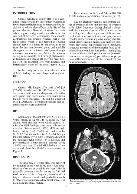
1.08%) were seen. Cranial MRI findings of two<br />
cases with clonic hemifacial spasms are seen in<br />
Figure 1.<br />
DISCUSSION<br />
The first case of clonic HFS was reported<br />
by Schultze in the year 1875, and it was described<br />
by Gowers in detail in the year 1884. It<br />
generally becomes manifest during the fifth and<br />
sixth decades of life. It frequently starts by affecting<br />
m. orbicularis oculi, and gradually spreads<br />
to other muscles innervated by facial nerve. It is<br />
not a painful clinical picture.<br />
Figure 1: The cranial MR samples of two cases with clonic HFS. Left:<br />
Small vessel disease (T2 FLAIR, axial); Right: Dolichoectasia of basilary<br />
arter (T2, axial).<br />
- 2 -