

Witmer, L. M., R. C. Ridgely, D. L. Dufeau, and M. C.
Witmer, L. M., R. C. Ridgely, D. L. Dufeau, and M. C.
Witmer, L. M., R. C. Ridgely, D. L. Dufeau, and M. C.

You also want an ePaper? Increase the reach of your titles
YUMPU automatically turns print PDFs into web optimized ePapers that Google loves.
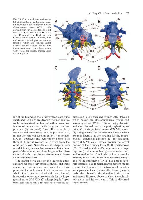
Fig. 6.8. Cranial endocast, endosseous<br />
labyrinth, <strong>and</strong> some endocranial vascular<br />
structures of the sauropod dinosaur,<br />
Camarasaurus lentus (CM 11338),<br />
derived from surface renderings of CT<br />
scan data. A, left lateral view. B, caudal<br />
view. C, ventral view. D, dorsal view.<br />
Color scheme: cranial endocast, blue;<br />
endosseous labyrinth, pink; nerve canals<br />
(most of which also transmit veins),<br />
yellow; smaller venous canals, dark<br />
blue; arterial canals, red; columella, pale<br />
yellow. Scale bar equals 2 cm (see Color<br />
Plates, Fig. 6.8).<br />
ing of the braincase, the olfactory tracts are quite<br />
short, <strong>and</strong> the bulbs are strongly inclined relative<br />
to the main axis of the brain. Another prominent<br />
feature of the endocast is the large <strong>and</strong> pendant<br />
pituitary (hypophyseal) fossa. The large bony<br />
fossa housed much more than the pituitary itself,<br />
in that the cerebral carotids enter it ventrolaterally,<br />
the abducens <strong>and</strong> oculomotor nerves pass<br />
through it, <strong>and</strong> it receives large veins from the<br />
orbit (see below). Nevertheless, as Edinger (1942)<br />
noted, it is very reasonable to assume that at least<br />
part of the reason that these large-bodied dinosaurs<br />
had such large pituitary fossae was to house<br />
an enlarged pituitary.<br />
The cranial nerve exits on the sauropod endocasts<br />
are generally very straightforward, <strong>and</strong> share<br />
a number of common features, many of which are<br />
primitive for archosaurs, if not sauropsids as a<br />
whole. Shared features, all of which are bilateral,<br />
include the following: (1) two canals for the hypoglossal<br />
nerve (CN XII); (2) a large ‘jugular’ aperture<br />
(sometimes called the ‘metotic foramen;’ see<br />
6. Using CT to Peer into the Past 77<br />
discussion in Sampson <strong>and</strong> <strong>Witmer</strong>, 2007) through<br />
which passed the glossopharyngeal, vagus, <strong>and</strong><br />
accessory nerves (CN IX–XI) <strong>and</strong> the jugular vein,<br />
<strong>and</strong> which housed part of the perilymphatic apparatus;<br />
(3) a single facial nerve (CN VII) canal;<br />
(4) a single canal for the trigeminal nerve which<br />
exp<strong>and</strong>s laterally as the swelling for the (extracranial)<br />
trigeminal ganglion; (5) the abducens<br />
nerve (CN VI) canal passes through the lateral<br />
portion of the pituitary fossa; (6) the oculomotor<br />
(CN III) <strong>and</strong> trochlear (IV) apertures are large,<br />
separate (or sharing an hour-glass-shaped fifissure),<br />
<strong>and</strong> located in the infundibular region (where the<br />
pituitary fossa joins the main endocranial cavity);<br />
<strong>and</strong> (7) the optic nerve (CN II) has a broad separate<br />
aperture. The trigeminal arrangement merits<br />
comment in that none of the trigeminal branches<br />
are separate in these (or any other known) sauropods,<br />
which is unlike the situation in the extant<br />
archosaurs discussed above in which the ophthalmic<br />
nerve had its own canal. This is discussed<br />
further below.


