

Witmer, L. M., R. C. Ridgely, D. L. Dufeau, and M. C.
Witmer, L. M., R. C. Ridgely, D. L. Dufeau, and M. C.
Witmer, L. M., R. C. Ridgely, D. L. Dufeau, and M. C.

Create successful ePaper yourself
Turn your PDF publications into a flip-book with our unique Google optimized e-Paper software.
70 L.M. <strong>Witmer</strong> et al.<br />
6.3 Methods<br />
CT scanning of the extant crocodile (OUVC<br />
10425), Camarasaurus (CM 11338), Diplodocus<br />
(CM 3452), <strong>and</strong> Tyrannosaurus (AMNH 5117)<br />
took place at O’Bleness Memorial Hospital<br />
(OBMH) in Athens, Ohio, using a General Electric<br />
(GE) LightSpeed Ultra Multislice CT scanner.<br />
This scanner was equipped with the Extended<br />
Hounsfi field<br />
option (which greatly improves<br />
resolvability of detail from dense objects such as<br />
fossils by extending the dynamic range of images<br />
as much as 16-fold) <strong>and</strong> a ‘bow-tie’ fifilter<br />
(which<br />
decreases beam-hardening artifacts). These specimens<br />
were scanned helically at a slice thickness of<br />
625 μm, 120 kV, <strong>and</strong> 200–300 mA. The other specimens<br />
of Diplodocus were scanned at OBMH with<br />
a GE HiSpeed FX/i Multislice helical scanner at<br />
a slice thickness of 1 mm (AMNH 694) or 2 mm<br />
(CM 11161), 120–140 kV, <strong>and</strong> 150 mA. The raw<br />
scan data were reconstructed using a bone algorithm.<br />
Data were output from the scanners in<br />
DICOM format, <strong>and</strong> then imported into Amira<br />
3.1.1 or 4.1.1 (Mercury-TGS, Chelmsford, MA) for<br />
viewing, analysis, <strong>and</strong> visualization.<br />
The extant owl skull (OUVC 10220) was scanned<br />
at the Ohio University MicroCT (OUμCT) facility<br />
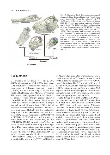
Fig. 6.2. Diagram of the phylogenetic relationships of<br />
the principal taxa discussed in the text. From left, the<br />
extant crocodilian Crocodylus johnstoni (OUVC<br />
10425), the diplodocoid sauropod Diplodocus longus<br />
(CM 11161), the macronarian sauropod Camarasaurus<br />
lentus (CM 11338), the basal coelurosaurian<br />
theropod Tyrannosaurus rex (AMNH 5117), <strong>and</strong> the<br />
avian theropod (bird) Bubo virginianus (OUVC<br />
10220). These sauropods <strong>and</strong> theropods are saurischian<br />
dinosaurs. The images are surface renderings of<br />
CT scan data. Bony skulls are depicted as transparent,<br />
revealing such internal structures as the brain cast,<br />
labyrinth, <strong>and</strong> pneumatic sinuses. All skulls are oriented<br />
in their ‘alert’ postures, determined by orienting<br />
the skull such that the lateral semicircular canal<br />
is horizontal. Scale bars equal 10 cm, except that for<br />
B. virginianus which equals 2 cm (see Color Plates,<br />
Fig. 6.2).<br />
in Athens, Ohio, using a GE eXplore Locus in vivo<br />
Small Animal MicroCT Scanner. It was scanned<br />
(with a domestic mouse, Mus musculus [OUVC<br />
10449], in its jaws; Fig. 6.1) at a slice thickness of<br />
92 μm, 80 kV, 450 μA. The resulting volume data (in<br />
VFF format) were exported from MicroView 2.1.2<br />
(open-source software developed by GE; microview.<br />
sourceforge.net) in DICOM format, which were<br />
then subsequently imported into Amira.<br />
Data derived from both OBMH <strong>and</strong> OUμCT<br />
were analyzed on 32- <strong>and</strong> 64-bit PC workstations<br />
with 4 GB of RAM <strong>and</strong> nVidia Quadro FX 3000<br />
or 4500 video cards <strong>and</strong> running Microsoft<br />
Windows XP Professional, Windows XP Professional<br />
x64, or Linux 2.6.18 (Debian 4.0 distribution).<br />
Structures of interest (e.g., cranial endocast,<br />
labyrinth, air sinuses, etc.) were highlighted <strong>and</strong><br />
digitally extracted using Amira’s segmentation<br />
tools for quantification fi <strong>and</strong> visualization. Both<br />
surfaces <strong>and</strong> volumes were generated, <strong>and</strong> were<br />
used to illustrate this paper. To facilitate discussion,<br />
we will refer to the digital casts of structures<br />
as if they were the structures themselves (e.g.,<br />
‘lateral semicircular canal’ versus ‘digital cast of<br />
lateral semicircular canal’). Additional visualizations<br />
of the CT scan data are available on www.<br />
ohio.edu/<strong>Witmer</strong>Lab.


