

Witmer, L. M., R. C. Ridgely, D. L. Dufeau, and M. C.
Witmer, L. M., R. C. Ridgely, D. L. Dufeau, and M. C.
Witmer, L. M., R. C. Ridgely, D. L. Dufeau, and M. C.

Create successful ePaper yourself
Turn your PDF publications into a flip-book with our unique Google optimized e-Paper software.
74 L.M. <strong>Witmer</strong> et al.<br />
6.6 Extant Outgroups: Bubo<br />
virginianus, Great Horned Owl<br />
Whereas the cranial endocast of the freshwater<br />
crocodile above bears only a superficial firesem- blance to the underlying brain, the endocast of<br />
Bubo virginianus (OUVC 10220; Fig. 6.7) closely<br />
resembles the form of the brain itself, as determined<br />
both by our dissection of a different specimen<br />
of B. virginianus (OUVC 10427) <strong>and</strong> Turner’s<br />
(1891) illustration of a B. virginianus brain (see<br />
also Stingelin 1957, for photographs of the brains<br />
of four other owl species). This close resemblance<br />
results from the fact that avian meninges are very<br />
thin, <strong>and</strong> thus the large brain essentially fills fi the<br />
endocranial cavity (Jerison 1973; Iwaniuk <strong>and</strong><br />
Nelson 2002). A major difference between the<br />
brain <strong>and</strong> endocast is that the expansive occipital<br />
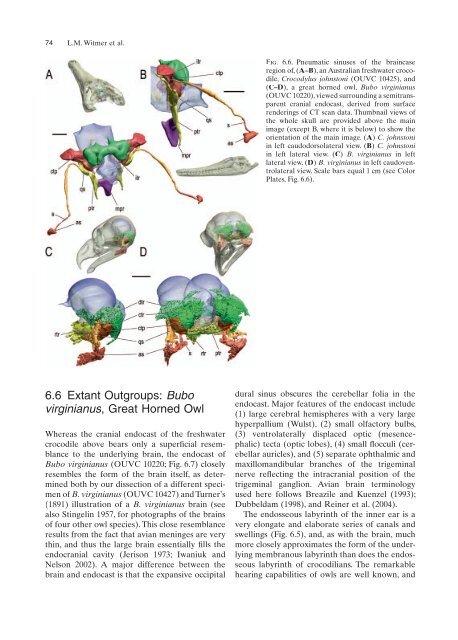
Fig. 6.6. Pneumatic sinuses of the braincase<br />
region of, (A–B), an Australian freshwater crocodile,<br />
Crocodylus johnstoni (OUVC 10425), <strong>and</strong><br />
(C–D), a great horned owl, Bubo virginianus<br />
(OUVC 10220), viewed surrounding a semitransparent<br />
cranial endocast, derived from surface<br />
renderings of CT scan data. Thumbnail views of<br />
the whole skull are provided above the main<br />
image (except B, where it is below) to show the<br />
orientation of the main image. (A) C. johnstoni<br />
in left caudodorsolateral view. (B) C. johnstoni<br />
in left lateral view. (C) B. virginianus in left<br />
lateral view. (D) B. virginianus in left caudoventrolateral<br />
view. Scale bars equal 1 cm (see Color<br />
Plates, Fig. 6.6).<br />
dural sinus obscures the cerebellar folia in the<br />
endocast. Major features of the endocast include<br />
(1) large cerebral hemispheres with a very large<br />
hyperpallium (Wulst), (2) small olfactory bulbs,<br />
(3) ventrolaterally displaced optic (mesencephalic)<br />
tecta (optic lobes), (4) small fl<br />
occuli (cer-<br />
ebellar auricles), <strong>and</strong> (5) separate ophthalmic <strong>and</strong><br />
maxillom<strong>and</strong>ibular branches of the trigeminal<br />
nerve reflecting fl the intracranial position of the<br />
trigeminal ganglion. Avian brain terminology<br />
used here follows Breazile <strong>and</strong> Kuenzel (1993);<br />
Dubbeldam (1998), <strong>and</strong> Reiner et al. (2004).<br />
The endosseous labyrinth of the inner ear is a<br />
very elongate <strong>and</strong> elaborate series of canals <strong>and</strong><br />
swellings (Fig. 6.5), <strong>and</strong>, as with the brain, much<br />
more closely approximates the form of the underlying<br />
membranous labyrinth than does the endosseous<br />
labyrinth of crocodilians. The remarkable<br />
hearing capabilities of owls are well known, <strong>and</strong>


