

Witmer, L. M., R. C. Ridgely, D. L. Dufeau, and M. C.
Witmer, L. M., R. C. Ridgely, D. L. Dufeau, and M. C.
Witmer, L. M., R. C. Ridgely, D. L. Dufeau, and M. C.

Create successful ePaper yourself
Turn your PDF publications into a flip-book with our unique Google optimized e-Paper software.
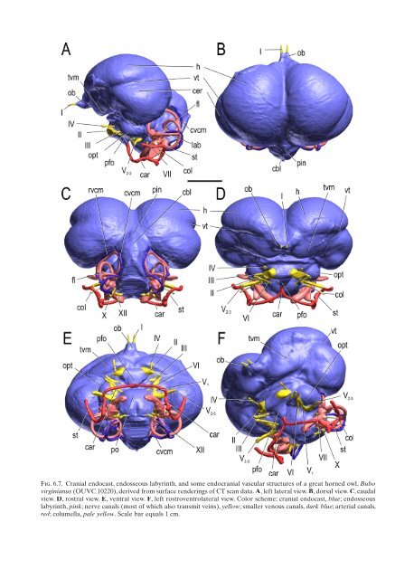
Fig. 6.7. Cranial endocast, endosseous labyrinth, <strong>and</strong> some endocranial vascular structures of a great horned owl, Bubo<br />
virginianus (OUVC 10220), derived from surface renderings of CT scan data. A, left lateral view. B, dorsal view. C, caudal<br />
view. D, rostral view. E, ventral view. F, left rostroventrolateral view. Color scheme: cranial endocast, blue; endosseous<br />
labyrinth, pink; nerve canals (most of which also transmit veins), yellow; smaller venous canals, dark blue; arterial canals,<br />
red; columella, pale yellow. Scale bar equals 1 cm.


