

Witmer, L. M., R. C. Ridgely, D. L. Dufeau, and M. C.
Witmer, L. M., R. C. Ridgely, D. L. Dufeau, and M. C.
Witmer, L. M., R. C. Ridgely, D. L. Dufeau, and M. C.

Create successful ePaper yourself
Turn your PDF publications into a flip-book with our unique Google optimized e-Paper software.
6.4 Results<br />
As noted earlier, we will rely more on illustration<br />
than text to convey anatomical form. We begin<br />
this section with a presentation of the extant taxa<br />
because the identifi fications<br />
of soft-tissue struc-<br />
tures, as well as their relationships to bony struc-<br />
tures, have been confirmed fi by other means (e.g.,<br />
dissection). Moreover, in the case of the crocodile,<br />
an intact specimen was scanned, <strong>and</strong> many softtissue<br />
components can be observed in the scan<br />
data. We follow with a presentation of the fossil<br />
taxa, beginning with the two sauropods <strong>and</strong> then<br />
Tyrannosaurus, basing our identifi fications<br />
in part<br />
on the crocodile <strong>and</strong> owl presented here but also<br />
on the dozens of other extant <strong>and</strong> fossil archosaurs<br />
we have analyzed as part of the broader<br />
project.<br />
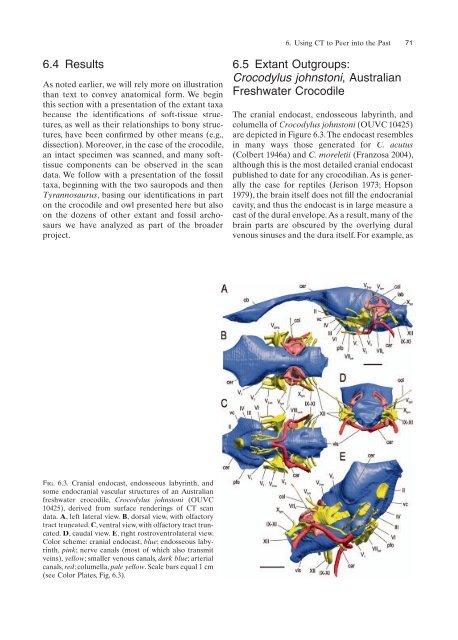
Fig. 6.3. Cranial endocast, endosseous labyrinth, <strong>and</strong><br />
some endocranial vascular structures of an Australian<br />
freshwater crocodile, Crocodylus johnstoni (OUVC<br />
10425), derived from surface renderings of CT scan<br />
data. A, left lateral view. B, dorsal view, with olfactory<br />
tract truncated. C, ventral view, with olfactory tract truncated.<br />
D, caudal view. E, right rostroventrolateral view.<br />
Color scheme: cranial endocast, blue; endosseous labyrinth,<br />
pink; nerve canals (most of which also transmit<br />
veins), yellow; smaller venous canals, dark blue; arterial<br />
canals, red; columella, pale yellow. Scale bars equal 1 cm<br />
(see Color Plates, Fig. 6.3).<br />
6. Using CT to Peer into the Past 71<br />
6.5 Extant Outgroups:<br />
Crocodylus johnstoni, Australian<br />
Freshwater Crocodile<br />
The cranial endocast, endosseous labyrinth, <strong>and</strong><br />
columella of Crocodylus johnstoni (OUVC 10425)<br />
are depicted in Figure 6.3. The endocast resembles<br />
in many ways those generated for C. acutus<br />
(Colbert 1946a) <strong>and</strong> C. moreletii (Franzosa 2004),<br />
although this is the most detailed cranial endocast<br />
published to date for any crocodilian. As is generally<br />
the case for reptiles (Jerison 1973; Hopson<br />
1979), the brain itself does not fill fi the endocranial<br />
cavity, <strong>and</strong> thus the endocast is in large measure a<br />
cast of the dural envelope. As a result, many of the<br />
brain parts are obscured by the overlying dural<br />
venous sinuses <strong>and</strong> the dura itself. For example, as


