Hydroids (Cnidaria, Hydrozoa) of the Danish expedition to
Hydroids (Cnidaria, Hydrozoa) of the Danish expedition to
Hydroids (Cnidaria, Hydrozoa) of the Danish expedition to

Create successful ePaper yourself
Turn your PDF publications into a flip-book with our unique Google optimized e-Paper software.
HYDROIDS OF THE DANISH EXPEDITION TO THE KEI ISLANDS<br />
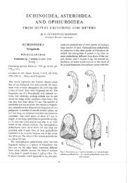
margin <strong>of</strong> hydro<strong>the</strong>ca, with three openings: one<br />
terminal, one on upper surface where becoming<br />
free, one just below <strong>the</strong> latter leading in<strong>to</strong> hydro<strong>the</strong>cal<br />
cavity (foramen). Lateral nema<strong>to</strong><strong>the</strong>cae<br />
tubular, inclined forward, over<strong>to</strong>pping somewhat<br />
hydro<strong>the</strong>cal margin, one or two openings:<br />
one terminal and one near base on upper surface,<br />
<strong>the</strong> latter not always visible.<br />
Gono<strong>the</strong>cae on modified hydrocladia (phylac<strong>to</strong>carps),<br />
one or two per phylac<strong>to</strong>carp. Phylac<strong>to</strong>carps<br />
composed <strong>of</strong> one hydrocladial segment<br />
with hydro<strong>the</strong>ca as in normal hydrocladia, followed<br />
by cylindrical segments with one or two<br />
nema<strong>to</strong><strong>the</strong>cae. Gono<strong>the</strong>ca attached <strong>to</strong> segment<br />
following hydro<strong>the</strong>cate segment, lens-shaped,<br />
diameter 0.6 mm; gonophore medusoid, with<br />
spadix, bell margin with granules.<br />
Remarks<br />
Hirohi<strong>to</strong>’s (1983, 1995) material allocated <strong>to</strong> this<br />
species had ra<strong>the</strong>r thin intra<strong>the</strong>cal septae, had<br />
several gono<strong>the</strong>cae per phylac<strong>to</strong>carp, and was<br />
apparently larviparous. I <strong>the</strong>refore doubt that Hirohi<strong>to</strong>’s<br />
samples belonged <strong>to</strong> M. philippina. The<br />
increased number <strong>of</strong> gono<strong>the</strong>cae and <strong>the</strong> lamellar<br />
intra<strong>the</strong>cal septae match better M. balei (Nutting,<br />
1905). Contrary <strong>to</strong> this, Leloup’s (1930b) specimen<br />
identified as M. balei appears indistinguishable<br />
from M. philippina.<br />
Distribution<br />
Circumglobal in tropical and subtropical waters.<br />
Type locality: Manila, Philippines.<br />
Macrorhynchia phoenicea (Busk, 1852)<br />
Figs 68–69.<br />
Plumularia aurita Busk, 1852: 397.<br />
Plumularia phoenicea Busk, 1852: 398.<br />
Aglaophenia rostrata Kirchenpauer, 1872: 45, pl. 1: fig. 25,<br />
pl. 6: fig. 25. – Weltner 1900: 588.<br />
Ly<strong>to</strong>carpus spectabilis Allman, 1883: 43, fig. 2, pl. 15: figs<br />
1–5.<br />
Aglaophenia phoenicea. – Bale 1884: 159, pl. 15: figs 1–5,<br />
pl. 17: figs 1–4, pl. 19: fig. 31.<br />
?Aglaophenia disjuncta Pictet, 1893: 59, pl. 3: figs 51–52.<br />
Ly<strong>to</strong>carpus phoeniceus. – Billard 1910: 48, fig. 22. – Billard<br />
1913: 74, figs 60–61. – Weltner 1900: 588. – Leloup<br />
1930b: 10, fig. 7, pl. 2: fig. 1. – Millard & Bouillon 1973:<br />
94. – Millard 1975: 451, fig. 137D.<br />
Macrorhynchia phoenicea. – Mammen 1967: 313, figs 108–<br />
109. – Rho 1967: 348, fig. 8. – Ryland & Gibbons 1991:<br />
555, fig. 23. – Hirohi<strong>to</strong> 1995: 299, fig. 106a–e.<br />
223<br />
Macrorhynchia phoenicia.– Watson 2000: 68, fig. 54A–E.<br />
Material examined:<br />
Kei Islands Expedition stations: 18. – 19, with gono<strong>the</strong>cae. –<br />
24. – 26, with gono<strong>the</strong>cae. – 57. – 67. – 69. – 71, with<br />
gono<strong>the</strong>cae. – 72. – 106. – 107. – Kei Islands Expedition, Kei<br />
Islands, Tual, 2 m, 28 Mar 1922, with gono<strong>the</strong>cae. – Kei<br />
Islands Expedition, Kei Islands, Tual, 22 Mar 1922, with<br />
gono<strong>the</strong>cae. – Kei Islands Expedition, Banda Islands, Neira<br />
Island, 25 m, 14 Jun 1922.<br />
Differential diagnosis<br />
Somewhat similar <strong>to</strong> Macrorhynchia philippina,<br />
but branching more regular, hydrocladia more<br />
bristly, denser, lengths quite homogenous, about<br />
12 hydro<strong>the</strong>cae per hydrocladium; abcauline horizontal<br />
shelf in hydro<strong>the</strong>ca thin and not triangular,<br />
height <strong>of</strong> hydro<strong>the</strong>ca smaller (0.22–0.25<br />
mm), free abcauline wall short, margin without<br />
abcauline <strong>to</strong>oth, lateral margin with two irregular<br />
cusps; majority <strong>of</strong> lateral nema<strong>to</strong><strong>the</strong>ca directed<br />
<strong>to</strong>wards above; both nema<strong>to</strong><strong>the</strong>cae <strong>of</strong> hydrocaulus<br />
with two openings <strong>of</strong> different size. Gono<strong>the</strong>ca<br />
lens-shaped, less flattened but also with<br />
sharp edge along circumference. Nema<strong>to</strong><strong>the</strong>cae<br />
<strong>of</strong> phylac<strong>to</strong>carps in three rows.<br />
Description<br />
See Millard (1975), Ryland & Gibbons (1991),<br />
and Watson (2000).<br />
Remarks<br />
The bristly, neatly regular hydrocladia <strong>of</strong> equal<br />
length (Fig. 68) make large and fully grown<br />
Macrorhynchia phoenicea (Busk, 1852) <strong>to</strong> some<br />
degree recognizable even without <strong>the</strong> aid <strong>of</strong> a<br />
microscope. The hydro<strong>the</strong>cae and <strong>the</strong> internodes<br />
are quite variable (Fig. 68C–E). Especially <strong>the</strong><br />
outline <strong>of</strong> <strong>the</strong> lateral rim <strong>of</strong> <strong>the</strong> hydro<strong>the</strong>ca is very<br />
variable. Bale (1884) discussed <strong>the</strong> variability <strong>of</strong><br />
this species.<br />
The gonophores seen in <strong>the</strong> present material<br />
are likely sessile sporosacs, female ones containing<br />
10–16 eggs.<br />
The samples from stations 24, 26, and 57<br />
deviate somewhat from <strong>the</strong> o<strong>the</strong>rs (Fig. 69). The<br />
colonies are smaller (6 cm), more gracile, <strong>the</strong>y<br />
have longer internodes (0.30–0.34 mm), thinner<br />
hydrocladia, <strong>the</strong> lateral nema<strong>to</strong><strong>the</strong>cae <strong>of</strong> <strong>the</strong><br />
proximal hydro<strong>the</strong>cae are directed in <strong>the</strong> direc-