

Signal Analysis Research (SAR) Group - RNet - Ryerson University
Signal Analysis Research (SAR) Group - RNet - Ryerson University
Signal Analysis Research (SAR) Group - RNet - Ryerson University

Create successful ePaper yourself
Turn your PDF publications into a flip-book with our unique Google optimized e-Paper software.
images contain localized heterogeneous texture elements,<br />
whereas normal images are smooth (uniform). Utilization of<br />
the SIDWT allowed for the extraction of consistent (i.e. shiftinvariant)<br />
features. Furthermore, due to the scale-invariant<br />
basis functions of the DWT, pathologies of varying sizes<br />
were captured within one transformation (i.e. the features<br />
were scale-invariant).<br />
The system is relatively robust to the different camera<br />
angles by design. Since the viewing angle is different from<br />
image to image, features were collected at various angles (0 ◦ ,<br />
45 ◦ , 90 ◦ , 135 ◦ ) in the respective subbands in order to account<br />
for the texture properties, regardless of the orientation. The<br />
feature set thus offered a semi-rotational invariant representation<br />
which could account for oriented textural properties at<br />
various angles within the gastrointestinal tract.<br />
Since this is the first work in the area of small bowel<br />
image classification, the results are promising and show<br />
great potential for applications such as CAD and CBIR. This<br />
is especially true since all features were extracted in a fullyautomated<br />
manner without any intervention or assistance<br />
from a gastroenterologist. This means that such a system<br />
could in fact be used as a tool which could either (1) sort<br />
the 8 hours of film and highlight suspicious regions or (2)<br />
automatically retrieve a specific region or mass, without<br />
having to use text annotations.<br />
Although the classification results are high, any misclassification<br />
can be accounted to cases where there is a lack<br />
of statistical differentiation between the texture uniformity<br />
of the abnormal and normal small bowel images. Additionally,<br />
normal tissue can sometimes assume the properties<br />
of abnormal regions; for example, consider a normal small<br />
bowel image which has more than the average amount of<br />
folds. This may be characterized as non-uniform texture and<br />
consequently would be misclassified.<br />
Another important consideration arises from the sizes of<br />
the databases. As was stated in Section II-E, the number of<br />
images used for classification can determine the accuracy<br />
of the estimated classifier parameters. Since only a modest<br />
number of images were used, misclassification could result<br />
due to the lack of proper estimation of the classifiers parameters<br />
(although the scheme tried to combat this with LOOM).<br />
Additionally, finding the right trade off between number of<br />
features and database size is an ongoing research topic and<br />
has yet to be perfectly defined [8].<br />
A last point for discussion is the fact that features were<br />
successfully extracted from the compressed domain. Since<br />
many forms of multi-media are being stored in lossy formats,<br />
it is important that classification systems may also be<br />
successful when utilized in the compressed domain.<br />
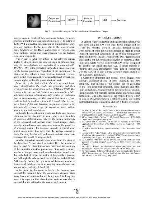
Fig. 2. System block diagram for the classification of small bowel images.<br />
4527<br />
13<br />
IV. CONCLUSIONS<br />
A unified feature extraction and classification scheme was<br />
developed using the DWT for small bowel images and this<br />
is the first reported work in the area. Textural features<br />
were extracted from the wavelet domain in order to obtain<br />
localized numerical descriptors of the relative homogeneity<br />
of the small bowel images. To ensure the DWT representation<br />
was suitable for the consistent extraction of features, a shiftinvariant<br />
discrete wavelet transform (SIDWT) was computed.<br />
To combat the small database size, a small number of<br />
features and LDA classification were used in conjunction<br />
with the LOOM to gain a more accurate approximation of<br />
the classifier’s parameters.<br />
Seventy-five abnormal and normal bowel images were<br />
correctly classified at rate of 85% specificity and 85%<br />
sensitivity. The success of the system can be accounted<br />
to the semi-rotational invariant, scale-invariant and shiftinvariant<br />
features, which permitted the extraction of discriminating<br />
features for multiple camera angles and various sized<br />
pathologies. Due to the success of the proposed work, it may<br />
be used in a CAD scheme or a CBIR application, to assist the<br />
gastroenterologists to diagnose and sort 8 hours of footage.<br />
REFERENCES<br />
[1] B. Kim, S. Park, C. Jee, and S. Yoon, in An earthworm-like locomotive<br />
mechanism for capsule endoscopes. International Conference on<br />
Intelligent Robots and Systems, Aug. 2005, pp. 2997 – 3002.<br />
[2] Given Imaging Ltd., PillCam TM SB Capsule Endoscopy. [ONLINE],<br />
2006, http://www.givenimaging.com/.<br />
[3] M. Unser and A. Aldroubi, “A review of wavelets in biomedical<br />
applications,” Proceedings of the IEEE, vol. 84, no. 4, pp. 626 – 638,<br />
Apr. 1996.<br />
[4] S. Mallat, Wavelet Tour of <strong>Signal</strong> Processing. USA: Academic Press,<br />
1998.<br />
[5] J.Liang and T. Parks, “Image coding using translation invariant wavelet<br />
transforms with symmetric extensions,” IEEE Transactions on Image<br />
Processing, vol. 7, pp. 762 – 769, May 1998.<br />
[6] A. Khademi, “Multiresolutional analysis for classification and compression<br />
of medical images,” Master’s thesis, 2006, ryerson <strong>University</strong>,<br />
Canada.<br />
[7] B. Julesz, “Textons, the elements of texture perception, and their<br />
interactions,” Nature, vol. 290, no. 5802, pp. 91–97, Mar. 1981.<br />
[8] K. Fukunaga and R. Hayes, “Effects of sample size in classifier design,”<br />
IEEE Transactions on Pattern <strong>Analysis</strong> and Machine Intelligence,<br />
vol. 11, no. 8, pp. 873 – 885, Aug. 1989.<br />
Authorized licensed use limited to: <strong>Ryerson</strong> <strong>University</strong> Library. Downloaded on July 7, 2009 at 11:53 from IEEE Xplore. Restrictions apply.