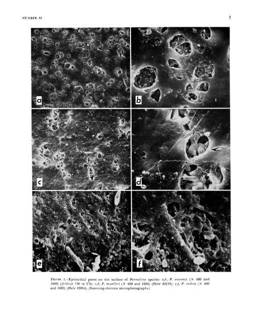
2 Morphology THALISIS STRLCTURE.-Pa?-melina basically has a closely appressed to adnate foliose thallus with narrow lobes (1-4 mm wide). Most species have more or less subirregular lobation and apically rotund lobes (Figures I&, 18b), although narrow lobed species are sublinear (Figures 12d, 16c,). There is, however, a wide range <strong>of</strong> variation in lobe configuration and width, much more than exists, for example, in Hypotmchynn (<strong>Hale</strong>, 1975a). The internal anatomy is similar to that <strong>of</strong> o<strong>the</strong>r epicorticate genera (<strong>Hale</strong>, 1973a). A thin, generally palisade plectenchymatous cortex is overlain by a pored epicortex (Figures 1 and 2). The medulla consists <strong>of</strong> loosely packed hyphae, <strong>of</strong>ten encrusted with lichen substances (Figure 2f). The lower cortex is p”p1ectenchymatous (Figure 20-d). The upper surface is strongly white maculate in Pwmelina con.son (Figure 3a), P. miielleii, and P. pilostr, less conspiciiously so in P. nzelanochaetn, P. pa.stillifei.a, P. qiiercina, and P. tiliacea, and faintly or not at all in <strong>the</strong> remaining species. The lower surface is black with only three exceptions. Pai.melina cnormis has a uniformly pale brown lower surface, and P. expallida and P. versiforrnis are darker brown tending toward black at <strong>the</strong> center. CILIA.-The marginal cilia which characterize Pnrrnelina are usually distinct but may be variably dispersed around <strong>the</strong> lobe margins. In many species <strong>the</strong>y occur more or less regularly both on lobe tips and in <strong>the</strong> axils (Figures 3a, lia, 18b). In o<strong>the</strong>rs cilia may be lacking at <strong>the</strong> lobe tips and confined to <strong>the</strong> axils (Figure 3b) or may be so sparse and inconspicuous in a few species, such as P. .rimplicioy, that <strong>the</strong>y are overlooked. In <strong>the</strong>se cases one might tend to classify <strong>the</strong>m in <strong>the</strong> genus Pseutlopaimelia (<strong>Hale</strong>, 1976a), which is differentiated from <strong>Parmelina</strong>, among o<strong>the</strong>r ways, by <strong>the</strong> total absence <strong>of</strong> marginal cilia. Great care must be taken to establish <strong>the</strong> presence or absence <strong>of</strong> cilia in <strong>the</strong> lobe axils. Ano<strong>the</strong>r example <strong>of</strong> this problem is Pa?-melin(i endoleum and P.rezidopnrmelin siibtiliacea (Nylander) <strong>Hale</strong>, two very similar species in Australia that differ in <strong>the</strong> production <strong>of</strong> cilia. Both have fatty acids and cannot be separated easily by a chemical test. Confusion may also arise with species <strong>of</strong> Hypot~a- SSIITHSONIAPi COSTRIBUTIONS TO BOTANY chyna that lack cilia but may have a very dense rhizinal mat below. The mat may assume a marginal position and resemble cilia. These “cilia,” however, will be dichotomously branched. Such a distinction fails in many specimens <strong>of</strong> H. rcvoluta (Floerke) <strong>Hale</strong> having ra<strong>the</strong>r sparsely branched rhizines and a few marginally positioned “cilia.” If incorrectly interpreted as having furcate rhizines and sparse marginal cilia, such a lichen might be identified as <strong>Parmelina</strong> cryptochlom, which has more powdery capitate soralia, or as pustulate P. spiimosn, which has a pale yellowish medulla. All <strong>of</strong> <strong>the</strong>se species have gvrophoric acid. One fur<strong>the</strong>r example <strong>of</strong> parallelism involving Pam e 1 in a rl is sec t a and Hypo t ru c h y n a n eod issec t a (<strong>Hale</strong>) <strong>Hale</strong> can be mentioned. The latter has clearly dichotomously branched rhizines and lacks cilia, yet both contain <strong>the</strong> same chemical, gyrophoric acid, and have virtually identical lobe configuration. It is difficult, if not impossible, to decide at this time whe<strong>the</strong>r intergeneric hybridization has occurred in <strong>the</strong>se cases and resulted in “hybrid” species. RHIzIms.-?’he rhiLines <strong>of</strong> <strong>Parmelina</strong> are <strong>of</strong> two types: simple to sparsely furcate (Figure 3c) and squarrose. Squarrose rhizines, that is, those with a main axis and short lateral branches, are confined mostly to some species in section Myelochroa. This pattern is also known in a few species <strong>of</strong> Parmelia (e.g., P. ,szilcata Taylor) and Pai.motl-ema (<strong>the</strong> P. 7.cticnlOtzLm group). ISIDIA AND LosuLEs.-Isidia are normally cylindrical and erect as in o<strong>the</strong>r parmelioid genera and occur in <strong>the</strong> following 13 species: P. antillensis, P. expallidla, P. di.csecta (Figure 34, P. horrescens, P. jamesii, P. indica, P. lindmanii, P. melanochaeta, P. obsessn, P. perisidians, P. tiliacea, P. usambarensis, and P. urnllichinna. Isidia are distinctly lobulate in P. degelii ant1 P. spatlziilata (Figure 44 and uniquely peltate in P. pastillifem (Figures 3e, 4b), a close relative <strong>of</strong> P. tiliacea, Apical cilia almost always occur in P. howescens and P. melanochaeta (Figure 4c) and resemble those in PaYmotTema crin i- tiinz (Acliarius) Choisy. Lobules not originating from isidia are found in P. schindle7.i and P. xantholepis (Figure 4e,f); <strong>the</strong>y are mostly marginal. PvsTuLEs.-These are characteristic <strong>of</strong> four species, P. Iiaynchinensis, P. leucotylizn (Figure 5g), P.
NUMBER 33 FIGURE 1.-Epicortical pores oti <strong>the</strong> surface <strong>of</strong> Parnze[irzn species: a,b, P. ettornzis (X 400 and 1600) (Jeilicoe 150 in US); c,d, P. muelleri (X 400 and 1600) (<strong>Hale</strong> 42219); e,!, P. indica (X 400 atid 1600) (<strong>Hale</strong> 43884). (Scatitiing-electron microphotographs.) 3