Screening of Sclerosing Agents Introduced Intraarticularly for ...
Screening of Sclerosing Agents Introduced Intraarticularly for ...
Screening of Sclerosing Agents Introduced Intraarticularly for ...

Create successful ePaper yourself
Turn your PDF publications into a flip-book with our unique Google optimized e-Paper software.
Alterations Associated with Minimal Change Nephrotic Syndrome<br />
<strong>Screening</strong> <strong>of</strong> <strong>Sclerosing</strong> <strong>Agents</strong> <strong>Introduced</strong><br />
<strong>Intraarticularly</strong> <strong>for</strong> Synoviorthesis in Experimental<br />
Chronic Synovitis. A Second Report<br />
Alfredas Staponas,<br />
Vida Graþienë<br />
Institute <strong>of</strong> Experimental and<br />
Clinical Medicine,<br />
Þygimantø 9,<br />
LT-2600 Vilnius, Lithuania<br />
INTRODUCTION<br />
Until now the treatment <strong>of</strong> chronic synovitis remains<br />
the main unsolved problem in rheumatology. Despite<br />
conservative treatment with aggressive immunosuppressive<br />
drugs, rather <strong>of</strong>ten exudation remains in<br />
one or more joints <strong>of</strong> patients with chronic synovitis.<br />
This is not only wearisome <strong>for</strong> the patients, but also<br />
deepening the destructive changes in joint hyaline<br />
cartilage by released inflammatory mediators from<br />
inflamed synovium. In those cases the intraarticular<br />
treatment by injections <strong>of</strong> dexametasone, cortivasole,<br />
triamcinolon, hydrocortison and other types <strong>of</strong><br />
corticosteroids (2, 3) as well as solutions <strong>of</strong> diclophenac<br />
and opioid analgesia (4), hyaluronic acid (5),<br />
radioactive and chemical synoviorthese (6, 7) and<br />
other kinds <strong>of</strong> treatments are used. Along with suppressing<br />
chronic inflammation in the synovium <strong>of</strong><br />
the joint, all a<strong>for</strong>enamed modes <strong>of</strong> intraarticular treatment<br />
<strong>of</strong> chronic synovitis (together with surgical<br />
synovectomy) induce destructive degenerative changes<br />
in hyaline cartilage and in other joint structures.<br />
ISSN 1392–0138. A cta medica Lituanica. 2001. T. 8, Nr. 3<br />
The aim <strong>of</strong> the study was to evaluate the effectiveness <strong>of</strong> reduced concentrations<br />
<strong>of</strong> preliminary selected sclerosing agents (1) having a minimal destructive effect<br />
on the structure and metabolism <strong>of</strong> articular cartilage <strong>for</strong> the intraarticular<br />
treatment <strong>of</strong> chronic synovitis in rabbits. High doses <strong>of</strong> sclerosants introduced<br />
intraarticularly in a preliminary screening study, together with a sclerosing effect<br />
<strong>of</strong> the inflammatory synovial layer, induced distinct destructive changes in<br />
articular cartilage. In a second study chronic synovitis was induced in 30 rabbits<br />
by a modified method <strong>of</strong> Dumonde and Glynn described in a preliminary report<br />
(1). The rabbits were sacrificed 1, 2, 3, 4 and 5 weeks after intraarticular injection<br />
<strong>of</strong> the sclerosant solution. Such a sequence <strong>of</strong> study permits to evaluate the<br />
dynamics <strong>of</strong> the development <strong>of</strong> histopathological changes induced by the<br />
sclerosing solution in the synovial layer and articular cartilage.<br />
The histopathological data on the synovial layer and articular cartilage in the<br />
second experimental study with intraarticular injections <strong>of</strong> reduced concentrations<br />
<strong>of</strong> sclerosing solutions in chronic arthritis were almost similar to the results<br />
<strong>of</strong> the preliminary study when high concentrations <strong>of</strong> sclerosants were used. In<br />
the future experimental study we would try to use a different time <strong>of</strong> introducing<br />
the sclerosants into a joint cavity, because the sclerosant concentration did not<br />
differ by its influence on the destructive consequences in the articular cartilage.<br />
Key words: experimental arthritis, sclerosants, synoviorthesis<br />
The aim <strong>of</strong> the study was to select a sclerosing<br />
agent <strong>for</strong> the intraarticular treatment <strong>of</strong> chronic synovitis,<br />
able to induce fibrosis <strong>of</strong> inflamed synovium<br />
and having a minimal destructive effect upon articular<br />
cartilage and metabolism in rabbits. In a preliminary<br />
study (screening <strong>of</strong> sclerosing solutions <strong>for</strong> intraarticular<br />
treatment) the sclerosants <strong>for</strong> sclerotherapy<br />
<strong>of</strong> testicular hydrocele, epidydimal, renal or hepatic<br />
cysts, <strong>of</strong> oesophageal, haemorrhoidal and leg varices<br />
(8–11) were used. Their influence on synovial<br />
inflammation and articular cartilage was unknown.<br />
The concentrations <strong>of</strong> sclerosing solutions were used<br />
as described by authors. A distinct sclerosing effect<br />
<strong>of</strong> sclerosing solutions introduced intraarticularly in<br />
inflamed synovium in chronic arthritis was found.<br />
There<strong>for</strong>e all solutions induce more or less destructive<br />
changes in the hyaline cartilage <strong>of</strong> the joint<br />
(1). For the second stage <strong>of</strong> the study, reduced concentrations<br />
<strong>of</strong> sclerosing agents selected in the preliminary<br />
study, having a minimal destructive effect on hyaline<br />
cartilage were used <strong>for</strong> the intraarticular treatment<br />
<strong>of</strong> experimental chronic arthritis.<br />
193
Asta Valanèiûtë, Petras Kaltenis, Augustina Jankauskienë, Rimantë Èerkauskienë, Regina Firantienë, Vytas Tamoðiûnas...<br />
MATERIALS AND METHODS<br />
The methods <strong>of</strong> induction <strong>of</strong> chronic arthritis and<br />
<strong>of</strong> evaluation <strong>of</strong> histopathological changes in synovial<br />
layer and articular cartilage were used as described<br />
in the preliminary report (1). Ten days following<br />
the last immunization active immune synovitis<br />
developed in all 30 rabbits. The rabbits were randomised<br />
into 6 groups <strong>of</strong> 5 animals each, and 1 ml <strong>of</strong><br />
intraarticular injection <strong>of</strong> different sclerosants was<br />
introduced into the right and 1 ml <strong>of</strong> sterile physiologic<br />
saline into the left knee joint <strong>of</strong> the same rabbit<br />
(control). Each experimental group (<strong>of</strong> 5 animals)<br />
was injected with the following solutions <strong>of</strong><br />
sclerosants: 1) Doxorubicini 1 mg; 2) Cisplastini 0.25<br />
mg; 3) 0.5% Dioxydini; 4) 16% natrium chloride;<br />
5) 0.05% Papaini and 6) Brulamycini 20 mg. The<br />
results were correlated to 3% Polidocanol, a sclerosant<br />
widely used in European countries <strong>for</strong> the<br />
treatment <strong>of</strong> chronic synovitis (7, 10). Because <strong>of</strong><br />
insufficient synoviorthesis and marked destruction <strong>of</strong><br />
cartilage, the two sclerosant solutions (96% ethanol<br />
and Cephasoline 0.25 mg) used in the preliminary<br />
study were excluded from the subsequent experimental<br />
studies. One rabbit from each group (6 animals)<br />
was sacrified 1, 2, 3, 4 and 5 weeks post sclerotherapy<br />
after 25 mg <strong>of</strong> natrium thiopental anaesthesia.<br />
Such a sequence <strong>of</strong> study permits to evaluate the<br />
dynamic <strong>of</strong> the development <strong>of</strong> histopathological<br />
signs in synovium and articular cartilage which were<br />
induced by sclerotherapy. A histopathological evaluation<br />
by grading the changes in synovium and articular<br />
hyaline cartilage <strong>of</strong> both knee joints after arthrotomy<br />
was done.<br />
RESULTS<br />
Grading <strong>of</strong> histopathological signs in the synovium<br />
and articular cartilage <strong>of</strong> the left knee joint <strong>of</strong> rabbits<br />
revealed distinct inflammatory changes charac-<br />
<strong>Sclerosing</strong><br />
solutions<br />
194<br />
teristic <strong>of</strong> acute synovitis, which developed in 3 weeks<br />
and were replaced by the signs <strong>of</strong> chronic arthritis<br />
5 weeks after immunization. In the hyaline<br />
cartilage <strong>of</strong> the left knee joint we observed <strong>for</strong>mation<br />
<strong>of</strong> inflammatory pannus, as well as erosions and<br />
other structural and metabolic changes <strong>of</strong> chondrocytes,<br />
leading to atrophy <strong>of</strong> cartilage, the changes<br />
typical <strong>of</strong> destructive arthritis. All sclerosants were<br />
compared by their effect to Polidocanol which exerts<br />
a marked antiproliferative and antiinflammatory<br />
effect (joint swelling, sites <strong>of</strong> synoviocyte-A<br />
hyperplasia (by necrosis) and villous proliferation (by<br />
desquamation <strong>of</strong> vilii), foci <strong>of</strong> connective tissue disorganisation<br />
disappear and necrosis <strong>of</strong> inflammatory cells<br />
in small foci <strong>of</strong> stromal and perivascular infiltrations<br />
is induced), and stromal and vascular fibrosis<br />
and lipomatosis were found. A destructive inflammatory<br />
pannus <strong>of</strong> the cartilage turned into fibrous,<br />
and only a slight destruction <strong>of</strong> chondrocyte structure<br />
and metabolism and no atrophy <strong>of</strong> hyaline<br />
cartilage after intraarticular injection <strong>of</strong> Polidocanol<br />
were found.<br />
A summary evaluation <strong>of</strong> fibrosis in synovium<br />
and destruction in articular cartilage upon intraarticular<br />
treatment <strong>of</strong> chronic synovitis with sclerosing<br />
solutions is presented in Table. The sclerosant solutions<br />
might be divided into two groups. The first<br />
one represents agents with deep synoviorthesis and<br />
evident destructive changes in articular cartilage without<br />
any signs <strong>of</strong> chondrocyte restoration. The action<br />
<strong>of</strong> the other one differs by the concentration <strong>of</strong> the<br />
agent introduced intraarticularly, as well as by the<br />
degree <strong>of</strong> synoviorthesis and destruction <strong>of</strong> the articular<br />
cartilage. From the first week following-intraarticular<br />
injection <strong>of</strong> Doxorubicin (2 mg/ml), in<br />
comparison to Polidocanol, the villi and synoviocyte-<br />
A necrobiosis and desquamation together with mild<br />
fibrosis and lipomatosis <strong>of</strong> the subsynovial layer <strong>of</strong><br />
synovium remained until week 5 <strong>of</strong> the experiment.<br />
After reduced doses (1 mg/ml) <strong>of</strong> intraarticular<br />
Table. Summary evaluation <strong>of</strong> fibrosis in synovium and destruction in articular cartilage upon intraarticular treatment<br />
<strong>of</strong> chronic synovitis with sclerosing solutions<br />
Fibrosis <strong>of</strong><br />
Destruction <strong>of</strong> articular cartilage<br />
synovium Slight Average Deep<br />
deep superficial recover unrecover recover unrecover recover unrecover<br />
Doxorubicini + (+) + (+)<br />
Dioxydini + (+) (+) +<br />
Papaini + (+) + (+)<br />
Cisplastini + (+) + (+)<br />
Sol. NaCl + (+) + (+)<br />
Brulamycini + +(+)<br />
+ – evaluation <strong>of</strong> preliminary results; (+) – evaluation <strong>of</strong> second study results.
Doxorubicini, renovation <strong>of</strong> synoviocyte-A proliferation<br />
after 5 weeks <strong>of</strong> postsclerosant injection was<br />
found. Both concentrations <strong>of</strong> Doxorubicini induced<br />
lysis <strong>of</strong> the nucleus <strong>of</strong> chondrocytes in all layers <strong>of</strong><br />
articular cartilage, and the structure <strong>of</strong> cartilage<br />
became indistinct and composed mainly <strong>of</strong> a homogeneous<br />
proteinaceous material at the end <strong>of</strong> the<br />
experimental study. Under the influence <strong>of</strong> both concentrations<br />
<strong>of</strong> intraarticular injections <strong>of</strong> Cisplatini,<br />
the histopathological picture was similar to that <strong>of</strong><br />
Doxorubicin. The sclerosing capacity <strong>of</strong> the agent<br />
was evident in the inflamed synovium, but the induced<br />
deep destruction <strong>of</strong> chondrocytes (picnosis <strong>of</strong><br />
nucleus) in all zones <strong>of</strong> articular cartilage with<br />
washed proteoglycans (GAGs) to deep cartilage from<br />
the first week was not restored until 5 weeks post<br />
sclerotherapy (Fig. 1 A, B). The fibrosing capacity<br />
<strong>of</strong> Brulamycini on inflammatory synovium was<br />
evident and manifested by a necrobiosis and<br />
desquamation <strong>of</strong> proliferated villi and <strong>of</strong> synoviocytes-A<br />
layer, as well as by an inflammatory cell<br />
Alterations Associated with Minimal Change Nephrotic Syndrome<br />
necrobiosis, fibrous vasculopathy and a cellular<br />
thrombus in the vascular lumen <strong>of</strong> the deep subsynovial<br />
layer. Deep usuration <strong>of</strong> the surface <strong>of</strong> cartilage,<br />
disappearance <strong>of</strong> chondrocytes having a<br />
picnotic nucleus, and an evident diminution <strong>of</strong><br />
GAGs in all layers induced an atrophy <strong>of</strong> the<br />
cartilage with no signs <strong>of</strong> the possibility to restore<br />
(Fig. 1 C, D).<br />
Another 3 sclerosant solutions, Dioxydini, Papaini<br />
and natrium chloride, in high concentrations induced<br />
an evident and deep fibrosis <strong>of</strong> inflamed synovium,<br />
and at reduced concentrations <strong>of</strong> the sclerosants<br />
only superficial fibrosis was found. The proliferation<br />
<strong>of</strong> villi was reduced, necrobiosis and desquamation<br />
<strong>of</strong> synoviocytes-A were evident, as well<br />
as a stromal and vascular fibrosis <strong>of</strong> synovium under<br />
the influence <strong>of</strong> high concentrations <strong>of</strong> sclerosants<br />
was found. We observed an average destruction<br />
<strong>of</strong> cartilage structure and metabolism under the<br />
influence <strong>of</strong> both concentrations <strong>of</strong> Papaini (Fig. 2<br />
E, F) and natrium chloride (Fig. 2 C, D), while<br />
A B<br />
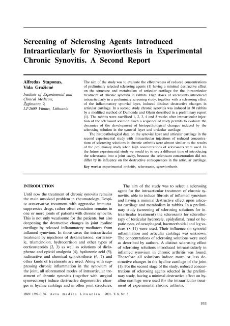
C D<br />
Fig. 1. Erosions and usuration ( ) <strong>of</strong> the surface, irregular disarrangement, picnosis <strong>of</strong> the nucleus ( ), and GAGs<br />
in matrix <strong>of</strong> cartilage ( ) after 3 and 5 weeks <strong>of</strong> intraarticular injections <strong>of</strong> Cisplatini 0.5 mg (A) and 0.25 mg (B),<br />
Brulamycini 40 mg (C) and 20 mg (D) in rabbits with chronic synovitis. HE, Toluidine blue X 800<br />
195
Asta Valanèiûtë, Petras Kaltenis, Augustina Jankauskienë, Rimantë Èerkauskienë, Regina Firantienë, Vytas Tamoðiûnas...<br />
196<br />
A B<br />
C D<br />
E F<br />
Fig. 2. Synovial plannus ( ), erosions and usuration <strong>of</strong> the surface ( ), picnosis <strong>of</strong> the nucleus and vacuolization<br />
<strong>of</strong> chondrocytes ( ), proliferated chondroblasts ( ) and GAGs ( ) in the articular cartilage <strong>of</strong> rabbits in chronic<br />
synovitis after 3 or 5 weeks <strong>of</strong> intraarticular injections <strong>of</strong>.: Dioxydini 1% (A), 0.5% (B), Sol. Natrium chloride 24%<br />
(C), 16% (D), Sol. Papaini 0.1% (E), 0.05% (F). HE, Toluidine blue. X 200, 800<br />
only slight destruction <strong>of</strong> cartilage was found upon<br />
injection <strong>of</strong> both concentrations <strong>of</strong> Dioxydini (Fig. 2<br />
A, B). A thicker or thinner synovial pannus and<br />
some erosions on the surface, pycnotic nucleus and<br />
cytoplasm vacuolization <strong>of</strong> disorderly situated chon-<br />
drocytes in all layers <strong>of</strong> the cartilage together with<br />
diminished ability to synthesise GAGs in Papaine<br />
and natrium chloride groups was seen, but 5 weeks<br />
following sclerotherapy the autolysed chondrocytes<br />
were slightly restored and GAGs production was
increased, especially in chondrocytes <strong>of</strong> the middle<br />
and deep layers. In all sclerosants groups clusters <strong>of</strong><br />
proliferated chondroblasts by the destroyed chondrocytes<br />
appeared, which spread into the middle zone<br />
<strong>of</strong> the cartilage from the synovial/cartilage junction.<br />
DISCUSSION<br />
Some sclerosants used in the study on intraarticular<br />
sclerotherapy <strong>of</strong> experimental chronic arthritis are<br />
used <strong>for</strong> clinical induction <strong>of</strong> sclerosis <strong>of</strong> testicular<br />
hydrocele, epidydimal, renal or hepatic cysts, <strong>of</strong><br />
oesophageal, haemorrhoidal and leg varices (12–14).<br />
Their influence on synovial inflammation and articular<br />
cartilage in scientific literature is unknown. Our<br />
preliminary report screened several sclerosing solutions<br />
<strong>for</strong> the intraarticular treatment <strong>of</strong> chronic arthritis.<br />
They had a minimal destructive effect on the<br />
articular cartilage (1), and reduced concentrations<br />
<strong>of</strong> them were tested in this study. Our results showed<br />
that a high as well as reduced concentrations<br />
<strong>of</strong> Doxorubicini, Cysplatini and Brulamycini introduced<br />
intraarticularly to rabbits with chronic arthritis<br />
induced an evident fibrosis <strong>of</strong> synovium (chemical<br />
synoviorthesis), with deep destructive changes and<br />
without any tendency to restore the structure and<br />
metabolism <strong>of</strong> the articular cartilage. Such destructive<br />
action <strong>of</strong> these sclerosants mainly depends on a close<br />
contact <strong>of</strong> chemically active solutions with the<br />
surface <strong>of</strong> the articular cartilage and compels us to<br />
exclude it from an arsenal <strong>of</strong> sclerosants to be valued<br />
<strong>for</strong> intraarticular treatment <strong>of</strong> chronic arthritis.<br />
Another group <strong>of</strong> tested sclerosant solutions Dioxydini,<br />
Papaini and natrium chloride in high concentrations<br />
induced a deep synoviorthesis with average<br />
degree <strong>of</strong> destructive changes in chondrocytes,<br />
but there remained a tendency to a slow restoration<br />
<strong>of</strong> cartilage structure (5 weeks post sclerotherapy).<br />
Reduced concentrations <strong>of</strong> these sclerosants<br />
had only a superficial and insufficient synoviorthesis<br />
capacity and some degree <strong>of</strong> destruction <strong>of</strong> chondrocytes<br />
with the remaining capacity to restore the<br />
cartilage structure by spreading the proliferated mesenchymal<br />
cells into the middle layer <strong>of</strong> cartilage<br />
from the synovium/cartilage junction. So, irrespective<br />
<strong>of</strong> the concentration <strong>of</strong> sclerosant solutions used<br />
in this study <strong>for</strong> intraarticular treatment <strong>of</strong> chronic<br />
arthritis, destructive changes <strong>of</strong> articular cartilage<br />
were revealed, but the reduced concentrations had<br />
an insufficient synoviorthesis capacity. To avoid or<br />
minimize destructive changes in the chondrocytes,<br />
different time <strong>of</strong> exposure <strong>of</strong> the articular cavity to<br />
sclerosing solutions chosen in the second study<br />
would be per<strong>for</strong>med in the last stage <strong>of</strong> the experimental<br />
study.<br />
Alterations Associated with Minimal Change Nephrotic Syndrome<br />
CONCLUSIONS<br />
1. Intraarticular injections <strong>of</strong> high concentrations <strong>of</strong><br />
sclerosing solutions used <strong>for</strong> the intraarticular treatment<br />
in experimental chronic arthritis induced a distinct<br />
fibrosis <strong>of</strong> synovium (chemical synoviorthesis).<br />
2. Independently <strong>of</strong> sclerosant concentration, evident<br />
destructive alterations <strong>of</strong> chondrocytes in the<br />
articular cartilage <strong>of</strong> knee joint were found. At least<br />
some <strong>of</strong> the solutions used in reduced concentrations<br />
<strong>for</strong> intraarticular treatment <strong>of</strong> chronic arthritis<br />
(Dioxydini, Papaini, natrium chloride) exhibited an<br />
ability to restore the cartilage by spreading proliferated<br />
mesenchymal cells into the middle layer from<br />
the synovial/cartilage junction.<br />
References<br />
Received 7 May 2001<br />
Accepted 1 June 2001<br />
1.Staponas A, Graþienë V. <strong>Screening</strong> <strong>of</strong> sclerosing agents<br />
introduced intraarticularly <strong>for</strong> synoviorthesis in experimental<br />
chronic synovitis. A preliminary report. Acta<br />
Medica Lituanica 2000; 7 (4): 166–70.<br />
2.Ravand P, Monlinier L, Girandeau B, Ayral X, Guerin<br />
C, Noel E, Thomas P, Fantrel B, Mazieres B,<br />
Dongados M. Effects <strong>of</strong> joint lavage and steroid injection<br />
in patients with osteoarthritis <strong>of</strong> the knee: results<br />
<strong>of</strong> multicenter, randomised, controlled trial. Arthr<br />
Rheum 1998 Mar; 82: 475–82.<br />
3.Papacrhiston G, Anagnoston S, Katsorhri T. The effect<br />
<strong>of</strong> intraarticular hydrocortisone injection on the<br />
articular cartilage <strong>of</strong> rabbits. Acta Orthop Scand Suppl<br />
1997 Oct; 275: 132–4.<br />
4.G^urkan Y, Kili^ckan L, Buluc L, M^nezzinoglu S,<br />
Toker K. Effects <strong>of</strong> dicl<strong>of</strong>enac and intraarticular morphine<br />
(bupivacaine) on postarthroscopic pain control.<br />
Minerva Anestesiol 1999 Oct; 39: 741–5.<br />
5.Roman JA, Chismol J, Morales M, Donderis JL. Intraarticular<br />
treatment with hyaluronic acid. Comparative<br />
study <strong>of</strong> Hyalgan and Adant. Clin Rheum 2000;<br />
19 (3): 204–6.<br />
6.De Vargas AF, Fernandez-Palazzi F. Cytogenetic studies<br />
in patients with haemophilic hemarthrosis treated<br />
by Au-198, Rh-186 and Y-90 radioactive synoviorthesis.<br />
J Paed Orthopaed Part B 2000 Jan; 9 (1):<br />
52–4.<br />
7.Rosenbaum M, Rottenstreich E. Synoviorthesis, chemical<br />
and radiation synovectomy in rheumatic disease.<br />
Harefuah 1990 Apr; 118 (7): 399–401.<br />
8.Dachlin L, Tisunder B, Kapstad L. Comparison <strong>of</strong><br />
polidocanol in sclerotherapy <strong>of</strong> testicular hydrocele and<br />
epidydimal cyst. Br J Urol 1997 Sept; 80 (3): 468–71.<br />
9.Zimmet SE. Treatment <strong>of</strong> varicose and teleangiectatic<br />
leg veins with hypertonic saline (letter, comments). J<br />
Dermatol Surg Oncol 1990 Sep; 16 (9): 876–7.<br />
10.El-Diarty TA, Shokeir A, Tawfeck HA et al. Ethanol<br />
sclerotherapy <strong>for</strong> symptomatic renal cysts. J Enurol<br />
1995 Jun; 9 (3): 273–6.<br />
197
Asta Valanèiûtë, Petras Kaltenis, Augustina Jankauskienë, Rimantë Èerkauskienë, Regina Firantienë, Vytas Tamoðiûnas...<br />
11.Zimmer T, Rucktøaschel F, Støolzel U, Luhr RM,<br />
Schuppan D, Stallmach A, Zeitz M, Weber E, Riecken<br />
EO. Endoscopic sclerotherapy with fibrin glue as<br />
compared with polidocanol to prevent early oesophageal<br />
variceal rebleeding. J Hepatol 1998 Feb; 28 (2):<br />
292–7.<br />
A. Staponas, V. Graþienë<br />
ANTROJI ATRANKA ÁSÀNARINIØ<br />
SKLEROZUOJANÈIØ AGENTØ SINOVIORTEZEI<br />
TRIUÐIØ LËTINIO SINOVITO EIGOJE<br />
Santrauka<br />
Darbo tikslas buvo iðtirti sumaþintø koncentracijø sklerozuojanèiø<br />
agentø ásànariná poveiká triuðiø lëtinio sinovito<br />
eigoje, pradinëje atrankoje (1) sukëlusiø silpniausius destrukcinius<br />
sànarinës kremzlës struktûros ir metabolizmo<br />
pokyèius. Didelës jø koncentracijos vartojamos autoriø ávairiems<br />
cistiniams dariniams sklerozuoti, suleistos á sànará lëtinio<br />
sinovito eigoje, ðalia ryðkaus sinovijos dangalà fibrozuojanèio<br />
poveikio sukëlë ryðkià sànarinës kremzlës destrukcijà.<br />
Antroje studijoje lëtiná sinovità sukëlëme 30-èiai<br />
triuðiø pagal mûsø modifikuotà Dumonde ir Glynn metodà,<br />
smulkiai apraðytà pradiniame darbe (1). Praëjus 1, 2,<br />
3, 4 ir 5 savaitëms po sklerozantø suleidimo á deðiná kelio<br />
198<br />
sànará, po anestezijos triuðiai buvo uþmuðti ir sinovinio<br />
dangalo bei kremzlës pavyzdþiai ið abiejø kelio sànariø<br />
imami histologiniam tyrimui. Tokia tyrimø seka leido ávertinti<br />
ásànario gydymo maþesnëmis sklerozantø koncentracijomis<br />
poveikio sinovijos dangalui ir kremzlei dinamikà<br />
triuðiams lëtinio sinovito eigoje. Sinovinio dangalo histopatologinis<br />
tyrimas parodë, kad sklerozuojanèiø agentø fibrozuojantis<br />
poveikis priklausë nuo koncentracijos: didelës sukëlë<br />
gilià subsinovinio sluoksnio fibrozæ, tuo tarpu maþesnës<br />
– pavirðinæ fibrozæ, bet abiem atvejais ryðkiai sumaþëjo<br />
sinoviocitø-A sluoksnio deskvamacija ir gaureliø proliferacija.<br />
Tuo tarpu maþesnës sklerozuojanèiø agentø koncentracijos<br />
sukëlë beveik analogiðkus vidutinio stiprumo kremzlës<br />
struktûros ir metabolizmo pokyèius, kaip ir suleidus didelæ<br />
koncentracijà. Vieni agentai rodë ðiokius tokius kremzlës<br />
regeneracijos poþymius po 4 ir 5 savaièiø gydymo (Dioxydini,<br />
Papaini, NaCl tirpalas) proliferuojant mezenchiminëms<br />
làstelëms ið sinovijos/kremzlës jungties, kiti suardë<br />
kremzlës struktûrà negráþtamai (Cisplatini,.Brulamycini,<br />
Doxorubicini). Norëdami iðvengti kremzlës destrukcijos gydant<br />
lëtiná sinovità ásànarinëmis sklerozantø injekcijomis,<br />
kitame ðios serijos darbe bandysime sumaþinti sklerozuojanèio<br />
agento ekspozijà sànario ertmëje.<br />
Raktaþodþiai: eksperimentinis artritas, sklerozuojantys<br />
agentai, sinoviortezë