Second branchial cyst in the parapharyngealspace - Université de ...
Second branchial cyst in the parapharyngealspace - Université de ...
Second branchial cyst in the parapharyngealspace - Université de ...

Create successful ePaper yourself
Turn your PDF publications into a flip-book with our unique Google optimized e-Paper software.
ANL-1153; No of Pages 4<br />
Abstract<br />
<strong>Second</strong> <strong>branchial</strong> <strong>cyst</strong> <strong>in</strong> <strong>the</strong> <strong>parapharyngealspace</strong>: A case report<br />
S. Saussez a,b, *, T. De Maesschalk a , V. Mahillon a , O. Filleul b , S. Louryan c<br />
a Department of Otorh<strong>in</strong>olaryngology, Head and Neck Surgery, CHU Sa<strong>in</strong>t-Pierre, 322 Rue Haute, 1000 Brussels, Belgium<br />
b Laboratory of Anatomy, Faculty of Medic<strong>in</strong>e and Pharmacy, University of Mons-Ha<strong>in</strong>aut, 7000 Mons, Belgium<br />
c Laboratory of Anatomy and Embryology, Faculty of Medic<strong>in</strong>e, <strong>Université</strong> Libre <strong>de</strong> Bruxelles, 808 route <strong>de</strong> Lennik, 1070 Brussels, Belgium<br />
Received 15 January 2008; accepted 10 June 2008<br />
Objective: We report <strong>the</strong> cl<strong>in</strong>ical f<strong>in</strong>d<strong>in</strong>gs and management of a large retro- and parapharyngeal <strong>branchial</strong> <strong>cyst</strong> <strong>in</strong> a 54-year-old man whose<br />
only compla<strong>in</strong>t was a 12-month history of snor<strong>in</strong>g.<br />
Method: Case report and a review of <strong>the</strong> world literature concern<strong>in</strong>g parapharyngeal <strong>cyst</strong>s are presented.<br />
Results: On computed tomography (CT) images, a well-marg<strong>in</strong>ated <strong>cyst</strong>ic mass was observed <strong>in</strong> <strong>the</strong> left retro- and parapharyngeal spaces,<br />
with displacement of <strong>the</strong> left <strong>in</strong>ternal and common carotid arteries. The <strong>cyst</strong> conta<strong>in</strong>ed thick, sterile, yellowish pus, without malignant cells.<br />
We performed a transoral resection without any surgical complications. No recurrence was observed 2 years later.<br />
Conclusion: Parapharyngeal <strong>branchial</strong> <strong>cyst</strong>s are rare and often paucisymptomatic. The transoral approach can provi<strong>de</strong> good exposure<br />
allow<strong>in</strong>g complete resection without significant post-operative complications or cervical scarr<strong>in</strong>g.<br />
# 2008 Elsevier Ireland Ltd. All rights reserved.<br />
Keywords: Branchial <strong>cyst</strong>; Parapharyngeal space; Transoral approach<br />
1. Introduction<br />
Auris Nasus Larynx xxx (2008) xxx–xxx<br />
Branchial <strong>cyst</strong>s are common neck masses <strong>in</strong> adults but<br />
very rarely <strong>de</strong>velop <strong>in</strong> <strong>the</strong> parapharyngeal space. The<br />
parapharyngeal space lies adjacent to <strong>the</strong> naso- and<br />
oropharynx between <strong>the</strong> base of <strong>the</strong> skull and <strong>the</strong> hyoid.<br />
The most common symptoms are hear<strong>in</strong>g loss due to middle<br />
ear effusion, dysphagia, dysarthria and dyspnea. Accord<strong>in</strong>g<br />
to Bailey and Proctor, second <strong>branchial</strong> <strong>cyst</strong>s can be divi<strong>de</strong>d<br />
<strong>in</strong>to four subtypes: (i) type 1 <strong>cyst</strong>s are located superficially<br />
along <strong>the</strong> anterior edge of <strong>the</strong> sternocleidomastoid muscle<br />
beneath <strong>the</strong> cervical fascia, (ii) type 2 <strong>cyst</strong>s lie on <strong>the</strong> great<br />
vessels beneath <strong>the</strong> envelop<strong>in</strong>g fascia of <strong>the</strong> neck, (iii) type 3<br />
<strong>cyst</strong>s pass between <strong>the</strong> great neck vessels to reach <strong>the</strong><br />
pharyngeal wall and (iv) type 4 <strong>cyst</strong>s are situated un<strong>de</strong>r <strong>the</strong><br />
pharyngeal wall medial to <strong>the</strong> great neck vessels [1,2]. Total<br />
excision of <strong>the</strong> <strong>cyst</strong>ic mass is <strong>the</strong> only way to prevent<br />
* Correspond<strong>in</strong>g author at: University of Mons-Ha<strong>in</strong>aut, Faculty of<br />
Medic<strong>in</strong>e, Department of Anatomy, Pentagone 1B, Avenue du Champ <strong>de</strong><br />
Mars, 6, 7000 Mons, Belgium. Tel.: +32 65 37 35 62; fax: +32 65 37 35 57.<br />
E-mail address: sven.saussez@umh.ac.be (S. Saussez).<br />
0385-8146/$ – see front matter # 2008 Elsevier Ireland Ltd. All rights reserved.<br />
doi:10.1016/j.anl.2008.06.005<br />
recurrence. Here, we <strong>de</strong>scribe a case of large retro- and<br />
parapharyngeal <strong>branchial</strong> <strong>cyst</strong> (type 4) revealed only by<br />
snor<strong>in</strong>g and that was successfully resected transorally. We<br />
discuss <strong>the</strong> embryological orig<strong>in</strong> of this parapharyngeal <strong>cyst</strong><br />
and <strong>the</strong> <strong>the</strong>rapeutic options <strong>de</strong>scribed <strong>in</strong> <strong>the</strong> literature,<br />
especially <strong>the</strong> transoral approach.<br />
2. Case report<br />
www.elsevier.com/locate/anl<br />
A 54-year-old man presented at <strong>the</strong> otolaryngological<br />
consultation with a 12-month history of snor<strong>in</strong>g. The patient<br />
did not <strong>de</strong>scribe any dysphagia but exhibited weight loss of<br />
6 kg dur<strong>in</strong>g <strong>the</strong> previous 3 months. He did not report alcohol<br />
abuse and had stopped smok<strong>in</strong>g 18 months earlier.<br />
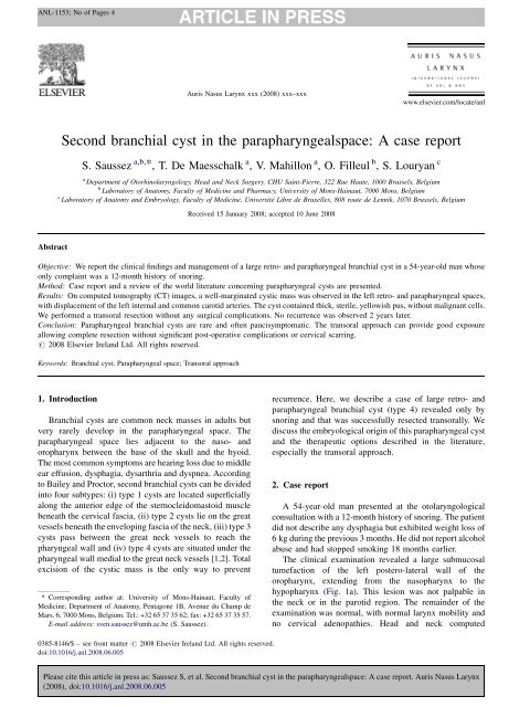
The cl<strong>in</strong>ical exam<strong>in</strong>ation revealed a large submucosal<br />
tumefaction of <strong>the</strong> left postero-lateral wall of <strong>the</strong><br />
oropharynx, extend<strong>in</strong>g from <strong>the</strong> nasopharynx to <strong>the</strong><br />
hypopharynx (Fig. 1a). This lesion was not palpable <strong>in</strong><br />
<strong>the</strong> neck or <strong>in</strong> <strong>the</strong> parotid region. The rema<strong>in</strong><strong>de</strong>r of <strong>the</strong><br />
exam<strong>in</strong>ation was normal, with normal larynx mobility and<br />
no cervical a<strong>de</strong>nopathies. Head and neck computed<br />
Please cite this article <strong>in</strong> press as: Saussez S, et al. <strong>Second</strong> <strong>branchial</strong> <strong>cyst</strong> <strong>in</strong> <strong>the</strong> <strong>parapharyngealspace</strong>: A case report. Auris Nasus Larynx<br />
(2008), doi:10.1016/j.anl.2008.06.005
ANL-1153; No of Pages 4<br />
2<br />
tomography (CT) revealed a 10-cm diameter <strong>cyst</strong> situated<br />
medially to <strong>the</strong> <strong>in</strong>ternal and common carotid arteries,<br />
enlarg<strong>in</strong>g <strong>the</strong> left para- and retropharyngeal spaces (Fig. 1b<br />
and c). An <strong>in</strong>traoral f<strong>in</strong>e-needle aspiration of <strong>the</strong> <strong>cyst</strong><br />
revealed thick, sterile, yellowish pus, without malignant<br />
cells.<br />
After aspiration and dra<strong>in</strong>age of <strong>the</strong> parapharyngeal <strong>cyst</strong>,<br />
we performed a complete transoral resection un<strong>de</strong>r general<br />
anes<strong>the</strong>sia. In fact, we placed <strong>the</strong> patient <strong>in</strong> sup<strong>in</strong>e <strong>de</strong>cubitus<br />
position with forced cervical extension and used an<br />
amygdalectomy autostatic retractor to realize this transoral<br />
S. Saussez et al. / Auris Nasus Larynx xxx (2008) xxx–xxx<br />
Fig. 1. The para- and retropharyngeal <strong>cyst</strong> was observed directly when <strong>the</strong> patient opened his mouth (A). Axial (B) and coronal (C) head and neck CT-scans<br />
show <strong>the</strong> <strong>branchial</strong> <strong>cyst</strong> before <strong>the</strong> resection. The <strong>cyst</strong> filled <strong>the</strong> retropharyngeal and parapharyngeal spaces and displaced <strong>the</strong> left common -black arrow- and<br />
<strong>in</strong>ternal carotid arteries laterally.<br />
resection. First of all, we performed a very superficial<br />
<strong>in</strong>cision – 7–8 cm length – of <strong>the</strong> lateral oropharyngeal wall.<br />
We prolonged this <strong>in</strong>cision <strong>in</strong> <strong>the</strong> lateral part of <strong>the</strong> soft<br />
palate which allowed one to dissect <strong>the</strong> superior –<br />
nasopharyngeal – part of <strong>the</strong> <strong>cyst</strong>. The wall <strong>cyst</strong> was very<br />
th<strong>in</strong> and located just below <strong>the</strong> pharyngeal wall. After that,<br />
we visualized <strong>the</strong> <strong>cyst</strong> wall and aspired this content – not<br />
completely – <strong>de</strong>creas<strong>in</strong>g significantly <strong>the</strong> <strong>cyst</strong> size but<br />
avoid<strong>in</strong>g complete collapsus. Then, us<strong>in</strong>g <strong>the</strong> electro<strong>the</strong>rmal<br />
bipolar vessel sealer, we dissected very carefully <strong>the</strong> space<br />
between <strong>the</strong> lateral <strong>cyst</strong> wall and <strong>the</strong> middle, <strong>in</strong>ferior<br />
Please cite this article <strong>in</strong> press as: Saussez S, et al. <strong>Second</strong> <strong>branchial</strong> <strong>cyst</strong> <strong>in</strong> <strong>the</strong> <strong>parapharyngealspace</strong>: A case report. Auris Nasus Larynx<br />
(2008), doi:10.1016/j.anl.2008.06.005
ANL-1153; No of Pages 4<br />
pharyngeal constrictor muscles. We have teared <strong>the</strong> wall of<br />
<strong>the</strong> <strong>cyst</strong> <strong>in</strong> two places but <strong>the</strong> resection was even complete.<br />
The <strong>in</strong>ferior part of <strong>the</strong> <strong>cyst</strong> was logically <strong>the</strong> more difficult<br />
to dissect. F<strong>in</strong>ally, <strong>the</strong> rest<strong>in</strong>g pharyngeal mucosa was<br />
sutured to <strong>the</strong>se pharyngeal constrictor muscles. We sutured<br />
also <strong>the</strong> <strong>in</strong>cision of <strong>the</strong> soft palate. The patient did not<br />
experience any surgical complications and was able to eat<br />
normally on <strong>the</strong> eighth day. Histopathological exam<strong>in</strong>ation<br />
of <strong>the</strong> resected <strong>cyst</strong> was consistent with a <strong>branchial</strong> <strong>cyst</strong>.<br />
Subsequent head and neck CT, at 2 and 12 months of followup,<br />
showed no recurrence of <strong>the</strong> <strong>cyst</strong> (Fig. 2B). The ENT<br />
exam<strong>in</strong>ation 2 years after surgery confirmed this lack of<br />
recurrence (Fig. 2).<br />
3. Discussion<br />
Parapharyngeal tumors are rare, account<strong>in</strong>g for about 0.5<br />
percent of all head and neck tumors. The ma<strong>in</strong> three groups<br />
of parapharyngeal tumors are salivary (most frequently,<br />
<strong>de</strong>ep-lobe parotid tumors) and neurogenic tumors, followed<br />
by paragangliomas [3]. The differential diagnoses of a<br />
parapharyngeal <strong>cyst</strong>ic mass <strong>in</strong>clu<strong>de</strong> <strong>cyst</strong>ic schwannomas,<br />
m<strong>in</strong>or salivary gland tumors and <strong>branchial</strong> <strong>cyst</strong>s. In this<br />
report, we discuss <strong>the</strong> difficult and challeng<strong>in</strong>g operative<br />
management of this lesion (<strong>branchial</strong> <strong>cyst</strong>s), which <strong>in</strong> our<br />
view must be managed differently from o<strong>the</strong>r solid<br />
parapharyngeal tumors.<br />
Parapharyngeal <strong>branchial</strong> <strong>cyst</strong>s usually cause <strong>the</strong><br />
enlargement of <strong>the</strong> pharynx, result<strong>in</strong>g <strong>in</strong> dysphagia,<br />
dysarthria and, rarely, dyspnea. Depend<strong>in</strong>g on <strong>the</strong> location<br />
of <strong>the</strong> <strong>cyst</strong>, it may compress <strong>the</strong> Eustachian tube,<br />
<strong>de</strong>term<strong>in</strong><strong>in</strong>g hear<strong>in</strong>g loss [4], or cranial nerves IX, X and<br />
XII [5]. More often, symptoms referable to masses <strong>in</strong> <strong>the</strong><br />
parapharyngeal space are m<strong>in</strong>imal: our patient’s only<br />
symptom of this large <strong>cyst</strong> was snor<strong>in</strong>g without any<br />
S. Saussez et al. / Auris Nasus Larynx xxx (2008) xxx–xxx 3<br />
dysphagia. Such a cl<strong>in</strong>ical presentation has never been<br />
<strong>de</strong>scribed <strong>in</strong> <strong>the</strong> literature.<br />
Branchial clefts, fistula and <strong>cyst</strong>s correspond to remnants<br />
of <strong>branchial</strong> grooves and/or pouches. The second, third and<br />
fourth grooves fuse to constitute <strong>the</strong> cervical s<strong>in</strong>us, closed by<br />
an expansion of <strong>the</strong> second arch. Branchial ecto<strong>de</strong>rmal <strong>cyst</strong>s<br />
anterior to <strong>the</strong> sternomastoid muscle <strong>de</strong>rive from a remnant<br />
of <strong>the</strong> cervical s<strong>in</strong>us. They can also <strong>de</strong>velop between this<br />
muscle and <strong>the</strong> <strong>in</strong>ternal jugular ve<strong>in</strong>, between <strong>the</strong> <strong>in</strong>ternal<br />
jugular ve<strong>in</strong> and <strong>the</strong> <strong>in</strong>ternal carotid artery, or f<strong>in</strong>ally<br />
medially to <strong>the</strong> carotid, as <strong>in</strong> our case [6,7]. The orig<strong>in</strong> of<br />
parapharyngeal <strong>cyst</strong>s is controversial. It could correspond to<br />
a remnant of <strong>the</strong> cervical s<strong>in</strong>us, as expla<strong>in</strong>ed above. But this<br />
k<strong>in</strong>d of <strong>cyst</strong> can also arise from endo<strong>de</strong>rmal pharyngeal<br />
tissue. The second <strong>branchial</strong> pouch gives rise to <strong>the</strong> palatal<br />
tonsil. Thus, it could be possible that <strong>the</strong> present <strong>cyst</strong><br />
corresponds to a vestige of this pouch [6,7]. Consi<strong>de</strong>r<strong>in</strong>g <strong>the</strong><br />
presence of subepi<strong>the</strong>lial lymphocytes, Wild and co-authors<br />
proposed a second hypo<strong>the</strong>sis, where parapharyngeal <strong>cyst</strong>s<br />
<strong>de</strong>rived from ectopic epi<strong>the</strong>lial cells <strong>in</strong> regional lymph no<strong>de</strong>s<br />
[8].<br />
From our po<strong>in</strong>t of view, <strong>the</strong> <strong>de</strong>cision to opt for ei<strong>the</strong>r a<br />
transcervical or a transoral approach is <strong>the</strong> real challenge<br />
with this pathology. We <strong>de</strong>ci<strong>de</strong>d to resect this large<br />
parapharyngeal <strong>cyst</strong> transorally, for several reasons that<br />
we <strong>de</strong>scribe hereafter.<br />
First, <strong>the</strong> <strong>cyst</strong> was located medially to <strong>the</strong> <strong>in</strong>ternal carotid<br />
artery, requir<strong>in</strong>g <strong>the</strong> dissection of carotid and jugular vessels<br />
and cranial nerves (l<strong>in</strong>gual, hypoglossal and superior<br />
laryngeal nerves) if a transcervical approach was chosen,<br />
potentially <strong>in</strong>creas<strong>in</strong>g <strong>the</strong> risk of post-operative complications.<br />
Such complications have never been reported when a<br />
transoral approach was used [4,10,11,14]. In our review of<br />
<strong>the</strong> literature, 36 cases have been reported until now and<br />
several approaches used: repeated aspiration and sclerosant<br />
<strong>in</strong>jection (5/36 cases), transcervical approach (20/36 cases),<br />
Fig. 2. Photograph (A) shows <strong>the</strong> patient’s oropharynx after <strong>the</strong> complete resection. (B) Axial head and neck CT confirmed <strong>the</strong> absence of recurrence 12 months<br />
after surgery.<br />
Please cite this article <strong>in</strong> press as: Saussez S, et al. <strong>Second</strong> <strong>branchial</strong> <strong>cyst</strong> <strong>in</strong> <strong>the</strong> <strong>parapharyngealspace</strong>: A case report. Auris Nasus Larynx<br />
(2008), doi:10.1016/j.anl.2008.06.005
ANL-1153; No of Pages 4<br />
4<br />
transcervical–transparotid approach (mandibular luxation<br />
vs. mandibular sw<strong>in</strong>g, 4/36 cases) transoral approach with<br />
total excision (5/36 cases) and marsupialisation (2/36 cases)<br />
[5,9–15]. Simple <strong>in</strong>cision, puncture-aspiration of <strong>the</strong> <strong>cyst</strong>, or<br />
<strong>in</strong>jection of sclerosant substances presents a high risk of<br />
recurrence. Complete resection us<strong>in</strong>g <strong>the</strong> transcervical<br />
approach seems to be <strong>the</strong> technique of choice but several<br />
neurovascular complications (palsies of cranial nerves IX, X<br />
and XII) are <strong>de</strong>scribed [9,12]. In our experience, <strong>the</strong> key<br />
po<strong>in</strong>t of <strong>the</strong> transoral approach was to aspirate <strong>the</strong> <strong>cyst</strong> –<br />
<strong>de</strong>creas<strong>in</strong>g very significantly <strong>the</strong> <strong>cyst</strong> size – before<br />
perform<strong>in</strong>g <strong>the</strong> lateral dissection with a long-electro<strong>the</strong>rmal<br />
bipolar vessel sealer. For a very large <strong>cyst</strong>, even us<strong>in</strong>g a<br />
transcervical approach, <strong>the</strong> reduction of <strong>the</strong> <strong>cyst</strong> size thanks<br />
to an aspiration could be an <strong>in</strong>terest<strong>in</strong>g solution. Moreover,<br />
we realized usually such aspiration for huge cervical <strong>cyst</strong><br />
(type 2) which allowed to <strong>de</strong>crease <strong>the</strong> <strong>in</strong>cision size.<br />
<strong>Second</strong>, <strong>the</strong> transoral approach provi<strong>de</strong>s <strong>the</strong> best<br />
es<strong>the</strong>tic results. Diaz-Manzano recently <strong>de</strong>scribed a<br />
transoral approach allow<strong>in</strong>g marsupialization with postoperative<br />
obliteration of <strong>the</strong> tract [15]. No visible scar was<br />
created us<strong>in</strong>g this transoral approach; this cosmetic<br />
consi<strong>de</strong>ration could be particularly significant for our<br />
young patients. The transparotid and transmandibular<br />
approaches presented <strong>the</strong> poorest aes<strong>the</strong>tic results. F<strong>in</strong>ally,<br />
<strong>the</strong> transcervical approach could be preferred when <strong>the</strong><br />
tumor has a palpable neck component, which <strong>in</strong> practice<br />
means that <strong>the</strong> <strong>cyst</strong> has to present a lateral cervical<br />
extension (type 3 <strong>cyst</strong>) [3,5].<br />
In short, parapharyngeal <strong>branchial</strong> <strong>cyst</strong>s are rare and<br />
often paucisymptomatic. The transoral approach can<br />
provi<strong>de</strong> good exposure allow<strong>in</strong>g complete resection<br />
without significant post-operative complications or cervical<br />
scarr<strong>in</strong>g.<br />
S. Saussez et al. / Auris Nasus Larynx xxx (2008) xxx–xxx<br />
References<br />
[1] Bailey H. The cl<strong>in</strong>ical aspects of <strong>branchial</strong> <strong>cyst</strong>s. Br J Surg<br />
1933;10:173–82.<br />
[2] Proctor B. Lateral vestigial <strong>cyst</strong>s and fistulas of <strong>the</strong> neck. Laryngoscope<br />
1955;65:355–401.<br />
[3] Maran AGD, Mackenzie J, Murray JAM. The parapharyngeal space. J<br />
Laryngol Otol 1984;98:371–80.<br />
[4] Güneri A, Günbay MU, Güneri EA, Ceryan K, Sütay S. Management<br />
of parapharyngeal space <strong>cyst</strong>s. J Laryngol Otol 1994;108:795–7.<br />
[5] Sh<strong>in</strong> HJ, Lee HK, Kim SY, Park HW, Khang SK, Choi CG, et al.<br />
Parapharyngeal second <strong>branchial</strong> <strong>cyst</strong> manifest<strong>in</strong>g as cranial nerve<br />
palsies: MR f<strong>in</strong>d<strong>in</strong>gs. Am J Neuroradiol 2001;22:510–2.<br />
[6] Carlson BM. Human embryology and <strong>de</strong>velopmental biology, 3rd ed.,<br />
Mosby: St. Louis; 2004.<br />
[7] Stevenson RE, Hall J, editors. Human malformations and related<br />
anomalies. 2nd ed., Oxford: Oxford University Press; 2006.<br />
[8] Wild G, Mischke D, Lobeck H, Kastenbauer E. The lateral <strong>cyst</strong> of <strong>the</strong><br />
neck: congenital or acquired. Acta Otolaryngol (Stockh)<br />
1987;103:546–50.<br />
[9] Ostfeld EJ, Wiesel JM, Rab<strong>in</strong>son S, Auslan<strong>de</strong>r L. Parapharyngeal<br />
(retrostyloid)-third <strong>branchial</strong> cleft <strong>cyst</strong>. J Laryngol Otol<br />
1991;105:790–2.<br />
[10] Shidara K, Uruma T, Yasuoka Y, Kamei T. Two cases of nasopharyngeal<br />
<strong>branchial</strong> <strong>cyst</strong>. J Laryngol Otol 1993;107:453–5.<br />
[11] Thaler ER, Tom LW, Handler SD. <strong>Second</strong> <strong>branchial</strong> cleft anomalies<br />
present<strong>in</strong>g as pharyngeal masses. Otolaryngol Head Neck Surg<br />
1993;109:941–4.<br />
[12] Papay FA, Kalucis C, Eliachar I, Tucker HM. Nasopharyngeal presentation<br />
of second <strong>branchial</strong> cleft <strong>cyst</strong>. Otolaryngol Head Neck Surg<br />
1994;110:232–4.<br />
[13] Durrant TJ, Sevick RJ, Lauryssen C, MacRae ME. Parapharyngeal<br />
<strong>branchial</strong> cleft <strong>cyst</strong> present<strong>in</strong>g with cranial nerve palsies. Can Assoc<br />
Radiol J 1994;45:134–6.<br />
[14] Chabot M, Fra<strong>de</strong>t G, Thériault R, Morrissette YP. The excision of<br />
<strong>branchial</strong> parapharyngeal <strong>cyst</strong>s by transbuccal or -cervical approach. J<br />
Otolaryngol 1995;25:108–12.<br />
[15] Diaz-Manzano JA, Sànchez-Mart<strong>in</strong>ez N, Iniesta-Alcàzar J, Med<strong>in</strong>a-<br />
Banegas. A conservative surgical treatment of parapharyngeal <strong>branchial</strong><br />
<strong>cyst</strong>. Auris Nasus Larynx 2008;35:161–4.<br />
Please cite this article <strong>in</strong> press as: Saussez S, et al. <strong>Second</strong> <strong>branchial</strong> <strong>cyst</strong> <strong>in</strong> <strong>the</strong> <strong>parapharyngealspace</strong>: A case report. Auris Nasus Larynx<br />
(2008), doi:10.1016/j.anl.2008.06.005