The Plant Vascular System: Evolution, Development and FunctionsF
The Plant Vascular System: Evolution, Development and FunctionsF
The Plant Vascular System: Evolution, Development and FunctionsF

You also want an ePaper? Increase the reach of your titles
YUMPU automatically turns print PDFs into web optimized ePapers that Google loves.
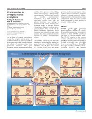
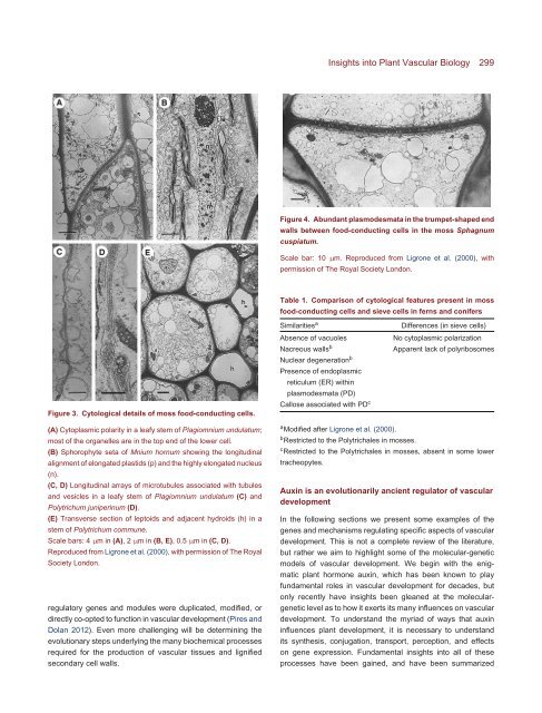
Figure 3. Cytological details of moss food-conducting cells.<br />
(A) Cytoplasmic polarity in a leafy stem of Plagiomnium undulatum;<br />
most of the organelles are in the top end of the lower cell.<br />
(B) Sphorophyte seta of Mnium hornum showing the longitudinal<br />
alignment of elongated plastids (p) <strong>and</strong> the highly elongated nucleus<br />
(n).<br />
(C, D) Longitudinal arrays of microtubules associated with tubules<br />
<strong>and</strong> vesicles in a leafy stem of Plagiomnium undulatum (C) <strong>and</strong><br />
Polytrichum juniperinum (D).<br />
(E) Transverse section of leptoids <strong>and</strong> adjacent hydroids (h) in a<br />
stem of Polytrichum commune.<br />
Scale bars: 4 µm in(A), 2µm in(B, E), 0.5µm in(C, D).<br />
Reproduced from Ligrone et al. (2000), with permission of <strong>The</strong> Royal<br />
Society London.<br />
regulatory genes <strong>and</strong> modules were duplicated, modified, or<br />
directly co-opted to function in vascular development (Pires <strong>and</strong><br />
Dolan 2012). Even more challenging will be determining the<br />
evolutionary steps underlying the many biochemical processes<br />
required for the production of vascular tissues <strong>and</strong> lignified<br />
secondary cell walls.<br />
Insights into <strong>Plant</strong> <strong>Vascular</strong> Biology 299<br />
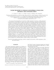
Figure 4. Abundant plasmodesmata in the trumpet-shaped end<br />
walls between food-conducting cells in the moss Sphagnum<br />
cuspiatum.<br />
Scale bar: 10 µm. Reproduced from Ligrone et al. (2000), with<br />
permission of <strong>The</strong> Royal Society London.<br />
Table 1. Comparison of cytological features present in moss<br />
food-conducting cells <strong>and</strong> sieve cells in ferns <strong>and</strong> conifers<br />
Similaritiesa Differences (in sieve cells)<br />
Absence of vacuoles No cytoplasmic polarization<br />
Nacreous wallsb Apparent lack of polyribosomes<br />
Nuclear degenerationb Presence of endoplasmic<br />
reticulum (ER) within<br />
plasmodesmata (PD)<br />
Callose associated with PDc a Modified after Ligrone et al. (2000).<br />
b Restricted to the Polytrichales in mosses.<br />
c Restricted to the Polytrichales in mosses, absent in some lower<br />
tracheopytes.<br />
Auxin is an evolutionarily ancient regulator of vascular<br />
development<br />
In the following sections we present some examples of the<br />
genes <strong>and</strong> mechanisms regulating specific aspects of vascular<br />
development. This is not a complete review of the literature,<br />
but rather we aim to highlight some of the molecular-genetic<br />
models of vascular development. We begin with the enigmatic<br />
plant hormone auxin, which has been known to play<br />
fundamental roles in vascular development for decades, but<br />
only recently have insights been gleaned at the moleculargenetic<br />
level as to how it exerts its many influences on vascular<br />
development. To underst<strong>and</strong> the myriad of ways that auxin<br />
influences plant development, it is necessary to underst<strong>and</strong><br />
its synthesis, conjugation, transport, perception, <strong>and</strong> effects<br />
on gene expression. Fundamental insights into all of these<br />
processes have been gained, <strong>and</strong> have been summarized