de la structure à la croissance cellulaire - Université Bordeaux 1
de la structure à la croissance cellulaire - Université Bordeaux 1
de la structure à la croissance cellulaire - Université Bordeaux 1

You also want an ePaper? Increase the reach of your titles
YUMPU automatically turns print PDFs into web optimized ePapers that Google loves.
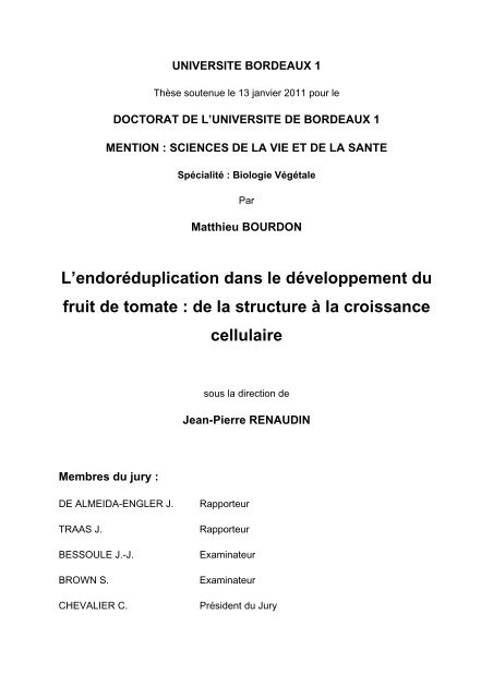
UNIVERSITE BORDEAUX 1<br />
Thèse soutenue le 13 janvier 2011 pour le<br />
DOCTORAT DE L’UNIVERSITE DE BORDEAUX 1<br />
MENTION : SCIENCES DE LA VIE ET DE LA SANTE<br />
Spécialité : Biologie Végétale<br />
Par<br />
Matthieu BOURDON<br />
L’endoréduplication dans le développement du<br />
fruit <strong>de</strong> tomate : <strong>de</strong> <strong>la</strong> <strong>structure</strong> <strong>à</strong> <strong>la</strong> <strong>croissance</strong><br />
Membres du jury :<br />
cellu<strong>la</strong>ire<br />
sous <strong>la</strong> direction <strong>de</strong><br />
Jean-Pierre RENAUDIN<br />
DE ALMEIDA-ENGLER J. Rapporteur<br />
TRAAS J. Rapporteur<br />
BESSOULE J.-J. Examinateur<br />
BROWN S. Examinateur<br />
CHEVALIER C. Prési<strong>de</strong>nt du Jury
Résumé<br />
RESUME<br />
Le développement du fruit <strong>de</strong> tomate s’accompagne d’un phénomène d’endopolyploïdisation<br />
(amplification <strong>de</strong> l’ADN en l'absence <strong>de</strong> mitose) associé <strong>à</strong> <strong>la</strong> <strong>croissance</strong> cellu<strong>la</strong>ire. Au sta<strong>de</strong> vert<br />
mature huit niveaux <strong>de</strong> ploïdie sont présents (2C <strong>à</strong> 256C) dans le péricarpe.<br />
Une première partie du travail a porté sur l’étu<strong>de</strong> <strong>de</strong> <strong>la</strong> distribution spatiale <strong>de</strong>s niveaux <strong>de</strong><br />
ploïdie dans ce tissu. Cet objectif a nécessité <strong>la</strong> mise au point d’une métho<strong>de</strong> originale <strong>de</strong><br />
détermination <strong>de</strong> <strong>la</strong> ploïdie in situ reposant sur <strong>la</strong> technique <strong>de</strong> BAC-FISH. Nous avons montré que les<br />
cellules les plus polyploï<strong>de</strong>s se situent dans les assises internes du péricarpe, et qu’elles sont aussi<br />
les plus gran<strong>de</strong>s. Ces cellules semblent déj<strong>à</strong> formées au moment <strong>de</strong> l’anthèse. Cette cartographie <strong>de</strong><br />
<strong>la</strong> ploïdie associée <strong>à</strong> une analyse <strong>de</strong> <strong>la</strong> taille cellu<strong>la</strong>ire a également montré que <strong>la</strong> taille finale <strong>de</strong>s<br />
cellules ne dépend pas uniquement <strong>de</strong> leur niveau <strong>de</strong> ploïdie mais également <strong>de</strong> leur position dans le<br />
péricarpe. Enfin, nos résultats suggèrent que l’endopolyploïdisation précè<strong>de</strong> <strong>la</strong> <strong>croissance</strong> cellu<strong>la</strong>ire.<br />
Dans une <strong>de</strong>uxième partie du travail, nous avons étudié <strong>la</strong> <strong>structure</strong> <strong>de</strong>s noyaux en<br />
microscopie <strong>à</strong> fluorescence et électronique. L’endopolyploïdisation affecte profondément <strong>la</strong> taille et <strong>la</strong><br />
forme <strong>de</strong>s noyaux, qui acquièrent un volume important et une forme complexe avec <strong>de</strong> profon<strong>de</strong>s<br />
invaginations. La taille du nucléole augmente avec celle du noyau, ce qui suggère une activité <strong>de</strong><br />
transcription accrue. De plus, <strong>la</strong> présence <strong>de</strong> nombreuses mitochondries <strong>à</strong> proximité <strong>de</strong>s noyaux<br />
polyploï<strong>de</strong>s suggère une forte activité métabolique en lien avec l’endopolyploïdisation. L’utilisation <strong>de</strong><br />
<strong>la</strong> métho<strong>de</strong> BAC-FISH a permis également <strong>de</strong> montrer que <strong>la</strong> polyploïdie se faisait par<br />
endoreduplication avec <strong>la</strong> formation <strong>de</strong> chromosomes polytènes.<br />
Dans une troisième partie nous avons cherché, en crib<strong>la</strong>nt une banque <strong>de</strong> mutants Micro-<br />
Tom, <strong>à</strong> i<strong>de</strong>ntifier <strong>de</strong>s lignées affectées dans l’endoreduplication afin d’étudier l’impact <strong>de</strong> ce<br />
phénomène sur <strong>la</strong> vitesse <strong>de</strong> <strong>croissance</strong> du fruit. Nous avons caractérisé plusieurs familles dont les<br />
niveaux moyens <strong>de</strong> ploïdie variaient par rapport <strong>à</strong> <strong>la</strong> lignée <strong>de</strong> référence. Une <strong>de</strong> ces familles présente<br />
un phénotype stable au cours <strong>de</strong> <strong>de</strong>ux générations, avec une augmentation d’au moins 30 % <strong>de</strong> <strong>la</strong><br />
ploïdie moyenne et une augmentation <strong>de</strong> <strong>la</strong> taille <strong>de</strong>s cellules du péricarpe. Cependant cette famille<br />
présentant aussi un développement re<strong>la</strong>tivement parthénocarpique <strong>de</strong> ses fruits, sa caractérisation n’a<br />
pas pu être poursuivie dans le cadre <strong>de</strong> ce travail.<br />
Mots-clés : Tomate, endoréduplication, <strong>croissance</strong> cellu<strong>la</strong>ire, <strong>structure</strong> du noyau<br />
ABSTRACT<br />
Tomato fruit <strong>de</strong>velopment inclu<strong>de</strong>s massive endopolyploidisation events (DNA duplication in<br />
the absence of mitoses) within pericarp cells, in which 8 DNA levels from 2 C to 256 C are <strong>de</strong>tected at<br />
mature green stage.<br />
The first part of this work <strong>de</strong>alt with the study of the spatial distribution of ploidy levels in<br />
pericarp. To achieve this purpose, a new method for in situ ploidy assessment was set up using a<br />
BAC-FISH protocol. The main results are 1/ the most polyploid cells are located in central mesocarp<br />
cell <strong>la</strong>yers; 2/ the most polyploid cells are also the <strong>la</strong>rgest cells; 3/ these cells are likely to be already<br />
present in ovary at anthesis. Ploidy mapping has also shown that the final cell size does not <strong>de</strong>pend<br />
only on ploidy level but also on cell location in pericarp, and that endopolyploidization is likely set up in<br />
tissues before cell expansion.<br />
The <strong>structure</strong> of the polyploid nucleus was studied by using fluorescence microscopy and<br />
electron microscopy. Endopolyploidization profoundly modifies the size and shape of nuclei, which<br />
become much <strong>la</strong>rger and acquire a complex shape with <strong>de</strong>ep invaginations. Nucleolus size increases,<br />
which is likely re<strong>la</strong>ted to transcriptional increase. Moreover, the presence of numerous mitochondria in<br />
the close vicinity of the nuclear membrane reinforces the hypothesis of increased nuclear and<br />
metabolic activity in polyploid cells. The BAC-FISH in situ method for ploidy assessment also revealed<br />
that endopolyploidization procee<strong>de</strong>d through polyteny.<br />
In the <strong>la</strong>st part of this work, we screened a tomato Micro-Tom tilling bank for mutants affected<br />
in endopolyploidization. The aim was to use tomato lines with distinct ploidy levels to check the<br />
influence of ploidy on fruit growth rate. Several mutant families were i<strong>de</strong>ntified with mo<strong>de</strong>rately<br />
increased ploidy levels. One of these families exhibited transmissible phenotype through 2<br />
generations, with ploidy increased by ca. 30 % and increased pericarp cell size. As these mutants had<br />
also a strongly pronounced parthenocarpic phenotype, their characterization could not be further<br />
advanced in the frame of this work.<br />
Keywords : Tomato, endoreduplication, cell growth, nuclei <strong>structure</strong>
Remerciements<br />
Ce travail a été réalisé <strong>à</strong> l’Institut National <strong>de</strong> <strong>la</strong> recherche agronomique, dans<br />
l’équipe Organogenèse du fruit et Endoréduplication au sein <strong>de</strong> l’UMR 619 Biologie<br />
du fruit. A ce titre j’aimerais vivement remercier Christian Chevalier et Dominique<br />
Rolin, pour avoir permis mon intégration au sein <strong>de</strong> leur équipe et <strong>la</strong>boratoire,<br />
respectivement.<br />
Je remercie aussi mon directeur <strong>de</strong> thèse, M. Jean-Pierre Renaudin, pour avoir<br />
encadré cette thèse.<br />
Je tiens aussi <strong>à</strong> remercier Mme J. De Almeida Engler et M. J. Traas, qui me<br />
font l’honneur <strong>de</strong> juger ce travail en tant que rapporteurs ainsi que M. S. Brown, J.-J.<br />
Bessoule et C. Chevalier en tant qu’examinateurs.<br />
Je tiens enfin <strong>à</strong> remercier tout particulièrement Mlle N. Frangne et Mme C.<br />
Cheniclet, pour leur encadrement actif <strong>de</strong> cette thèse et pour les modèles<br />
scientifiques et humains qu’elles ont représentés pour moi durant cette thèse. Je les<br />
remercie aussi chaleureusement ainsi que M. C. Chevalier pour l’ai<strong>de</strong> inestimable<br />
qu’ils ont pu m’apporter lors <strong>de</strong> <strong>la</strong> rédaction <strong>de</strong> ce manuscrit.<br />
De même, je tiens <strong>à</strong> apporter mes plus grands remerciements <strong>à</strong> :<br />
* Olivier Coriton, pour son enseignement en cytogénétique, sa participation<br />
active dans nos nombreuses réflexions scientifiques et sa réactivité légendaire <strong>à</strong><br />
toutes nos questions…<br />
* Spencer Brown, pour son ai<strong>de</strong> inestimable en cytométrie/microscopie et son<br />
approche si unique <strong>de</strong> <strong>la</strong> science et <strong>de</strong> <strong>la</strong> vie. Il reste <strong>à</strong> ce jour trop peu <strong>de</strong><br />
scientifiques aussi mordus que lui étant capables <strong>de</strong> vous transmettre et <strong>de</strong> vous<br />
faire vivre sa passion en moins <strong>de</strong> 2 phrases…<br />
* Ronan Piriou, ami, coloc, <strong>à</strong> qui revient l’immense mérite <strong>de</strong> m’avoir supporté,<br />
porté et aidé <strong>à</strong> tous niveaux dans cette entreprise qui a connu <strong>de</strong>s hauts mais aussi<br />
bien <strong>de</strong>s bas… Merci mille fois mon ami !<br />
*Mireia Noguera, quien me ha ayudado a sobrellevar <strong>la</strong> soledad en <strong>la</strong> etapa <strong>de</strong><br />
redacción, consiguiendo siempre hacerme sonreír <strong>de</strong><strong>la</strong>nte <strong>de</strong> mi or<strong>de</strong>nador… y<br />
aportándome más, mucho más…
* El Tomate, el cual me ha permitido disminuir mi estrés durante <strong>la</strong> redacción y<br />
a<strong>de</strong>más ver este fruto <strong>de</strong>s<strong>de</strong> un punto <strong>de</strong> vista más divertido, sobre todo en los<br />
peores momentos…<br />
* Audrey Abot, pour les longs entretiens téléphoniques <strong>de</strong> vidage <strong>de</strong> sac et <strong>de</strong><br />
tête…<br />
* Yoan Jacquemin, ami <strong>de</strong> toujours, pour tous les moments passés <strong>de</strong>puis le<br />
premier jour sur les bancs <strong>de</strong> <strong>la</strong> Fac jusqu’au diplôme final.<br />
* Stève <strong>de</strong> Bossoreille <strong>de</strong> Ribou et son in<strong>de</strong>scriptible calme olympien sans qui<br />
je n’aurais jamais connu Fe<strong>la</strong>.<br />
* Isaias, pour sa compagnie inestimable lors <strong>de</strong> <strong>la</strong> rédaction en tant que 2 e<br />
coloc’ et son remp<strong>la</strong>cement effectif pour quasiment toutes mes tâches ménagères<br />
durant cette pério<strong>de</strong>…<br />
* Mes parents (Martine et Jean-Michel Bourdon) et mon petit frère (Alexandre<br />
Bourdon) ; jamais je n’aurais pu réaliser ce travail sans leur soutien et leur confiance<br />
indéfectibles.<br />
* Mes collègues : Duyen Prodhomme et Lisa Boureau pour les interminables<br />
discussions entreprises ; Je n’oublierais pas non plus Nico<strong>la</strong>s Viron et son côté pince<br />
sans rire et pince tout court ; Antoine Monier, Calimero <strong>de</strong>vant l’éternel ; Christian<br />
Kappel, pour ces dégustations vins et fromage inoubliables ; les Fred pour leurs<br />
insondables conneries ; Guil<strong>la</strong>ume Ménard pour ces b<strong>la</strong>gues douteuses <strong>à</strong> souhait et<br />
ses changements <strong>de</strong> fond d’écran inopinés ; Mehdi Nafati et Thomas Guiraud pour<br />
les moments les plus studieux entrecoupés <strong>de</strong> discussions tout aussi studieuses ;<br />
Michel Hernould pour son inimitable Humour (oui oui avec un grand « H » !), Yves<br />
Gibon pour son apport ineffable <strong>à</strong> <strong>la</strong> vie sociale du <strong>la</strong>boratoire via sa passion pour le<br />
baby-foot et enfin encore Nathalie Frangne pour son soutien tant scientifique que<br />
moral durant cette thèse et pour toutes nos digressions sans fin lors <strong>de</strong> nos<br />
« pseudo » réunions scientifiques.<br />
* Aux colocs successifs <strong>de</strong> l’inimitable « 46 rue Brémontier » : Julie, Bogdan,<br />
Jérem’, Pierrot, Orane, Alex, Camille, Fanny et Théo.<br />
* Enfin <strong>à</strong> tous mes amis <strong>de</strong> Bor<strong>de</strong>aux : Marine, Dimitri, Evelyne, Stéphane,<br />
Camille, David, Thibaut, Aurel’, Cous’, Thierry, Sarah, Mathieu, et tous les autres…
Sommaire<br />
Abréviations………………………………………………………………………………………………...p1<br />
CHAPITRE 1 : INTRODUCTION……………………………………………….……….…...p2<br />
PARTIE 1 : Influence <strong>de</strong>s variations <strong>de</strong> ploïdie dans l’évolution………………….………...……….p3<br />
1.1. Reproduction sexuée, ploïdie et cycle <strong>de</strong> vie ……………………………………….....………....p3<br />
1.2. Reproduction sexuée, polyploïdisation et spéciation……………………………………..….…..p6<br />
1.3. Cycle cellu<strong>la</strong>ire, endopolyploïdisation et <strong>croissance</strong>…………………………………….……….p9<br />
REFERENCES INTRODUCTION PARTIE 1……………………………………………………………...p15<br />
PARTIE 2 : Chapitre d’ouvrage : Endoreduplication and growth of fleshy fruits…………….….p18<br />
PARTIE 3 : Endopolyploïdisation : conséquences structurales et implications…………….…..p51<br />
fonctionnelles<br />
3.1. Endopolyploïdisation, différenciation, <strong>croissance</strong> et modalités d’action…………..………….p51<br />
3.2. Influence <strong>de</strong> l’endopolyploïdisation sur l’expression génétique…………….…..…………..…..p53<br />
3.2.1. Augmentation globale du niveau <strong>de</strong> transcription...................................................p53<br />
via l’amplification fonctionnelle du génome.<br />
3.2.2. Endopolyploïdie et régu<strong>la</strong>tion <strong>de</strong> l’expression génétique…………………..………….p54<br />
3.2.3. Spatialisation du génome et expression génétique…………………………….………p56<br />
3.3. Caractéristiques cellu<strong>la</strong>ires <strong>de</strong> <strong>la</strong> cellule polyploï<strong>de</strong>………………………………………….…..p62<br />
3.3.1. Organisation et différenciation <strong>de</strong> l’enveloppe …………………………………..……..p62<br />
nucléaire lors <strong>de</strong> l’endopolyploïdisation<br />
3.3.2. Organisation et évolution du cytop<strong>la</strong>sme en …………………………………….……..p64<br />
re<strong>la</strong>tion avec l’endopolyploïdie<br />
3.4. Objectifs du travail <strong>de</strong> thèse…………………………………………………..………………..…..p66<br />
REFERENCES INTRODUCTION PARTIE 3……………………………………………….……………..p68
CHAPITRE 2 : RESULTATS………………………………………………………………….p74<br />
PARTIE 1 : ARTICLE 1 :.............................................................................................................p75<br />
In p<strong>la</strong>nta quantification of endoreduplication using Fluorescent In Situ Hybridization (FISH)<br />
PARTIE 2 : ARTICLE 2……………………………………………………………………………...……..p109<br />
Structural analysis of endopolyploid nuclei from tomato (So<strong>la</strong>num lycopersicum) fruit cells<br />
PARTIE 3 : CRIBLAGE DE LA BANQUE DE MUTANTS TILLING MICRO-TOM……………..…..p140<br />
Recherche <strong>de</strong> mutants affectés dans l’endoréduplication<br />
CHAPITRE 3 : DISCUSSION – CONCLUSION..............................................p165<br />
1. Contexte et problématique……………………………………………………………..…………….p165<br />
2. Implication <strong>de</strong> l’endopolyploïdisation dans <strong>la</strong> <strong>croissance</strong> cellu<strong>la</strong>ire………..……………….p166<br />
3. Amplification fonctionnelle du génome et profils d’expression……………………..………..p171<br />
associés <strong>à</strong> l’endopolyploïdisation<br />
4. Supra-organisation et ergonomie <strong>de</strong> <strong>la</strong> cellule endopolyploï<strong>de</strong>……………………...……….p172<br />
5. CONCLUSION……………………………………………………………………………………..……p178<br />
REFERENCES DISCUSSION – CONCLUSION……………………………………………..…......…..p179<br />
ANNEXES : PRODUCTION SCIENTIFIQUE.......................................................p181
Abréviations<br />
Aci<strong>de</strong>s nucléiques et nucléoti<strong>de</strong>s<br />
ADN Aci<strong>de</strong> désoxyribonucléique<br />
ADNg Aci<strong>de</strong> désoxyribonucléique génomique<br />
ARN Aci<strong>de</strong> ribonucléique<br />
ARNm Aci<strong>de</strong> ribonucléique messager<br />
ARNr Aci<strong>de</strong> ribonucléique ribosomal<br />
ARNt Aci<strong>de</strong> ribonucléique <strong>de</strong> transfert<br />
ATP A<strong>de</strong>nosine 5’ triphosphate<br />
Kb, Mb kilobase, mégabase<br />
kDa, MDa Kilodalton, Mégadalton<br />
RNA Pol II RNA polymérase II<br />
TC Territoire chromosomique<br />
Unités<br />
°C <strong>de</strong>gré Celsius<br />
g accélération<br />
rpm round per minute<br />
s, min, h secon<strong>de</strong>, minute, heure<br />
EI Endoreduplication In<strong>de</strong>x<br />
MCV : Mean C Value<br />
Divers<br />
2-D , 3-D <strong>de</strong>ux dimensions; troiss dimensions<br />
GFP Green Fluorescent Protein<br />
BY-2 lignée cellu<strong>la</strong>ire <strong>de</strong> Tabac ayant pour origine le cultivar BY-2 (Bright Yellow<br />
– 2)<br />
RE Réticulum Endop<strong>la</strong>smique<br />
EMS Ethyl Methyl Sulfonate<br />
BSA Bovine Serum Albumin<br />
cv Cultivar<br />
DEPC Diethylpyrocarbonate<br />
DAPI 4’,6-diamino-2-phenylindole<br />
FISH Fluorescent in situ Hybridization<br />
SSC tampon ”saline-sodium citrate”<br />
TBS tampon “tris buffer sakine”<br />
NOR Nucleo<strong>la</strong>r Organizing Regions<br />
DIOC6(3), Dihexyloxacarbocyanine iodi<strong>de</strong><br />
TEM Transmission Electron Microscopy<br />
- 1 -
CHAPITRE 1 : INTRODUCTION<br />
Chez les eucaryotes, <strong>la</strong> réplication <strong>de</strong> l’information génétique est assurée au<br />
cours d’un cycle cellu<strong>la</strong>ire aboutissant <strong>à</strong> 2 processus <strong>de</strong> division nucléaire: <strong>la</strong> mitose<br />
et <strong>la</strong> méiose. Quel que soit le processus, le maintien conservatif <strong>de</strong> cette information<br />
passe alors par l’alternance <strong>de</strong> phases <strong>de</strong> ploïdies distinctes n et 2n. En<br />
conséquence, les eucaryotes ont développé au cours <strong>de</strong> l’évolution une tolérance<br />
envers les changements <strong>de</strong> ploïdie (Gerstein and Otto 2009). Cette tolérance a sans<br />
doute permis l’émergence d’autres évènements conduisant <strong>à</strong> <strong>de</strong>s modifications <strong>de</strong><br />
ploïdie, pérennisés soit au sein d’organes ou <strong>de</strong> lignées cellu<strong>la</strong>ires<br />
(endopolyploïdisation), soit au sein d’organismes entiers (auto- et allo-<br />
polyploïdisation). Ce faisant, les variations <strong>de</strong> ploïdie ont une importance non<br />
négligeable dans l’histoire évolutive <strong>de</strong>s eucaryotes, influençant <strong>la</strong> mise en p<strong>la</strong>ce<br />
d’un cycle <strong>de</strong> reproduction particulier, d’un phénotype ou d’un organe, voire<br />
favorisant l’apparition <strong>de</strong> nouvelles espèces.<br />
Nous développerons dans une première partie l’influence évolutive <strong>de</strong>s<br />
variations <strong>de</strong> ploïdie les plus représentatives chez les eucaryotes. Dans une<br />
<strong>de</strong>uxième partie, nous insisterons sur une forme particulière <strong>de</strong> changement <strong>de</strong><br />
ploïdie, l’endopolyploïdisation, et sur le rôle qu’elle pourrait jouer au sein d’un organe<br />
végétal particulier, <strong>à</strong> savoir le fruit. Nous ferons ensuite le point sur l’impact<br />
molécu<strong>la</strong>ire et cellu<strong>la</strong>ire <strong>de</strong> ce phénomène, et nous terminerons cette introduction en<br />
présentant les objectifs <strong>de</strong> ce travail, visant <strong>à</strong> mieux cerner le rôle <strong>de</strong><br />
l’endopolyploidisation au cours du développement du fruit <strong>de</strong> Tomate.<br />
- 2 -
PARTIE 1 : Influence évolutive <strong>de</strong>s variations <strong>de</strong> ploïdie<br />
dans l’évolution<br />
1.1. Reproduction sexuée, ploïdie et cycle <strong>de</strong> vie<br />
L’apparition <strong>de</strong> <strong>la</strong> méiose, et donc <strong>de</strong> <strong>la</strong> reproduction sexuée, outre <strong>la</strong> variabilité<br />
génétique qu’elle apporte par le jeu <strong>de</strong>s recombinaisons entre chromosomes, a<br />
entraîné l’alternance <strong>de</strong> générations (cycles <strong>de</strong> vie) haploï<strong>de</strong>s et diploï<strong>de</strong>s (Mable<br />
and Otto 1998) (Figure 1).<br />
Chez les Embryophytes l’évolution tend vers une réduction <strong>de</strong> <strong>la</strong> durée <strong>de</strong> <strong>la</strong><br />
phase haploï<strong>de</strong> et une dominance <strong>de</strong> <strong>la</strong> phase diploï<strong>de</strong> (Figure 2). Une telle<br />
observation suggère donc un avantage sélectif <strong>de</strong> <strong>la</strong> diploïdie envers l’haploïdie. En<br />
effet, étant donné que <strong>la</strong> plupart <strong>de</strong>s mutations affectant négativement <strong>la</strong> valeur<br />
adaptative (fitness) sont partiellement récessives, et que l’apparition d’allèles<br />
mutants dans une popu<strong>la</strong>tion est rare, alors il est improbable qu’un individu diploï<strong>de</strong><br />
provenant d’un croisement aléatoire porte les <strong>de</strong>ux copies mutantes du même allèle.<br />
A l’inverse, <strong>de</strong>s individus haploï<strong>de</strong>s expriment chacune <strong>de</strong>s mutations <strong>de</strong> leur<br />
génome. De cette manière, <strong>de</strong>s individus <strong>à</strong> phase diploï<strong>de</strong> dominante auraient une<br />
meilleure valeur adaptative, et <strong>la</strong> génération diploï<strong>de</strong> serait sélectivement favorisée.<br />
Cependant, <strong>la</strong> contrepartie <strong>à</strong> une telle « stratégie » évolutive, bénéfique pour <strong>la</strong><br />
survie <strong>de</strong> l’individu, est représentée par <strong>la</strong> fixation d’allèles mutants dans <strong>la</strong><br />
<strong>de</strong>scendance. Par conséquent, les popu<strong>la</strong>tions haploï<strong>de</strong>s ten<strong>de</strong>nt <strong>à</strong> porter moins <strong>de</strong><br />
mutations délétères dans leur génome et auraient alors une meilleure valeur<br />
adaptative <strong>à</strong> l’équilibre que les popu<strong>la</strong>tions diploï<strong>de</strong>s.<br />
- 3 -
- 4 -
Une autre hypothèse avancée pour expliquer l’avantage <strong>de</strong> <strong>la</strong> diploïdie<br />
concerne <strong>la</strong> vitesse d’évolution. Un individu diploï<strong>de</strong> possè<strong>de</strong> <strong>de</strong>ux copies du<br />
génome haploï<strong>de</strong>, donc si <strong>la</strong> probabilité <strong>de</strong> mutation est constante, alors un génome<br />
diploï<strong>de</strong> subira <strong>de</strong>ux fois plus d’évènements <strong>de</strong> mutations que son pendant haploï<strong>de</strong>.<br />
Cependant, le bénéfice <strong>de</strong> ces mutations pourrait aussi être masqué par le jeu <strong>de</strong><br />
dominance/récessivité imposé par <strong>la</strong> paire d’allèles présente chez les diploï<strong>de</strong>s. En<br />
ce sens, le gain apporté par <strong>la</strong> mutation ne pourrait accroître <strong>la</strong> vitesse d’évolution du<br />
diploï<strong>de</strong> contre celle <strong>de</strong> l’haploï<strong>de</strong> que si <strong>la</strong> mutation bénéfique s’avère dominante.<br />
Malheureusement, peu <strong>de</strong> données expérimentales permettent d’expliquer <strong>de</strong><br />
façon satisfaisante l’adoption d’un cycle <strong>de</strong> vie plutôt qu’un autre. La sauvegar<strong>de</strong> <strong>de</strong><br />
- 5 -
cycles <strong>de</strong> vie très variés au cours <strong>de</strong> l’évolution <strong>de</strong>s eucaryotes suggère qu’il n’existe<br />
pas d’avantage évolutif définitif d’une phase chromosomique sur une autre. En<br />
revanche, le <strong>la</strong>rge <strong>de</strong>gré <strong>de</strong>s variations observées suggère que <strong>la</strong> stratégie <strong>de</strong> cycle<br />
<strong>de</strong> vie peut être vue comme un caractère variable encore soumis <strong>à</strong> <strong>de</strong>s modifications<br />
évolutives. Ce faisant, il offre l’opportunité pour une espèce donnée d’évoluer vers<br />
une tendance plutôt qu’une autre en fonction <strong>de</strong> <strong>la</strong> pression sélective du milieu, <strong>la</strong><br />
tendance retenue étant <strong>la</strong> forme <strong>la</strong> plus <strong>à</strong> même d’assurer <strong>la</strong> survie <strong>de</strong> l’espèce au<br />
sein <strong>de</strong> ce milieu.<br />
1.2. Reproduction sexuée, polyploïdisation et spéciation<br />
Outre l’alternance <strong>de</strong> phases haploï<strong>de</strong> et diploï<strong>de</strong> au cours du cycle <strong>de</strong> vie d’un<br />
organisme, d’autres situations peuvent aboutir <strong>à</strong> l’apparition d’évènements <strong>de</strong><br />
polyploïdisation produisant <strong>de</strong>s individus avec une ou plusieurs copies du ou <strong>de</strong>s<br />
génomes parentaux. Deux types <strong>de</strong> polyploïdisations sont distingués dans ce cadre :<br />
l’autopolyploïdisation et l’allopolyploïdisation, selon qu’elles concernent le<br />
rassemblement <strong>de</strong> génomes diploï<strong>de</strong>s semb<strong>la</strong>bles ou différents (hybridation intra- ou<br />
inter-spécifique) (Otto 2007, Otto and Whitton 2000). Elles peuvent provenir d’une<br />
non-réduction gamétique (erreur <strong>de</strong> déroulement <strong>de</strong> <strong>la</strong> méiose), d’un doublement<br />
génomique (erreur <strong>de</strong> déroulement <strong>de</strong> <strong>la</strong> mitose) ou <strong>de</strong> polyspermie (plusieurs<br />
gamètes mâles fécondant un ovule) (Gerstein and Otto 2009). Le mo<strong>de</strong> <strong>de</strong><br />
polyploïdisation privilégié chez les p<strong>la</strong>ntes semble être <strong>la</strong> non-réduction gamétique,<br />
alors qu’au sein du règne animal, ce mécanisme semble aussi dû <strong>à</strong> <strong>la</strong> polyspermie.<br />
Cependant, chez les mammifères et les oiseaux, il semble moins toléré en raison <strong>de</strong><br />
problèmes d’empreintes génétiques et <strong>de</strong> développement p<strong>la</strong>cental. Chez les<br />
p<strong>la</strong>ntes, <strong>la</strong> fréquence d’apparition <strong>de</strong> ce phénomène semble importante. Afin<br />
d’estimer le nombre d’évènements <strong>de</strong> polyploïdisation ancestraux, le nombre <strong>de</strong><br />
chromosomes au sein <strong>de</strong> chaque espèce considérée a été mesuré et l’excès d’un<br />
- 6 -
nombre pair <strong>de</strong> chromosomes par rapport aux nombres impairs est considéré<br />
comme un bon indicateur d’évènements <strong>de</strong> polyploïdisation passés (Otto, 2007). En<br />
effet, si l’on part du postu<strong>la</strong>t que le nombre <strong>de</strong> chromosomes n d’un génome a autant<br />
<strong>de</strong> chances d’être un chiffre pair ou impair, alors l’observation d’un excès <strong>de</strong><br />
nombres pairs dans un lot d’espèces donné reflète un nombre important<br />
d’évènements <strong>de</strong> polyploïdisations. Selon ce critère, 42 % <strong>de</strong>s fougères, 32 % <strong>de</strong>s<br />
monocotylédones et 18 % <strong>de</strong>s dicotylédones auraient subi au moins un évènement<br />
<strong>de</strong> polyploïdisation au cours <strong>de</strong> leur évolution.<br />
L’apport évolutif <strong>de</strong> ces mécanismes reste, comme les variations <strong>de</strong>s cycles <strong>de</strong><br />
vie évoquées plus haut, difficile <strong>à</strong> déterminer. On peut reprendre une partie <strong>de</strong>s<br />
arguments cités dans <strong>la</strong> partie précé<strong>de</strong>nte comme le masquage <strong>de</strong>s mutations<br />
délétères et l’augmentation <strong>de</strong> <strong>la</strong> vitesse d’évolution, cependant les contreparties<br />
évoquées restent les mêmes. Ainsi, <strong>la</strong> vitesse d’évolution <strong>de</strong>s polyploï<strong>de</strong>s serait<br />
accrue seulement si les mutations bénéfiques y ayant lieu présentent un caractère<br />
dominant.<br />
On peut alors se <strong>de</strong>man<strong>de</strong>r pourquoi l’évolution semble avoir conservé autant<br />
d’organismes présentant <strong>de</strong>s évènements <strong>de</strong> polyploïdisation ? Tout d’abord par un<br />
apport <strong>de</strong> nouveaux gènes et <strong>de</strong> nouveaux allèles, favorisant <strong>la</strong> variabilité génétique<br />
et potentiellement le pouvoir adaptatif <strong>de</strong> l’individu néo-formé. Même si l’apport <strong>de</strong><br />
variabilité semble plus important dans le cas <strong>de</strong> l’allopolyploïdisation puisque<br />
rassemb<strong>la</strong>nt <strong>de</strong>s génomes d’espèces différentes, l’autopolyploïdisation n’est pas en<br />
reste en termes <strong>de</strong> variabilité apportée en réalisant une combinaison complexe et<br />
inédite d’allèles provenant <strong>de</strong>s <strong>de</strong>ux parents et favorisant ainsi l’hétérozygotie<br />
(Osborn et al. 2003). Ensuite, le doublement du génome offre <strong>la</strong> potentialité <strong>de</strong> néo-<br />
fonctionnalisation <strong>de</strong> gènes tout en conservant les fonctions d’origine. L’apparition <strong>de</strong><br />
- 7 -
nouvelles fonctions pourrait donc permettre une meilleure adaptation <strong>à</strong><br />
l’environnement via un spectre <strong>de</strong> réponses biologiques plus <strong>la</strong>rge.<br />
Enfin, les polyploï<strong>de</strong>s nouvellement formés ont un génome plutôt instable et<br />
subissent beaucoup <strong>de</strong> réarrangements chromosomiques (Wen<strong>de</strong>l, 2000). En effet,<br />
<strong>de</strong>s recombinaisons non-homologues sont hautement probables entre chromosomes<br />
homéologues (cas <strong>de</strong>s allopolyploï<strong>de</strong>s). En altérant ainsi le contexte génomique <strong>de</strong><br />
certains gènes, ces réarrangements peuvent alors augmenter <strong>la</strong> variabilité génétique<br />
<strong>de</strong> <strong>la</strong> lignée polyploï<strong>de</strong> nouvellement formée. On observe aussi une forte variation<br />
dans l’expression génétique, souvent associée <strong>à</strong> <strong>la</strong> divergence <strong>de</strong>s génomes<br />
parentaux (et donc <strong>à</strong> l’allopolyploïdie). Inversement, une plus faible variation <strong>de</strong><br />
l’expression est attendue chez les autopolyploï<strong>de</strong>s (Galitski et al. 1999, Guo et al.<br />
1996)<br />
Ainsi, le rôle <strong>de</strong> <strong>la</strong> polyploïdisation dans l’évolution pourrait s’effectuer par <strong>la</strong><br />
néo-fonctionnalisation <strong>de</strong> gènes par duplication du génome, et par <strong>la</strong> variabilité<br />
génétique apportée par l’allopolyploïdisation et l’autopolyploïdisation. Le potentiel<br />
évolutif offert par ce phénomène, engendrant <strong>de</strong>s lignées génétiquement différentes<br />
<strong>de</strong> leurs géniteurs capables d’envahir <strong>de</strong> nouvelles niches écologiques serait alors <strong>à</strong><br />
l’origine <strong>de</strong> <strong>la</strong> réussite évolutive <strong>de</strong>s polyploï<strong>de</strong>s. En ce sens, les évènements <strong>de</strong><br />
polyploïdisation sont considérés comme un mécanisme majeur dans <strong>la</strong> création <strong>de</strong><br />
nouvelles espèces, et plus particulièrement les évènements d’allopolyploïdisation<br />
(Soltis and Soltis 2009).<br />
Nous avons ainsi vu que le génome eucaryote rece<strong>la</strong>it une certaine p<strong>la</strong>sticité lui<br />
permettant <strong>de</strong> gérer <strong>de</strong>s doublements génomiques. Nous terminerons cette première<br />
partie en abordant un mo<strong>de</strong> <strong>de</strong> polyploïdisation dont l’impact adaptatif et/ou évolutif a<br />
été moins discuté : l’endopolyploïdisation.<br />
- 8 -
1.3. Cycle cellu<strong>la</strong>ire, endopolyploïdisation et <strong>croissance</strong><br />
L’existence <strong>de</strong> l’endopolyploïdisation illustre <strong>la</strong> notion <strong>de</strong> flexibilité du cycle<br />
nucléaire eucaryote. Ce phénomène consiste en l’augmentation locale du nombre <strong>de</strong><br />
copies d’un génome par cellule. Il résulte d’un arrêt programmé du cycle cellu<strong>la</strong>ire<br />
postérieurement <strong>à</strong> <strong>la</strong> phase <strong>de</strong> synthèse <strong>de</strong> l’ADN et antérieurement <strong>à</strong> <strong>la</strong> phase <strong>de</strong><br />
mitose, doub<strong>la</strong>nt ainsi <strong>la</strong> quantité d’ADN <strong>à</strong> chaque endocycle et conduisant donc <strong>à</strong><br />
l’apparition <strong>de</strong> plusieurs niveaux <strong>de</strong> ploïdie au sein d’un même organisme (Barow<br />
2006, Edgar and Orr-Weaver 2001, Joubès and Chevalier 2000) (Figure 3). Ce type<br />
<strong>de</strong> polyploïdie ayant lieu au sein <strong>de</strong> tissus somatiques, elle est aussi appelée<br />
polyploïdie terminale et serait fortement impliquée dans <strong>la</strong> <strong>croissance</strong> cellu<strong>la</strong>ire et <strong>la</strong><br />
différenciation (Edgar and Orr-Weaver 2001, Hulskamp 2004). On <strong>la</strong> retrouve dans<br />
<strong>de</strong> nombreux taxons pluricellu<strong>la</strong>ires incluant les algues, les champignons, les p<strong>la</strong>ntes<br />
et les animaux (Figure 4).<br />
Chez les animaux, (Gregory and Hebert 1999) mentionnent <strong>la</strong> présence <strong>de</strong><br />
l’endopolyploïdisation dans chaque espèce animale testée avec une prévalence chez<br />
les arthropo<strong>de</strong>s. En effet, les insectes paraissent les plus concernés par ce<br />
phénomène, avec les exemples bien connus <strong>de</strong>s g<strong>la</strong>n<strong>de</strong>s salivaires et <strong>de</strong>s cellules<br />
nourricières <strong>de</strong>s <strong>la</strong>rves <strong>de</strong> drosophile et <strong>de</strong> Bombyx mori. Chez les mammifères, les<br />
tissus et/ou cellules les plus étudiés ayant recours <strong>à</strong> ce mécanisme sont les<br />
mégakaryocytes (précurseurs <strong>de</strong>s p<strong>la</strong>quettes sanguines, (Raslova et al. 2007)), les<br />
hépatocytes (cellules du foie (Lu et al. 2007)) et les cellules du trophob<strong>la</strong>ste (tissu<br />
nourricier <strong>de</strong> l’embryon, (Varmuza et al. 1988)). Les <strong>de</strong>grés d’endopolyploïdisation<br />
sont variables et dépen<strong>de</strong>nt <strong>de</strong> l’organisme et même du tissu considéré.<br />
- 9 -
En effet, alors que les mégakaryocytes et les hépatocytes atteignent <strong>de</strong>s niveaux <strong>de</strong><br />
ploïdie assez faibles (32C et 8C, (Raslova, et al. 2007, Vinogradov et al. 2001)), les<br />
cellules <strong>de</strong>s g<strong>la</strong>n<strong>de</strong>s salivaires <strong>de</strong> <strong>la</strong>rve <strong>de</strong> drosophile et du trophob<strong>la</strong>ste <strong>de</strong><br />
mammifères peuvent atteindre <strong>de</strong>s valeurs <strong>de</strong> 1024C (Smith and Orr-Weaver 1991,<br />
Zybina and Zybina 1996) et le cas le plus extrême est celui <strong>de</strong>s cellules <strong>de</strong>s g<strong>la</strong>n<strong>de</strong>s<br />
salivaires productrices <strong>de</strong> soie <strong>de</strong> Bombyx mori atteignant 10 6 C (D'Amato and<br />
Durante 2001, Gregory and Hebert 1999). En fonction <strong>de</strong> l’étape <strong>à</strong> <strong>la</strong>quelle le cycle<br />
cellu<strong>la</strong>ire est stoppé, on peut distinguer plusieurs types d’endocycles : l’arrêt est<br />
antérieur <strong>à</strong> <strong>la</strong> métaphase, les chromati<strong>de</strong>s filles restent attachées au même<br />
centromère et l’endocycle est appelé cycle endoréduplicatif (Figure 3a et 3b,<br />
- 10 -
exemples <strong>de</strong>s cellules nourricières <strong>de</strong> l’ovaire <strong>de</strong> drosophile et du trophob<strong>la</strong>ste<br />
mammifère) ; l’arrêt est postérieur <strong>à</strong> <strong>la</strong> métaphase, les chromati<strong>de</strong>s filles sont<br />
séparées les unes <strong>de</strong>s autres et l’endocycle est nommé alors cycle endomitotique<br />
(Figure 3c, exemple <strong>de</strong>s mégakaryocytes mammifères) (Edgar and Orr-Weaver<br />
2001).<br />
Chez les p<strong>la</strong>ntes, l’endopolyploïdie semble aussi <strong>la</strong>rgement représentée, bien<br />
que quasiment absente chez les gymnospermes et les fougères ((Barow M. and A.<br />
2003), (Jillian D. Bainard 2010). Cependant, <strong>la</strong> représentation <strong>de</strong> ce phénomène a<br />
- 11 -
été récemment étudiée au sein <strong>de</strong>s Bryophytes (Mousses, Hépatiques et<br />
Anthocérotes), groupe représentant les p<strong>la</strong>ntes les plus proches <strong>de</strong>s premiers<br />
Embryophytes, dans le but <strong>de</strong> mieux comprendre l’évolution <strong>de</strong> l’endopolyploïdie<br />
(Jillian D. Bainard 2010). Les auteurs ont remarqué une forte présence <strong>de</strong><br />
l’endopolyploïdisation chez les mousses, excepté pour <strong>la</strong> famille <strong>de</strong>s sphaignes, et<br />
une quasi absence chez les Hépatiques. De cette étu<strong>de</strong>, les auteurs ont alors conclu<br />
que l’endopolyploïdie aurait probablement évolué <strong>de</strong> manière indépendante dans les<br />
différents taxons végétaux, au vu <strong>de</strong>s différences observées chez les Bryophytes<br />
ainsi que chez les angiospermes. En effet, les angiospermes recèlent un gran<strong>de</strong><br />
représentation <strong>de</strong> mécanisme, et ce <strong>à</strong> divers <strong>de</strong>grés (Figure 5, (Barow 2006)).<br />
Cependant, (Barow M. and A. 2003) n’ont pu mettre en évi<strong>de</strong>nce une filiation<br />
phylogénétique <strong>de</strong> l’endopolyploïdisation au-<strong>de</strong>l<strong>à</strong> <strong>de</strong> cette échelle taxonomique.<br />
Chez les p<strong>la</strong>ntes, comme chez les animaux, les cellules et tissus<br />
s’endopolyploïdisant sont multiples et les niveaux <strong>de</strong> ploïdie retrouvés dans ces<br />
<strong>structure</strong>s dépen<strong>de</strong>nt autant <strong>de</strong> l’organisme que <strong>de</strong> l’organe considéré.<br />
L’endopolyploïdie a ainsi été rapportée dans l’hypocotyle (Arabidopsis thaliana, 8-<br />
16C, (Gendreau et al. 1997)), le cortex racinaire (Arisamea et Zea, jusqu’<strong>à</strong> 32C, (List<br />
1963) les tiges (Bryona dioica, 16C, (Barow 2006)), les feuilles (Arabidopsis thaliana,<br />
8-16C, (Galbraith et al. 1991)) les trichomes (Arabidopsis thaliana, 64C, (Hulskamp<br />
2004)), l’albumen (Zea mays, 96C, (Kowles and Phillips 1985)) et les cellules du sac<br />
embryonnaire (Haustaria, jusqu’<strong>à</strong> 24576C, (Nagl 1975)). Il a aussi été montré que le<br />
fruit pouvait atteindre <strong>de</strong>s niveaux <strong>de</strong> ploïdie très élevés (So<strong>la</strong>num lycopersicum,<br />
512C, (Cheniclet et al. 2005)).<br />
- 12 -
D’un point <strong>de</strong> vue évolutif, <strong>la</strong> mise en évi<strong>de</strong>nce d’une corré<strong>la</strong>tion négative entre<br />
<strong>la</strong> taille du génome et <strong>la</strong> probabilité d’apparition <strong>de</strong> l’endopolyploïdie a conduit Nagl<br />
(1975) <strong>à</strong> considérer l’endopolyploïdisation comme une alternative évolutive <strong>à</strong><br />
l’augmentation <strong>de</strong> <strong>la</strong> taille du génome. En effet, cet auteur émet l’hypothèse d’une<br />
- 13 -
quantité minimale d’ADN nécessaire <strong>à</strong> certaines fonctions cellu<strong>la</strong>ires. Cependant,<br />
cette hypothèse a été récemment remise en cause en démontrant <strong>la</strong> faiblesse <strong>de</strong> <strong>la</strong><br />
corré<strong>la</strong>tion citée plus haut sur un plus grand échantillonnage (Barow and Meister<br />
2003). Ces auteurs mettent alors plutôt en avant une implication <strong>de</strong><br />
l’endopolyploïdisation dans <strong>la</strong> vitesse <strong>de</strong> <strong>croissance</strong> <strong>de</strong> l’organe considéré ou dans<br />
l’accomplissement rapi<strong>de</strong> d’un cycle <strong>de</strong> vie. Malgré tout, <strong>la</strong> corré<strong>la</strong>tion négative<br />
observée entre endopolyploïdisation et durée du cycle <strong>de</strong> vie s’est elle aussi révélée<br />
assez faible (Barow and Meister 2003), indice que l’endopolyploïdisation aurait un<br />
rôle plus étendu que l’accomplissement rapi<strong>de</strong> d’un cycle <strong>de</strong> vie.<br />
Nous émettrons alors une hypothèse plus générale. En effet, <strong>la</strong> fréquente<br />
apparition <strong>de</strong> l’endopolyploïdie au sein <strong>de</strong>s différents taxons eucaryotes suggère<br />
qu’elle apporte une amélioration <strong>de</strong> <strong>la</strong> valeur adaptative (fitness) via l’amélioration <strong>de</strong><br />
<strong>la</strong> fonction <strong>de</strong> l’organe dans lequel elle a lieu. Contrairement aux variations <strong>de</strong> ploïdie<br />
impliquant une variation constitutive <strong>de</strong> <strong>la</strong> taille du génome (cf parties précé<strong>de</strong>ntes),<br />
l’impact <strong>de</strong> l’endopolyploïdie sur le phénotype sera plus localisé, étant restreinte <strong>à</strong><br />
certains organes ou types cellu<strong>la</strong>ires. Cependant, les caractères mis en jeu ont<br />
souvent un rôle important dans <strong>la</strong> valeur adaptative <strong>de</strong> l’individu (Gregory and Hebert<br />
1999). Ces auteurs citent ainsi les exemples <strong>de</strong> <strong>la</strong> formation <strong>de</strong> cellules nourricières<br />
chez l’embryon <strong>de</strong> nombreux arthropo<strong>de</strong>s ou même celui du changement <strong>de</strong><br />
morphologie <strong>de</strong> <strong>la</strong> tête chez Daphnia – qui mime <strong>la</strong> tête d’un prédateur - faisant suite<br />
<strong>à</strong> l’endopolyploïdisation. Chez <strong>la</strong> tomate et plusieurs autres angiospermes, <strong>la</strong> mise<br />
en p<strong>la</strong>ce <strong>de</strong> très hauts niveaux <strong>de</strong> ploïdie dans le fruit et son impact sur le<br />
développement <strong>de</strong> l’organe ont alors sans doute permis l’amélioration <strong>de</strong> <strong>la</strong> valeur<br />
adaptative <strong>de</strong> <strong>la</strong> p<strong>la</strong>nte via l’amélioration du rôle préexistant que jouait le fuit : <strong>la</strong><br />
protection <strong>de</strong> l’embryon en développement et sa dispersion dans l’environnement<br />
(Bourdon, et al. 2010, Gil<strong>la</strong>spy, et al. 1993).<br />
- 14 -
Cependant, l’implication <strong>de</strong> l’endopolyploïdie est restée moins étudiée dans le<br />
développement du fruit que chez l’hypocotyle, <strong>la</strong> feuille et le trichome d’Arabidopsis<br />
thaliana, ou l’albumen <strong>de</strong> certaines monocotylédones qui constituent les principaux<br />
modèles d’étu<strong>de</strong>s <strong>de</strong> ce phénomène chez les p<strong>la</strong>ntes.<br />
REFERENCES INTRODUCTION PARTIE 1<br />
Barow, M. (2006) Endopolyploidy in seed p<strong>la</strong>nts. BioEssays, 28, 271-281.<br />
Barow, M. and Meister, A. (2003) Endopolyploidy in seed p<strong>la</strong>nts is differently<br />
corre<strong>la</strong>ted to systematics, organ, life strategy and genome size. P<strong>la</strong>nt Cell and<br />
Environment, 26, 571-584.<br />
Barow M. and A., M. (2003) Endopolyploidy in seed p<strong>la</strong>nts is differently corre<strong>la</strong>ted to<br />
systematics, organ, life strategy and genome size. P<strong>la</strong>nt, Cell & Environment,<br />
26, 571-584.<br />
Bourdon, M., Frangne, N., Mathieu-Rivet, E., Nafati, M., Cheniclet, C., Renaudin,<br />
J.-P. and Chevalier, C. (2010) Endoreduplication and Growth of Fleshy<br />
Fruits. In Progress in Botany 71, pp. 101-132.<br />
Cheniclet, C., Rong, W.Y., Causse, M., Frangne, N., Bolling, L., Car<strong>de</strong>, J.-P. and<br />
Renaudin, J.-P. (2005) Cell Expansion and Endoreduplication Show a Large<br />
Genetic Variability in Pericarp and Contribute Strongly to Tomato Fruit Growth.<br />
P<strong>la</strong>nt Physiol., 139, 1984-1994.<br />
D'Amato, F. and Durante, M. (2001) Polyploidy: John Wiley & Sons, Ltd.<br />
Edgar, B.A. and Orr-Weaver, T.L. (2001) Endoreplication Cell Cycles: More for<br />
Less. Cell, 105, 297-306.<br />
Galbraith, D.W., Harkins, K.R. and Knapp, S. (1991) Systemic endopolyploidy in<br />
Arabidopsis thaliana. Journal Name: P<strong>la</strong>nt Physiology; (United States);<br />
Journal Volume: 96:3, Medium: X; Size: Pages: 985-989.<br />
Galitski, T., Saldanha, A.J., Styles, C.A., Lan<strong>de</strong>r, E.S. and Fink, G.R. (1999)<br />
Ploidy Regu<strong>la</strong>tion of Gene Expression. Science, 285, 251-254.<br />
Gendreau, E., Traas, J., Desnos, T., Grandjean, O., Caboche, M. and Hofte, H.<br />
(1997) Cellu<strong>la</strong>r Basis of Hypocotyl Growth in Arabidopsis thaliana. P<strong>la</strong>nt<br />
Physiol., 114, 295-305.<br />
Gerstein, A.C. and Otto, S.P. (2009) Ploidy and the Causes of Genomic Evolution.<br />
Journal of Heredity.<br />
- 15 -
Gil<strong>la</strong>spy, G., Bendavid, H. and Gruissem, W. (1993) FRUITS - A<br />
DEVELOPMENTAL PERSPECTIVE. P<strong>la</strong>nt Cell, 5, 1439-1451.<br />
Gregory, T.R. and Hebert, P.D.N. (1999) The Modu<strong>la</strong>tion of DNA Content:<br />
Proximate Causes and Ultimate Consequences. Genome Research, 9, 317-<br />
324.<br />
Guo, M., Davis, D. and Birchler, J.A. (1996) Dosage Effects on Gene Expression in<br />
a Maize Ploidy Series. Genetics, 142, 1349-1355.<br />
Hulskamp, M. (2004) P<strong>la</strong>nt trichomes: a mo<strong>de</strong>l for cell differentiation. Nat Rev Mol<br />
Cell Biol, 5, 471-480.<br />
Jillian D. Bainard, S.G.N. (2010) Endopolyploidy in Bryophytes: Wi<strong>de</strong>spread in<br />
Mosses and Absent in Liverworts. Journal of Botany, 2010.<br />
Joubès, J. and Chevalier, C. (2000) Endoreduplication in higher p<strong>la</strong>nts. P<strong>la</strong>nt<br />
Molecu<strong>la</strong>r Biology, 43, 735-745.<br />
Kowles, R.V. and Phillips, R.L. (1985) DNA amplification patterns in maize<br />
endosperm nuclei during kernel <strong>de</strong>velopment. Proceedings of the National<br />
Aca<strong>de</strong>my of Sciences of the United States of America, 82, 7010-7014.<br />
List, A., Jr. (1963) Some Observations on DNA Content and Cell and Nuclear<br />
Volume Growth in the Developing Xylem Cells of Certain Higher P<strong>la</strong>nts.<br />
American Journal of Botany, 50, 320-329.<br />
Lu, P., Prost, S., Caldwell, H., Tugwood, J., Betton, G. and Harrison, D. (2007)<br />
Microarray analysis of gene expression of mouse hepatocytes of different<br />
ploidy. Mammalian Genome, 18, 617-626.<br />
Nagl, W. (1975) DNA endoreduplication and polyteny un<strong>de</strong>rstood as evolutionary<br />
strategies. Nature, UK, 261, 614-615.<br />
Osborn, T.C., Chris Pires, J., Birchler, J.A., Auger, D.L., Jeffery Chen, Z., Lee,<br />
H.-S., Comai, L., Madlung, A., Doerge, R.W., Colot, V. and Martienssen,<br />
R.A. (2003) Un<strong>de</strong>rstanding mechanisms of novel gene expression in<br />
polyploids. Trends in Genetics, 19, 141-147.<br />
Otto, S.P. (2007) The Evolutionary Consequences of Polyploidy. Cell, 131, 452-462.<br />
Otto, S.P. and Whitton, J. (2000) Polyploid inci<strong>de</strong>nce and evolution. Annual Review<br />
of Genetics, 34, 401-437.<br />
Raslova, H., Kauffmann, A., Sekkai, D., Ripoche, H., Larbret, F., Robert, T., Le<br />
Roux, D.T., Kroemer, G., Debili, N., Dessen, P., Lazar, V. and<br />
Vainchenker, W. (2007) Interre<strong>la</strong>tion between polyploidization and<br />
megakaryocyte differentiation: a gene profiling approach. Blood, 109, 3225-<br />
3234.<br />
- 16 -
Smith, A.V. and Orr-Weaver, T.L. (1991) The regu<strong>la</strong>tion of the cell cycle during<br />
Drosophi<strong>la</strong> embryogenesis: the transition to polyteny. Development, 112, 997-<br />
1008.<br />
Soltis, P.S. and Soltis, D.E. (2009) The role of hybridization in p<strong>la</strong>nt speciation.<br />
Annual Review of P<strong>la</strong>nt Biology, 60, 561-588.<br />
Varmuza, S., Pri<strong>de</strong>aux, V., Kothary, R. and Rossant, J. (1988) Polytene<br />
chromosomes in mouse trophob<strong>la</strong>st giant cells. Development, 102, 127-134.<br />
Vinogradov, A.E., Anatskaya, O.V. and Kudryavtsev, B.N. (2001) Re<strong>la</strong>tionship of<br />
hepatocyte ploidy levels with body size and growth rate in mammals. Genome,<br />
44, 350-360.<br />
Zybina, E.V. and Zybina, T.G. (1996) Polytene chromosomes in mammalian cells.<br />
International Review of Cytology - a Survey of Cell Biology, Vol 165, 165, 53-<br />
119.<br />
- 17 -
PARTIE 2 : Chapitre d’ouvrage : Endoreduplication and<br />
growth of fleshy fruits<br />
- 18 -
Endoreduplication and Growth of Fleshy Fruits<br />
Matthieu Bourdon, Nathalie Frangne, Elodie Mathieu-Rivet, Mehdi Nafati,<br />
Catherine Cheniclet, Jean-Pierre Renaudin, and Christian Chevalier<br />
Contents<br />
1 Introduction . . . . . . . . . . . . . . . . . . . . . . . . . . . . . . . . . . . . . . . . . . . . . . . . . . . . . . . . . . . . . . . . . . . . . . . . . . . . . . . 102<br />
2 Fruit Development and Growth . . . . . . . . . . . . . . . . . . . . . . . . . . . . . . . . . . . . . . . . . . . . . . . . . . . . . . . . . . . 103<br />
2.1 Carpel Morphogenesis and Fruit Set . . . . . . . . . . . . . . . . . . . . . . . . . . . . . . . . . . . . . . . . . . . . . . . . 103<br />
2.2 Fruit Growth . . . . . . . . . . . . . . . . . . . . . . . . . . . . . . . . . . . . . . . . . . . . . . . . . . . . . . . . . . . . . . . . . . . . . . . . 104<br />
2.3 Cell Division During Fruit Growth . . . . . . . . . . . . . . . . . . . . . . . . . . . . . . . . . . . . . . . . . . . . . . . . . 106<br />
2.4 Cell Expansion During Fruit Growth . . . . . . . . . . . . . . . . . . . . . . . . . . . . . . . . . . . . . . . . . . . . . . . 108<br />
3 Endopolyploidization . . . . . . . . . . . . . . . . . . . . . . . . . . . . . . . . . . . . . . . . . . . . . . . . . . . . . . . . . . . . . . . . . . . . . 109<br />
3.1 Definitions . . . . . . . . . . . . . . . . . . . . . . . . . . . . . . . . . . . . . . . . . . . . . . . . . . . . . . . . . . . . . . . . . . . . . . . . . . 109<br />
3.2 Occurrence of Endopolyploidization in Fruit Species . . . . . . . . . . . . . . . . . . . . . . . . . . . . . . 110<br />
3.3 Cellu<strong>la</strong>r Aspects of Endoreduplication . . . . . . . . . . . . . . . . . . . . . . . . . . . . . . . . . . . . . . . . . . . . . . 112<br />
4 Proposed Physiological Roles for Endoreduplication . . . . . . . . . . . . . . . . . . . . . . . . . . . . . . . . . . . . . 114<br />
4.1 Endoreduplication and the Determination of Cell- and Organ Size . . . . . . . . . . . . . . . . . 114<br />
4.2 Endoreduplication and Cell Differentiation . . . . . . . . . . . . . . . . . . . . . . . . . . . . . . . . . . . . . . . . . 116<br />
4.3 Endoreduplication and Metabolism . . . . . . . . . . . . . . . . . . . . . . . . . . . . . . . . . . . . . . . . . . . . . . . . . 116<br />
4.4 Endoreduplication in Response to Environmental Factors . . . . . . . . . . . . . . . . . . . . . . . . . . 117<br />
4.5 Endoreduplication and Growth Rate . . . . . . . . . . . . . . . . . . . . . . . . . . . . . . . . . . . . . . . . . . . . . . . . 117<br />
5 Molecu<strong>la</strong>r Control of Endoreduplication . . . . . . . . . . . . . . . . . . . . . . . . . . . . . . . . . . . . . . . . . . . . . . . . . . 118<br />
5.1 The Canonical Cell Cycle . . . . . . . . . . . . . . . . . . . . . . . . . . . . . . . . . . . . . . . . . . . . . . . . . . . . . . . . . . . 118<br />
5.2 The Endocycle . . . . . . . . . . . . . . . . . . . . . . . . . . . . . . . . . . . . . . . . . . . . . . . . . . . . . . . . . . . . . . . . . . . . . . 119<br />
6 Concluding Remarks . . . . . . . . . . . . . . . . . . . . . . . . . . . . . . . . . . . . . . . . . . . . . . . . . . . . . . . . . . . . . . . . . . . . . . 125<br />
References . . . . . . . . . . . . . . . . . . . . . . . . . . . . . . . . . . . . . . . . . . . . . . . . . . . . . . . . . . . . . . . . . . . . . . . . . . . . . . . . . . . . 126<br />
Abstract The fruit is a specialized organ, which results from the <strong>de</strong>velopment of<br />
the ovary after successful flower pollination and fertilization, and provi<strong>de</strong>s a<br />
suitable environment for seed maturation and seed dispersal mechanisms. Due to<br />
M. Bourdon, E. Mathieu-Rivet, M. Nafati, C. Cheniclet and C. Chevalier ð*Þ<br />
INRA (Institut National <strong>de</strong> <strong>la</strong> Recherche Agronomique), UMR619 Biologie du Fruit, F-33883,<br />
Villenave d’Ornon, France<br />
e-maill: chevalie@bor<strong>de</strong>aux.inra.fr<br />
M. Bourdon, N. Frangne, E. Mathieu-Rivet, M. Nafati and J.-P. Renaudin<br />
<strong>Université</strong> <strong>de</strong> Bor<strong>de</strong>aux, UMR619 Biologie du Fruit, F-33883, Villenave d’Ornon, France<br />
U. L€uttge et al. (eds.), Progress in Botany 71,<br />
DOI 10.1007/978-3-642-02167-1_4, # Springer‐Ver<strong>la</strong>g Berlin Hei<strong>de</strong>lberg 2010<br />
101
102 M. Bourdon et al.<br />
their importance in human nutrition and their economic inference, fleshy fruit<br />
species have been the subject of <strong>de</strong>velopmental studies, mostly <strong>de</strong>voted to ovary<br />
formation, fruit set, and fruit maturation. The growth phase of the fruit has been<br />
much less addressed, although the complex interp<strong>la</strong>y between cell division and cell<br />
expansion during this period is a crucial <strong>de</strong>terminant of the final size, weight and<br />
shape of fruits. This chapter aims at reviewing our current knowledge on fleshy fruit<br />
<strong>de</strong>velopment and addresses the cellu<strong>la</strong>r and molecu<strong>la</strong>r mechanisms involved in<br />
their growth, with a special emphasis on the cell expansion associated process of<br />
endoreduplication, with tomato fruit as the mo<strong>de</strong>l species for fleshy fruits.<br />
1 Introduction<br />
The fruit is a p<strong>la</strong>nt organ specific to Angiosperms, which typically contains the<br />
seeds. At the botanical level, most fruits <strong>de</strong>velop from mature ovaries and, therefore,<br />
inclu<strong>de</strong> carpel tissues in part or whole. Additionally, many species <strong>de</strong>velop<br />
mature fruit tissues from extracarpel<strong>la</strong>ry floral components, e.g., strawberry, pineapple,<br />
mulberry, and pome fruits (apple, pear), in which the receptacle, bracts,<br />
calyx, and floral tube (the fused base of floral organs), respectively constitute the<br />
majority of mature fruit tissue (Coombe 1976; Gil<strong>la</strong>spy et al. 1993; Nitsch 1953).<br />
The fruit has evolved in Angiosperms to fulfill ovule and seed protection during<br />
embryo <strong>de</strong>velopment, and seed dispersal after maturation. This important physiological<br />
function accounts for a significant part of the adaptive success of Angiosperms.<br />
As such, the fruit has been un<strong>de</strong>r strong selective pressure, which accounts<br />
for the very wi<strong>de</strong> diversity of fruit size, form and composition, and of seed and fruit<br />
dispersion mechanisms. These mechanisms range from the small, non<strong>de</strong>hiscent<br />
akene dry fruit, dispersed by wind, to the <strong>la</strong>rge, fleshy, and juicy berry and drupe<br />
fruits, which have to be eaten by animals, such as mammals or birds for seed<br />
dispersion and germination. As an example, the So<strong>la</strong>naceae family, which encompasses<br />
nearly 10,000 species, has very diverse types of fruits, with capsules, drupes,<br />
pyrenes, berries, and several sorts of <strong>de</strong>hiscent noncapsu<strong>la</strong>r fruits occurring in more<br />
than 90 genera (Knapp 2002).<br />
According to common use, fruit refers to the fleshy, edible fruits, such as grape,<br />
banana, tomato, citrus, cucurbits, pomes, stone fruit, and mango. These species are<br />
subjected to major agricultural production, which rely on permanent selection and<br />
improvement in yield and quality. Their common feature is tissues that accumu<strong>la</strong>te<br />
water and many organic compounds, such as sugars, organic acids, pigments, f<strong>la</strong>vor<br />
and aromas, and vitamins, which bestow their juiciness and attractiveness. Most<br />
fundamental knowledge exists about the control of maturation of fleshy fruit<br />
(Giovannoni 2001, 2004), and their postharvest handling (Brecht et al. 2003;<br />
Soliva-Fortuny and Martin-Belloso 2003), and more recently, about the processes<br />
involved in carpel morphogenesis and fruit set by using the mo<strong>de</strong>l dry fruit of
Endoreduplication and Growth of Fleshy Fruits 103<br />
Arabidopsis thaliana (Ferrandiz 2002; Ferrandiz et al. 1999; Roe<strong>de</strong>r and Yanofsky<br />
2006). Surprisingly, in the past 50 years, only a few reviews have addressed the<br />
molecu<strong>la</strong>r, cellu<strong>la</strong>r, and physiological events that control growth and differentiation<br />
in fleshy fruits (Bol<strong>la</strong>rd 1970; Coombe 1976; Gil<strong>la</strong>spy et al. 1993; Nitsch 1953,<br />
1965, 1970; Varga and Bruinsma 1986). However, this <strong>de</strong>velopmental phase<br />
represents by far most of the total duration of fruit <strong>de</strong>velopment, and is obviously<br />
associated with many parameters <strong>de</strong>termining fruit quality, such as fruit size, shape,<br />
and composition.<br />
Most recent studies on fleshy fruit growth have focused on tomato, So<strong>la</strong>num<br />
lycopersicum (Gil<strong>la</strong>spy et al. 1993; Giovannoni 2004; Srivastava and Handa 2005;<br />
Tanksley 2004), a crop of the So<strong>la</strong>naceae, which produces a multicarpel<strong>la</strong>r<br />
berry. This crop is of strong economical importance, and for which numerous<br />
genetic (http://tgrc.ucdavis.edu/) and molecu<strong>la</strong>r (http://compbio.dfci.harvard.edu/<br />
tgi/) resources have been <strong>de</strong>veloped. In tomato, high levels of endopolyploidy occur<br />
in the course of fruit growth (Bergervoet et al. 1996; Bertin et al. 2007; Cheniclet<br />
et al. 2005; Joubès et al. 1999). This review <strong>de</strong>als with the cellu<strong>la</strong>r and molecu<strong>la</strong>r<br />
mechanisms involved in the growth of fleshy fruit, with a special emphasis on<br />
endopolyploidy and tomato fruit <strong>de</strong>velopment.<br />
2 Fruit Development and Growth<br />
2.1 Carpel Morphogenesis and Fruit Set<br />
Fruits typically <strong>de</strong>velop from pre-existing organs, such as carpels insi<strong>de</strong> flowers.<br />
The first phase of fruit <strong>de</strong>velopment represents the morphogenesis and growth of<br />
carpels and ovules, from flower initiation to the double fecundation occurring in<br />
ovules (Gil<strong>la</strong>spy et al. 1993; Tanksley 2004). The ontogenic re<strong>la</strong>tionship between<br />
carpel and leaf has been emphasized (Gil<strong>la</strong>spy et al. 1993) and the genetic network<br />
responsible for carpel and ovule <strong>de</strong>velopment has been thoroughly analyzed in<br />
A. thaliana (Ferrandiz et al. 1999). In grape, tomato, and apple, carpels are formed<br />
by ca. 17–20 rounds of cell divisions during this prebloom period, with virtually no<br />
cell expansion (Coombe 1976; Ho 1992). These divisions occur in the L3 <strong>la</strong>yer of<br />
the floral meristem in a coordinated <strong>de</strong>velopmental pattern.<br />
The number of cells formed in the ovary before anthesis is an important<br />
<strong>de</strong>terminant of the potential final size in many fruits. This exp<strong>la</strong>ins why fruits<br />
from early opening flowers are <strong>la</strong>rger at maturity than those from <strong>la</strong>ter blooms<br />
(Coombe 1976). In addition, in many species including tomato (Bohner and<br />
Bangerth 1988a, 1988b; Frary et al. 2000; Tanksley 2004) and kiwi fruit<br />
(Cruz-Castillo et al. 2002), ovary size at anthesis and mature fruit size are frequently<br />
corre<strong>la</strong>ted positively. More work is nee<strong>de</strong>d to unravel the effect of internal cues,
104 M. Bourdon et al.<br />
e.g., sink effects (Ho 1992), and external ones, e.g., temperature (Bertin 2005;<br />
Higashi et al. 1999) on this phenomenon.<br />
2.2 Fruit Growth<br />
Shortly before anthesis, growth usually stops in the ovary. The second phase of fruit<br />
<strong>de</strong>velopment, i.e., fruit growth, resumes only after pollination by compatible pollen<br />
and then fertilization (i.e., at fruit set). A major issue for un<strong>de</strong>rstanding fruit growth<br />
is to <strong>de</strong>cipher the signals and their mo<strong>de</strong> of action, which simultaneously induce<br />
corol<strong>la</strong> and stamen senescence and fruit set after pollination and fertilization.<br />
Recent results in tomato suggest a function of abscisic acid (ABA) and ethylene<br />
before fruit set, to keep the ovary in a temporally protected and dormant state<br />
(Balbi and Lomax 2003; Vriezen et al. 2008).<br />
The involvement of p<strong>la</strong>nt hormones in fruit set has long been postu<strong>la</strong>ted from the<br />
efficiency of externally applied auxins or gibberellins to rep<strong>la</strong>ce pollination and<br />
fertilization in inducing fruit set and growth (Crane 1964; Nitsch 1965; Nitsch<br />
1970; Vivian-Smith and Koltunow 1999). When the auxin signaling pathway is<br />
altered, fruit set and <strong>de</strong>velopment can occur without fertilization, i.e., parthenocarpy<br />
(Carmi et al. 2003; Goetz et al. 2006; Goetz et al. 2007; Pandolfini et al. 2007; Wang<br />
et al. 2005). Auxin action on fruit set may also be through gibberellic acid (GA)<br />
synthesis (Serrani et al. 2008) and GA action (Martí et al. 2007). Fruit set has also<br />
been re<strong>la</strong>ted to the strong increase of sucrose import capacity in young tomato fruit<br />
(Ho 1996; D’Aoust et al. 1999).<br />
Fruit growth is by far the longest phase of fruit <strong>de</strong>velopment. It ranges from 1 week<br />
for A. thaliana, 3–5 weeks for strawberry, 5–8 weeks for tomato, to 60 weeks for many<br />
citrus fruits with an average of 15 weeks for most fleshy fruits (Coombe 1976)<br />
(Table 1). Two distinctive types of fruit growth curves have been reported on cumu<strong>la</strong>tive<br />
and/or rate bases according to fruit species (Coombe 1976). Single S-shaped<br />
(sigmoidal) growth curves occur in most species including tomato. In few-see<strong>de</strong>dfruit,<br />
such as stone fruit (drupes) and some berries, such as grape, growth curves fit a<br />
double-sigmoid pattern, which involves two successive phases of growth with physiologically<br />
distinct sink activity and a transition in between (DeJong and Goudriaan<br />
1989). The type of growth curve for several fruit species is indicated in Table 1.<br />
In general, fruit growth starts by a period of intense cell divisions. Then, before<br />
the frequency of cell division <strong>de</strong>clines, cells begin to en<strong>la</strong>rge rapidly; the final<br />
period of fruit growth relies uniquely on cell expansion. As already pointed out<br />
more than 50 years ago, “fruit growth curves do not indicate when the transition of<br />
cell multiplication to cell en<strong>la</strong>rgement occurs, so that, from the point of view of the<br />
growth process, distinction between both is not consi<strong>de</strong>red to be important. The<br />
important entity seems to be the organ rather than its constituents” (Nitsch 1953).<br />
Thus, although they offer convenient data for phenological studies, most growth<br />
curves are ina<strong>de</strong>quate for the analysis of the components of growth (Coombe 1976).
Table 1 Characteristics of fruit growth in 22 species un<strong>de</strong>rgoing or not endopolyploidization, and sorted according to fruit growth duration<br />
Family Speciesenglish Speciesbotanical name DNA<br />
name<br />
contentpg/<br />
1C a<br />
Dev.<br />
Curve b<br />
Growth<br />
duration c<br />
Max nb endocycles<br />
d<br />
Cell<br />
diameter<br />
(m) e<br />
References f<br />
Endoreduplication and Growth of Fleshy Fruits 105<br />
Brassicaceae Arabidopsis Arabidopsis thaliana 0.16 S 1 3 25 Vivian-Smith and Koltunow<br />
(1999)<br />
Rosaceae Strawberry Fragaria sp. 0.4* S 3–5 5 50 Suutarinen et al. (1998)<br />
Cucurbitaceae Melon Cucumis melo 1.05 S 5–6 6 450 Kano (2007)<br />
So<strong>la</strong>naceae Potato So<strong>la</strong>num tuberosum 0.88 or 2.10 n.d. 6 4 n.d. -<br />
Fabaceae Common Bean Phaseolus vulgaris 1.43 n.d. 6 4–5 n.d. -<br />
Cucurbitaceae Cucumber Cucumis sativus 1.77 S 6 6 >200 Boonkorkaew et al. (2008)<br />
So<strong>la</strong>naceae Tomato So<strong>la</strong>num lycopersicum 1.0* S 5–8 7–8 600–1,000 Cheniclet et al. (2005)<br />
So<strong>la</strong>naceae Pepper Capsicum annuum 4.00 S 8 6 800 Rygol and L€uttge (1983)<br />
Rosaceae Cherry Prunus avium 0.35 D 9–11 4–5 65 Stern et al. (2007)<br />
Rosaceae Raspberry Rubus sp. 0.3* D 12 5 n.d. -<br />
Vitacaee Grape Vitis vinifera 0.43 D 14 0 350 Schlosser et al. (2008)<br />
Rosaceae Apricot Prunus armeniaca 0.30 D 16 5 n.d. -<br />
Rosaceae Peach Prunus persica 0.28 D 16–26 5 120 Ognjanov et al. (1995)<br />
Musaceae Banana Musa sp. 0.6* Other 17–18 0 n.d.<br />
Rosaceae Plum Prunus domestica 0.93 D 19–22 4 100 Stern et al. (2007)<br />
Rosaceae Pear Pyrus communis 0.55 S 18–25 0 n.d. -<br />
Rosaceae Apple Malus domesticaor 4.50 S 21–22 0 n.d. -<br />
M. communis<br />
Ebenaceae Persimmon Diospyros kaki 1.2–1.7* D 21–28 0 220 Hamada et al. (2008)<br />
Actinidiaceae Kiwi Actinidia chinensis 4.19 D 23–26 0 200 Hopping (1976)Cruz-<br />
Castillo et al. (2002)<br />
Moraceae Fig tree Ficus carica 0.70 D 25 0 n.d. -<br />
Lauraceae Avocado Persea americana 0.93 S 40 0 n.d. -<br />
Rutaceae Orange Citrus x sinensis 0.63 S 60 0 n.d. -<br />
a<br />
DNA content per haploid genome (major source: P<strong>la</strong>nt DNA C-values database, Royal Botanical Gar<strong>de</strong>ns, Kew, http://data.kew.org).<br />
*mean of different varieties or data<br />
b<br />
Type of <strong>de</strong>velopment growth curve (S ¼ single sigmoid, D ¼ double-sigmoid)<br />
c<br />
Length of fruit <strong>de</strong>velopment from anthesis to ripe stage (in weeks) (literature and our personal data)<br />
d<br />
Personal data on maximum number of endocycles un<strong>de</strong>rgone during fruit <strong>de</strong>velopment. n.d.: non <strong>de</strong>termined<br />
e<br />
Approximate cell diameter of the <strong>la</strong>rgest mesocarp cells in mature fruit<br />
f<br />
Main source of cell diameter data (p.d.: our personal data)
106 M. Bourdon et al.<br />
Fruit growth relies on a spatially and temporally organized pattern of cell<br />
division and of cell expansion. Remarkably, fleshy fruit tissues may have very<br />
various ontogenic origins (Coombe 1976), although they all share simi<strong>la</strong>r characteristics,<br />
with <strong>la</strong>rge, highly vacuo<strong>la</strong>ted cells with thin walls. In tomato, the ovary<br />
wall has seemingly a simple organization at anthesis, with ca. 11 <strong>la</strong>yers of small<br />
isodiametric cells including two epi<strong>de</strong>rmal cell <strong>la</strong>yers, and vascu<strong>la</strong>r bundles in the<br />
central pericarp (Cheniclet et al. 2005). During fruit organogenesis and growth, the<br />
fertilized ovary acquires a complex pattern of cells with various sizes and shapes,<br />
and various metabolic differentiations (Cheniclet et al. 2005; Gil<strong>la</strong>spy et al. 1993;<br />
Mohr and Stein 1969; Smith 1935) (Fig. 1a). How this spatio-temporal pattern of<br />
<strong>de</strong>velopment is re<strong>la</strong>ted to gene expression, metabolic profiles and cellu<strong>la</strong>r characteristics,<br />
such as endopolyploidy has only just started to be <strong>de</strong>scribed (Lemaire-<br />
Chamley et al. 2005). This apparent slow progress has been due to the difficulty in<br />
corre<strong>la</strong>ting various cellu<strong>la</strong>r and molecu<strong>la</strong>r data at the level of given cell types in<br />
three-dimensionally complex organs, such as fruits. How this complexity re<strong>la</strong>tes to<br />
hormonal and nutritional regu<strong>la</strong>tion during fruit growth also remains <strong>la</strong>rgely ignored.<br />
Many data have been reported on the kinetics of hormone content in various<br />
fruit materials (reviewed in Gil<strong>la</strong>spy et al. 1993; Srivastava and Handa 2005), but<br />
their use for a proper un<strong>de</strong>rstanding of their action in re<strong>la</strong>tion to growth mechanisms<br />
remains rather elusive. Fruit growth requires the combined presence of several<br />
growth-promoting p<strong>la</strong>nt hormones, such as auxin, gibberellins, cytokinins and<br />
brassinosteroids (Cowan et al. 2005; Ozga et al. 2002; Srivastava and Handa<br />
2005). Many of these hormones appear to originate from the <strong>de</strong>veloping seeds,<br />
with a particu<strong>la</strong>r role for the endosperm in the secretion of these compounds.<br />
2.3 Cell Division During Fruit Growth<br />
Active cell division within the flesh is usually restricted to an initial period of<br />
1–2 weeks after pollination and fertilization (e.g., cucurbits, tomato), 3 weeks in<br />
apple, 4–7 weeks in Japanese pear, peach, and plum. Cell divisions do not occur in<br />
the pericarp of Corynth grape, Rubus, or some Ribes species. However, divisions<br />
continue in avocado and in strawberry throughout the life of the fruit (Coombe<br />
1976; Crane 1964; Nitsch 1965). In tomato and as in many fleshy fruits, cell<br />
divisions occur in various cells and with various division p<strong>la</strong>nes to allow fruit<br />
growth, but they also occur in discrete cell <strong>la</strong>yers with <strong>de</strong>finite division p<strong>la</strong>nes for<br />
specific purposes. This is the case for tomato, where the two epi<strong>de</strong>rmal cell <strong>la</strong>yers of<br />
the pericarp un<strong>de</strong>rgo anticlinal divisions, whereas the two subepi<strong>de</strong>rmal <strong>la</strong>yers<br />
un<strong>de</strong>rgo several rounds of periclinal divisions leading to an increase in the number<br />
of pericarp cell <strong>la</strong>yers to a varying extent according to the tomato line. These<br />
various types of cell divisions are differently regu<strong>la</strong>ted because cell-<strong>la</strong>yer<br />
forming cell divisions occur only within 5–8 days post-anthesis in various tomato<br />
lines (Cheniclet et al. 2005; Cong et al. 2002; Mazzucato et al. 1998), whereas
Endoreduplication and Growth of Fleshy Fruits 107<br />
a<br />
b<br />
c<br />
d<br />
Numberof nuclei<br />
4 8 16 32 64 128 256<br />
10 µm<br />
vb<br />
DNA content (C-value)<br />
Fig. 1 Cellu<strong>la</strong>r aspects of endoreduplication in tomato fruit. (a) Pericarp histology in mature green<br />
fruits from two distinct tomato lines (left: Gar<strong>de</strong>ner’s <strong>de</strong>light; right: Montfavet), showing the<br />
variability in pericarp tissue patterning resulting from differences in cell expansion. vb: vascu<strong>la</strong>r<br />
bundles; ie: inner epi<strong>de</strong>rmis. (b) Flow cytometry analysis of mature green fruit pericarp from a<br />
<strong>la</strong>rge-fruited line, showing the distribution of nuclei according to DNA content (C-value).<br />
(c) DAPI-stained nuclei iso<strong>la</strong>ted from mature green tomato pericarp (cherry line) and sorted<br />
according to their fluorescence intensity ; from left to right: 4C, 8C, 16C, 32C, 64C, 128C) ;<br />
note the increase in size and the increasing complexity of con<strong>de</strong>nsed chromatin distribution<br />
revealed by DAPI fluorescence. (d) FISH on two nuclei iso<strong>la</strong>ted from mature green pericarp<br />
tomato and flow cytometry sorted according to their ploidy c<strong>la</strong>ss (left: 2C, right: 64C); DAPIstained<br />
DNA appear in blue, and hybridization spots of a BAC probe specific for chromosome 7<br />
appear in red after Texas-red reve<strong>la</strong>tion<br />
512<br />
ie
108 M. Bourdon et al.<br />
randomly-oriented cell divisions occur for longer periods up to 10–18 days postanthesis<br />
(Gil<strong>la</strong>spy et al. 1993; Tanksley 2004). Moreover, two different modified<br />
genetic backgrounds affect cell divisions in tomato pericarp, excluding cell-<strong>la</strong>yer<br />
forming divisions (Cong et al. 2002; Jones et al. 2002). Commonly, tissues closest<br />
to the ovules (e.g., p<strong>la</strong>centa in tomato or fruit surface in strawberry) cease division<br />
earlier than other tissues (Coombe 1976). All these data indicate spatially and<br />
temporally complex regu<strong>la</strong>tion of cell divisions in growing fruit.<br />
After anthesis, the locu<strong>la</strong>r cavities in fruit are usually filled as a result of intense<br />
cell division activity from one or more of the locule surface areas (e.g., p<strong>la</strong>centa in<br />
tomato, septum in banana and grape, endo<strong>de</strong>rmis in banana and orange, and seed<br />
aril in lychee). Cell expansion then contributes to the filling of the locule, which<br />
behaves in concert with the neighboring flesh to form fruit pulp or, as in tomato, to<br />
form a jelly-like tissue with distinct properties from the pericarp (Coombe 1976).<br />
In conclusion, the fruit as a whole is composed of cells, which were present at<br />
anthesis and of newly formed cells during fruit growth. The ratio between both kinds<br />
of cells is a function of the number of doublings at these two phases. After anthesis,<br />
80–97% of fruit cells in apple, strawberry, peach, apricot and tomato are produced,<br />
whereas ca. 70% of fruit cells are formed before anthesis in cucumber and blueberry<br />
(Cano-Medrano and Darnell 1997). The modu<strong>la</strong>tion of cell division in fruit either preor<br />
post-anthesis has repeatedly been associated with strong variations in fruit size.<br />
As an illustration, strong differences in overall anticlinal, but not periclinal, cell<br />
division in the pericarp are associated with varying levels of fw2.2 transcripts<br />
corresponding to the major quantitative trait locus (QTL) for tomato fruit size,<br />
which accounts for as much as a 30% difference in fruit fresh weight between<br />
small-fruited and <strong>la</strong>rge-fruited tomatoes (Cong et al. 2002; Liu et al. 2003).<br />
2.4 Cell Expansion During Fruit Growth<br />
In eukaryotic cells, cell en<strong>la</strong>rgement results from two processes: cell growth by<br />
increase in cytop<strong>la</strong>smic volume and cell expansion through vacuo<strong>la</strong>tion. Cell<br />
growth by cytop<strong>la</strong>smic volume increase may occur in all types of cells and<br />
is responsible for mo<strong>de</strong>rate increases in cell volume, by less than tenfold<br />
(Sugimoto-Shirasu and Roberts 2003). Cell expansion through vacuo<strong>la</strong>tion is a<br />
specific property of p<strong>la</strong>nt cells because of their <strong>la</strong>rge vacuo<strong>la</strong>r compartment, and<br />
it leads to an increase in cell volume by more than one hundredfold (Sugimoto-<br />
Shirasu and Roberts 2003). Cell expansion through vacuo<strong>la</strong>tion typically starts in<br />
young organs, once cells have stop dividing, exit the cell cycle, and differentiate.<br />
Some of the <strong>la</strong>rgest cells found in p<strong>la</strong>nts occur in the flesh of ripe fruit, with cell<br />
length between 150 and 700 mm, and in some cases more than 1 mm. The volume of<br />
cells in ovary wall is only ca. 10 3 –10 2 nL at anthesis whereas it is in the range of<br />
1–10 nL, up to 100 nL in mature fruit (Coombe 1976). Because cell volume may be<br />
increased by 10 2 –10 4 times during the growth of fleshy fruit, this phenomenon
Endoreduplication and Growth of Fleshy Fruits 109<br />
makes by far the greatest contribution to the total expansion of the fruit. Cell size<br />
has been recognized as a critical component of fruit size in cucumber, blueberry,<br />
and grape (Cano-Medrano and Darnell 1997).<br />
When expressed on an arithmetic scale, cell expansion, by several or<strong>de</strong>rs of<br />
magnitu<strong>de</strong>, has often been consi<strong>de</strong>red to occur after the cell division phase in fleshy<br />
fruit (Gil<strong>la</strong>spy et al. 1993). In fact, cell expansion starts in the very few days after fruit<br />
set, concomitantly with cell division (Boonkorkaew et al. 2008; Cheniclet et al. 2005),<br />
and it <strong>la</strong>sts for the entire period of fruit growth. However, whether initial cell growth<br />
occurs through cytop<strong>la</strong>smic growth while the cells are actively cycling in the mitotic<br />
cycle remains poorly un<strong>de</strong>rstood. The most intense cell growth phase then occurs<br />
through dramatic increases in the vacuo<strong>la</strong>r volume and vacuo<strong>la</strong>tion in<strong>de</strong>x of fruit cells.<br />
Cell size is typically <strong>de</strong>scribed by linear cell dimensions, e.g., diameter or perimeter<br />
in fruit sections, which gives only an approximation of cell volume, and thus, requires<br />
<strong>de</strong>eper sectional analyses to take cell shape into account. The absolute measurement of<br />
cell size and shape in the growing grape fruit revealed that cell shape was irregu<strong>la</strong>r and<br />
cell volumes in the inner mesocarp of a grape berry exhibited a 14-fold range variation,<br />
with polysigmoidal distribution and clusterings around specific cell size c<strong>la</strong>sses<br />
(Gray et al. 1999). Obviously, more measurements of this type are required to fully<br />
un<strong>de</strong>rstand the patterning of fleshy parenchymatous fruit tissues.<br />
In the course of fleshy fruit <strong>de</strong>velopment, the extent of cell expansion is<br />
influenced by cell wall behavior, turgor, and constraints imposed upon the flesh<br />
by the extensibility of the skin. Auxin is generally consi<strong>de</strong>red responsible for cell<br />
expansion during fruit growth, although this effect may not be direct but mediated<br />
through ill-<strong>de</strong>fined compounds mediating sink activity.<br />
3 Endopolyploidization<br />
3.1 Definitions<br />
Polyploidy can be <strong>de</strong>fined as the addition of a complete set of chromosomes to<br />
one genome, which results from either sexual reproduction via 2n gametes or<br />
somatic chromosome doubling. According to the mo<strong>de</strong> of polyploidy formation,<br />
allopolyploidy and autopolyploidy can be distinguished as originating respectively<br />
from interspecific hybridization between divergent progenitor species, thus giving<br />
rise to the presence of distinct subgenomes, and from intraspecific hybridization<br />
(or self-fertilization) or somatic chromosome doubling, thus resulting in i<strong>de</strong>ntical<br />
subgenomes (Otto 2007). Re<strong>la</strong>ted to autopolyploidy, endopolyploidy corresponds<br />
to the occurrence of different ploidy levels within an organism.<br />
Endopolyploidy can result from the generation of multinucleate cells originating<br />
from acytokinetic mitosis, from nuclear fusion, from endomitosis, or from endoreduplication.<br />
Multinucleate cells in p<strong>la</strong>nts are found during the formation of the
110 M. Bourdon et al.<br />
syncitial female gametophyte (Ya<strong>de</strong>gari and Drews 2004), in anther tapetum<br />
(D’Amato 1984) and seed endosperm (Berger 2003), while nuclear fusion may<br />
also contribute to polyploidization of the cha<strong>la</strong>zal domain of the endosperm in<br />
Arabidopsis (Baroux et al. 2004). Endomitosis corresponds to a doubling of the<br />
chromosome number in nucleus. Chromosomes double and con<strong>de</strong>nse, sister chromatids<br />
separate normally, but return to the interphase state within an intact nuclear<br />
envelope, thus generating nuclear endopolyploidy. Although endomitosis mainly<br />
occurrs in animals, it is only rarely encountered in p<strong>la</strong>nts (D’Amato 1984).<br />
Endoreduplication represents the most common mo<strong>de</strong> of cell endopolyploidization<br />
in p<strong>la</strong>nts and is estimated to occur in over 90% of Angiosperms (Nagl 1976; D’Amato<br />
1984). This process is an endonuclear chromosome duplication, which occurs in the<br />
absence of any obvious con<strong>de</strong>nsation and <strong>de</strong>con<strong>de</strong>nsation steps leading to the production<br />
of chromosomes with 2n chromatids without any change in chromosome number<br />
(Joubès and Chevalier 2000; Edgar and Orr-Weaver 2001). As a consequence,<br />
hypertrophying nuclei arise from successive cycles of DNA replication without<br />
segregation of sister chromatids, and in extreme cases “giant” polytene chromosomes<br />
are generated as observed for embryo suspensor cells of Phaseolus species (D’Amato<br />
1984) or giant hair cells of Bryonia anthers (Barlow 1975).<br />
3.2 Occurrence of Endopolyploidization in Fruit Species<br />
As stated by D’Amato (1984), endopolyploidy is “of such a wi<strong>de</strong>spread occurrence<br />
in p<strong>la</strong>nts, that it can be regar<strong>de</strong>d as the rule rather than the exception.” A recent<br />
survey performed on several vegetative organs of 54 seed p<strong>la</strong>nt species different in<br />
term of genome size and belonging to two Gymnosperm and 14 Angiosperm<br />
families by Barow and Meister (2003) indicated that endopolyploidy occurred in<br />
33 species taken from ten different families. Though absent in Gymnosperms,<br />
the frequent occurrence of endopolyploidy within Angiosperms seems restricted,<br />
however, to certain phylogenetic groups (Barow 2006).<br />
Though <strong>la</strong>rgely documented in vegetative organs (Barow and Meister 2003), the<br />
occurrence of endopolyploidy in reproductive organs, especially in fruit tissues, and<br />
its extent in different species has gained little attention so far. Endopolyploidization<br />
was found in ovu<strong>la</strong>r tissues (antipodal cells, synergids, endosperm and embryo<br />
suspensor cells) and in anther cells (anther hair, g<strong>la</strong>ndu<strong>la</strong>r hair and anther tapetal<br />
cells), where endoreduplication seems to be the preferential way of polyploidization<br />
(D’Amato 1984; Carvalheira 2000; Bauer and Birchler 2006).<br />
More than 60 years ago, endopolyploidization was reported initially in cucumber<br />
and other fruit materials (cited in Coombe 1976 and Barow 2006) and then in the<br />
mesocarp parenchyma cells in apricot (Bradley and Crane 1955). However, in this<br />
<strong>la</strong>tter report the natural and/or physiological occurrence of the phenomenon in this<br />
fruit could be questioned as these authors observed polysomaty (increase in chromosome<br />
number according to endomitosis), after an auxin treatment and subsequent
Endoreduplication and Growth of Fleshy Fruits 111<br />
needle wounding of the fruit mesocarp. More recently, some reports <strong>de</strong>scribed the<br />
occurrence of endopolyploidization in the pericarp of Sorghum bicolor (K<strong>la</strong>dnik<br />
et al. 2006), Ornithogalum umbel<strong>la</strong>tum ovary epi<strong>de</strong>rmal cells (Kwiatkowska et al.<br />
2007), and sugar-beet pericarp (up to 32C in diploid and to 16C in triploid and<br />
tetraploid p<strong>la</strong>nts) (Lukaszewska and Sliwinska 2007). Most of the reports <strong>de</strong>aling<br />
with endopolyploidy in fruit so far concerned tomato (Bergervoet et al. 1996; Bertin<br />
et al. 2007; Cheniclet et al. 2005; Joubès et al. 1999) (Fig. 1b). On the contrary,<br />
endopolyploidy has been reported neither for grape (Ojeda et al. 1999) nor for apple<br />
(Harada et al. 2005).<br />
To provi<strong>de</strong> more data about the occurrence of endopolyploidization in fruits, we<br />
initiated a <strong>la</strong>rge scale analysis of ploidy levels in fruit of different species, focusing<br />
mainly on fleshy fruits. DNA content of nuclei iso<strong>la</strong>ted from whole ovaries at<br />
anthesis and from pericarp of fully <strong>de</strong>veloped fruits were analyzed by flow cytometry,<br />
and the maximum number of endocycles (corresponding to the number of DNA<br />
duplications, cf. Sect. 5.2) ma<strong>de</strong> during fruit <strong>de</strong>velopment was <strong>de</strong>termined. These<br />
data are presented in Table 1, together with additional parameters, such as the DNA<br />
content per haploid genome, the average cell diameter in fruit flesh (avai<strong>la</strong>ble<br />
data from the literature), and the type and duration of growth. The or<strong>de</strong>ring of<br />
phylogenetic families and species within the table is based on the duration of growth<br />
accordingly.<br />
From this table and additional data not shown, endopolyploidization in fruit<br />
appears to be <strong>de</strong>pen<strong>de</strong>nt in part from the phylogenetic position of species, as<br />
observed for vegetative organs by Barow and Meister (2003). In some families<br />
(e.g., Rutaceae), no endopolyploidization was observed, while in others, such as in<br />
Brassicaceae, Cucurbitaceae, Fabaceae, and So<strong>la</strong>naceae, most of the fruit species<br />
disp<strong>la</strong>yed a high <strong>de</strong>gree of endopolyploidization. According to Barow and Meister<br />
(2003), a high frequency of endopolyploidization was also reported in vegetative<br />
organs of these families. Amongst Rosaceae, pome fruits (apple, pear) do not<br />
exhibit endopolyploidization, while stone fruits (Prunus sp.) as well as strawberry<br />
and raspberry un<strong>de</strong>rgo several rounds of DNA duplication.<br />
Of the vegetative organs reported to disp<strong>la</strong>y endopolyploidization, the number of<br />
endocycles that cells un<strong>de</strong>rgo is predominantly, 2–3, less frequently 4 and rarely 5<br />
(corresponding respectively to ploidy levels of 8C, 16C, 32C and 64C for diploid<br />
species, where C is the DNA content of the unreplicated haploid genome of a<br />
gamete). Higher ploidy levels appear to occur more frequently in fleshy fruits for<br />
which 4–5 endocycles can often be observed (Table 1). In maize endosperm,<br />
fertilization gives rise to 3C cells, which un<strong>de</strong>rgo up to 5 endocycles in the course<br />
of <strong>de</strong>velopment, resulting in a maximum ploidy level of 96C (Leiva-Neto et al.<br />
2004). In fruits of Cucurbitaceae and So<strong>la</strong>naceae, mesocarp cells commonly un<strong>de</strong>rgo<br />
6 endocycles, the highest ploidy levels for these cells being reached in tomato<br />
where 8 endocycles (up to 512C) can be observed. This high level of endopolyploidy<br />
in tomato and the numerous data reported on this process in this species<br />
(Bergervoet et al. 1996; Joubès et al. 1999; Cheniclet et al. 2005) makes it an<br />
outstanding mo<strong>de</strong>l for studying endopolyploidization and its physiological role<br />
during fruit <strong>de</strong>velopment.
112 M. Bourdon et al.<br />
3.3 Cellu<strong>la</strong>r Aspects of Endoreduplication<br />
3.3.1 Cell Size<br />
Endopolyploidization and cell expansion often occur simultaneously in <strong>de</strong>veloping<br />
organs, and high ploidy level have long been reported as associated with <strong>la</strong>rge cell<br />
size (Joubès and Chevalier 2000; Sugimoto-Shirasu and Roberts 2003). An ultimate<br />
<strong>de</strong>monstration of a direct corre<strong>la</strong>tion between ploidy level and size in single cells<br />
requires simultaneous in situ <strong>de</strong>termination of DNA ploidy level and size in<br />
individual cells. This has been achieved only in a limited number of vegetative<br />
tissues, such as leaf epi<strong>de</strong>rmis (Me<strong>la</strong>ragno et al. 1993), hypocotyl (Gendreau et al.<br />
1997), and symbiotic root nodules (Cebol<strong>la</strong> et al. 1999), and of floral tissues (Kudo<br />
and Kimura 2002; Lee et al. 2004). In tomato, B€unger-Kibler and Bangerth (1982)<br />
showed that the ploidy level in fruit pericarp cells increases from 4 to 10 days after<br />
anthesis. At this stage, small cells localized around vascu<strong>la</strong>r bundles and hypo<strong>de</strong>rmis<br />
disp<strong>la</strong>y nuclei with a 4C level, while in the <strong>la</strong>rge parenchyma cells, the ploidy<br />
level was between 16 and 32C, thus arguing for cell en<strong>la</strong>rgement that is corre<strong>la</strong>ted<br />
with DNA content. Subsequently, such an observation was also reported by<br />
Bergervoet et al. (1996) and Joubès et al. (1999), while Cheniclet et al. (2005)<br />
established clearly a direct corre<strong>la</strong>tion between mean cell size and mean C-level<br />
during the <strong>de</strong>velopment of tomato fruit pericarp.<br />
3.3.2 Nuclear Size<br />
Obviously as the nuclear DNA content un<strong>de</strong>rgoes exponential amplification<br />
through endoreduplication, the nuclear volume is expected to increase accordingly.<br />
In tomato fruit, an increase in nuclear size was observed in most pericarp cell-<strong>la</strong>yers<br />
in the course of <strong>de</strong>velopment (up to 6-fold in diameter) (Bergervoet et al. 1996;<br />
Joubès et al. 1999). Nuclei sorted by flow cytometry according to their DNA<br />
content show a strong positive corre<strong>la</strong>tion between nuclear size and ploidy level<br />
as far as the lower ploidy levels are concerned, but this corre<strong>la</strong>tion becomes weaker<br />
for higher ploidy levels, due to a <strong>la</strong>rger variation of nuclear sizes (Bourdon et al.<br />
personal communication) (Fig. 1c). The heterogeneity in nuclear size of highly<br />
polyploid nuclei suggests that the nuclear volume <strong>de</strong>pends not only on its DNA<br />
content, but also on other parameters, such as the amount and conformation of<br />
nuclear proteins, RNA molecules, and the <strong>de</strong>gree of chromatin con<strong>de</strong>nsation.<br />
Therefore, one cannot <strong>de</strong>duce the precise ploidy level of a nucleus merely from<br />
its re<strong>la</strong>tive volume in a given tissue. It is noteworthy that a positive re<strong>la</strong>tionship<br />
between nuclear size and ploidy level has also been <strong>de</strong>monstrated in various<br />
endoreduplicating p<strong>la</strong>nt tissues, such as metaxylem in maize roots, Aloë arborea<br />
(Agavaceae) and Zebrina pendu<strong>la</strong> (Commelinaceae) (List 1963), endosperm in<br />
S. bicolor (K<strong>la</strong>dnik et al. 2006), and in species encompassing a wi<strong>de</strong> variation in<br />
endopolyploidy (Barow 2006).
Endoreduplication and Growth of Fleshy Fruits 113<br />
3.3.3 Nuclear Morphology<br />
In most studies <strong>de</strong>aling with histological aspects of endopolyploidy, the overall<br />
morphology of nuclei has been observed in whole tissue or epi<strong>de</strong>rmal <strong>la</strong>yers, but<br />
quite often the fine <strong>structure</strong> of nuclei was not <strong>de</strong>scribed.<br />
In tomato fruit, observations were ma<strong>de</strong> on mature pericarp nuclei by confocal<br />
microscopy, bright field microscopy and transmission electron microscopy<br />
(Frangne et al. personal communication). A gradual increase in nuclear size is<br />
observed between the epi<strong>de</strong>rmal cell-<strong>la</strong>yer and the central cell-<strong>la</strong>yers of pericarp.<br />
Nuclei also present differences in term of shape. In the epi<strong>de</strong>rmal cell <strong>la</strong>yer, the<br />
nuclei disp<strong>la</strong>y a regu<strong>la</strong>r shape, whilst in the central cell <strong>la</strong>yers they acquire a<br />
complex shape, with numerous <strong>de</strong>ep grooves and digitations. When these nuclei<br />
are observed with conventional fluorescence microscopy after DAPI-staining, the<br />
grooves in their DNA are barely visible, but are suggested by the complex distribution<br />
of con<strong>de</strong>nsed chromatin (Fig. 1c). The gradient of nuclear size and shape<br />
observed in situ from the epi<strong>de</strong>rmis to the central <strong>la</strong>yers of pericarp is simi<strong>la</strong>r to the<br />
gradient observed in ploidy-sorted nuclei, thus strongly suggesting that the most<br />
central nuclei are the most polyploid. Whether or not this particu<strong>la</strong>r morphology of<br />
nuclei observed in tomato fruit results from their high ploidy level has not yet been<br />
<strong>de</strong>termined. Such a variation in the shape of the nucleus according to ploidy levels<br />
has also been reported in different cell types of A. thaliana (Barow 2006).<br />
3.3.4 Chromatid Organization in Fruit Polyploid Nuclei<br />
As pointed out in Table 1, endopolyploidization is a wi<strong>de</strong>spread phenomenon in<br />
various fruit species. However to our knowledge, not a single report focused so far<br />
on elucidating the chromosome organization in endopolyploid fruits’ cells, except<br />
the study of Bradley and Crane (1955), who <strong>de</strong>monstrated the occurrence of<br />
endomitoses in the mesocarp of apricot. In an effort to i<strong>de</strong>ntify chromosome<br />
organization in polyploidy tissues of tomato fruit, we performed a fluorescence in<br />
situ hybridization (FISH) analysis of tomato pericarp nuclei sorted by flow cytometry<br />
according to their DNA content. A biotin-<strong>la</strong>beled bacterial artificial chromosome<br />
(BAC) DNA sequence specific for tomato chromosome 7 (O. Coriton,<br />
personal communication) was used as a probe.<br />
As illustrated in Fig. 1d, a 2C nucleus disp<strong>la</strong>yed two spots of hybridization as<br />
expected for the two copies of chromosome 7, while the hybridization on a 64C<br />
nucleus resulted in a uneven distribution of spots throughout the whole nuclear<br />
volume, but a clustering in two separated areas. The spatial proximity of the<br />
hybridization spots within these two areas suggests that the sister chromatids stay<br />
closely associated during endopolyploidization, thus accounting for a polytene<br />
<strong>structure</strong> of chromosomes and consequently for endoreduplication in tomato fruit.<br />
Such polytenic <strong>structure</strong>s were also observed in maize endosperm (Bauer and<br />
Birchler 2006), Arabidopsis root tips and mature leaves (Lermontova et al. 2006),<br />
and cabbage root tips (Sesek et al. 2005).
114 M. Bourdon et al.<br />
Even if polyteny seems to be wi<strong>de</strong>spread in various p<strong>la</strong>nt organs, differences in<br />
chromatin organization can be observed between cell types during endoreduplication.<br />
These differences lie mainly in a more or less complete pairing of sister<br />
chromatids and in the state of chromatin con<strong>de</strong>nsation. Sister chromatid pairing<br />
can be observed along the entire length of chromosomes, such as for giant hair cells<br />
in Bryona anthers (Barlow 1975) or just in part of them, such as the embryo<br />
suspensor cells in Phaseolus coccineus (Nenno et al. 1994). Recently, Schubert<br />
et al. (2006) provi<strong>de</strong>d evi<strong>de</strong>nce for a differential alignment of sister chromatids<br />
pairing along Arabidopsis chromosome 1, and thus, for polyteny in Arabidopsis<br />
vegetative tissues. In addition, the con<strong>de</strong>nsation state of centromeres in Arabidopsis<br />
endoreduplicated cells was shown to be variable and cell type-<strong>de</strong>pen<strong>de</strong>nt (Fang and<br />
Spector 2005). Decon<strong>de</strong>nsation could also account for a greater range of chromatin<br />
movement in endoreduplicated cells (Kato and Lam 2003). Hence, further investigations<br />
are nee<strong>de</strong>d to <strong>de</strong>termine the biological significance of chromosome and<br />
chromatin organization in endoreduplicated cells.<br />
Since endoreduplication appears to be the preferred mo<strong>de</strong> of endopolyploidization<br />
in most p<strong>la</strong>nt organs and notably in tomato fruit, we shall use this term from<br />
here onwards.<br />
4 Proposed Physiological Roles for Endoreduplication<br />
4.1 Endoreduplication and the Determination<br />
of Cell- and Organ Size<br />
The frequent positive corre<strong>la</strong>tion between endoreduplication and cell size in<br />
many different p<strong>la</strong>nt species, organs, and cell types (Joubès and Chevalier 2000;<br />
Sugimoto-Shirasu and Roberts 2003; Kondorosi and Kondorosi 2004) is commonly<br />
interpreted as endoreduplication as the driver for cell expansion. The successive<br />
rounds of DNA synthesis during endoreduplication induce a consequent hypertrophy<br />
of the nucleus. This can influence the final size of the cell, which, therefore,<br />
may adjust its cytop<strong>la</strong>smic volume with respect to the DNA content of the nucleus<br />
(according to the “karyop<strong>la</strong>smic ratio” theory; Sugimoto-Shirasu and Roberts<br />
2003). Cell expansion proceeds further through vacuo<strong>la</strong>tion, to adjust the size of<br />
the vacuo<strong>la</strong>r compartment in accordance with the cytop<strong>la</strong>smic volume, and even to<br />
increase the vacuo<strong>la</strong>tion in<strong>de</strong>x of the p<strong>la</strong>nt cell.<br />
Cheniclet et al. (2005) provi<strong>de</strong>d evi<strong>de</strong>nce for such a positive corre<strong>la</strong>tion between<br />
cell size and ploidy level in pericarp tissue during fruit <strong>de</strong>velopment. However<br />
insufficient comparison has been performed so far in other fruit species, and even in<br />
tomato, opposing results were reported in two tomato isogenic lines differing by<br />
fruit weight and sugar content QTLs (Bertin et al. 2003) and between <strong>la</strong>rge fruits of
Endoreduplication and Growth of Fleshy Fruits 115<br />
lines cultivated at different temperatures (Bertin 2005). To gain further insight<br />
about the possible re<strong>la</strong>tionship between endoreduplication and cell size in fruit, we<br />
collected data from the literature and performed additional measurements of cell<br />
size in fully-grown fruit disp<strong>la</strong>ying or not disp<strong>la</strong>ying endopolyploidization<br />
(Table 1). This analysis indicated no clear-cut re<strong>la</strong>tionship between these two<br />
parameters. For example, nonendopolyploidizing fruits like kiwi, persimmon, and<br />
grape disp<strong>la</strong>y cell diameters of ca. 200 mm, 220 mm, and 350 mm respectively, while<br />
smaller cell diameters are observed in endopolyploidizing fruits, such as the<br />
Arabidopsis silique (25 mm), strawberry (50 mm), cherry (65 mm), and peach<br />
(120 mm). Therefore the ability to <strong>de</strong>velop <strong>la</strong>rge cells as measured by their<br />
diameters, is not restricted to endopolyploidizing fruits. Nevertheless when endopolyploidizing<br />
fruits are compared, it also appears that the <strong>la</strong>rgest cells are present in<br />
fruits that un<strong>de</strong>rgo the highest number of endocycles (diameters of 600–1,000 mmin<br />
tomato, 800 mm in pepper, and 450 mm in melon), which suggests that endopolyploidization<br />
might be necessary for p<strong>la</strong>nt cells to reach very <strong>la</strong>rge sizes.<br />
However, various examples show that endoreduplication can occur in the<br />
absence of dramatic cell expansion. For instance, Gendreau et al. (1998) reported<br />
that a small cell size in some Arabidopsis mutants did not prevent the occurrence of<br />
the same number of endocycles as in <strong>la</strong>rger cells present in wild-type. Endoreduplication<br />
is, thus, obviously not the only parameter either signifying or favoring<br />
cell expansion.<br />
The mean level of endoreduplication of various p<strong>la</strong>nt organs has repeatedly been<br />
found corre<strong>la</strong>ted with organ size, as exemplified in Arabidopsis leaves (Cookson<br />
et al. 2006), pea cotyledons (Lemontey et al. 2000) and tomato fruit (Cheniclet et al.<br />
2005). However, the function of endoreduplication in organ growth remains open to<br />
various hypotheses. Organ growth can be consi<strong>de</strong>red as the result either of a given<br />
ba<strong>la</strong>nce of cell-based (autonomous) mechanisms relying on division, expansion,<br />
and endoreduplication or according to the opposite, of organismal level of regu<strong>la</strong>tion<br />
(Mizukami 2001; John and Qi 2008). In view of the organismal control, the<br />
synthesis of cytop<strong>la</strong>sm is the primary process and DNA endoreduplication would<br />
come second to maintain the karyop<strong>la</strong>smic ratio (Cookson et al. 2006; John and Qi<br />
2008). This <strong>la</strong>tter parameter is difficult to estimate because of technical constraints,<br />
although it is a common observation that fruits with endopolyploidy such as tomato<br />
have <strong>la</strong>rger protein contents linked to a <strong>la</strong>rger cytop<strong>la</strong>smic compartment, than fruits<br />
without endopolyploidy, such as apple (Harada et al. 2005).<br />
Although the corre<strong>la</strong>tion between cell size and endoreduplication is obvious,<br />
which process triggers the other is still a matter of <strong>de</strong>bate, as a kind of Chicken or<br />
the egg causality dilemma. Since endoreduplication corresponds to successive<br />
rounds of DNA duplication in the absence of mitosis as <strong>de</strong>fined below (cf. Sect. 5),<br />
it is therefore likely that a minimal cell size must be required to commit to the<br />
following round of DNA replication, thus implying cell growth. However, once<br />
DNA synthesis is completed, the doubling of the DNA quantity can in turn promote<br />
cell growth, according to the “karyop<strong>la</strong>smic ratio” theory (Sugimoto-Shirasu and<br />
Roberts 2003).
116 M. Bourdon et al.<br />
4.2 Endoreduplication and Cell Differentiation<br />
In the mo<strong>de</strong>l p<strong>la</strong>nt Arabidopsis, the influence of endoreduplication in forming<br />
<strong>la</strong>rge specialized cells was best characterized in epi<strong>de</strong>rmal cells of mature leaves<br />
(Me<strong>la</strong>ragno et al. 1993), during hypocotyl <strong>de</strong>velopment in which the ploidy levels<br />
vary according to light conditions (Gendreau et al. 1997), and in leaf single-celled<br />
trichomes (Larkin et al. 2007). The growth of trichomes was shown to be <strong>de</strong>pen<strong>de</strong>nt<br />
on the succession of endocycles. The formation of a two-branched trichome cell<br />
requires three rounds of endocycle, leading to a 16C DNA ploidy level. A supplementary<br />
endocycle may eventually occur to give rise to the formation of a third<br />
branch and 32C DNA content. Moreover, cell growth and differentiation of trichomes<br />
is a genetically regu<strong>la</strong>ted process, since mutants affecting the nuclear<br />
ploidy level impacts positively or negatively on trichome cell size.<br />
As illustrated for trichomes, endoreduplication often occurs during the differentiation<br />
of cells that are highly specialized in their morphology or metabolism. This<br />
is the case for cells from tomato fruit pericarp and jelly-like locu<strong>la</strong>r tissues<br />
(Cheniclet et al. 2005; Lemaire-Chamley et al. 2005; Chevalier 2007) as <strong>de</strong>scribed<br />
above, for symbiotic host cells during the formation of nitrogen-fixing root nodules<br />
in legumes (Cebol<strong>la</strong> et al. 1999) and/or for the endosperm cells of maize kernels<br />
(Kowles and Phillips 1985; Kowles et al. 1990).<br />
4.3 Endoreduplication and Metabolism<br />
There are instances where endoreduplication is linked to endogenous metabolism.<br />
For example, nodule <strong>de</strong>velopment on legume roots is initiated in response to interaction<br />
with the symbiotic bacterium Sinorhizobium meliloti. During their differentiation<br />
process, symbiotic nodule cells, programmed to fix nitrogen, <strong>de</strong>velop into very <strong>la</strong>rge<br />
and highly endoreduplicated cells (Cebol<strong>la</strong> et al. 1999; Vinar<strong>de</strong>ll et al. 2003) and<br />
disp<strong>la</strong>y an important transcriptional activity that is remarkably specific to the nodule<br />
(Mergaert et al. 2003). Also, in Zea mays, endosperm cells accumu<strong>la</strong>te <strong>la</strong>rge amounts<br />
of starch and storage proteins, concomitantly with multiple and successive endocycles<br />
during seed <strong>de</strong>velopment (Lopes and Larkins 1993).<br />
Since a corre<strong>la</strong>tion exists between endoreduplication and cell differentiationspecific<br />
metabolism, it is tempting to specu<strong>la</strong>te that one role of endoreduplication<br />
would be to modu<strong>la</strong>te transcriptional activity by increasing the avai<strong>la</strong>bility of DNA<br />
temp<strong>la</strong>tes for gene expression as the gene copy number is obviously multiplied, and<br />
therefore, to modu<strong>la</strong>te subsequent trans<strong>la</strong>tional and metabolic activities. However,<br />
this hypothesis has neither been convincingly <strong>de</strong>monstrated nor negated in p<strong>la</strong>nt<br />
cells. For example, Leiva-Neto et al. (2004) showed that endoreduplication levels<br />
did not clearly impact on the expression level of some endosperm-specific genes,<br />
which led them to propose that endoreduplication in maize endosperm functions<br />
primarily to provi<strong>de</strong> a store of nitrogen and nucleoti<strong>de</strong>s during embryogenesis<br />
and/or germination.
Endoreduplication and Growth of Fleshy Fruits 117<br />
4.4 Endoreduplication in Response to Environmental Factors<br />
An important physiological role of endoreduplication might be in the adaptation to<br />
adverse environmental factors, especially maintenance of growth un<strong>de</strong>r stress<br />
conditions. For instance, amplification of DNA may provi<strong>de</strong> a means to protect<br />
the genome from DNA damaging conditions, such as uneven chromosome segregation<br />
or UV damage. In Arabidopsis, the UV-B-insensitive 4 mutation (uvi4)<br />
promotes the progression of endoreduplication in hypocotyl and leaf <strong>de</strong>velopment,<br />
and confers an increased UV-B tolerance (Hase et al. 2006). Endoreduplication was<br />
also <strong>de</strong>monstrated to be an adaptation factor in p<strong>la</strong>nt responses to high salt concentration<br />
(Ceccarelli et al. 2006). Moreover, an increase in the extent of endoreduplication<br />
reduced the impact of water <strong>de</strong>ficit on epi<strong>de</strong>rmal cell size, leaf expansion rate<br />
and final leaf size (Cookson et al. 2006), and facilitated growth at low temperatures<br />
(Barow 2006), thus suggesting that an increase in DNA content can be of advantage<br />
in a given environment.<br />
4.5 Endoreduplication and Growth Rate<br />
As <strong>de</strong>scribed above (cf. Sect. 4.1) endoreduplication does not seem to be necessary<br />
for cell expansion. However, it could participate in modu<strong>la</strong>ting the rate or organ<br />
growth and/or cell expansion. We previously <strong>de</strong>monstrated a clear corre<strong>la</strong>tion<br />
between mean ploidy level in fully <strong>de</strong>veloped pericarp and final fruit size and<br />
weight, when 20 tomato lines disp<strong>la</strong>ying a <strong>la</strong>rge range in fruit weight were compared<br />
(Cheniclet et al. 2005).<br />
To investigate whether endoreduplication influences the fruit growth rate, we<br />
recor<strong>de</strong>d the time of anthesis (full bloom in the case of trees) and fruit maturity<br />
(harvest time) for different fruit species and compared the <strong>de</strong>termined length of<br />
<strong>de</strong>velopment with the extent of endoreduplication (Table 1). From this analysis, it<br />
has been conclu<strong>de</strong>d that (1) in all species where fruit <strong>de</strong>velopment <strong>la</strong>sts for a very<br />
long period of time (over 17 weeks), endoreduplication does not occur in fruit<br />
tissues; (2) in species disp<strong>la</strong>ying middle-length fruit <strong>de</strong>velopment (10–16 weeks),<br />
some fruits do not disp<strong>la</strong>y endoreduplication (e.g., grape), while others un<strong>de</strong>rgo 4–5<br />
endocycles (e.g., fruits of many Prunus sp.); (3) in fruits, which <strong>de</strong>velop rapidly (in<br />
less than 10 weeks) un<strong>de</strong>rgo several rounds of endocycle (3–8). Moreover, the<br />
formation of cells of simi<strong>la</strong>r size, and even <strong>la</strong>rger ones, takes far less time in<br />
endopolyploidizing fruits than in nonendopolyploidizing fruits. Assuming that the<br />
ovary cells disp<strong>la</strong>y roughly simi<strong>la</strong>r sizes at anthesis in the different species, these<br />
observations support the assertion that endoreduplication is likely to influence not<br />
only the rate of fruit <strong>de</strong>velopment but also the rate of cell expansion in species<br />
exhibiting rapid fruit <strong>de</strong>velopment. This would be consistent with the analysis of<br />
Barow (2006), who suggested that endoreduplication contributes to a greater<br />
extensive growth than in nonendopolyploid p<strong>la</strong>nts.
118 M. Bourdon et al.<br />
The rate of organ growth could be also un<strong>de</strong>r the influence of genome size. From<br />
the data presented in Table 1, no obvious re<strong>la</strong>tionship between genome size and<br />
length of fruit growth could be observed. For instance, pear and apple (nonendoreduplicating<br />
species) have respectively a small (C-value ¼ 0.55 pg) and <strong>la</strong>rge (Cvalue<br />
¼ 4.50 pg) genome size and disp<strong>la</strong>y simi<strong>la</strong>r fruit growth lengths of around 21<br />
weeks. The same observation is also true for endoreduplicating species, such as<br />
tomato (C-value ¼ 1 pg) and pepper (C-value ¼ 4 pg), whose fruit both <strong>de</strong>velops in<br />
6–8 weeks. Thus, the positive corre<strong>la</strong>tion between genome size and life cycle<br />
shown by Bennett (1972) may not be applicable if confined to fruit <strong>de</strong>velopment,<br />
at least in this range of genome size.<br />
We also examined if the ability to exhibit endoreduplication was favored by a<br />
small genome size, owing to the fact that a higher nuclear DNA content as a result<br />
of autopolyploidy requires more time to replicate (Bennett 1972). From the 22<br />
different fruit species analyzed in Table 1, it seems that the occurrence and extent of<br />
endoreduplication is not re<strong>la</strong>ted to genome size, since endopolyploidizing fruits<br />
disp<strong>la</strong>y genome sizes ranging from 0.16 to 4 pg and nonendopolyploidizing ones<br />
from 0.43 to 4.50 pg.This result is in accordance with the findings of Barow and<br />
Meister (2003) who observed only a low negative corre<strong>la</strong>tion between genome size<br />
and endopolyploidization, which was contrary to a previous c<strong>la</strong>im based on fewer<br />
species (Nagl 1976).<br />
5 Molecu<strong>la</strong>r Control of Endoreduplication<br />
5.1 The Canonical Cell Cycle<br />
The canonical eukaryotic cell cycle is composed of four distinct phases: an undifferentiated<br />
DNA presynthetic phase with a 2C nuclear DNA content, termed the<br />
G1 phase; the S-phase, during which DNA is synthesized, with a nuclear DNA<br />
content intermediate between 2C and 4C; a second undifferentiated phase (DNA<br />
postsynthetic phase) with a 4C nuclear DNA content, termed the G2 phase; and the<br />
ultimate M-phase or mitosis. Mitotic cell division is the ultimate step in the cell<br />
cycle, and leads to the transmission of the genetic information from one mother<br />
cell to two daughter cells. The c<strong>la</strong>ssical cell cycle thus involves the accurate<br />
duplication of the chromosomal DNA stock in S-phase and its subsequent equal<br />
segregation in each nascent cell as a result of mitosis.<br />
The progression within the four phases of the p<strong>la</strong>nt cell cycle is regu<strong>la</strong>ted by a<br />
c<strong>la</strong>ss of conserved heterodimeric protein complexes consisting of a catalytic subunit<br />
referred to as cyclin-<strong>de</strong>pen<strong>de</strong>nt kinase (CDK) and a regu<strong>la</strong>tory cyclin (CYC)<br />
subunit whose association <strong>de</strong>termines the activity of the complex, its stability, its<br />
localization and substrate specificity (Inzé and De Veyl<strong>de</strong>r 2006). The canonical<br />
A-type CDK (CDKA; Joubès et al. 2000) harboring the PSTAIRE hallmark in the<br />
cyclin-binding domain disp<strong>la</strong>ys a pivotal role during the cell cycle as it participates
Endoreduplication and Growth of Fleshy Fruits 119<br />
in different CDKA/CYC complexes to trigger the specific phosphory<strong>la</strong>tion of<br />
numerous protein substrates at the boundaries between the G1 and the S phases,<br />
and between the G2 and M phases, thus allowing the commitment to DNA replication<br />
and mitosis respectively. The avai<strong>la</strong>bility and binding of the regu<strong>la</strong>tory cyclin<br />
subunit to a CDK partner is thus of prime importance for the regu<strong>la</strong>tion of the cell<br />
cycle. However, other levels of complexity in the regu<strong>la</strong>tion occur at the posttrans<strong>la</strong>tional<br />
level affecting the components of the CDK/CYC complexes. The kinase<br />
activity of the complexes is <strong>de</strong>pen<strong>de</strong>nt on the phosphory<strong>la</strong>tion/<strong>de</strong>phosphory<strong>la</strong>tion<br />
status of the kinase itself, and also on the binding of CDK inhibitors and/or<br />
recruitment of additional regu<strong>la</strong>tory factors (Inzé and De Veyl<strong>de</strong>r 2006).<br />
5.2 The Endocycle<br />
The endoreduplication cycle (endocycle) is ma<strong>de</strong> of the succession of S- and<br />
undifferentiated G-phase without mitosis, thus accounting for the cessation of cell<br />
division and the increase in ploidy level (Joubès and Chevalier 2000; Edgar and<br />
Orr-Weaver 2001; Vlieghe et al. 2007). As a result, part of the molecu<strong>la</strong>r control<br />
existing for the c<strong>la</strong>ssical cell cycle regu<strong>la</strong>tion is conserved in the endocycle.<br />
5.2.1 The Mitosis-to-Endoreduplication Transition<br />
The commitment to endocycle and the consequent <strong>la</strong>ck of mitosis has been proposed<br />
to occur in the absence of a mitosis inducing factor (MIF) which normally<br />
governs the passage through the G2-M transition. In addition, down-regu<strong>la</strong>tion of<br />
M-phase-associated CDK activity is sufficient to drive cells into the endoreduplication<br />
cycle (Vlieghe et al. 2007). The M-phase CDKB1;1 activity is required to<br />
prevent a premature entry in the endocycle. Hence, CDKB1;1 is the likely candidate<br />
kinase to be part of the MIF. Though the cyclin partner of CDKB1;1 insi<strong>de</strong><br />
MIF still awaits a <strong>de</strong>finite i<strong>de</strong>ntification, convincing in p<strong>la</strong>nta functional analyses<br />
highlighted the A-type cyclin CYCA2;3 to be the most appropriate candidate (Yu<br />
et al. 2003; Imai et al. 2006).<br />
5.2.2 Relicensing of Origins of Replication<br />
The control of MIF activity cannot account solely for the progression within the<br />
endocycle, since it requires the fluctuation in the activity of S-phase CDK between<br />
DNA synthesis and the undifferentiated G-phase as to allow relicensing of origins of<br />
replication. In eukaryotes, the initiation of DNA replication occurs only once during<br />
each cell cycle from multiple sites throughout each chromosome (Kelly and Brown<br />
2000). These origins of replication trigger the assembly of prereplication complex<br />
(pre-RC) harboring several components, such as the ORC, CDC6, CDT1, and MCMs<br />
proteins (Bryant and Francis 2008). During the cell cycle, the control of the G1-to-S
120 M. Bourdon et al.<br />
transition in p<strong>la</strong>nt cells is exerted through the retinob<strong>la</strong>stoma-re<strong>la</strong>ted protein (RBR)<br />
pathway (<strong>de</strong> Jager and Murray 1999; Gutierrez et al. 2002), where the hypophosphory<strong>la</strong>ted<br />
form of RBR binds to the E2F-DP dimeric transcription factor, thereby<br />
repressing the E2F-responsive genes required for the commitment to the S-phase,<br />
such as those encoding the pre-RC components. Interestingly, the ectopic expressions<br />
of either CDC6 (Castel<strong>la</strong>no et al. 2001) or CDT1 (Castel<strong>la</strong>no et al. 2004), or E2Fa<br />
with its dimerization partner DPa (De Veyl<strong>de</strong>r et al. 2002) are sufficient to trigger<br />
extra endocycles, as well as the inhibition of RBR function, consistent with the<br />
inhibitory function of RBR on E2F (Park et al. 2005). It is noteworthy that the<br />
activation of DNA replication in all these transgenic p<strong>la</strong>nts results in extra endocycles<br />
and also causes prolonged cell proliferation activity. These data illustrate the conservation<br />
of molecu<strong>la</strong>r controls between the endocycle and the canonical cell cycle, but<br />
more importantly the importance of regu<strong>la</strong>ting the CDK activity, including the<br />
pivotal role of MIF at the onset of endoreduplication. It was thus proposed as a<br />
quantitative mo<strong>de</strong>l that the amount of CDK activity controls the differentiation – and<br />
obviously the endoreduplication – status of a cell (De Veyl<strong>de</strong>r et al. 2007).<br />
5.2.3 Regu<strong>la</strong>tion of CDK Activity in Endoreduplication During<br />
Fruit Development<br />
Among the potential mechanisms regu<strong>la</strong>ting the CDK activities in endoreduplicating<br />
tissues, three distinct mechanisms are proposed to be affecting the components<br />
of the CDK/CYC complexes at the posttrans<strong>la</strong>tional level: (1) the WEE1<br />
kinase regu<strong>la</strong>tes negatively the CDK activity by phosphory<strong>la</strong>tion of residue Tyr15<br />
before the commitment to mitosis as to ensure that DNA replication and repair on<br />
damaged DNA have been completed; (2) the active CDKA/CYCD complexes may<br />
be inhibited by specific CDK inhibitors, termed ICK/KRPs (for Interactor of cyclin<strong>de</strong>pen<strong>de</strong>nt<br />
kinase/Kip-re<strong>la</strong>ted protein); (3) loss of CDK activity occurs upon the<br />
proteolytic <strong>de</strong>struction of the cyclin subunits via the ubiquitin proteasome pathway,<br />
involving the activation of the anaphase-promoting complex (APC) through its<br />
association with the CCS52.<br />
Since endoreduplication p<strong>la</strong>ys such an important part during fruit <strong>de</strong>velopment<br />
(Chevalier 2007), the re<strong>la</strong>tive contribution of these different control mechanisms on<br />
CDK activity has been addressed in tomato.<br />
Role of WEE1<br />
Homologues to WEE1 have been iso<strong>la</strong>ted from various p<strong>la</strong>nt species (Shimotohno<br />
and Umeda 2007). While functional analyses performed in Schizosaccharomyces<br />
pombe indicated that expression of the maize or Arabidopsis gene led to the inhibition<br />
of cell division and significant cell en<strong>la</strong>rgement (Sun et al. 1999; Sorrell et al. 2002),<br />
Arabidopsis knock-out mutants for WEE1 grow perfectly well un<strong>de</strong>r nonstress conditions<br />
(De Schutter et al. 2007). Neither cell division nor endoreduplication was<br />
affected in these mutants, thus indicating that WEE1 is not rate-limiting for cell cycle
Endoreduplication and Growth of Fleshy Fruits 121<br />
progression un<strong>de</strong>r normal growth conditions. However, WEE1 was shown to be a<br />
critical target of the DNA replication and DNA damage checkpoints, which operates<br />
in the G2 phase by arresting the cell cycle in response to DNA damage.<br />
Since the WEE1 gene is significantly expressed in endoreduplicating cells of<br />
maize endosperm (Sun et al. 1999) and tomato fruit tissues (Gonzalez et al. 2004),<br />
the function of the WEE1 kinase in the onset of endoreduplication in p<strong>la</strong>nta appears<br />
to conflict with the work of De Schutter et al. (2007). During tomato fruit <strong>de</strong>velopment,<br />
i.e., in a highly endoreduplicating cell context, we provi<strong>de</strong>d evi<strong>de</strong>nce that<br />
WEE1 is involved in the <strong>de</strong>termination of endoreduplication and thus, participates<br />
in the control of cell size during tomato fruit <strong>de</strong>velopment through its expected<br />
negative regu<strong>la</strong>tion on CDK activity (Gonzalez et al. 2007).<br />
In Arabidopsis, the WEE1 Kinase controls the cell cycle arrest in response to<br />
activation of the DNA integrity checkpoint, thus targeting the CDKA/Cyclin complex,<br />
resulting in a stop of the cell cycle in the G2 phase until DNA is repaired or replication<br />
is completed (De Schutter et al. 2007). Unlike its Arabidopsis counterpart, the downregu<strong>la</strong>tion<br />
of tomato WEE1 induced a small-fruit phenotype originating from a<br />
reduction in cell size associated with a lowering of endoreduplication (Gonzalez<br />
et al. 2007), suggesting that WEE1 acts on endoreduplication in a species-<strong>de</strong>pen<strong>de</strong>nt<br />
manner, especially in the context of highly endoreduplicating tissues. Within the<br />
endocycles, a minimal cell size must be required to pass the checkpoint in the<br />
G-phase prior to the following round of DNA replication. Hence, WEE1 activity<br />
could contribute to inhibit the CDK/cyclin complexes driving the G-to-S transition,<br />
preventing a premature entry into the S-phase of the following endocycle by regu<strong>la</strong>ting<br />
the length of the G-phase and thus allowing cell en<strong>la</strong>rgement (Gonzalez et al. 2007).<br />
Role of ICK/KRP<br />
P<strong>la</strong>nt specific CDK inhibitors called ICK/KRPs bind and inhibit or sequester CDKs<br />
(Verkest et al. 2005b). In A. thaliana, when ICK/KRPs are constitutively expressed<br />
slightly above their endogenous level, only mitotic cell cycle specific CDKA;1<br />
complexes are affected, thus blocking the G2/M transition, while the endoreduplication<br />
cycle specific CDKA;1 complexes are unaffected (Verkest et al. 2005a;<br />
Weinl et al. 2005). The fine tuning of the ICK/KRP protein abundance was<br />
<strong>de</strong>monstrated as a key feature for cell cycle control, and especially to trigger the<br />
onset of the endoreduplication cycle (Verkest et al. 2005a). The activity of the<br />
G2-to-M specific CDKB1;1 in dividing cells triggers the phosphory<strong>la</strong>tion of Arath;<br />
KRP2, thus mediating its <strong>de</strong>gradation by the proteasome, and consequently controls<br />
the level of CDKA;1 activity. Therefore, when CDKB1;1 activity <strong>de</strong>creases, Arath;<br />
KRP2 protein level increases, leading cells to enter the endoreduplication cycle.<br />
Four different KRP inhibitors have been i<strong>de</strong>ntified so far in tomato, namely<br />
Solly;KRP1 to Solly;KRP4. The respective expression of Solly;KRP1 and Solly;<br />
KRP2 disp<strong>la</strong>yed distinct behaviors, since Solly;KRP1 is preferentially expressed at<br />
20 DPA (both at the transcription and trans<strong>la</strong>tion levels), when the endoreduplication<br />
process is maximal in the pericarp and the gel tissues, while Solly; KRP2
122 M. Bourdon et al.<br />
Fig. 2 Molecu<strong>la</strong>r and functional analysis of genes involved in the control of endoreduplication<br />
during tomato fruit <strong>de</strong>velopment. (a) Schemed profile of mRNA expression for the four tomato<br />
KRP genes. The stages of fruit <strong>de</strong>velopment are indicated in days post-anthesis. MG: Mature<br />
Green. (b) Phenotypic and molecu<strong>la</strong>r analysis of tomato CCS52A down-regu<strong>la</strong>ted Pro35S:<br />
SlCCS52AAS p<strong>la</strong>nts. Mature fruits (red ripe stage) were compared with untransformed control
Endoreduplication and Growth of Fleshy Fruits 123<br />
expression peaks during fruit maturation (Bisbis et al. 2006). Using quantitative<br />
PCR following reverse transcription (RT-qPCR), we showed that the four tomato<br />
KRPs disp<strong>la</strong>y unique expression profiles during tomato fruit <strong>de</strong>velopment (Fig. 2a).<br />
Interestingly, Solly;KRP4 is mostly expressed during the very early <strong>de</strong>velopment of<br />
the fruit, i.e., when cell divisions predominantly drive fruit growth, while the<br />
expression of Solly;KRP3 increases at the <strong>la</strong>test stages of fruit <strong>de</strong>velopment, i.e.,<br />
when cell expansion predominantly accounts for fruit growth. These data suggest<br />
that these KRPs may disp<strong>la</strong>y distinct physiological and functional roles during the<br />
cell cycle and eventually, during the endoreduplication cycle.<br />
During tomato fruit <strong>de</strong>velopment, the <strong>la</strong>rge and hypervacuo<strong>la</strong>rized cells constituting<br />
the jelly-like (gel) locu<strong>la</strong>r tissue un<strong>de</strong>rgo multiple rounds of endoreduplication.<br />
Within this particu<strong>la</strong>r tissue, mitosis is arrested after 20 DPA and only<br />
endoreduplication occurs until then concomitantly with a strong posttrans<strong>la</strong>tional<br />
inhibitory regu<strong>la</strong>tion of the CDKA activity (Joubès et al. 1999). The origin of this<br />
posttrans<strong>la</strong>tional regu<strong>la</strong>tory mechanism resi<strong>de</strong>s in part in the accumu<strong>la</strong>tion of Solly;<br />
KRP1, which accounts for the inhibition of CDK/CYC kinase activity during the<br />
<strong>de</strong>velopment of the gel tissue in tomato fruit (Bisbis et al. 2006).<br />
Role of CCS52<br />
The ubiquitin-<strong>de</strong>pen<strong>de</strong>nt proteolysis of mitotic cyclins requires the involvement<br />
of a specific E3-type ubiquitin ligase named the anaphase-promoting complex/<br />
cyclosome (APC/C). In p<strong>la</strong>nts, the APC/C is activated by the CCS52A protein,<br />
homologous to the mammalian CDH1 and Drosophi<strong>la</strong> FZR, which binds to cyclins<br />
to drive them towards the <strong>de</strong>gradation process by the 26S proteasome (Capron et al.<br />
2003). Like its eukaryotic counterparts, CCS52A was found to promote the onset<br />
and progression of endoreduplication (Cebol<strong>la</strong> et al. 1999; Vinar<strong>de</strong>ll et al. 2003).<br />
As far as the Arabidopsis CCS52A2 isoform is concerned, its transcriptional<br />
activity is un<strong>de</strong>r the control of the atypical E2Fe/DEL1 factor, which acts as a<br />
repressor of premature endocycle onset (Vlieghe et al. 2005; Lammens et al. 2008).<br />
We have investigated the functional role of CCS52 genes during tomato fruit<br />
<strong>de</strong>velopment. Transgenic p<strong>la</strong>nts un<strong>de</strong>rexpressing SlCCS52A or SlCCS52B using the<br />
Fig. 2 (continued) (WT) of the same age. Fruit mass measurements of 25 DPA-fruits harvested<br />
from Slccs52A AS lines and WT. The corresponding transcript levels of 25 DPA-fruits were<br />
measured by real-time PCR, and the re<strong>la</strong>tive abundance of mRNA was normalized to Slbtubulin<br />
and SleiF4A. Data are mean standard <strong>de</strong>viation (n ¼ 3). The comparison of 25 DPA-pericarps<br />
from WT and Slccs52A AS fruits reveals an alteration in cell size induced by the down-regu<strong>la</strong>tion of<br />
SlCCS52A, together with a shift towards lower DNA ploidy levels. (c) Phenotypic and molecu<strong>la</strong>r<br />
analysis of Pro35S:SlCCS52AOE p<strong>la</strong>nts overexpressing tomato CCS52A. Mature fruits (red ripe<br />
stage) and fully <strong>de</strong>veloped leaves from line Pro35S:SlCCS52A OE3 were compared with untransformed<br />
control (WT). In line Pro35S:SlCCS52A OE3 DNA ploidy levels in leaves were shifted<br />
towards greater levels as a result of CCS52A overexpression. The growth kinetics of fruits<br />
harvested from WT p<strong>la</strong>nts and Pro35S:SlCCS52A OE <<br />
p<strong>la</strong>nts (lines OE1, OE25, OE27, OE29 and<br />
OE30) was established by measuring fruit mass daily up to 35 DPA
124 M. Bourdon et al.<br />
CaMV 35S promoter as a primary approach were generated. While the Pro35S:<br />
SlCCS52B AS p<strong>la</strong>nts showed no evi<strong>de</strong>nt phenotype at all, the Pro35S:SlCCS52A AS<br />
p<strong>la</strong>nts disp<strong>la</strong>yed smaller fruits than wild-type p<strong>la</strong>nts (Fig. 2b). The ploidy level of<br />
the Pro35S:SlCCS52A AS fruits was reduced and corre<strong>la</strong>ted with a <strong>de</strong>crease in mean<br />
cell size and an increased cell number.<br />
Gain-of-function transgenic tomato p<strong>la</strong>nts overexpressing SlCCS52A were also<br />
generated. In the most extreme case, the phenotype of the Pro35S:SlCCS52A OE line<br />
OE3 exhibited gross changes in leaf <strong>de</strong>velopment and morphology (Fig. 2c). The<br />
p<strong>la</strong>nt phenotype was characterized by the appearance of un<strong>de</strong>r-<strong>de</strong>veloped (small and<br />
curly) leaves, resembling those of Arabidopsis p<strong>la</strong>nts overexpressing CDK inhibitors<br />
(Wang et al. 2000; De Veyl<strong>de</strong>r et al. 2001; Schnittger et al. 2003), thus suggesting<br />
that cell division was <strong>de</strong>eply impaired in these p<strong>la</strong>nts. Interestingly, the ploidy level in<br />
these p<strong>la</strong>nts was increased towards high DNA levels. Five more SlCCS52A overexpressing<br />
p<strong>la</strong>nts were analyzed for which the overall phenotypes were less affected<br />
allowing subsequent analyses, especially fruit growth characteristics (Fig. 2c).<br />
In accordance with the level of SlCCS52A expression, which was not significantly<br />
different from that of WT, fruits from line OE25 did behave like WT. On the<br />
contrary, the growth of fruits from the four other lines expressing greater levels of<br />
SlCCS52A transcripts disp<strong>la</strong>yed a slower kinetics. However, fruits from these transgenic<br />
lines at 35 DPA ten<strong>de</strong>d to reach almost the same size as WT fruit.<br />
To exp<strong>la</strong>in these data, the effects of a SlCSS52A overexpression during very<br />
early fruit <strong>de</strong>velopment are essentially, likely to affect the cell division process thus<br />
leading to very small fruits. Thereafter, and in accordance with the functional<br />
specificity of SlCSS52A in the control of endoreduplication, fruit growth is then<br />
accelerated during the endoreduplication-driven cell expansion phase to recover an<br />
optimum final fruit size close to that of WT. A kinetic study of the appearance of<br />
highly polyploid nuclei during fruit growth in these Pro35S:SlCCS52A OE lines does<br />
support strongly this hypothesis (Mathieu-Rivet et al. submitted).<br />
The functional analysis of endoreduplication-promoting genes, such as WEE1<br />
and CCS52A thus <strong>de</strong>monstrates the physiological role of endoreduplication in<br />
fleshy fruit growth, since a reduction in cell size originating from a <strong>de</strong>crease in<br />
DNA ploidy levels impacts the whole fruit <strong>de</strong>velopment and final fruit size.<br />
We cannot make short work of the above stated causality dilemma, and whether<br />
endoreduplication is as a driving regu<strong>la</strong>tor for cell expansion. Nonetheless WEE1<br />
is thought to control the endocycle G-phase length as to allow the sufficient cell<br />
growth prior to commitment to the next nuclear DNA amplification, and accordingly<br />
cell en<strong>la</strong>rgement becomes <strong>de</strong>termining for subsequent DNA reduplication.<br />
At the organ level, manipu<strong>la</strong>ting endoreduplication through CCS52A overexpression<br />
highlights the function of endoreduplication as an ultimate driving force for<br />
fruit growth, and even an enhancer of cell growth rate facilitating and/or accelerating<br />
fruit growth or bigger fruit size. As shown in our cytological analysis in<br />
Table 1, endoreduplication is strong when fruit <strong>de</strong>velopment is short and, therefore,<br />
endoreduplication is a way to get big fruits rapidly as supported by our<br />
molecu<strong>la</strong>r data.
Endoreduplication and Growth of Fleshy Fruits 125<br />
6 Concluding Remarks<br />
Fleshy fruit organogenesis represents an interesting p<strong>la</strong>nt system to study at the<br />
molecu<strong>la</strong>r and functional level the regu<strong>la</strong>tion of cell division and cell expansion<br />
processes, and especially, the interp<strong>la</strong>y between the c<strong>la</strong>ssical cell cycle and the<br />
endoreduplication cycle. Among the different p<strong>la</strong>nt mo<strong>de</strong>ls, tomato is especially<br />
well suited to address this question. In<strong>de</strong>ed, the final size of the fruit <strong>de</strong>pends<br />
upon both the cell number and cell size, which is strongly associated with endoreduplication.<br />
The originality of the tomato fruit mo<strong>de</strong>l resi<strong>de</strong>s in the cellu<strong>la</strong>r and<br />
structural diversity of tissues that compose the fruit, and very importantly final<br />
cell size (cell diameter higher than 0.6 mm at the end of fruit <strong>de</strong>velopment)<br />
and ploidy levels (up to 512C in some genotypes) (Cheniclet et al. 2005). Such<br />
levels of endopolyploidy represent values unmatched in other mo<strong>de</strong>l p<strong>la</strong>nts, such<br />
as Arabidopsis or maize where the maximum physiological ploidy is limited<br />
respectively to 32C (epi<strong>de</strong>rmal cells of the hypocotyls) or 96C (seed endosperm).<br />
Why such high levels of endopolyploidy in tomato represents a crucial<br />
interrogation.<br />
As reviewed in this chapter, several physiological traits have been attributed<br />
to endoreduplication in p<strong>la</strong>nts, and as far as fleshy fruits are concerned, the<br />
complete elucidation of the functional role of endoreduplication during their<br />
growth still awaits us. As discussed herein, we believe that endoreduplication<br />
facilitates the growth of fleshy fruits by driving an exten<strong>de</strong>d cell expansion<br />
process. Obviously, it is also tempting to specu<strong>la</strong>te that endoreduplication is<br />
associated with cell differentiation and metabolic specialization cell in fruit cells<br />
in the course of fleshy fruit <strong>de</strong>velopment. The biochemical composition of fruit<br />
is mainly <strong>de</strong>termined during the growth period sustained by cell expansion:<br />
starch, organic acids, secondary metabolites and aroma precursors accumu<strong>la</strong>te<br />
and thus contribute to the organoleptic and nutritional quality of fruit such as<br />
tomato. These metabolic modifications during fruit growth originate from a<br />
profound change in the cellu<strong>la</strong>r behavior and gene expression program of fruit<br />
cells and are concomitant with endoreduplication as recently analyzed (e.g.,<br />
Lemaire-Chamley et al. 2005; Mounet et al. 2009). However, the direct link<br />
between endoreduplication and gene expression control still awaits a <strong>de</strong>finitive<br />
<strong>de</strong>monstration.<br />
Acknowledgements Part of the research <strong>de</strong>scribed in this chapter was supported by the 6th<br />
Framework Program of the European Commission, within the European So<strong>la</strong>naceae Integrated<br />
project, EU-SOL (grant no. FOOD-CT-2006-016214), and by fundings from the Region Aquitaine.<br />
M. B., E.M.-R. and M.N. were supported respectively by grants n 26855-2007, n 19061-2005 and<br />
n 24220–2006 from the Ministère <strong>de</strong> l’Enseignement Supérieur et <strong>de</strong> <strong>la</strong> Recherche (France).<br />
We express our <strong>de</strong>epest thanks to Dr Theresa Barreneche and Dr Hélène Christmann (Research<br />
Unit 419 on Fruit Species, INRA Bor<strong>de</strong>aux, France), for having provi<strong>de</strong>d fruit samples of<br />
25 varieties from the Prunus species and the corresponding data for bloom and maturity time.<br />
The Prunus species are conserved in the Prunus, Castanea, Jug<strong>la</strong>ns Genetic Resources Centre of<br />
INRA Bor<strong>de</strong>aux.
126 M. Bourdon et al.<br />
Dr Spencer Brown and Olivier Catrice (P<strong>la</strong>nt Science Institute, UPR 2355, CNRS, Gif-sur-Yvette,<br />
France) are acknowledged for flow cytometry sorting of tomato nuclei and helpful discussions, and<br />
Dr Olivier Coriton (P<strong>la</strong>nt Cytogenetics P<strong>la</strong>teform, INRA Le Rheu, France) for his precious help in<br />
performing FISH analyses.<br />
We acknowledge the excellent technical assistance from Valérie Rouyère. Part of the cytological<br />
work was done on the Imaging Cytology facility (P<strong>la</strong>teau Technique Imagerie Cytologie, IFR103)<br />
at INRA Bor<strong>de</strong>aux with the help of Martine Peypelut for confocal image acquisition of hybridized<br />
nuclei.<br />
References<br />
Balbi V, Lomax TL (2003) Regu<strong>la</strong>tion of early tomato fruit <strong>de</strong>velopment by the Diageotropica<br />
gene. P<strong>la</strong>nt Physiol 131:186–197<br />
Barlow PW (1975) The polytene nucleus of the giant cell of Bryonia anthers. Protop<strong>la</strong>sma 83:339–349<br />
Baroux C, Fransz P, Grossnik<strong>la</strong>us U (2004) Nuclear fusions contribute to polyploidization of the<br />
gigantic nuclei in the cha<strong>la</strong>zal endosperm of Arabidopsis. P<strong>la</strong>nta 220:38–46<br />
Barow M (2006) Endopolyploidy in seed p<strong>la</strong>nts. BioEssays 28:271–281<br />
Barow M, Meister A (2003) Endopolyploidy in seed p<strong>la</strong>nts is differently corre<strong>la</strong>ted to systematics,<br />
organ, life strategy and genome size. P<strong>la</strong>nt Cell Environ 26:571–584<br />
Bauer MJ, Birchler JA (2006) Organization of endoreduplicated chromosomes in the endosperm<br />
of Zea mays L. Chromosoma 115:383–394<br />
Bennett MD (1972) Nuclear DNA content and minimum generation time in herbaceous p<strong>la</strong>nts.<br />
Proc R Soc Lond Ser B Biol Sci 181:109–135<br />
Berger F (2003) Endosperm: the crossroad of seed <strong>de</strong>velopment. Curr Opin P<strong>la</strong>nt Biol 6: 42–50<br />
Bergervoet JHW, Verhoeven HA, Gilissen LJW, Bino RJ (1996) High amounts of nuclear DNA in<br />
tomato (Lycopersicon esculentum Mill.) pericarp. P<strong>la</strong>nt Sci 116:141–145<br />
Bertin N (2005) Analysis of the tomato fruit growth response to temperature and p<strong>la</strong>nt fruit<br />
load in re<strong>la</strong>tion to cell division, cell expansion and DNA endoreduplication. Ann Bot<br />
95:439–447<br />
Bertin N, Borel C, Brunel B, Cheniclet C, Causse M (2003) Do genetic make-up and growth<br />
manipu<strong>la</strong>tion affect tomato fruit size by cell number, or cell size and endoreduplication? Ann<br />
Bot 92:415–424<br />
Bertin N, Lecomte A, Brunel B, Fishman S, Genard M (2007) A mo<strong>de</strong>l <strong>de</strong>scribing cell polyploidization<br />
in tissues of growing fruit as re<strong>la</strong>ted to cessation of cell proliferation. J Exp Bot<br />
58:1903–1913<br />
Bisbis B, Delmas F, Joubès J, Sicard A, Hernould M, Inzé D, Mouras A, Chevalier C (2006)<br />
Cyclin-Depen<strong>de</strong>nt Kinase Inhibitors are involved in endoreduplication during tomato fruit<br />
<strong>de</strong>velopment. J Biol Chem 281:7374–7383<br />
Bohner J, Bangerth F (1988a) Cell number, cell size and hormone levels in semi-isogenic mutants<br />
of Lycopersicon pimpinellifolium differing in frut size. Physiol P<strong>la</strong>nt 72:316–320<br />
Bohner J, Bangerth F (1988b) Effect of fruit-set sequence and <strong>de</strong>foliation on cell number, cell size<br />
and hormone levels of tomato fruits (Lycopersicon esculentum Mill.) within a truss. P<strong>la</strong>nt<br />
Growth Regul 7:141–155<br />
Bol<strong>la</strong>rd EG (1970) The physiology and nutrition of <strong>de</strong>veloping fruits. In: Hulme AC (ed) The<br />
biochemistry of fruit and their products, vol 1. Aca<strong>de</strong>mic, London, pp 387–425<br />
Boonkorkaew P, Hikosaka S, Sugiyama N (2008) Effect of pollination on cell division, cell<br />
en<strong>la</strong>rgement, and endogenous hormones in fruit <strong>de</strong>velopment in a gynoecious cucumber. Sci<br />
Hortic 116:1–7<br />
Bradley MV, Crane JC (1955) The effect of 2,4,5-trichlorophenoxyacetic acid on cell and nuclear<br />
size and endopolyploidy in parenchyma of apricot fruits. Am J Bot 42:273–281
Endoreduplication and Growth of Fleshy Fruits 127<br />
Brecht JK, Chau KV, Fonseca SC, Oliveira FAR, Silva FM, Nunes MCN, Ben<strong>de</strong>r RJ (2003)<br />
Maintaining optimal atmosphere conditions for fruits and vegetables throughout the postharvest<br />
handling chain. Postharvest Biol Technol 27:87–101<br />
Bryant JA, Francis D (2008) Initiation of DNA replication In: Bryant JA, Francis D (eds) The<br />
eukaryotic cell cycle. Taylor and Francis, Abingdon, UK, pp 29–44<br />
B€unger-Kibler S, Bangerth F (1982) Re<strong>la</strong>tionship between cell number, cell size and fruit size of<br />
see<strong>de</strong>d fruits of tomato (Lycoperpsicon esculentum Mill.), and those induced parthenocarpically<br />
by the application of p<strong>la</strong>nt growth regu<strong>la</strong>tors. P<strong>la</strong>nt Growth Regul 1:143–154<br />
Cano-Medrano R, Darnell RL (1997) Cell number and cell size in parthenocarpic vs. pollinated<br />
Blueberry (Vaccinium ashei) fruits. Ann Bot 80:419–425<br />
Capron A, Okrész L, Genschik P (2003) First g<strong>la</strong>nce at the p<strong>la</strong>nt APC/C, a highly conserved<br />
ubiquitin-protein ligase. Trends P<strong>la</strong>nt Sci 8:83–89<br />
Carmi N, Salts Y, Dedicova B, Shabtai S, Barg R (2003) Induction of parthenocarpy in tomato via<br />
specific expression of the rolB gene in the ovary. P<strong>la</strong>nta 217:726–735<br />
Carvalheira GMG (2000) P<strong>la</strong>nt polytene chromosomes. Genet Mol Biol 3:1043–1050<br />
Castel<strong>la</strong>no MM, <strong>de</strong>l Pozo JC, Ramirez-Parra E, Brown S, Gutierrez C (2001) Expression and<br />
stability of Arabidopsis CDC6 are associated with endoreduplication. P<strong>la</strong>nt Cell 13:2671–2686<br />
Castel<strong>la</strong>no MM, Boniotti MB, Caro E, Schnittger A, Gutierrez C (2004) DNA replication<br />
licensing affects cell proliferation or endoreplication in a cell type–specific manner. P<strong>la</strong>nt<br />
Cell 16:2380–2393<br />
Cebol<strong>la</strong> A, Vinar<strong>de</strong>ll JM, Kiss E, O<strong>la</strong>h B, Roudier F, Kondorosi A, Kondorosi E (1999) The<br />
mitotic inhibitor ccs52 is required for endoreduplication and ploidy-<strong>de</strong>pen<strong>de</strong>nt cell en<strong>la</strong>rgement<br />
in p<strong>la</strong>nts. EMBO J 18:4476–4484<br />
Ceccarelli M, Sanantonio E, Marmottini F, Amzal<strong>la</strong>g GN, Cionini PG (2006) Chromosome<br />
endoreduplication as a factor of salt adaptation in Sorghum bicolor. Protop<strong>la</strong>sma 227:113–118<br />
Cheniclet C, Rong WY, Causse M, Bolling L, Frangne N, Car<strong>de</strong> JP, Renaudin JP (2005) Cell<br />
expansion and endoreduplication show a <strong>la</strong>rge genetic variability in pericarp and contribute<br />
strongly to tomato fruit growth. P<strong>la</strong>nt Physiol 139:1984–1994<br />
Chevalier C (2007) Cell cycle control and fruit <strong>de</strong>velopment. In: Inzé D (ed) Cell cycle control and<br />
p<strong>la</strong>nt <strong>de</strong>velopment. Annual p<strong>la</strong>nt reviews, vol 32. B<strong>la</strong>ckwell, Oxford, pp 269–293<br />
Cong B, Liu J, Tanksley SD (2002) Natural alleles of a tomato QTL modu<strong>la</strong>te fruit size through<br />
heterochronic regu<strong>la</strong>tory mutations. Proc Natl Acad Sci U S A 99:13606–13611<br />
Cookson SJ, Radziejwoski A, Granier C (2006) Cell and leaf size p<strong>la</strong>sticity in Arabidopsis: what is<br />
the role of endoreduplication? P<strong>la</strong>nt Cell Environ 29:1273–1283<br />
Coombe B (1976) The <strong>de</strong>velopment of fleshy fruits. Annu Rev P<strong>la</strong>nt Physiol 27:507–528<br />
Cowan AK, Taylor NJ, van Sta<strong>de</strong>n J (2005) Hormone homeostasis and induction of the small-fruit<br />
phenotype in a “Hass” avocado. P<strong>la</strong>nt Growth Regul 45:11–19<br />
Crane JC (1964) Growth substances in fruit setting and <strong>de</strong>velopment. Annu Rev P<strong>la</strong>nt Physiol<br />
15:303–326<br />
Cruz-Castillo JG, Woolley DJ, Lawes GS (2002) Kiwifruit size and CPPU response are influenced<br />
by the time of anthesis. Sci Hortic 95:23–30<br />
D’Amato F (1984) Role of polyploidy in reproductive organs and tissues. In: Johri BM (ed)<br />
Embryology of Angiosperms. Springer, New York, pp 519–566<br />
D’Aoust MA, Yelle S, Nguyen-Quoc B (1999) Antisense inhibition of tomato fruit sucrose<br />
synthase <strong>de</strong>creases fruit setting and the sucrose unloading capacity of young fruit. P<strong>la</strong>nt Cell<br />
11:2407–2418<br />
De Schutter K, Joubès J, Cools T, Verkest A, Corellou F, Babiychuk E, Van Der Schueren E,<br />
Beeckman T, Kushnir S, Inzé D, De Veyl<strong>de</strong>r L (2007) Arabidopsis WEE1 kinase controls cell<br />
cycle arrest in response to activation of the DNA integrity checkpoint. P<strong>la</strong>nt Cell 19:211–25<br />
De Veyl<strong>de</strong>r L, Beeckman T, Beemster GTS, Krols L, Terras F, Landrieu I, Van Der Schueren E,<br />
Maes S, Naudits M, Inzé D (2001) Functional analysis of cyclin-<strong>de</strong>pen<strong>de</strong>nt kinase inhibitors of<br />
Arabidopsis. P<strong>la</strong>nt Cell 13:1–15
128 M. Bourdon et al.<br />
De Veyl<strong>de</strong>r L, Beeckman T, Beemster GTS, <strong>de</strong> Almeida Engler J, Ormenese S, Maes S, Naudts M,<br />
Van Der Schueren E, Jaqmard A, Engler G, Inzé D (2002) Control of proliferation, endoreduplication<br />
and differentiation by the Arabidopsis E2Fa-DPa transcription factor. EMBO J<br />
21:1360–1368<br />
De Veyl<strong>de</strong>r L, Beeckman T, Inzé D (2007) The ins and outs of the p<strong>la</strong>nt cell cycle. Nat Rev Mol<br />
Cell Biol 8:655–665<br />
DeJong T, Goudriaan J (1989) Mo<strong>de</strong>ling peach fruit growth and carbohydrate requirements:<br />
reevaluation of the double-sigmoid growth pattern. J Am Soc Hortic Sci 114:800–804<br />
Edgar BA, Orr-Weaver TL (2001) Endoreplication cell cycles: more for less. Cell 105:297–306<br />
Fang Y, Spector DL (2005) Centromere positioning and dynamics in living Arabidopsis p<strong>la</strong>nts.<br />
Mol Biol Cell 16:5710–5718<br />
Ferrandiz C (2002) Regu<strong>la</strong>tion of fruit <strong>de</strong>hiscence in Arabidopsis. J Exp Bot 53:2031–2038<br />
Ferrandiz C, Pe<strong>la</strong>z S, Yanofsky MF (1999) Control of carpel and fruit <strong>de</strong>velopment in Arabidopsis.<br />
Annu Rev Biochem 68:321–354<br />
Frary A, Nesbitt TC, Frary A, Grandillo D, van <strong>de</strong>r Knapp E, Cong B, Liu J, Meller J, Elber R,<br />
Alpert KB, Tanksley SD (2000) fw2.2: a quantitative trait locus key to the evolution of tomato<br />
fruit size. Science 289:85–88<br />
Gendreau E, Traas J, Desnos T, Grandjean O, Caboche M, Höfte H (1997) Cellu<strong>la</strong>r basis of<br />
hypocotyl growth in Arabidopsis thaliana. P<strong>la</strong>nt Physiol 114:295–305<br />
Gendreau E, Höfte H, Grandjean O, Brown S, Traas J (1998) Phytochrome controls the number of<br />
endoreduplication cycles in the Arabidopsis thaliana hypocotyl. P<strong>la</strong>nt J 13:221–230<br />
Gil<strong>la</strong>spy G, Ben-David H, Gruissem W (1993) Fruits: a <strong>de</strong>velopmental perspective. P<strong>la</strong>nt Cell<br />
5:1439–1451<br />
Giovannoni J (2001) Molecu<strong>la</strong>r biology of fruit maturation and ripening. Annu Rev P<strong>la</strong>nt Physiol<br />
P<strong>la</strong>nt Mol Biol 52:725–749<br />
Giovannoni J (2004) Genetic regu<strong>la</strong>tion of fruit <strong>de</strong>velopment and ripening. P<strong>la</strong>nt Cell 16:<br />
S170–S180<br />
Goetz M, Vivian-Smith A, Johnson SD, Koltunow AM (2006) Auxin response factor8 is a<br />
negative regu<strong>la</strong>tor of fruit initiation in Arabidopsis. P<strong>la</strong>nt Cell 18:1873–1886<br />
Goetz M, Hooper LC, Johnson SD, Rodrigues JCM, Vivian-Smith A, Koltunow AM (2007)<br />
Expression of aberrant forms of AUXIN RESPONSE FACTOR8 stimu<strong>la</strong>tes parthenocarpy<br />
in Arabidopsis and tomato. P<strong>la</strong>nt Physiol 145:351–366<br />
Gonzalez N, Hernould M, Delmas F, Gévaudant F, Duffe P, Causse M, Mouras A, Chevalier C<br />
(2004) Molecu<strong>la</strong>r characterization of a WEE1 gene homologue in tomato (Lycopersicon<br />
esculentum Mill.). P<strong>la</strong>nt Mol Biol 56:849–861<br />
Gonzalez N, Gévaudant F, Hernould CC, Mouras A (2007) The cell cycle-associated protein<br />
kinase WEE1 regu<strong>la</strong>tes cell size in re<strong>la</strong>tion to endoreduplication in <strong>de</strong>veloping tomato fruit.<br />
P<strong>la</strong>nt J 51:642–655<br />
Gray JD, Kolesik P, Hoj PB, Coombe BG (1999) Confocal measurement of the three-dimensional<br />
size and shape of p<strong>la</strong>nt parenchyma cells in a <strong>de</strong>veloping fruit tissue. P<strong>la</strong>nt J 19:229–236<br />
Gutierrez C, Ramirez-Parra E, Mar Castel<strong>la</strong>no M, <strong>de</strong>l Pozo JC (2002) G1 to S transition: more than<br />
a cell cycle engine switch. Curr Opin P<strong>la</strong>nt Biol 5:480–486<br />
Hamada K, Hasegawa K, Ogata T (2008) Strapping and a synthetic cytokinin promote cell<br />
en<strong>la</strong>rgement in ‘Hiratanenashi’ Japanese persimmon. P<strong>la</strong>nt Growth Regul 54:225–230<br />
Harada T, Kurahashi W, Yanai M, Wakasa Y, Satoh T (2005) Involvement of cell proliferation and<br />
cell en<strong>la</strong>rgement in increasing the fruit size of Malus species. Sci Hortic 105:447–456<br />
Hase Y, Trung KH, Matsunaga T, Tanaka A (2006) A mutation in the uvi4 gene promotes<br />
progression of endoreduplication and confers increased tolerance towards ultraviolet B light.<br />
P<strong>la</strong>nt J 46:317–326<br />
Higashi K, Hosoya K, Ezura H (1999) Histological analysis of fruit <strong>de</strong>velopment between two<br />
melon (Cucumis melo L. reticu<strong>la</strong>tus) genotypes setting a different size of fruit. J Exp Bot<br />
50:1593–1597
Endoreduplication and Growth of Fleshy Fruits 129<br />
Ho L (1992) Fruit growth and sink strength. In Marshall C and Grace J (eds) Fruit and seed<br />
production. Aspect of <strong>de</strong>velopment, environmental physiology and ecology. Cambridge<br />
University Press, Cambridge, pp 101–124<br />
Ho LC (1996) The mechanism of assimi<strong>la</strong>te partitioning and carbohydrate compartmentation in<br />
fruit in re<strong>la</strong>tion to the quality and yield of tomato. J Exp Bot 47:1239–1243<br />
Hopping ME (1976) Structure and <strong>de</strong>velopment of fruit and seeds in chinese gooseberry (Actinidia<br />
chinensis P<strong>la</strong>nch.). N Z J Bot 14:63–68<br />
Imai KK, Ohashi Y, Tsuge T, Yoshizumi T, Matsui M, Oka A, Aoyama T (2006) The A-type<br />
cyclin CYCA2;3 is a key regu<strong>la</strong>tor of ploidy levels in Arabidopsis endoreduplication. P<strong>la</strong>nt<br />
Cell 18:382–396<br />
Inzé D, De Veyl<strong>de</strong>r L (2006) Cell cycle regu<strong>la</strong>tion in p<strong>la</strong>nt <strong>de</strong>velopment. Annu Rev Genet<br />
40:77–105<br />
John PCL, Qi R (2008) Cell division and endoreduplication: doubtful engines of vegetative<br />
growth. Trends P<strong>la</strong>nt Sci 13:121–127<br />
Jones B, Frasse P, Olmos E, Zegzouti H, Li ZG, Latche A, Pech JC, Bouzayen M (2002) Downregu<strong>la</strong>tion<br />
of DR12, an auxin-response-factor homolog, in the tomato results in a pleiotropic<br />
phenotype including dark green and blotchy ripening fruit. P<strong>la</strong>nt J 32:603–613<br />
Joubès J, Chevalier C (2000) Endoreduplication in higher p<strong>la</strong>nts. P<strong>la</strong>nt Mol Biol 43:737–747<br />
Joubès J, Phan T-H, Just D, Rothan C, Bergounioux C, Raymond P, Chevalier C (1999) Molecu<strong>la</strong>r<br />
and biochemical characterization of the involvement of Cyclin-<strong>de</strong>pen<strong>de</strong>nt kinase CDKA<br />
during the early <strong>de</strong>velopment of tomato fruit. P<strong>la</strong>nt Physiol 121:857–869<br />
Joubès J, Chevalier C, Dudits D, Heberle-Bors E, Inzé D, Umeda M, Renaudin J-P (2000) Cyclin<strong>de</strong>pen<strong>de</strong>nt<br />
kinases re<strong>la</strong>ted protein kinases in p<strong>la</strong>nts. P<strong>la</strong>nt Mol Biol 43:607–621<br />
Kano Y (2007) Comparison of Cell Size and Kind of Sugars Accumu<strong>la</strong>ted in Grape Berries vs<br />
Melon Fruits. Environ Control Biol 45:95–101<br />
Kato N, Lam E (2003) Chromatin of endoreduplicated pavement cells has greater range of<br />
movement than that of diploid guard cells in Arabidopsis thaliana. J Cell Sci 116:2195–2201<br />
Kelly TJ, Brown GW (2000) Regu<strong>la</strong>tion of chromosome replication. Annu Rev Biochem<br />
69:829–880<br />
K<strong>la</strong>dnik A, Chourey PS, Pring DR, Dermastia M (2006) Development of the endosperm of<br />
Sorghum bicolor during the endoreduplication-associated growth phase. J Cereal Sci<br />
43:209–215<br />
Knapp S (2002) Tobacco to tomatoes: a phylogenetic perspective on fruit diversity in the<br />
So<strong>la</strong>naceae. J Exp Bot 53:2001–2022<br />
Kondorosi E, Kondorosi A (2004) Endoreduplication and activation of the anaphase-promoting<br />
complex during symbiotic cell <strong>de</strong>velopment. FEBS Lett 567:152–157<br />
Kowles RV, Phillips RL (1985) DNA amplification patterns in maize endosperm nuclei during<br />
kernel <strong>de</strong>velopment. Proc Natl Acad Sci USA 82:7010–7014<br />
Kowles RV, Srienc F, Phillips RL (1990) Endoreduplication of nuclear DNA in the <strong>de</strong>veloping<br />
maize endosperm. Dev Genet 11:125–132<br />
Kudo N, Kimura Y (2002) Nuclear DNA endoreduplication during petal <strong>de</strong>velopment in cabbage:<br />
re<strong>la</strong>tionship between ploidy levels and cell size. J Exp Bot 53:1017–1023<br />
Kwiatkowska M, Popłonska K, Kazmierczak A, Stepinski D, Roga<strong>la</strong> K, Polewczyk K (2007) Role<br />
of DNA endoreduplication, lipotubuloids, and gibberellic acid in epi<strong>de</strong>rmal cell growth during<br />
fruit <strong>de</strong>velopment of Ornithogalum umbel<strong>la</strong>tum. J Exp Bot 58:2023–2031<br />
Lammens T, Boudolf V, Kheibarshekan L, Panagiotis Zalmas L, Gaamouche T, Maes S, Vanstraelen<br />
M, Kondorosi E, La Thangue NB, Govaerts W, Inze D, De Veyl<strong>de</strong>r L (2008) Atypical<br />
E2F activity restrains APC/CCCS52A2 function obligatory for endocycle onset. Proc Nat Acad<br />
Sci U S A 105:14721–14726<br />
Larkin JC, Brown ML, Churchman ML (2007) Insights into he endocycle from trichome <strong>de</strong>velopment.<br />
In: Inzé D (ed) Cell cycle control and p<strong>la</strong>nt <strong>de</strong>velopment. Annual p<strong>la</strong>nt reviews, vol 32.<br />
B<strong>la</strong>ckwell, Oxford, pp 249–268
130 M. Bourdon et al.<br />
Lee HC, Chiou DW, Chen WH, Markhart AH, Chen YH, Lin TY (2004) Dynamics of cell growth<br />
and endoreduplication during orchid flower <strong>de</strong>velopment. P<strong>la</strong>nt Sci 166:659–667<br />
Leiva-Neto JT, Grafi G, Sabelli PA, Dante RA, Woo YM, Maddock S, Gordon-Kamm WJ, Larkins<br />
BA (2004) A dominant negative mutant of Cyclin-Depen<strong>de</strong>nt Kinase A reduces endoreduplication<br />
but not cell size or gene expression in maize endosperm. P<strong>la</strong>nt Cell 16:1854–1869<br />
Lemaire-Chamley M, Petit J, Garcia V, Just D, Bal<strong>de</strong>t P, Germain V, Fagard M, Mouassite M,<br />
Cheniclet C, Rothan C (2005) Changes in transcriptional profiles are associated with early fruit<br />
tissue specialization in tomato. P<strong>la</strong>nt Physiol 139:292–299<br />
Lemontey C, Mousset-Dec<strong>la</strong>s C, Munier-Jo<strong>la</strong>in N, Boutin JP (2000) Maternal genotype influences<br />
pea seed size by controlling both mitotic activity during early embryogenesis and final<br />
endoreduplication level/cotyledon cell size in mature seed. J Exp Bot 51:167–175<br />
Lermontova I, Schubert V, Fuchs J, K<strong>la</strong>tte S, Macas J, Schubert I (2006) Loading of Arabidopsis<br />
centromeric histone CENH3 occurs mainly during G2 and requires the presence of the histone<br />
fold domain. P<strong>la</strong>nt Cell 18:2443–2451<br />
List A (1963) Some observations on DNA content and cell and nuclear volume growth in the<br />
<strong>de</strong>veloping xylem cells of certain higher p<strong>la</strong>nts. Am J Bot 50:320–329<br />
Liu J, Cong B, Tanksley SD (2003) Generation and analysis of an artificial gene dosage series in<br />
tomato to study the mechanisms by which the cloned quantitative trait locus fw2.2 controls fruit<br />
size. P<strong>la</strong>nt Physiol 132:292–299<br />
Lopes MA, Larkins BA (1993) Endosperm origin, <strong>de</strong>velopment, and function. P<strong>la</strong>nt Cell<br />
5:1383–1399<br />
Lukaszewska E, Sliwinska E (2007) Most organs of sugar-beet (Beta vulgaris L.) p<strong>la</strong>nts at the<br />
vegetative and reproductive stages of <strong>de</strong>velopment are polysomatic. Sexual P<strong>la</strong>nt Reprod<br />
20:99–107<br />
Martí C, Orzáez D, Ellul P, Moreno V, Carbonell J, Granell A (2007) Silencing of DELLA induces<br />
facultative parthenocarpy in tomato fruits. P<strong>la</strong>nt J 52:865–876<br />
Mazzucato A, Tad<strong>de</strong>i AR, Soressi GP (1998) The parthenocarpic fruit (pat) mutant of tomato<br />
(Lycopersicon esculentum Mill.) sets seedless fruits and has aberrant anther and ovule <strong>de</strong>velopment.<br />
Development 125:107–114<br />
Me<strong>la</strong>ragno JE, Mehrotra B, Coleman AW (1993) Re<strong>la</strong>tionship between endopolyploidy and cell<br />
size in epi<strong>de</strong>rmal tissue of Arabidopsis. P<strong>la</strong>nt Cell 5:1661–1668<br />
Mergaert P, Nikovics K, Kelemen Z, Maunoury N, Vaubert D, Kondorosi A, Kondorosi E (2003)<br />
A novel family in Medicago truncatu<strong>la</strong> consisting of more than 300 nodule-specific genes<br />
coding for small, secreted polypepti<strong>de</strong>s with conserved cysteine motifs. P<strong>la</strong>nt Physiol<br />
132:161–173<br />
Mizukami Y (2001) A matter of size: <strong>de</strong>velopmental control of organ size in p<strong>la</strong>nts. Curr Opin<br />
P<strong>la</strong>nt Biol 4:533–539<br />
Mohr WP, Stein M (1969) Fine <strong>structure</strong> of fruit <strong>de</strong>velopment in tomato. Can J P<strong>la</strong>nt Sci<br />
49:549–553<br />
Mounet F, Moing A, Garcia V, Petit J, Maucourt M, Debor<strong>de</strong> C, Bernillon S, Le Gall G,<br />
Colquhoun I, Defernez M, Girau<strong>de</strong>l JL, Rolin D, Rothan C, Lemaire-Chamley M (2009)<br />
Gene and metabolite regu<strong>la</strong>tory network analysis of early <strong>de</strong>veloping fruit tissues highlights<br />
new candidate genes for the control of tomato fruit composition and <strong>de</strong>velopment. P<strong>la</strong>nt<br />
Physiol 149:1505–1528<br />
Nagl W (1976) DNA endoreduplication and polyteny un<strong>de</strong>rstood as evolutionary strategies.<br />
Nature 261:614–645<br />
Nenno M, Schumann K, Nagl W (1994) Detection of rRNA and phaseolin genes on polytene<br />
chromosomes of Phaseolus coccineus by fluorescence in situ hybridization after pepsin pretreatment.<br />
Genome 37:1018–1021<br />
Nitsch JP (1953) The physiology of fruit growth. Annu Rev P<strong>la</strong>nt Physiol 4:199<br />
Nitsch JP (1965) Physiology of flower and fruit <strong>de</strong>velopment. In: Ruh<strong>la</strong>nd W (ed) Encyclopedia of<br />
p<strong>la</strong>nt physiology. Springer, Berlin, Hei<strong>de</strong>lberg, New York, pp 1537–1647
Endoreduplication and Growth of Fleshy Fruits 131<br />
Nitsch JP (1970) Hormonal factors in growth and <strong>de</strong>velopment. In: Hulme AC (ed) The biochemistry<br />
of fruits and their products 2. Aca<strong>de</strong>mic, London, pp 427–472<br />
Ognjanov V, Vujanic-Varga D, Misic PD, Veresbaranji I, Macet K, Tesovic Z, Krstic M, Petrovic<br />
N (1995) Anatomical and biochemical studies of fruit <strong>de</strong>velopment in peach. Sci Hort<br />
64:33–48<br />
Ojeda H, Deloire A, Carbonneau A, Ageorges A, Romieu C (1999) Berry <strong>de</strong>velopment of grapevines:<br />
Re<strong>la</strong>tions between the growth of berries and their DNA content indicate cell multiplication<br />
and en<strong>la</strong>rgement. Vitis 38:145–150<br />
Otto SP (2007) The evolutionary consequences of polyploidy. Cell 131:452–462<br />
Ozga JA, van Huizen R, Reinecke DM (2002) Hormone and seed-specific regu<strong>la</strong>tion of pea fruit<br />
growth. P<strong>la</strong>nt Physiol 128:1379–1389<br />
Pandolfini T, Molesini B, Spena A (2007) Molecu<strong>la</strong>r dissection of the role of auxin in fruit<br />
initiation. Trends P<strong>la</strong>nt Sci 12:327–329<br />
Park JA, Ahn JW, Kim YK, Kim SJ, Kim JK, Kim WT, Pai HS (2005) Retinob<strong>la</strong>stoma protein<br />
regu<strong>la</strong>tes cell proliferation, differentiation, and endoreduplication in p<strong>la</strong>nts. P<strong>la</strong>nt J 42:153–163<br />
Roe<strong>de</strong>r AHK, Yanofsky MF (2006) Fruit <strong>de</strong>velopment in Arabidopsis. The Arabidopsis Book<br />
52:1–50<br />
Rygol J, L€uttge U (1983) Water-re<strong>la</strong>tion parameters of giant and normal cells of Capsicum<br />
annuum pericarp. P<strong>la</strong>nt Cell Environ 6:545–553<br />
Schlosser J, Olsson N, Weis M, Reid K, Peng F, Lund S, Bowen P (2008) Cellu<strong>la</strong>r expansion and<br />
gene expression in the <strong>de</strong>veloping grape (Vitis vinifera L.). Protop<strong>la</strong>sma 232:255–265<br />
Schnittger A, Weinl C, Bouyer D, Schöbinger U, H€ulskamp M (2003) Misexpression of the cyclin<strong>de</strong>pen<strong>de</strong>nt<br />
kinase inhibitor ICK1/KRP1 in single-celled Arabidopsis trichomes reduces endoreduplication<br />
and cell size and induces cell <strong>de</strong>ath. P<strong>la</strong>nt Cell 15:303–315<br />
Schubert V, K<strong>la</strong>tte M, Pecinka A, Meister A, Jasencakova Z, Schubert I (2006) Sister chromatids<br />
are often incompletely aligned in meristematic and endopolyploid interphase nuclei of Arabidopsis<br />
thaliana. Genetics 172:467–475<br />
Serrani J, Ruiz-Rivero O, Fos M, Garcia-Martinez JM (2008) Auxin-induced fruit-set in tomato is<br />
mediated in part by gibberellins. P<strong>la</strong>nt J 56:922–934<br />
Sesek P, Kump B, Bohanec B (2005) Interphase <strong>structure</strong> of endoreduplicated nuclei in diploid<br />
and tetraploid Brassica oleracea L. Acta Biol Cracov Ser Bot 47:93–99<br />
Shimotohno A, Umeda M (2007) CDK phosphory<strong>la</strong>tion. In: Inzé D (ed) Cell cycle control and<br />
p<strong>la</strong>nt <strong>de</strong>velopment, Annual P<strong>la</strong>nt Reviews, vol 32. B<strong>la</strong>ckwell, Oxford, pp 114–137<br />
Smith O (1935) Pollination and life-history studies of the tomato (Lycopersicon esculentum Mill.).<br />
Cornell Univ Agric Exp Stn Mem 184:3–16<br />
Soliva-Fortuny RC, Martin-Belloso O (2003) New advances in extending the shelf-life of fresh-cut<br />
fruits: a review. Trends Food Sci Technol 14:341–353<br />
Sorrell DA, Marchbank A, McMahon K, Dickinson JR, Rogers HJ, Francis D (2002) A WEE1<br />
homologue from Arabidopsis thaliana. P<strong>la</strong>nta 215:518–522<br />
Srivastava A, Handa AK (2005) Hormonal regu<strong>la</strong>tion of tomato fruit <strong>de</strong>velopment: a molecu<strong>la</strong>r<br />
perspective. J P<strong>la</strong>nt Growth Regul 24:67–82<br />
Stern RA, F<strong>la</strong>ishman M, Applebaum S, Ben-Arie R (2007) Effect of synthetic auxins on fruit<br />
<strong>de</strong>velopment of ‘Bing’ cherry (Prunus avium L.). Sci Hortic 114:275–280<br />
Sugimoto-Shirasu K, Roberts K (2003) “Big it up”: endoreduplication and cell-size control in<br />
p<strong>la</strong>nts. Curr Opin P<strong>la</strong>nt Biol 6:544–553<br />
Sun Y, Dilkes BP, Zhang C, Dante RA, Carneiro NP, Lowe KS, JUNG R, Gordon-Kamm WJ,<br />
Larkins BA (1999) Characterization of maize (Zea mays L.) Wee1 and its activity in <strong>de</strong>veloping<br />
endosperm. Proc Nat Acad Sci U S A 96:4180–4185<br />
Suutarinen J, Änäkäinen L, Autio K (1998) Comparison of Light Microscopy and Spatially<br />
Resolved Fourier Transform Infrared (FT-IR) Microscopy in the Examination of Cell Wall<br />
Components of Strawberries. Lebensm Wiss Technol 31:595–601<br />
Tanksley SD (2004) The genetic, <strong>de</strong>velopmental, and molecu<strong>la</strong>r bases of fruit size and shape<br />
variation in tomato. P<strong>la</strong>nt Cell 16:S181–S189
132 M. Bourdon et al.<br />
Varga A, Bruinsma J (1986) Tomato. In: Monselise SP (ed) CRC Handbook of fruit set and<br />
<strong>de</strong>velopment. CRC, Boca Raton, FL, pp 461–480<br />
Verkest A, Weinl C, Inzé D, De Veyl<strong>de</strong>r L, Schnittger A (2005a) Switching the cell cycle. Kipre<strong>la</strong>ted<br />
proteins in p<strong>la</strong>nt cell cycle control. P<strong>la</strong>nt Physiol 139:1099–1106<br />
Verkest A, <strong>de</strong> Manes CLO, Vercruysse S, Maes S, Van Der Schueren E, Beeckman T, Genschik P,<br />
Kuiper M, Inzé D, De Veyl<strong>de</strong>r L (2005a) The Cyclin-Depen<strong>de</strong>nt Kinase inhibitor KRP2<br />
controls the onset of endoreduplication cycle during Arabidopsis leaf <strong>de</strong>velopment through<br />
inhibition of mitotic CDKA;1 kinase complexes. P<strong>la</strong>nt Cell 17:1723–1736<br />
Vinar<strong>de</strong>ll JM, Fedorova E, Cebol<strong>la</strong> A, Kevei Z, Horvath G, Kelemen Z, Tarayre S, Roudier F,<br />
Mergaert P, Kondorosi A, Kondorosi E (2003) Endoreduplication mediated by the Anaphase-<br />
Promoting Complex Activator CCS52A is required for symbiotic cell differentiation in<br />
Medicago truncatu<strong>la</strong> nodules. P<strong>la</strong>nt Cell 15:2093–2105<br />
Vivian-Smith A, Koltunow AM (1999) Genetic analysis of growth-regu<strong>la</strong>tor-induced parthenocarpy<br />
in Arabidopsis. P<strong>la</strong>nt Physiol 121:437–451<br />
Vlieghe K, Boudolf V, Beemster GTS, Maes S, Magyar Z, Atanassova A, <strong>de</strong> Almeida Engler J, De<br />
Groodt R, Inzé D, De Veyl<strong>de</strong>r L (2005) The DP-E2F-like gene DEL1 controls the endocycle in<br />
Arabidopsis thaliana. Curr Biol 15:59–63<br />
Vlieghe K, Inzé D, De Veyl<strong>de</strong>r L (2007) Physiological relevance and molecu<strong>la</strong>r control of the<br />
endocycle in p<strong>la</strong>nts. In: Inzé D (ed) Cell cycle control and P<strong>la</strong>nt <strong>de</strong>velopment. Annual p<strong>la</strong>nt<br />
reviews, vol 32. B<strong>la</strong>ckwell, Oxford, pp 227–248<br />
Vriezen WH, Feron R, Maretto F, Keijman J, Mariani C (2008) Changes in tomato ovary<br />
transcriptome <strong>de</strong>monstrate complex hormonal regu<strong>la</strong>tion of fruit set. New Phytol 177:60–76<br />
Wang H, Zhou Y, Gilmer S, Whitwill S, Fowke LC (2000) Expression of the cyclin-<strong>de</strong>pen<strong>de</strong>nt<br />
protein kinase inhibitor ICK1 affects cell division, p<strong>la</strong>nt growth and morphology. P<strong>la</strong>nt J<br />
24:613–623<br />
Wang H, Jones B, Li Z, Frasse P, De<strong>la</strong><strong>la</strong>n<strong>de</strong> C, Regad F, Chaabouni S, Latché A, Pech J-C,<br />
Bouzayen M (2005) The tomato Aux/IAA transcription factor IAA9 is involved in fruit<br />
<strong>de</strong>velopment and leaf morphogenesis. P<strong>la</strong>nt Cell 17:2676–2692<br />
Weinl C, Marquardt S, Kuijt SJH, Nowack MK, Jakoby MJ, H€ulskamp M, Schnittger A (2005)<br />
Novel functions of p<strong>la</strong>nt Cyclin-Depen<strong>de</strong>nt Kinase inhibitors, ICK1/KRP1, can act non-cel<strong>la</strong>utonomously<br />
and inhibit entry into mitosis. P<strong>la</strong>nt Cell 17:1704–1722<br />
Ya<strong>de</strong>gari R, Drews GN (2004) Female gametophyte <strong>de</strong>velopment. P<strong>la</strong>nt Cell 16:S133–S141<br />
Yu Y, Steinmetz A, Meyer D, Brown S, Shen WH (2003) The tobacco A-type cyclin, Nicta;<br />
CycA2;3, at the nexus of cell division and differentiation. P<strong>la</strong>nt Cell 15:2763–2777
PARTIE 3 : Endopolyploïdisation : conséquences<br />
structurales et implications fonctionnelles<br />
3.1. Endopolyploïdisation, différenciation, <strong>croissance</strong> et modalités<br />
d’action.<br />
Nous avons mentionné lors <strong>de</strong>s parties précé<strong>de</strong>ntes <strong>de</strong> cette introduction que<br />
l’endopolyploïdisation se produit au cours <strong>de</strong> <strong>la</strong> <strong>croissance</strong> et <strong>de</strong> <strong>la</strong> différenciation<br />
d’organes ou <strong>de</strong> lignées cellu<strong>la</strong>ires particulières. Par conséquent, ce phénomène<br />
peut être considéré comme partie intégrante du programme <strong>de</strong> développement.<br />
Il n’est alors pas surprenant <strong>de</strong> constater que ce phénomène est régulé par les<br />
mêmes signaux permettant le développement et <strong>la</strong> différenciation. En effet, dans<br />
l’hypocotyle d’Arabidopsis, les gibbérellines régulent positivement<br />
l’endopolyploïdisation et l’élongation cellu<strong>la</strong>ire, avec <strong>de</strong>s concentrations optimales<br />
différentes (Gendreau et al. 1999). Chez les mégakaryocytes, précurseurs <strong>de</strong>s<br />
p<strong>la</strong>quettes sanguines, l’endopolyploïdisation et <strong>la</strong> différenciation sont régulées<br />
positivement via l’action <strong>de</strong> <strong>la</strong> thrombopoïétine (Zimmet and Ravid 2000). De même,<br />
l’induction du développement du fruit <strong>de</strong> tomate est soumise <strong>à</strong> une régu<strong>la</strong>tion<br />
hormonale complexe faisant suite <strong>à</strong> <strong>la</strong> pollinisation (Vriezen et al. 2008) et certaines<br />
<strong>de</strong> ces hormones (gibbérellines) sont <strong>à</strong> mêmes d’influer sur <strong>la</strong> ploïdie moyenne du<br />
fruit (Serrani et al. 2007).<br />
L’hypothèse évoquée dans (Bourdon et al. 2010) selon <strong>la</strong>quelle<br />
l’endopolyploïdisation joue un rôle dans <strong>la</strong> différenciation cellu<strong>la</strong>ire provient<br />
majoritairement du constat <strong>de</strong> <strong>la</strong> mise en p<strong>la</strong>ce <strong>de</strong> ce phénomène au sein <strong>de</strong> cellules<br />
ou d’organes spécialisés (trichome, feuille, fruit). Dans ce cadre, il a été récemment<br />
prouvé que l’endopolyploïdisation a un rôle actif dans le <strong>de</strong>venir cellu<strong>la</strong>ire <strong>de</strong>s<br />
- 51 -
trichomes chez Arabidopsis (Bramsiepe et al. 2010). L’initiation et <strong>la</strong> différenciation<br />
<strong>de</strong>s trichomes chez <strong>de</strong>s doubles mutants gl2-gl3 (codant <strong>de</strong>s facteurs <strong>de</strong><br />
transcription promouvant l’initiation du trichome) a été partiellement restaurée via <strong>la</strong><br />
surexpression <strong>de</strong> CCS52, gène déterminant dans <strong>la</strong> régu<strong>la</strong>tion et <strong>la</strong> promotion <strong>de</strong><br />
l’endopolyploïdisation (Larson-Rabin et al. 2009, Mathieu-Rivet et al. 2010).<br />
Nous avons aussi évoqué le lien entre endopolyploïdisation et <strong>croissance</strong><br />
cellu<strong>la</strong>ire (Bourdon, et al. 2010), en mentionnant <strong>la</strong> théorie du rapport nucléo-<br />
cytop<strong>la</strong>smique arguant pour un équilibre entre quantité d’ADN et volume du<br />
cytop<strong>la</strong>sme (Sugimoto-Shirasu and Roberts 2003). En augmentant <strong>la</strong> teneur en ADN,<br />
l’endopolyploïdisation favoriserait <strong>la</strong> <strong>croissance</strong> subséquente du cytop<strong>la</strong>sme et donc<br />
<strong>de</strong> <strong>la</strong> cellule. Les mécanismes sous-jacents <strong>à</strong> cette régu<strong>la</strong>tion nucléo-cytop<strong>la</strong>smique<br />
sont inconnus, mais une explication pourrait être constituée par le contrôle <strong>de</strong> <strong>la</strong><br />
biogenèse <strong>de</strong>s ribosomes via l’endopolyploïdisation, et donc le nombre d’acteurs<br />
disponibles pour <strong>la</strong> production <strong>de</strong> cytop<strong>la</strong>sme (Sugimoto-Shirasu and Roberts 2003).<br />
Une autre hypothèse intéressante pour expliquer le rôle <strong>de</strong><br />
l’endopolyploïdisation dans le développement a été émise par (Anisimov 2005). La<br />
cellule polyploï<strong>de</strong> y est vue comme un « endoclone oligomérique ». Les fonctions <strong>de</strong><br />
plusieurs cellules sont réunies au sein d’une seule <strong>à</strong> l’organisation unifiée via une<br />
centralisation <strong>structure</strong>lle et fonctionnelle. Anisimov propose alors<br />
l’endopolyploïdisation comme une simplification du fonctionnement <strong>de</strong> l’organe via <strong>la</strong><br />
centralisation <strong>de</strong> fonctions cellu<strong>la</strong>ires équivalentes au sein <strong>de</strong> cellules dont<br />
l’ergonomie et les re<strong>la</strong>tions avec son environnement cellu<strong>la</strong>ire proche seraient <strong>de</strong> fait<br />
optimisées. Cette hypothèse d’ergonomie serait donc en accord avec l’augmentation<br />
<strong>de</strong> <strong>la</strong> vitesse <strong>de</strong> <strong>croissance</strong> suggérée plus haut, l’organe endopolyploïdisé contenant<br />
<strong>de</strong>s cellules <strong>à</strong> <strong>la</strong> fonctionnalité accrue. En ce sens, les avantages conférés par<br />
- 52 -
l’oligomérisation du système cellu<strong>la</strong>ire pourraient alors être considérés comme une<br />
<strong>de</strong>s caractéristiques <strong>de</strong> <strong>la</strong> stratégie <strong>de</strong> <strong>croissance</strong> liée <strong>à</strong> <strong>la</strong> polyploïdie.<br />
Au vu <strong>de</strong>s données évoquées dans les parties précé<strong>de</strong>ntes, nous assumons<br />
que les rôles principaux <strong>de</strong> l’endopolyploïdisation consistent en une implication dans<br />
les phénomènes <strong>de</strong> <strong>croissance</strong> et <strong>de</strong> différenciation cellu<strong>la</strong>ire et que cette implication<br />
passe alors probablement par une action sur le profil d’expression génétique. Nous<br />
abor<strong>de</strong>rons alors un peu plus en détail dans les prochaines parties l’implication <strong>de</strong><br />
l’endopolyploïdisation dans l’expression génétique et son organisation structurale <strong>à</strong><br />
l’échelle cellu<strong>la</strong>ire et sub-cellu<strong>la</strong>ire en re<strong>la</strong>tion avec l’hypothèse d’ergonomie citée ci-<br />
<strong>de</strong>ssus.<br />
3.2. Influence <strong>de</strong> l’endopolyploïdisation sur l’expression génétique<br />
Le lien entre endopolyploïdie et expression génétique peut être abordé sous<br />
<strong>de</strong>ux angles différents :<br />
3.2.1. Augmentation globale du niveau <strong>de</strong> transcription via l’amplification<br />
fonctionnelle du génome.<br />
Cet aspect est lié au maintien du rapport nucléo-cytop<strong>la</strong>smique. En effet, nous<br />
avons mentionné dans <strong>la</strong> partie précé<strong>de</strong>nte (§ 3.1) que (Sugimoto-Shirasu and<br />
Roberts 2003) proposent le maintien <strong>de</strong> ce rapport via un contrôle <strong>de</strong> <strong>la</strong> biogenèse<br />
<strong>de</strong>s ribosomes pour <strong>la</strong> production <strong>de</strong> cytop<strong>la</strong>sme. Or cette hypothèse ne semble<br />
avoir <strong>de</strong> sens que si <strong>la</strong> quantité <strong>de</strong> transcrits <strong>à</strong> traduire suit l’augmentation du nombre<br />
<strong>de</strong> ribosomes. Cette hypothèse d’augmentation du niveau global <strong>de</strong> <strong>la</strong> transcription<br />
en corré<strong>la</strong>tion directe avec le niveau <strong>de</strong> ploïdie a été émise <strong>de</strong>puis longtemps.<br />
Cependant, peu <strong>de</strong> travaux l’ont confirmée. (Raslova et al. 2003) ont montré<br />
l’expression <strong>de</strong> plusieurs loci dupliqués chez les mégakaryocytes. Cette observation,<br />
- 53 -
par extrapo<strong>la</strong>tion, permet d’émettre l’hypothèse que si toutes les copies respectives<br />
<strong>de</strong> chaque gène sont fonctionnelles lors du processus d’endopolyploïdisation, <strong>la</strong><br />
quantité globale <strong>de</strong> transcrits doit varier <strong>de</strong> façon proportionnelle au niveau <strong>de</strong><br />
ploïdie. (Guo et al. 1996) ont quant a eux abordé <strong>la</strong> question par <strong>de</strong>s approches<br />
quantitatives comparatives <strong>de</strong> lignées <strong>de</strong> Maïs haploï<strong>de</strong>, diploï<strong>de</strong>, triploï<strong>de</strong> et<br />
tétraploï<strong>de</strong>. Ils ont ainsi montré par northern blot une constance <strong>de</strong>s ratios <strong>de</strong>s<br />
quantité ARNr/ADNg, révé<strong>la</strong>nt l’amplification fonctionnelle <strong>de</strong>s gènes codant les ARN<br />
ribosomaux, en accord avec l’hypothèse précitée. Ensuite, ils ont aussi pu montrer<br />
que <strong>la</strong> plupart <strong>de</strong>s niveaux d’expression <strong>de</strong> 18 gènes candidats présentaient un<br />
niveau <strong>de</strong> transcrits constant par unité <strong>de</strong> génome, correspondant donc <strong>à</strong> une<br />
augmentation proportionnelle <strong>de</strong> <strong>la</strong> quantité <strong>de</strong> transcrits re<strong>la</strong>tivement au niveau <strong>de</strong><br />
ploïdie.<br />
3.2.2. Endopolyploïdie et régu<strong>la</strong>tion <strong>de</strong> l’expression génétique.<br />
Certains auteurs ont relevé le cas <strong>de</strong> gènes régulés positivement ou<br />
négativement en fonction du niveau <strong>de</strong> ploïdie, ouvrant l’hypothèse <strong>de</strong> <strong>la</strong> régu<strong>la</strong>tion<br />
spécifique <strong>de</strong> <strong>la</strong> transcription par <strong>la</strong> mise en p<strong>la</strong>ce <strong>de</strong> <strong>la</strong> polyploïdie. Cet aspect,<br />
quant <strong>à</strong> lui, semble plus relié <strong>à</strong> une implication <strong>de</strong> l’endopolyploïdie dans les<br />
processus <strong>de</strong> différenciation, puisqu’influençant directement le profil d’expression<br />
génétique en cours. La publication <strong>la</strong> plus représentative <strong>de</strong> ce type <strong>de</strong> régu<strong>la</strong>tion a<br />
été publiée en 1999, chez <strong>la</strong> levure, par (Galitski et al. 1999) qui, utilisant une<br />
approche par microarrays, ont démontré que certains gènes présentaient une<br />
augmentation ou une diminution <strong>de</strong> leur niveau d’expression proportionnelle au<br />
niveau <strong>de</strong> ploïdie. Dans ce cas, et contrairement aux exemples cités dans le<br />
paragraphe précé<strong>de</strong>nt, certains gènes ne présentent plus un rapport constant<br />
ARNm/ADN, mais une baisse ou une hausse <strong>de</strong> ce rapport proportionnelle <strong>à</strong> <strong>la</strong><br />
quantité d’ADN par cellule. Ce travail a aussi suggéré que ces variations spécifiques<br />
- 54 -
d’expression génétique pouvaient expliquer <strong>la</strong> variation du phénotype avec le niveau<br />
<strong>de</strong> ploïdie. Plus récemment, (Raslova et al. 2007), en utilisant le même type<br />
d’approche, ont montré que <strong>la</strong> thrombopoïétine induisait chez <strong>de</strong>s cellules animales<br />
en cultures <strong>la</strong> mise en p<strong>la</strong>ce <strong>de</strong> niveaux <strong>de</strong> ploïdie avec <strong>de</strong>s profils d’expression<br />
génétique spécifiques associés. De plus, les gènes dont l’expression variait en<br />
fonction du niveau <strong>de</strong> ploïdie étaient pour <strong>la</strong> plupart impliqués dans <strong>la</strong> différenciation<br />
indiquant que le phénomène d’endomitose chez les mégakaryocytes prend<br />
directement part <strong>à</strong> leur différenciation terminale. En effet, dans cas, <strong>la</strong><br />
polyploïdisation promeut l’expression coordonnée <strong>de</strong> gènes impliqués dans <strong>la</strong><br />
production et le fonctionnement <strong>de</strong>s p<strong>la</strong>quettes sanguines. En revanche, le même<br />
type d’approche menée chez <strong>de</strong>s hépatocytes se polyploïdisant avec l’âge (Lu et al.<br />
2007), n’a pas montré d’implication <strong>de</strong> ce phénomène dans <strong>la</strong> différenciation <strong>de</strong> ces<br />
cellules, les profils d’expression génétique semb<strong>la</strong>nt simi<strong>la</strong>ires quel que soit le niveau<br />
<strong>de</strong> ploïdie. La ploïdie semble dans ce cas contribuer <strong>à</strong> une protection contre les<br />
stress oxydatifs. La régu<strong>la</strong>tion du profil d’expression génétique via<br />
l’endopolyploïdisation peut donc être dépendante du modèle considéré. On peut<br />
alors émettre l’hypothèse que <strong>la</strong> mise en p<strong>la</strong>ce <strong>de</strong> l’endopolyploïdisation dans un<br />
organe au cours <strong>de</strong> l’évolution a pu tisser <strong>de</strong>s liens d’interdépendance plus ou moins<br />
forts avec le programme <strong>de</strong> développement et <strong>de</strong> différenciation au cours duquel elle<br />
a lieu. En ce sens, chaque cas d’endopolyploïdisation pourrait avoir une influence<br />
différente sur le profil d’expression génétique. Cependant, les mécanismes par<br />
lesquels l’endopolyploïdisation peut influer sur le programme d’expression génétique<br />
sont encore <strong>la</strong>rgement méconnus mais nous donnerons quelques pistes<br />
d’investigation possibles dans le paragraphe suivant.<br />
- 55 -
3.2.3. Spatialisation du génome et expression génétique.<br />
La duplication du génome lors <strong>de</strong> l’endopolyploïdisation peut revêtir différentes<br />
formes, résultant en différentes conformations chromosomiques, relevant<br />
principalement chez les Eucaryotes <strong>de</strong> l’endomitose et <strong>de</strong> l’endoréduplication<br />
(D'Amato 1984, Edgar and Orr-Weaver 2001). Dans le cas <strong>de</strong> l’endoréduplication, nous<br />
avons évoqué dans (Bourdon, et al. 2010) que <strong>la</strong> con<strong>de</strong>nsation <strong>de</strong>s chromosomes<br />
pouvait être dépendante du type cellu<strong>la</strong>ire. Chez <strong>la</strong> drosophile, l’état <strong>de</strong> con<strong>de</strong>nsation<br />
<strong>de</strong>s chromosomes polytènes semble provenir d’un alignement plus ou moins<br />
important <strong>de</strong>s chromati<strong>de</strong>s filles conditionné par <strong>la</strong> phase <strong>de</strong> l’endocycle et le nombre<br />
d’endocycles, respectivement (Dej and Spradling 1999). Même si l’étu<strong>de</strong> <strong>de</strong>scriptive<br />
du mécanisme d’endopolyploïdisation a été réalisée chez plusieurs modèles<br />
(D'Amato 1984, Therman 1995), peu d’informations sont disponibles quant aux<br />
implications fonctionnelles <strong>de</strong>s mo<strong>de</strong>s d’endopolyploïdisation et leurs niveaux <strong>de</strong><br />
con<strong>de</strong>nsation chromosomique. Nous pouvons cependant émettre un certain nombre<br />
d’hypothèses grâce <strong>à</strong> certaines données concernant l’interaction entre chromati<strong>de</strong>s<br />
homologues et <strong>la</strong> spatialisation du génome.<br />
Chez <strong>la</strong> drosophile, il a été reporté un appariement fréquent <strong>de</strong>s chromosomes<br />
homologues durant l’interphase. De plus ces interactions entre séquences<br />
homologues semblent avoir une influence non négligeable dans l’expression <strong>de</strong><br />
certains loci, un processus nommé transvection (Duncan 2002, Tartof and Henikoff<br />
1991). Lors <strong>de</strong> l’endoréduplication, les chromati<strong>de</strong>s filles peuvent s’aligner sur toute<br />
leur longueur et atteindre un haut <strong>de</strong> niveau <strong>de</strong> con<strong>de</strong>nsation. Récemment, (Hartl et<br />
al. 2008) ont pu montrer que <strong>la</strong> surexpression <strong>de</strong> <strong>la</strong> con<strong>de</strong>nsine II promouvait le<br />
désappariement <strong>de</strong> ces chromati<strong>de</strong>s chez les chromosomes polytènes <strong>de</strong> drosophile.<br />
Ces auteurs ont montré que ce désappariement régu<strong>la</strong>it négativement le processus<br />
<strong>de</strong> transvection, et donc <strong>la</strong> modu<strong>la</strong>tion <strong>de</strong> l’expression <strong>de</strong> gènes par appariement<br />
- 56 -
homologue. Chez les p<strong>la</strong>ntes, ce processus <strong>de</strong> transvection n’a, <strong>à</strong> notre<br />
connaissance, jamais été décrit. De plus, un état <strong>de</strong> con<strong>de</strong>nsation comparable aux<br />
formes les plus con<strong>de</strong>nsées <strong>de</strong>s chromosomes polytènes <strong>de</strong> drosophile n’a pu être<br />
observé, malgré un alignement important <strong>de</strong>s chromosomes polytènes <strong>de</strong> cellules<br />
géantes d’anthères (Barlow 1975) ou <strong>de</strong> cellules <strong>de</strong> suspenseurs embryonnaires<br />
(Carvalheira 2000). Cependant, une étu<strong>de</strong> récente rapporte un appariement<br />
différentiel <strong>de</strong>s chromati<strong>de</strong>s sœurs <strong>à</strong> différents loci le long du chromosome 1<br />
d’Arabidopsis et le fait que cet appariement diminue avec le niveau <strong>de</strong> ploïdie<br />
(Schubert et al. 2006). De plus, (Kato and Lam 2003), en utilisant un marquage in<br />
vivo <strong>de</strong> <strong>la</strong> GFP <strong>à</strong> un locus donné, rapportent une plus gran<strong>de</strong> dynamique <strong>de</strong>s loci<br />
taggés au sein <strong>de</strong>s cellules endorédupliquées. Au vu <strong>de</strong> ces observations, <strong>la</strong><br />
compréhension <strong>de</strong>s interactions entre chromati<strong>de</strong>s homologues au sein <strong>de</strong> cellules<br />
endopolyploïdisées semble importer pour appréhen<strong>de</strong>r <strong>la</strong> régu<strong>la</strong>tion <strong>de</strong> l’expression<br />
génétique lors <strong>de</strong> <strong>la</strong> mise en p<strong>la</strong>ce <strong>de</strong> ce mécanisme.<br />
Certaines données récentes liées <strong>à</strong> <strong>la</strong> notion et <strong>à</strong> <strong>la</strong> signification biologique <strong>de</strong><br />
territoires chromosomiques peuvent permettre certaines hypothèses quant <strong>à</strong><br />
l’influence <strong>de</strong> <strong>la</strong> spatialisation du génome au sein <strong>de</strong> cellules polyploï<strong>de</strong>s. Les<br />
chromosomes s’organisent dans le volume nucléaire en territoires définis et non<br />
aléatoires (Cremer and Cremer 2006) (Cremer and Cremer 2010). Même si <strong>la</strong> notion<br />
<strong>de</strong> territoire chromosomique est aujourd’hui <strong>la</strong>rgement acceptée, <strong>de</strong>s différences<br />
d’organisation fonctionnelle subsistent entre les auteurs, donnant lieu <strong>à</strong> plusieurs<br />
modèles quelque peu différents. Ceci est principalement dû aux résolutions limitées<br />
offertes par <strong>la</strong> microscopie optique et <strong>à</strong> <strong>la</strong> difficulté d’obtenir <strong>de</strong>s données en trois<br />
dimensions <strong>à</strong> l’ai<strong>de</strong> <strong>de</strong> <strong>la</strong> microscopie électronique (Cremer and Cremer 2010). Trois<br />
modèles ont émergé (Branco and Pombo 2007) :<br />
- 57 -
1) Le modèle CT-IC (Chromosome Territories – Inter Chromosome domain ; Figure<br />
6a et 6b) : il propose l’organisation en territoires chromosomiques où les gènes actifs<br />
sont localisés <strong>à</strong> <strong>la</strong> périphérie <strong>de</strong> ces territoires (le domaine interchromosomique,<br />
Figure 6a), <strong>de</strong>venant alors accessibles aux acteurs <strong>de</strong> <strong>la</strong> transcription et <strong>de</strong><br />
l’épissage. Le domaine interchromosomique est alors considéré comme un canal ou<br />
les gènes sont transcrits et épissés. Cependant, suite <strong>à</strong> <strong>la</strong> démonstration <strong>de</strong> sites <strong>de</strong><br />
transcription actifs au cœur <strong>de</strong>s territoires chromosomiques, ce modèle a évolué vers<br />
une variante territoire chromosomique – domaine interchromatinien (Figure 6b).<br />
L’explication théorique <strong>de</strong> ce modèle repose alors sur une organisation<br />
ultrastructurale <strong>de</strong> <strong>la</strong> chromatine <strong>à</strong> 2 échelles (domaines <strong>de</strong> 1Mb eux-mêmes<br />
composés <strong>de</strong> sous-domaines chromatiniens <strong>de</strong> 100 kb) (Figure 6b).<br />
2) Le modèle « treillis » (<strong>la</strong>ttice mo<strong>de</strong>l ; Figure 6c) :<br />
Ce modèle propose une organisation générale <strong>de</strong> <strong>la</strong> chromatine en un réseau<br />
enchevêtré <strong>de</strong> fibres <strong>de</strong> 10 et 30 nanomètres dans les territoires chromosomiques.<br />
Les canaux interchromatiniens évoqués dans le modèle CT-IC <strong>de</strong>viennent <strong>de</strong>s zones<br />
d’interactions plus ou moins prononcées aux frontières <strong>de</strong>s territoires<br />
chromosomiques. Il suggère que toutes les zones du mail<strong>la</strong>ge ainsi organisé restent<br />
accessibles aux macromolécules par diffusion et que <strong>la</strong> con<strong>de</strong>nsation <strong>de</strong> <strong>la</strong><br />
chromatine en fibres <strong>de</strong> 30 nanomètres serait suffisante pour réguler l’activité <strong>de</strong><br />
gènes en limitant l’accessibilité <strong>de</strong> <strong>la</strong> machinerie transcriptionnelle <strong>à</strong> l’intérieur <strong>de</strong> <strong>la</strong><br />
fibre. Ainsi, ce modèle se passe d’une super-organisation en sous-domaines <strong>de</strong> 100<br />
kb évoqué plus haut.<br />
3) Le modèle réseau interchromatinien (ICN, Inter-Chromatin Network ; Figure 6c) :<br />
Il <strong>la</strong>isse un peu <strong>de</strong> côté <strong>la</strong> notion <strong>de</strong> différences ultrastructurales entre chromatine<br />
active et inactive pour se concentrer sur l’influence <strong>de</strong>s processus <strong>de</strong> transcription<br />
- 58 -
sur l’organisation du génome. Ce modèle repose sur l’observation récente<br />
d’interactions longue distance intrachromosomiques et même interchromosomiques.<br />
Des « foci » <strong>de</strong> RNA Pol II actives ont pu être détectés au niveau <strong>de</strong>s zones<br />
d’enchevêtrement entre les territoires, et l’inhibition <strong>de</strong> <strong>la</strong> transcription a pour<br />
conséquence un changement du profil d’enchevêtrement entre 2 chromosomes.<br />
Donc, si <strong>la</strong> présence d’usines <strong>de</strong> transcription stabilise l’interaction<br />
interchromatinienne, ce<strong>la</strong> suggère qu’elle joue un rôle prépondérant dans<br />
l’organisation <strong>de</strong>s chromosomes dans le noyau. (Schoenfel<strong>de</strong>r et al. 2010), ont<br />
récemment montré <strong>de</strong> manière très élégante que sur 33 gènes exprimés dans les<br />
cellules érythroï<strong>de</strong>s et répartis sur 15 chromosomes différents, ceux étant co-régulés<br />
étaient préférentiellement recrutés au sein <strong>de</strong>s mêmes usines <strong>de</strong> transcription. De<br />
plus, ils ont montré que Klf1, facteur <strong>de</strong> transcription exprimé dans les cellules<br />
érythroï<strong>de</strong>s, était capable <strong>de</strong> favoriser <strong>la</strong> localisation <strong>de</strong>s gènes qu’il régu<strong>la</strong>it au sein<br />
d’un nombre restreint d’usines <strong>de</strong> transcription. Ces auteurs concluent que <strong>la</strong> mise<br />
en p<strong>la</strong>ce d’usines <strong>de</strong> transcription spécialisées augmenterait l’efficacité <strong>de</strong><br />
transcription <strong>de</strong> gènes co-régulés via <strong>la</strong> centralisation <strong>de</strong> facteurs <strong>de</strong> transcription<br />
spécifiques requis pour l’augmentation et <strong>la</strong> coordination <strong>de</strong> leur transcription. Cette<br />
augmentation <strong>de</strong> <strong>la</strong> concentration locale <strong>de</strong> facteurs <strong>de</strong> transcription et <strong>de</strong> leurs sites<br />
<strong>de</strong> liaison pourrait ainsi augmenter <strong>la</strong> probabilité <strong>de</strong> ré-initiation <strong>de</strong> <strong>la</strong> transcription.<br />
Toutes ces considérations pointent le fait que non seulement l’organisation et <strong>la</strong><br />
<strong>structure</strong> <strong>de</strong> <strong>la</strong> chromatine joue un rôle dans <strong>la</strong> régu<strong>la</strong>tion <strong>de</strong> <strong>la</strong> transcription, mais<br />
que <strong>la</strong> transcription elle-même joue un rôle prépondérant dans l’organisation du<br />
génome dans le noyau. Ces découvertes récentes mettent en exergue l’extrême<br />
complexité <strong>de</strong> l’interre<strong>la</strong>tion entre positionnement du génome et régu<strong>la</strong>tion <strong>de</strong> <strong>la</strong><br />
transcription.<br />
- 59 -
- 60 -
Partant <strong>de</strong> ce constat, l’endopolyploïdisation pourrait donc ne pas seulement<br />
avoir un simple effet quantitatif sur <strong>la</strong> transcription, en re<strong>la</strong>tion directe avec le nombre<br />
d’endocycles. L’endomitose, avec l’augmentation du nombre <strong>de</strong> chromosomes <strong>à</strong><br />
chaque endocycle, et l’endoréduplication, produisant <strong>de</strong>s chromosomes polytènes,<br />
pourraient avoir <strong>de</strong>s impacts différents sur l’organisation chromosomique dans le<br />
noyau et donc sur <strong>la</strong> régu<strong>la</strong>tion qualitative <strong>de</strong> <strong>la</strong> transcription. A priori, <strong>la</strong> <strong>structure</strong><br />
polytène présenterait un avantage au vu <strong>de</strong> l’hypothèse <strong>de</strong> centralisation <strong>de</strong>s<br />
fonctions émise par Anisimov (2005). En effet, si toutes les chromati<strong>de</strong>s sœurs sont<br />
proches dans l’espace, le recrutement d’un gène au sein d’une usine <strong>de</strong> transcription<br />
semble plus facile <strong>à</strong> mettre en œuvre. Ainsi, récemment (Berr and Schubert 2007)<br />
ont pu montrer que l’organisation en territoires chromosomiques était maintenu<br />
durant le processus d’endoréduplication chez Arabidopsis. On peut alors se<br />
<strong>de</strong>man<strong>de</strong>r si les gènes dupliqués sont recrutés au sein <strong>de</strong> « super-usines » <strong>de</strong><br />
transcription (cf. modèle ICN ci-<strong>de</strong>ssus). L’existence <strong>de</strong> telles usines irait dans le<br />
sens d’une centralisation <strong>de</strong>s mécanismes <strong>de</strong> transcription et <strong>de</strong> leurs mécanismes<br />
<strong>de</strong> régu<strong>la</strong>tion, pouvant ainsi améliorer l’ergonomie <strong>de</strong> fonctionnement du noyau.<br />
Toutes ces observations pointent donc le fait que les interactions entre<br />
chromati<strong>de</strong>s, <strong>la</strong> spatialisation du génome sont autant <strong>de</strong> données <strong>à</strong> prendre <strong>à</strong><br />
compte pour appréhen<strong>de</strong>r les mécanismes d’endopolyploïdisation et leur rôle dans<br />
l’expression génétique.<br />
- 61 -
3.3. Caractéristiques cellu<strong>la</strong>ires <strong>de</strong> <strong>la</strong> cellule polyploï<strong>de</strong><br />
3.3.1. Organisation et différenciation <strong>de</strong> l’enveloppe nucléaire lors <strong>de</strong><br />
l’endopolyploïdisation<br />
L’organisation et le fonctionnement dynamique du génome dépen<strong>de</strong>nt<br />
étroitement <strong>de</strong>s échanges entre le cytop<strong>la</strong>sme et le noyau. A ce titre, il semble donc<br />
intéressant <strong>de</strong> considérer aussi l’impact possible <strong>de</strong> l’endopolyploïdie sur l’enveloppe<br />
nucléaire et l’organisation cytop<strong>la</strong>smique.<br />
L’enveloppe nucléaire est une double membrane constituée <strong>de</strong> <strong>la</strong> membrane<br />
nucléaire interne et <strong>de</strong> <strong>la</strong> membrane nucléaire externe. Un autre système<br />
membranaire spécifique, opérant <strong>la</strong> jonction entre ces <strong>de</strong>ux membranes au niveau<br />
<strong>de</strong>s pores nucléaires, a aussi pu être i<strong>de</strong>ntifié (Evans et al. 2009). Elle crée un<br />
environnement biochimique distinct du cytop<strong>la</strong>sme, et offre une surface d’ancrage <strong>de</strong><br />
<strong>la</strong> chromatine (Rose et al. 2004) et jouerait un rôle important dans <strong>la</strong> régu<strong>la</strong>tion <strong>de</strong><br />
l’expression génétique (Akhtar and Gasser 2007). La membrane nucléaire régule<br />
aussi les échanges avec le cytop<strong>la</strong>sme grâce <strong>à</strong> une <strong>structure</strong> protéique complexe, le<br />
pore nucléaire, composé d’environ trente polypepti<strong>de</strong>s différents pour un poids<br />
molécu<strong>la</strong>ire total <strong>de</strong> 40 <strong>à</strong> 70 MDa et entouré <strong>de</strong> <strong>la</strong> membrane du pore. Celui-ci forme<br />
un canal aqueux <strong>de</strong> 40 nm <strong>de</strong> diamètre par lequel les petites molécules (masse < 20-<br />
40 kDa) diffusent librement, alors que les molécules plus grosses sont transportées<br />
via <strong>de</strong>s processus actifs et signaux-dépendants (Brandizzi et al. 2004, Hetzer 2010).<br />
Les principaux mouvements du nucléop<strong>la</strong>sme vers le cytop<strong>la</strong>sme concernent <strong>de</strong>s<br />
ARNm, <strong>de</strong>s ARNt et <strong>de</strong>s protéines, et ceux du cytop<strong>la</strong>sme vers le noyau consistent<br />
majoritairement en l’import <strong>de</strong> protéines nucléaires. Des motifs spécifiques d’aci<strong>de</strong>s<br />
aminés déterminent <strong>la</strong> capacité <strong>à</strong> l’importation ou <strong>à</strong> l’exportation nucléaire (Brandizzi,<br />
et al. 2004).<br />
- 62 -
L’étu<strong>de</strong> <strong>de</strong> l’enveloppe nucléaire végétale a moins progressé que celle <strong>de</strong> son<br />
pendant animal. Plusieurs caractéristiques différencient ces <strong>de</strong>ux enveloppes. Tout<br />
d’abord, les <strong>la</strong>mines, famille <strong>de</strong> protéines nucléaires formant un réseau enchevêtré<br />
<strong>de</strong> fibres sur <strong>la</strong> face interne <strong>de</strong> l’enveloppe nucléaire <strong>de</strong>s métazoaires et permettant<br />
l’accrochage <strong>de</strong> <strong>la</strong> chromatine, semblent absentes chez les végétaux et les levures.<br />
La microscopie électronique a révélé <strong>la</strong> présence <strong>de</strong> <strong>structure</strong>s simi<strong>la</strong>ires chez les<br />
p<strong>la</strong>ntes supérieures (Rose, et al. 2004). De plus, les p<strong>la</strong>ntes ne possè<strong>de</strong>nt pas <strong>de</strong><br />
centrosomes c<strong>la</strong>irement défini, le rôle <strong>de</strong> centre organisateur <strong>de</strong> microtubules<br />
(« MTOC », « microtubule organizing center »), présent chez lez animaux, semb<strong>la</strong>nt<br />
assuré par l’enveloppe nucléaire (Meier 2007).<br />
Durant <strong>la</strong> différenciation, il a été montré que <strong>la</strong> membrane nucléaire pouvait<br />
subir d’importantes modifications chez les animaux et chez les végétaux. Chez les<br />
animaux, (Fricker et al. 1997) ont montré <strong>la</strong> présence <strong>de</strong> nombreuses invaginations,<br />
ou canaux membranaires dérivés du réticulum endop<strong>la</strong>smique, pouvant traverser<br />
tout le noyau. Chez les végétaux, (Collings et al. 2000) ont aussi révélé <strong>la</strong> présence<br />
<strong>de</strong> profonds sillons et d’invaginations chez les noyaux <strong>de</strong> cellules d’épi<strong>de</strong>rme<br />
d’oignon et <strong>de</strong> <strong>la</strong> lignée cellu<strong>la</strong>ire BY-2 <strong>de</strong> tabac. Le terme « invagination », désignait<br />
ici <strong>de</strong>s <strong>structure</strong>s semb<strong>la</strong>bles <strong>à</strong> celles décrites par Fricker, <strong>à</strong> savoir <strong>de</strong> profonds<br />
canaux tubu<strong>la</strong>ires dérivés du RE. Ils ont aussi pu co-localiser ces invaginations avec<br />
<strong>de</strong>s fi<strong>la</strong>ments d’actine. Cependant, l’apparition <strong>de</strong> ces sillons ne semble pas<br />
dépendante <strong>de</strong> <strong>la</strong> polymérisation <strong>de</strong>s fibres d’actine, car elle se maintient en<br />
présence <strong>de</strong> cytocha<strong>la</strong>sine D, drogue dépolymérisante <strong>de</strong>s fi<strong>la</strong>ments d’actine. Dans<br />
cette étu<strong>de</strong> (Collings, et al. 2000), ces <strong>structure</strong>s se sont révélées stables dans le<br />
temps, contrairement aux observations <strong>de</strong> Fricker et al. (1997), et aux changements<br />
<strong>de</strong> forme <strong>de</strong>s noyaux observés chez Arabidopsis (Chytilova et al. 2000). Expliquant<br />
<strong>la</strong> fonctionnalité <strong>de</strong> ces invaginations par une augmentation <strong>de</strong> <strong>la</strong> surface d’échange<br />
et par l’export putatif d’ARN via les fibres d’actine observées, Collings et al. (2000)<br />
- 63 -
suggèrent <strong>la</strong> concomitance <strong>de</strong> ce phénomène avec l’endopolyploïdisation, car <strong>la</strong><br />
mise en p<strong>la</strong>ce <strong>de</strong> ces invaginations, comme l’endopolyploïdisation, semble réservée<br />
<strong>à</strong> certains types cellu<strong>la</strong>ires. Cependant, cette suggestion n’a pas encore été vérifiée,<br />
et très peu <strong>de</strong> données existent quant <strong>à</strong> l’influence <strong>de</strong> l’endopolyploïdie sur<br />
l’ultra<strong>structure</strong> du noyau. Les seules observations notables sur <strong>la</strong> <strong>structure</strong> <strong>de</strong>s<br />
noyaux durant l’endopolyploïdisation proviennent du modèle <strong>de</strong>s cellules du<br />
trophob<strong>la</strong>ste <strong>de</strong>s mammifères, où le noyau endorédupliqué subit un phénomène<br />
particulier <strong>de</strong> dépolyploïdisation en se subdivisant en plusieurs sous-<strong>structure</strong>s<br />
(Zybina and Zybina 2008).<br />
3.3.2. Organisation et évolution du cytop<strong>la</strong>sme en re<strong>la</strong>tion avec<br />
l’endopolyploïdie<br />
Si beaucoup d’étu<strong>de</strong>s font état <strong>de</strong> <strong>la</strong> régu<strong>la</strong>tion fine du rapport nucléo-<br />
cytop<strong>la</strong>smique chez les animaux, peu <strong>de</strong> données existent quant <strong>à</strong> ce phénomène<br />
chez les p<strong>la</strong>ntes, du fait <strong>de</strong> <strong>la</strong> difficulté <strong>à</strong> mesurer le volume cytop<strong>la</strong>smique, qui<br />
découle <strong>de</strong> l’importance et <strong>de</strong> <strong>la</strong> variabilité du compartiment vacuo<strong>la</strong>ire chez les<br />
cellules végétales (Sugimoto-Shirasu and Roberts 2003).<br />
Si le volume du cytop<strong>la</strong>sme suit <strong>la</strong> quantité d’ADN selon l’hypothèse du rapport<br />
nucléo-cytop<strong>la</strong>smique, <strong>la</strong> question se pose <strong>de</strong> l’évolution <strong>de</strong>s organelles lors <strong>de</strong> <strong>la</strong><br />
<strong>croissance</strong> cytop<strong>la</strong>smique. En effet, les mitochondries et les chlorop<strong>la</strong>stes sont le<br />
siège <strong>de</strong> fonctions physiologiques majeures comme <strong>la</strong> respiration, <strong>la</strong> photosynthèse,<br />
ainsi que <strong>de</strong>s programmes d’expression génétique coordonnés avec ceux du noyau.<br />
On peut donc s’attendre <strong>à</strong> ce que leur popu<strong>la</strong>tion, leur volume ou leur contenu en<br />
ADN s’adapte au niveau <strong>de</strong> ploïdie nucléaire au cours <strong>de</strong> <strong>la</strong> <strong>croissance</strong> cellu<strong>la</strong>ire.<br />
Peu <strong>de</strong> données sont disponibles <strong>à</strong> ce sujet. (Raynaud et al., 2005) ont montré <strong>la</strong><br />
présence d’une régu<strong>la</strong>tion commune aux divisions cellu<strong>la</strong>ire et p<strong>la</strong>stidiale, en<br />
montrant que <strong>la</strong> diminution d’expression du gène AtCDT1 affectait ces 2 processus.<br />
- 64 -
(Schwacke et al. 2007) ont révélé récemment <strong>la</strong> présence <strong>de</strong> facteurs <strong>de</strong><br />
transcription <strong>à</strong> double localisation, nucléaire et p<strong>la</strong>stidiale, ou nucléaire et<br />
mitochondriale, ce qui constitue un argument en faveur d’une coordination dans <strong>la</strong><br />
régu<strong>la</strong>tion <strong>de</strong> l’expression génétique au sein <strong>de</strong> ces organelles.<br />
Nous avons déj<strong>à</strong> évoqué l’hypothèse d’un apport <strong>de</strong> l’endopolyploïdie <strong>à</strong> <strong>la</strong><br />
<strong>croissance</strong> cellu<strong>la</strong>ire et tissu<strong>la</strong>ire via une centralisation <strong>de</strong>s fonctions au sein d’une<br />
seule cellule. Dans ce cas on peut se <strong>de</strong>man<strong>de</strong>r si le gain d’ergonomie<br />
potentiellement acquis dans <strong>la</strong> cellule polyploï<strong>de</strong> passe par une structuration<br />
cytop<strong>la</strong>smique particulière. Il a été évoqué plus haut <strong>la</strong> présence <strong>de</strong> fi<strong>la</strong>ments<br />
d’actine au sein <strong>de</strong> replis membranaires du noyau chez l’oignon et une re<strong>la</strong>tion<br />
putative <strong>de</strong> ces <strong>structure</strong>s avec l’endopolyploïdie (Collings, et al. 2000). Or, il a été<br />
que <strong>la</strong> myosine XI est fortement impliquée dans <strong>la</strong> motilité <strong>de</strong>s organelles tel que le<br />
réticulum endop<strong>la</strong>smique, l’appareil <strong>de</strong> golgi, les peroxysomes, les mitochondries et<br />
le noyau lui-même (cité par (Thomas et al. 2009)). Dans ce cas, y-a-t-il <strong>de</strong>s<br />
interactions privilégiées entre le noyau polyploï<strong>de</strong> et son cytop<strong>la</strong>sme environnant,<br />
interactions notamment opérées via le cytosquelette ? En effet, il a déj<strong>à</strong> été rapporté<br />
chez le tabac <strong>de</strong>s rapprochements et même un regroupement <strong>de</strong>s p<strong>la</strong>stes autour <strong>de</strong><br />
l’enveloppe nucléaire, rapprochement supposé améliorer les interactions<br />
fonctionnelles entre ces organelles. De plus, les p<strong>la</strong>stes ont été co-localisés avec les<br />
repliements <strong>de</strong> l’enveloppe nucléaire (Kwok and Hanson 2004). Cependant, <strong>à</strong> ce<br />
jour, aucune étu<strong>de</strong> n’a mis en évi<strong>de</strong>nce un lien entre polyploïdie et <strong>la</strong> mise en p<strong>la</strong>ce<br />
d’une organisation spécifique du cytop<strong>la</strong>sme <strong>de</strong> <strong>la</strong> cellule polyploï<strong>de</strong> et plus<br />
particulièrement <strong>à</strong> proximité du noyau.<br />
- 65 -
3.4. Objectifs du travail <strong>de</strong> thèse<br />
Nous avons souligné <strong>la</strong> forte représentation <strong>de</strong> l’endopolyploïdisation chez les<br />
Eucaryotes au sein <strong>de</strong> nombreux tissus, et mis en avant l’importance que pourrait<br />
jouer ce mécanisme, en particulier durant le développement du fruit, via une<br />
probable implication dans <strong>la</strong> <strong>croissance</strong> et <strong>la</strong> différenciation cellu<strong>la</strong>ire. Son implication<br />
dans <strong>la</strong> <strong>croissance</strong> cellu<strong>la</strong>ire chez <strong>la</strong> tomate a entre autres été démontré en révé<strong>la</strong>nt<br />
une corré<strong>la</strong>tion positive entre taille cellu<strong>la</strong>ire moyenne et ploïdie moyenne au sein <strong>de</strong><br />
20 fonds génétiques différents (Cheniclet et al. 2005). De plus, cette étu<strong>de</strong> a aussi<br />
mis en avant une corré<strong>la</strong>tion positive entre le poids moyen du fruit et <strong>la</strong> taille<br />
cellu<strong>la</strong>ire moyenne dans le péricarpe. Afin d’aller plus loin dans <strong>la</strong> compréhension <strong>de</strong><br />
ce mécanisme et <strong>de</strong> son implication dans le développement et <strong>la</strong> différenciation du<br />
fruit <strong>de</strong> tomate, nous avons réalisé une étu<strong>de</strong> <strong>à</strong> différentes échelles en définissant 2<br />
axes principaux <strong>de</strong> ce travail :<br />
I – Etu<strong>de</strong> <strong>de</strong> l’endopolyploïdisation <strong>à</strong> l’échelle cellu<strong>la</strong>ire et sub-cellu<strong>la</strong>ire :<br />
implication dans <strong>la</strong> <strong>croissance</strong>, <strong>la</strong> différenciation cellu<strong>la</strong>ire et intégration<br />
dans le développement du péricarpe.<br />
Comme il a été mentionné au cours <strong>de</strong> cette introduction, les thématiques <strong>de</strong><br />
<strong>croissance</strong> cellu<strong>la</strong>ire et <strong>de</strong> différenciation ont souvent été mises en lien avec le<br />
phénomène d’endopolyploïdisation. Cependant, il existe peu d’étu<strong>de</strong>s faisant cas <strong>de</strong><br />
l’établissement <strong>de</strong> liens directs entre niveau <strong>de</strong> ploïdie et taille cellu<strong>la</strong>ire (Cebol<strong>la</strong> et<br />
al. 1999, Gendreau et al. 1997, Kudo and Kimura 2002, Lee et al. 2004, Me<strong>la</strong>ragno<br />
et al. 1993). De plus, il n’existe pas, <strong>à</strong> notre connaissance, d’étu<strong>de</strong> mettant en lien<br />
endopolyploïdie et phénotype cellu<strong>la</strong>ire, tant sur les aspects organisationnels que sur<br />
les aspects <strong>de</strong> différenciation interne <strong>de</strong> <strong>la</strong> cellule. C’est dans cet objectif qu’une<br />
mise au point d’un outil <strong>de</strong> détermination <strong>de</strong> <strong>la</strong> ploïdie in situ a été réalisée dans ce<br />
travail. Cet outil nous a permis d’étudier <strong>la</strong> ploïdie directement dans son contexte<br />
- 66 -
cellu<strong>la</strong>ire et ainsi d’étudier finement les caractères liés <strong>à</strong> l’endopolyploïdisation. Un<br />
intérêt particulier a été porté sur l’étu<strong>de</strong> structurale et son environnement. De même,<br />
nous avons voulu améliorer grâce cette métho<strong>de</strong> nos connaissances concernant <strong>la</strong><br />
mise en p<strong>la</strong>ce <strong>de</strong> l’endopolyploïdisation <strong>à</strong> l’échelle tissu<strong>la</strong>ire, et ainsi mieux<br />
appréhen<strong>de</strong>r son intégration dans le programme <strong>de</strong> développement du fruit.<br />
II) Impact <strong>de</strong>s variations <strong>de</strong> ploïdie sur le développement du fruit :<br />
implication dans <strong>la</strong> <strong>croissance</strong> cellu<strong>la</strong>ire et <strong>la</strong> cinétique <strong>de</strong> développement<br />
du fruit.<br />
Nous avons aussi mis en avant lors <strong>de</strong> cette introduction (Bourdon, et al. 2010)<br />
une hypothèse re<strong>la</strong>tivement récente concernant le rôle <strong>de</strong> l’endopolyploïdisation<br />
dans <strong>la</strong> vitesse <strong>de</strong> <strong>croissance</strong> (Barow 2006, Barow and Meister 2003). Afin <strong>de</strong> tester<br />
l’influence du niveau <strong>de</strong> ploïdie sur ce paramètre, nous avons alors cherché, au sein<br />
d’une popu<strong>la</strong>tion <strong>de</strong> lignées génétiquement proches, <strong>de</strong>s p<strong>la</strong>ntes présentant <strong>de</strong>s<br />
fruits affectées dans le niveau <strong>de</strong> ploïdie. Pour ce faire, nous avons entrepris le<br />
crib<strong>la</strong>ge d’une banque <strong>de</strong> mutants EMS <strong>de</strong> tomate (cv Micro-Tom) afin d’isoler <strong>de</strong>s<br />
mutants présentant <strong>de</strong>s profils <strong>de</strong> ploïdie altérés dans le but d’étudier leur cinétique<br />
<strong>de</strong> <strong>croissance</strong> cellu<strong>la</strong>ire et organismique.<br />
- 67 -
REFERENCES INTRODUCTION PARTIE 3<br />
Akhtar, A. and Gasser, S.M. (2007) The nuclear envelope and transcriptional<br />
control. Nat Rev Genet, 8, 507-517.<br />
Anisimov, A.P. (2005) Endopolyploidy as a morphogenetic factor of <strong>de</strong>velopment.<br />
Cell Biology International, 29, 993-1004.<br />
Barlow, P.W. (1975) The polytene nucleus of the giant hair cell<br />
of<i>Bryonia</i> anthers. Protop<strong>la</strong>sma, 83, 339-349.<br />
Barow, M. (2006) Endopolyploidy in seed p<strong>la</strong>nts. BioEssays, 28, 271-281.<br />
Barow, M. and Meister, A. (2003) Endopolyploidy in seed p<strong>la</strong>nts is differently<br />
corre<strong>la</strong>ted to systematics, organ, life strategy and genome size. P<strong>la</strong>nt Cell and<br />
Environment, 26, 571-584.<br />
Berr, A. and Schubert, I. (2007) Interphase Chromosome Arrangement in<br />
Arabidopsis thaliana Is Simi<strong>la</strong>r in Differentiated and Meristematic Tissues and<br />
Shows a Transient Mirror Symmetry After Nuclear Division. Genetics, 176,<br />
853-863.<br />
Bourdon, M., Frangne, N., Mathieu-Rivet, E., Nafati, M., Cheniclet, C., Renaudin,<br />
J.-P. and Chevalier, C. (2010) Endoreduplication and Growth of Fleshy<br />
Fruits. In Progress in Botany 71, pp. 101-132.<br />
Bramsiepe, J., Wester, K., Weinl, C., Roodbarke<strong>la</strong>ri, F., Kasili, R., Larkin, J.C.,<br />
Hülskamp, M. and Schnittger, A. (2010) Endoreplication Controls Cell Fate<br />
Maintenance. PLoS Genet, 6, e1000996.<br />
Branco, M.R. and Pombo, A. (2007) Chromosome organization: new facts, new<br />
mo<strong>de</strong>ls. Trends in Cell Biology, 17, 127-134.<br />
Brandizzi, F., Irons, S.L. and Evans, D.E. (2004) The p<strong>la</strong>nt nuclear envelope: new<br />
prospects for a poorly un<strong>de</strong>rstood <strong>structure</strong>. New Phytologist, 163, 227-246.<br />
Carvalheira, G.M.G. (2000) P<strong>la</strong>nt polytene chromosomes. Genetics and Molecu<strong>la</strong>r<br />
Biology, 23, 1043-1050.<br />
Cebol<strong>la</strong>, A., Maria Vinar<strong>de</strong>ll, J., Kiss, E., O<strong>la</strong>h, B., Roudier, F., Kondorosi, A. and<br />
Kondorosi, E. (1999) The mitotic inhibitor ccs52 is required for<br />
- 68 -
endoreduplication and ploidy-<strong>de</strong>pen<strong>de</strong>nt cell en<strong>la</strong>rgement in p<strong>la</strong>nts. EMBO J,<br />
18, 4476-4484.<br />
Cheniclet, C., Rong, W.Y., Causse, M., Frangne, N., Bolling, L., Car<strong>de</strong>, J.-P. and<br />
Renaudin, J.-P. (2005) Cell Expansion and Endoreduplication Show a Large<br />
Genetic Variability in Pericarp and Contribute Strongly to Tomato Fruit Growth.<br />
P<strong>la</strong>nt Physiol., 139, 1984-1994.<br />
Chytilova, E., Macas, J., Sliwinska, E., Rafelski, S.M., Lambert, G.M. and<br />
Galbraith, D.W. (2000) Nuclear Dynamics in Arabidopsis thaliana. Mol. Biol.<br />
Cell, 11, 2733-2741.<br />
Collings, D.A., Carter, C.N., Rink, J.C., Scott, A.C., Wyatt, S.E. and Allen, N.S.<br />
(2000) P<strong>la</strong>nt Nuclei Can Contain Extensive Grooves and Invaginations. P<strong>la</strong>nt<br />
Cell, 12, 2425-2440.<br />
Cremer, T. and Cremer, C. (2006) Rise, fall and resurrection of chromosome<br />
territories: a historical perspective Part II. Fall and resurrection of chromosome<br />
territories during the 1950S to 1980s. Part III. Chromosome territories and the<br />
functional nuclear architecture: experiments and mo<strong>de</strong>ls from the 1990s to the<br />
present. European Journal of Histochemistry, 50, 223-272.<br />
Cremer, T. and Cremer, M. (2010) Chromosome Territories. Cold Spring Harbor<br />
Perspectives in Biology, 2.<br />
D'Amato (1984) Role of polyploidy in reproductive organs and tissues. Embryology<br />
of angiosperms, 519-566.<br />
Dej, K.J. and Spradling, A.C. (1999) The endocycle controls nurse cell polytene<br />
chromosome <strong>structure</strong> during Drosophi<strong>la</strong> oogenesis. Development, 126, 293-<br />
303.<br />
Duncan, I.W. (2002) Transvection effects in Drosophi<strong>la</strong>. Annual Review of Genetics,<br />
36, 521-556.<br />
Edgar, B.A. and Orr-Weaver, T.L. (2001) Endoreplication Cell Cycles: More for<br />
Less. Cell, 105, 297-306.<br />
Evans, D., Graumann, S.L.I.K. and Runions, J. (2009) The P<strong>la</strong>nt Nuclear<br />
Envelope. In Functional Organization of the P<strong>la</strong>nt Nucleus, pp. 9-28.<br />
- 69 -
Fricker, M., Hollinshead, M., White, N. and Vaux, D. (1997) Interphase Nuclei of<br />
Many Mammalian Cell Types Contain Deep, Dynamic, Tubu<strong>la</strong>r Membrane-<br />
bound Invaginations of the Nuclear Envelope. The Journal of Cell Biology,<br />
136, 531-544.<br />
Galitski, T., Saldanha, A.J., Styles, C.A., Lan<strong>de</strong>r, E.S. and Fink, G.R. (1999)<br />
Ploidy Regu<strong>la</strong>tion of Gene Expression. Science, 285, 251-254.<br />
Gendreau, E., Orbovic, V., Höfte, H. and Traas, J. (1999) Gibberellin and ethylene<br />
control endoreduplication levels in the <i>Arabidopsis thaliana</i><br />
hypocotyl. P<strong>la</strong>nta, 209, 513-516.<br />
Gendreau, E., Traas, J., Desnos, T., Grandjean, O., Caboche, M. and Hofte, H.<br />
(1997) Cellu<strong>la</strong>r Basis of Hypocotyl Growth in Arabidopsis thaliana. P<strong>la</strong>nt<br />
Physiol., 114, 295-305.<br />
Guo, M., Davis, D. and Birchler, J.A. (1996) Dosage Effects on Gene Expression in<br />
a Maize Ploidy Series. Genetics, 142, 1349-1355.<br />
Hartl, T.A., Smith, H.F. and Bosco, G. (2008) Chromosome Alignment and<br />
Transvection Are Antagonized by Con<strong>de</strong>nsin II. Science, 322, 1384-1387.<br />
Hetzer, M.W. (2010) The Nuclear Envelope. Cold Spring Harbor Perspectives in<br />
Biology, 2, Article No.: a000539.<br />
Kato, N. and Lam, E. (2003) Chromatin of endoreduplicated pavement cells has<br />
greater range of movement than that of diploid guard cells in Arabidopsis<br />
thaliana. J Cell Sci, 116, 2195-2201.<br />
Kudo, N. and Kimura, Y. (2002) Nuclear DNA endoreduplication during petal<br />
<strong>de</strong>velopment in cabbage: re<strong>la</strong>tionship between ploidy levels and cell size. J.<br />
Exp. Bot., 53, 1017-1023.<br />
Kwok, E.Y. and Hanson, M.R. (2004) P<strong>la</strong>stids and stromules interact with the<br />
nucleus and cell membrane in vascu<strong>la</strong>r p<strong>la</strong>nts. P<strong>la</strong>nt Cell Reports, 23, 188-<br />
195.<br />
Larson-Rabin, Z., Li, Z., Masson, P.H. and Day, C.D. (2009) FZR2/CCS52A1<br />
Expression Is a Determinant of Endoreduplication and Cell Expansion in<br />
Arabidopsis. P<strong>la</strong>nt Physiol., 149, 874-884.<br />
- 70 -
Lee, H.-C., Chiou, D.-W., Chen, W.-H., Markhart, A.H., Chen, Y.-H. and Lin, T.-Y.<br />
(2004) Dynamics of cell growth and endoreduplication during orchid flower<br />
<strong>de</strong>velopment. P<strong>la</strong>nt Science, 166, 659-667.<br />
Lu, P., Prost, S., Caldwell, H., Tugwood, J., Betton, G. and Harrison, D. (2007)<br />
Microarray analysis of gene expression of mouse hepatocytes of different<br />
ploidy. Mammalian Genome, 18, 617-626.<br />
Mathieu-Rivet, E., Gevaudant, F., Sicard, A., Sa<strong>la</strong>r, S., Do, P.T., Mouras, A.,<br />
Fernie, A.R., Gibon, Y., Rothan, C., Chevalier, C. and Hernould, M. (2010)<br />
Functional analysis of the anaphase promoting complex activator CCS52A<br />
highlights the crucial role of endo-reduplication for fruit growth in tomato. P<strong>la</strong>nt<br />
Journal, 62, 727-741.<br />
Meier, I. (2007) Composition of the p<strong>la</strong>nt nuclear envelope: theme and variations.<br />
Journal of Experimental Botany, 58, 27-34.<br />
Me<strong>la</strong>ragno, J.E., Mehrotra, B. and Coleman, A.W. (1993) Re<strong>la</strong>tionship between<br />
Endopolyploidy and Cell Size in Epi<strong>de</strong>rmal Tissue of Arabidopsis. P<strong>la</strong>nt Cell,<br />
5, 1661-1668.<br />
Raslova, H., Kauffmann, A., Sekkai, D., Ripoche, H., Larbret, F., Robert, T., Le<br />
Roux, D.T., Kroemer, G., Debili, N., Dessen, P., Lazar, V. and<br />
Vainchenker, W. (2007) Interre<strong>la</strong>tion between polyploidization and<br />
megakaryocyte differentiation: a gene profiling approach. Blood, 109, 3225-<br />
3234.<br />
Raslova, H., Roy, L., Vourc'h, C., Le Couedic, J.P., Brison, O., Metivier, D.,<br />
Feunteun, J., Kroemer, G., Debili, N. and Vainchenker, W. (2003)<br />
Megakaryocyte polyploidization is associated with a functional gene<br />
amplification. Blood, 101, 541-544.<br />
Raynaud, C., Perennes, C., Reuzeau, C., Catrice, O., Brown, S. and<br />
Bergounioux, C. (2005) Cell and p<strong>la</strong>stid division are coordinated through the<br />
prereplication factor AtCDT1. Proceedings of the National Aca<strong>de</strong>my of<br />
Sciences of the United States of America, 102, 8216-8221.<br />
Rose, A., Patel, S. and Meier, I. (2004) The p<strong>la</strong>nt nuclear envelope. P<strong>la</strong>nta, 218,<br />
327-336.<br />
- 71 -
Schoenfel<strong>de</strong>r, S., Sexton, T., Chakalova, L., Cope, N.F., Horton, A., Andrews, S.,<br />
Kurukuti, S., Mitchell, J.A., Um<strong>la</strong>uf, D., Dimitrova, D.S., Eskiw, C.H., Luo,<br />
Y., Wei, C.-L., Ruan, Y., Bieker, J.J. and Fraser, P. (2010) Preferential<br />
associations between co-regu<strong>la</strong>ted genes reveal a transcriptional interactome<br />
in erythroid cells. Nat Genet, 42, 53-61.<br />
Schubert, V., K<strong>la</strong>tte, M., Pecinka, A., Meister, A., Jasencakova, Z. and Schubert,<br />
I. (2006) Sister Chromatids Are Often Incompletely Aligned in Meristematic<br />
and Endopolyploid Interphase Nuclei of Arabidopsis thaliana. Genetics, 172,<br />
467-475.<br />
Schwacke, R., Fischer, K., Ketelsen, B., Krupinska, K. and Krause, K. (2007)<br />
Comparative survey of p<strong>la</strong>stid and mitochondrial targeting properties of<br />
transcription factors in Arabidopsis and rice. Molecu<strong>la</strong>r Genetics and<br />
Genomics, 277, 631-646.<br />
Serrani, J., Fos, M., Atarés, A. and García-Martínez, J. (2007) Effect of Gibberellin<br />
and Auxin on Parthenocarpic Fruit Growth Induction in the cv Micro-Tom of<br />
Tomato. Journal of P<strong>la</strong>nt Growth Regu<strong>la</strong>tion, 26, 211-221.<br />
Sugimoto-Shirasu, K. and Roberts, K. (2003) "Big it up": endoreduplication and<br />
cell-size control in p<strong>la</strong>nts. Current Opinion in P<strong>la</strong>nt Biology, 6, 544-553.<br />
Tartof, K.D. and Henikoff, S. (1991) Trans-sensing effects from Drosophi<strong>la</strong> to<br />
humans. Cell, 65, 201-203.<br />
Therman, E. (1995) Chromosome behavior in cell differentiation: a field ripe for<br />
exploration? Genetics, 141, 799-804.<br />
Thomas, C., Tholl, S., Moes, D., Dieterle, M., Papuga, J., Moreau, F. and<br />
Steinmetz, A. (2009) Actin bundling in p<strong>la</strong>nts. Cell Motility and the<br />
Cytoskeleton, 66, 940-957.<br />
Vriezen, W.H., Feron, R., Maretto, F., Keijman, J. and Mariani, C. (2008) Changes<br />
in tomato ovary transcriptome <strong>de</strong>monstrate complex hormonal regu<strong>la</strong>tion of<br />
fruit set. New Phytologist, 177, 60-76.<br />
Zimmet, J. and Ravid, K. (2000) Polyploidy: Occurrence in nature, mechanisms,<br />
and significance for the megakaryocyte-p<strong>la</strong>telet system. Experimental<br />
hematology, 28, 3-16.<br />
- 72 -
Zybina, E.V. and Zybina, T.G. (2008) Modifications of nuclear envelope during<br />
differentiation and <strong>de</strong>polyploidization of rat trophob<strong>la</strong>st cells. Micron, 39, 593-<br />
606.<br />
- 73 -
CHAPITRE 2 : RESULTATS<br />
- 74 -
PARTIE 1 : ARTICLE 1<br />
In p<strong>la</strong>nta quantification of endoreduplication<br />
using Fluorescent In Situ Hybridization (FISH)<br />
Matthieu Bourdon 1,2 , Olivier Coriton 3 , Catherine Cheniclet 1,2 , Spencer Brown 4 ,<br />
Christel Poujol 5 , Christian Chevalier 1,* , Jean-Pierre Renaudin 2 and Nathalie Frangne 2<br />
1 Institut National <strong>de</strong> <strong>la</strong> Recherche Agronomique (INRA), Unité Mixte <strong>de</strong> Recherche<br />
619 sur <strong>la</strong> Biologie du Fruit, BP 81, F-33883 Villenave d'Ornon ce<strong>de</strong>x, France;<br />
2 <strong>Université</strong> Bor<strong>de</strong>aux 1, Unité Mixte <strong>de</strong> Recherche 619 sur <strong>la</strong> Biologie du Fruit, BP<br />
81, F-33883 Villenave d'Ornon ce<strong>de</strong>x, France;<br />
3 Institut National <strong>de</strong> <strong>la</strong> Recherche Agronomique (INRA), Unité Mixte <strong>de</strong> Recherche<br />
118 d’Amélioration <strong>de</strong>s P<strong>la</strong>ntes et Biotechnologies Végétales, BP 35327, F-35653 Le<br />
Rheu ce<strong>de</strong>x, France.<br />
4 Centre National <strong>de</strong> <strong>la</strong> Recherche Scientifique (CNRS), Institut <strong>de</strong>s Sciences du<br />
Végétal, UPR 2355, bâtiment 23/24; F-91198 Gif-sur-Yvette, France<br />
5 <strong>Université</strong> V. Segalen Bor<strong>de</strong>aux 2, Photonic Imaging P<strong>la</strong>tform of BIC (Bor<strong>de</strong>aux<br />
Imaging Center), Institut <strong>de</strong>s neurosciences François Magendie, <strong>Université</strong> V<br />
Segalen Bor<strong>de</strong>aux2, 33077 Bor<strong>de</strong>aux e<strong>de</strong>x, France<br />
* Author for correspon<strong>de</strong>nce: Christian Chevalier<br />
Institut National <strong>de</strong> <strong>la</strong> Recherche Agronomique (INRA), <strong>Université</strong> <strong>de</strong> Bor<strong>de</strong>aux,<br />
Unité Mixte <strong>de</strong> Recherche 619 sur <strong>la</strong> Biologie du Fruit, BP 81, F-33883 Villenave<br />
d'Ornon Ce<strong>de</strong>x, France<br />
Tel: +33 557122693; Fax: +33 557122541; e-mail: chevalie@bor<strong>de</strong>aux.inra.fr<br />
- 75 -
Running title: In situ endoreduplication assessment for p<strong>la</strong>nt nuclei<br />
Key words: Endoreduplication, polyteny, pericarp, nuclei, FISH, Tomato<br />
ABSTRACT<br />
Endopolyploidy, i.e. amplification of the genome in absence of mitosis, occurs in<br />
many p<strong>la</strong>nt species and goes along with organ and cell differentiation. Deciphering<br />
the functional roles of endopolyploidy is hampered by the fact that polyploid tissues<br />
generally comprise cells with various ploidy levels. In some fleshy fruits (amongst<br />
them tomato fruit) the ploidy levels present at the end of <strong>de</strong>velopment range from 2C<br />
to 256C in the same tissue.<br />
For investigating the temporal and spatial distribution of endopolyploidy it is<br />
necessary to address the DNA content of individual nuclei in situ. Conventional<br />
methods such as fluorometry or <strong>de</strong>nsitometry can be used for some tissues<br />
disp<strong>la</strong>ying favorable characteristics: small cells, small nuclei, organization in<br />
mono<strong>la</strong>yer. But high levels of varying polyploidy are usually associated with <strong>la</strong>rge<br />
sizes of nuclei and cells, in a complex 3D-organization of the tissues. The<br />
conventional methods are ina<strong>de</strong>quate for such tissue, becoming semi-quantitative<br />
and imprecise.<br />
We have <strong>de</strong>veloped a new method based on BAC-FISH that allows the in situ<br />
<strong>de</strong>termination of DNA ploidy level of individual nuclei. This method relies on the<br />
counting of hybridization signals and not on intensity measurements and is expected<br />
to provi<strong>de</strong> an alternative method to map endopolyploidy patterns in mature, 3-D<br />
organized p<strong>la</strong>nt tissues as illustrated by the analysis of ploidy level and cell size in<br />
pericarp from tomato mature green fruit.<br />
- 76 -
INTRODUCTION<br />
Endopolyploidy is a wi<strong>de</strong>spread process which corresponds to the amplification of<br />
the genome in absence of mitosis. It can be restricted to certain cell types or tissues<br />
but most of the differentiated cells in numerous higher p<strong>la</strong>nt species un<strong>de</strong>rgo several<br />
rounds of DNA synthesis leading to endopolyploidy (Nagl, 1975). Endopolyploidy has<br />
been <strong>de</strong>scribed to be associated with increased gene expression (Galitski et al.,<br />
1999; Raslova et al., 2003) and increased cell growth (Lee et al., 2009). In p<strong>la</strong>nts, a<br />
strong corre<strong>la</strong>tion between cell size and endoreduplication has been reported in a<br />
broad range of species (Me<strong>la</strong>ragno et al., 1993; Gendreau et al., 1998; Vilhar et al.,<br />
2002; Cheniclet et al., 2005; Jovtchev et al., 2006; Bourdon et al., 2010).<br />
Deciphering the functional roles of endopolyploidy is hampered by the fact that<br />
polyploid tissues are often ma<strong>de</strong> of cells with various <strong>de</strong>grees of endopolyploidy (e.g.<br />
in tomato fruit; Cheniclet et al., 2005). The extent of this mechanism is often<br />
assessed by global methods such as flow cytometry, which <strong>de</strong>scribes the frequency<br />
distribution of the different ploidy c<strong>la</strong>sses of nuclei present in a tissue, but does not<br />
provi<strong>de</strong> any information on the tissue-specific localization of these nuclei. Therefore<br />
linking the ploidy level of each individual nucleus to its cellu<strong>la</strong>r and tissu<strong>la</strong>r<br />
environment is hardly possible.<br />
Since endopolyploidy is involved in organ and cell differentiation and in many<br />
<strong>de</strong>velopmental and physiological processes (Lee et al., 2009), investigating the<br />
temporal and spatial distribution of this phenomenon in a tissue is an important step<br />
towards un<strong>de</strong>rstanding its role. It is therefore necessary to address the DNA content<br />
of individual nuclei in situ.<br />
Two kinds of methods are avai<strong>la</strong>ble to measure the DNA content of individual<br />
nuclei in vivo: <strong>de</strong>nsitometry and fluorometry. Both methods are based on DNA<br />
staining, the former one using Schiff’s reagent according to the Feulgen reaction<br />
(Greilhuber, 2008) and the <strong>la</strong>tter using a fluorochrome such as 4',6-diamino-2-<br />
- 77 -
phenylindole (DAPI) or propidium iodi<strong>de</strong> (Dolezel and Bartos, 2005; Johnston et al.,<br />
1999). In both cases the signal can be quantified either by a photometer equipped<br />
with diaphragms suited to the nuclear size (photometric cytometry) or by analysis of<br />
images generally acquired with a CCD camera (image cytometry).<br />
In the Feulgen method the DNA content is proportional to the nuclear absorbance<br />
(or optical <strong>de</strong>nsity) calcu<strong>la</strong>ted from measurement of the transmitted light intensity.<br />
The use of the Feulgen method requires a strict standardization of sample<br />
preparation and staining procedure and a precise calibration of the instrument in<br />
or<strong>de</strong>r to guarantee reliable measurements of DNA amounts (Vilhar and Desmastia,<br />
2002). The Feulgen method is wi<strong>de</strong>ly used in medical sciences for diagnostic<br />
purposes (Haroske et al., 1998). In p<strong>la</strong>nt sciences it has been used mainly for the<br />
<strong>de</strong>termination of p<strong>la</strong>nt genome size (Bennett and Leitch, 1995) and now tends to be<br />
rep<strong>la</strong>ced by flow cytometry. The <strong>de</strong>termination of genome size was usually<br />
performed on iso<strong>la</strong>ted nuclei or on meristematic tissues suitable for squash<br />
preparations, allowing the obtention of a mono<strong>la</strong>yer of cells on a microscope g<strong>la</strong>ss<br />
sli<strong>de</strong>. This method cannot be applied if the maintenance of tissue integrity is required<br />
for a spatial localization of the observed nuclei. Few studies report simultaneous<br />
measurement of ploidy level by Feulgen <strong>de</strong>nsitometry and spatial localization of the<br />
observed nuclei in “intact” tissues, such as maize caryopsis (Vilhar et al., 2002;<br />
Dermastia et al., 2009) or sorghum caryopsis (K<strong>la</strong>dnik et al., 2006). In the <strong>la</strong>tter case<br />
the measurements were ma<strong>de</strong> using 20 µm paraffin sections, i.e. thick enough to<br />
contain whole nuclei (5-20 µm in diameter).<br />
In the fluorometric method, the DNA content is estimated from the intensity of<br />
fluorescence emitted by the stained nuclei. The in situ measurement of ploidy levels<br />
was achieved in Arabidopsis leaf and hypocotyls-epi<strong>de</strong>rmis, using a<br />
microspectrofluorometer (Me<strong>la</strong>ragno et al., 1993). The fluorometric method was also<br />
used in combination with image analysis, to measure directly the re<strong>la</strong>tive nuclear<br />
- 78 -
DNA content of individual cells from DAPI-stained Arabidopsis hypocotyls in situ<br />
(Gendreau et al., 1998) or in Arabidopsis meristematic cells treated with different<br />
drugs (Grandjean et al., 2004). So far the in situ measurement of ploidy levels by<br />
<strong>de</strong>nsitometric or fluorometric methods has been applied only to young tissues with<br />
small cells and nuclei, either in mono<strong>la</strong>yers or in paraffin sections.<br />
In fleshy fruits of many species, very high ploidy levels (256C) are reached at the<br />
end of fruit <strong>de</strong>velopment, concomitant with very <strong>la</strong>rge cell sizes (Bourdon et al.,<br />
2010). As a consequence the above-mentioned methods may not be appropriate to<br />
perform in situ measurements of ploidy levels in fully <strong>de</strong>veloped fruit tissues in which<br />
cell and nuclear size can range over two or<strong>de</strong>rs due to endopolyploidy.<br />
In or<strong>de</strong>r to investigate endopolyploidy in tomato fruit at the cellu<strong>la</strong>r level, we have<br />
<strong>de</strong>veloped a new method based on fluorescent in situ hybridization (FISH) that allows<br />
a direct <strong>de</strong>termination of in situ DNA ploidy level of individual nuclei. This method is<br />
based on the counting of hybridization signals, and it is therefore free from<br />
constraints re<strong>la</strong>ted to measurement of staining intensities. It is thus expected to<br />
provi<strong>de</strong> an alternative way to <strong>de</strong>nsitometric and fluorometric methods to analyze<br />
endopolyploidy patterns in mature differentiated, 3-D organized p<strong>la</strong>nt tissues. We<br />
applied this method to map ploidy level in pericarp from mature green fruit. Our<br />
results indicate that ploidy level is not only <strong>de</strong>pen<strong>de</strong>nt on cell size but also relies on<br />
cell position within the pericarp.<br />
- 79 -
RESULTS AND DISCUSSION<br />
Current methods for in situ ploidy assessment are not suited to tomato<br />
pericarp<br />
Tomato fruit pericarp disp<strong>la</strong>ys an or<strong>de</strong>red pattern of parenchymatic cell <strong>la</strong>yers<br />
(mesocarp) surroun<strong>de</strong>d by the first outer and inner epi<strong>de</strong>rmal cell <strong>la</strong>yers and crossed<br />
by vascu<strong>la</strong>r bundles in the central zone (Figure 1) (Cheniclet et al., 2005). In tomato,<br />
fruit growth proceeds according to cell divisions and to cell expansion. From anthesis<br />
to mature green stage the number of cell <strong>la</strong>yers is roughly doubled by means of<br />
periclinal divisions which occur mainly in outer and inner subepi<strong>de</strong>rmis <strong>la</strong>yers. In the<br />
cherry tomato (Wva106 variety) (Figure 1), 12 to 15 cell <strong>la</strong>yers are present at the end<br />
of the growth period (mature green stage) and a dramatic difference in cell size is<br />
observed between these <strong>la</strong>yers. Small cells (20-50 µm in diameter) are encountered<br />
in the outer epi<strong>de</strong>rmis and in the first 2-4 cell <strong>la</strong>yers of exocarp, as well as in the<br />
vascu<strong>la</strong>r bundles, while much <strong>la</strong>rger cells (100-300 µm in diameter) are present in the<br />
central part of mesocarp (Figure 1). Concomitantly to cell expansion, pericarp cells<br />
un<strong>de</strong>rgo multiple cycles of endopolyploidization (Bergervoet et al., 1996; Joubès and<br />
Chevalier, 2000; Cheniclet et al., 2005) and disp<strong>la</strong>y a broad range of C values from<br />
2C to 128C and even 256C at mature green stage (Figure 2a).<br />
Our initial attempts to analyze the in situ polyploidization pattern in tomato pericarp<br />
relied on DAPI fluorescence intensity measurement on digital images, using a<br />
method simi<strong>la</strong>r to that of Gendreau et al. (1998). However, we were rapidly<br />
confronted with difficulties re<strong>la</strong>ted to the structural characteristics of tomato pericarp.<br />
Because of the <strong>la</strong>rge sizes of cell and nuclei within mesocarp (see below), we had to<br />
use tissue sections thicker than 150 µm to be able to visualize a sufficient number of<br />
whole nuclei (Figure S1). However for <strong>de</strong>ep-located nuclei, part of the fluorescence<br />
was lost or diffused by interca<strong>la</strong>ry cell components with different refractive in<strong>de</strong>xes,<br />
especially cell walls or amylop<strong>la</strong>sts which are abundant in the inner mesocarp (Figure<br />
- 80 -
1, Figure S1). It was therefore not possible to normalize fluorescence values between<br />
nuclei dispersed throughout this heterogeneous tissue volume.<br />
Figure 1. Cellu<strong>la</strong>r organization of tomato pericarp at mature green stage. Semithin<br />
section (1.5 µm thick) of tomato pericarp stained by toluidine blue. Oe: outer epi<strong>de</strong>rmis,<br />
vb: vascu<strong>la</strong>r bundle, ie: inner epi<strong>de</strong>rmis; 1-15 represents the cell <strong>la</strong>yers; b<strong>la</strong>ck arrowheads<br />
point to amylop<strong>la</strong>sts. Scale bar: 500 µm<br />
Thus, the quantification of DAPI fluorescence intensity could not be retained as a<br />
reliable method to analyze tomato pericarp ploidy patterns in situ.<br />
The size of nuclei has often been reported to increase according to ploidy level<br />
(D’Amato, 1984; Joubès et al., 1999; Grandjean et al., 2004; Bourdon et al., 2010).<br />
This parameter was thus investigated as a quantitative marker of endopolyploidy in<br />
tomato pericarp. Pericarp nuclei from 2C to 128C DNA content were sorted by flow<br />
cytometry according to ploidy levels and collected on microscope sli<strong>de</strong>s. For each<br />
nucleus an image was acquired at the focal point corresponding to its maximal<br />
section and its area was measured. The nuclear area significantly increased<br />
according to ploidy levels (Figure 2b) and a positive corre<strong>la</strong>tion (R² = 0.84) was<br />
observed between the two parameters (Figure 2c), with the mean equatorial area of<br />
128C nuclei being 24-fold that of 2C nuclei.<br />
- 81 -
Figure 2. Ploidy levels in tomato pericarp at mature green stage and nuclear<br />
surface measurements. (a) Flow cytometry histogram of tomato pericarp at mature green<br />
stage. Y-axis represents object count and X-axis DAPI fluorescence intensity in arbitrary<br />
units. Ploidy levels from 2C to 128C are encountered at this <strong>de</strong>velopment stage. (b)<br />
Epifluorescence images of DAPI-stained interphase nuclei sorted according to their ploidy<br />
level. Fluorescence was recovered with a band pass filter 435-485 nm un<strong>de</strong>r excitation<br />
wavelength from 340 to 380 nm. Scale bar: 10µm. (c) Area of the equatorial p<strong>la</strong>ne (µm², Yaxis)<br />
of interphase nuclei sorted according to their ploidy level.<br />
- 82 -
Since the shape of the nuclei is very variable in pericarp we could not extrapo<strong>la</strong>te<br />
the nuclear volume from the measurement of their sectional area and therefore were<br />
not able to assess the expected proportionality between nuclear volume and ploidy<br />
level. Despite the corre<strong>la</strong>tion disp<strong>la</strong>yed between nuclear area and ploidy level, the<br />
<strong>la</strong>rge standard <strong>de</strong>viations led to partial over<strong>la</strong>pping of surface values corresponding<br />
to adjacent ploidy levels. Thus the area measurements of nuclei allowed only to<br />
discriminate low, medium or high ploidy c<strong>la</strong>sses, but did not allow the <strong>de</strong>termination<br />
of the precise ploidy level of a nucleus. Additionally, these results from iso<strong>la</strong>ted nuclei<br />
could not be simply extrapo<strong>la</strong>ted to nuclei observed in situ. First, a variation in the<br />
volume and shape of nuclei could occur during nuclei extraction and sorting. Second,<br />
the measured area of the nuclei <strong>de</strong>pends on their orientation: iso<strong>la</strong>ted nuclei tend to<br />
lie on their <strong>la</strong>rgest si<strong>de</strong> on microscope sli<strong>de</strong>s, while in situ nuclei are immobilized in a<br />
fixed orientation, most often f<strong>la</strong>ttened between a rigid cell wall and a turgescent<br />
vacuole (Figure S1). The in situ <strong>de</strong>termination of nuclear sizes from 2-D images is<br />
therefore not robust. The estimation of ploidy levels from in situ nuclear size<br />
measurements was thus not retained as the parameter was too imprecise.<br />
In situ <strong>de</strong>termination of ploidy using FISH on purified and sorted nuclei<br />
Hence we aimed at <strong>de</strong>veloping a reliable method less sensitive towards<br />
heterogeneity of cell size, form and content as well as location and orientation of the<br />
nucleus. To achieve this, the measured parameters have to be highly corre<strong>la</strong>ted to<br />
ploidy levels. The number of a given DNA locus in endopolyploid cells is directly<br />
connected to the ploidy level. Thus, we <strong>de</strong>veloped a method for ploidy <strong>de</strong>termination<br />
based on the <strong>de</strong>tection of a given locus by Fluorescent In Situ Hybridization (FISH).<br />
Each copy of the locus is visualized by a fluorescent signal in the nuclear volume and<br />
the number of signals is <strong>de</strong>termined. This method offers the advantage to rely on<br />
object counting rather than on integrating fluorescence intensities.<br />
- 83 -
Thus, two BACs clones, LE_HBa0048M11 (118kb) and LE_HBa0037G17 (111kb)<br />
located on chromosome 7 and 12 respectively (SGN; http://solgenomics.net and O.<br />
Coriton, personal communication), have been selected as in situ BAC hybridizations<br />
(BAC-FISH) showing a unique and chromosome-specific <strong>la</strong>beling (Figure S2, S3).<br />
For both probes, each of the two homologous chromosomes disp<strong>la</strong>yed two close<br />
hybridization signals, located at a sub-telomeric region of the two duplicated<br />
chromatids present during metaphase. This result confirmed not only the mapping of<br />
these two probes, but more importantly the re<strong>la</strong>tionship between the number of<br />
chromatids and the number of loci <strong>de</strong>tected, even if they were spatially close to each<br />
other. The two probes were then used on diploid and polyploid interphase nuclei,<br />
where the number of loci corresponds to the ploidy level (x loci corresponding to xC).<br />
LE_HBa0048M11 was used to set up the method, and LE_HBa0037G17 was used<br />
for further confirmation.<br />
The method was first assessed on nuclei sorted by flow cytometry according to<br />
<strong>de</strong>fined ploidy levels from 2C to 128C (Figure 3). As the method does not require any<br />
uniform fluorescence quantification, microscopic parameters could be set for each<br />
nucleus for optimal fluorescence recovery. Image acquisition was performed on<br />
hybridized sli<strong>de</strong>s with confocal <strong>la</strong>ser scanning microscopy as Z-series to retrieve all<br />
fluorescent signals in the nuclear volume. FISH patterns showed one or two clusters<br />
of hybridization dots for each nucleus whatever the ploidy level (Figure 3). The<br />
overall number of hybridization dots increased in each nucleus according to its ploidy<br />
level (Figure 3). Simi<strong>la</strong>r results were obtained with the two BAC probes (Figure 3;<br />
Figure S4). As previously stated with FISH on metaphase spreads (Figure S2, Figure<br />
S3), such a distribution strongly suggests that each hybridization dot corresponds to<br />
a single chromatid, and that sister chromatids remain attached during<br />
endoreduplication which leads to the generation of polytenic chromosomes<br />
- 84 -
(D’Amato, 1984; Bourdon et al., 2010, Edgar and Orr Weaver, 2001; Joubès and<br />
Chevalier, 2000).<br />
Figure 3. FISH on sorted interphase nuclei. FISH was done on mature green tomato<br />
pericarp interphase nuclei sorted according to their ploidy level. The probe was BAC<br />
LE_HBa0048M11 and the hybridization signal is visualized as red spots (Texas Red). The<br />
blue color corresponds to DNA staining with DAPI. The images are maximal projections of<br />
confocal z-series. Scale bar: 10 µm.<br />
The number of hybridization dots was then counted on these sorted nuclei using<br />
the method <strong>de</strong>scribed in experimental procedures and the results are shown in<br />
Figure 4. A strong, nearly linear corre<strong>la</strong>tion (R 2 = 0.92) was found between ploidy<br />
levels and dot numbers. The slope of the regression curve was nearly constant for<br />
ploidy levels ranging from 2C to 128C. Contrary to the 2D nuclear area<br />
measurements, the standard <strong>de</strong>viation for dot number counts was low enough to<br />
allow assignment of the ploidy level of individual iso<strong>la</strong>ted nuclei by this FISH method.<br />
This result also shows that our dot counting method is reliable and robust, even for<br />
highly endoreplicated nuclei where the dots are very numerous.<br />
- 85 -
Figure 4. Corre<strong>la</strong>tion between ploidy levels and LE_HBa0048M11 FISH locus<br />
counts. Results of LE_HBa0048M11 FISH dot counts on tomato mature green pericarp<br />
interphase nuclei sorted according to their ploidy level. Thirty nuclei were acquired per ploidy<br />
level.<br />
The re<strong>la</strong>tionship between the ploidy level and the number of loci <strong>de</strong>tected by BAC-<br />
FISH having been validated on sorted nuclei, we then applied the method to analyze<br />
endopolyploidy patterns in tomato mature green pericarp.<br />
Application of the FISH method to sectioned and fixed pericarp tissues<br />
The application of this BAC-FISH method to pericarp tissue has necessitated the<br />
<strong>de</strong>velopment of a new protocol adapted from that of Prieto et al. (2007). We had to<br />
optimize several parameters: tissue preservation, section thickness, method for<br />
handling the sections and permeabilization steps.<br />
The efficiency of the fixation <strong>de</strong>pends on the <strong>de</strong>pth within the pericarp fragment<br />
and the fixation duration. After 1 h of fixation in the presence of 4%<br />
paraformal<strong>de</strong>hy<strong>de</strong>, serial sections through the pericarp fragment were observed<br />
un<strong>de</strong>r the microscope in or<strong>de</strong>r to check for tissue integrity and for the presence of<br />
nuclei after DAPI staining. The effective penetration of the fixative was <strong>de</strong>duced from<br />
the presence of nuclei. In<strong>de</strong>ed, a progressive loss of nuclei may occur as the<br />
strength of the fixation <strong>de</strong>creases with tissue <strong>de</strong>pth. Yet efficient retention of nuclei in<br />
situ also <strong>de</strong>pends on the section thickness so this parameter was tested. Finally, for<br />
tomato pericarp, a series of six 150 µm-thick sections, over a total of ~900 µm,<br />
- 86 -
appeared to be the best compromise between an efficient recovery of whole nuclei<br />
and a clear visualization of the different cell <strong>la</strong>yers.<br />
Two slightly different protocols were set up for processing tissue sections. In<strong>de</strong>ed,<br />
we had major difficulties to maintain the 150 µm-thick tissue sections on the surface<br />
of g<strong>la</strong>ss sli<strong>de</strong>s. A <strong>la</strong>rge panel of in-house or commercially coated sli<strong>de</strong>s was tested in<br />
or<strong>de</strong>r to avoid the loss of tissue sections during hybridization and washing steps, but<br />
none of them improved significantly the adhesion of tissue sections. A first successful<br />
FISH protocol (Protocol 1) (see Experimental procedures) inclu<strong>de</strong>d a thin <strong>la</strong>yer of bi-<br />
compoun<strong>de</strong>d epoxy Araldite® glue to achieve strong adhesion of the sections onto<br />
the sli<strong>de</strong>. Although providing consistent results (Figure 5a,c,e), the reproducibility of<br />
Protocol 1 was somewhat hampered by the difficulty to obtain a homogeneous<br />
spread of the glue. Moreover, the use of the glue could increase the natural<br />
autofluorescence of tomato tissue which complicated visual assessment of the FISH<br />
signals.<br />
To circumvent these constraints, a second FISH protocol (Protocol 2) was<br />
performed, using immersed tissue sections in microtubes. But the cell wall<br />
permeabilization step of Protocol 1 (0.1% cellu<strong>la</strong>se-pectolyase, see Experimental<br />
procedures), when performed in microtubes, was very <strong>de</strong>leterious to tissue integrity<br />
and led to difficulties in retrieving whole sections or to <strong>de</strong>posit them f<strong>la</strong>t on<br />
microscope sli<strong>de</strong>s for observation (data not shown). After testing several enzyme<br />
concentrations we found that <strong>de</strong>spite omitting this step, probe access to the genomic<br />
DNA was efficient. Probably, the proteinase treatment (pepsin), which came <strong>la</strong>ter in<br />
the protocol, was sufficient to give the probe access to the nuclei. This simplified<br />
Protocol 2 conserved tissue <strong>structure</strong>s well, gave highly reproducible hybridizations<br />
and offered convenient tissue handling (Figure 5b,d,f). When we compared the<br />
protocols 1 and 2, simi<strong>la</strong>r results were obtained (Figure 5). Moreover, FISH patterns<br />
- 87 -
(dots distributed in one or two clusters) were also simi<strong>la</strong>r to those obtained on sorted<br />
nuclei, which clearly validates the reliability of these in situ FISH protocols.<br />
Figure 5. Setting up the method in situ. FISH on tomato mature green pericarps with<br />
LE_HBa0048M11 probe, using two protocols. (a,c,e) Protocol 1: pericarps glued on<br />
microscope sli<strong>de</strong>s. The nuclei are located in <strong>la</strong>yers 1, 9 and 13 respectively. (b,d,f) Protocol<br />
2: pericarp hybridized in microtubes. The nuclei are located in <strong>la</strong>yers 1, 10 and 13<br />
respectively. The images are maximal projections of confocal z-series. Blue: DAPI, red:<br />
Texas Red. Scale bars: a, 5 µm; b-f, 10 µm.<br />
The attribution of ploidy levels to in situ analyzed nuclei is illustrated in Figure 6,<br />
which shows an application of the whole method for two nuclei in a pericarp section,<br />
located in the central mesocarp. Following FISH acquisition, image analyses resulted<br />
in the <strong>de</strong>termination of 76 and 36 hybridization dots in these two nuclei, respectively<br />
(Figure 6b and 6c). In or<strong>de</strong>r to convert the dot number measured on pericarp<br />
sections into ploidy level, we first had to <strong>de</strong>termine the dot number thresholds<br />
corresponding to the limits of each ploidy c<strong>la</strong>ss. We used the ordinates of the<br />
arithmetical means of two successive ploidy levels (i.e. 3; 6; 12; 24; 48; 96; 192)<br />
according to the experimental curve obtained on sorted nuclei (Figure 4) (i.e. 3.2; 6.5;<br />
12.8; 25.3; 49.0; 92.1; 160.2). We chose to use the experimental curve rather than<br />
the theoretical re<strong>la</strong>tion of dot number to ploidy (equation y=x) to better taking into<br />
account experimental variations inherent to our system. With this method we<br />
attributed ploidy levels of 64C and 32C respectively to the above mentioned nuclei<br />
(Figure 6b-c). It is un<strong>de</strong>rlined that the nucleus in Figure 6c was c<strong>la</strong>ssified<br />
- 88 -
unambiguously <strong>de</strong>spite its localization in the inner mesocarp, where cells contain<br />
many amylop<strong>la</strong>sts.<br />
Figure 6. Method application in situ. The in situ FISH protocol 2 with LE_HBa0048M11<br />
probe on tomato mature green pericarps. Two hybridized nuclei (b, c; scale bars: 10 µm) and<br />
their location in pericarp (a, scale bar: 500 µm). Maximal projections of confocal z-series.<br />
Blue: DAPI, red: Texas Red.<br />
By this means, ploidy levels were attributed to all nuclei disp<strong>la</strong>yed in Figure 5. In<br />
Figure 5a a nucleus located in cell <strong>la</strong>yer #1 showed 4 hybridization dots and was<br />
attributed a 4C ploidy level. In Figure 5b another nucleus also in <strong>la</strong>yer #1 had two<br />
dots by visual examination but by image analysis, taking into account the unit area of<br />
a standard object, 4 dots were counted. The nucleus was thus also attributed a 4C<br />
ploidy level. In Figure 5c and 5d the nuclei located in cell <strong>la</strong>yers #9 and #10 (mid-<br />
mesocarp) contained 61 and 116 dots, corresponding to 64C and 128C respectively.<br />
Finally, in Figure 5e and 5f the nuclei, both located in cell <strong>la</strong>yer #13 close to the inner<br />
epi<strong>de</strong>rmis (endocarp), were c<strong>la</strong>ssified 32C (40 dots) and 128C (147 dots),<br />
- 89 -
espectively. These examples show that ploidy levels can be assigned<br />
unambiguously to nuclei located in different pericarp areas, whatever the occurrence<br />
of autofluorescence, presence of amylop<strong>la</strong>sts or nuclear orientation.<br />
To investigate the contribution of endopolyploidy in pericarp <strong>de</strong>velopment, we<br />
performed this FISH-based method for <strong>de</strong>termining the ploidy levels reached at<br />
mature green stage in the different cells <strong>la</strong>yers and establishing a ploidy map of the<br />
tomato pericarp.<br />
The ploidy distribution in tomato pericarp follows a specific but heterogeneous<br />
pattern<br />
In or<strong>de</strong>r to summarize the results obtained on different pericarp fragments we<br />
have drawn a scheme representing a theoretical pericarp (Figure 7). On this scheme<br />
only 13 cell <strong>la</strong>yers figure, even if the pericarp can contain up to 16 cell <strong>la</strong>yers. In fact,<br />
it is difficult to distinguish the tiny outer subepi<strong>de</strong>rmal cell <strong>la</strong>yers within such thick<br />
pericarp sections. In or<strong>de</strong>r to built this ploidy map, fifteen to twenty-one nuclei were<br />
analyzed for each cell <strong>la</strong>yer excepted for the #2 (11 nuclei), #8 (12 nuclei) and the<br />
cells close to vascu<strong>la</strong>r bundles (9 nuclei). The two protocols <strong>de</strong>scribed above were<br />
equally used in terms of sampling.<br />
- 90 -
Figure 7. Ploidy mapping on tomato mature green pericarp. (a) Representative<br />
Technovit semi-thin section of mature green pericarp. Cell <strong>la</strong>yers are indicated above the<br />
section. Yellow arrows point to amylop<strong>la</strong>sts in the inner mesocarp. oe: outer epi<strong>de</strong>rmis; ie:<br />
inner epi<strong>de</strong>rmis; m: mesocarp; vb: vascu<strong>la</strong>r bundle. Scale bar: 500 µm. (b) Distribution of<br />
mean Endoreduplication In<strong>de</strong>x (EI) in the cell <strong>la</strong>yers. (EI, see colour scale on the right.). (c)<br />
Ploidy level occurrence and representation for each cell <strong>la</strong>yer. Ploidy levels are <strong>de</strong>fined on<br />
the left. Proportion of each ploidy level in each cell <strong>la</strong>yer is indicated as percentage values<br />
(%) co<strong>de</strong>d in gray scale (on the right).<br />
For each nucleus, the ploidy level was measured using our FISH dot counting<br />
method. Then the Endoreduplication In<strong>de</strong>x (EI: mean number of endoreduplication<br />
- 91 -
cycles per nucleus) was calcu<strong>la</strong>ted for each cell <strong>la</strong>yer. As shown in Figure 7b the EI<br />
was found to increase gradually from the outer epi<strong>de</strong>rmis (EI near 0) to the sixth cell<br />
<strong>la</strong>yer (EI around 5). Throughout the middle mesocarp the EI is high (5-6), except for a<br />
slight <strong>de</strong>crease in the 11 th cell <strong>la</strong>yer disp<strong>la</strong>ying an EI value between 4 and 5. In the<br />
<strong>la</strong>st cell <strong>la</strong>yer (#13) forming the inner epi<strong>de</strong>rmis, the EI is lower (around 3). The cells<br />
close to vascu<strong>la</strong>r bundles disp<strong>la</strong>y intermediate EI values, comprised between 2 and<br />
4.<br />
Figure 7c shows the frequency distribution of the ploidy levels in each cell <strong>la</strong>yer.<br />
The lower ploidy level (2C) is restricted to the outer epi<strong>de</strong>rmis and the subepi<strong>de</strong>rmal<br />
<strong>la</strong>yer (#1 and #2 respectively), while the highest one (256C) is reached only in some<br />
<strong>la</strong>yers between the 7 th to the 13 th position. Figure 7 also shows that most cell <strong>la</strong>yers<br />
contain cells with 3 or 4 different ploidy levels, the most heterogeneous one being the<br />
inner subepi<strong>de</strong>rmis (#12), with 5 ploidy levels ranging from 16C to 256C.<br />
All the ploidy levels, ranging from 2C to 256C, which are present at the mature<br />
green stage (Cheniclet et al., 2005) could be allocated to this mo<strong>de</strong>l pericarp. Our<br />
method allowed us to <strong>de</strong>tect ploidy levels as high as 256C, although this c<strong>la</strong>ss is<br />
sometimes missed by flow cytometry (Figure 2a) because of the low frequency of<br />
such nuclei.<br />
The possibility of measuring for each cell both ploidy level and cell size allowed us<br />
to <strong>de</strong>termine at the level of cell <strong>la</strong>yer the re<strong>la</strong>tionship existing between ploidy level<br />
and cell size which, in tomato pericarp, had been investigated so far only on the<br />
basis of mean cell ploidy and size (Cheniclet et al., 2005). The area of each cell was<br />
measured on the Z-section corresponding to its maximum. Figure 8a compares for<br />
each cell <strong>la</strong>yer the mean cell area and the endoreduplication in<strong>de</strong>x. The cell size<br />
increases from ~350 µm² in <strong>la</strong>yers #1 and #2 (outer epi<strong>de</strong>rmis and subepi<strong>de</strong>rmis) to<br />
reach a maximum of ~48000 µm² in <strong>la</strong>yer #9 (toward the middle of mesocarp) and<br />
<strong>de</strong>creases to ~3000 µm² in <strong>la</strong>yer #13 (inner epi<strong>de</strong>rmis). This curve reflects the fact<br />
- 92 -
that the cell <strong>la</strong>yers located in the middle of pericarp (#8, #9, #10) were already<br />
present at anthesis and were the first to enter the growth phase while the<br />
intermediate <strong>la</strong>yers (#3 to #7 and #11, #12) which were formed successively in the<br />
course of fruit <strong>de</strong>velopment entered the growth phase <strong>la</strong>ter. The global distribution of<br />
Endoreduplication In<strong>de</strong>x according to cell <strong>la</strong>yer is simi<strong>la</strong>r to that of cell area,<br />
confirming, at the cell <strong>la</strong>yer scale, the link between endoreduplication and cell growth<br />
which has been reported in several p<strong>la</strong>nt organs (Sugimoto-Shirasu and Roberts,<br />
2003, Kondorosi and Kondorosi, 2004). However a high Endoreduplication In<strong>de</strong>x is<br />
observed in the small cells of the external <strong>la</strong>yers #2 and #3, suggesting that<br />
endoreduplication can takes p<strong>la</strong>ce in the absence of cell growth or before the onset<br />
of cell growth phase.<br />
At the cellu<strong>la</strong>r level (Figure 8b), a positive corre<strong>la</strong>tion (R²=0.60) between ploidy<br />
level and cell size was observed <strong>de</strong>spite the <strong>la</strong>tter’s high heterogeneity at each ploidy<br />
level. Part of this variation can be attributed to a cell <strong>la</strong>yer effect (Figure 8c). For<br />
example, 32C ploidy levels were found in all pericarp <strong>la</strong>yers (excepted #1 and #2)<br />
and the size of these cells was very heterogeneous. Simi<strong>la</strong>rly, 16C ploidy levels are<br />
found in small cells (<strong>la</strong>yer #2 and 3), intermediate cells (#4, #5, #13 and close to<br />
vascu<strong>la</strong>r bundles) and <strong>la</strong>rge cells (#12). This indicates that cell size is not only<br />
<strong>de</strong>pen<strong>de</strong>nt on ploidy levels but also upon cell position within the pericarp.<br />
- 93 -
Figure 8 Corre<strong>la</strong>tion between cell area and ploidy levels in tomato mature green<br />
pericarp. (a) Endoreduplication In<strong>de</strong>x (blue, right y-axis) and mean cell area (red, left yaxis)<br />
according to cell <strong>la</strong>yer (x-axis). The <strong>la</strong>yer 1 corresponds to outer epi<strong>de</strong>rmis and 13 to<br />
inner epi<strong>de</strong>rmis. Vb corresponds to cells close to the vascu<strong>la</strong>r bundles. (b) Individual cell<br />
areas (y-axis) according to their ploidy level (x-axis). (c) For each cell <strong>la</strong>yer the rectangles<br />
represent the mean area of the cells disp<strong>la</strong>ying the same ploidy level (colour co<strong>de</strong>d).<br />
- 94 -
CONCLUSION<br />
Despite the wi<strong>de</strong>spread occurrence of endopolyploidy in p<strong>la</strong>nts, the avai<strong>la</strong>ble<br />
methods to investigate its spatial distribution (“mapping”) in a tissue context are<br />
clearly limited, notably in complex, 3-D organized p<strong>la</strong>nt tissues with disparate cells,<br />
such as fruit tissues. Our study <strong>de</strong>monstrates an innovative method to <strong>de</strong>termine<br />
ploidy levels directly in p<strong>la</strong>nt tissue samples, making use of the BAC-FISH technique.<br />
The FISH Protocol 2 <strong>de</strong>scribed herein is efficient, reliable, and easily reproducible. It<br />
requires the use of a BAC clone specific marker as probe to attribute a single locus<br />
as a single chromosome. The locus should be preferably distant enough from the<br />
centromere to allow discrimination of the dots in polyploid nuclei. This method has<br />
allowed discriminating a broad range of ploidy levels (2C to 256C) and circumvents<br />
non-uniform tissue features that interfere with quantitative imaging. Compared to<br />
conventional fluorometry already used with p<strong>la</strong>nt tissues (Me<strong>la</strong>ragno et al, 1993), the<br />
BAC-FISH dot counting is a method <strong>de</strong>void of the constraints of assessing and<br />
integrating total nuclear fluorescence intensity. It appears more robust for<br />
endopolyploidy studies in tissues containing <strong>la</strong>rge cells; an important point since<br />
endopolyploidy has been <strong>de</strong>scribed as being corre<strong>la</strong>ted with cell size (Cheniclet et al,<br />
2005; Sugimoto-Shirasu and Roberts, 2003). This method is also compatible with the<br />
study of endopolyploidy in tissues containing compounds emitting autofluorescence.<br />
Ploidy <strong>de</strong>termination by local dot count rather than by re<strong>la</strong>tive integral nuclear<br />
fluorescence is an important progress. It should allow consi<strong>de</strong>ration of<br />
endopolyploidy which occurs in many p<strong>la</strong>nt species (Barrow, 2006), even in highly<br />
diffusive tissues such as seeds, cotyledons and nodules.<br />
This method has allowed us to draw the ploidy map of pericarp from mature green<br />
tomato fruits. Moreover these results are in agreement with the observations ma<strong>de</strong><br />
by Nafati et al. (2011). In<strong>de</strong>ed, by overexpressing SlKRP1 during the expansion<br />
- 95 -
phase of tomato fruit, the authors hypothesized that endoreduplication would rather<br />
support a range of cell size than a <strong>de</strong>fined one and that it would prece<strong>de</strong> cell<br />
expansion. Our present observations from normal fruit concur these two<br />
assumptions, un<strong>de</strong>rlining the partial in<strong>de</strong>pen<strong>de</strong>nce of the two cellu<strong>la</strong>r processes. The<br />
method will now be used for analyzing the evolution of endoreduplication pattern in<br />
tomato pericarp in the course of fruit <strong>de</strong>velopment, and its connection with cell<br />
division, expansion and differentiation processes.<br />
EXPERIMENTAL PROCEDURES<br />
P<strong>la</strong>nt material and growth conditions<br />
Tomato p<strong>la</strong>nts (So<strong>la</strong>num lycopersicum L. cv. Wva106, 2n=2x=24) were grown in a<br />
greenhouse during the autumn season with a 15 h light (approximately 270 W.m -2 ) / 9<br />
h dark photoperiod. On average, the temperature was 25°C during the day and 16°C<br />
during the night. Individual flowers were tagged on the day of anthesis (flower<br />
opening). Fruits were harvested at mature green stage (29-31 dpa).<br />
For chromosomal preparation, tomato seeds (cv. MicroTom) were germinated in a<br />
Petri dish containing a wet Whatman paper at room temperature and in darkness.<br />
Forty-eight hours <strong>la</strong>ter, the dishes were transferred to 4°C (for 12 to 24 h) in or<strong>de</strong>r to<br />
synchronize mitoses and subsequently transferred to room temperature (for another<br />
12 to 24 h).<br />
Cytological analysis<br />
For cytological analysis pericarp from 30 dpa fruits was prepared by a resin-<br />
embedding method. Thin fragments from equatorial zone were fixed in 2.5%<br />
glutaral<strong>de</strong>hy<strong>de</strong> in 100 mM pH 7.2 phosphate buffer for 4 h at 20°C, <strong>de</strong>hydrated<br />
through an ethanol series and embed<strong>de</strong>d in Technovit 7100 (Kulzer). Sections (1-3<br />
- 96 -
µm thick) were ma<strong>de</strong> on a Reichert 2040 microtome Leica Microsystems (www.leica-<br />
microsystems.com) and stained with 0.04% toluidine blue. Images were acquired<br />
with an Axiophot microscope (Carl Zeiss SAS, www.zeiss.fr) and a SPOT digital<br />
camera (Diagnostic Instruments Inc, www.diaginc.com).<br />
Chromosome Preparation<br />
Sli<strong>de</strong> preparations were performed as <strong>de</strong>scribed in Nico<strong>la</strong>s et al. (2007) on meiotic<br />
chromosome preparations. Briefly tomato root tips of 0.5 - 1.5 cm in length were<br />
treated with 0.04% 8-hydroxiquinoleine to accumu<strong>la</strong>te metaphase events, fixed in 3:1<br />
ethanol-g<strong>la</strong>cial acetic acid, washed in 0.01 M citric acid-sodium citrate pH 4.5 and<br />
then permeabilized with a solution of 5% cellu<strong>la</strong>se RS Onozuka (Yakult Hansha<br />
Tokyo, Japan) and 1% Y23 pectolyase (Duchefa Biochemie, The Nether<strong>la</strong>nds). One<br />
root tip was transferred to a sli<strong>de</strong> and was squashed in a drop of 3:1 ethanol: acetic<br />
acid. After air-drying, sli<strong>de</strong>s were stored at -20°C until FISH was performed.<br />
Iso<strong>la</strong>tion of tomato nuclei and flow cytometry<br />
Nuclei were prepared from about 2 mm thick sections of tomato pericarp (without<br />
septa) at mature green stage by chopping with a razor b<strong>la</strong><strong>de</strong> in 2 ml of Galbraith’s<br />
extraction buffer (Galbraith et al., 1983) complemented with 5 mM sodium<br />
metabisulfite and 1% (w/v) polyethylene glycol 10000. The suspension was filtered<br />
twice through a 48 µm nylon mesh and nuclei were stained with DAPI (4’,6-<br />
diamidino-2-phenylindole, Sigma-Aldrich, St-Louis, USA) at a final concentration of 5<br />
µg.ml -1 . Nuclei were sorted with a MoFlo XDP cytometer (Beckman Coulter, Roissy,<br />
France; www.beckmancoulter.com) using standard isotonic sheath at 30 psi, 3 drop<br />
sorting and coinci<strong>de</strong>nce abort. DAPI was excited with a 405 nm <strong>la</strong>ser, using light-<br />
scatter to gate on nuclei, height/area discrimination against doublets and signal area<br />
of the 455 nm emission, When sorting for subsequent imaging of nuclear size, three<br />
- 97 -
fruits were used and 500 nuclei of each ploidy level (2C to 128C) were sorted directly<br />
onto 3-well microscope g<strong>la</strong>ss sli<strong>de</strong>s (Superfrost® CML France, www.cml.fr) prepared<br />
with 20 µl of a cushion ma<strong>de</strong> of 500 mM sucrose, 50% Galbraith’s buffer, 2%<br />
paraformal<strong>de</strong>hy<strong>de</strong> and 50 µg.ml -1 chromomycin A3. (Chromomycin A3 was used for<br />
heterochromatin staining experiments whose data are not presented here.) For FISH<br />
experiments, nine fruits were used for preparing sli<strong>de</strong>s containing 200 to 2000 nuclei<br />
of a given ploidy level (2C to 128C). Nuclei were sorted on sli<strong>de</strong>s covered with 10 µl<br />
of a 2 M sucrose cushion, allowed to dry at room temperature and stored at -20°C up<br />
to several months. To maintain nuclear morphology when sorting onto sli<strong>de</strong>s, a<br />
slower flow was used with sheath at 15 psi. DAPI and chromomycin A3 were from<br />
Sigma-Aldrich (St. Louis, USA).<br />
Image acquisition<br />
Images of sorted nuclei were acquired with an epifluorescence microscope Nikon®<br />
Eclipse 800 (www.nikon.fr) equipped with a Spot RTKE digital camera (Diagnostic<br />
Instruments Inc, www.diaginc.com). For image acquisition of DAPI/chromomycin A3<br />
stained interphasic nuclei sorted according to their ploidy level, the fluorescence was<br />
recovered with a band pass filter from 435 to 485 nm un<strong>de</strong>r excitation wavelength<br />
from 340 to 380 nm. FISH images were recor<strong>de</strong>d as Z-series (Z step: 0.2 µm) at the<br />
maximal resolution (pixel size X, Y, Z: 80, 80, 198 nm) of a confocal <strong>la</strong>ser-scanning<br />
microscope (Scanhead TCS-SP2 on a DMRXA2, Leica Microsystems,<br />
http://www.leica-microsystems.com/) with a 63x/NA1.4 HCX PL APO objective, using<br />
for Texas Red a Helium/Neon 543 nm <strong>la</strong>ser for excitation and 570-675 nm as<br />
emission range and for DAPI the 405 nm dio<strong>de</strong> for excitation and 430-490 nm as<br />
emission range.<br />
- 98 -
Image analysis<br />
The surface of the sorted nuclei was measured with Image ProPlus software (Media<br />
Cybernetics Inc.,www.mediacy.com) on 2D epifluorescence images. In or<strong>de</strong>r to count<br />
the hybridization dots on confocal Z-series, we used the Metamorph software<br />
(Molecu<strong>la</strong>r Devices, http://www.molecu<strong>la</strong>r<strong>de</strong>vices.com) and particu<strong>la</strong>rly the<br />
Multidimensional Image Analysis (MIA) module <strong>de</strong>veloped by Racine et al. (2006,<br />
2007). The method is based on locally-adaptive noise filtering and 2D segmentation<br />
by wavelet transformation (Starck et al., 1998) followed by maximal projection of the<br />
segmented images. The number of dots was estimated on this projection by<br />
measuring the surface of the segmented objects and taking into account the area of<br />
a standard object previously <strong>de</strong>fined from multiple 2D images. In or<strong>de</strong>r to minimize<br />
noise, for each Z-series of a nucleus two regions of interest corresponding to the two<br />
clusters of signals were drawn, and the analysis was restricted to these regions and<br />
to the range of Z-p<strong>la</strong>nes containing specific signal. The re<strong>la</strong>tion between nuclear<br />
endoploidy (from sorted nuclei) and dot number was <strong>de</strong>termined (cf. Figure 4), and<br />
c<strong>la</strong>ss thresholds were established (see section Application of the FISH method to<br />
sectioned and fixed pericarp tissues). The Endoreduplication In<strong>de</strong>x (EI) is <strong>de</strong>fined as<br />
the number of endoreduplication cycles per nucleus and is calcu<strong>la</strong>ted from the ploidy<br />
level (P) by the formu<strong>la</strong>: EI=Log2(P)-1. The EI is 0 for 2C nuclei, 1 for 4C nuclei, 2 for<br />
8C nuclei, and so forth.<br />
FISH procedure on iso<strong>la</strong>ted interphase nuclei<br />
Two BAC clones LE_HBa0048M11 (118 kb) and LE_HBa0037G17 (111 kb) obtained<br />
from tomato Heinz 1706 HindIII BAC Library (Budiman et al., 2000) were <strong>la</strong>beled by<br />
random priming with biotin-14-dUTP according to manufacturer’s recommendations<br />
(Invitrogen, Life Technologies, Paisley, UK).<br />
FISH was performed as follows:<br />
- 99 -
Step 1 - RNAse treatment: Nuclei-coated sli<strong>de</strong>s obtained as <strong>de</strong>scribed above were<br />
briefly washed with 2X SSC at 42°C and then incubated in 100 µg.ml -1 RNAse A<br />
(Sigma Aldrich, St Louis, USA) (in 2X SSC) for 1 h at 37°C and washed twice in 2X<br />
SSC at 42°C for 3 min. Step 2 - Pepsin digestion: Sli<strong>de</strong>s were incubated with<br />
0.05% pepsin (Sigma Aldrich, St. Louis, USA) in 10 mM HCl for 20 min at 37°C, and<br />
washed twice in 2X SSC at 42°C for 3 min. Step 3 - Post-fixation: Sli<strong>de</strong>s were fixed<br />
10 min with 4% paraformal<strong>de</strong>hy<strong>de</strong> (Sigma-Aldrich) in 0.1 M phosphate buffer (pH 7)<br />
at room temperature and washed twice in 2X SSC at room temperature for 3 min.<br />
Step 4 – Denaturation: Sli<strong>de</strong>s were <strong>de</strong>natured 2 min at 70°C in 70% formami<strong>de</strong> in<br />
2X SSC. Step 5 – Dehydration: Immediately after <strong>de</strong>naturation, sli<strong>de</strong>s were<br />
<strong>de</strong>hydrated in ethanol series (70, 90, and 100%) for 3 min each at -20°C and finally<br />
air-dried. Step 6 – Hybridization: The hybridization mixture consisted of 50%<br />
<strong>de</strong>ionized formami<strong>de</strong>, 10% <strong>de</strong>xtran sulfate, 2X SSC, 0.25% SDS, 0.2 µg.ml -1 of<br />
sonicated salmon sperm DNA and 100 ng per sli<strong>de</strong> of <strong>la</strong>beled probes previously<br />
<strong>de</strong>natured at 92°C for 6 min and cooled in ice bath for 15 min. In situ hybridization<br />
was done overnight at 37°C. Step 7 - Detection and amplification of the FISH<br />
signal: Post-hybridization washes were in 2X SSC for 5 min at 42°C, 5 min in 50%<br />
formami<strong>de</strong> in 2X·SSC at 42°C, 5 min in 2X SSC at 42°C, 5 min in 4X SSC-0.2%<br />
Tween 20 at 42°C, 5 min in 4X SSC-0.2% at room temperature. Sli<strong>de</strong>s were then<br />
saturated with 200 µl of 5% BSA in 4X SSC-0.2% Tween 20 solution for 5 min at<br />
room temperature. Biotiny<strong>la</strong>ted probes were <strong>de</strong>tected by avidin-Texas Red, and<br />
fluorescence was subsequently amplified with biotiny<strong>la</strong>ted anti-avidin D and avidin-<br />
Texas Red (Vector Laboratories, Burlingame, USA). Each <strong>de</strong>tection/amplification<br />
step was followed by a double wash in 4X SSC-0.2% Tween 20 at 42°C for 3 min<br />
and a second blocking step with 5%BSA was performed before fluorescence<br />
amplification. Nuclei were finally mounted and counterstained in Vectashield (Vector<br />
Laboratories, Burlingame, USA), containing 1.5 µg.ml -1 DAPI. Each 37°C incubation<br />
- 100 -
step was realized in a moist chamber and each 42°C wash step was done un<strong>de</strong>r<br />
gentle agitation.<br />
FISH procedure on vibratome sections<br />
Pericarp pieces from fruit equatorial region were fixed 1 h in 4% paraformal<strong>de</strong>hy<strong>de</strong> in<br />
0.1 M Phosphate Buffer un<strong>de</strong>r vacuum infiltration at 20 kPa. Fixed tissues were then<br />
washed for 10 min in 50 mM Tris, 150 mM Nacl (pH 7) (TBS) and 150 µm sections<br />
were obtained using a vibrating b<strong>la</strong><strong>de</strong> microtome Microm HM 650V (Microm<br />
MicroTech France, http://mm-france.fr/). Very slow and <strong>la</strong>rge amplitu<strong>de</strong> sectioning<br />
was performed as recommen<strong>de</strong>d by Prieto et al. (2007). The pericarp sections were<br />
treated according to two slightly different protocols:<br />
For Protocol 1, sections were <strong>de</strong>posited on a thin <strong>la</strong>yer of bi-compoun<strong>de</strong>d epoxy<br />
Araldite® rapid glue (Hunstman Advanced Materials, Düsserldorf, Germany) and<br />
allowed to dry a minimum of 2 h at room temperature. Tissues prepared in this way<br />
can be stored for months at -20°C. Sections were then <strong>de</strong>hydrated through a series<br />
of methanol (30, 50, 70, and 100%) baths for 3 min each at room temperature and<br />
allowed to dry at room temperature. The permeabilization step with 0.1% cellu<strong>la</strong>se<br />
RS Onozuka (Yakult Hansha Tokyo, Japan) and 0.1% pectolyase Y23 (Duchefa<br />
Biochemie, The Nether<strong>la</strong>nds) was done for 30 min in TBS at 37°C. Sli<strong>de</strong>s were<br />
subsequently washed for 10 min in TBS at room temperature and <strong>de</strong>hydrated<br />
through another methanol bath series (30, 50, 70, and 100%).<br />
For Protocol 2, freshly obtained sections were <strong>de</strong>posited in 2 ml microcentrifuge<br />
tubes. Sections were then <strong>de</strong>hydrated through methanol bath series (30, 50, 70, and<br />
100%) for 3 min each at room temperature and allowed to dry at room temperature<br />
un<strong>de</strong>r vacuum at 20 kPa to ensure drying. The permeabilization step with enzymes<br />
was omitted.<br />
- 101 -
The next steps of these two protocols refer to the FISH procedure on iso<strong>la</strong>ted<br />
nuclei (above),.but performed in this or<strong>de</strong>r: step 1, 2, 3, 5, 4, 6 and 7. In<strong>de</strong>ed,<br />
<strong>de</strong>hydration (step 5) was performed before <strong>de</strong>naturation (step 4) which was realized<br />
with the following modifications: after <strong>de</strong>hydration, <strong>de</strong>naturation of samples and<br />
probes was achieved simultaneously at 78°C during 8 min (according to Prieto et al.,<br />
2007). This longer <strong>de</strong>naturation step appeared to be much more effective than the<br />
70% formami<strong>de</strong> treatment for 2 min at 70°C used for iso<strong>la</strong>ted nuclei.<br />
- 102 -
SUPPORTING INFORMATION<br />
Figure S1. DAPI-stained nuclei in tomato mature green pericarp section. (a)<br />
Tomato pericarp from mature green fruit was fixed with ethanol/acid acetic (3:1), rinsed<br />
rapidly in 70% ethanol, rehydrated, hand sectioned in the equatorial p<strong>la</strong>ne, stained with 1<br />
µg.ml-1 DAPI in 0.5 M EDTA and observed in both epifluorescence and transmitted light.<br />
Note the presence on numerous amylop<strong>la</strong>sts in inner mesocarp (arrows). Oe: outer<br />
epi<strong>de</strong>rmis, vb: vascu<strong>la</strong>r bundle, ie: inner epi<strong>de</strong>rmis Scale bar: 100 µm. (b) The same pericarp<br />
section observed by epifluorescence only (excitation 340-380 nm; emission 435-485 nm).<br />
The white boxes 1-5 correspond to the nuclei presented at higher magnification in c. Scale<br />
bar 100 µm. (c) Three-fold higher magnification of pericarp nuclei. (c1) Exocarp nuclei with<br />
close cuticle autofluorescence. (c2, c3) External mesocarp nuclei located close to the<br />
surface of the section, which would be suitable for fluorescence intensity measurement.<br />
These two nuclei are located in the same <strong>la</strong>yer and probably have a simi<strong>la</strong>r size and lens<br />
shape, but their measured area would differ due to their different orientation. (c4) Mesocarp<br />
nucleus surroun<strong>de</strong>d by amylop<strong>la</strong>sts (arrow). (c5) Mesocarp nucleus f<strong>la</strong>ttened against cell<br />
wall. Cell walls and amylop<strong>la</strong>sts induce artifactual images of the nuclei. Scale bars: 20 µm.<br />
- 103 -
Figure S2. FISH on metaphase chromosomes with LE_HBa0048M11 probe.<br />
Fluorescence in situ hybridization (FISH) with BAC LE_HBa0048M11 (red) on root tip<br />
metaphase chromosomes of tomato So<strong>la</strong>num lycopersicum cv. MicroTom. Specific locus<br />
staining is indicated by arrowheads and chromosomes carrying FISH dots are twice<br />
magnified below in the left and right corners. Chromosomes were counterstained with DAPI<br />
(blue). Scale bar: 10 µm.<br />
Figure S3. FISH on metaphase chromosomes with LE_HBa0037G17 probe. FISH<br />
with BAC LE_HBa0037G17 (red) on root tip metaphase chromosomes of tomato So<strong>la</strong>num<br />
lycopersicum cv. MicroTom. Specific locus staining is indicated by arrowheads and<br />
chromosomes carrying FISH dots are twice magnified in the left corner. Chromosomes were<br />
counterstained with DAPI (blue). Scale bar: 10 µm.<br />
- 104 -
Figure S4. FISH on interphase nuclei with LE_HBa0037G17 probe. FISH was done<br />
on mature green pericarp interphase nuclei sorted according to their ploidy level. The probe<br />
was BAC LE_HBa0037G17 (red). The images are maximal projections of z-series for 32C<br />
and 128C ploidy levels. Red: hybridization signal, blue: DAPI. Scale bar: 10 µm.<br />
ACKNOWLEDGMENTS<br />
The image acquisition and morphometric analysis were performed on the P<strong>la</strong>nt<br />
Imaging Pole of the Bor<strong>de</strong>aux Imaging Center (BIC) (http://www.bic.u-bor<strong>de</strong>aux2.fr)<br />
with the participation of M. Peypelut. The FISH dot counting was performed on the<br />
Photonic Imaging Pole of the BIC. Flow cytometry sorting was performed on the<br />
Imagif P<strong>la</strong>tform, Gif-sur-Yvette, for which the authors thank O. Catrice, N. Maunoury<br />
and M. Bourge (IFR87). Part of the FISH method was <strong>de</strong>veloped on the Molecu<strong>la</strong>r<br />
Cytogenetic P<strong>la</strong>tform of Rennes with the technical assistance of V. Huteau. The<br />
authors thank V. Rouyère for growing tomato p<strong>la</strong>nts, helping for sample preparation<br />
and flow cytometry analysis.<br />
- 105 -
REFERENCES<br />
Bennett, M.D. and Leitch, I.J. (1995) Nuclear DNA amounts in angiosperms. Annals<br />
of Botany, 76, 113-176.<br />
Budiman, M.A., Mao, L., Wood, T.C. and Wing, R.A. (2000) A <strong>de</strong>ep-coverage<br />
tomato BAC library and prospects toward <strong>de</strong>velopment of an STC framework for<br />
genome sequencing. Genome Research, 10, 129-136.<br />
Bergervoet, J., Verhoeven, H., Gilissen, L.J.W. and Bino, R. (1996) High amounts<br />
of nuclear DNA in tomato (Lycopersicon esculentum Mill.) pericarp. P<strong>la</strong>nt Science,<br />
116, 141-145.<br />
Bourdon, M., Frangne, N., Mathieu-Rivet, E., Nafati, M., Cheniclet, C., Renaudin,<br />
J.-P. and Chevalier, C. (2010) Endoreduplication and Growth of Fleshy Fruits.<br />
Prog. Bot. 71, 101-132.<br />
Cheniclet, C., Rong, W.Y., Causse, M., Frangne, N., Bolling, L., Car<strong>de</strong>, J.-P. and<br />
Renaudin, J.-P. (2005) Cell Expansion and Endoreduplication Show a Large<br />
Genetic Variability in Pericarp and Contribute Strongly to Tomato Fruit Growth.<br />
P<strong>la</strong>nt Physiol. 139, 1984-1994.<br />
D'Amato, F. (1984) Role of polyploidy in reproductive organs and tissues. In<br />
Embryology of Angiosperms (BM, J., ed.) New York: Springer, pp. 519–566.<br />
Dermastia, M., K<strong>la</strong>dnik, A., Koce, J.D. and Chourey, P.S. (2009) A cellu<strong>la</strong>r study of<br />
teosinte Zea mays subsp. parviglumis (Poaceae) caryopsis <strong>de</strong>velopment showing<br />
several processes conserved in mayze. Am. J. Bot., 96, 1798-1807.<br />
Dolezel, J. and Bartos, J. (2005) P<strong>la</strong>nt DNA Flow Cytometry and Estimation of<br />
Nuclear Genome Size. Ann Bot, 95, 99-110.<br />
Edgar, B.A. and Orr-Weaver, T.L. (2001) Endoreplication Cell Cycles: More for<br />
Less. Cell, 105, 297-306.<br />
Galbraith, D.W., Harkins, K.R., Maddox, J.M., Ayres, N.M., Sharma, D.P. and<br />
Firoozabady, E. (1983) Rapid Flow Cytometric Analysis of the Cell Cycle in Intact<br />
P<strong>la</strong>nt Tissues. Science, 220, 1049-1051.<br />
Galitski, T., Saldanha, A.J., Styles, C.A., Lan<strong>de</strong>r, E.S. and Fink, G.R. (1999)<br />
Ploidy Regu<strong>la</strong>tion of Gene Expression. Science, 285, 251-254.<br />
Gendreau, E., Hofte, H., Grandjean, O., Brown, S. and Traas, J. (1998)<br />
Phytochrome controls the number of endoreduplication cycles in the Arabidopsis<br />
thaliana hypocotyl. P<strong>la</strong>nt J, 13, 221-230.<br />
- 106 -
Grandjean, O., Vernoux, T., Laufs, P., Belcram, K., Mizukami, Y. and Traas, J.<br />
(2004) In Vivo Analysis of Cell Division, Cell Growth, and Differentiation at the<br />
Shoot Apical Meristem in Arabidopsis. P<strong>la</strong>nt Cell, 16, 74-87.<br />
Greilhuber, J. (2008) Cytochemistry and C-values: The Less-well-known World of<br />
Nuclear DNA Amounts. Ann. Bot. 101, 791-804.<br />
Haroske, G., Giroud, F., Reith, A. and Böcking, A. (1998) 1997 ESACP consensus<br />
report on diagnostic DNA image cytometry. Part I: Basic consi<strong>de</strong>rations and<br />
recommendations for preparation, measurement and interpretation. European<br />
Society for Analytical Cellu<strong>la</strong>r Pathology. Analytical Cellu<strong>la</strong>r Pathology, 17, 189-<br />
200.<br />
Johnston, J.S., Bennett, M.D., Rayburn, A.L., Galbraith, D.W. and Price, H.J.<br />
(1999) Reference standards for <strong>de</strong>termination of DNA content of p<strong>la</strong>nt nuclei. Am.<br />
J. Bot., 86, 609-613.<br />
Joubés J., Phan T-H., Just D., Rothan C., Bergounioux C., Raymond P.,<br />
Chevalier C (1999) Molecu<strong>la</strong>r and biochemical characterization of the involvement<br />
of Cyclin-<strong>de</strong>pen<strong>de</strong>nt kinase CDKA during the early <strong>de</strong>velopment of tomato fruit.<br />
P<strong>la</strong>nt Physiol 121,857–869<br />
Joubès, J. and Chevalier, C. (2000) Endoreduplication in higher p<strong>la</strong>nts. P<strong>la</strong>nt Mol.<br />
Biol. 43, 735-745.<br />
Jovtchev, G., Schubert, V., Meister, A. and Schubert, I. (2006) Nuclear DNA<br />
content and nuclear and cell volume are positively corre<strong>la</strong>ted in angiosperms.<br />
Cytogenetic and Genome Research, 114, 77-82.<br />
K<strong>la</strong>dnik, A., Chourey, P.S., Pring, D.R. and Dermastia, M. (2006) Development of<br />
the endosperm of Sorghum bicolor during the endoreduplication-associated<br />
growth phase. J. Cereal Sci. 43, 209-215.<br />
Kondorosi, E. and Kondorosi, A. (2004) Endoreduplication and activation of the<br />
anaphase-promoting complex during symbiotic cell <strong>de</strong>velopment. FEBS Lett. 567,<br />
152-157.<br />
Lee, H.O., Davidson, J.M. and Duronio, R.J. (2009) Endoreplication: polyploidy<br />
with purpose. Genes & Development, 23, 2461-2477.<br />
Me<strong>la</strong>ragno, J.E., Mehrotra, B. and Coleman, A.W. (1993) Re<strong>la</strong>tionship between<br />
Endopolyploidy and Cell Size in Epi<strong>de</strong>rmal Tissue of Arabidopsis. P<strong>la</strong>nt Cell, 5,<br />
1661-1668.<br />
Nafati, M., Cheniclet, C., Hernould, M., Do, P.T., Fernie, A., Chevalier, C.,<br />
Gévaudant, F. (2011) The specific overexpression of a Cyclin Depen<strong>de</strong>nt Kinase<br />
- 107 -
Inhibitor in tomato fruit mesocarp cells uncouples endoreduplication and cell<br />
growth. P<strong>la</strong>nt J., in press.<br />
Nico<strong>la</strong>s, S.D, Le Mignon, G., Eber. F., Coriton. O., Monod. H., Clouet. V., Huteau.<br />
V., Lostanlen. A., Delourme. R., Chalhoub. B., Ry<strong>de</strong>r. C.D., Chèvre. A.M.and<br />
Jenczewski. E. (2007) Homeologous recombination p<strong>la</strong>ys a major role in<br />
chromosome rearrangements that occur during meiosis of Brassica napus<br />
haploids. Genetics. Feb;175(2):487‐503<br />
Prieto, P., Moore, G. and Shaw, P. (2007) Fluorescence in situ hybridization on<br />
vibratome sections of p<strong>la</strong>nt tissues. Nature. Prot. 2, 1831-1838.<br />
Racine, V., Hertzog, A., Jouanneau, J., Sa<strong>la</strong>mero, J., Kervrann, C. and Sibarita,<br />
J.-B. (2006) Multiple-target tracking of 3D fluorescent objects based on simu<strong>la</strong>ted<br />
annealing. IEEE International Symposium on Biomedical Imaging, 1020-1023.<br />
Racine, V., Sachse, M., Sa<strong>la</strong>mero, J., Fraisier, V., Trubuil, A. and Sibarita, J.B.<br />
(2007) Visualization and quantification of vesicle trafficking on a three-dimensional<br />
cytoskeleton network in living cells. J.Microscopy, 225, 214-228.<br />
Raslova, H., Roy, L., Vourc'h, C., Le Couedic, J.P., Brison, O., Metivier, D.,<br />
Feunteun, J., Kroemer, G., Debili, N. and Vainchenker, W. (2003)<br />
Megakaryocyte polyploidization is associated with a functional gene amplification.<br />
Blood, 101, 541-544.<br />
Starck, J.L., Murtagh, F. and Bijaoui, A. (1998) Image processing and<br />
dataanalysis: The multiscale approach. New York, USA: Cambridge University<br />
Press.<br />
Sugimoto-Shirasu, K. and Roberts, K. (2003) "Big it up": endoreduplication and<br />
cell-size control in p<strong>la</strong>nts. Curr. Opin. P<strong>la</strong>nt Biol. 6, 544-553.<br />
Vilhar, B. and Dermastia, M. (2002) Standardisation of instrumentation in p<strong>la</strong>nt DNA<br />
image cytometry. Acta Bot. Croat., 61, 11-26.<br />
Vilhar, B., K<strong>la</strong>dnik, A., Blejec, A., Chourey, P.S. and Dermastia, M. (2002a)<br />
Cytometrical Evi<strong>de</strong>nce That the Loss of Seed Weight in the miniature1 Seed<br />
Mutant of Maize Is Associated with Reduced Mitotic Activity in the Developing<br />
Endosperm. P<strong>la</strong>nt Physiol., 129, 23-30.<br />
- 108 -
PARTIE 2 : ARTICLE 2<br />
Structural analysis of endopolyploid nuclei<br />
from tomato (So<strong>la</strong>num lycopersicum) fruit cells<br />
Matthieu Bourdon 2 , Catherine Cheniclet 1,2 , Mehdi Nafati 1,2 , Olivier Coriton 3 , Spencer Brown 5 ,<br />
Martine Peypelut 4 , Valérie Rouyére 2 , A<strong>de</strong>line Moïse 1 , Jean-Pierre Renaudin 2 , Christian<br />
Chevalier 1,* , Nathalie Frangne 2<br />
1 Institut National <strong>de</strong> <strong>la</strong> Recherche Agronomique (INRA), Unité Mixte <strong>de</strong> Recherche 619 sur<br />
<strong>la</strong> Biologie du Fruit, BP 81, F-33883 Villenave d'Ornon Ce<strong>de</strong>x, France ;<br />
2 <strong>Université</strong> Bor<strong>de</strong>aux 1, Unité Mixte <strong>de</strong> Recherche 619 sur <strong>la</strong> Biologie du Fruit, BP 81, F-<br />
33883 Villenave d'Ornon Ce<strong>de</strong>x, France ;<br />
3 Institut National <strong>de</strong> <strong>la</strong> Recherche Agronomique (INRA), Unité Mixte <strong>de</strong> Recherche 118<br />
d’Amélioration <strong>de</strong>s P<strong>la</strong>ntes et Biotechnologies Végétales, BP 35327, F-35653 Le Rheu<br />
Ce<strong>de</strong>x, France ;<br />
4 Bor<strong>de</strong>aux Imaging Center, Pôle Imagerie du Végétal, 71 av. E. Bour<strong>la</strong>ux, F-33883 Villenave<br />
d’Ornon, France;<br />
5 Centre National <strong>de</strong> <strong>la</strong> Recherche Scientifique (CNRS), Institut <strong>de</strong>s Sciences du Végétal,<br />
UPR 2355, bâtiment 23/24 ; F-91198 Gif-sur-Yvette, France.<br />
* Author for correspon<strong>de</strong>nce: Christian Chevalier<br />
Institut National <strong>de</strong> <strong>la</strong> Recherche Agronomique (INRA), <strong>Université</strong> <strong>de</strong> Bor<strong>de</strong>aux, Unité Mixte<br />
<strong>de</strong> Recherche 619 sur <strong>la</strong> Biologie du Fruit, BP 81, F-33883 Villenave d'Ornon Ce<strong>de</strong>x, France<br />
Tel: +33 557122693; Fax: +33 557122541; e-mail: chevalie@bor<strong>de</strong>aux.inra.fr<br />
Running title:<br />
Key words: nucleus, endopolyploidy, polyteny, tomato, FISH<br />
- 109 -
ABSTRACT<br />
Endopolyploidy is a wi<strong>de</strong>spread process which corresponds to the amplification of<br />
the genome in the absence of mitosis. In tomato fruits, very high ploidy levels (up to<br />
256C) are reached at the end of fruit <strong>de</strong>velopment, concomitant with very <strong>la</strong>rge cell<br />
sizes (Bourdon et al., 2010). By using cellu<strong>la</strong>r approaches (Fluorescence and<br />
electron microscopy) we provi<strong>de</strong> a structural analysis of endoreduplicated nuclei at<br />
the level of chromatin and nucleolus organization, nuclear shape and re<strong>la</strong>tion with<br />
other cellu<strong>la</strong>r organelles such as mitochondria. We <strong>de</strong>monstrate that<br />
endopolyploidization affects <strong>de</strong>eply the nuclear <strong>structure</strong> in tomato. In mesocarp<br />
cells, disp<strong>la</strong>ying high ploidy levels, nuclei possess a highly complex shape with<br />
numerous <strong>de</strong>ep grooves, allowing them to maintain a constant area to perimeter ratio<br />
<strong>de</strong>spite of their increase in volume. Numerous mitochondria are observed in the<br />
close vicinity of polyploid nuclei. FISH method <strong>de</strong>monstrates that endopolyploidy<br />
leads to the formation of polytene chromosomes and to increased rDNA transcription.<br />
Our results enlighten the role of endoreduplication in the establishment of a highly<br />
<strong>structure</strong>d supra-cellu<strong>la</strong>r system required for the promotion of cell growth during<br />
tomato fruit <strong>de</strong>velopment.<br />
- 110 -
INTRODUCTION<br />
Endopolyploidy is a wi<strong>de</strong>spread phenomenon in higher p<strong>la</strong>nts. It can result from<br />
the generation of multinucleate cells originating from acytokinetic mitosis, such as in<br />
the formation of the syncitial female gametophyte (Ya<strong>de</strong>gari and Drews, 2004), in<br />
anther tapetum (D’Amato 1984) or in seed endosperm (Berger, 2003) or from nuclear<br />
fusion, such as in the cha<strong>la</strong>zal domain of the endosperm in Arabidopsis (Baroux et<br />
al., 2004). Endopolyploidy can also originate from endomitosis or from<br />
endoreduplication. During endomitosis, the chromosome number within the nucleus<br />
is doubled as chromosomes double and con<strong>de</strong>nse, sister chromatids separate<br />
normally, but return to the interphase state within an intact nuclear envelope, thus<br />
generating nuclear endopolyploidy. However this process is rather unusual in p<strong>la</strong>nts<br />
(D’Amato, 1984). Endoreduplication is the major source of endopolyploidy in higher<br />
p<strong>la</strong>nts, particu<strong>la</strong>rly in Angiosperms where it arises in most tissues (Nagl, 1976;<br />
D’Amato, 1984). During endoreduplication, endonuclear DNA duplication occurs in<br />
the absence of any obvious con<strong>de</strong>nsation and <strong>de</strong>con<strong>de</strong>nsation steps leading to the<br />
production of chromosomes with 2n chromatids without any change in chromosome<br />
number (Joubès and Chevalier, 2000).<br />
So far the physiological role of endoreduplication during p<strong>la</strong>nt <strong>de</strong>velopment is not<br />
fully un<strong>de</strong>rstood. Re<strong>la</strong>ted to the nuclear DNA amplification and the consequent<br />
multiplication of the gene copy number, endoreduplication has been proposed to<br />
support an increase in transcriptional and metabolic activities (Galitski et al., 1999;<br />
Kondorosi and Kondorosi, 2004) or to provi<strong>de</strong> a means to protect the genome<br />
against DNA damage caused by adverse environmental factors (Hase et al., 2006).<br />
However none of these theories has been undisputedly <strong>de</strong>monstrated in p<strong>la</strong>nts.<br />
The most obvious role for endoreduplication is associated with the <strong>de</strong>termination<br />
of cell size, as positive corre<strong>la</strong>tions between endoreduplication and cell size are<br />
- 111 -
c<strong>la</strong>ssically observed in many different p<strong>la</strong>nt species, organs and cell types (Chevalier<br />
et al., 2011, in press). During endoreduplication, hypertrophying nuclei arise from<br />
successive cycles of DNA replication. According to the “karyop<strong>la</strong>smic ratio” theory,<br />
this hypertrophy then influences the final size of the cell that tends to adjust its<br />
cytop<strong>la</strong>smic volume with respect to the nuclear DNA content (Sugimoto-Shirasu and<br />
Roberts, 2003).<br />
In tomato, evi<strong>de</strong>nce for such a positive corre<strong>la</strong>tion between mean cell size and<br />
mean ploidy level has in<strong>de</strong>ed been provi<strong>de</strong>d during fruit <strong>de</strong>velopment (Cheniclet et<br />
al., 2005), and investigated at the molecu<strong>la</strong>r level in the course of tomato fruit<br />
<strong>de</strong>velopment (Chevalier et al., 2011, in press). The ba<strong>la</strong>nce between mitosis and<br />
commitment to endoreduplication is exerted at the level of Cyclin Depen<strong>de</strong>nt Kinase<br />
(CDK) activity control (De Veyl<strong>de</strong>r et al., 2007). Altering the expression of cell cycle<br />
genes that affect the components of the CDK/CYC complexes at the post-<br />
trans<strong>la</strong>tional level resulted in tomato fruit in a <strong>de</strong>crease in DNA ploidy levels and<br />
consequent reduction in both cell size and fruit size (Gonzalez et al., 2007; Mathieu-<br />
Rivet et al., 2010). Unexpectedly, the fruit-specific overexpression of a CDK inhibitor<br />
during the cell expansion period of growth in tomato induced the uncoupling of<br />
endoreduplication from cell size, as the <strong>de</strong>crease in ploidy levels occurred without<br />
affecting cell size (Nafati et al., 2011 in press). Hence endoreduplication does not<br />
exert a direct control on cell growth, but would rather support a range of cell size<br />
rather than a <strong>de</strong>fined one, modu<strong>la</strong>ting the rate of cell expansion and/or organ growth,<br />
such as in fruit <strong>de</strong>velopment.<br />
Investigating endoreduplication in tomato (So<strong>la</strong>num lycopersicum Mill.) is of<br />
consi<strong>de</strong>rable significance, not only because high levels of endopolyploidy occur in<br />
the course of fruit <strong>de</strong>velopment, but also because it does contribute to fruit growth in<br />
a <strong>de</strong>velopmentally and genetically regu<strong>la</strong>ted manner. However our knowledge of the<br />
- 112 -
cellu<strong>la</strong>r and nuclear modifications induced by endoreduplication in a particu<strong>la</strong>r<br />
tissu<strong>la</strong>r context (such as in fruit pericarp) remains scarce.<br />
Due to the difficulty to <strong>de</strong>termine ploidy level at the cellu<strong>la</strong>r level in the tissue<br />
context, we have recently <strong>de</strong>veloped a FISH method which allowed us to establish a<br />
ploidy map of tomato pericarp at mature green stage (Bourdon et al., submitted).<br />
Here we provi<strong>de</strong> a structural analysis of endoreduplicated nuclei, at the level of<br />
chromatin and nucleolus organization, nuclear shape and re<strong>la</strong>tion with other cellu<strong>la</strong>r<br />
organelles such as mitochondria. Our data will be discussed re<strong>la</strong>tive to the<br />
hypothesized functional roles of endoreduplication in p<strong>la</strong>nt <strong>de</strong>velopment.<br />
RESULTS<br />
Endopolyploidization in tomato leads to polytene chromosomes<br />
As a first step, we investigated the chromosomal organization within polyploid<br />
nuclei, applying a Fluorescent In Situ Hybridization (FISH) technique, as recently<br />
<strong>de</strong>scribed (Bourdon et al., submitted). Using flow cytometry-sorted nuclei at different<br />
ploidy levels, we used a wheat genomic DNA clone specific for the 45S rDNA locus<br />
which is located on chromosome 2 in tomato (Jo et al., 2009). Whatever the ploidy<br />
level, only two clustering hybridization signals were obtained (Figure 1a), thus<br />
<strong>de</strong>monstrating that endopolyploidization in tomato occurs according to the<br />
endoreduplication process, i.e. the production of chromosomes with 2 n chromatids<br />
without change in the chromosome number, and not according to endomitosis which<br />
leads to an increase in the chromosome number. Simi<strong>la</strong>r results were observed when<br />
using a specific probe for the 5S rDNA gene cluster which is located on chromosome<br />
1 (Lapitan et al., 1991; Chang et al., 2008) (Supplemental Figure 1).<br />
These results were confirmed by using Chromomycin A3, a DNA-specific dye<br />
(Arumuganathan et al. 1994) which preferentially stains GC-rich regions such as the<br />
Nucleo<strong>la</strong>r Organizing Regions (NOR) located on chromosome 2 (Chang et al., 2008).<br />
- 113 -
In accordance with the 45S rDNA hybridization and the endoreduplicated nature of<br />
chromosomes in tomato, Chromomycin A3 stained two clusters corresponding to the<br />
NOR region (Nucleo<strong>la</strong>r Organizer Region) in the vicinity of the nucleolus (Figure 1b).<br />
Figure 1. Structural analysis of chromosomes organization in endoreduplicated nuclei from<br />
tomato fruit pericarp. (a) FISH analysis on flow cytometry-sorted nuclei according to their<br />
ploidy level, using a 45S rDNA probe from wheat. DNA was counterstained with DAPI (blue)<br />
Arrows point out the specific FISH DNA hybridization spots (red) revealing chromosome 2.<br />
Scale bar: 10µm. (b) Staining of flow cytometry-sorted nuclei according to their ploidy level<br />
using Chromomycin A3. Arrows point out the specifically stained Nucleo<strong>la</strong>r Organizing<br />
Region on chromosome 2. Scale bar: 10 µm.<br />
Endopolyploidy affects <strong>de</strong>eply the nuclear <strong>structure</strong><br />
Taking advantage of the <strong>de</strong>termination of ploidy levels at the cellu<strong>la</strong>r level recently<br />
established in tomato pericarp (Bourdon et al., submitted), we then analysed the<br />
nuclear <strong>structure</strong> in re<strong>la</strong>tion to ploidy levels insi<strong>de</strong> the three most contrasted pericarp<br />
areas in term of ploidy level, i.e. the outer epi<strong>de</strong>rmis with very low ploidy levels of 2C<br />
and 4C, the inner epi<strong>de</strong>rmis with mid-ploidy levels of 8C to 32C and the mid- to inner<br />
mesocarp with high ploidy levels of 32C to 128C. The nuclear <strong>structure</strong> was analysed<br />
using two different methods of observation. First we used fluorescence microscopy<br />
on tissues stained with DIOC6(3), a lipophilic fluorescent dye <strong>la</strong>belling intracellu<strong>la</strong>r<br />
membranes (Terasaki and Reese, 1992) and with DAPI, a DNA-specific dye (Figure<br />
- 114 -
2a-d). Second we used transmission electron microscopy (TEM) on Epon embed<strong>de</strong>d<br />
tissues (Figure 2e-h).<br />
Figure 2. Structural analysis of tomato fruit pericarp nuclei.<br />
Nuclei located in various positions across the tomato fruit pericarp were observed using<br />
confocal and transmission electronic microscopy (TEM). (a and e) Nucleus from the outer<br />
epi<strong>de</strong>rmis (OE); (b and f)) Nucleus from the inner epi<strong>de</strong>rmis (IE); (c,d,g and h) Nucleus from<br />
the mesocarp (M). (a-d) Structural analysis using confocal microscopy. Dioc6(3) was used to<br />
stain the nuclear envelope in green and DAPI to counterstain the DNA in blue. Dioc6(3) also<br />
reveals endop<strong>la</strong>smic reticulum and mitochondria. (e-h) Ultrastructural analysis using TEM. (i)<br />
Analysis of nuclear shape according to the position across tomato fruit pericarp, expressed<br />
as the roundness parameter from the ImagePro Plus software. Roundness of tomato<br />
pericarp nuclei acquired from TEM pictures was expressed as the result of L².(4πS) -1 , where<br />
- 115 -
L represents the nucleus perimeter and S the nucleus area. Roundness values close to one<br />
correspond to a perfectly round nucleus. (j) Evolution of the nuclear exchange surface of<br />
pericarp nuclei (expressed as the nuclear area to perimeter ratio re<strong>la</strong>ted to nucleus radius),<br />
compared to the evolution of this parameter for a circle (theorical area/perimeter ratio). The<br />
significance of the constant mitochondria distributions was confirmed by a Mann & Whitney<br />
test (α = 2.5%). White arrows point to mitochondria. Scale bar: 10µm.<br />
Round shaped nuclei were found in the outer epi<strong>de</strong>rmis (Figures 2a and 2e),<br />
elongated and slightly grooved nuclei were observed in the inner epi<strong>de</strong>rmis, (Figures<br />
2b and 2f), and nuclei with a very complex shape and numerous <strong>de</strong>ep grooves were<br />
found in mid- to inner mesocarp (Figure 2c, 2d, 2g and 2h). These observations show<br />
a clear re<strong>la</strong>tionship between the complexity of the nucleus shape and the ploidy level.<br />
In or<strong>de</strong>r to asses more precisely the nucleus shape complexity, we measured on<br />
TEM pictures the nuclear area (A) and perimeter (P) corresponding to length of<br />
nuclear envelope and we calcu<strong>la</strong>ted the roundness factor according to the formu<strong>la</strong> P²<br />
x (4πA) -1 (Figure 2i). This value is equal to 1 for a circle and increases for objects<br />
diverging from this shape. High values for roundness were found in pericarp zones of<br />
high ploidy levels (mesocarp (M)) while low values were measured in outer epi<strong>de</strong>rmis<br />
(OE) (low ploidy level) and intermediate ones in inner epi<strong>de</strong>rmis (IE) where ploidy<br />
levels are intermediate (Figure 2i), showing that the extent of grooves seems to rely<br />
on that of ploidy. We also observed an increase in nuclear size; small nuclei are<br />
observed in outer epi<strong>de</strong>rmis, intermediate ones in inner epi<strong>de</strong>rmis and <strong>la</strong>rge nuclei in<br />
mesocarp (Figure 2 a-h).<br />
As a consequence of increase in DNA content the nucleus size increases with<br />
ploidy level (Bourdon et al., 2010, submitted ). In or<strong>de</strong>r to evaluate the consequence<br />
of this increase in size on the exchange ability between nucleus content and<br />
cytop<strong>la</strong>sm, we then plotted the nuclear A to P ratio against the nuclear radius (Figure<br />
2j). If the nuclei increased in size while keeping their original round shape, their A/P<br />
ratio would increase linearly with their radius, as is illustrated in the theoretical<br />
area/perimeter ratio graph calcu<strong>la</strong>ted for a circle. This would result in a lower<br />
- 116 -
exchange ability. On the contrary, the <strong>de</strong>termined ratios for pericarp nuclei appeared<br />
to be re<strong>la</strong>tively constant, indicating that the nuclei could maintain the extent of their<br />
exchange ability during endopolyploidization (Figure 2j).<br />
Mitochondria surround polyploid nuclei within nuclear grooves<br />
It is commonly inferred that endopolyploidy is associated to metabolically active<br />
cells (Kudo and Kimura, 2002; Larkins et al., 2001; Sugimoto-Shirasu and Roberts,<br />
2003), even though no clear evi<strong>de</strong>nce at the cellu<strong>la</strong>r level could be provi<strong>de</strong>d. When<br />
investigating the nuclear <strong>structure</strong> using DioC6(3) as a dye, we were able to reveal<br />
the presence of numerous mitochondria in the close vicinity of polyploid nuclei<br />
(Figure 2d).<br />
We investigated whether this close re<strong>la</strong>tionship between nucleus and mitochondria<br />
was re<strong>la</strong>ted to the extent of endopolyploidy, using fluorescence microscopy on fresh<br />
handma<strong>de</strong> tissue sections stained with Rhodamine 123, a fluorescent cationic dye<br />
used to <strong>la</strong>bel mitochondria in living cells (Emaus et al., 1986). Numerous<br />
mitochondria were <strong>de</strong>tected in the vicinity of polyploid nuclei from the mesocarp<br />
(Figure 3b) or in inner epi<strong>de</strong>rmis (Figure 3c), while only few mitochondria were seen<br />
close to the nuclei in the outer epi<strong>de</strong>rmis (Figure 3a). Moreover, a significant amount<br />
of mitochondria co-localized with nuclear grooves as <strong>de</strong>scribed above. Such an<br />
observation could also be ma<strong>de</strong> on TEM pictures (Figure 2h). We then quantitatively<br />
assessed the number of mitochondria in re<strong>la</strong>tion to ploidy levels, by counting the<br />
organelles in the three pericarp areas <strong>de</strong>fined according to the ploidy mo<strong>de</strong>l map<br />
established by Bourdon et al. (2010 submitted). From the z-series of confocal<br />
acquired images the mean total number of mitochondria surrounding a nucleus was<br />
found to be around 23 in the outer epi<strong>de</strong>rmis, 237 in the central mesocarp and 58 in<br />
the inner epi<strong>de</strong>rmis (Figure 4a). Mitochondria were also counted in the vicinity of<br />
- 117 -
TEM observed nuclei (Figure 4b). These values, which correspond to a single p<strong>la</strong>ne<br />
section of the nucleus, are lower but disp<strong>la</strong>y the same pattern in re<strong>la</strong>tion to pericarp<br />
zones. Both methods show that the number of mitochondria associated to the nuclei<br />
increased according to the extent of ploidy.<br />
Figure 3. Mitochondria presence in the vicinity of pericarp nuclei. Mitochondria were stained<br />
with rhodamine 123 (green). DNA was counterstained with DAPI (blue). Images correspond<br />
to confocal single p<strong>la</strong>ne. (a) nucleus in outer epi<strong>de</strong>rmis. (b) nucleus in mesocarp. (c) nucleus<br />
in inner epi<strong>de</strong>rmis. Scale bar: 10µm.<br />
- 118 -
We next calcu<strong>la</strong>ted the ratio of mitochondria per nuclear area to assess the<br />
re<strong>la</strong>tive distribution of mitochondria around the nucleus (Figure 4c and d). In contrast<br />
to the above <strong>de</strong>scribed measurements, no significant differences could be observed<br />
within the different pericarp areas (Mann & Whitney test, α = 2.5%), whatever the<br />
mo<strong>de</strong> of observation (confocal microscopy or TEM). This result thus suggested that<br />
the number of mitochondria reported to a nuclear area unit remains constant<br />
whatever the ploidy level.<br />
Figure 4. Number of mitochondria surrounding nuclei in tomato fruit pericarp. Mitochondria<br />
(Mt) counting on confocal microscope images (a,c) and electron microscope TEM images<br />
(b,d) of nuclei in 3 pericarp zones (OE: outer epi<strong>de</strong>rmis, M: mesocarp, IE: inner epi<strong>de</strong>rmis).<br />
The DAPI-stained maximal projection of each nucleus for confocal images. (a) Mean<br />
mitochondria number per nucleus (integration of all the confocal z-p<strong>la</strong>nes); (b) Mean<br />
mitochondria number per nucleus section (TEM 2D images); (c) Ratio between the total<br />
mitochondria number and the nucleus area measured on the maximal projection of the<br />
confocal z-series. (d) Ratio between the mitochondria number and the nucleus area in the<br />
TEM section. No significant differences could be observed between the different pericarp<br />
zones when comparing re<strong>la</strong>tive area c<strong>la</strong>sses values of c, and d, respectively (Mann &<br />
Whitney test, α = 2,5%).<br />
- 119 -
Endopolyploidization leads to increased rDNA transcription<br />
The <strong>de</strong>velopmental process of cell growth obviously needs a sustained protein<br />
synthesis which itself requires a sustained ribosome genesis. Consequently, the<br />
control of cell growth somehow <strong>de</strong>pends upon the control of ribosome synthesis<br />
(Rudra and Warner, 2004). Ribosomal RNAs are transcribed and assembled into<br />
ribosomal subunits at the nucleolus within the nucleus (Hernan<strong>de</strong>z-Verdun, 2006). To<br />
investigate part of this process, we inten<strong>de</strong>d to assess the transcriptional activity<br />
associated to the nucleolus in the course of endopolyploidization by performing a<br />
RNA FISH experiment using the 5.8S rRNA as a probe (Figure 5).<br />
Figure 5. RNA FISH analysis on tomato pericarp iso<strong>la</strong>ted nuclei using a 5.8S RNA probe.<br />
(a) Hybridization pattern of the 5.8S RNA probe in green. DAPI-stained DNA appears in blue.<br />
Scale bar: 10 µm. (b) Measurement of the nucleo<strong>la</strong>r surface re<strong>la</strong>tive to the nuclear surface. A<br />
total number of 209 nuclei were used for counting.<br />
- 120 -
As shown in Figure 5a, the 5.8S rRNA hybridization signal was intimately<br />
associated to the nucleolus. The increase in the nucleolus area was found positively<br />
corre<strong>la</strong>ted to that of the nucleus area (Figure 5b), which itself is corre<strong>la</strong>ted to ploidy<br />
level (Bourdon et al. submitted), thus indicating an enhanced transcriptional activity<br />
for 5.8S of the nucleolus in the course of endopolyploidization.<br />
DISCUSSION<br />
During tomato fruit <strong>de</strong>velopment, we established clear positive corre<strong>la</strong>tions<br />
between the mean cell size within the fruit pericarp and the mean ploidy level, as well<br />
as between the mean ploidy level and final fruit size (Cheniclet et al., 2005; Bourdon<br />
et al., 2010 submitted). Therefore endopolyploidization is a major <strong>de</strong>terminant for the<br />
final size of the cell, which can exp<strong>la</strong>in the observed gradation in cell size in tomato<br />
fruit but contributes also in part to the variation in fruit size.<br />
Endopolyploidization in tomato produces polytene chromosomes<br />
Various mechanisms can account for the occurrence of endopolyploidy in p<strong>la</strong>nts: it<br />
may originate from the generation of multinucleate cells originating from acytokinetic<br />
mitosis, from nuclear fusion, from endomitosis or from endoreduplication (Joubès and<br />
Chevalier, 2000). We provi<strong>de</strong>d the evi<strong>de</strong>nce that endoreduplication per se is the main<br />
mo<strong>de</strong> of cell endopolyploidization in tomato. Using a Fluorescence In Situ<br />
Hybridization (FISH) approach on pericarp nuclei sorted by flow cytometry according<br />
to their DNA content and rDNA-specific probes (Figure 1a; Supplemental Figure 1) or<br />
a dye specifically <strong>de</strong>tecting the chromosome 2-located NOR (Figure 1b), we showed<br />
that endopolyploidization in tomato fruit tissues does not lead to any increase in the<br />
chromosome number as expected for endoreduplication, contrary to what would<br />
result from endomitosis. The probes that were used in FISH assays enco<strong>de</strong> the 45S<br />
and 5S rDNA, two highly tan<strong>de</strong>m repeated DNA sequences (2300 and 1000 copies,<br />
- 121 -
espectively; Chang et al., 2008). The use of non-repeated DNA sequences enco<strong>de</strong>d<br />
by BAC probes specifically hybridizing to chromosome 7 and 11 allowed us to<br />
confirm that endopolyploidization in tomato produces chromosomes with 2n<br />
chromatids (Bourdon et al., 2010; submitted). In<strong>de</strong>ed, we could obtain in this study<br />
FISH signals that were organized in two clusters disp<strong>la</strong>ying a number of hybridizing<br />
dots (thus corresponding to the <strong>de</strong>tected chromatids) that were nicely corre<strong>la</strong>ted to<br />
the various ploidy levels. Altogether these results <strong>de</strong>monstrate the polytene<br />
organization of tomato chromosomes in endopolyploid cells.<br />
Polytene chromosomes are thus characterized by the pairing of sister chromatids<br />
that stay attached to the same centromere. However, such a chromatid pairing is not<br />
always evi<strong>de</strong>nt (Carvalheira, 2000), and c<strong>la</strong>ssic vs. nonc<strong>la</strong>ssic polyteny was even<br />
<strong>de</strong>scribed (Zybina and Zybina, 1996). In tomato fruit nuclei, chromosomes are not<br />
even visible and indirect techniques such as FISH or Chromomycin A3 staining could<br />
reveal restricted areas of chromosomes that <strong>de</strong>monstrated the occurrence of<br />
polyteny (Figure 1). The biological significance of such a chromosomal organisation<br />
is <strong>la</strong>rgely unknown. In Arabidopsis, it has been shown that GFP-tagged loci have a<br />
greater range of movement in endoreduplicated cells compared to their diploid<br />
counterparts (Kato and Lam, 2003), while the centromeres of endoreduplicated<br />
chromosomes are intimately linked to the nuclear envelope, thus constraining<br />
movement (Fang and Spector, 2005). Even if these observed chromosomal<br />
organisation and potential movement are not well un<strong>de</strong>rstood, it may be interpreted<br />
in light of specific chromosomal territories. In<strong>de</strong>ed such a positioning of the genome<br />
in the nuclear volume is thought to be of great importance in the regu<strong>la</strong>tion of gene<br />
expression (Schoenfel<strong>de</strong>r et al., 2010). Hence, polyteny, by restraining chromosome<br />
to specific nuclear areas (Figure 1) could maintain chromosomal territories during<br />
endoreduplication, and thus offer a better efficiency for gene transcription by<br />
grouping co-regu<strong>la</strong>ted genes to restricted “transcription factories”.<br />
- 122 -
Endoreduplication triggers the formation of a highly <strong>structure</strong>d supra-cellu<strong>la</strong>r<br />
system during fruit <strong>de</strong>velopment.<br />
The tomato fruit pericarp is composed of a broad variety of cell types from the<br />
inner epi<strong>de</strong>rmis to outer epi<strong>de</strong>rmis according to a complex differentiation pattern<br />
during fruit <strong>de</strong>velopment (Bourdon et al., 2010, submitted). Endoreduplication has<br />
often been associated with the cell differentiation process, because it is associated<br />
with the final differentiation of cells or organs, such as in leaf trichomes, suspensor<br />
cells or hypocotyls (D'Amato, 1984; Gendreau et al., 1997; Hulskamp et al., 1999).<br />
However, the direct implication of endoreduplication in driving cell differentiation is<br />
still a matter of <strong>de</strong>bate. So far the best established mo<strong>de</strong>l for cell differentiation<br />
re<strong>la</strong>ted to endoreduplication is the Arabidopsis leaf trichome, where the number of<br />
endoreduplication cycles <strong>de</strong>termines the branching shape of the cell (Ishida et al.,<br />
2008). Moreover, Bramsiepe et al. (2010) showed recently that endoreduplication is<br />
directly implicated in trichome cell fate maintenance as the promotion of<br />
endoreduplication in g<strong>la</strong>brous patterning mutants restores trichome initiation.<br />
In tomato, our previous analysis of the ploidy level distribution and cell fate across<br />
the pericarp tissue showed no evi<strong>de</strong>nce for any positive corre<strong>la</strong>tion between<br />
endoreduplication and cell fate, since a <strong>de</strong>finite ploidy level or a <strong>de</strong>finite ploidy extent<br />
cannot be assigned to a precise cell fate or cell size (Bourdon et al., 2010,<br />
submitted). Although cell size has been c<strong>la</strong>ssically <strong>de</strong>scribed as being positively<br />
corre<strong>la</strong>ted to endoreduplication in wild type tomato fruits (Cheniclet et al., 2005;<br />
Nafati et al., 2011, in press), we have shown that there is a great discrepancy of cell<br />
sizes in the pericarp with partially over<strong>la</strong>pping ploidy levels (Bourdon et al., 2010,<br />
submitted). These data were in full agreement with the proposed hypothesis from<br />
Nafati et al. (2011, in press) stating that endoreduplication would rather support a<br />
range of cell size than a precise <strong>de</strong>finite one, thus supporting the i<strong>de</strong>a that it would<br />
serve as a trigger for cell growth (Mathieu-Rivet et al., 2010) .<br />
- 123 -
The nuclear <strong>structure</strong> of endoreduplicated cells in tomato pericarp is <strong>de</strong>eply<br />
affected by endopolyploidization. Nuclei appear as complex organelles, with highly<br />
invaginated and groovy nuclear envelopes (Figure 2). Such nuclear grooves were<br />
already reported for p<strong>la</strong>nt and animal nuclei (Fricker et al., 1997; Collings et al.,<br />
2000). Even if this structural feature was suggested to be associated with polyploidy<br />
(Collings et al., 2000), it has been neither reported elsewhere, to our knowledge, nor<br />
directly linked to the extent of DNA ploidy. We were able to show that<br />
endoreduplicated nuclei within the fruit pericarp maintain a constant area to perimeter<br />
ratio (Figure 2j), suggesting that the profound invaginations characterizing the<br />
endoreduplicated nuclei operate to ensure the maintenance of efficient nucleus to<br />
cytop<strong>la</strong>sm communications, <strong>de</strong>spite the increase in cell volume. Interestingly,<br />
numerous mitochondia were found to be localized in the grooves of the<br />
endoreduplicated nuclear envelope (Figure 3), i.e. at the level of the cytop<strong>la</strong>sm-to-<br />
nucleus exchange surface. Therefore the presence of a <strong>la</strong>rge popu<strong>la</strong>tion of<br />
mitochondria close to the endoreduplicated nuclear envelope could be exp<strong>la</strong>ined by<br />
an absolute need of ATP synthesis, required as an energy supply for nuclear<br />
biochemical processes such as DNA replication, and even transcription and<br />
trans<strong>la</strong>tion activities during endoreduplication.<br />
Endoreduplication impacts the nucleolus activity<br />
The functional role of endoreduplication on the differentiation of metabolically-<br />
specialized cells has been commonly stated (but still not <strong>de</strong>monstrated at the cellu<strong>la</strong>r<br />
level) (Chevalier et al., 2011, in press). The highly polyploid endosperm cells of<br />
maize kernels which accumu<strong>la</strong>te <strong>la</strong>rge amounts of starch and storage proteins<br />
(Sabelli and Larkins, 2009), and the nitrogen-fixing symbiotic root nodule cells in<br />
legumes (Cebol<strong>la</strong> et al., 1999) are quoted as metabolically specialized and highly<br />
endoreduplicated cells. Interestingly an important transcriptional activity is tightly<br />
- 124 -
linked to the endoreduplication-associated nitrogen-fixing nodules (Mergaert et al.,<br />
2003). As far as tomato fruit is concerned, only <strong>de</strong>scriptive analyses for<br />
transcriptional and metabolic profiling in endoreduplicated tissues were provi<strong>de</strong>d<br />
(Lemaire-Chamley et al., 2005; Mounet et al., 2009; Schauer et al., 2006;<br />
Steinhauser et al., 2010), without any direct link with the extent of endoreduplication.<br />
Using a 5.8S rDNA probe in RNA FISH experiment, we here provi<strong>de</strong> evi<strong>de</strong>nce that<br />
not only the size of the nucleolus was positively corre<strong>la</strong>ted with the nucleus size and<br />
consequently with ploidy levels (Figure 5), but more importantly that the transcription<br />
of 5.8S rDNA was enhanced during endoreduplication. These data are consistent<br />
with our observation of the en<strong>la</strong>rgement of the Nucleo<strong>la</strong>r Organizing Region revealed<br />
by chromomycin A3 staining (Figure 1b). Such a nucleolus en<strong>la</strong>rgement has been<br />
reported in ro<strong>de</strong>nt trophob<strong>la</strong>sts (Zybina and Zybina, 1996) and drosophi<strong>la</strong> (Pierce et<br />
al., 2004), in light of increased transcriptional and trans<strong>la</strong>tional activities in the<br />
context of endopolyploidization. Interestingly, this feature was also associated with<br />
the rate of proliferating activity in cancer cells (Derenzini et al., 2000). It is therefore<br />
tempting to draw a parallel with a putative increased rate of growth induced by<br />
endoreduplication. The adjustment of a nuclear-to-cytop<strong>la</strong>smic ratio to drive cell<br />
growth (according to the karyop<strong>la</strong>smic theory; Sugimoto-Shirasu and Roberts, 2003)<br />
would thus require to control the ribosomal RNA and protein synthesis as to enhance<br />
the ribosome biogenesis, in or<strong>de</strong>r to control the number of “cytop<strong>la</strong>smic factories”<br />
essential for a sustained cell growth.<br />
By manipu<strong>la</strong>ting the expression of a gene that is specifically associated with<br />
endoreduplication, Mathieu-Rivet et al. (2010) <strong>de</strong>monstrated the biological<br />
significance of endoreduplication as a <strong>de</strong>terminant for cell and fruit growth in tomato.<br />
In addition, the manipu<strong>la</strong>tion of endoreduplication and consequent induction of fruit<br />
growth also impacted greatly nitrogen metabolism in <strong>de</strong>veloping fruits, establishing a<br />
link between endoreduplication-induced growth and primary metabolism.<br />
- 125 -
Interestingly, it was recently reported in yeast that polyploidy-associated differential<br />
gene regu<strong>la</strong>tion is regu<strong>la</strong>ted by the increase in cell size (Wu et al., 2010): a causal<br />
re<strong>la</strong>tionship between cell size and transcription was in<strong>de</strong>ed <strong>de</strong>monstrated,<br />
suggesting that cells adjust their transcriptional capacities to their geometric<br />
properties including cell size.<br />
CONCLUSION<br />
According to Anisimov (2005), endopolyploidization can be seen as a mean to<br />
provi<strong>de</strong> a highly functional supra-cellu<strong>la</strong>r oligomerized system, centralizing<br />
physiological functions aimed at improving the ergonomy of the cell and simplifying<br />
intracellu<strong>la</strong>r regu<strong>la</strong>tions. Alongsi<strong>de</strong> with previous studies (Cheniclet et al., 2005;<br />
Bourdon et al., 2010 submitted; Mathieu-Rivet et al., 2010; Nafati et al., in press), the<br />
present report has contributed to enlighten the functional role of endoreduplication in<br />
the establishment of a highly <strong>structure</strong>d supra-cellu<strong>la</strong>r system required for the<br />
promotion of cell growth during tomato fruit <strong>de</strong>velopment. In cooperation with the<br />
adjustment of the nuclear-to-cytop<strong>la</strong>smic volume ratio, different levels of specific<br />
structural (chromosomal, nuclear) organization in endoreduplicated cells were<br />
observed which could account for the involvement of endoreduplication in the<br />
<strong>de</strong>termination of cell growth.<br />
- 126 -
EXPERIMENTAL PROCEDURES<br />
P<strong>la</strong>nt material and growth conditions<br />
Cherry tomato (So<strong>la</strong>num lycopersicum Mill. cv Wva106) p<strong>la</strong>nts were grown in a<br />
greenhouse un<strong>de</strong>r a thermoperiod of 25°C/20°C and a photoperiod of 14/10 h<br />
(day/night). Tomato fruits were harvested at mature green stage (29-32 dpa (days<br />
post anthesis)).<br />
Flow cytometry<br />
Nuclei were prepared from about 2 mm thick sections of tomato pericarp (without<br />
septa) from equatorial region by gentle chopping with a razor b<strong>la</strong><strong>de</strong> in 2 ml of<br />
Galbraith’s extraction buffer (Galbraith et al., 1983). The suspension was filtered<br />
twice through a 48 µm nylon mesh and nuclei were stained with DAPI at a final<br />
concentration of 5 µg.ml-1. With a 405 nm <strong>la</strong>ser, light-scatter to gate on nuclei,<br />
height/area discrimination against doublets and signal area of the 455 nm emission,<br />
nuclei were sorted with a MoFlo XDP cytometer (Beckman Coulter, Roissy, France;<br />
www.beckmancoulter.com) using standard isotonic sheath, 3 drop sorting and<br />
coinci<strong>de</strong>nce abort. For chromomycin A3 <strong>la</strong>belling experiments three fruits were used<br />
to prepare sli<strong>de</strong>s containing 500 nuclei of each ploidy level (2C to 128C) sorted<br />
directly onto 3-well microscope g<strong>la</strong>ss sli<strong>de</strong>s (Superfrost®; CML France, www.cml.fr)<br />
prepared with 20 µl of a cushion ma<strong>de</strong> of 500 mM sucrose, in 50% Galbraith’s buffer,<br />
(Galbraith et al., 1983) 2% paraformal<strong>de</strong>hy<strong>de</strong> and 50 µg.ml -1 chromomycin A3. Image<br />
acquisition was performed in a short <strong>de</strong><strong>la</strong>y after nucleus sorting. For FISH<br />
experiments, 3 fruits were used for preparing sli<strong>de</strong>s containing up to 2000 nuclei of a<br />
given ploidy level (2C to 128C). Nuclei were sorted on sli<strong>de</strong>s covered with 10 µl of a<br />
500 mM sucrose cushion, allowed to dry at room temperature and stored at -20°C.<br />
To maintain nuclear morphology when sorting onto sli<strong>de</strong>s, a slower flow was used,<br />
- 127 -
with sheath at 15 psi. DAPI and chromomycin A3 were from Sigma-Aldrich (St. Louis,<br />
USA).<br />
Probes for Fluorescent In Situ Hybridization (FISH)<br />
A 45S rDNA ribosomal probe was used for DNA FISH experiments. The pTa71<br />
p<strong>la</strong>smid contains a 9 kbp-long EcoRI fragment encoding the 45S rDNA repeat unit<br />
(18S-5.8S-26S genes and spacers) iso<strong>la</strong>ted from Triticum aestivum (Ger<strong>la</strong>ch and<br />
Bedbrook, 1979). The 5S rDNA ribosomal probe from tomato was as <strong>de</strong>scribed in Xu<br />
and Earle (1996). Probes were <strong>la</strong>beled by random priming with biotin-14-dUTP<br />
according to manufacturer’s recommendations (In Vitrogen, Life Technologies,<br />
Paisley, UK). For RNA FISH, the 5.8S sequence of the 45S (5.8S, 18S and 25S)<br />
rDNA transcription unit was amplified by PCR. The sense probe used as a negative<br />
control was amplified using the following set of 5’ and 3’ primers respectively:<br />
TAATACGACTCACTATAGGGACAAACGACTCTCGGCAAC and<br />
CGTGACGCCCAGGCAGACGTGCCCTCGGCCAAATGGCTTC. The antisense<br />
probe was amplified using the following set of 5’ and 3’ primers respectively:<br />
ACAAACGACTCTCGGCAACGGATATCTCGGCTCTCGCAT and<br />
TAATACGACTCACTATAGGGAGGCAGACGTGCCCTCG. For both combinations of<br />
primers, the sequence of the T7 promoter (indicated as un<strong>de</strong>rlined) was inclu<strong>de</strong>d in<br />
the 5’ primer (synthesis of the sense probe) and in the 3’ primer (synthesis of the<br />
antisense probe) to allow the specific transcription by the T7 RNA polymerase. PCR<br />
were performed using tomato (cv Microtom) genomic DNA. Amplicons were then<br />
purified on agarose gel with Wizard® PCR Preps DNA Purification System<br />
(Promega, Madison, Wisconsin, USA). Then RNA Digoxigenin <strong>la</strong>beled probes were<br />
synthesized by in vitro transcription assays with the SP6/T7 DIG RNA <strong>la</strong>beling kit<br />
(Roche Diagnostics, Mey<strong>la</strong>n France) according to manufacturer’s instructions. The<br />
- 128 -
DIG <strong>la</strong>beling efficiency was then checked by dot blot using Anti-Digoxigenin-AP, Fab<br />
fragments (Roche Diagnostics, Mey<strong>la</strong>n France ).<br />
DNA FISH procedures<br />
DNA FISH was performed as <strong>de</strong>scribed in Bourdon et al. (2010, submitted) using<br />
flow-cytometry sorted nuclei according to their ploidy levels<br />
Preparation of iso<strong>la</strong>ted nuclei for RNA FISH<br />
Nuclei were prepared on ice from about 2 mm thick sections of 30 dpa tomato<br />
pericarp (without septa) by gentle chopping with a razor b<strong>la</strong><strong>de</strong> in 1 mL of CyStain UV<br />
ploidy buffer (Partec). The suspension was filtered through a 30 µm nylon mesh<br />
directly on 400 µl of 2M sucrose cushion in the same buffer and centrifuged at 1200 g<br />
for 10 min at 4°C in or<strong>de</strong>r to pellet the amylop<strong>la</strong>sts. 150µL of the centrifuged<br />
suspension were then recovered just over the cushion interface, taked and were<br />
gently homogenized by pipetting up and down. Finally, 10µL of this homogenate<br />
were <strong>de</strong>posited and spread out on pasteurized microscope g<strong>la</strong>ss sli<strong>de</strong>s and allowed<br />
to dry.<br />
RNA FISH<br />
RNA FISH was performed directly after nuclei iso<strong>la</strong>tion as <strong>de</strong>scribed in Jolly et<br />
al.(1997), using a protocol adapted from Popp et al.(1990). Nuclei prepared as<br />
<strong>de</strong>scribed above were prefixed in 4% paraformal<strong>de</strong>hy<strong>de</strong>/PBS during 10 min, then<br />
washed twice in Tris-HCl 0.1M during 5 min, permeabilized in HCl 0.1M for 10 min,<br />
then in 0.5% saponin/triton X100 in PBS three times for 5 min, protected in 20%<br />
glycerol in PBS, once again permeabilized in liquid nitrogen 3 times for 3 s, and<br />
finally <strong>de</strong>hydrated in ascending ethanol baths of 5 min each (70%, 90%, 100% at -<br />
20°C) and allowed to dry for 15 min. The hybridization mixture consisted of 50%<br />
- 129 -
<strong>de</strong>ionized formami<strong>de</strong>, 10% <strong>de</strong>xtran sulfate, 2X SSC, 0.25% SDS, 0.2 µg.ml -1 of<br />
sonicated salmon sperm DNA and 100 ng per sli<strong>de</strong> of <strong>la</strong>beled probes previously<br />
<strong>de</strong>natured at 92°C for 6 min and cooled in ice bath for 15 min. In situ hybridization<br />
was carried out overnight at 37°C. Post-hybridization washes were in 2X SSC for 5<br />
min at 42°C, 5 min in 50% formami<strong>de</strong> in 2X·SSC at 42°C, 5 min in 2X SSC at 42°C, 5<br />
min in 4X SSC-0.2% Tween 20 at 42°C, 5 min in 4X SSC-0.2% at room temperature.<br />
Sli<strong>de</strong>s were then saturated with 200 µl of 5% BSA in 4X SSC-0.2% Tween 20<br />
solution for 5 min at room temperature. Digoxigenin <strong>la</strong>beled probes were <strong>de</strong>tected by<br />
Anti-Digoxigenin-Fluorescein (Roche Diagnostics, Mey<strong>la</strong>n France). The Nuclei were<br />
finally mounted and counterstained in Vectashield (Vector Laboratories, Burlingame,<br />
USA), containing 1.5 µg.mL -1 DAPI. Three negative controls were performed to<br />
confirm the specificity of this protocol: 1) hybridization with the T7 transcribed 5.8S<br />
sense probe <strong>de</strong>scribed above; 2) inclu<strong>de</strong> a 1 h treatment with RNAse right after the<br />
nuclei iso<strong>la</strong>tion step; 3) hybridization with a mock treatment (DEPC treated water<br />
instead of the DIG-<strong>la</strong>beled probe).<br />
Dioc6(3) <strong>la</strong>belling<br />
Equatorial hand ma<strong>de</strong> slices of 30 dpa fruit pericarp were cut into small pieces and<br />
fixed un<strong>de</strong>r vacuum infiltration at 20 kPa for 1 hour at 20°C in 4% paraformal<strong>de</strong>hy<strong>de</strong><br />
in 0.1M phosphate buffer pH7.2. Fixed tissues were left in the fixative solution<br />
overnight, finally washed for 10 min in 0.1M phosphate buffer. 150 µm sections were<br />
then obtained using a vibrating b<strong>la</strong><strong>de</strong> microtome (Microm HM 650V; MicroTech<br />
France, http://mm-france.fr/), un<strong>de</strong>r very slow and <strong>la</strong>rge amplitu<strong>de</strong> sectioning.<br />
Staining of the nuclear envelope was then performed for 10 min in a 1.7 nM 3,3'-<br />
Dihexyloxacarbocyanine iodi<strong>de</strong> (DioC6(3)) solution in 0.1M phosphate buffer pH7.2.<br />
Finally, the sections were briefly washed in 0.1M phosphate buffer pH7.2 and<br />
mounted on microscope g<strong>la</strong>ss sli<strong>de</strong>s in the same buffer.<br />
- 130 -
Mitochondria staining<br />
All steps were performed in a medium consisting in 0.1 M phosphate buffer pH7.2<br />
with 0.3 M sorbitol. Fresh 30 dpa pericarp hand-ma<strong>de</strong> sections from fruit equatorial<br />
region were stained for 1 min with 0.5 µM Rhodamine 123 (Emaus et al., 1986).<br />
Sections were then briefly washed for 1 min, stained 1 min with 10 µg.mL -1 DAPI<br />
(Sigma-Aldrich, St. Louis, USA), washed again for 1min and finally mounted in 0.1M<br />
phosphate buffer pH7.2 with 0.3M sorbitol and immediatly observed with confocal<br />
microscope.<br />
Fluorescence microscopy<br />
Fluorescent images of the nuclei were recor<strong>de</strong>d as z-series on a confocal <strong>la</strong>ser-<br />
scanning microscope (Leica, Scanhead TCS-SP2 on a DMRXA2, Leica<br />
Microsystems, http://www.leica-microsystems.com/) with a 63x/NA1.4 HCX PL APO<br />
objective. The excitation (Ex) and emission (Em) wavelengths were as follow: for<br />
Dioc6(3) Ex 488 nm (argon <strong>la</strong>ser) Em 505-555, for Rhodamine 123 Ex 496 nm (argon<br />
<strong>la</strong>ser) Em 500-560 nm, for DAPI Ex 405 nm (dio<strong>de</strong>) Em 430-490 nm, for 45S and 5S<br />
rDNA FISH, (Texas red <strong>la</strong>beled probes) Ex 543nm (helium/neon <strong>la</strong>ser) Em 570-675,<br />
for 5.8S RNA FISH (Fluorescein <strong>la</strong>beled probes) Ex 488nm (argon <strong>la</strong>ser) Ex 500-550<br />
nm. Images of nuclei stained with chromomycin A3 were acquired on a Leica<br />
DMI6000 microscope with a 20x objective and with Ex 450-490 Em long pass 515<br />
nm.<br />
Electron microscopy<br />
Pericarp fragments excised from fruit equatorial p<strong>la</strong>ne were fixed in 2.5%<br />
glutaral<strong>de</strong>hy<strong>de</strong> in 150 mM phosphate buffer pH 7.2 for 4-6 hr at 4°C. During the first<br />
30-40 min after immersion in the fixative the samples were submitted to a partial<br />
vacuum (20 kPa) to extract intercellu<strong>la</strong>r gas. After 3 washes in phosphate buffer<br />
- 131 -
(successively 200 mM, 150 mM and 100 mM), the samples were post-fixed in 1%<br />
osmium tetroxi<strong>de</strong> in 100 mM phosphate buffer for 3 hr at 4°C, rinsed again with<br />
phosphate buffer (100 mM then 50 mM) and water. An additional fixation step was<br />
performed in 1% tannic acid (BDH) in water for 30 min at room temperature. Tissues<br />
were <strong>de</strong>hydrated at 4°C through an ethanol series, infiltrated successively with<br />
propylene oxi<strong>de</strong> (2 x 10min), propylene oxi<strong>de</strong>/Epon (1:1, v/v) (2 hr in capped vials<br />
then 2 hr in open vials un<strong>de</strong>r venti<strong>la</strong>ted hood) and finally in Epon overnight. The<br />
samples were embed<strong>de</strong>d in Epon in ge<strong>la</strong>tine capsules and polymerization achieved<br />
for 3 days at 60°C.<br />
Semi-thin sections (1 µm) were ma<strong>de</strong> with g<strong>la</strong>ss knives on a Reichert 2040<br />
microtome, stained with toluidine blue and photographed on a Zeiss Axiophot<br />
microscope with a Spot camera (Diagnostics Instruments). The most interesting<br />
nuclei were located and their position in pericarp recor<strong>de</strong>d on printed images.<br />
Ultrathin sections (50 nm) of the selected areas (containing interesting nuclei) were<br />
then ma<strong>de</strong> with a diamond knife on a Reichert Ultracut S ultramicrotome collected on<br />
naked 600 mesh hexagonal grids or parlodion-coated 100 or 200 mesh grids, and<br />
stained with 7% uranyl acetate (ethanol 50%) and optionally with lead citrate. Digital<br />
images were acquired on a CM10 (FEI) transmission electron microscope with an<br />
AMT camera (Elexience).<br />
Image analysis<br />
All measurements and counting were ma<strong>de</strong> with Image ProPlus 6.2 software<br />
(Media Cybernetics Inc., www.mediacy.com).<br />
For measurements of nuclear grooves on TEM images, the nuclear radius was<br />
calcu<strong>la</strong>ted as the mean value of Feret min and Feret max, being respectively the<br />
width and length of the smallest rectangle enclosing the nucleus. The nuclear<br />
perimeter was measured as the length of nuclear envelope, including that of<br />
- 132 -
enclosed invaginations. Counting of mitochondria on TEM images was performed by<br />
tagging the mitochondria close to the nucleus, i.e. at a maximal distance of ca. 2 µm<br />
from the nuclear envelope (the mean size of mitochondria in this tissue being 1<br />
µmx0.5 µm).<br />
For mitochondria (stained with Rhodamine) counting the z-series were treated by<br />
blind <strong>de</strong>convolution using the AutoQuant X software (Roper Scientific). The outline of<br />
the nucleus was <strong>de</strong>termined by image segmentation on the maximal projection of the<br />
DAPI series. This outline was applied onto the Rhodamine series and the<br />
mitochondria located within this outline or in the ca. 1 µm width surrounding area<br />
were manually tagged on each z-p<strong>la</strong>ne with Image Pro Plus. This method ensures<br />
that mitochondria which are visible on two successive p<strong>la</strong>nes due to the poor<br />
resolution in z-axis are not counted twice.<br />
Since a small sampling was used for image analyses, significantly different<br />
distributions between pericarp areas (i.e. inner- and outer epi<strong>de</strong>rmis, mesocarp) were<br />
tested with a non-parametric Mann & Whitney test (α = 2.5%).<br />
- 133 -
SUPPORTING INFORMATION<br />
Supplemental figure S1. DNA FISH (red) on iso<strong>la</strong>ted nuclei using 5S rDNA tomato probe.<br />
Two nuclei of different size are represented. The hybridization signal is distributed in two<br />
groups of red dots (indicated by yellow arrows) and the difference in dot number indicates<br />
contrasted ploidy levels for the two nuclei. Scale bar: 10 µm.<br />
ACKNOWLEDGEMENTS<br />
The image acquisition and image analysis were performed on the P<strong>la</strong>nt Imaging Pole<br />
the Bor<strong>de</strong>aux Imaging Center (http://www.bic.u-bor<strong>de</strong>aux2.fr). Flow cytometry<br />
sorting was performed on the Imagif P<strong>la</strong>tform (IFR87, CNRS, Gif-sur-Yvette, France),<br />
with the unvaluable help from Mickaël Bourge. Julien Pirello (UMR 619 Biologie du<br />
Fruit, INRA Bor<strong>de</strong>aux, France) is thanked for 5S rDNA FISH experiments and<br />
Philippe Gallusci (UMR 619 Biologie du Fruit, INRA Bor<strong>de</strong>aux, France ) for providing<br />
us the 5S rDNA probe.<br />
- 134 -
REFERENCES<br />
Anisimov, A.P. (2005) Endopolyploidy as a morphogenetic factor of <strong>de</strong>velopment.<br />
Cell Biology International, 29, 993-1004.<br />
Arumuganathan, K., Martin, G.B., Telenius, H., Tanksley, S.D. and Earle, E.D.<br />
(1994) Chromosome 2-specific DNA clones from flow-sorted chromosomes of<br />
tomato. Molecu<strong>la</strong>r and General Genetics MGG, 242, 551-558.<br />
Baroux, C., Fransz, P. and Grossnik<strong>la</strong>us, U. (2004) Nuclear fusions contribute to<br />
polyploidization of the gigantic nuclei in the cha<strong>la</strong>zal endosperm of<br />
Arabidopsis. P<strong>la</strong>nta, 220, 38-46.<br />
Berger, F. (2003) Endosperm: the crossroad of seed <strong>de</strong>velopment. Current Opinion<br />
in P<strong>la</strong>nt Biology, 6, 42-50.<br />
Bourdon, M., Coriton, O., Cheniclet, C., Brown, S., Poujol, C., Chevalier, C.,<br />
Renaudin, J.-P., Frangne, N. (2010) In p<strong>la</strong>nta quantification of<br />
endopolyploidy using Fluorescent In Situ Hybridization (FISH).<br />
SubmittedBramsiepe, J., Wester, K., Weinl, C., Roodbarke<strong>la</strong>ri, F., Kasili,<br />
R., Larkin, J.C., Hülskamp, M. and Schnittger, A. (2010) Endoreplication<br />
Controls Cell Fate Maintenance. PLoS Genet, 6, e1000996.<br />
Carvalheira, G.M.G. (2000) P<strong>la</strong>nt polytene chromosomes. Genetics and Molecu<strong>la</strong>r<br />
Biology, 23, 1043-1050.<br />
Cebol<strong>la</strong>, A., Maria Vinar<strong>de</strong>ll, J., Kiss, E., O<strong>la</strong>h, B., Roudier, F., Kondorosi, A. and<br />
Kondorosi, E. (1999) The mitotic inhibitor ccs52 is required for<br />
endoreduplication and ploidy-<strong>de</strong>pen<strong>de</strong>nt cell en<strong>la</strong>rgement in p<strong>la</strong>nts. EMBO J,<br />
18, 4476-4484.<br />
Chang, S.-B., Yang, T.-J., Datema, E., van Vugt, J., Vosman, B., Kuipers, A.,<br />
Meznikova, M., Szinay, D., Lankhorst, R., Jacobsen, E. and <strong>de</strong> Jong, H.<br />
(2008) FISH mapping and molecu<strong>la</strong>r organization of the major repetitive<br />
sequences of tomato. Chromosome Research, 16, 919-933.<br />
Cheniclet, C., Rong, W.Y., Causse, M., Frangne, N., Bolling, L., Car<strong>de</strong>, J.-P. and<br />
Renaudin, J.-P. (2005) Cell Expansion and Endoreduplication Show a Large<br />
Genetic Variability in Pericarp and Contribute Strongly to Tomato Fruit Growth.<br />
P<strong>la</strong>nt Physiol., 139, 1984-1994.<br />
Chevalier, C., Nafati, M., Mathieu-Rivet, E., Bourdon M., Frangne, N., Cheniclet,<br />
C., Renaudin J.-P. Gévaudant, F., Hernould, M. (2011) Elucidating the<br />
functional role of endoreduplication in tomato fruit <strong>de</strong>velopment. Ann. Bot., in<br />
press.<br />
- 135 -
Collings, D.A., Carter, C.N., Rink, J.C., Scott, A.C., Wyatt, S.E. and Allen, N.S.<br />
(2000) P<strong>la</strong>nt Nuclei Can Contain Extensive Grooves and Invaginations. P<strong>la</strong>nt<br />
Cell, 12, 2425-2440.<br />
D'Amato, F. (1984) Role of polyploidy in reproductive organs and tissues. Johri, BM<br />
(ed) . Embryology of angiosperms . Springer, New York, 519-566.<br />
De Veyl<strong>de</strong>r, L., Beeckman, T. and Inze, D. (2007) The ins and outs of the p<strong>la</strong>nt cell<br />
cycle. Nature Reviews Molecu<strong>la</strong>r Cell Biology, 8, 655-665.<br />
Derenzini, M., Trere, D., Pession, A., Govoni, M., Sirri, V. and Chieco, P. (2000)<br />
Nucleo<strong>la</strong>r size indicates the rapidity of cell proliferation in cancer tissues.<br />
Journal of Pathology, 191, 181-186.<br />
Emaus, R.K., Grunwald, R. and Lemasters, J.J. (1986) RHODAMINE-123 AS A<br />
PROBE OF TRANSMEMBRANE POTENTIAL IN ISOLATED RAT-LIVER<br />
MITOCHONDRIA - SPECTRAL AND METABOLIC PROPERTIES. Biochimica<br />
Et Biophysica Acta, 850, 436-448.<br />
Fang, Y. and Spector, D.L. (2005) Centromere Positioning and Dynamics in Living<br />
Arabidopsis P<strong>la</strong>nts. Mol. Biol. Cell, 16, 5710-5718.<br />
Fricker, M., Hollinshead, M., White, N. and Vaux, D. (1997) Interphase Nuclei of<br />
Many Mammalian Cell Types Contain Deep, Dynamic, Tubu<strong>la</strong>r Membrane-<br />
bound Invaginations of the Nuclear Envelope. The Journal of Cell Biology,<br />
136, 531-544.<br />
Galbraith, D.W., Harkins, K.R., Maddox, J.M., Ayres, N.M., Sharma, D.P. and<br />
Firoozabady, E. (1983) Rapid Flow Cytometric Analysis of the Cell Cycle in<br />
Intact P<strong>la</strong>nt Tissues. Science, 220, 1049-1051.<br />
Galitski, T., Saldanha, A.J., Styles, C.A., Lan<strong>de</strong>r, E.S. and Fink, G.R. (1999)<br />
Ploidy Regu<strong>la</strong>tion of Gene Expression. Science, 285, 251-254.<br />
Gendreau, E., Traas, J., Desnos, T., Grandjean, O., Caboche, M. and Hofte, H.<br />
(1997) Cellu<strong>la</strong>r Basis of Hypocotyl Growth in Arabidopsis thaliana. P<strong>la</strong>nt<br />
Physiol., 114, 295-305.<br />
Ger<strong>la</strong>ch, W.L. and Bedbrook, J.R. (1979) Cloning and characterization of ribosomal<br />
RNA genes from wheat and barley. Nucleic Acids Research, 7, 1869-1885.<br />
Gonzalez, N., Gevaudant, F., Hernould, M., Chevalier, C. and Mouras, A. (2007)<br />
The cell cycle-associated protein kinase WEE1 regu<strong>la</strong>tes cell size in re<strong>la</strong>tion to<br />
endoreduplication in <strong>de</strong>veloping tomato fruit. P<strong>la</strong>nt Journal, 51, 642-655.<br />
Hase, Y., Trung, K.H., Matsunaga, T. and Tanaka, A. (2006) A mutation in the uvi4<br />
gene promotes progression of endo-reduplication and confers increased<br />
tolerance towards ultraviolet B light. The P<strong>la</strong>nt Journal, 46, 317-326.<br />
- 136 -
Hernan<strong>de</strong>z-Verdun, D. (2006) Nucleolus: from <strong>structure</strong> to dynamics. Histochemistry<br />
and Cell Biology, 125, 127-137.<br />
Hulskamp, M., Schnittger, A. and Folkers, U. (1999) Pattern formation and cell<br />
differentiation: Trichomes in Arabidopsis as a genetic mo<strong>de</strong>l system.<br />
International Review of Cytology - a Survey of Cell Biology, Vol 186, 186, 147-<br />
178.<br />
Ishida, T., Kurata, T., Okada, K., Wada, T. (2008) A genetic regu<strong>la</strong>tory network in<br />
the <strong>de</strong>velopment of trichomes and root hairs. Annual Review in P<strong>la</strong>nt Biology,<br />
59, 365-386.<br />
Jo, S.-H., Koo, D.-H., Kim, J., Hur, C.-G., Lee, S., Yang, T.-j., Kwon, S.-Y. and<br />
Choi, D. (2009) Evolution of ribosomal DNA-<strong>de</strong>rived satellite repeat in tomato<br />
genome. BMC P<strong>la</strong>nt Biology, 9, 42.<br />
Jolly, C., Monge<strong>la</strong>rd, F., Robert-Nicoud, M. and Vourc'h, C. (1997) Optimization of<br />
Nuclear Transcript Detection by FISH and Combination with Fluorescence<br />
Immunocytochemical Detection of Transcription Factors. J. Histochem.<br />
Cytochem., 45, 1585-1592.<br />
Joubès, J. and Chevalier, C. (2000) Endoreduplication in higher p<strong>la</strong>nts. P<strong>la</strong>nt<br />
Molecu<strong>la</strong>r Biology, 43, 735-745.<br />
Kato, N. and Lam, E. (2003) Chromatin of endoreduplicated pavement cells has<br />
greater range of movement than that of diploid guard cells in Arabidopsis<br />
thaliana. J Cell Sci, 116, 2195-2201.<br />
Kondorosi, E. and Kondorosi, A. (2004) Endoreduplication and activation of the<br />
anaphase-promoting complex during symbiotic cell <strong>de</strong>velopment. FEBS<br />
Letters, 567, 152-157.<br />
Kudo, N. and Kimura, Y. (2002) Nuclear DNA endoreduplication during petal<br />
<strong>de</strong>velopment in cabbage: re<strong>la</strong>tionship between ploidy levels and cell size. J.<br />
Exp. Bot., 53, 1017-1023.<br />
Lapitan, N.L.V., Ganal, M.W. and Tanksley, S.D. (1991) ORGANIZATION OF THE<br />
5S-RIBOSOMAL RNA GENES IN THE GENOME OF TOMATO. Genome, 34,<br />
509-514.<br />
Larkins, B.A., Dilkes, B.P., Dante, R.A., Coelho, C.M., Woo, Y.m. and Liu, Y.<br />
(2001) Investigating the hows and whys of DNA endoreduplication. Journal of<br />
Experimental Botany, 52, 183-192.<br />
Lemaire-Chamley, M., Petit, J., Garcia, V., Just, D., Bal<strong>de</strong>t, P., Germain, V.,<br />
Fagard, M., Mouassite, M., Cheniclet, C. and Rothan, C. (2005) Changes in<br />
- 137 -
Transcriptional Profiles Are Associated with Early Fruit Tissue Specialization in<br />
Tomato. P<strong>la</strong>nt Physiol., 139, 750-769.<br />
Mathieu-Rivet, E., Gevaudant, F., Sicard, A., Sa<strong>la</strong>r, S., Do, P.T., Mouras, A.,<br />
Fernie, A.R., Gibon, Y., Rothan, C., Chevalier, C. and Hernould, M. (2010)<br />
Functional analysis of the anaphase promoting complex activator CCS52A<br />
highlights the crucial role of endo-reduplication for fruit growth in tomato. P<strong>la</strong>nt<br />
Journal, 62, 727-741.<br />
Mergaert, P., Nikovics, K., Kelemen, Z., Maunoury, N., Vaubert, D., Kondorosi,<br />
A. and Kondorosi, E. (2003) A novel family in Medicago truncatu<strong>la</strong> consisting<br />
of more than 300 nodule-specific genes coding for small, secreted<br />
polypepti<strong>de</strong>s with conserved cysteine motifs. P<strong>la</strong>nt Physiology, 132, 161-173.<br />
Mounet, F., Moing, A., Garcia, V., Petit, J., Maucourt, M., Debor<strong>de</strong>, C., Bernillon,<br />
S., Le Gall, G., Colquhoun, I., Defernez, M., Girau<strong>de</strong>l, J.-L., Rolin, D.,<br />
Rothan, C. and Lemaire-Chamley, M. (2009) Gene and Metabolite<br />
Regu<strong>la</strong>tory Network Analysis of Early Developing Fruit Tissues Highlights New<br />
Candidate Genes for the Control of Tomato Fruit Composition and<br />
Development. P<strong>la</strong>nt Physiol., 149, 1505-1528.<br />
Nafati, M., Cheniclet, C., Hernould, M., Do, P.T., Fernie, A., Chevalier, C.,<br />
Gévaudant, F. (2011) The specific overexpression of a Cyclin Depen<strong>de</strong>nt<br />
Kinase Inhibitor in tomato fruit mesocarp cells uncouples endoreduplication<br />
and cell growth. P<strong>la</strong>nt J., in press.<br />
Nagl, W. (1975) DNA endoreduplication and polyteny un<strong>de</strong>rstood as evolutionary<br />
strategies. Nature, UK, 261, 614-615.<br />
Pierce, S.B., Yost, C., Britton, J.S., Loo, L.W.M., Flynn, E.M., Edgar, B.A. and<br />
Eisenman, R.N. (2004) dMyc is required for <strong>la</strong>rval growth and endoreplication<br />
in Drosophi<strong>la</strong>. Development, 131, 2317-2327.<br />
Popp, S., Scholl, H.P., Loos, P., Jauch, A., Stelzer, E., Cremer, C. and Cremer, T.<br />
(1990) Distribution of chromosome 18 and X centric heterochromatin in the<br />
interphase nucleus of cultured human cells. Experimental Cell Research, 189,<br />
1-12.<br />
Rudra, D. and Warner, J.R. (2004) What better measure than ribosome synthesis?<br />
Genes & Development, 18, 2431-2436.<br />
Sabelli, P.A. and Larkins, B.A. (2009) The contribution of cell cycle regu<strong>la</strong>tion to<br />
endosperm <strong>de</strong>velopment. Sexual P<strong>la</strong>nt Reproduction, 22, 207-219.<br />
- 138 -
Schauer, N., Semel, Y., Roessner, U., Gur, A., Balbo, I., Carrari, F., Pleban, T.,<br />
Perez-Melis, A., Bruedigam, C., Kopka, J., Willmitzer, L., Zamir, D. and<br />
Fernie, A.R. (2006) Comprehensive metabolic profiling and phenotyping of<br />
interspecific introgression lines for tomato improvement. Nature<br />
Biotechnology, 24, 447-454.<br />
Schoenfel<strong>de</strong>r, S., Sexton, T., Chakalova, L., Cope, N.F., Horton, A., Andrews, S.,<br />
Kurukuti, S., Mitchell, J.A., Um<strong>la</strong>uf, D., Dimitrova, D.S., Eskiw, C.H., Luo,<br />
Y., Wei, C.-L., Ruan, Y., Bieker, J.J. and Fraser, P. (2010) Preferential<br />
associations between co-regu<strong>la</strong>ted genes reveal a transcriptional interactome<br />
in erythroid cells. Nat Genet, 42, 53-61.<br />
Steinhauser, M.C., Steinhauser, D., Koehl, K., Carrari, F., Gibon, Y., Fernie, A.R.<br />
and Stitt, M. (2010) Enzyme Activity Profiles during Fruit Development in<br />
Tomato Cultivars and So<strong>la</strong>num pennellii. P<strong>la</strong>nt Physiology, 153, 80-98.<br />
Sugimoto-Shirasu, K. and Roberts, K. (2003) "Big it up": endoreduplication and<br />
cell-size control in p<strong>la</strong>nts. Current Opinion in P<strong>la</strong>nt Biology, 6, 544-553.<br />
Terasaki, M. and Reese, T.S. (1992) Characterization of endop<strong>la</strong>smic reticulum by<br />
co-localization of BiP and dicarbocyanine dyes. J Cell Sci, 101, 315-322.<br />
Xu, J. and Earle, E.D. (1996) High resolution physical mapping of 45S (5.8S, 18S<br />
and 25S) rDNA gene loci in the tomato genome using a combination of<br />
karyotyping and FISH of pachytene chromosomes. Chromosoma, 104, 545-<br />
550.<br />
Ya<strong>de</strong>gari, R. and Drews, G.N. (2004) Female gametophyte <strong>de</strong>velopment. P<strong>la</strong>nt Cell,<br />
16, S133-S141.<br />
Zybina, E.V. and Zybina, T.G. (1996) Polytene chromosomes in mammalian cells.<br />
International Review of Cytology - a Survey of Cell Biology, Vol 165, 165, 53-<br />
119.<br />
- 139 -
PARTIE 3 :<br />
RECHERCHE DE MUTANTS AFFECTES DANS<br />
L’ENDOREDUPLICATION : CRIBLAGE DE LA BANQUE DE<br />
INTRODUCTION<br />
MUTANTS TILLING MICRO-TOM<br />
L’endopolyploïdisation, augmentation du nombre <strong>de</strong> copies d’un génome dans une<br />
cellule, est un phénomène extrêmement répandu chez les eucaryotes, qui<br />
concernerait plus <strong>de</strong> 90 % <strong>de</strong>s Angiospermes (D'Amato 1984). Ce processus se<br />
caractérise par l’apparition au sein d’organes spécifiques <strong>de</strong> cellules avec <strong>de</strong>s<br />
gammes <strong>de</strong> ploïdie variées (Barow 2006). Il est souvent associé <strong>à</strong> <strong>la</strong> <strong>croissance</strong><br />
cellu<strong>la</strong>ire ou <strong>de</strong> l’organe et au métabolisme via une amplification fonctionnelle du<br />
génome dans les cellules où il a lieu. Chez les p<strong>la</strong>ntes, il atteint <strong>de</strong>s proportions<br />
moyennes (jusqu’<strong>à</strong> 32 C environ) dans les parties végétatives telles que les feuilles<br />
(Galbraith et al. 1991), l’hypocotyle (Gendreau et al. 1997), alors que les niveaux<br />
atteints dans les organes reproducteurs peuvent être beaucoup plus élevés (jusqu’<strong>à</strong><br />
24576 C, (Nagl 1975)). Chez <strong>la</strong> tomate, on remarque <strong>la</strong> mise en p<strong>la</strong>ce <strong>de</strong> forts<br />
niveaux <strong>de</strong> ploïdie (256 C <strong>à</strong> 512 C) au cours du développement du fruit (Bergervoet<br />
et al. 1996, Cheniclet et al. 2005). Une corré<strong>la</strong>tion positive a été démontrée entre <strong>la</strong><br />
ploïdie moyenne <strong>de</strong> fruit, <strong>la</strong> taille cellu<strong>la</strong>ire moyenne et le poids moyen du fruit au<br />
sein <strong>de</strong> lignées génétiquement différentes <strong>de</strong> tomate (Cheniclet, et al. 2005). Il a<br />
aussi été récemment suggéré (Barow and Meister 2003, Bourdon et al. 2010) une<br />
implication <strong>de</strong> l’endopolyploïdisation dans <strong>la</strong> vitesse <strong>de</strong> <strong>croissance</strong> <strong>de</strong> l’organe ou<br />
l’espèce considérée. Dans le but <strong>de</strong> tester cette hypothèse, nous avons cherché <strong>à</strong><br />
- 140 -
comparer <strong>de</strong>s lignées <strong>de</strong> tomate significativement affectées dans l’endoreduplication<br />
et génétiquement proches. Du fait <strong>de</strong> l’absence, au début <strong>de</strong> ce travail, <strong>de</strong> lignées<br />
disponibles (mutants ou p<strong>la</strong>ntes transgéniques), nous avons engagé le crib<strong>la</strong>ge<br />
d’une banque <strong>de</strong> mutants <strong>de</strong> tomate, variété Micro-Tom, pour rechercher <strong>de</strong>s<br />
mutants potentiellement affectés dans l’endoreduplication.<br />
RESULTATS<br />
La banque <strong>de</strong> mutants TILLING <strong>de</strong> <strong>la</strong> variété <strong>de</strong> tomate Micro-Tom <strong>de</strong> l’UMR 619<br />
Biologie du Fruit a été utilisée pour <strong>la</strong> recherche <strong>de</strong> mutants affectés dans le<br />
phénomène d’endoréduplication. Elle contient approximativement 3000 familles <strong>de</strong><br />
mutants m2 conservées sous formes <strong>de</strong> graines issues <strong>de</strong> l’autofécondation <strong>de</strong><br />
p<strong>la</strong>ntes m1 qui correspon<strong>de</strong>nt aux graines ayant subi le traitement mutagène (EMS)<br />
(Figure 1). Les p<strong>la</strong>ntes ont été annotées lors <strong>de</strong> leur <strong>croissance</strong> pour plusieurs<br />
aspects <strong>de</strong> leur phénotype, notamment les caractéristiques morphologiques <strong>de</strong>s<br />
fruits, dont <strong>la</strong> taille et l’épaisseur du péricarpe, et ce parfois sur plusieurs générations<br />
(Just D., communication personnelle).<br />
Choix <strong>de</strong>s familles <strong>à</strong> étudier<br />
La première étape a consisté <strong>à</strong> choisir parmi toutes les familles <strong>de</strong> <strong>la</strong> banque<br />
celles présentant <strong>la</strong> plus gran<strong>de</strong> probabilité d’être affectées dans l’endoreduplication.<br />
La liste et <strong>la</strong> <strong>de</strong>scription <strong>de</strong>s familles <strong>de</strong> toute <strong>la</strong> banque est présente <strong>à</strong> l’adresse<br />
suivante : http://bor<strong>de</strong>aux.inra.fr/umr619/live/MMDB/<br />
- 141 -
Figure 1. Mo<strong>de</strong> d’obtention <strong>de</strong>s familles <strong>de</strong> <strong>la</strong> banque <strong>de</strong> mutants microtom. Les graines N0<br />
subissent le traitement EMS afin <strong>de</strong> générer <strong>de</strong>s mutations et prennent alors l’appel<strong>la</strong>tion<br />
« m1 ». Les graines « m1 » mises <strong>à</strong> germer donneront <strong>de</strong>s p<strong>la</strong>ntes donnant <strong>de</strong>s fruits dont<br />
les graines, une fois poolés, constitueront une famille <strong>de</strong> génération « m2 ». Chaque<br />
nouvelle génération incrémentera <strong>de</strong> un <strong>la</strong> valeur <strong>de</strong> l’indice <strong>de</strong> « m ».<br />
Chez <strong>la</strong> tomate <strong>de</strong>ux paramètres ont été montrés comme ayant un lien avec le<br />
<strong>de</strong>gré d’endoréduplication : <strong>la</strong> taille du fruit et l’épaisseur du péricarpe (Bourdon, et<br />
al. 2010, Cheniclet, et al. 2005). Nous avons donc sélectionné <strong>de</strong>s familles annotées<br />
comme présentant <strong>de</strong>s gros fruits, <strong>de</strong>s petits fruits et/ou un péricarpe épais. Afin <strong>de</strong><br />
limiter le nombre <strong>de</strong> gènes impliqués dans le phénotype <strong>de</strong> <strong>la</strong> p<strong>la</strong>nte et du fruit, nous<br />
avons cherché <strong>à</strong> éviter les familles présentant <strong>de</strong>s phénotype développementaux<br />
délétères au niveau <strong>de</strong>s parties végétatives et florales, et nous avons aussi privilégié<br />
les familles dont le phénotype du fruit avait été confirmé sur plusieurs générations<br />
successives (jusqu’<strong>à</strong> m4 pour certaines familles). De plus, nous avons essayé <strong>de</strong><br />
réduire au minimum le nombre <strong>de</strong> familles contenant <strong>de</strong>s fruits <strong>à</strong> carpelles<br />
surnuméraires ou sous-numéraires, étant donné l’influence <strong>de</strong> ce paramètre sur <strong>la</strong><br />
taille finale du fruit (Cheniclet, et al. 2005). De cette manière, 62 familles ont été<br />
sélectionnées:<br />
- 142 -
- gros fruits : 35 familles.<br />
- petits fruits : 13 familles.<br />
- péricarpe épais : 13 familles.<br />
- gros fruit et péricarpe épais : 1 famille.<br />
Nous avons réalisé un premier cycle <strong>de</strong> culture (appelé « génération A ») <strong>à</strong> partir <strong>de</strong><br />
18 graines <strong>de</strong> chacune <strong>de</strong> ces familles.<br />
Figure 2. Répartition <strong>de</strong>s niveaux <strong>de</strong> ploïdie au sein <strong>de</strong> <strong>la</strong> popu<strong>la</strong>tion témoin Micro-Tom,<br />
génération A. (a), Cytogramme représentatif <strong>de</strong> <strong>la</strong> popu<strong>la</strong>tion témoin, variété Micro-tom, au<br />
sta<strong>de</strong> breaker. En ordonnées sont mentionnés le nombre d’objets comptés et en abscisses<br />
l’intensité <strong>de</strong> fluorescence en unités arbitraires (UA). Les niveaux <strong>de</strong> ploïdie sont indiqués au<br />
<strong>de</strong>ssus <strong>de</strong> chaque pic. (b), Répartition moyenne <strong>de</strong>s niveaux <strong>de</strong> ploïdie au sein <strong>de</strong> <strong>la</strong><br />
popu<strong>la</strong>tion témoin (génération A). Les proportions <strong>de</strong> chaque c<strong>la</strong>sse <strong>de</strong> ploïdie sont<br />
indiquées en ordonnées (en %). En abscisses sont indiqués les différents niveaux <strong>de</strong> ploïdie<br />
A partir <strong>de</strong> ces valeurs <strong>la</strong> ploïdie moyenne (MCV) <strong>à</strong> été calculée. (56) fruits ont été analysés.<br />
- 143 -
Etu<strong>de</strong> <strong>de</strong> <strong>la</strong> variabilité <strong>de</strong> <strong>la</strong> popu<strong>la</strong>tion sauvage<br />
Nous avons tout d’abord cherché <strong>à</strong> apprécier <strong>la</strong> variabilité <strong>de</strong> l’endoréduplication<br />
au sein <strong>de</strong> <strong>la</strong> variété Micro-Tom « sauvage », afin <strong>de</strong> pouvoir par <strong>la</strong> suite juger <strong>de</strong><br />
différences significatives chez les lignées mutantes. Nous avons choisi <strong>de</strong> récolter et<br />
d’analyser le premier fruit <strong>de</strong> chaque p<strong>la</strong>nte pour nous affranchir <strong>de</strong>s effets <strong>de</strong> <strong>la</strong><br />
compétition entre les fruits (Bal<strong>de</strong>t et al. 2006, Serrani et al. 2007). Ensuite, dans un<br />
souci <strong>de</strong> facilitation <strong>de</strong> <strong>la</strong> récolte d’un grand nombre d’individus <strong>à</strong> un sta<strong>de</strong><br />
comparable, les fruits ont été prélevés au sta<strong>de</strong> breaker, sta<strong>de</strong> observé pendant un <strong>à</strong><br />
<strong>de</strong>ux jours <strong>à</strong> <strong>la</strong> fin <strong>de</strong> <strong>la</strong> <strong>croissance</strong>, juste avant <strong>la</strong> maturation, et facilement<br />
reconnaissable grâce au changement <strong>de</strong> couleur du fruit (<strong>la</strong> couleur verte s’éc<strong>la</strong>ircit<br />
par disparition <strong>de</strong> <strong>la</strong> chlorophylle et tend vers le jaune pâle). Un histogramme <strong>de</strong><br />
cytométrie représentatif <strong>de</strong>s fruits <strong>de</strong> <strong>la</strong> popu<strong>la</strong>tion témoin est représenté en Figure<br />
2a, et <strong>la</strong> répartition moyenne <strong>de</strong>s niveaux <strong>de</strong> ploïdie <strong>de</strong>s fruits <strong>de</strong> cette popu<strong>la</strong>tion est<br />
indiqué en Figure 2b. Le pic du niveau 2 C étant faible et plus ou moins masqué par<br />
le bruit <strong>de</strong> fond, nous avons fait le choix <strong>de</strong> ne pas le quantifier afin <strong>de</strong> ne pas biaiser<br />
l’estimation <strong>de</strong> <strong>la</strong> ploïdie moyenne, qui avoisine 20 C au sein <strong>de</strong> <strong>la</strong> popu<strong>la</strong>tion témoin.<br />
Cependant, les valeurs <strong>de</strong> ploïdie moyenne <strong>de</strong>s 55 fruits témoins analysés montrent<br />
une forte variabilité, puisqu’elles varient entre 12 et 32 C (Figure 3). Nous avons<br />
ensuite comparé <strong>la</strong> taille du fruit (diamètre ou poids) avec son niveau <strong>de</strong> ploïdie<br />
moyen. Aucune corré<strong>la</strong>tion significative n’a été observée (Figures 4 et 5).<br />
- 144 -
Figure 3. Distribution <strong>de</strong>s valeurs <strong>de</strong> ploïdie moyenne <strong>de</strong> <strong>la</strong> popu<strong>la</strong>tion témoin, variété<br />
Micro-Tom, génération A. La fréquence d’apparition <strong>de</strong> chaque c<strong>la</strong>sse <strong>de</strong> ploïdie moyenne<br />
est indiquée en ordonnées (en %). Les c<strong>la</strong>sses <strong>de</strong> ploïdie moyenne (MCV) sont indiquées en<br />
abscisses. L’intervalle <strong>de</strong> confiance <strong>à</strong> environ 95% (94,3%) <strong>de</strong> <strong>la</strong> distribution <strong>de</strong>s valeurs <strong>de</strong><br />
ploïdie moyenne est représenté par l’acco<strong>la</strong><strong>de</strong> en rouge.<br />
Figure 4. Diamètre du fruit en fonction du niveau <strong>de</strong> ploïdie moyen au sein <strong>de</strong> <strong>la</strong> popu<strong>la</strong>tion<br />
témoin, variété Micro-Tom, génération A. Le diamètre (en mm) est indiqué en ordonnée<br />
contre le niveau <strong>de</strong> ploïdie moyen (MCV) en abscisses.<br />
- 145 -
Figure 5. Poids du fruit en fonction du niveau <strong>de</strong> ploïdie moyen au sein <strong>de</strong> <strong>la</strong> popu<strong>la</strong>tion<br />
témoin, variété Micro-Tom, génération A. Le poids (en g) est indiqué en ordonnée contre le<br />
niveau <strong>de</strong> ploïdie moyen (MCV) en abscisses.<br />
I<strong>de</strong>ntification <strong>de</strong>s familles affectées dans l’endoréduplication<br />
Nous avons ensuite cherché <strong>à</strong> mettre en évi<strong>de</strong>nce les familles <strong>de</strong> mutants<br />
présentant un niveau <strong>de</strong> ploïdie moyen du fruit significativement différent <strong>de</strong> celui <strong>de</strong>s<br />
fruits <strong>de</strong> <strong>la</strong> lignée sauvage. Nous avons pris en compte le pourcentage d’apparition<br />
par famille <strong>de</strong> valeurs significativement différentes <strong>de</strong> <strong>la</strong> popu<strong>la</strong>tion témoin. Comme<br />
les génotypes et <strong>la</strong> répartition <strong>de</strong>s phénotypes ne sont pas homogènes au sein <strong>de</strong><br />
chaque famille <strong>de</strong> mutants, nous avons aussi pris en compte les valeurs individuelles<br />
différant significativement <strong>de</strong> <strong>la</strong> répartition <strong>de</strong>s ploïdies moyennes <strong>de</strong> <strong>la</strong> popu<strong>la</strong>tion<br />
témoin. Pour ce faire, nous avons mis en p<strong>la</strong>ce un filtre simple basé sur <strong>la</strong> distribution<br />
<strong>de</strong> <strong>la</strong> popu<strong>la</strong>tion témoin (Figure 3). L’intervalle <strong>de</strong> confiance <strong>à</strong> 95 % <strong>de</strong> <strong>la</strong> popu<strong>la</strong>tion<br />
témoin est délimité approximativement par les bornes <strong>de</strong> ploïdie moyenne 15 C et 30<br />
C. Une valeur est alors jugée significativement différente <strong>de</strong> <strong>la</strong> popu<strong>la</strong>tion témoin si<br />
elle se situe hors <strong>de</strong> ces bornes. Nous avons choisi <strong>de</strong> ne retenir que les familles<br />
ayant au moins 25 % <strong>de</strong> leur effectif présentant <strong>de</strong>s valeurs en <strong>de</strong>hors <strong>de</strong> ces<br />
- 146 -
ornes. Sur les 62 familles analysées, 9 correspondaient <strong>à</strong> ces critères (Figure 6).<br />
Parmi celles-ci, <strong>la</strong> famille P30A9 est <strong>de</strong> loin <strong>la</strong> plus représentée puisqu’elle comporte<br />
10 p<strong>la</strong>ntes avec <strong>de</strong>s fruits <strong>de</strong> ploïdie moyenne plus élevée que <strong>la</strong> popu<strong>la</strong>tion<br />
sauvage, dont <strong>de</strong>ux p<strong>la</strong>ntes donnant chacune un fruit <strong>de</strong> ploïdie moyenne très<br />
élevée, d’environ 50 C. (Figure 6). Le cytogramme d’un <strong>de</strong> ces <strong>de</strong>ux fruits est<br />
représenté en Figure 7b en comparaison d’un témoin (Figure 7a). On remarque ici<br />
une forte proportion <strong>de</strong>s noyaux 16 C par rapport au témoin, mais aussi une forte<br />
proportion <strong>de</strong> 128 C, ainsi que l’apparition <strong>de</strong>s niveaux 256 C (quasi inexistant chez<br />
le sauvage) et 512 C.<br />
Figure 6. Choix <strong>de</strong>s familles présentant <strong>de</strong>s valeurs <strong>de</strong> ploïdies moyennes significativement<br />
altérées. Le diamètre du fruit (en mm) est représenté en ordonnées. Le niveau <strong>de</strong> ploïdie<br />
moyen (MCV) est représenté en abscisses. Sont représentés sur le graphe les 9 familles<br />
ayant montré plus <strong>de</strong> 25% <strong>de</strong> leur effectif en <strong>de</strong>hors <strong>de</strong>s bornes <strong>de</strong> ploïdie moyenne<br />
(MCV30), hormis pour <strong>la</strong> popu<strong>la</strong>tion témoin (sauvage). Les 5 familles semées<br />
pour confirmation en génération B sont encadrées en rouge dans <strong>la</strong> légen<strong>de</strong>. Les p<strong>la</strong>ntes<br />
utilisées pour le semis <strong>de</strong> génération B sont indiquées par <strong>de</strong>s flèches, hormis pour <strong>la</strong> famille<br />
P30A9 ou les graines issues <strong>de</strong> plusieurs p<strong>la</strong>ntes ont été semées en génération B.<br />
Nous avons voulu confirmer lors d’une nouvelle génération (génération B) le<br />
phénotype <strong>de</strong>s familles jugées les plus intéressantes. Pour ce faire, nous avons<br />
- 147 -
semé les graines <strong>de</strong>s p<strong>la</strong>ntes ayant présenté les phénotypes <strong>de</strong> ploïdie les plus<br />
marqués. Malheureusement en ce qui concerne <strong>la</strong> famille P30A9 les fruits <strong>de</strong>s<br />
p<strong>la</strong>ntes présentant les plus forts niveaux <strong>de</strong> ploïdie possédaient très peu <strong>de</strong> graines,<br />
voire aucune et nous n’avons pu générer <strong>la</strong> <strong>de</strong>scendance que d’une partie <strong>de</strong>s<br />
p<strong>la</strong>ntes, ayant présenté en génération A <strong>de</strong>s niveaux <strong>de</strong> ploïdie remarquables. Outre<br />
P30A9, 4 autres familles parmi les 9 retenues en génération A ont été utilisées pour<br />
confirmation en génération B : P1B3, P45A5, P17F12 et P4E2 (encadrées en rouge<br />
dans <strong>la</strong> légen<strong>de</strong> <strong>de</strong> <strong>la</strong> Figure 6). Le choix <strong>de</strong> ces 4 familles a été fondé sur l’existence<br />
dans <strong>la</strong> génération A d’un nombre re<strong>la</strong>tivement élevé <strong>de</strong> fruits présentant un fort<br />
niveau <strong>de</strong> ploïdie (P1B3, P45A5 et P17F12) et/ou une taille plus élevée (P4E2). Pour<br />
ces familles, seules ont été semées les graines <strong>de</strong> <strong>la</strong> p<strong>la</strong>nte jugée <strong>la</strong> plus<br />
intéressante, tel qu’indiqué par <strong>de</strong>s flèches sur <strong>la</strong> Figure 6.<br />
- 148 -<br />
Figure 7. Exemple <strong>de</strong> cytogramme<br />
d’un péricarpe <strong>de</strong> fruit (P30A9)<br />
présentant une ploïdie moyenne<br />
significativement altérée en<br />
comparaison d’un péricarpe <strong>de</strong> fruit<br />
témoin. En ordonnées sont<br />
mentionnés le nombre d’objets<br />
(noyaux) comptés et en abscisses<br />
l’intensité <strong>de</strong> fluorescence en unités<br />
arbitraires (UA). Les niveaux <strong>de</strong> ploïdie<br />
sont indiqués au <strong>de</strong>ssus <strong>de</strong> chaque<br />
pic. (a), Cytogramme représentatif <strong>de</strong><br />
<strong>la</strong> popu<strong>la</strong>tion témoin, variété Microtom,<br />
au sta<strong>de</strong> breaker. (b),<br />
Cytogramme présentant <strong>de</strong>s niveaux<br />
<strong>de</strong> ploïdie très élevés, provenant <strong>de</strong> <strong>la</strong><br />
famille P30A9 au sta<strong>de</strong> breaker. Les<br />
pics remarquables sont indiqués par<br />
<strong>de</strong>s flèches.
Chaque donnée <strong>de</strong> <strong>la</strong> génération B a été comparée <strong>à</strong> son équivalent au sein <strong>de</strong> <strong>la</strong><br />
génération précé<strong>de</strong>nte et vis-<strong>à</strong>-vis d’un témoin (var. Micro-Tom « sauvage ») <strong>de</strong> <strong>la</strong><br />
même culture (Figure 8). Aucune donnée n’a pu être recueillie en génération B pour<br />
<strong>la</strong> famille P17F12 en raison d’un problème <strong>de</strong> germination. La famille P4E2 ne<br />
confirme pas en génération B sa taille plus importante du fruit. Les 3 familles P30A9,<br />
P1B3 et P45A5 confirment par contre en génération B un phénotype <strong>de</strong> ploïdie<br />
moyenne élevée, supérieure <strong>à</strong> 30 C, le résultat le plus robuste en termes statistiques<br />
étant celui <strong>de</strong> <strong>la</strong> famille P30A9 du fait du nombre important <strong>de</strong> p<strong>la</strong>ntes semées (89).<br />
On notera aussi <strong>la</strong> hausse significative <strong>de</strong> <strong>la</strong> ploïdie moyenne <strong>de</strong> <strong>la</strong> lignée sauvage<br />
lors <strong>de</strong> <strong>la</strong> culture B, passant <strong>de</strong> 20 C <strong>à</strong> 26 C, avec une distribution plus étalée sans<br />
doute due <strong>à</strong> un effectif moindre (18 p<strong>la</strong>ntes contre 56 en génération A) (Figure 8a).<br />
En ce qui concerne le diamètre et le poids du fruit, <strong>la</strong> lignée sauvage apparaît<br />
homogène entre les <strong>de</strong>ux générations. La famille P1B3 confirme un phénotype <strong>de</strong><br />
gros fruit, avec un diamètre moyen avoisinant les 25 mm pour les <strong>de</strong>ux générations<br />
et un poids moyen approchant les 8 g. Pour <strong>la</strong> famille P30A9, le phénotype gros fruit<br />
parfois retrouvé en première génération (Figure 8b) a disparu et les diamètre et poids<br />
moyens <strong>de</strong>s fruits sont significativement inférieurs <strong>à</strong> ceux du témoin. La famille<br />
P45A5 montre un phénotype <strong>de</strong> ploïdie élevé et un diamètre supérieur <strong>à</strong> celui du<br />
témoin mais ces observations sont <strong>à</strong> pondérer compte tenu <strong>de</strong> <strong>la</strong> faiblesse <strong>de</strong><br />
l’échantillonnage en génération B (n = 2).<br />
- 149 -
Figure 8. Taille du fruit et ploïdie moyenne chez 5 familles <strong>de</strong> mutants. Ploïdie moyenne<br />
(MCV), diamètre et poids moyens pour les 5 familles sélectionnées pour confirmation. La<br />
génération A est représentée en bleu, <strong>la</strong> B en jaune. Ploïdie moyenne (MCV, a), Diamètre<br />
(en mm, b) et Poids (en g, c) sont représentés en ordonnées. Les familles sont représentées<br />
en abscisses.<br />
- 150 -
Figure 9. Structure <strong>de</strong>s <strong>de</strong>mi-fruits <strong>de</strong> génération A. (a), <strong>de</strong>mi- fruits <strong>de</strong> p<strong>la</strong>ntes sauvages<br />
(WT) ; 3 exemples sont représentés pour illustrer <strong>la</strong> variabilité <strong>de</strong> taille <strong>de</strong>s fruits du témoin.<br />
(b), <strong>de</strong>mi- fruits <strong>de</strong> p<strong>la</strong>ntes <strong>de</strong> <strong>la</strong> famille P30A9 ; 3 exemples <strong>de</strong> fruits fortement polyploï<strong>de</strong>s<br />
sont représentées. (c),(d),(e) et (f) <strong>de</strong>mi- fruits <strong>de</strong> p<strong>la</strong>ntes <strong>de</strong>s familles :respectivement<br />
P17F12, P1B3, P45A5, P4E2. Echelle : 20 mm.<br />
Morphologie <strong>de</strong>s fruits <strong>de</strong>s lignées affectées dans l’endoréduplication<br />
Nous avons comparé <strong>la</strong> morphologie <strong>de</strong>s fruits <strong>de</strong>s lignées remarquables avec<br />
celle <strong>de</strong>s fruits témoins (Figure 9). Sur un petit nombre <strong>de</strong> fruits <strong>de</strong>s images du<br />
péricarpe coloré au bleu <strong>de</strong> toluidine ont été acquises afin d’analyser <strong>la</strong> morphologie<br />
du péricarpe au niveau cellu<strong>la</strong>ire (Figures 9 et 10). Bien que l’ensemble <strong>de</strong>s images<br />
n’ait pu être analysé <strong>de</strong> façon quantitative, certaines observations peuvent être<br />
- 151 -
dégagées <strong>de</strong> ces <strong>de</strong>ux types d’images. Les fruits <strong>de</strong> <strong>la</strong> famille P30A9 (Figure 9b et<br />
10b, c, d) présentent un phénotype fréquent <strong>de</strong> parthénocarpie (absence <strong>de</strong> graines),<br />
associé <strong>à</strong> une absence <strong>de</strong> gel locu<strong>la</strong>ire, une taille réduite du fruit mais une<br />
hypertrophie re<strong>la</strong>tive du péricarpe et particulièrement <strong>de</strong>s septa (cloisons entre les<br />
loges carpel<strong>la</strong>ires). Les cellules du péricarpe sont plus allongées, en particulier dans<br />
le mésocarpe interne, que celles <strong>de</strong>s fruits témoins. Cette observation concerne<br />
aussi les cellules <strong>de</strong>s septa, qui ont un p<strong>la</strong>n d’élongation différent <strong>de</strong> celui du témoin<br />
(Figure 10, flèches). Les mesures effectuées sur les images <strong>de</strong> quelques <strong>de</strong>mi-fruits<br />
révèlent un nombre d’assises équivalent <strong>à</strong> celui du témoin mais une taille cellu<strong>la</strong>ire<br />
sensiblement plus élevée (Figure 11a et 11b). Cependant, compte tenu du faible<br />
échantillonnage, <strong>la</strong> différence ne s’est pas révélée significative (Mann & Whitney, α =<br />
2,5%). On notera tout <strong>de</strong> même que <strong>la</strong> surface cellu<strong>la</strong>ire moyenne d’un <strong>de</strong>s fruits les<br />
plus affectés (mCV ≈ 50) représente le double <strong>de</strong> <strong>la</strong> moyenne du témoin (environ<br />
0,05 mm² contre 0,025 mm², donnée non montrée). La famille P1B3 (Figures 9d et<br />
10e) semble quant <strong>à</strong> elle présenter un phénotype <strong>de</strong> fruit plus normal et se distingue<br />
du témoin essentiellement par l’épaisseur plus gran<strong>de</strong> <strong>de</strong> son péricarpe (septa<br />
compris). Les mesures réalisées montrent un nombre d’assises plus faible que dans<br />
les témoins et une taille cellu<strong>la</strong>ire moyenne plus élevée (Figure 11a et 11b)). Dans<br />
les familles P45A5 et P4E2 les fruits se distinguent peu en moyenne <strong>de</strong>s fruits<br />
témoins. La présence parfois observée <strong>de</strong> cellules allongées dans le mésocarpe<br />
interne semble associée <strong>à</strong> une réduction du nombre <strong>de</strong> graines dans <strong>la</strong> loge<br />
concernée. Ce phénomène peut également être observé chez le témoin. On notera<br />
cependant un nombre d’assises cellu<strong>la</strong>ires plus faibles et une taille cellu<strong>la</strong>ire<br />
moyenne plus élevée chez les 2 fruits mesurés.<br />
- 152 -
- 153 -
Figure 10. (page précé<strong>de</strong>nte) Phénotype cellu<strong>la</strong>ire <strong>de</strong>s fruits <strong>de</strong>s familles sélectionnées<br />
pour confirmation après marquage au bleu <strong>de</strong> toluidine. Pour chaque <strong>de</strong>mi-fruit, un<br />
agrandissement d’un morceau <strong>de</strong> péricarpe est représenté. (a), Sauvage (WT). (b), (c), (d),<br />
P30A9, générations A, B, A, respectivement. Les flèches pointent le p<strong>la</strong>n d’allongement<br />
cellu<strong>la</strong>ire altéré au niveau <strong>de</strong>s septa. (e), P1B3 génération B. (f), P45A5 génération A. (g),<br />
P4E2 génération A. Echelle : 2 mm pour les <strong>de</strong>mi-fruits et 1mm pour les agrandissements <strong>de</strong><br />
péricarpe.<br />
Figure 11. Caractérisation cellu<strong>la</strong>ire <strong>de</strong>s fruits <strong>de</strong>s familles P1B3, P30A9 et P45A5. Analyse<br />
du nombre d’assises cellu<strong>la</strong>ires et surfaces cellu<strong>la</strong>ires moyennes <strong>de</strong>s péricarpes <strong>de</strong>s<br />
différentes familles en comparaison du témoin. Le nombre d’assises (a) et <strong>la</strong> surface<br />
cellu<strong>la</strong>ire moyenne (mm², b) sont indiqués en ordonnées. Les familles sont représentées en<br />
abscisses. Les étoiles représentent une distribution significativement différente du témoin<br />
(Mann & Whitney, α = 2,5%).<br />
DISCUSSION<br />
Variabilité <strong>de</strong> <strong>la</strong> popu<strong>la</strong>tion sauvage<br />
Nous avons été confronté dans cette étu<strong>de</strong> <strong>à</strong> une forte variabilité <strong>de</strong> <strong>la</strong> popu<strong>la</strong>tion<br />
témoin en terme <strong>de</strong> taille <strong>de</strong> fruit et <strong>de</strong> niveau <strong>de</strong> ploïdie (du simple au double pour<br />
chacun <strong>de</strong> ces <strong>de</strong>ux paramètres chez le sauvage), et ce malgré un prélèvement<br />
systématique du premier fruit <strong>de</strong> chaque p<strong>la</strong>nte et <strong>à</strong> sta<strong>de</strong> physiologique équivalent<br />
(breaker). Cette observation impose alors d’effectuer <strong>de</strong>s cribles assez stringents<br />
pour isoler <strong>de</strong>s familles présentant <strong>de</strong>s critères significativement affectés. Il apparaît<br />
que <strong>la</strong> question <strong>de</strong>s conditions <strong>de</strong> culture <strong>à</strong> adopter dans <strong>de</strong> telles étu<strong>de</strong>s, compte<br />
tenu <strong>de</strong> <strong>la</strong> variabilité observée, doit être considérée avec le plus grand soin. En effet,<br />
- 154 -
nous pensons qu’une partie <strong>de</strong> <strong>la</strong> variabilité observée pourrait être due <strong>à</strong> certaines<br />
conditions culturales. Tout d’abord, les p<strong>la</strong>nts étaient cultivés dans <strong>de</strong>s pots<br />
re<strong>la</strong>tivement petits, pour permettre une forte <strong>de</strong>nsité <strong>de</strong> p<strong>la</strong>ntes. Cette contrainte<br />
retentit sur <strong>la</strong> <strong>croissance</strong> et est susceptible d’induire <strong>de</strong> <strong>la</strong> variabilité. De plus, <strong>la</strong><br />
<strong>de</strong>nsité <strong>de</strong>s p<strong>la</strong>ntes n’a pas été <strong>la</strong> même <strong>à</strong> chaque endroit <strong>de</strong> <strong>la</strong> table <strong>de</strong> culture<br />
durant <strong>la</strong> génération A, le taux <strong>de</strong> germination n’étant pas homogène partout. En<br />
effet, ce taux va dépendre principalement <strong>de</strong>s familles mises en culture et <strong>de</strong> leur<br />
capacité <strong>à</strong> germer en fonction <strong>de</strong>s mutations qu’elles portent, potentiellement létales<br />
ou délétères pour une bonne germination en terre. Par conséquent, certaines p<strong>la</strong>ntes<br />
se sont retrouvées en environnement plus confiné que d’autres, environnement qui a<br />
pu favoriser <strong>la</strong> survenue <strong>de</strong> plusieurs attaques d’oïdium en augmentant localement le<br />
taux d’humidité. Ensuite, on sait que le bon développement du fruit dépend du taux<br />
<strong>de</strong> fécondation <strong>de</strong>s ovules. Or, pour ce<strong>la</strong>, les fleurs doivent être « vibrées »<br />
efficacement <strong>à</strong> <strong>la</strong> date optimale, point difficile <strong>à</strong> réaliser <strong>à</strong> l’échelle d’un telle culture.<br />
Enfin, nous ajouterons <strong>à</strong> ce<strong>la</strong>, que Micro-Tom, contrairement <strong>à</strong> d’autres variétés <strong>de</strong><br />
tomate (ex : WVA106, utilisée dans les autres parties <strong>de</strong> <strong>la</strong> thèse), ne forme pas <strong>de</strong><br />
grappes bien déterminées <strong>de</strong> fleurs. Conséquemment, le nombre <strong>de</strong> fleurs varie<br />
d’une p<strong>la</strong>nte <strong>à</strong> l’autre (et ce au sein d’une même famille), conduisant <strong>à</strong><br />
l’établissement d’une charge en fruits différentes d’une p<strong>la</strong>nte <strong>à</strong> l’autre et donc<br />
potentiellement <strong>à</strong> <strong>de</strong> grosses variations quant <strong>à</strong> <strong>la</strong> taille finale du premier fruit. En ce<br />
sens, il aurait été préférable <strong>de</strong> limiter le nombre <strong>de</strong> fleurs par fruits, comme suggéré<br />
par (Serrani, et al. 2007).<br />
- 155 -
Etu<strong>de</strong> corré<strong>la</strong>tive <strong>de</strong>s paramètres mesurés pour l’ensemble <strong>de</strong> <strong>la</strong> génération A<br />
Contrairement aux résultats obtenus au sein <strong>de</strong> plusieurs lignées <strong>de</strong> tomate<br />
(Cheniclet, et al. 2005), présentant une diversité génétique notable, nous n’avons<br />
pas observé <strong>de</strong> corré<strong>la</strong>tion positive significative entre les paramètres mesurés<br />
(ploïdie, diamètre, poids du fruit) ni <strong>à</strong> l’échelle <strong>de</strong> <strong>la</strong> popu<strong>la</strong>tion sauvage ni <strong>à</strong> celle <strong>de</strong>s<br />
popu<strong>la</strong>tions mutantes sélectionnées. Ceci pointe le fait que l’endoréduplication n’est<br />
qu’un <strong>de</strong>s multiples paramètres biologiques pouvant influer sur <strong>la</strong> <strong>croissance</strong> et <strong>la</strong><br />
taille finale du fruit. En conséquence, son effet strict peut être, <strong>à</strong> l’échelle d’une<br />
popu<strong>la</strong>tion mutante, difficilement discernable compte tenu du nombre <strong>de</strong> gènes<br />
potentiellement affectés au sein <strong>de</strong> <strong>la</strong> banque et <strong>de</strong>s variations environnementales<br />
évoquées plus haut. En revanche, le fait <strong>de</strong> n’avoir i<strong>de</strong>ntifié aucune lignée mutante<br />
significativement et négativement affectée dans ses niveaux d’endopolyploïdisation<br />
serait en faveur d’un caractère obligatoire <strong>de</strong> ce mécanisme lors du développement<br />
du fruit. Il faut aussi re<strong>la</strong>tiviser cette hypothèse du fait que notre échantillonnage ne<br />
comportait que 13 familles annotées pour le caractère « petits fruits », susceptible<br />
d’être associé <strong>à</strong> une faible endoréduplication.<br />
I<strong>de</strong>ntification <strong>de</strong> lignées faiblement affectées dans le phénomène<br />
d’endoréduplication<br />
Notre objectif initial était <strong>de</strong> détecter <strong>de</strong>s lignées drastiquement affectées dans <strong>la</strong><br />
mise en p<strong>la</strong>ce <strong>de</strong> l’endopolyploïdisation, c’est-<strong>à</strong>-dire présentant au moins une ou<br />
<strong>de</strong>ux c<strong>la</strong>sses <strong>de</strong> ploïdie <strong>de</strong> plus ou <strong>de</strong> moins par rapport <strong>à</strong> celles existant chez le<br />
sauvage. Cet objectif n’a pas été entièrement rempli dans <strong>la</strong> mesure où les lignées<br />
les plus affectées comportaient au maximum un niveau <strong>de</strong> ploïdie supplémentaire<br />
(256 C), <strong>à</strong> l’exception <strong>de</strong> rares fruits montrant également un faible pourcentage <strong>de</strong><br />
- 156 -
noyaux <strong>à</strong> 512 C (Figure 7). Le fait que nous n’ayons pas trouvé <strong>de</strong> lignée très<br />
affectée pour l’endoreduplication dans notre échantillonnage <strong>de</strong> 62 familles ne<br />
prouve pas que <strong>de</strong> telles lignées n’existent pas parmi les 3000 familles <strong>de</strong> <strong>la</strong><br />
collection. Les critères retenus pour le choix <strong>de</strong>s familles <strong>à</strong> tester (taille du fruit et du<br />
péricarpe) n’ayant pas permis <strong>de</strong> détecter <strong>de</strong> fortes variations <strong>de</strong> ploïdie, on peut<br />
envisager que <strong>de</strong> telles variations existent en l’absence <strong>de</strong> phénotype morphologique<br />
notable au niveau du fruit. En effet, nous avons choisi comme familles <strong>à</strong> semer celles<br />
présentant un phénotype liée <strong>à</strong> une <strong>croissance</strong> modifiée (gros fruits, petits fruits,<br />
péricarpe épais), nous basant donc sur l’a priori que les familles présentant une<br />
<strong>croissance</strong> modifiée auraient plus <strong>de</strong> chances <strong>de</strong> montrer une variation <strong>de</strong> ploïdie<br />
moyenne. Or, s’il est vrai que <strong>la</strong> mise en p<strong>la</strong>ce <strong>de</strong> l’endopolyploïdisation est très<br />
souvent associée aux phénomènes <strong>de</strong> <strong>croissance</strong> dans <strong>la</strong> littérature (John and Qi<br />
2008, Sugimoto-Shirasu and Roberts 2003), <strong>de</strong>s découp<strong>la</strong>ges entre ces <strong>de</strong>ux<br />
phénomènes ont déj<strong>à</strong> été observé (Leiva-Neto et al., 2004 ; Nafati, 2011 et al., sous<br />
presse). En ce sens, effectuer un crib<strong>la</strong>ge <strong>de</strong> familles non associées <strong>à</strong> <strong>la</strong> <strong>croissance</strong><br />
du fruit aurait peut-être été plus fructueux pour isoler une lignée affectée <strong>de</strong> manière<br />
importante dans niveau moyen d’endopolyploïdisation. Cependant, les moyens<br />
humains et <strong>la</strong> p<strong>la</strong>ce en serre étant limités, effectuer un choix <strong>de</strong> familles parmi <strong>la</strong><br />
banque <strong>de</strong> mutants était nécessaire. Il convient aussi <strong>de</strong> redéfinir ce qui pourrait être<br />
un niveau <strong>de</strong> ploïdie moyen significativement altéré. En effet, les variations <strong>de</strong> ploïdie<br />
dont il est fait état dans <strong>la</strong> littérature dépassent, <strong>à</strong> notre connaissance, rarement le<br />
doublement ou <strong>la</strong> division par <strong>de</strong>ux <strong>de</strong> <strong>la</strong> valeur <strong>de</strong> <strong>la</strong> ploïdie moyenne (Leiva-Neto et<br />
al. 2004). En fait, nous sommes confrontés ici <strong>à</strong> un mécanisme dépendant <strong>de</strong><br />
multiples facteurs, <strong>à</strong> <strong>la</strong> croisée <strong>de</strong>s chemins entre division, <strong>croissance</strong> et<br />
différenciation. En ce sens, il apparaît presque illusoire <strong>de</strong> pouvoir isoler <strong>de</strong>s lignées<br />
- 157 -
complètement inhibées dans <strong>la</strong> mise en p<strong>la</strong>ce <strong>de</strong> l’endopolyploïdisation ou<br />
présentant au moins <strong>de</strong>ux niveaux <strong>de</strong> ploïdie supplémentaires.<br />
P30A9 : une ploïdie élevée associée <strong>à</strong> un phénotype partiellement<br />
parthénocarpique<br />
La famille <strong>la</strong> plus affectée que nous ayons pu i<strong>de</strong>ntifier (P30A9) présente un<br />
phénotype marqué <strong>de</strong> parthénocarpie. Ceci rend donc difficile d’éventuelles analyses<br />
futures dans le cadre <strong>de</strong> cinétiques <strong>de</strong> <strong>croissance</strong> nécessitant un développement<br />
d’apparence normal du fruit. En ce sens, <strong>la</strong> famille qui correspond le plus <strong>à</strong> ces<br />
critères serait P1B3, associant un fruit significativement plus gros, contenant <strong>de</strong>s<br />
graines viables et ayant une ploïdie modérément plus élevée, qui semble associée <strong>à</strong><br />
une réduction du nombre d’assises et une augmentation <strong>de</strong> <strong>la</strong> taille cellu<strong>la</strong>ire dans le<br />
péricarpe. Nous pouvons cependant émettre quelques hypothèses concernant<br />
l’origine du phénotype du fruit P30A9. Il est fait état dans <strong>la</strong> littérature <strong>de</strong> plusieurs<br />
étu<strong>de</strong>s dans lesquelles le développement du fruit peut être induit via un apport<br />
d’hormones exogènes, donnant lieu <strong>à</strong> <strong>la</strong> formation <strong>de</strong> fruits parthénocarpiques<br />
(Bünger-Kibler and Bangerth 1982, Serrani, et al. 2007). Le phénotype <strong>de</strong><br />
développement cellu<strong>la</strong>ire observé chez P30A9 ressemble étrangement au phénotype<br />
pouvant être observé sur <strong>de</strong>s lignées dont le développement du fruit a été déclenché<br />
par un apport exogène <strong>de</strong> gibbérellines (Bünger-Kibler and Bangerth 1982, Serrani,<br />
et al. 2007). En effet, le développement <strong>de</strong>s cellules du mésocarpe interne semble<br />
affecté <strong>de</strong> <strong>la</strong> même manière, présentant une surface plus gran<strong>de</strong> et un allongement<br />
cellu<strong>la</strong>ire fortement privilégié dans un seul axe (perpendicu<strong>la</strong>ire <strong>à</strong> <strong>la</strong> surface du fruit).<br />
En ce sens on pourrait émettre l’hypothèse que <strong>la</strong> mutation responsable du<br />
phénotype observé chez <strong>la</strong> famille P30A9 provienne d’un métabolisme ou d’une voie<br />
<strong>de</strong> signalisation altérée <strong>de</strong>s gibbérellines. Le phénotype <strong>de</strong> ploïdie plus marqué<br />
- 158 -
observé ans cette famille serait alors compatible avec l’observation <strong>de</strong> l’implication<br />
<strong>de</strong>s gibbérellines dans le contrôle <strong>de</strong> l’endopolyploïdie chez Arabidopsis thaliana<br />
(Gendreau et al. 1999).<br />
Confirmation <strong>de</strong> l’augmentation <strong>de</strong> <strong>la</strong> taille cellu<strong>la</strong>ire moyenne chez les p<strong>la</strong>ntes<br />
significativement altérée dans leur niveau <strong>de</strong> ploïdie moyen<br />
Malgré le fait que les familles isolées lors <strong>de</strong> cette étu<strong>de</strong> n’ont pas totalement<br />
satisfait nos attentes en termes <strong>de</strong> variations <strong>de</strong> ploïdie, nous avons pu lors <strong>de</strong> cette<br />
étu<strong>de</strong> confirmer <strong>la</strong> re<strong>la</strong>tion ploïdie moyenne et taille cellu<strong>la</strong>ire moyenne<br />
précé<strong>de</strong>mment relevé dans (Cheniclet, et al. 2005) chez trois familles mutantes <strong>de</strong> <strong>la</strong><br />
banque tilling Micro-Tom. Ce résultat va donc dans le sens d’une <strong>croissance</strong><br />
cellu<strong>la</strong>ire favorisée par <strong>la</strong> mise en p<strong>la</strong>ce <strong>de</strong> <strong>la</strong> ploïdie.<br />
Nous avons pu lors <strong>de</strong> ce travail isoler plusieurs familles <strong>de</strong> mutants affectées<br />
dans leur niveau <strong>de</strong> ploïdie moyen. Cependant, les phénotypes les plus marqués<br />
relevés chez une famille (P30A9) ont été associés <strong>à</strong> <strong>la</strong> présence <strong>de</strong> parthénocarpie.<br />
Cette observation, couplée <strong>à</strong> <strong>la</strong> présence <strong>de</strong> phénotypes moins marqués au sein <strong>de</strong>s<br />
autres familles isolées, a donc constitué un frein <strong>à</strong> <strong>la</strong> poursuite <strong>de</strong> cette étu<strong>de</strong>.<br />
MATERIEL ET METHODES<br />
Conditions <strong>de</strong> culture<br />
Génération A:<br />
Dix-huit graines <strong>de</strong> chacune <strong>de</strong>s 62 familles sélectionnées ont été semées et<br />
cultivées en petites alvéoles (environ 6 cm <strong>de</strong> diamètre) dans du terreau Agrofino®<br />
(Peltracom) avec un arrosage en sub-irrigation 2 fois par semaine avec une solution<br />
nutritive <strong>à</strong> pH 5,8 (KNO3 3,5 mM, K2SO4 1 mM, KH2PO4 2 mM, Ca(NO3)2 6 mM,<br />
- 159 -
MgSO4 2 mM) jusqu’<strong>à</strong> <strong>la</strong> mise <strong>à</strong> fruit. Par <strong>la</strong> suite, <strong>la</strong> fréquence d’arrosage reste<br />
i<strong>de</strong>ntique mais <strong>la</strong> solution nutritive est composée <strong>de</strong> KNO3 4 mM, K2SO4 1,5 mM,<br />
KH2PO4 1,5 mM, Ca(NO3)2 4 mM, MgSO4 1,5 mM. Nous avons ajouté 4 lignes <strong>de</strong><br />
18 graines <strong>de</strong> p<strong>la</strong>ntes sauvages réparties uniformément au sein <strong>de</strong> <strong>la</strong> culture afin<br />
d’éprouver d’éventuels effets positions sur <strong>la</strong> table <strong>de</strong> culture. Les semis ont été<br />
réalisés le 07 février 2008 et les fruits récoltés au sta<strong>de</strong> breaker entre le 14 avril et le<br />
23 mai. Le taux moyen <strong>de</strong> germination <strong>de</strong> toute <strong>la</strong> table <strong>de</strong> culture a été <strong>de</strong> 64%.<br />
Génération B:<br />
La famille P30A9 ayant révélé les phénotypes les plus contrastés en terme <strong>de</strong> ploïdie<br />
moyenne, jusqu’<strong>à</strong> 18 graines produites par chaque p<strong>la</strong>nte intéressante <strong>de</strong> <strong>la</strong><br />
génération A (en fonction du nombre <strong>de</strong> graines disponibles) ont été semées. Pour<br />
les 4 autres familles sélectionnées (cf résultats), jusqu’<strong>à</strong> 12 graines produites par <strong>la</strong><br />
p<strong>la</strong>nte <strong>la</strong> plus intéressante ont été semées. 21 p<strong>la</strong>ntes sauvages ont été réparties <strong>à</strong><br />
trois endroits différents dans <strong>la</strong> table <strong>de</strong> culture. Les conditions <strong>de</strong> culture pour <strong>la</strong><br />
génération B ont été volontairement choisies plus favorables afin que chaque p<strong>la</strong>nte<br />
puisse avoir une <strong>croissance</strong> moins contrainte. Pour ce faire <strong>de</strong>s pots carrés <strong>de</strong> 9x9<br />
cm <strong>de</strong> côté ont été utilisés avec un espacement entre les p<strong>la</strong>ntes plus important<br />
(environ 4 cm). Le terreau, les conditions d’éc<strong>la</strong>irage et les solutions nutritives ont été<br />
les mêmes que pour <strong>la</strong> génération A. Le taux moyen <strong>de</strong> germination <strong>de</strong> toute <strong>la</strong> table<br />
<strong>de</strong> culture a été <strong>de</strong> 70%.<br />
Confirmation du phénotype morphologique<br />
Compte tenu du grand nombre <strong>de</strong> p<strong>la</strong>ntes (760 germées), nous avons choisi<br />
d’analyser <strong>la</strong> ploïdie uniquement <strong>de</strong>s fruits correspondant au phénotype <strong>de</strong> taille<br />
attendu selon nos critères <strong>de</strong> présélection <strong>de</strong>s familles (480 p<strong>la</strong>ntes). Le diamètre<br />
- 160 -
moyen <strong>de</strong>s fruits sauvages <strong>de</strong> Micro-Tom varie <strong>de</strong> 20 <strong>à</strong> 24 mm environ (D. Just,<br />
communication personnelle). Nous avons donc sélectionné les fruits <strong>à</strong> analyser par<br />
cytométrie comme suit :<br />
- petit fruit : diamètre inférieur <strong>à</strong> 20 mm.<br />
- gros fruit : diamètre supérieur <strong>à</strong> 24 mm.<br />
- péricarpe épais : tous les fruits ont été analysés puisque nous ne pouvions avoir un<br />
<strong>à</strong> priori <strong>de</strong> l’épaisseur du péricarpe avant ouverture du fruit. De plus le caractère<br />
épaisseur du péricarpe constitue une valeur re<strong>la</strong>tive (surface du péricarpe sur<br />
surface du fruit) qu’il aurait été difficile <strong>de</strong> mesurer avant chaque analyse<br />
cytométrique.<br />
Récolte et photographie <strong>de</strong>s fruits<br />
Les fruits on été récoltés au sta<strong>de</strong> breaker. Chaque fruit <strong>à</strong> analyser a été mesuré<br />
selon son grand diamètre, pesé, et photographié <strong>à</strong> l’ai<strong>de</strong> d’un appareil Canon EOS<br />
400D, entier puis coupé en <strong>de</strong>ux.<br />
Cytométrie en flux.<br />
Un morceau <strong>de</strong> péricarpe <strong>de</strong> chaque loge carpel<strong>la</strong>ire au niveau équatorial du fruit<br />
a été prélevé afin que l’analyse cytométrique reflète au mieux les différentes parties<br />
du péricarpe du fruit. Ces fragments ont été hachés <strong>à</strong> l’ai<strong>de</strong> d’une <strong>la</strong>me <strong>de</strong> rasoir<br />
dans 1 mL <strong>de</strong> tampon CyStain® UV ploidy (Partec, Münster, Allemagne). Le produit<br />
du hachage est ensuite filtré sur une maille en nylon <strong>de</strong> 100 µm et analysé <strong>à</strong> l’ai<strong>de</strong><br />
d’un cytomètre en flux PAS-II flow (Partec, Münster, Allemagne). Les analyses<br />
quantitatives <strong>de</strong>s histogrammes <strong>de</strong> ploïdie ont été réalisées grâce au logiciel DPAC<br />
(Partec, Münster, Allemagne) et grâce <strong>à</strong> une macro développée sur le logiciel<br />
- 161 -
d’analyse d’images ImagePro Plus 6.2 (Media Cybernetics, Bethesda, EU). La<br />
ploïdie moyenne <strong>de</strong> chaque histogramme <strong>de</strong> ploïdie a été calculée selon <strong>la</strong> somme<br />
<strong>de</strong> toutes les « C-value » pondérées par leur fréquence respective.<br />
Analyse phénotypique <strong>de</strong>s fruits <strong>à</strong> <strong>la</strong> loupe binocu<strong>la</strong>ire<br />
L’analyse <strong>de</strong>s <strong>de</strong>mi-fruits a été réalisé après 1 minute <strong>de</strong> marquage dans une<br />
solution <strong>de</strong> 0,04% <strong>de</strong> bleu <strong>de</strong> toluidine dans 0,1 M <strong>de</strong> tampon phosphate. Les <strong>de</strong>mi-<br />
fruits ont alors été rapi<strong>de</strong>ment rincés <strong>à</strong> l’eau et observés sous un stéréomicroscope<br />
MZLeica FLIII. Les images ont été acquises avec une caméra couleur Leica DC300F.<br />
Le nombre d’assises par fruit est déterminé visuellement dans 3 loges carpel<strong>la</strong>ires<br />
différentes. Les analyses <strong>de</strong> taille cellu<strong>la</strong>ire moyenne ont été réalisé grâce au logiciel<br />
d’analyse d’images ImagePro Plus 6.2 (Media Cybernetics, Bethesda, EU) sur 3<br />
loges carpel<strong>la</strong>ires différentes pour chaque fruit analysé.<br />
REFERENCES<br />
Bal<strong>de</strong>t, P., Hernould, M., Laporte, F., Mounet, F., Just, D., Mouras, A., Chevalier,<br />
C. and Rothan, C. (2006) The expression of cell proliferation-re<strong>la</strong>ted genes in<br />
early <strong>de</strong>veloping flowers is affected by a fruit load reduction in tomato p<strong>la</strong>nts.<br />
Journal of Experimental Botany, 57, 961-970.<br />
Barow, M. (2006) Endopolyploidy in seed p<strong>la</strong>nts. BioEssays, 28, 271-281.<br />
Barow, M. and Meister, A. (2003) Endopolyploidy in seed p<strong>la</strong>nts is differently<br />
corre<strong>la</strong>ted to systematics, organ, life strategy and genome size. P<strong>la</strong>nt Cell and<br />
Environment, 26, 571-584.<br />
Bergervoet, J.H.W., Berhoeven, H.A., Gilissen, L.J.W. and Bino, R.J. (1996) High<br />
amounts of nuclear DNA in tomato (Lycopersicon esculentum Mill.) pericarp.<br />
P<strong>la</strong>nt Science, 116, 141-145.<br />
Bourdon, M., Frangne, N., Mathieu-Rivet, E., Nafati, M., Cheniclet, C., Renaudin,<br />
J.-P. and Chevalier, C. (2010) Endoreduplication and Growth of Fleshy Fruits.<br />
In Progress in Botany 71, pp. 101-132.<br />
- 162 -
Bünger-Kibler, S. and Bangerth, F. (1982) Re<strong>la</strong>tionship between cell number, cell<br />
size and fruit size of see<strong>de</strong>d fruits of tomato (Lycopersicon esculentum Mill.),<br />
and those induced parthenocarpically by the application of p<strong>la</strong>nt growth<br />
regu<strong>la</strong>tors. P<strong>la</strong>nt Growth Regu<strong>la</strong>tion, 1, 143-154.<br />
Cheniclet, C., Rong, W.Y., Causse, M., Frangne, N., Bolling, L., Car<strong>de</strong>, J.-P. and<br />
Renaudin, J.-P. (2005) Cell Expansion and Endoreduplication Show a Large<br />
Genetic Variability in Pericarp and Contribute Strongly to Tomato Fruit Growth.<br />
P<strong>la</strong>nt Physiol., 139, 1984-1994.<br />
D'Amato, F. (1984) Role of polyploidy in reproductive organs and tissues. Johri, BM<br />
(ed) . Embryology of angiosperms . Springer, New York, 519-566.<br />
Galbraith, D.W., Harkins, K.R. and Knapp, S. (1991) Systemic endopolyploidy in<br />
Arabidopsis thaliana. Journal Name: P<strong>la</strong>nt Physiology; (United States); Journal<br />
Volume: 96:3, Medium: X; Size: Pages: 985-989.<br />
Gendreau, E., Orbovic, V., Höfte, H. and Traas, J. (1999) Gibberellin and ethylene<br />
control endoreduplication levels in the <i>Arabidopsis thaliana</i><br />
hypocotyl. P<strong>la</strong>nta, 209, 513-516.<br />
Gendreau, E., Traas, J., Desnos, T., Grandjean, O., Caboche, M. and Hofte, H.<br />
(1997) Cellu<strong>la</strong>r Basis of Hypocotyl Growth in Arabidopsis thaliana. P<strong>la</strong>nt<br />
Physiol., 114, 295-305.<br />
John, P.C.L. and Qi, R. (2008) Cell division and endoreduplication: doubtful engines<br />
of vegetative growth. Trends in P<strong>la</strong>nt Science, 13, 121-127.<br />
Leiva-Neto, J.T., Grafi, G., Sabelli, P.A., Dante, R.A., Woo, Y.-m., Maddock, S.,<br />
Gordon-Kamm, W.J. and Larkins, B.A. (2004) A Dominant Negative Mutant<br />
of Cyclin-Depen<strong>de</strong>nt Kinase A Reduces Endoreduplication but Not Cell Size or<br />
Gene Expression in Maize Endosperm. P<strong>la</strong>nt Cell, 16, 1854-1869.<br />
Nafati, M., Cheniclet, C., Hernould, M., Do, P.T., Fernie, A., Chevalier, C.,<br />
Gévaudant, F. (2011) The specific overexpression of a Cyclin Depen<strong>de</strong>nt<br />
Kinase Inhibitor in tomato fruit mesocarp cells uncouples endoreduplication<br />
and cell growth. P<strong>la</strong>nt J., in press.<br />
Nagl, W. (1975) DNA endoreduplication and polyteny un<strong>de</strong>rstood as evolutionary<br />
strategies. Nature, UK, 261, 614-615.<br />
- 163 -
Serrani, J., Fos, M., Atarés, A. and García-Martínez, J. (2007) Effect of Gibberellin<br />
and Auxin on Parthenocarpic Fruit Growth Induction in the cv Micro-Tom of<br />
Tomato. Journal of P<strong>la</strong>nt Growth Regu<strong>la</strong>tion, 26, 211-221.<br />
Sugimoto-Shirasu, K. and Roberts, K. (2003) "Big it up": endoreduplication and<br />
cell-size control in p<strong>la</strong>nts. Current Opinion in P<strong>la</strong>nt Biology, 6, 544-553.<br />
- 164 -
CHAPITRE 3 : DISCUSSION - CONCLUSION<br />
1. Contexte et problématique<br />
Nous avons souligné dans ce travail <strong>la</strong> gran<strong>de</strong> représentation du phénomène<br />
d’endopolyploïdisation chez les eucaryotes. Chez les p<strong>la</strong>ntes, ce phénomène semble<br />
tellement répandu (D'Amato 1984, Joubès and Chevalier 2000, Nagl 1975) que cette<br />
observation légitime <strong>à</strong> elle seule l’intérêt que nous pouvons porter <strong>à</strong> son implication<br />
lors du développement. Dans <strong>la</strong> littérature, ce phénomène est majoritairement<br />
associé <strong>à</strong> <strong>la</strong> <strong>croissance</strong> cellu<strong>la</strong>ire (Sugimoto-Shirasu and Roberts 2003) et <strong>à</strong> <strong>la</strong><br />
différentiation (Bramsiepe et al. 2010, Hulskamp 2004). Il est aussi mis en re<strong>la</strong>tion<br />
avec le métabolisme :<br />
- premièrement <strong>de</strong> par l’observation <strong>de</strong> son existence dans un grand nombre<br />
<strong>de</strong> cellules productrices spécialisées ou <strong>de</strong> soutien <strong>à</strong> <strong>la</strong> <strong>croissance</strong> (cellules<br />
<strong>de</strong>s g<strong>la</strong>n<strong>de</strong>s productrices <strong>de</strong> soie chez Bombyx mori (D'Amato and Durante<br />
2001), cellules du trophob<strong>la</strong>stes chez les mammifères (Zybina and Zybina<br />
1996), cellules du suspenseur <strong>de</strong> l’embryon <strong>de</strong>s Spermaphytes (Carvalheira<br />
2000, D'Amato 1984)).<br />
- <strong>de</strong>uxièmement par l’hypothèse d’une amplification fonctionnelle du génome<br />
(Bourdon et al. 2010).<br />
Nous avons aussi mis en avant l’hypothèse <strong>de</strong> l’influence <strong>de</strong> l’endopolyploïdie sur<br />
<strong>la</strong> vitesse <strong>de</strong> <strong>croissance</strong> dans le fruit (Bourdon, et al. 2010), et ce <strong>à</strong> partir <strong>de</strong> données<br />
chiffrées <strong>de</strong> durée <strong>de</strong> <strong>croissance</strong> <strong>de</strong> différentes espèces <strong>de</strong> fruits. Nous avons voulu<br />
aller plus loin dans l’étu<strong>de</strong> <strong>de</strong> l’endopolyploïdie au sein du fruit <strong>de</strong> tomate, par <strong>la</strong><br />
<strong>de</strong>scription <strong>de</strong> sa mise en p<strong>la</strong>ce <strong>à</strong> l’échelle chromosomique, cellu<strong>la</strong>ire ainsi que sa<br />
- 165 -
distribution tissu<strong>la</strong>ire. Nous avons ainsi apporté une série d’informations nouvelles<br />
sur ce phénomène, informations qui <strong>de</strong>vraient permettre <strong>de</strong> mieux comprendre son<br />
inci<strong>de</strong>nce sur le fonctionnement cellu<strong>la</strong>ire et, <strong>à</strong> ,une échelle plus intégrée, sur <strong>la</strong><br />
<strong>croissance</strong> et <strong>la</strong> différentiation <strong>de</strong>s organes polyploï<strong>de</strong>s.<br />
2. Implication <strong>de</strong> l’endopolyploïdisation dans <strong>la</strong> <strong>croissance</strong><br />
cellu<strong>la</strong>ire<br />
Dans un premier temps nous avons étudié <strong>la</strong> re<strong>la</strong>tion existant entre ploïdie et taille<br />
cellu<strong>la</strong>ire directement <strong>à</strong> l’échelle <strong>de</strong> <strong>la</strong> cellule au sein du péricarpe. Cette analyse a<br />
été rendue possible grâce <strong>à</strong> <strong>la</strong> mise au point d’un outil <strong>de</strong> détermination <strong>de</strong> <strong>la</strong> ploïdie<br />
in situ basé sur <strong>de</strong>s métho<strong>de</strong>s <strong>de</strong> FISH (Bourdon, article 2, soumis). Cet outil nous a<br />
permis <strong>de</strong> confirmer directement <strong>à</strong> l’échelle cellu<strong>la</strong>ire <strong>la</strong> corré<strong>la</strong>tion positive entre<br />
niveau <strong>de</strong> ploïdie et taille cellu<strong>la</strong>ire précé<strong>de</strong>mment observée <strong>à</strong> l’échelle tissu<strong>la</strong>ire<br />
(Cheniclet et al. 2005). De plus, ce résultat a pu être confirmé lors du crib<strong>la</strong>ge <strong>de</strong> <strong>la</strong><br />
banque <strong>de</strong> mutants Micro-Tom. En effet, 3 <strong>de</strong>s familles <strong>de</strong> mutants isolées lors du<br />
crib<strong>la</strong>ge et arborant une ploïdie moyenne plus élevée ont montré une augmentation<br />
<strong>de</strong> leur taille cellu<strong>la</strong>ire moyenne. Cependant, nous avons aussi pu montrer que le<br />
niveau <strong>de</strong> ploïdie ne déterminait pas précisément <strong>la</strong> taille cellu<strong>la</strong>ire, et que d’autres<br />
paramètres tels que l’âge <strong>de</strong> <strong>la</strong> cellule et sa position dans le péricarpe semb<strong>la</strong>ient<br />
influer sur l’acquisition d’une taille cellu<strong>la</strong>ire donnée. Malgré tout, le paramètre ploïdie<br />
semble affecter le potentiel global <strong>de</strong> <strong>croissance</strong> cellu<strong>la</strong>ire, les cellules les plus<br />
gran<strong>de</strong>s ayant invariablement les plus hauts niveaux <strong>de</strong> ploïdie. La métho<strong>de</strong> <strong>de</strong><br />
mesure in situ <strong>de</strong> <strong>la</strong> ploïdie a aussi permis l’établissement d’une cartographie<br />
re<strong>la</strong>tivement précise <strong>de</strong>s niveaux <strong>de</strong> ploïdie au sein du péricarpe <strong>de</strong> tomate.<br />
- 166 -
- 167 -
Figure 1 (cf page précé<strong>de</strong>nte). Dynamique <strong>de</strong>s division périclines au cours du<br />
développement du péricarpe <strong>de</strong> tomate et association avec les phénomènes<br />
d’endopolyploïdisation et <strong>de</strong> <strong>croissance</strong> cellu<strong>la</strong>ire. (a) : péricarpe au sta<strong>de</strong> anthèse (0 jour<br />
après anthèse, jaa) ; (b) : péricarpe au sta<strong>de</strong> 8 jaa ; (c) : péricarpe au sta<strong>de</strong> 29 jaa (vert<br />
mature). (d) : niveau d’endoréduplication moyen et surface cellu<strong>la</strong>ire moyenne en fonction <strong>de</strong><br />
l’assise cellu<strong>la</strong>ire ; <strong>la</strong> surface cellu<strong>la</strong>ire est donnée en µm² (axe <strong>de</strong>s ordonnée, en rouge); le<br />
niveau d’endoréduplication moyen par assise est donné par l’in<strong>de</strong>x d’endoréduplication (axe<br />
<strong>de</strong>s ordonnée (en bleu)); les assises cellu<strong>la</strong>ires sont indiquées en abscisses. (e) : Surface<br />
cellu<strong>la</strong>ire moyenne en fonction <strong>de</strong> du niveau <strong>de</strong> ploïdie (16C <strong>à</strong> 128C) et <strong>de</strong> l’assise cellu<strong>la</strong>ire<br />
; <strong>la</strong> surface cellu<strong>la</strong>ire est donnée en µm² (axe <strong>de</strong>s ordonnée); les assises cellu<strong>la</strong>ires sont<br />
indiquées en abscisses ; les niveaux <strong>de</strong> ploïdie sont indiqués dans <strong>la</strong> légen<strong>de</strong> <strong>à</strong> droite du<br />
graphique. F : faisceau vascu<strong>la</strong>ire. Les coupes <strong>de</strong> péricarpe représentées sont <strong>de</strong>s coupes<br />
semi fines incluses en technovit et observées en microscopie optique. Echelle : 0 jaa = 50<br />
µm; 8 jaa = 200 µm; 29 jaa = 500 µm.<br />
Cette cartographie, bien que statique puisqu’établie <strong>à</strong> un unique sta<strong>de</strong> <strong>de</strong><br />
développement (vert mature, environ 30 jours après anthèse), permet malgré tout<br />
d’avancer ci-<strong>de</strong>ssous <strong>de</strong>s hypothèses quant <strong>à</strong> <strong>la</strong> mise en p<strong>la</strong>ce dynamique <strong>de</strong><br />
l’endopolyploïdisation en re<strong>la</strong>tion étroite avec le profil spatio-temporel <strong>de</strong>s divisions<br />
cellu<strong>la</strong>ires. Dans <strong>la</strong> paroi <strong>de</strong> l’ovaire, environ 7 assises cellu<strong>la</strong>ires sont présentes <strong>à</strong><br />
l’anthèse (Figure 1a). La fécondation, une fois réalisée, induit le développement du<br />
fruit (Gil<strong>la</strong>spy et al. 1993) et le péricarpe subit une première pério<strong>de</strong> <strong>de</strong> <strong>croissance</strong> <strong>à</strong><br />
travers <strong>de</strong> nombreuses divisions cellu<strong>la</strong>ires. Ces divisions ont lieu aussi bien selon<br />
les axes anticlinaux (selon le rayon du fruit) que périclinaux (parallèles <strong>à</strong> <strong>la</strong> surface<br />
du fruit). Lors <strong>de</strong> cette phase <strong>de</strong> division, se dérou<strong>la</strong>nt principalement durant les 8<br />
premiers jours suivant l’anthèse, le nombre d’assises cellu<strong>la</strong>ires du péricarpe<br />
augmente jusqu’<strong>à</strong> atteindre un nombre compris entre 13 et 16 assises (Figure 1b) via<br />
<strong>de</strong>s divisions périclinales, dites histogéniques. Ensuite ces divisions sont presque<br />
stoppées durant le reste du développement du fruit, le nombre d’assises restant<br />
quasiment constant entre 8 et 29 jours après anthèse (Figure 1c). Il semble que <strong>la</strong><br />
gran<strong>de</strong> majorité <strong>de</strong> ces nouvelles assises cellu<strong>la</strong>ires proviennent <strong>de</strong> divisions au sein<br />
<strong>de</strong>s assises sous-épi<strong>de</strong>rmiques (E2 et I2, Figure 1a et 1b) et particulièrement du<br />
sous-épi<strong>de</strong>rme externe (E2) (Cheniclet, et al. 2005). En observant les p<strong>la</strong>ns <strong>de</strong><br />
- 168 -
division <strong>à</strong> différents sta<strong>de</strong>s <strong>de</strong> développement, nous avons nommé les nouvelles<br />
assises formées après l’anthèse en fonction <strong>de</strong> l’assise dont elles semblent issues et<br />
<strong>de</strong> leur ordre d’apparition chronologique (Figure 1b et 1c). Ainsi, les assises<br />
provenant <strong>de</strong>s divisions <strong>de</strong> <strong>la</strong> couche sous-épi<strong>de</strong>rmique externe (E2) portent les<br />
noms E2a, E2b, et ainsi <strong>de</strong> suite (l’assise E2a étant <strong>la</strong> première formée), et les<br />
assises provenant <strong>de</strong>s divisions <strong>de</strong> <strong>la</strong> couche sous-épi<strong>de</strong>rmique interne (I2) sont<br />
nommées I2a, I2b, etc. Au fur et <strong>à</strong> mesure <strong>de</strong> <strong>la</strong> formation <strong>de</strong> nouvelles assises, les<br />
premières formées sont repoussées vers <strong>la</strong> partie centrale du mésocarpe. La<br />
dynamique <strong>de</strong> division cellu<strong>la</strong>ire pourrait ainsi expliquer une partie <strong>de</strong> <strong>la</strong> répartition<br />
<strong>de</strong>s niveaux <strong>de</strong> ploïdie que nous avons mise en évi<strong>de</strong>nce dans ce travail. En effet<br />
l’augmentation graduelle du niveau <strong>de</strong> ploïdie <strong>de</strong> l’épi<strong>de</strong>rme externe vers le milieu du<br />
mésocarpe (E1 <strong>à</strong> E3/E4, Figure 1d) pourrait s’expliquer par le gradient d’âge <strong>de</strong>s<br />
cellules. De plus les cellules sont le siège d’un nombre variable <strong>de</strong> cycles <strong>de</strong><br />
divisions avant d’entrer en endocycle (Cheniclet, communication personnelle ; (Bertin<br />
et al. 2007)), ce qui pourrait expliquer l’hétérogénéité <strong>de</strong>s niveaux <strong>de</strong> ploïdie<br />
observée au sein d’une même assise. Comparons maintenant l’évolution <strong>de</strong>s niveaux<br />
<strong>de</strong> ploïdie en fonction <strong>de</strong> <strong>la</strong> taille cellu<strong>la</strong>ire (Figure 1e). Si l’on considère les niveaux<br />
<strong>de</strong> ploïdie 32C entre les assises E2e (néo-formée) et E3 (présente <strong>à</strong> l’anthèse), <strong>la</strong><br />
taille cellu<strong>la</strong>ire est beaucoup plus gran<strong>de</strong> en E3 qu’en E2e. Ceci <strong>la</strong>isserait suggérer<br />
que, contrairement aux hypothèses avancées par (John and Qi 2008), <strong>la</strong> mise en<br />
p<strong>la</strong>ce <strong>de</strong> l’endoréduplication précè<strong>de</strong>rait <strong>la</strong> <strong>croissance</strong> cellu<strong>la</strong>ire, puisqu’<strong>à</strong> niveau <strong>de</strong><br />
ploïdie égal, <strong>la</strong> surface cellu<strong>la</strong>ire est plus gran<strong>de</strong> au sein <strong>de</strong> l’assise <strong>la</strong> plus âgée<br />
(E3). Le même type <strong>de</strong> remarque peut être faite en comparant le niveau 16C entre<br />
les assises E2e et E2c.<br />
- 169 -
Lorsque l’on considère l’évolution <strong>de</strong> <strong>la</strong> ploïdie <strong>à</strong> partir <strong>de</strong> l’assise génératrice<br />
interne (I2), on s’aperçoit que cette assise présente un niveau <strong>de</strong> ploïdie moyen<br />
beaucoup plus élevé que E2 et avec une hétérogénéité <strong>de</strong>s valeurs. Ceci est en<br />
accord avec l’observation que dans I2 les divisions cellu<strong>la</strong>ires sont moins fréquentes<br />
et ne concerneraient sans doute pas toutes les cellules <strong>de</strong> cette assise (C. Cheniclet,<br />
communication personnelle).<br />
Par conséquent, plusieurs cellules <strong>de</strong> cette assise entreraient dès l’anthèse dans<br />
l’endocycle (aboutissant <strong>à</strong> <strong>de</strong>s niveaux élevés <strong>de</strong> ploïdie au sta<strong>de</strong> vert mature) alors<br />
que d’autres se diviseraient une ou <strong>de</strong>ux fois avant d’entrer en endocycle donnant<br />
<strong>de</strong>s niveaux moins élevés. Par conséquent, le fait que certaines cellules n’atteignent<br />
pas les niveaux <strong>de</strong> ploïdie les plus élevés durant le développement du péricarpe<br />
pourrait être expliqué en gran<strong>de</strong> partie par un dé<strong>la</strong>i <strong>de</strong> transition entre le cycle<br />
cellu<strong>la</strong>ire et l’endopolyploïdisation. De plus, (Bünger-Kibler and Bangerth 1982)<br />
décrivent les faisceaux vascu<strong>la</strong>ires comme <strong>de</strong>s « centres <strong>de</strong> division cellu<strong>la</strong>ire ». En<br />
accord avec cette observation et notre hypothèse, <strong>la</strong> zone entourant les faisceaux<br />
vascu<strong>la</strong>ires présente <strong>de</strong>s niveaux <strong>de</strong> ploïdie plus faibles que les cellules adjacentes<br />
<strong>de</strong>s mêmes assises (cf. E3, E4, I3 et F, Figure 1d). Il a été aussi montré que<br />
certaines cellules <strong>de</strong> <strong>la</strong> partie centrale du mésocarpe entrent en division cellu<strong>la</strong>ire<br />
lors <strong>de</strong>s premiers sta<strong>de</strong>s <strong>de</strong> développement (lors <strong>de</strong>s 15 premiers jaa), selon <strong>de</strong>s<br />
p<strong>la</strong>ns <strong>de</strong> division plus ou moins aléatoires (Cheniclet, et al. 2005), ce qui pourrait<br />
expliquer l’hétérogénéité <strong>de</strong>s niveaux <strong>de</strong> ploïdie retrouvés au sein <strong>de</strong> ces assises.<br />
Cependant, <strong>de</strong>s expérimentations supplémentaires sont nécessaires, et ce <strong>à</strong><br />
plusieurs sta<strong>de</strong>s <strong>de</strong> développement, afin <strong>de</strong> comprendre <strong>la</strong> dynamique précise <strong>de</strong><br />
mise en p<strong>la</strong>ce <strong>de</strong> l’endopolyploïdisation dans le péricarpe <strong>de</strong> tomate et <strong>la</strong> transition<br />
du cycle cellu<strong>la</strong>ire vers l’endopolyploïdisation. En effet, Nafati et al. (2011, sous<br />
- 170 -
presse) ont montré que l’intégration <strong>de</strong>s phénomènes <strong>de</strong> division et<br />
d’endoréduplication pouvait être modélisée <strong>à</strong> l’échelle du tissu et du fruit au cours du<br />
développement. Il sera alors intéressant d’implémenter ces informations avec <strong>de</strong>s<br />
données <strong>de</strong> dynamique <strong>de</strong> division cellu<strong>la</strong>ire et d’endoréduplication <strong>à</strong> l’échelle <strong>de</strong>s<br />
différentes assises cellu<strong>la</strong>ires du péricarpe. Pour ce faire, il pourrait être envisagé<br />
une cartographie dynamique <strong>de</strong>s cellules en division aux premiers sta<strong>de</strong>s <strong>de</strong><br />
développement du fruit, couplé <strong>à</strong> une cartographie <strong>de</strong> cellules endopolyploïdisées.<br />
Un blocage <strong>de</strong>s cellules en métaphase pourrait être réalisé comme pour les p<strong>la</strong>ques<br />
métaphasiques obtenues <strong>à</strong> partir <strong>de</strong> racines (Bourdon et al., 2010, soumis), et les<br />
échantillons ainsi obtenus seraient utilisés pour étudier <strong>la</strong> répartition <strong>de</strong>s niveaux <strong>de</strong><br />
ploïdie comme détaillé dans (Bourdon et al., 2010, soumis). De cette façon une<br />
cartographie <strong>de</strong>s cellules en cours <strong>de</strong> division cellu<strong>la</strong>ire ainsi qu’une cartographie <strong>de</strong>s<br />
cellules endopolyploïdisées pourrait être obtenue en même temps.<br />
3. Amplification fonctionnelle du génome et profils d’expression<br />
associés <strong>à</strong> l’endopolyploïdisation<br />
Lors <strong>de</strong> cette étu<strong>de</strong> nous nous sommes aussi intéressé <strong>à</strong> l’amplification<br />
fonctionnelle du génome durant l’endoréduplication. En effet, c’est une <strong>de</strong>s<br />
hypothèses majeures pour expliquer <strong>la</strong> stratégie <strong>de</strong> <strong>croissance</strong> liée <strong>à</strong> <strong>la</strong> mise en<br />
p<strong>la</strong>ce <strong>de</strong> l’endoréduplication. La duplication du génome entraînerait une<br />
augmentation proportionnelle du nombre global <strong>de</strong> transcrits et ainsi du métabolisme<br />
général <strong>de</strong> <strong>la</strong> cellule polyploï<strong>de</strong>. Nous avons ainsi obtenu <strong>de</strong>s données al<strong>la</strong>nt dans le<br />
sens d’une augmentation <strong>de</strong> <strong>la</strong> transcription <strong>de</strong>s gènes codant certains ARN<br />
ribosomaux, molécules <strong>à</strong> l’interface <strong>de</strong>s phénomènes <strong>de</strong> transcription et <strong>de</strong><br />
traduction, par leur intégration au sein <strong>de</strong>s ribosomes. Nous avons aussi entrepris<br />
- 171 -
une analyse plus ciblée afin <strong>de</strong> montrer une amplification fonctionnelle <strong>à</strong> l’échelle <strong>de</strong><br />
gènes candidats dupliqués en coup<strong>la</strong>nt <strong>de</strong>s métho<strong>de</strong>s <strong>de</strong> RNA FISH et <strong>de</strong> DNA<br />
FISH. Brièvement, ce type d’expérimentation consiste <strong>à</strong> montrer que toutes les<br />
copies d’un gène sont dupliquées en co-localisant dans le noyau <strong>la</strong> position <strong>de</strong> ses<br />
loci (DNA FISH) et <strong>de</strong>s ARNm naissants produits <strong>à</strong> partir <strong>de</strong> ces loci (RNA FISH).<br />
Cependant, nous avons rencontré <strong>de</strong>s difficultés méthodologiques liées <strong>à</strong> <strong>la</strong><br />
réalisation du FISH <strong>à</strong> l’échelle du gène, que ce soit pour les métho<strong>de</strong>s <strong>de</strong> DNA FISH<br />
ou <strong>de</strong> RNA FISH. En effet, les différents essais n’ont pas permis <strong>de</strong> détecter <strong>de</strong><br />
signaux fluorescents. Il est <strong>à</strong> noter qu’<strong>à</strong> notre connaissance peu <strong>de</strong> travaux font état<br />
<strong>de</strong> <strong>la</strong> faisabilité <strong>de</strong> cette métho<strong>de</strong> chez les p<strong>la</strong>ntes (Wegel and Shaw 2005). Nous<br />
envisageons aussi <strong>de</strong> coupler ces approches qualitatives <strong>à</strong> <strong>de</strong>s métho<strong>de</strong>s <strong>de</strong><br />
quantification <strong>de</strong>s ARNm <strong>de</strong>s candidats choisis en re<strong>la</strong>tion avec le niveau <strong>de</strong> ploïdie.<br />
Des étu<strong>de</strong>s <strong>de</strong> RT-PCR seront réalisées prochainement <strong>à</strong> partir <strong>de</strong> popu<strong>la</strong>tions <strong>de</strong><br />
noyaux triés en fonction du niveau <strong>de</strong> ploïdie. Enfin une étu<strong>de</strong> globale <strong>de</strong><br />
l’expression <strong>de</strong>s gènes, toujours sur noyaux triés par cytométrie en flux sera aussi<br />
réalisée par l’utilisation <strong>de</strong> <strong>la</strong> métho<strong>de</strong> <strong>de</strong> RNA-Seq (Wang et al. 2009). Nous<br />
espérons ainsi améliorer notre compréhension <strong>de</strong> l’amplification fonctionnelle du<br />
génome et <strong>de</strong>s profils d’expression mis en p<strong>la</strong>ce durant l’endoréduplication.<br />
4. Supra-organisation et ergonomie <strong>de</strong> <strong>la</strong> cellule endopolyploï<strong>de</strong><br />
En parallèle <strong>de</strong>s hypothèses <strong>de</strong> <strong>la</strong> stratégie <strong>de</strong> <strong>croissance</strong> via une amplification<br />
fonctionnelle du génome sont aussi avancées les hypothèses d’économie d’énergie<br />
via l’endoréduplication. En effet, <strong>la</strong> cellule endorédupliquée n’ayant plus <strong>à</strong> effectuer<br />
<strong>de</strong> réorganisations structurales importantes liées <strong>à</strong> <strong>la</strong> division cellu<strong>la</strong>ire (répartition du<br />
cytop<strong>la</strong>sme en <strong>de</strong>ux cellules filles, mise en p<strong>la</strong>ce d’un fuseau mitotique et d’une paroi<br />
- 172 -
intermédiaire), le gain d’énergie résultant serait un <strong>de</strong>s avantages majeurs <strong>de</strong><br />
l’endoréduplication sur <strong>la</strong> division cellu<strong>la</strong>ire lors <strong>de</strong> <strong>la</strong> <strong>croissance</strong> <strong>de</strong> l’organe. En effet,<br />
un organe en développement, <strong>à</strong> taille finale égale, dépenserait <strong>à</strong> priori moins<br />
d’énergie en assurant <strong>la</strong> <strong>croissance</strong> d’un nombre limité <strong>de</strong> gran<strong>de</strong>s cellules<br />
polyploï<strong>de</strong>s qu’en assurant <strong>la</strong> division et <strong>la</strong> <strong>croissance</strong> d’un nombre important <strong>de</strong><br />
cellules diploï<strong>de</strong>s (Nagl, 1976). Cependant, on peut aller plus loin dans<br />
l’interprétation <strong>de</strong> l’économie d’énergie via l’endoréduplication. En effet, selon<br />
l’hypothèse formulé par (Anisimov, 2005), <strong>la</strong> centralisation <strong>de</strong>s fonctions cellu<strong>la</strong>ires<br />
au sein d’une cellule polyploï<strong>de</strong> et <strong>la</strong> structuration particulière <strong>de</strong> ces différentes<br />
fonctions conduirait <strong>à</strong> un meilleure ergonomie <strong>de</strong> <strong>la</strong> cellule polyploï<strong>de</strong>, ce qui pourrait<br />
entraîner par <strong>la</strong> même occasion une économie d’énergie considérable lors du<br />
développement cellu<strong>la</strong>ire et tissu<strong>la</strong>ire. Cette hypothèse est donc en accord avec les<br />
implications déj<strong>à</strong> citées <strong>de</strong> l’endopolyploïdisation dans <strong>la</strong> <strong>croissance</strong> et <strong>la</strong> vitesse <strong>de</strong><br />
<strong>croissance</strong>. Cependant aucune donnée <strong>à</strong> notre connaissance ne fait mention d’une<br />
économie d’énergie substantielle via une organisation structurale particulière. Or<br />
nous avons ici obtenu <strong>de</strong>s indices pouvant aller dans ce sens. Tout d’abord, nous<br />
avons montré par l’utilisation <strong>de</strong> plusieurs son<strong>de</strong>s utilisées en FISH et du marquage <strong>à</strong><br />
<strong>la</strong> chromomycine A3 que l’endopolyploïdisation procédait chez <strong>la</strong> tomate par<br />
endoréduplication, créant ainsi une forme <strong>de</strong> polyténie. Ce phénomène a pour<br />
conséquence principale le regroupement dans l’espace nucléaire <strong>de</strong> chromati<strong>de</strong>s<br />
filles par rattachement au même centromère et pourrait ainsi opérer un maintien <strong>de</strong>s<br />
territoires chromosomiques lors <strong>de</strong> l’endoréduplication. L’influence d’une telle<br />
conformation spatiale est inconnue mais nous émettons l’hypothèse que cette<br />
proximité <strong>de</strong>s chromati<strong>de</strong>s filles entre elles pourrait améliorer l’efficacité <strong>de</strong>s<br />
processus <strong>de</strong> transcription en cours. En effet, il a récemment été montré que <strong>de</strong>s<br />
- 173 -
gènes co-régulés pouvaient être préférentiellement acheminés et transcrits au sein<br />
d’usines <strong>de</strong> transcription spécialisées contenant <strong>de</strong>s facteurs <strong>de</strong> transcription<br />
spécifiques (Schoenfel<strong>de</strong>r et al. 2010). Dans ce cas, si l’endoréduplication promeut le<br />
maintien <strong>de</strong> territoires chromosomiques, elle pourrait aussi par <strong>la</strong> même occasion<br />
restreindre <strong>la</strong> localisation <strong>de</strong> ces usines <strong>de</strong> transcription spécialisées <strong>à</strong> certaines<br />
zones du noyau, améliorant ainsi l’efficacité <strong>de</strong> transcription d’un nombre important<br />
<strong>de</strong> gènes dupliqués. Cette hypothèse reste cependant <strong>à</strong> démontrer.<br />
Figure 2. Gradients d’ATP putatifs au sein <strong>de</strong> noyaux <strong>de</strong> péricarpe <strong>de</strong> tomate. a : noyau <strong>de</strong><br />
l’épi<strong>de</strong>rme (2C, 4C) externe. b : noyau polyploï<strong>de</strong> (>32C) du mésocarpe. L’évolution <strong>de</strong><br />
gradients putatifs d’ATP <strong>à</strong> partir <strong>de</strong> mitochondries i<strong>de</strong>ntifiées (entourées en jaune) est<br />
représentée en fonction <strong>de</strong> <strong>la</strong> distance entre 2 parties <strong>de</strong> l’enveloppe nucléaire. Echelle :<br />
10µm.<br />
Ensuite, nous avons montré que <strong>la</strong> présence d’invaginations était liée <strong>à</strong> <strong>la</strong> mise en<br />
p<strong>la</strong>ce <strong>de</strong> l’endoréduplication. Nous avons aussi pu révéler <strong>la</strong> présence <strong>de</strong><br />
mitochondries au sein <strong>de</strong> ces invaginations, argument en faveur d’une forte activité<br />
métabolique en lien avec l’endoréduplication. Même si les mesures ne semblent pas<br />
montrer une augmentation du nombre <strong>de</strong> mitochondries par unité <strong>de</strong> surface<br />
- 174 -
nucléaire, un avantage non négligeable pourrait provenir d’une telle structuration via<br />
l’augmentation locale <strong>de</strong> <strong>la</strong> concentration en ATP au sein <strong>de</strong>s noyaux. En effet, <strong>à</strong><br />
l’observation <strong>de</strong>s images <strong>de</strong> microscopie électronique (Figure 2), Il semble que,<br />
malgré l’augmentation (considérable) <strong>de</strong> volume du noyau en fonction <strong>de</strong> <strong>la</strong> ploïdie,<br />
<strong>la</strong> distance maximale entre les point situés « au cœur » du noyau et l’enveloppe<br />
nucléaire n’augmente pas, voire diminue, avec <strong>la</strong> ploïdie. On peutalors émettre<br />
l’hypothèse que l’ATP produit par les mitochondries pourrait, <strong>à</strong> coefficient <strong>de</strong> diffusion<br />
égal, se retrouver <strong>à</strong> <strong>de</strong>s concentrations locales plus élevées, comme illustré dans <strong>la</strong><br />
Figure 2. En effet, si l’on considère une mitochondrie donnée (entourées en jaune<br />
sur <strong>la</strong> figure), celle-ci va créer un gradient local d’ATP dans son environnement<br />
proche. Si <strong>la</strong> distance entre <strong>de</strong>ux enveloppes diminue, alors <strong>la</strong> « longueur » du<br />
gradient diminue et <strong>la</strong> valeur minimum du gradient <strong>de</strong> concentration sera plus élevée.<br />
Ce faisant, l’activité biologique du noyau pourrait alors être améliorée, l’efficacité <strong>de</strong>s<br />
mécanismes dépendant <strong>de</strong> l’ATP au sein du noyau étant favorisée. Dans le même<br />
ordre d’idée, on peut penser que <strong>la</strong> formation d’invaginations permet <strong>de</strong> faciliter<br />
l’export <strong>de</strong>s ARNs messagers vers le cytop<strong>la</strong>sme et d’une manière générale <strong>de</strong><br />
favoriser tous les échanges entre le noyau et le cytop<strong>la</strong>sme. De même, on peut alors<br />
se <strong>de</strong>man<strong>de</strong>r si le reste du cytop<strong>la</strong>sme subit une organisation particulière ayant pour<br />
conséquence un fonctionnement « facilité » <strong>de</strong> <strong>la</strong> cellule.<br />
- 175 -
Figure 3. Noyaux du mésocarpe <strong>de</strong> tomate <strong>à</strong> proximité d’amincissements <strong>de</strong> <strong>la</strong> paroi. (a) :<br />
<strong>de</strong>ux noyaux distincts (n1 et n2) proches dans l’espace via un amincissement <strong>de</strong> <strong>la</strong> paroi,<br />
vraisemb<strong>la</strong>blement lieu <strong>de</strong> p<strong>la</strong>smo<strong>de</strong>smes. (b) : <strong>de</strong>ux noyaux distincts (n3 et n4) proches<br />
dans l’espace via un amincissement <strong>de</strong> <strong>la</strong> paroi, vraisemb<strong>la</strong>blement lieu <strong>de</strong> p<strong>la</strong>smo<strong>de</strong>smes.<br />
Les flèches jaunes pointent <strong>la</strong> jonction entre noyaux. pa : paroi. Echelles : 10µm. Sta<strong>de</strong> : vert<br />
mature (30 jaa).<br />
Anisimov (2005) émet aussi l’hypothèse d’une simplification <strong>de</strong>s systèmes<br />
récepteurs <strong>de</strong>s cellules et d’échanges entre les cellules. Nous avons pu obtenir<br />
quelques indices préliminaires <strong>à</strong> <strong>de</strong> telles simplifications. En effet, un petit nombre<br />
d’images acquises lors <strong>de</strong> <strong>la</strong> cartographie <strong>de</strong> <strong>la</strong> ploïdie obtenue par les métho<strong>de</strong>s <strong>de</strong><br />
FISH ont révélé une proximité entre certains noyaux polyploï<strong>de</strong>s et <strong>de</strong>s<br />
amincissements <strong>de</strong> <strong>la</strong> paroi cellu<strong>la</strong>ire, vraisemb<strong>la</strong>blement lieu <strong>de</strong> regroupement <strong>de</strong><br />
p<strong>la</strong>smo<strong>de</strong>smes (Figure 3). Une étu<strong>de</strong> systématique <strong>de</strong> cette proximité doit cependant<br />
être entreprise en re<strong>la</strong>tion avec le niveau <strong>de</strong> ploïdie afin <strong>de</strong> montrer un éventuel lien<br />
avec <strong>la</strong> mise en p<strong>la</strong>ce <strong>de</strong> l’endoréduplication chez <strong>la</strong> tomate. On peut alors se<br />
<strong>de</strong>man<strong>de</strong>r pourquoi <strong>de</strong>s cellules mettraient en p<strong>la</strong>ce un rapprochement <strong>de</strong> leur noyau<br />
- 176 -
dans l’espace via ces zones d’amincissements pariétaux. Malgré tout, cette<br />
observation serait en accord avec une communication intercellu<strong>la</strong>ire facilitée via une<br />
amélioration <strong>de</strong>s échanges symp<strong>la</strong>stiques. Toutes ces observations ten<strong>de</strong>nt vers <strong>la</strong><br />
mise en p<strong>la</strong>ce d’une cellule <strong>à</strong> l’organisation et au fonctionnement optimisés. En effet,<br />
même si nos observations vont plutôt dans le sens d’un maintien du nombre<br />
d’organites par unité <strong>de</strong> génome ou d’un maintien <strong>de</strong> <strong>la</strong> capacité globale d’échange<br />
du noyau, on ne peut écarter l’hypothèse que les structurations observées, que ce<br />
soit <strong>à</strong> l’échelle chromosomique, nucléaire ou cellu<strong>la</strong>ire, servent une meilleure<br />
ergonomie <strong>de</strong> <strong>la</strong> cellule polyploï<strong>de</strong>. Ce faisant, <strong>la</strong> mise en p<strong>la</strong>ce <strong>de</strong><br />
l’endopolyploïdisation soutiendrait l’hypothèse <strong>de</strong> l’établissement d’une cellule au<br />
ren<strong>de</strong>ment, plus élevés. Cependant, <strong>la</strong> problématique énergétique <strong>de</strong> <strong>la</strong> cellule<br />
polyploï<strong>de</strong> reste très peu discutée dans <strong>la</strong> littérature, et, <strong>à</strong> notre connaissance,<br />
aucune approche expérimentale n’a été tentée en vue d’étudier cette question<br />
spécifique. Or, on pourrait imaginer comparer <strong>la</strong> production d’ATP <strong>de</strong>s mitochondries<br />
<strong>de</strong> cellules polyploï<strong>de</strong>s <strong>de</strong> cellules diploï<strong>de</strong>s en utilisant un compétiteur <strong>de</strong> l’oxygène<br />
en tant qu’accepteur final d’électron produisant une réaction colorée et mesurable<br />
par spectrophotométrie (ex : TTC,( Triphényl 2.3.5 tétrazolium chlorure) (Ruf and<br />
Brunner 2003)). A ces données pourraient être couplées <strong>de</strong>s analyses cytométriques<br />
et <strong>de</strong>s dosages protéiques permettant d’effectuer <strong>de</strong>s corré<strong>la</strong>tions entre énergie<br />
produite (et donc potentiellement consommée) par unité <strong>de</strong> temps, quantité d’ADN<br />
(et donc niveau <strong>de</strong> ploïdie moyen) et quantité totale <strong>de</strong> protéines produites (par unité<br />
<strong>de</strong> temps). Ainsi, l’on pourrait mieux appréhen<strong>de</strong>r les notions <strong>de</strong> consommation et<br />
d’économie d’énergie chez les cellules endopolyploïdisées, notions qui peuvent être<br />
mises en re<strong>la</strong>tion directe avec les problématiques <strong>de</strong> <strong>croissance</strong> et <strong>de</strong> vitesse <strong>de</strong><br />
<strong>croissance</strong>.<br />
- 177 -
5. CONCLUSION<br />
Nous avons lors <strong>de</strong> cette étu<strong>de</strong> mis au point une métho<strong>de</strong> <strong>de</strong> détermination <strong>de</strong> <strong>la</strong><br />
ploïdie in situ. Celle-ci nous a permis d’étudier les caractéristiques structurales <strong>de</strong> <strong>la</strong><br />
cellule en lien direct avec l’endopolyploïdisation et d’émettre l’hypothèse que <strong>la</strong> mise<br />
en p<strong>la</strong>ce <strong>de</strong> l’endopolyploïdisation pouvait être reliée <strong>à</strong> <strong>de</strong>s problématiques<br />
d’ergonomie cellu<strong>la</strong>ire et d’économie d’énergie. De plus, nous avons pu confirmer<br />
que l’endopolyploïdisation était étroitement liée <strong>à</strong> <strong>la</strong> <strong>croissance</strong> cellu<strong>la</strong>ire et ce via<br />
<strong>de</strong>ux approches : le crib<strong>la</strong>ge <strong>de</strong> <strong>la</strong> banque <strong>de</strong> mutants Micro-Tom et <strong>la</strong> ploïdie in situ.<br />
La première a révélé que <strong>la</strong> plupart <strong>de</strong>s phénotypes d’augmentation <strong>de</strong> ploïdie<br />
montraient une augmentation <strong>de</strong> leur taille cellu<strong>la</strong>ire moyenne. De plus, aucun fruit<br />
mutant dépourvu d’endopolyploïdisation n’a pu être i<strong>de</strong>ntifié, ce qui peut nous<br />
permettre <strong>de</strong> penser que le bon déroulement du développement du fruit <strong>de</strong> tomate<br />
passe obligatoirement par <strong>la</strong> mise en p<strong>la</strong>ce <strong>de</strong> l’endopolyploïdisation. La secon<strong>de</strong> a<br />
révélé cette même corré<strong>la</strong>tion mais directement <strong>à</strong> l’échelle cellu<strong>la</strong>ire. Elle nous a<br />
aussi indiqué que l’endopolyploïdisation, plutôt que <strong>de</strong> déterminer <strong>la</strong> taille cellu<strong>la</strong>ire<br />
finale, permettait plutôt une potentialisation <strong>de</strong> <strong>la</strong> <strong>croissance</strong> cellu<strong>la</strong>ire. Enfin, nous<br />
avons aussi pu obtenir, en corré<strong>la</strong>nt les résultats <strong>de</strong> <strong>la</strong> répartition <strong>de</strong> <strong>la</strong> ploïdie in situ<br />
et <strong>la</strong> dynamique <strong>de</strong> division cellu<strong>la</strong>ire durant le développement du péricarpe <strong>de</strong><br />
tomate, <strong>de</strong>s indices en faveur d’une mise en p<strong>la</strong>ce <strong>de</strong> l’endopolyploïdisation<br />
précédant <strong>la</strong> <strong>croissance</strong> cellu<strong>la</strong>ire.<br />
- 178 -
REFERENCES DISCUSSION - CONCLUSION<br />
Anisimov, A.P. (2005) Endopolyploidy as a morphogenetic factor of <strong>de</strong>velopment.<br />
Cell Biology International, 29, 993-1004.<br />
Bertin, N., Lecomte, A., Brunel, B., Fishman, S. and Genard, M. (2007) A mo<strong>de</strong>l<br />
<strong>de</strong>scribing cell polyploidization in tissues of growing fruit as re<strong>la</strong>ted to<br />
cessation of cell proliferation. Journal of Experimental Botany, 58, 1903-1913.<br />
Bourdon, M., Frangne, N., Mathieu-Rivet, E., Nafati, M., Cheniclet, C., Renaudin,<br />
J.-P. and Chevalier, C. (2010) Endoreduplication and Growth of Fleshy Fruits.<br />
In Progress in Botany 71, pp. 101-132.<br />
Bourdon, M., Coriton, O., Cheniclet, C., Brown, S., Poujol, C., Chevalier, C.,<br />
Renaudin, J.-P., Frangne, N. (2010) In p<strong>la</strong>nta quantification of endopolyploidy<br />
using Fluorescent In Situ Hybridization (FISH). Submitted<br />
Bramsiepe, J., Wester, K., Weinl, C., Roodbarke<strong>la</strong>ri, F., Kasili, R., Larkin, J.C.,<br />
Hülskamp, M. and Schnittger, A. (2010) Endoreplication Controls Cell Fate<br />
Maintenance. PLoS Genet, 6, e1000996.<br />
Bünger-Kibler, S. and Bangerth, F. (1982) Re<strong>la</strong>tionship between cell number, cell<br />
size and fruit size of see<strong>de</strong>d fruits of tomato (Lycopersicon esculentum Mill.),<br />
and those induced parthenocarpically by the application of p<strong>la</strong>nt growth<br />
regu<strong>la</strong>tors. P<strong>la</strong>nt Growth Regu<strong>la</strong>tion, 1, 143-154.<br />
Carvalheira, G.M.G. (2000) P<strong>la</strong>nt polytene chromosomes. Genetics and Molecu<strong>la</strong>r<br />
Biology, 23, 1043-1050.<br />
Cheniclet, C., Rong, W.Y., Causse, M., Frangne, N., Bolling, L., Car<strong>de</strong>, J.-P. and<br />
Renaudin, J.-P. (2005) Cell Expansion and Endoreduplication Show a Large<br />
Genetic Variability in Pericarp and Contribute Strongly to Tomato Fruit Growth.<br />
P<strong>la</strong>nt Physiol., 139, 1984-1994.<br />
D'Amato, F. (1984) Role of polyploidy in reproductive organs and tissues. Johri, BM<br />
(ed) . Embryology of angiosperms . Springer, New York, 519-566.<br />
D'Amato, F. and Durante, M. (2001) Polyploidy: John Wiley & Sons, Ltd.<br />
Gil<strong>la</strong>spy, G., Bendavid, H. and Gruissem, W. (1993) FRUITS - A<br />
DEVELOPMENTAL PERSPECTIVE. P<strong>la</strong>nt Cell, 5, 1439-1451.<br />
Hulskamp, M. (2004) P<strong>la</strong>nt trichomes: a mo<strong>de</strong>l for cell differentiation. Nat Rev Mol<br />
Cell Biol, 5, 471-480.<br />
- 179 -
John, P.C.L. and Qi, R. (2008) Cell division and endoreduplication: doubtful engines<br />
of vegetative growth. Trends in P<strong>la</strong>nt Science, 13, 121-127.<br />
Joubès, J. and Chevalier, C. (2000) Endoreduplication in higher p<strong>la</strong>nts. P<strong>la</strong>nt<br />
Molecu<strong>la</strong>r Biology, 43, 735-745.<br />
Nafati, M., Cheniclet, C., Hernould, M., Do, P.T., Fernie, A., Chevalier, C.,<br />
Gévaudant, F. (2011) The specific overexpression of a Cyclin Depen<strong>de</strong>nt<br />
Kinase Inhibitor in tomato fruit mesocarp cells uncouples endoreduplication<br />
and cell growth. P<strong>la</strong>nt J., in press.<br />
Nagl, W. (1975) DNA endoreduplication and polyteny un<strong>de</strong>rstood as evolutionary<br />
strategies. Nature, UK, 261, 614-615.<br />
Nagl, W. (1976) NUCLEAR ORGANIZATION. Annual Review of P<strong>la</strong>nt Physiology<br />
and P<strong>la</strong>nt Molecu<strong>la</strong>r Biology, 27, 39-69.<br />
Ruf, M. and Brunner, I. (2003) Vitality of tree fine roots: reevaluation of the<br />
tetrazolium test. Tree Physiology, 23, 257-263.<br />
Schoenfel<strong>de</strong>r, S., Sexton, T., Chakalova, L., Cope, N.F., Horton, A., Andrews, S.,<br />
Kurukuti, S., Mitchell, J.A., Um<strong>la</strong>uf, D., Dimitrova, D.S., Eskiw, C.H., Luo,<br />
Y., Wei, C.-L., Ruan, Y., Bieker, J.J. and Fraser, P. (2010) Preferential<br />
associations between co-regu<strong>la</strong>ted genes reveal a transcriptional interactome<br />
in erythroid cells. Nat Genet, 42, 53-61.<br />
Sugimoto-Shirasu, K. and Roberts, K. (2003) "Big it up": endoreduplication and<br />
cell-size control in p<strong>la</strong>nts. Current Opinion in P<strong>la</strong>nt Biology, 6, 544-553.<br />
Wang, Z., Gerstein, M. and Sny<strong>de</strong>r, M. (2009) RNA-Seq: a revolutionary tool for<br />
transcriptomics. Nat Rev Genet, 10, 57-63.<br />
Wegel, E. and Shaw, P. (2005) Gene activation and <strong>de</strong>activation re<strong>la</strong>ted changes in<br />
the three-dimensional <strong>structure</strong> of chromatin. Chromosoma, 114, 331-337.<br />
Zybina, E.V. and Zybina, T.G. (1996) Polytene chromosomes in mammalian cells.<br />
International Review of Cytology - a Survey of Cell Biology, Vol 165, 165, 53-<br />
119.<br />
- 180 -
PRODUCTION SCIENTIFIQUE<br />
Revues <strong>à</strong> comité <strong>de</strong> lecture<br />
ANNEXES<br />
Bourdon, M., Coriton, O., Cheniclet, C., Brown, S., Poujol, C., Chevalier, C.,<br />
Renaudin, J.-P., Frangne, N. (2011) In p<strong>la</strong>nta quantification of<br />
endoreduplication using Fluorescent In Situ Hybridization (FISH). P<strong>la</strong>nt<br />
Journal, 66 : 1089-1099.<br />
Chevalier, C., Nafati, M., Mathieu-Rivet, E., Bourdon M., Frangne, N., Cheniclet,<br />
C., Renaudin J.-P. Gévaudant, F., Hernould, M. (2011) Elucidating the<br />
functional role of endoreduplication in tomato fruit <strong>de</strong>velopment. Ann. Bot.<br />
Domergue F., Vishwanath S.J., Joubès J., Ono J., Lee J., Bourdon M., Alhattab<br />
R., Lowe C., Pascal S., Lessire R., and Row<strong>la</strong>nd O. (2010) Three<br />
Arabidopsis fatty acyl-CoA reductases, FAR1, FAR4, and FAR5, generate<br />
primary fatty alcohols associated with suberin <strong>de</strong>position. P<strong>la</strong>nt Physiol, 153:<br />
1539-1554.<br />
Chapitres d’ouvrage<br />
Bourdon, M., Frangne, N., Mathieu-Rivet, E., Nafati, M., Cheniclet, C., Renaudin,<br />
J.-P. and Chevalier, C. (2010) Endoreduplication and Growth of Fleshy Fruits.<br />
In Progress in Botany 71, pp. 101-132.<br />
Communications <strong>à</strong> <strong>de</strong>s congrès<br />
2010 - Bourdon M., Coriton O., Cheniclet C., Brown S., Peypelut M.,<br />
Rouyère V., Renaudin J.P., Chevalier C., Frangne N. Toward a<br />
- 181 -
comprehensive mo<strong>de</strong>l for setting up endopolyploidization during tomato<br />
fruit <strong>de</strong>velopment. XVII Congress of the FESPB, 4-9 July 2010,<br />
Valencia, Spain (Best Poster Price)<br />
2010 - Bourdon M., Coriton O., Cheniclet C., Brown S., Peypelut M.,<br />
Chevalier C., Renaudin J.P., Frangne N. Endopolyploïdisation et fruit<br />
<strong>de</strong> tomate. Journées <strong>de</strong> l’école doctorale, 28 avril 2010, Arcachon,<br />
France (communication orale).<br />
2009 - Bourdon M., Coriton O., Cheniclet C., Brown S., Peypelut M.,<br />
Chevalier C., Renaudin J.P., Frangne N. Cytogenetics for<br />
un<strong>de</strong>rstanding endopolyploidization in tomato. 8 th National Meeting of<br />
the Société Française <strong>de</strong> Biologie Végétale (SFBV), 8-10 July 2009,<br />
Strasbourg, France (Poster).<br />
2009 - Bourdon M., Coriton O., Cheniclet C., Brown S., Peypelut M.,<br />
Rouyère V., Chevalier C., Renaudin J.P., Frangne N. A Cytogenetics<br />
approach to un<strong>de</strong>rstand endopolyploidization in tomato. Journées <strong>de</strong><br />
l’école doctorale, avril 2009, Arcachon, France (Poster).<br />
2008 - Bourdon M., Coriton O., Cheniclet C., Brown S., Peypelut M.,<br />
Chevalier C., Renaudin J.P., Frangne N. Endopolyploidy in tomato<br />
fruit (So<strong>la</strong>num lycopersicum) : Endomitosis vs Endoreduplication?<br />
Réunion du groupe Cytogénétique & Polyploïdie, 2-4 avril 2008,<br />
Angers, France (communication orale).<br />
- 182 -