

10 1 Full Volume (PDF)(jcmb.halic.edu.tr) - Journal of Cell and ...
10 1 Full Volume (PDF)(jcmb.halic.edu.tr) - Journal of Cell and ...
10 1 Full Volume (PDF)(jcmb.halic.edu.tr) - Journal of Cell and ...

Create successful ePaper yourself
Turn your PDF publications into a flip-book with our unique Google optimized e-Paper software.
46 Raziye MOHAMMADPOUR et al.<br />
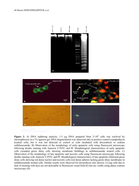
Figure 2. A) DNA laddering analysis. 1-3 µg DNA prepared from 2×<s<strong>tr</strong>ong>10</s<strong>tr</strong>ong> 6 cells was resolved by<br />
elec<strong>tr</strong>ophoresis in a 1% agarose gel. DNA fragmentation was observed only in positive con<strong>tr</strong>ol (camptothecin<br />
<strong>tr</strong>eated) cells, but it was not detected in con<strong>tr</strong>ol or cells incubated with doxorubicin or sodium<br />
sulfabenzamide. B) Observation <strong>of</strong> the morphology <strong>of</strong> early apoptotic cells using fluorescent microscopy<br />
following double staining with Annexin V-FITC <strong>and</strong> PI. Morphological characteristics <strong>of</strong> early apoptotic<br />
cells (rounded green shiny cells showing membrane blebbing) in sulfabenzamide <strong>tr</strong>eated cells. C)<br />
Observation <strong>of</strong> the morphology <strong>of</strong> late apoptotic <strong>and</strong> necrotic cells using fluorescent microscopy following<br />
double staining with Annexin V-FITC <strong>and</strong> PI. Morphological characteristics <strong>of</strong> late apoptotic (flattened green<br />
shiny cells showing red dense nuclei) <strong>and</strong> necrotic cells (red dense spheres lacking green shiny membrane) in<br />
sulfabenzamide <strong>tr</strong>eated cells. Similar results were observed for doxorubicin (not shown). Living cells due to<br />
lack <strong>of</strong> staining with dyes are not detectable in fluorescent visual field (E) but are visible using phase con<strong>tr</strong>ast<br />
microscopy (D).


