Download the full report (116 p.) - KCE
Download the full report (116 p.) - KCE
Download the full report (116 p.) - KCE

Create successful ePaper yourself
Turn your PDF publications into a flip-book with our unique Google optimized e-Paper software.
12 Multislice CT in Coronary Heart Disease <strong>KCE</strong> Reports 82<br />
Comparable figures are <strong>report</strong>ed in <strong>the</strong> most recently published ACC/AHA guidelines<br />
for <strong>the</strong> clinical application of echocardiography. 13 From <strong>the</strong>se data, we (crudely)<br />
calculated positive and negative LRs of 5.0 and 0.24 respectively. These figures<br />
26, 27<br />
correspond closely to those <strong>report</strong>ed in more recent literature.<br />
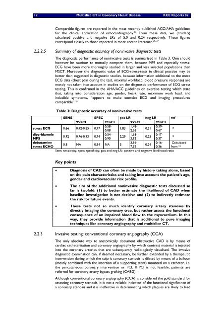
2.2.2.5 Summary of diagnostic accuracy of noninvasive diagnostic tests<br />
The diagnostic performance of noninvasive tests is summarised in Table 3. One should<br />
however be cautious to mutually compare <strong>the</strong>m, because MPS and especially stress-<br />
ECG have been more thoroughly studied in larger and less selected populations than<br />
MSCT. Moreover <strong>the</strong> diagnostic value of ECG-stress-tests in clinical practice may be<br />
better than suggested in diagnostic studies, because information additional to <strong>the</strong> mere<br />
ECG data (chest pain during <strong>the</strong> test, maximal workload, blood pressure response) are<br />
mostly not taken into account in studies on <strong>the</strong> diagnostic performance of ECG stress<br />
testing. This is confirmed in <strong>the</strong> AHA/ACC guidelines on exercise testing which state<br />
that, taking into consideration age, gender, heart rate, maximum work load, and<br />
inducible symptoms, “appears to make exercise ECG and imaging procedures<br />
comparable”. 16<br />
Table 3: Diagnostic accuracy of noninvasive tests<br />
SENS SPEC pos LR neg LR ref<br />
95%CI 95%CI 95%CI 95%CI<br />
stress ECG 0,66 0,42-0,85 0,77<br />
0,58-<br />
0,88<br />
1,83<br />
1,48-<br />
2,26<br />
0,51<br />
0,39-<br />
0,67<br />
14<br />
dipyridamole<br />
MPS<br />
0,92 0,76-0,93 0,74<br />
0,54-<br />
0,90<br />
2,29<br />
1,68-<br />
3,12<br />
0,25<br />
0,17-<br />
0,37<br />
14<br />
dobutamine<br />
stress ECHO<br />
0,8 NA 0,84 NA 5<br />
3,16-<br />
7,92<br />
0,24<br />
0,16-<br />
0,36<br />
Calculated<br />
from 25<br />
Sens: sensitivity, spec; specificity; pos and neg LR: positive and negative likelihood ratio<br />
Key points<br />
• Diagnosis of CAD can often be made by history taking alone, based<br />
on <strong>the</strong> pain characteristics and taking into account <strong>the</strong> patient’s age,<br />
gender and cardiovascular risk profile.<br />
• The aim of <strong>the</strong> additional noninvasive diagnostic tests discussed so<br />
far is twofold: (1) to better estimate <strong>the</strong> likelihood of CAD when<br />
baseline investigation is not decisive and (2) to indirectly estimate<br />
<strong>the</strong> risk for future events.<br />
• These tests not so much identify coronary artery stenoses by<br />
directly imaging <strong>the</strong> coronary tree, but ra<strong>the</strong>r assess <strong>the</strong> functional<br />
consequence of an impaired blood flow to <strong>the</strong> myocardium. In this<br />
way, <strong>the</strong>y provide information that is additional to pure imaging<br />
techniques like coronary angiography and multislice CT.<br />
2.2.3 Invasive testing: conventional coronary angiography (CCA)<br />
The only absolute way to anatomically document obstructive CAD is by means of<br />
cardiac ca<strong>the</strong>terisation and coronary angiography by which contrast material is injected<br />
into <strong>the</strong> coronary arteries that are subsequently radiologicaly visualised. The invasive<br />
diagnostic examination can, if deemed necessary, be fur<strong>the</strong>r extended by a <strong>the</strong>rapeutic<br />
intervention during which <strong>the</strong> culprit coronary stenosis is dilated by means of a balloon<br />
(mostly combined with <strong>the</strong> insertion of a supporting stent) mounted on a ca<strong>the</strong>ter, i.e.<br />
<strong>the</strong> percutaneous coronary intervention or PCI. If PCI is not feasible, patients are<br />
referred for coronary artery bypass grafting (CABG).<br />
Although conventional coronary angiography (CCA) is considered <strong>the</strong> gold standard for<br />
assessing coronary stenosis, it is not a reliable indicator of <strong>the</strong> functional significance of<br />
a coronary stenosis and it is ineffective in determinating which plaques are likely to lead