Download the full report (112 p.) - KCE
Download the full report (112 p.) - KCE
Download the full report (112 p.) - KCE
Create successful ePaper yourself
Turn your PDF publications into a flip-book with our unique Google optimized e-Paper software.
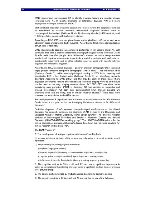
10 Interventions in Alzheimer’s Disease <strong>KCE</strong> Reports 111<br />
EFNS recommends non-contrast CT to identify treatable lesions and vascular disease<br />
(evidence Level A). In specific situations of differential diagnosis MRI is a more<br />
appropriate technique (evidence Level A). 13<br />
SBU concludes that after a baseline assessment, in cases where <strong>the</strong> diagnosis could not<br />
be established by classical methods, biochemical diagnostic markers such as<br />
cerebrospinal fluid analysis (Evidence Grade 1) effectively identify (> 80% sensitivity and<br />
> 80% specificity) people with Alzheimer’s disease.<br />
According to EFNS CSF total tau, phospho-tau and amyloid-beta1-42 can be used as an<br />
adjunct in cases of diagnostic doubt (Level B). According to NICE more standardization<br />
of CSF tests is required. 1<br />
EFNS recommends cognitive assessment is performed in all patients (level A). SBU<br />
concludes that after a baseline assessment, neuropsychological testing (Evidence Grade<br />
1) effectively identifies people with Alzheimer’s disease. A more comprehensive<br />
standardised cognitive assessments is particularly useful in patients with with mild or<br />
questionable impairment, and in o<strong>the</strong>r selected cases to assist with specific subtype<br />
diagnosis and differential diagnosis. 1<br />
According to SBU, functional diagnosis – positron emission tomography (PET scan) and<br />
single photon emission computed tomography (SPECT scan) – has moderate value<br />
(Evidence Grade 2), while neurophysiological testing – EEG brain mapping and<br />
quantitative EEG – has limited value (Evidence Grade 3) for identifying dementia<br />
disorders. According to EFNS SPECT and PET may be useful in those cases where<br />
diagnostic uncertainty remains after clinical and structural imaging work up, and should<br />
not be used as <strong>the</strong> only imaging measure (Level B). 14 FDG PET may show some<br />
superiority over perfusion SPECT in detecting AD but remains an expensive and<br />
invasive investigation. 1 PET scan tests demonstrating brain amyloid deposits are<br />
promising tools and are being used in clinical research studies. 16 These tests were<br />
however not yet included in <strong>the</strong> HTA <strong>report</strong>s.<br />
The Apolipoprotein E (ApoE) e4 allele is known to increase <strong>the</strong> risk for AD (Evidence<br />
Grade 1) but it is a poor marker for identifying Alzheimer’s disease or for differential<br />
diagnosis. 14<br />
Definitive diagnosis of AD requires histopathological confirmation of <strong>the</strong> clinical<br />
diagnosis. For research purposes, <strong>the</strong> diagnosis of AD is given in <strong>the</strong> Diagnostic and<br />
Statistical Manual of Mental Disorders, fourth edition (DSM-IV-TR) 17 and <strong>the</strong> National<br />
Institute of Neurological Disorders and Stroke – Alzheimer Disease and Related<br />
Disorders (NINCDS-ADRDA) working group. 18 The NINCDS-ADRDA criteria for <strong>the</strong><br />
clinical diagnosis of probable Alzheimer’s disease have been <strong>the</strong> reference standard for<br />
clinical research studies since 1984.<br />
The DSM-IV criteria 17<br />
A. The development of multiple cognitive deficits manifested by both:<br />
(1) memory impairment (impaired ability to learn new information or to recall previously learned<br />
information)<br />
(2) one (or more) of <strong>the</strong> following cognitive disturbances:<br />
(a) aphasia (language disturbance)<br />
(b) apraxia (impaired ability to carry out motor activities despite intact motor function)<br />
(c) agnosia (failure to recognize or identify objects despite intact sensory function)<br />
(d) disturbance in executive functioning (ie, planning, organizing, sequencing, abstracting).<br />
B. The cognitive deficits in Criteria A1 and A2 each cause significant impairment in<br />
social or occupational functioning and represent a significant decline from a previous<br />
level of functioning.<br />
C. The course is characterized by gradual onset and continuing cognitive decline.<br />
D. The cognitive deficits in Criteria A1 and A2 are not due to any of <strong>the</strong> following:


