

Complete issue - IMA Fungus
Complete issue - IMA Fungus
Complete issue - IMA Fungus

Create successful ePaper yourself
Turn your PDF publications into a flip-book with our unique Google optimized e-Paper software.
Zain, Moss & El-Sheikh<br />
ARTICLE<br />
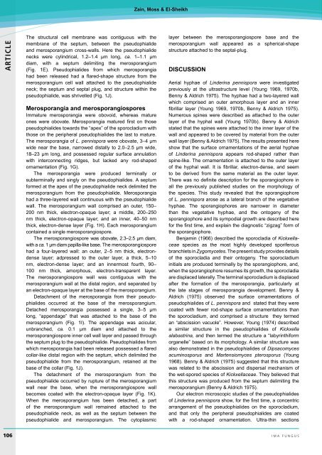
The structural cell membrane was contiguous with the<br />
membrane of the septum, between the pseudophialide<br />
and merosporangium cross-walls. Here the pseudophialide<br />
necks were cylindrical, 1.2–1.4 µm long, ca. 1–1.1 µm<br />
diam, with a septum delimiting the merosporangium<br />
(Fig. 1E). Pseudophialides from which merosporangia<br />
had been released had a flared-shape structure from the<br />
merosporangium cell wall attached to the pseudophialide<br />
neck; the septum and septal plug, and structure within the<br />
pseudophialide, was shrivelled (Fig. 1J).<br />
Merosporangia and merosporangiospores<br />
Immature merosporangia were obovoid, whereas mature<br />
ones were obovate. Merosporangia matured first on those<br />
pseudophialides towards the “apex” of the sporocladium with<br />
those on the peripheral pseudophialides the last to mature.<br />
The merosporangia of L. pennispora were obovate, 3–4 µm<br />
wide near the base, narrowed distally to 2.0–2.5 µm wide,<br />
18–23 µm long, and possessed regular surface annulation<br />
with interconnecting ridges, but lacked any rod-shaped<br />
ornamentation (Fig. 1G).<br />
The merosporangia were produced terminally or<br />
subterminally and singly on the pseudophialides. A septum<br />
formed at the apex of the pseudophialide neck delimited the<br />
merosporangium from the pseudophialide. Merosporangia<br />
had a three-layered wall continuous with the pseudophialide<br />
wall. The merosporangium wall comprised an outer, 150–<br />
200 nm thick, electron-opaque layer; a middle, 200–250<br />
nm thick, electron-opaque layer; and an inner, 40–50 nm<br />
thick, electron-dense layer (Fig. 1H). Each merosporangium<br />
contained a single merosporangiospore.<br />
The merosporangiospore was obovate, 2.3–2.5 µm diam,<br />
with a ca. 1 µm diam papilla-like base. The merosporangiospore<br />
had a four-layered wall: an outer, 2–5 nm thick, electrondense<br />
layer; adpressed to the outer layer, a thick, 5–10<br />
nm, electron-dense layer; and an innermost fourth, 90–<br />
100 nm thick, amorphous, electron-transparent layer.<br />
The merosporangiospore wall was contiguous with the<br />
merosporangium wall at the distal region, and separated by<br />
an electron-opaque layer at the base of the merosporangium.<br />
Detachment of the merosporangia from their pseudophialides<br />
occurred at the base of the merosporangium.<br />
Detached merosporangia possessed a single, 3–5 µm<br />
long, “appendage” that was attached to the base of the<br />
merosporangium (Fig. 1I). The appendage was acicular,<br />
unbranched, ca. 0.1 µm diam and attached to the<br />
merosporangiospore inner cell wall layer and passed through<br />
the septum plug to the pseudophialide. Pseudophialides from<br />
which merosporangia had been released possessed a flared<br />
collar-like distal region with the septum, which delimited the<br />
pseudophialide from the merosporangium, retained at the<br />
base of the collar (Fig. 1J).<br />
The detachment of the merosporangium from the<br />
pseudophialide occurred by rupture of the merosporangium<br />
wall near the base, when the merosporangiospore wall<br />
becomes coated with the electron-opaque layer (Fig. 1K).<br />
When the merosporangium has been detached, a part<br />
of the merosporangium wall remained attached to the<br />
pseudophialide neck, as well as the septum between the<br />
pseudophialide and merosporangium. The cytoplasmic<br />
layer between the merosporangiospore base and the<br />
merosporangium wall appeared as a spherical-shape<br />
structure attached to the septal-plug.<br />
DISCUSSION<br />
Aerial hyphae of Linderina pennispora were investigated<br />
previously at the ultrastructure level (Young 1969, 1970b,<br />
Benny & Aldrich 1975). The hyphae had a two-layered wall<br />
which comprised an outer amorphous layer and an inner<br />
fibrillar layer (Young 1969, 1970b, Benny & Aldrich 1975).<br />
Numerous spines were described as attached to the outer<br />
layer of the hyphal wall (Young 1970b). Benny & Aldrich<br />
stated that the spines were attached to the inner layer of the<br />
wall and appeared to be covered by material from the outer<br />
wall layer (Benny & Aldrich 1975). The results presented here<br />
show that the surface ornamentations of the aerial hyphae<br />
of Linderina pennispora appears rod-shaped rather than<br />
spine-like. The ornamentation is attached to the outer layer<br />
of the hyphal wall. It is fibrillar, electron-dense, and seem<br />
to be derived from the same material as the outer layer.<br />
There was no definite description for the sporangiophore in<br />
all the previously published studies on the morphology of<br />
the species. This study revealed that the sporangiophore<br />
of L. pennispora arose as a lateral branch of the vegetative<br />
hyphae. The sporangiophores are narrower in diameter<br />
than the vegetative hyphae, and the ontogeny of the<br />
sporangiophore and its sympodial growth are described here<br />
for the first time, and explain the diagnostic “zigzag” form of<br />
the sporangiophore.<br />
Benjamin (1966) described the sporocladia of Kickxellaceae<br />
species as the most highly developed sporiferous<br />
branchlets in Zygomycetes. The present study provides details<br />
of the sporocladia and their ontogeny. The sporocladium<br />
initials are produced terminally by the sporangiophore, and,<br />
when the sporangiophore resumes its growth, the sporocladia<br />
are displaced laterally. The terminal sporocladium is displaced<br />
after the formation of the merosporangia, particularly at<br />
the late stages of merosporangia development. Benny &<br />
Aldrich (1975) observed the surface ornamentations of<br />
pseudophialides of L. pennispora and stated that they were<br />
coated with fewer rod-shape surface ornamentations than<br />
the sporocladium, and comprised a structure they termed<br />
an “abscission vacuole”. However, Young (1974) described<br />
a similar structure in the pseudophialides of Kickxella<br />
alabastrina, and then termed the structure a “labyrinthiform<br />
organelle” based on its morphology. A similar structure was<br />
also demonstrated in the pseudophialides of Dipsacomyces<br />
acuminosporus and Martensiomyces pterosporus (Young<br />
1968). Benny & Aldrich (1975) suggested that this structure<br />
was related to the abscission and dispersal mechanism of<br />
the wet-spored species of Kickxellaceae. They believed that<br />
this structure was produced from the septum delimiting the<br />
merosporangium (Benny & Aldrich 1975).<br />
Our electron microscopic studies of the pseudophialides<br />
of Linderina pennispora show, for the first time, a concentric<br />
arrangement of the pseudophialides on the sporocladium,<br />
and that only the peripheral pseudophialides are coated<br />
with a rod-shaped ornamentation. Ultra-thin sections<br />
106 <br />
ima fUNGUS


