Paralytic Strabismus: Third, Fourth, and Sixth Nerve Palsy
Paralytic Strabismus: Third, Fourth, and Sixth Nerve Palsy
Paralytic Strabismus: Third, Fourth, and Sixth Nerve Palsy

You also want an ePaper? Increase the reach of your titles
YUMPU automatically turns print PDFs into web optimized ePapers that Google loves.
<strong>Paralytic</strong> <strong>Strabismus</strong> 815<br />
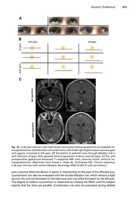
Fig. 10. A 36-year-old man with right fourth nerve palsy following resection of cerebellar hemangioblastoma.<br />
(A) Essentially normal ductions, with small right hypertropia in primary gaze<br />
<strong>and</strong> upgaze, increased in left gaze. (B) Simulation of patient’s view through Maddox rod in<br />
each direction of gaze. Note greatest vertical separation in down-<strong>and</strong>-left gaze. (C) Pre- <strong>and</strong><br />
postoperative gadolinium-enhanced T1-weighted MRI scans, showing fourth ventricle hemangioblastoma.<br />
(Reprinted from Prasad S, Volpe NJ, Tamhankar MA. Clinical reasoning:<br />
a 36-year-old man with vertical diplopia. Neurology 2009;72:e93–9; with permission.)<br />
<strong>and</strong> a second tilted line above or below it, intersecting on the side of the affected eye.<br />
Cyclotorsion can also be evaluated with the double Maddox rod, which refracts a light<br />
source into one red line (seen by the right eye) <strong>and</strong> one white line (seen by the left eye).<br />
The degree of relative cyclotorsion is measured by rotating the filters until the subject<br />
reports that the lines are parallel. Cyclotorsion can also be evaluated during dilated