Paralytic Strabismus: Third, Fourth, and Sixth Nerve Palsy
Paralytic Strabismus: Third, Fourth, and Sixth Nerve Palsy
Paralytic Strabismus: Third, Fourth, and Sixth Nerve Palsy

Create successful ePaper yourself
Turn your PDF publications into a flip-book with our unique Google optimized e-Paper software.
<strong>Paralytic</strong> <strong>Strabismus</strong> 825<br />
Mobius syndrome describes congenital facial diplegia that is frequently associated<br />
with sixth nerve palsy (Fig. 17). 74 Conjugate gaze paresis may be present, typically in<br />
cases with more severe abduction deficit. Other abnormalities such as complete<br />
external ophthalmoplegia, third nerve palsy, or ptosis occur less frequently. The cause<br />
of these deficits is believed to be hypoplasia of the relevant brainstem structures.<br />
Other inflammatory, infectious, <strong>and</strong> neoplastic processes that may affect the sixth<br />
nerve in the subarachnoid space are the same as those that can cause third <strong>and</strong> fourth<br />
nerve palsies <strong>and</strong> other cranial nerve deficits, <strong>and</strong> are discussed later.<br />
Diagnostic Testing<br />
Many patients with acute sixth nerve palsy require MRI to exclude structural, inflammatory,<br />
or neoplastic causes. All children <strong>and</strong> young adults with sixth nerve palsy<br />
should undergo imaging because of a higher prevalence of tumors <strong>and</strong> demyelinating<br />
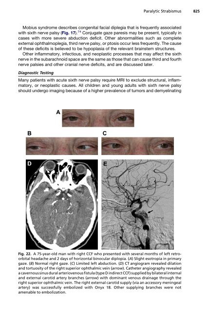
Fig. 22. A 75-year-old man with right CCF who presented with several months of left retroorbital<br />
headache <strong>and</strong> 2 days of horizontal binocular diplopia. (A) Slight esotropia in primary<br />
gaze. (B) Normal right gaze. (C) Limited left abduction. (D) CT angiogram revealed dilation<br />
<strong>and</strong> tortuosity of the right superior ophthalmic vein (arrow). Catheter angiography revealed<br />
a cavernous sinus dural arteriovenous fistula (type D indirect CCF) supplied by bilateral internal<br />
<strong>and</strong> external carotid artery branches (arrow) with dominant venous drainage through the<br />
right superior ophthalmic vein. The right external carotid supply (via an accessory meningeal<br />
artery) was successfully embolized with Onyx 18. Other supplying branches were not<br />
amenable to embolization.