808 Prasad & Volpe
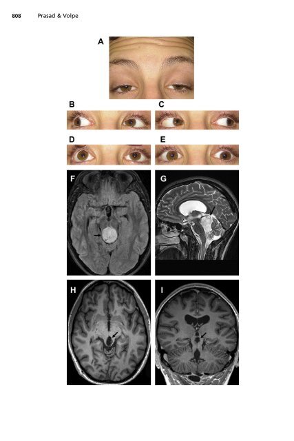
<strong>Paralytic</strong> <strong>Strabismus</strong> 809 with subarachnoid hemorrhage from a PComm aneurysm initially present with a third nerve palsy. 21,22 These aneurysms commonly project posterolaterally to compress the third nerve <strong>and</strong> involve the pupillary fibers in most cases. When the motility deficits are complete, pupillary involvement is virtually always present. If the motility deficit is partial, then the pupil may initially be spared. 23 Sparing of pupillomotor fibers may occur because they are resistant to evenly distributed compression, or because they are positioned dorsally, <strong>and</strong> in some cases compression is limited to the inferior aspect of the nerve. 24 A PComm aneurysm presenting acutely as a third nerve palsy represents a true neurosurgical emergency <strong>and</strong> may be treated by surgical clipping or endovascular coiling. 25 Microvascular third nerve palsy is commonly associated with risk factors including hypertension, diabetes, hyperlipidemia, advanced age, <strong>and</strong> smoking (see Fig. 4). This disorder results from impairment of microcirculation leading to circumscribed, ischemic demyelination of axons at the core of the nerve, typically in the cavernous sinus portion where a watershed territory exists. 5,26 Most of these patients exhibit pupillary sparing, because the pupillary fibers are located peripherally, closest to the blood supply provided by the surrounding vasa nervorum. However, some pupillary involvement may occur, typically with less than 1 mm (up to a maximum of 2.5 mm) of anisocoria found in approximately 40% of cases. 27 A microvascular third nerve palsy is frequently associated with orbital pain, which can be severe. Although it remains uncertain, the pain may result from ischemia of trigeminal sensory fibers that join the third nerve within the cavernous sinus. 28 There is an excellent prognosis for recovery of motility deficits from microvascular third nerve palsy, typically in 8 to 12 weeks. 29 Severe trauma is another common cause of third nerve palsies, involving traction at the skull base or fracture of the bones of the orbit or skull base (Fig. 9). 30,31 A third nerve palsy that follows minor head trauma may indicate an underlying structural lesion. 32,33 Although there is good prognosis for recovery following traumatic third nerve palsy, there is a high incidence of secondary aberrant regeneration. Slowly progressive third nerve palsies occasionally occur due to growth of a primary tumor of the nerve or nerve sheath. These lesions include neurinomas, neurofibromas, neurilemmomas, <strong>and</strong> schwannomas. 34 Neuroimaging will identify an enlarged, enhancing nerve in these cases. Uncommonly, a malignant meningioma, glioblastoma multiforme, or lymphoma may directly affect the third nerve. 35 Uncal herniation can cause direct compression of the third nerve against the free edge of the tentorium. In addition to third nerve deficits, these patients will have depressed mental status among other prominent neurologic deficits. In this situation, isolated pupil dilation may be the earliest manifestation of third nerve dysfunction. However, an isolated dilated pupil is never a manifestation of third nerve dysfunction in an awake <strong>and</strong> alert patient. : Fig. 5. A 15-year-old boy with bilateral nuclear third nerve palsies following resection of a midline juvenile pilocytic astrocytoma. (A) Severe bilateral ptosis in primary gaze. Note compensatory contraction of the frontalis muscle. (B) Reduced left adduction. Mydriasis of the left pupil is observed. (C) Slightly reduced right adduction. (D) Severe elevation limitation on attempted upgaze. The vertical gaze limitation was not overcome by the oculocephalic maneuver. (E) Bilateral depression deficit, greater on the right than on the left. Preoperative axial fluid-attenuated inversion-recovery (FLAIR) (F) <strong>and</strong> sagittal T2-weighted brain MRI (G) revealed a large heterogeneous midline mass (arrow) compressing the dorsal midbrain <strong>and</strong> causing hydrocephalus. Axial (H) <strong>and</strong> coronal MRI (I) 2 years following surgical resection showing focal volume loss in the dorsal midbrain (arrow).